Review

Xanthotoxin (8-Methoxypsoralen): A Review of Biological Activity and Potential Antitumor Properties


by
Anastasia A. Deryabina
1,2,
Matvey M. Tsyganov
1,2,*,
Marina K. Ibragimova
1,2,3,
Irina A. Tsydenova
1,
Olga Y. Rybalkina
4,
Arina K. Shagabudinova
1,
Pavel E. Nikiforov
4,
Maria V. Filonova
3,4 and
Alexey A. Churin
3,4
1
Cancer Research Institute, Tomsk National Research Medical Center, Russian Academy of Sciences, Tomsk 634050, Russia
2
Biochemistry and Molecular Biology Division, Siberian State Medical University, Tomsk 634050, Russia
3
Biological Institute, National Research Tomsk State University, Tomsk 634050, Russia
4
Goldberg’s Research Institute of Pharmacology and Regenerative Medicine, Tomsk National Research Medical Center, Russian Academy of Sciences, Tomsk 634050, Russia
*
Author to whom correspondence should be addressed.
Future Pharmacol. 2026, 6(3), 36; https://doi.org/10.3390/futurepharmacol6030036
Submission received: 14 May 2026 / Revised: 17 June 2026 / Accepted: 26 June 2026 / Published: 30 June 2026
(This article belongs to the Special Issue Feature Papers in Future Pharmacology 2026)

Abstract

Xanthotoxin (8-methoxypsoralen) belongs to the group of naturally occurring furanocoumarin (furocoumarin) compounds and is a product of plant secondary metabolism. Analysis of the available literature indicates that xanthotoxin exhibits a broad spectrum of pharmacological activities, including anti-inflammatory, antioxidant, immunomodulatory, and antibacterial effects. Xanthotoxin has been shown to stimulate autophagy via inhibition of the AKT/mTOR pathway, as well as to block cell migration by modulating RIG-1 and NF-κB signaling. Moreover, its effects on JNK/MAPK, PI3K/AKT, Calcium–CaMYK/PYK2, and other signaling cascades have been confirmed. Among its most promising properties is the ability to inhibit ABC transporters, thereby preventing the reduction of chemotherapeutic agent concentrations within tumor cells and enhancing their intracellular accumulation. Thus, the aim of this study was to evaluate xanthotoxin as a potential anticancer agent. The literature review was based on publications indexed in Google Scholar, Scopus, Web of Science, and PubMed and published between 2010 and 2026. Studies describing the biological properties of xanthotoxin, its toxicity, anticancer mechanisms of action, and modulation of ABC transporters were included. This literature review summarizes the pharmacological profile of xanthotoxin, and its biological activities and therapeutic potential, as well as its antitumor effects in various cancer cell lines. The available evidence may provide a foundation for the future development of xanthotoxin as a lead compound for anticancer drug discovery.

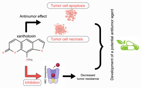
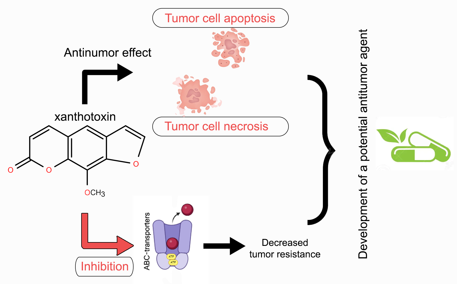
Graphical Abstract

1. Introduction

Despite advances in early diagnosis, disease progression remains the primary cause of high mortality rates in various cancers. This progression often occurs alongside ineffective or insufficiently potent therapeutic strategies [1]. A major factor contributing to this therapeutic failure is the development of chemoresistance [2]. The acquisition of resistance to anti-cancer drugs by tumor cells is a critical obstacle, leading to poor chemotherapy outcomes and, consequently, reduced patient survival rates [2].
A key mechanism driving this resistance involves ATP-binding cassette (ABC) transporters. These transmembrane proteins can actively pump chemotherapeutic agents out of cancer cells, thereby reducing their intracellular concentration and efficacy [3]. Additionally, enhanced DNA repair systems within tumors can mitigate the damage inflicted by these drugs, further contributing to a resistant phenotype [4]. The selective pressure exerted by chemotherapy agents inevitably fosters the emergence of resistant cell populations [5]. This dynamic underscores the critical need for research into combination therapies, specifically those that pair conventional chemotherapeutics with potential inhibitors of resistance mechanisms, such as ABC transporters.
In this context, compounds from the furanocoumarin (FC) group have emerged as promising candidates. Current data, derived from various in vitro and in vivo models, suggest that FCs possess a range of pharmacological effects, including spasmolytic, analgesic, and, notably, anticancer activities [6]. For instance, the FC xanthotoxin (also known as methoxsalen) has demonstrated pro-apoptotic properties, which can prevent the accumulation of genetically altered cells and inhibit their transformation [7]. Similarly, bergapten, another furanocoumarin, has been shown to initiate metabolic reprogramming in human breast cancer cells, leading to both anti-proliferative effects and the induction of apoptosis [8]. These findings suggest that bergapten may serve as a potentially valuable adjunct to targeted chemotherapy regimens [9]. The therapeutic potential of FCs may be significantly enhanced through their synergistic interaction with existing anticancer drugs [6]. Specifically, both bergapten and xanthotoxin have been reported to inhibit ABC transporters encoded by multidrug resistance (MDR) genes [10]. Research has shown that treating resistant cancer cells with xanthotoxin markedly increases the cytotoxicity of co-administered drugs like cisplatin, daunorubicin, and mitoxantrone [10]. Beyond direct effects on drug efflux, FCs also exhibit anti-inflammatory activity. This is achieved, in part, by inhibiting the NF-κB signaling pathway, which regulates genes involved in inflammation, cell proliferation, and apoptosis [11,12].
Given the often severe side effects associated with conventional chemotherapy, there is a growing focus on identifying effective anticancer agents from natural sources that may offer a better safety profile [13]. Xanthotoxin is emerging as a particularly compelling candidate in this regard, with demonstrated anti-tumor activity against a variety of cancers, including colorectal [7], hepatocellular [14], skin [15], T-cell lymphoma [16], prostate, and glioma [17]. The anticancer mechanisms of xanthotoxin appear to involve two major mechanisms: the induction of programmed cell death and the inhibition of uncontrolled cell proliferation [18]. In vitro studies have revealed that xanthotoxin can arrest the cell cycle, suppress proliferation, and direct cells toward apoptosis or autophagy [19]. Furthermore, it reduces cell viability and impedes division by triggering the mitochondrial apoptotic pathway, generating reactive oxygen species (ROS), and inhibiting the ERK1/2 signaling cascade, ultimately disrupting cellular metabolism [20]. This multifaceted activity highlights the potential of xanthotoxin to interfere with key processes of tumor cell division, differentiation, and proliferation [21].
Our previous research has contributed to this body of evidence. We found that course-based intragastric administration of a furanocoumarin-containing extract (comprising 42.97% isopimpinelline, 35.18% bergapten, and 15.41% xanthotoxin) to mice bearing Lewis lung carcinoma, alongside cisplatin treatment, resulted in a significant reduction in both the number and area of metastases. Furthermore, this combination therapy exerted a corrective effect on the hemostasis system, an effect similar to that of the anticoagulant warfarin [22]. In light of these findings and the supporting literature, xanthotoxin, as a key representative of the furanocoumarin group, warrants further detailed investigation as a promising agent with multi-faceted anticancer activity. Thus, the primary aim of this review was to evaluate the potential of xanthotoxin as an anticancer agent, with particular emphasis on its ability to overcome tumor cell drug resistance through modulation of ABC transporter activity.

2. Chemical Structure of Xanthotoxin

Furanocoumarins (FCs) are a class of naturally occurring chemical compounds, primarily known for the phototoxicity of several of their members, including bergapten, psoralen, and xanthotoxin. These compounds can cause skin burns upon exposure to ultraviolet radiation [23]. Chemically, FCs consist of a coumarin two-ring core fused with a furan ring. Currently, FCs are being investigated as potential therapeutic agents, particularly for their antiseptic, neuroprotective, osteoprotective, and antioxidant properties [24]. In plants, FCs are synthesized in response to stress or as a defense mechanism against biological threats such as fungal or bacterial infections and insect attacks [25]. Their biosynthesis involves the fusion of coumarin with a furan ring, yielding either linear or angular isomers depending on the position of the furan ring attachment [26] (Figure 1).
Linear (psoralens) furanocoumarins are characterized by a furan ring attached to the coumarin core at the 6–7 or 7–8 positions (Figure 1A), resulting in an elongated, linear molecular shape. This group includes xanthotoxin (8-methoxypsoralen), as well as psoralen and bergapten.
Angular (angelicins) furanocoumarins feature a furan ring attached at the 5–6 or 8–9 positions, giving the molecule a bent, angular configuration (e.g., angelicin; Figure 1B).
Among FCs, psoralen and its derivative 8-methoxypsoralen (xanthotoxin, 8-MOP) are the most prevalent compounds [27]. Xanthotoxin exhibits a broad spectrum of pharmacological activities, including neuroprotective, wound-healing, anti-inflammatory, antibacterial, antioxidant, and insecticidal effects [24]. Due to its lipophilic nature, xanthotoxin can cross the blood–brain barrier (BBB), facilitating its distribution into the central nervous system [18]. In addition, xanthotoxin regulates key cellular processes such as apoptosis and proliferation in both normal and malignant tissues. For instance, it has been shown to induce apoptosis in neuroblastoma and colorectal cancer cells by inactivating the PI3K/Akt signaling pathway [7], with similar effects observed in prostate cancer cells [28]. Furthermore, xanthotoxin isolated from plants has been reported to suppress the release of interleukin-6 (IL-6) and tumor necrosis factor-alpha (TNF-α) in RAW 264.7 cells through inhibition of the IKK/IκB/NF-κB pathway [29].
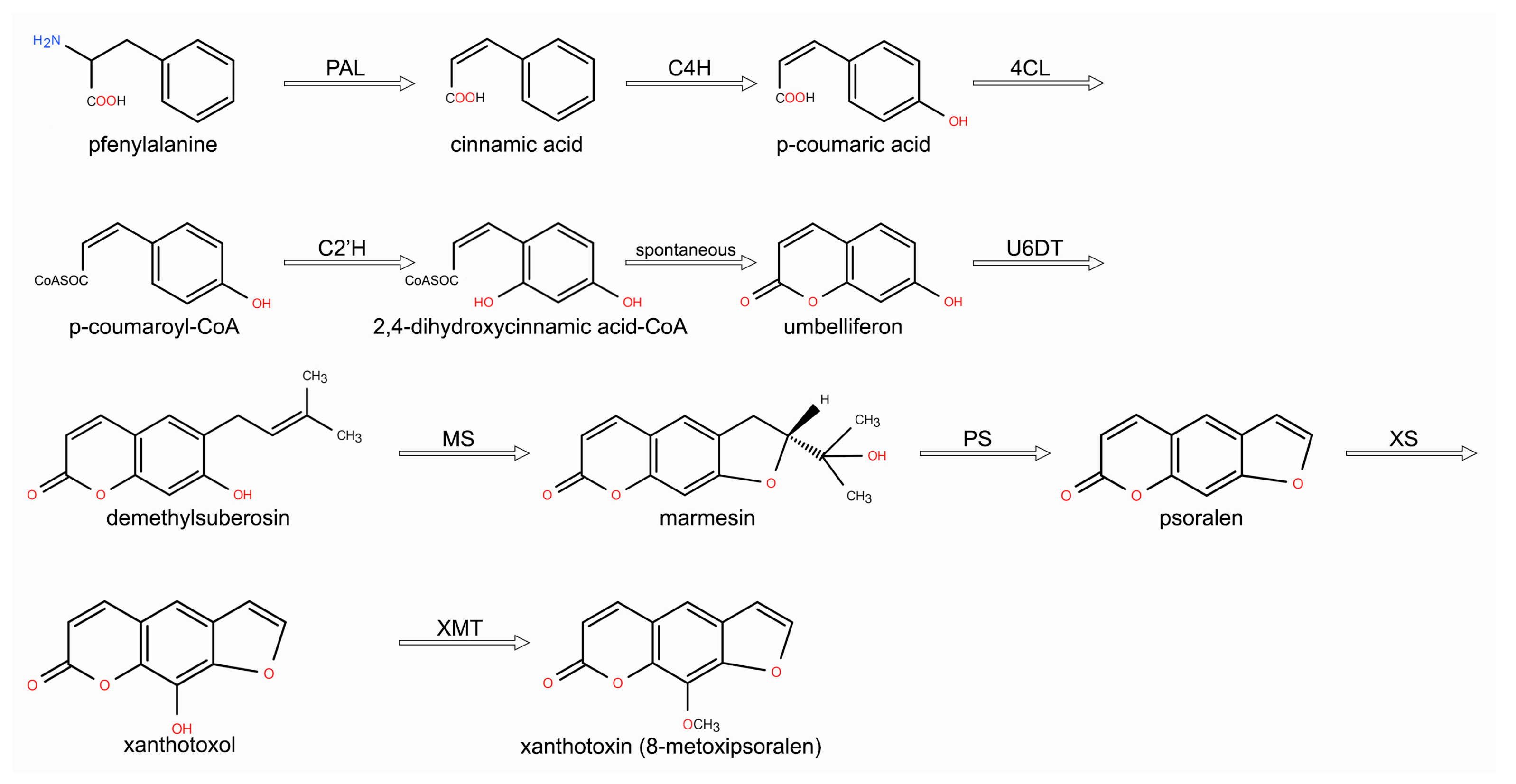
Research into furanocoumarins dates back to the 1930s. By the 1970s and 1980s, Canadian and Italian researchers had proposed a biosynthetic paradigm centered on umbelliferone. However, a complete biosynthetic pathway accounting for the full structural diversity of FCs remains elusive [30]. Contemporary studies on these compounds have shifted focus from discovering novel structures toward investigating their biological activity using in silico approaches. Attempts to produce FCs biotechnologically in microorganisms remain limited due to an incomplete understanding of the enzymatic pathways involved [30]. Nevertheless, a biosynthetic system for their precursor, umbelliferone, has been successfully established [31]. Umbelliferone is synthesized via the shikimate pathway from the amino acid phenylalanine. Phenylalanine ammonia lyase (PAL) deaminates phenylalanine to cinnamic acid (Figure 2). Cinnamic acid is then oxidized by cinnamate 4-hydroxylase (C4H) to yield p-coumaric acid, which contains a hydroxyl group at the C4 para-position. Subsequently, 4-coumarate-CoA ligase (4CL) attaches coenzyme A to the acid group. In the final enzymatic step, p-coumaroyl-CoA 2′-hydroxylase (C2′H) introduces a second hydroxyl group at the 2-position of the aromatic ring. A spontaneous rearrangement then closes the pyrone ring, forming umbelliferone [32,33]. In its glycosylated form, umbelliferone is transported within leaves to sites of furanocoumarin synthesis, where it undergoes deglycosylation [34]. A key step determining linear structure formation is prenylation at the C-6 position, catalyzed by umbelliferone 6-prenyltransferase (U6DT), which produces demethylsuberosin. Subsequent cyclization yields marmesin, which is oxidized to psoralen [35]—the immediate precursor of xanthotoxin. The critical step in xanthotoxin formation is hydroxylation of psoralen at the 5-position by psoralen 5-monooxygenase (P5M), generating xanthotoxol [36] (Figure 2).
The final reaction involves methylation of the xanthotoxol hydroxyl group by xanthotoxol O-methyltransferase (XMT), producing the target compound, xanthotoxin [37]. Demethoxylation reactions can also occur, converting xanthotoxin or bergapten back to psoralen [38].
Currently, the largest number of plant species containing furanocoumarins belong to the Apiaceae family, although these compounds are also found in other families [19,23]. Natural coumarins are widely distributed across at least 13 plant families, including Rutaceae, Apiaceae, Asteraceae, and Fabaceae. Xanthotoxin is most abundant in the fruits of Zanthoxylum bungeanum, as well as in the roots of Scholtzia umbellifera, Angelica sinensis, Apium graveolens (celery), and Vicia lens (lentil) [39]. Trace amounts of xanthotoxin have also been detected in certain food plants such as grapefruit, lime, and lemon, although these typically contain higher levels of other furanocoumarins, particularly bergapten [9]. Additionally, small quantities of xanthotoxin have been identified in animal and microbial metabolites [40]. Current methods for coumarin extraction include: matrix solid-phase dispersion (MSPD) [41]; supercritical fluid chromatography (SFC); and pressurized fluid extraction (PLE) [42]. However, coumarin components in plants often exist as mixtures of structurally similar compounds with comparable polarities, making their separation by traditional extraction and crystallization methods challenging. Chromatographic techniques are therefore commonly employed for their purification.
Due to the complex composition and low abundance of coumarins in plant material, the overall yield of xanthotoxin extracted from plant sources remains very low. Although the biosynthetic pathway to xanthotoxin was first described in 1936, the structural complexity, multi-step nature, low efficiency of chemical synthesis, and stability of intermediates render chemical synthesis impractical [43,44]. Currently, the most promising approaches to overcoming this challenge involve the use of biocatalysts, microbiological synthesis, or semi-synthetic strategies [45,46].

3. Biological Activity of Xanthotoxin

Xanthotoxin is a multifunctional compound that exerts a wide range of biological effects through its interactions with various organs, cellular structures, and metabolic pathways.

3.1. Neuroprotective Properties

Xanthotoxin has demonstrated promising therapeutic potential in animal models of several neurodegenerative conditions, including neurotoxicity, cognitive impairment induced by hypoxia-ischemia, Alzheimer’s disease, and Parkinson’s disease [47]. In addition to its neuroprotective effects, xanthotoxin exhibits anticonvulsant activity. In a mouse model of maximal electroshock (MES)-induced seizures, intraperitoneal administration of xanthotoxin was most effective when given 60 min prior to seizure induction (ED50 = 219.1 ± 4.7 mg/kg), an effect comparable to that of conventional antiepileptic drugs [48]. Furthermore, even at lower doses (50–100 mg/kg), xanthotoxin displayed anticonvulsant activity and potentiated the effects of standard antiepileptic agents such as levetiracetam and valproate [49]. Studies have also revealed the antidepressant potential of xanthotoxin. In experimental models, the compound alleviated depressive-like behavior and improved mood by enhancing serotonergic neurotransmission, primarily through inhibition of monoamine oxidase activity [50,51].

3.2. Dermatological Applications

Activated by UVA radiation, xanthotoxin is used clinically to treat proliferative skin disorders. The combination of xanthotoxin and UVA radiation, known as PUVA therapy (psoralen + UVA), remains an established therapeutic option for psoriasis [52]. The underlying mechanism involves intercalation of xanthotoxin into DNA; upon UVA exposure, it selectively forms photoadducts with the DNA of hyperproliferative cells, leading to cell damage and apoptosis. PUVA therapy also modulates microRNA expression, upregulating has-miR-4516, which in turn increases levels of the apoptotic proteins p53 and BAX. Apoptosis is closely linked to p53 activity, and miR-4516-mediated suppression of ubiquitin-conjugating enzyme E2N (UBE2N) promotes nuclear translocation of p53 [53]. PUVA-induced apoptosis has also been shown to block cell migration through multiple signaling pathways, including RIG-1 and NF-κB [54], as well as through inhibition of the AKT/mTOR pathway, which triggers autophagy [55]. Beyond its effects on DNA synthesis and fibroblast activity [56], PUVA therapy neutralizes reactive oxygen species (ROS) via malate dehydrogenase, succinate dehydrogenase, and ubiquinone oxidoreductase, potentially protecting surrounding tissues from oxidative damage [18]. The clinical efficacy of this approach has been demonstrated in the treatment of resistant alopecia areata. Topical application of 0.1% xanthotoxin followed by phototoxic UVA doses resulted in significant improvement in 57% of patients, with better responses observed in those with shorter disease duration [57]. Overall, PUVA photochemotherapy remains a well-established treatment modality for selected dermatological disorders for treating skin diseases [58].

3.3. Bone Tissue

Inhibiting the hypertrophy and aberrant proliferation of resting chondrocytes is a key strategy in the treatment of osteoporosis. Xanthotoxin exerts protective effects on bone tissue through two primary mechanisms: first, by suppressing the activation of pro-inflammatory signaling pathways, thereby limiting inflammation; and second, by increasing calcium ion (Ca2+) levels in bone tissue, leading to enhanced bone density [18]. Xanthotoxin has been shown to inhibit the expression of RUNX2, a transcription factor that serves as a marker of chondrocyte hypertrophy [59], as well as matrix metalloproteinase 13, via the p38-MAPK/HDAC4 pathway. Mechanistically, xanthotoxin reduces activation of p38 mitogen-activated protein kinase, which in turn upregulates histone deacetylase 4 (HDAC4), thereby suppressing RUNX2 production. In an ethanol-induced osteoporosis model, xanthotoxin increased serum levels of estradiol and osteocalcin, as well as bone alkaline phosphatase activity. Concurrently, it downregulated RANKL and inhibited activation of the PI3K/AKT, JNK/MAPK, and NF-κB signaling pathways. Collectively, these effects promote osteoblast differentiation and preserve bone structure [60].

3.4. Kidney

The nephroprotective effects of xanthotoxin are mediated primarily through two mechanisms: suppression of NF-κB pathway activation, which alleviates inflammation, and restoration of renal excretory function [18]. Xanthotoxin has been shown to inhibit the production of various pro-inflammatory cytokines by activated macrophages, including tumor necrosis factor-alpha (TNF-α), and interleukins (ILs), as well as inflammatory mediators such as nitric oxide (NO) and prostaglandins (PGs). This broad anti-inflammatory activity results from inhibition of the NF-κB signaling pathway. NF-κB is a eukaryotic transcription factor that regulates numerous genes involved in innate immunity and acute inflammatory responses [61]. As a potential nephroprotective agent, xanthotoxin inhibits NF-κB activation at multiple steps, including activation of IKKα/β, phosphorylation and degradation of IκBα, and phosphorylation and nuclear translocation of the p65 NF-κB subunit [61].

3.5. Lungs

In a mouse model of lipopolysaccharide (LPS)-induced acute lung injury, oral administration of xanthotoxin at a dose of 25 µM reduced the number of macrophages and neutrophils in lung tissue and attenuated pathological changes. This effect was attributed to the ability of xanthotoxin to significantly suppress the JAK/STAT signaling pathway, leading to reduced levels of interleukin-6 (IL-6) and nitric oxide (NO) [61]. Among the components of tobacco smoke, particular attention has been given to NNK (4-(methylnitrosamino)-1-(3-pyridyl)-1-butanone), a tobacco-specific N-nitrosamine with potent carcinogenic activity in the lung, as demonstrated by its ability to induce lung tumors in rats and hamsters. Notably, xanthotoxin at a dose of 12.5 mg/kg protected YG7108 cells from the damaging effects of NNK present in tobacco smoke, primarily by inhibiting cytochrome CYP2A activity [62].

3.6. Liver

The transcription factors DEC1 and DEC2 are known to regulate apoptosis, cell proliferation, circadian rhythms, and the progression of various malignancies, including hepatocellular carcinoma [63]. Treatment of human hepatocellular carcinoma (HepG2) cells with xanthotoxin (30 µM) for 48 h reduced cell viability to approximately 55% of control levels, as assessed by the MTT assay [64]. Quantitative real-time PCR (qRT-PCR) analysis revealed a significant decrease in DEC1 mRNA transcript levels following xanthotoxin treatment, implicating DEC1 in the apoptotic response induced by this compound in HepG2 cells [65]. These findings suggest that the hepatoprotective effects of xanthotoxin are mediated, at least in part, through the modulation of apoptotic pathways.

3.7. Cardiovascular System

In the early stages of atherosclerosis, cytokines and other activate endothelial cells, prompting them to secrete intercellular adhesion molecule-1 (ICAM-1) and vascular cell adhesion molecule-1 (VCAM-1). This promotes monocyte migration into the vascular wall and initiates an inflammatory cascade. Xanthotoxin has demonstrated therapeutic potential in cardiovascular disease by reducing the expression of these adhesion molecules, thereby protecting against endothelial damage and exerting anti-atherosclerotic effects [18]. Nitric oxide (NO) plays a key regulatory role in the vascular system, exhibiting anti-inflammatory, anti-tumor, and anti-proliferative activities [66]. Studies on human umbilical vein endothelial cells (HUVECs) have shown that xanthotoxin (20 µM) activates voltage-gated potassium (Kv) channels, inhibits L-type calcium channels, and induces vasodilation via the Akt–eNOS–cGMP signaling pathway [67]. Moreover, endothelial nitric oxide synthase (eNOS) levels are inversely correlated with systemic blood pressure. By upregulating eNOS expression, xanthotoxin may contribute to blood pressure normalization in hypertensive patients. Thus, xanthotoxin promotes vasodilation and supports vascular repair, conferring cardioprotective effects [67].

3.8. Anti-Inflammatory Activity

Recent years have seen a growing body of evidence supporting the use of xanthotoxin in the treatment of inflammatory conditions [68]. The anti-inflammatory activity of xanthotoxin was evaluated in LPS-stimulated (50 µg/mL) brain microvascular endothelial cells (BMECs) and RAW 264.7 macrophages [69]. Optimal anti-inflammatory effects were observed at an xanthotoxin concentration of 25 µg/mL. Western blot analysis revealed that xanthotoxin suppressed activation of the NF-κB transcription factor and the JNK/STAT signaling pathway. Additional in vivo studies demonstrated that xanthotoxin also inhibits the release of pro-inflammatory cytokines including IL-1β, IL-6, IL-8, and TNF-α from immune cells such as neutrophils, macrophages, and lymphocytes, thereby protecting cells from inflammatory damage [61].

3.9. Antibacterial Activity

Xanthotoxin exhibits not only enzyme inhibitory activity but also pronounced antibacterial effects. Its antibacterial activity was evaluated against the Gram-positive bacterium Staphylococcus aureus and the Gram-negative bacteria Escherichia coli and Pseudomonas aeruginosa using the microdilution method, with ampicillin serving as a positive control. Xanthotoxin potently inhibited acetylcholinesterase activity (IC50 = 39.64 µM) and displayed strong antibacterial activity (MIC = 7.5 mg/mL) against Gram-positive bacteria, particularly S. aureus [70]. The antibacterial mechanism is thought to involve penetration of the bacterial cell membrane, leading to DNA damage and impaired replication [71].

3.10. Toxicity Profile

Phototoxicity. Exposure to light potentiates the toxicity of xanthotoxin, as it indirectly affects DNA replication [9]. Recent in vivo studies, ROS assays, and pharmacokinetic analyses have confirmed that xanthotoxin (50 µg/mL) functions as a phototoxin [72]. Xanthotoxin exhibited maximal phototoxicity in Jurkat and HaCaT cells (IC50 = 18 µM). It is important to note that xanthotoxin itself is not inherently harmful; however, upon UVA exposure, it reacts with skin proteins, leading to pigment accumulation and lesion formation [73].
Organ-Specific Toxicity. Despite its therapeutic efficacy, xanthotoxin is associated with several dose-dependent toxic effects affecting various organ systems. Long-term PUVA therapy correlates with an increased risk of cataract development [74]. Prolonged UVA exposure in the presence of xanthotoxin can damage ocular epithelial cells, resulting in inflammatory infiltration, edema, and subsequent macular degeneration [75].
Reproductive Toxicity. Xanthotoxin has been shown to adversely affect the female reproductive system. In mouse studies, ovariotoxicity was observed, characterized by a significant reduction in the number and diameter of corpora lutea, thinning of the granulosa cell layer, and decreased follicle counts. These morphological changes were accompanied by reduced plasma levels of luteinizing hormone (LH), follicle-stimulating hormone (FSH), estrogen, and progesterone [76].
Hepatotoxicity. Hepatotoxicity manifests at high doses of xanthotoxin (40 mg/kg), potentially inducing liver injury characterized by jaundice and a marked elevation in total bilirubin levels [77].
Mutagenicity and Carcinogenicity. The mutagenic activity of xanthotoxin is primarily attributable to its photochemical properties. Upon UVA activation, it intercalates into DNA, forming monoadducts and interstrand crosslinks, which underpin its potent cytotoxic effects. However, PUVA therapy has been associated with an increased risk of skin malignancies. Specifically, treatment is linked to the development of basal cell carcinoma and, to a moderate degree, squamous cell carcinoma [78]. Long-term follow-up studies have demonstrated that more than half of patients receiving at least 400 PUVA treatments developed at least one squamous cell carcinoma within 25 years, and nearly one-third of those undergoing 200 treatments developed basal cell carcinoma [79,80]. Furthermore, the risk of malignant melanoma increases more than fivefold approximately 15 years after the first PUVA session, with the highest risk observed in patients receiving 250 or more treatments [78].
Thus, despite the promising therapeutic potential of xanthotoxin, its toxicity profile remains one of the key factors determining its future clinical applicability. The high risk of phototoxicity associated with light-induced activation may limit its therapeutic use, necessitating careful dose optimization and appropriate clinical precautions. In addition, the potential for organ toxicity underscores the need for rigorous safety measures and close patient monitoring. Particular attention should be paid to its reproductive and hepatotoxic effects, which require further large-scale preclinical and clinical studies to establish safe dosing regimens and fully characterize the associated risks. Overall, the safety and toxicity profile of xanthotoxin will play a critical role in its translation from an experimental compound to a clinically applicable anticancer agent. Further studies aimed at minimizing adverse effects and improving its therapeutic index are essential to expand its potential clinical use.

4. Potential Anti-Cancer Properties

Despite the potential toxicity observed in certain contexts, xanthotoxin demonstrates a clear ability to inhibit tumor cell growth, activate apoptotic mechanisms, and modulate immune responses. Its anticancer effects can be broadly categorized into photoactivated and non-photoactivated mechanisms.
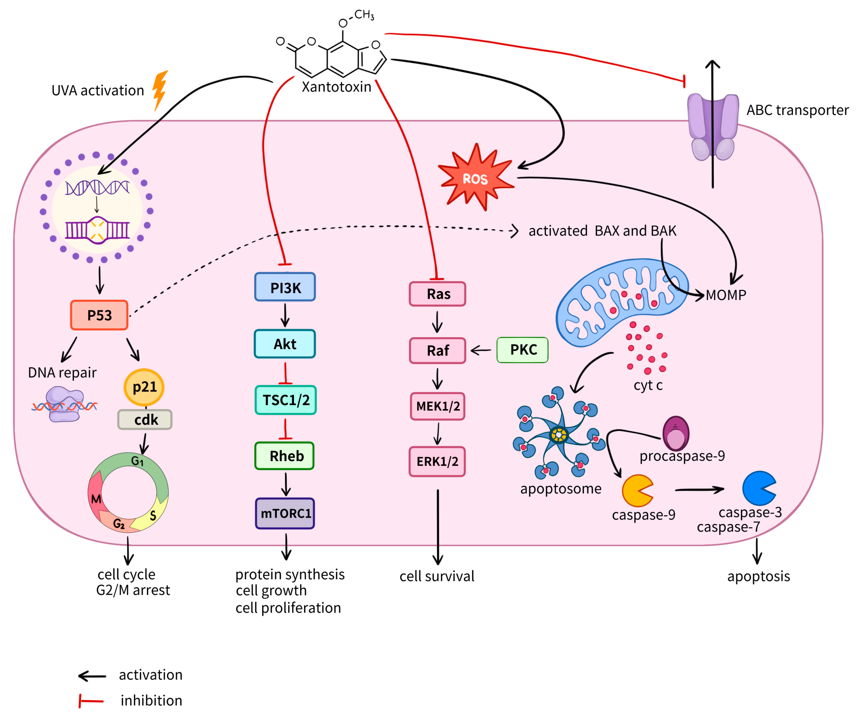
Photoactivated Mechanism: DNA Damage. In its simplest form, the anticancer effect of xanthotoxin under photoactivation involves direct interaction with DNA. Upon UVA irradiation, xanthotoxin acts as a classic photosensitizer: it intercalates into DNA and forms monoadducts and interstrand crosslinks with pyrimidine bases, primarily thymine. This blocks replication and transcription, leading to cell cycle arrest and activation of apoptosis, which directly underpins its anti-proliferative effect (Figure 3) [81,82].
Non-Photoactivated Mechanisms: Multiple Signaling Pathways. Critically, from a therapeutic perspective, xanthotoxin exhibits pronounced cytotoxic and pro-apoptotic activity even in the absence of photoactivation, through several distinct mechanisms.
  • Inhibition of the PI3K/AKT Survival Pathway. Xanthotoxin dose-dependently reduces phosphorylation of AKT at Thr308 (pAKT308) without affecting total AKT protein levels. Given that the PI3K/AKT pathway is hyperactive in many cancers and is critical for cell survival, proliferation, and apoptosis resistance, its suppression represents a potent anticancer effect. Inhibiting AKT directly promotes apoptosis by releasing its inhibitory effect on pro-apoptotic proteins such as BAD, caspase-9, and Bax [65,83] (Figure 3).
  • Induction of Apoptosis via the Bax/Bcl-2 Ratio. In human hepatocellular carcinoma (HepG2) cells, xanthotoxin (25–100 µM) exhibited dose-dependent pro-apoptotic activity. The mechanism involved downregulation of the anti-apoptotic protein Bcl-2 and upregulation of the pro-apoptotic protein Bax. This markedly increased the Bax/Bcl-2 ratio, destabilizing the mitochondrial membrane, promoting cytochrome c release, and activating caspase-9. Consequently, xanthotoxin induced dose-dependent increases in the active forms of caspases-8, -9, and -3, indicating simultaneous activation of both the extrinsic (receptor-mediated) and intrinsic (mitochondrial) apoptotic pathways [7,84] (Figure 3).
  • ROS-Mediated Oxidative Stress. Another mechanism involves the ability of xanthotoxin to induce reactive oxygen species (ROS) generation, leading to oxidative stress and apoptosis. Accumulation of ROS damages cellular structures and serves as an additional trigger for mitochondrial apoptosis while inhibiting survival pathways [7].
  • Inhibition of Proliferative Signaling and Cell Cycle Arrest. Xanthotoxin suppresses tumor cell growth by inducing programmed cell death, which is also linked to suppression of survival signals via the Ras–Raf–MEK–ERK pathway. Moreover, xanthotoxin can directly bind to DNA, thereby inhibiting the cell cycle. This is accompanied by increased levels of active p53 protein, which induces transcription of the p21 gene. The p21 protein (also known as WAF1 or CIP1) is a key effector of the p53 tumor suppressor and a potent inhibitor of cyclin-dependent kinases (Cdks). p21 binds to and inhibits fully formed cyclin–Cdk complexes:
Cyclin D–Cdk4/Cdk6: Responsible for progression through the G1 phase.
Cyclin E–Cdk2: Critical for the G1/S transition.
Cyclin A–Cdk2: Involved in S phase progression.
Cyclin B–Cdc2: Required for G2 phase progression and entry into mitosis [85].
Thus, p21-mediated cell cycle arrest at the G2/M phase contributes to the anti-proliferative effects of xanthotoxin [84] (Figure 3). In response to DNA damage, cellular signaling events include upregulation of the p21WAF1/CIP1 complex and p53 activation, which together induce cytochrome c release from mitochondria and subsequent cell death.
5.
Inhibition of Migration and Epithelial–Mesenchymal Transition. Studies have shown that psoralen can inhibit the cell cycle and suppress proliferation by inducing programmed cell death via apoptosis or autophagy. Furthermore, psoralen exhibits anti-migratory properties by suppressing NF-κB activation, thereby blocking epithelial–mesenchymal transition (EMT). Additional mechanisms include inhibition of anti-apoptotic proteins (c-FLIP, IAP) and activation of pro-apoptotic factors (Bax, JNK) [86].
The presence of a methoxy group enhances the anticancer properties of psoralen, independent of its position, as evidenced by the similar activity of 5-MOP and xanthotoxin. In HL-60 leukemia cells, both compounds (100 µM) exhibited pro-apoptotic and anti-proliferative effects and arrested the cell cycle at G1 phase with equal potency. Notably, replacing the methoxy group with an isopentenyloxy moiety at the C5 position reduced the pro-apoptotic properties of the compound [87]. Similar results were observed in HeLa S3 (human cervical cancer) and MK-1 (gastric adenocarcinoma) cell lines [88].
Current research has revealed that xanthotoxin exhibits cytotoxic activity against several tumor cell lines, although sensitivity varies depending on the cell type (Supplementary Materials Table S1 [89]). This suggests a cell-specific action of xanthotoxin, likely related to the distinct oncogenic pathways operating in different tumor cell lines [84]. As summarized in Table S1, xanthotoxin has been shown to suppress the viability of a broad range of tumor cell lines [7]. Among the most sensitive were SK-N-AS neuroblastoma cells (IC50 = 56.3 µM) and SW620 metastatic colorectal adenocarcinoma cells (IC50 = 88.5 µM). In contrast, RPMI 8226 and U266 multiple myeloma cells exhibited pronounced resistance, with an IC50 of 309.3 µM reported for U266 [7]. The mechanisms of cell death induced by xanthotoxin also appear to be cell-type dependent. Apoptosis was the predominant mode of death in sensitive lines, reaching 59.6% in SK-N-AS cells treated with 200 µM xanthotoxin, whereas necrosis levels remained low and concentration-independent. By comparison, doxorubicin, used as a positive control, induced substantial necrosis (31%), suggesting that xanthotoxin may trigger a more controlled, and potentially less pro-inflammatory, mechanism of cell death [7].
A critical finding is the selective toxicity of xanthotoxin toward malignant cells. At concentrations up to 100 µM—which already exert significant anti-proliferative effects on tumor cells—xanthotoxin was completely non-toxic to normal CCD 841 CoTr colonic epithelial cells and normal human skin fibroblasts (HSF). Even at a high concentration of 400 µM, viability of these normal cells was reduced by only ~40%, whereas the same concentration reduced viability by 90% in most tumor lines tested [7]. This selectivity was further supported by studies on glioblastoma: de Oliveira et al. (2016) demonstrated significantly higher cytotoxic activity of xanthotoxin against human GL-15 glioblastoma cells compared to non-mutant rat astrocytes [90].
Interestingly, while xanthotoxin clearly affected proliferation in some models, cell death was not always observed under the conditions used for proliferation assays, nor did treatment consistently induce cell cycle arrest. One plausible explanation is that the compound may slow cell division by modulating genes involved in cell cycle regulation, a phenomenon supported by observed changes in cell cycle-related protein levels in patients undergoing PUVA therapy [91]. Additionally, xanthotoxin induced morphological changes in tumor cells, such as the formation of filopodia-like structures, suggesting possible involvement of the actin cytoskeleton in its mechanism of action [92]. Particularly potent activity was observed in HepG2 human hepatocellular carcinoma cells, with an IC50 of 6.9 ± 1.07 µg/mL [93]. Treatment of HepG2 cells with this concentration induced marked cell cycle alterations, including an increase in the pre-G1 and G2/M apoptotic phases and a corresponding decrease in S-phase cells, while maintaining good biocompatibility with normal human cells [93]. This differential effect may be explained by the high expression of topoisomerase II in tumors, an enzyme involved in DNA recombination, replication, transcription, and repair [94,95]. Molecular docking analysis revealed that xanthotoxin interacts with the crystal structure of topoisomerase II with a binding energy of −5.72 kcal/mol, slightly higher than that of the known topoisomerase II inhibitor etoposide (−7.31 kcal/mol) [93]. Importantly, xanthotoxin interacts with the key amino acid Asp479 in a manner similar to etoposide, suggesting potential as a topoisomerase II inhibitor.
According to the available literature (Table S1), the antitumor activity of xanthotoxin varies considerably depending on the cancer cell line. Xanthotoxin exhibits high efficacy against neuroblastoma (SK-N-AS), colorectal cancer (SW620), multiple myeloma (RPMI 8226 and U266), and glioblastoma (T98G), reducing tumor cell viability by up to 90% at relatively low IC50 values. In contrast, its activity is substantially lower in melanoma (A375, FM55P and FM55M2) and lung cancer (A549 and NCI-H322) cell lines. These findings suggest that tumor cell sensitivity to xanthotoxin differs markedly among cancer types and is likely influenced by their molecular characteristics and underlying mechanisms of drug resistance. The relative resistance of normal CCD 841 CoTr and HSF cells (only 40% viability reduction at high concentrations) supports the notion of selective anticancer activity. However, it should be noted that the available evidence is derived primarily from in vitro studies and should therefore be regarded as preliminary until validated in more physiologically relevant experimental models. It is also important to emphasize that relatively high concentrations of xanthotoxin were required to achieve antitumor activity in several cancer cell lines, which may limit the clinical translatability of these findings. Consequently, further in vivo studies and well-designed clinical investigations are needed to establish the therapeutic efficacy, safety, and optimal dosing of xanthotoxin.
The effects of combining xanthotoxin with conventional chemotherapeutics are context-dependent. Synergistic effects were observed with cisplatin in melanoma cells, as well as in gastric (EPG85.257) and ovarian (A2780) adenocarcinoma. Similar synergy was noted with daunorubicin and mitoxantrone (Table S1). It is also noteworthy that, in melanoma models (FM55P and FM55M2), the combination of xanthotoxin with cisplatin produced a substantially greater reduction in tumor cell viability than either agent alone, suggesting a potential synergistic interaction. In contrast, although xanthotoxin has also been investigated in combination with mitoxantrone in acute promyelocytic leukemia cell lines, the magnitude of the combined effect was not quantitatively assessed. Therefore, the clinical implications of these findings remain uncertain, as the currently available evidence is limited and, in some cases, inconsistent. If synergistic interactions between xanthotoxin and conventional chemotherapeutic agents are confirmed, such combination strategies could improve antitumor efficacy while allowing dose reduction of individual drugs, thereby minimizing treatment-related toxicity and expanding the therapeutic potential of existing anticancer regimens. Conversely, if antagonistic interactions occur, the opposite outcome may be observed, including reduced therapeutic efficacy or an increased risk of adverse effects. Consistent with these findings, other furanocoumarins have also demonstrated anti-proliferative activity. Xanthotoxin (10 µg/mL) showed activity against MCF-7 breast cancer cells [96]. Reported IC50 values for xanthotoxin vary considerably: >50 µM for lung and colon cancer lines, 46.8 µM for prostate cancer, 44 µM for A375 melanoma, and 37.8 µM for A431 squamous cell carcinoma. Xanthotoxol, a related compound, exhibited cytotoxicity against MCF-7 cells with an IC50 of 11.92 mg/mL [97], with IC50 values of approximately 25 µM in lung cancer cells, 37.3 µM in prostate cancer, and >50 µM in A431 and A375 lines [97]. Imperatorin shows even lower IC50 values, ranging from 12.3 µg/mL in CNS tumors (XF498) to 19.4 µg/mL in colon cancer (HCT-15) [98].
It is important to note the dual nature of the combined action of xanthotoxin with other drugs. Both types of interaction—synergism and antagonism—can be explained from the perspective of the molecular mechanisms of anti-proliferative action affecting the cell cycle. As a general rule, if two drugs synergistically inhibit proliferation, they likely act on different phases or sites of the cell cycle, ultimately promoting a more rapid apoptotic response. Conversely, an antagonistic interaction between two anticancer drugs can be explained by the fact that one drug may affect different phases of the cell cycle. In such a case, one of the drugs blocks the cell cycle, rendering the second drug ineffective. That is, if the first drug arrests the cell cycle, the second cannot exert its anti-tumor effect, particularly if its molecular mechanisms are linked to the transition of cells into another phase of the cell cycle. For example, the combination of xanthotoxin and imperatorin has been shown to exert an antagonistic effect in melanoma cell lines [99].
Furthermore, it is important to note that the reported xanthotoxin concentrations in most studies (up to 400 μM, Table S1) appear relatively high. This can primarily be justified by assessing the potency and cytotoxicity of xanthotoxin. Furthermore, according to literature data, the IC50 value for some tumor cell lines reaches high values, which may indicate higher therapeutic doses of xanthotoxin for specific tumor cells (Table S1). Thus, the reported concentration ranges in the reviewed literature reflect possible therapeutically relevant levels. This approach allows us to understand how closely the selected concentrations (from the lowest to the highest) approximate potential clinical application and underscores the significance of the obtained data for drug development.

5. Impact on ABC-Transporters

Multidrug resistance (MDR) remains one of the primary causes of anticancer therapy failure. A key mechanism underlying MDR development is the active efflux of chemotherapeutic agents by transmembrane ABC transporters against their concentration gradient [100]. Most frequently, resistance arises from increased expression of ABC transporter genes, with certain family members capable of extruding a broad spectrum of amphiphilic compounds [101]. Importantly, clinical studies indicate that ABC transporters may influence not only MDR development but also tumor progression, invasion, and metastasis [102]. Our previous research has demonstrated that chemotherapy-induced changes in the expression of specific ABC transporter genes including ABCB1, ABCC1, ABCC2, ABCC5, ABCG1, and ABCG2 are associated with treatment response in breast cancer and non-small cell lung cancer patients [103,104]. Specifically, decreased expression correlated with objective response to treatment, whereas increased expression was associated with lack of response. Further studies revealed that these expression changes may be regulated by various aberrant states of the genes themselves or their chromosomal loci. For instance, deletions in MDR gene loci were shown to reduce expression in response to chemotherapy and were associated with favorable outcomes following neoadjuvant treatment [103]. Despite considerable progress in identifying molecular-genetic markers within ABC genes, these findings remain largely associative and may be confounded by interpatient variability. One approach to overcoming MDR involves identifying or developing effective agents that are not transport substrates [100]. A minority of currently used chemotherapeutics—including certain alkylating agents, antimetabolites, and anthracycline derivatives—fall into this category. An alternative strategy is the combined use of ABC transporter inhibitors—compounds that are not themselves toxins—alongside conventional chemotherapeutics. Ideally, such an inhibitor would be non-toxic, highly potent and specific for its target transporter, and devoid of adverse effects on the pharmacokinetics of the co-administered therapeutic agent [101]. In this context, xanthotoxin has been shown to act as an inhibitor of ABC transporters [10]. In LLC-PK1/BCRP cells—which overexpress the BCRP (ABCG2) gene—xanthotoxin treatment increased sensitivity to mitoxantrone [105]. Furthermore, xanthotoxin has been shown to inhibit ABC transporter-mediated efflux of daunorubicin, mitoxantrone, and cisplatin, thereby increasing their intracellular accumulation.
A comprehensive investigation by Mirzaei et al. (2017) examined the effects of xanthotoxin on ABC transporter activity using several cell line models, A2780RCIS (human ovarian epithelial carcinoma cells overexpressing MRP2), EPG85.257RDB (human gastric adenocarcinoma cells overexpressing MDR1), and MCF7MX (human breast epithelial carcinoma cells overexpressing BCRP), alongside their respective parental lines lacking ABC gene overexpression [10]. Xanthotoxin treatment did not significantly affect daunorubicin accumulation in parental A2780 and EPG85.257 cells, nor mitoxantrone accumulation in parental MCF7 cells. However, in A2780RCIS and EPG85.257RDB cells, xanthotoxin significantly enhanced daunorubicin accumulation within the first 72 h post-treatment (p < 0.01), with maximal accumulation observed at 24 and 48 h (p < 0.001). Although MRP-dependent daunorubicin efflux returned to control levels after 72 h in A2780RCIS cells, intracellular drug accumulation remained elevated. In MCF7MX cells, xanthotoxin treatment immediately reduced BCRP expression and increased mitoxantrone accumulation at 24, 48, and 72 h (p < 0.001) [10]. The inhibitory mechanisms of furanocoumarins, including xanthotoxin, on ABC transporters may vary. Studies suggest that the inhibitory effects of bergapten and xanthotoxin involve direct interaction with ABC transporters, preventing drug efflux and increasing intracellular accumulation [106]. Psoralen has been shown to increase the bioavailability and accumulation of MDR1 substrates, partially through transporter inhibition [107,108]. This may result from competitive blockade of MDR proteins, wherein furanocoumarins occupy drug-binding sites or interfere with ATP hydrolysis within the transmembrane transporter structure [109,110].
Kubrak et al. (2017) reported that furanocoumarin monotherapy induced upregulation of certain ABC genes: BCRP increased 2.850-fold (MX1/HL-60), LRP 1.358-fold (MX1/HL-60), MDR1 2.513-fold (MX2/HL-60), and MRP1 0.841-fold (CCRF/CEM) [111]. This may reflect protective cellular responses recognizing furanocoumarins as xenobiotics [112]. Notably, when combined with mitoxantrone, BCRP expression was suppressed to −1.657-fold (MX1/HL-60), LRP to −1.176-fold (CEM/C1), and MRP1 to −1.213-fold (CEM/C1), while MDR1 expression increased up to 2.325-fold (CEM/C1). Regarding other furanocoumarin derivatives, a phenylfuranocoumarin compound increased sensitivity of HCT-116/BCRP cells (human colorectal carcinoma cells overexpressing BCRP) to SN-38 [113]. Importantly, this derivative did not affect ABCG2 expression levels, suggesting functional inhibition of the transporter rather than transcriptional regulation [113]. Additionally, oxypeucedanin has been identified as a P-glycoprotein (ABCB1) substrate and significantly increased daunorubicin accumulation in Caco-2 human colorectal adenocarcinoma cells, likely through competitive binding to P-glycoprotein [114]. Imperatorin, another furanocoumarin, did not substantially affect cytotoxicity of colchicine, paclitaxel, or vincristine in ABCB1-overexpressing lines but significantly enhanced sensitivity of ABCG2-overexpressing lines to mitoxantrone, SN-38, and topotecan [115].
In summary, the available literature supports the role of xanthotoxin as a natural inhibitor of ABC transporters, including ABCB1, ABCG2, ABCC1, ABCC2 and others. Its primary mechanism appears to involve direct binding to these transporters, thereby reducing their functional activity and preventing chemotherapeutic agent efflux from tumor cells. However, several questions remain unresolved. Data on xanthotoxin’s effects on ABC gene expression patterns in tumor cells are limited. In addition, the molecular interactions between xanthotoxin and ABC transporter proteins remain largely unexplored. Research has largely focused on a few major ABC genes associated with specific cell lines, potentially overlooking indirect mechanisms of drug resistance mediated by other family members, such as ABCA1 [6]. Furthermore, tissue-specific activity of individual ABC genes has not been adequately considered. These knowledge gaps underscore the need for further investigation to fully elucidate the mechanistic profile, spectrum of activity, and therapeutic potential of xanthotoxin as an ABC transporter inhibitor in oncology.

6. Conclusions

Xanthotoxin is a naturally occurring furanocoumarin compound. Furanocoumarins represent plant secondary metabolites whose biosynthesis is triggered in response to environmental stressors as a defense mechanism. Alongside psoralen, xanthotoxin is considered the most widespread representative of linear (psoralen-type) furanocoumarins, classified according to the mode of furan ring attachment to the coumarin core. Research on furanocoumarins, initiated in the 1930s, has successfully elucidated the biosynthetic pathways leading to xanthotoxin. However, challenges remain regarding the efficiency of chemical synthesis and the stability of intermediate products. Currently, the most promising approach for obtaining pure xanthotoxin in substantial quantities involves a combination of semi-synthetic and biocatalytic strategies. Xanthotoxin, together with psoralen, is employed clinically in the treatment of proliferative skin disorders through PUVA therapy (psoralen + UVA). The clinical efficacy of this modality relies on the photochemical activity of xanthotoxin, which intercalates into DNA and triggers a pronounced cytotoxic effect. Additionally, by modulating the RIG-1 and NF-κB signaling pathways, xanthotoxin blocks cell migration, while inhibition of the AKT/mTOR pathway stimulates autophagy. Further studies have demonstrated the ability of xanthotoxin to influence additional signaling cascades, including JNK/MAPK, PI3K/AKT, and Calcium-CaMK/PYK2. These properties underpin the broad spectrum of pharmacological activities exhibited by xanthotoxin, encompassing anti-inflammatory, antioxidant, immunomodulatory, and antibacterial effects. Of particular interest among the biological activities of xanthotoxin are its potential anticancer properties. Xanthotoxin effectively inhibits tumor cell growth through mechanisms involving apoptosis induction and modulation of immune responses. Efficacy has been demonstrated across a range of tumor cell lines (Table S1), although considerable variability in sensitivity has been observed. This differential responsiveness may reflect the ability of xanthotoxin to target specific oncogenic pathways. One notable effect of xanthotoxin deserving particular attention is its capacity to inhibit ABC transporters, including ABCB1 and ABCG2. These transporters are known to contribute not only to multidrug resistance in tumors but also to disease progression, invasion, and metastasis. Xanthotoxin has been shown to reduce the functional activity of these transmembrane transporters, thereby preventing chemotherapeutic agent efflux from tumor cells and increasing intracellular drug accumulation. However, the existing evidence is derived from limited experimental models in vitro and does not yet provide a comprehensive understanding of the precise molecular targets and mechanisms underlying this activity. At present, this approach does not allow a comprehensive assessment of the clinical efficacy and safety of xanthotoxin, nor does it adequately characterize its pharmacokinetic and pharmacodynamic properties. Furthermore, the available literature demonstrates considerable variability in the sensitivity of different cancer cell lines to xanthotoxin, making it difficult to predict its therapeutic efficacy in clinical settings. Another important limitation is the limited understanding of the molecular mechanisms of action of xanthotoxin and its specific molecular targets, highlighting a critical gap in current knowledge.
Despite these limitations, the therapeutic potential of xanthotoxin remains highly promising. A growing body of experimental evidence indicates that xanthotoxin exhibits a broad spectrum of pharmacological activities in both in vitro and in vivo models, demonstrating significant biological and antitumor activity. These properties make xanthotoxin a promising lead compound for drug development, particularly in oncology. Moreover, its ability to inhibit ABC transporter activity and modulate multiple oncogenic signaling pathways provides new opportunities to enhance the efficacy of both conventional chemotherapy and combination therapeutic strategies.
Overall, elucidating the precise molecular targets and mechanisms of action of xanthotoxin remains a key priority for future research and will be essential for translating its promising preclinical activity into clinical applications.

Supplementary Materials

The following supporting information can be downloaded at https://www.mdpi.com/article/10.3390/futurepharmacol6030036/s1, Table S1: Effects of xanthotoxin on various tumor cell lines in mono- and combination therapy regimens.

Author Contributions

Conceptualization, M.M.T. and A.A.C.; methodology, A.A.D. and M.M.T.; resources, M.K.I., O.Y.R., A.K.S., I.A.T. and M.V.F.; writing—original draft preparation, A.A.D.; data curation, O.Y.R. and A.K.S.; visualization, I.A.T.; writing—review and editing, M.K.I. and M.V.F.; validation, P.E.N.; funding acquisition, M.M.T.; project administration, A.A.C. All authors have read and agreed to the published version of the manuscript.

Funding

This study was supported by the Tomsk National Research Medical Center of the Russian Academy of Sciences through an interdisciplinary grant: “Investigation of chemoresistance formation in tumor cells under the selective action of xanthotoxin and conventional chemotherapeutic agents in vivo models” (Principal Investigator: M.M. Tsyganov).

Data Availability Statement

No new data were created or analysed in this study. Data sharing is not applicable to this article.

Acknowledgments

Graphic annotation copyright: Icons used in creating the graphic annotation are made by Freepik from www.flaticon.com.

Conflicts of Interest

The authors declare no apparent or potential conflicts of interest related to the publication of this article.

References

  1. Mazingi, D.; Lakhoo, K. Cancer development and progression and the “Hallmarks of Cancer”. In Pediatric Surgical Oncology; Springer: Berlin/Heidelberg, Germany, 2025; pp. 379–393. [Google Scholar]
  2. Gu, Y.; Yang, R.; Zhang, Y.; Guo, M.; Takehiro, K.; Zhan, M.; Yang, L.; Wang, H. Molecular mechanisms and therapeutic strategies in overcoming chemotherapy resistance in cancer. Mol. Biomed. 2025, 6, 2. [Google Scholar] [CrossRef] [PubMed]
  3. Hlavata, I.; Mohelnikova-Duchonova, B.; Vaclavikova, R.; Liska, V.; Pitule, P.; Novak, P.; Bruha, J.; Vycital, O.; Holubec, L.; Treska, V. The role of ABC transporters in progression and clinical outcome of colorectal cancer. Mutagenesis 2012, 27, 187–196. [Google Scholar] [CrossRef] [PubMed]
  4. Helleday, T. Homologous recombination in cancer development, treatment and development of drug resistance. Carcinogenesis 2010, 31, 955–960. [Google Scholar] [CrossRef] [PubMed]
  5. Tsyganov, M.M.; Frolova, A.A.; Kravtsova, E.A.; Tsydenova, I.A.; Ibragimova, M.K. Modulation of homologous recombination gene activity in breast tumor cells in an in vitro model. Uspekhi Mol. Onkol. = Adv. Mol. Oncol. 2024, 11, 116–129. (In Russian) [Google Scholar] [CrossRef]
  6. Ahmed, S.; Khan, H.; Aschner, M.; Mirzae, H.; Küpeli Akkol, E.; Capasso, R. Anticancer potential of furanocoumarins: Mechanistic and therapeutic aspects. Int. J. Mol. Sci. 2020, 21, 5622. [Google Scholar] [CrossRef] [PubMed]
  7. Bartnik, M.; Sławińska-Brych, A.; Żurek, A.; Kandefer-Szerszeń, M.; Zdzisińska, B. 8-methoxypsoralen reduces AKT phosphorylation, induces intrinsic and extrinsic apoptotic pathways, and suppresses cell growth of SK-N-AS neuroblastoma and SW620 metastatic colon cancer cells. J. Ethnopharmacol. 2017, 207, 19–29. [Google Scholar] [CrossRef] [PubMed]
  8. Bruni, R.; Barreca, D.; Protti, M.; Brighenti, V.; Righetti, L.; Anceschi, L.; Mercolini, L.; Benvenuti, S.; Gattuso, G.; Pellati, F. Botanical sources, chemistry, analysis, and biological activity of furanocoumarins of pharmaceutical interest. Molecules 2019, 24, 2163. [Google Scholar] [CrossRef] [PubMed]
  9. Melough, M.M.; Cho, E.; Chun, O.K. Furocoumarins: A review of biochemical activities, dietary sources and intake, and potential health risks. Food Chem. Toxicol. 2018, 113, 99–107. [Google Scholar] [CrossRef] [PubMed]
  10. Mirzaei, S.A.; Dehkordi, N.G.; Ghamghami, M.; Amiri, A.H.; Abdolahinia, E.D.; Elahian, F. ABC-transporter blockage mediated by xanthotoxin and bergapten is the major pathway for chemosensitization of multidrug-resistant cancer cells. Toxicol. Appl. Pharmacol. 2017, 337, 22–29. [Google Scholar] [CrossRef] [PubMed]
  11. Gaptulbarova, K.; Tsyganov, M.; Pevzner, A.; Ibragimova, M.; Litviakov, N. NF-kB as a potential prognostic marker and a candidate for targeted therapy of cancer. Exp. Oncol. 2020, 42, 263–269. [Google Scholar] [PubMed]
  12. Pynam, H.; Dharmesh, S.M. Antioxidant and anti-inflammatory properties of marmelosin from Bael (Aegle marmelos L.); Inhibition of TNF-α mediated inflammatory/tumor markers. Biomed. Pharmacother. 2018, 106, 98–108. [Google Scholar] [CrossRef] [PubMed]
  13. Lu, J.; Xie, L.; Liu, K.; Zhang, X.; Wang, X.; Dai, X.; Liang, Y.; Cao, Y.; Li, X. Bilobalide: A review of its pharmacology, pharmacokinetics, toxicity, and safety. Phytother. Res. 2021, 35, 6114–6130. [Google Scholar] [CrossRef] [PubMed]
  14. Palacharla, R.C.; Molgara, P.; Panthangi, H.R.; Boggavarapu, R.K.; Manoharan, A.K.; Ponnamaneni, R.K.; Ajjala, D.R.; Nirogi, R. Methoxsalen as an in vitro phenotyping tool in comparison with 1-aminobenzotriazole. Xenobiotica 2019, 49, 169–176. [Google Scholar] [PubMed]
  15. Marrelli, M.; Perri, M.R.; Amodeo, V.; Giordano, F.; Statti, G.A.; Panno, M.L.; Conforti, F. Assessment of photo-induced cytotoxic activity of Cachrys sicula and Cachrys libanotis enriched-coumarin extracts against human melanoma cells. Plants 2021, 10, 123. [Google Scholar] [CrossRef] [PubMed]
  16. Čunderlíková, B.; Vasovič, V.; Randeberg, L.; Christensen, E.; Warloe, T.; Nesland, J.; Peng, Q. Modification of extracorporeal photopheresis technology with porphyrin precursors. Comparison between 8-methoxypsoralen and hexaminolevulinate in killing human T-cell lymphoma cell lines in vitro. Biochim. Biophys. Acta (BBA)-Gen. Subj. 2014, 1840, 2702–2708. [Google Scholar] [CrossRef]
  17. Lu, Y.-C.; Chou, C.-T.; Liang, W.-Z.; Kuo, C.-C.; Hsu, S.-S.; Wang, J.-L.; Jan, C.-R. Effect of Methoxsalen on ca (2)(+) homeostasis and viability in human osteosarcoma cells. Chin. J. Physiol. 2017, 60, 174–182. [Google Scholar] [CrossRef] [PubMed]
  18. Wu, A.; Lu, J.; Zhong, G.; Lu, L.; Qu, Y.; Zhang, C. Xanthotoxin (8-methoxypsoralen): A review of its chemistry, pharmacology, pharmacokinetics, and toxicity. Phytother. Res. 2022, 36, 3805–3832. [Google Scholar] [CrossRef] [PubMed]
  19. Maciejczyk, A.; Zając, A.P.; Sumorek-Wiadro, J.; Jakubowicz-Gil, J. Furanocoumarins in anticancer therapy-For and against. Fitoterapia 2020, 142, 104492. [Google Scholar] [CrossRef]
  20. Feng, M.; Zhang, H.; Cao, B.; Liu, S.; Mao, J.; Zhang, Q. Effects of 8-methoxypsoralen on the metabolism of 4-(methylnitrosamino)-1-(3-pyridyl)-1-butanone in mice. Drug Metab. Pharmacokinet. 2015, 30, 314–320. [Google Scholar] [CrossRef] [PubMed]
  21. Bellamri, M.; Yao, L.; Bonala, R.; Johnson, F.; Von Weymarn, L.B.; Turesky, R.J. Bioactivation of the tobacco carcinogens 4-aminobiphenyl (4-ABP) and 2-amino-9 H-pyrido (2, 3-b) indole (AαC) in human bladder RT4 cells. Arch. Toxicol. 2019, 93, 1893–1902. [Google Scholar] [CrossRef] [PubMed]
  22. Churin, A.A.; Zueva, E.P.; Udut, V.V. Sposob Usileniya Protivometastaticheskogo Deystviya Tsisplatina i Snizheniya Yego Pobochnogo Deystviya na Sistemu Gemostaza (Method for Enhancing the Antimetastatic Effect of Cisplatin and Reducing Its Adverse Effects on the Hemostasis System). Patent RF 2825825, 30 August 2024. [Google Scholar] [CrossRef]
  23. Shtratnikova, V.Y. Furanocoumarins: History of research, diversity, synthesis, physiological role in the plant, and medical application. Russ. J. Plant Physiol. 2023, 70, 169. [Google Scholar] [CrossRef]
  24. Li, J.; Zhang, Z.; Qiu, J.; Huang, X. 8-Methoxypsoralen has anti-inflammatory and antioxidant roles in osteoarthritis through SIRT1/NF-κB pathway. Front. Pharmacol. 2021, 12, 692424. [Google Scholar] [CrossRef] [PubMed]
  25. Martin, J.; Baker, E.; Byrde, R. The fungitoxicities of plant furocoumarins. Ann. Appl. Biol. 1966, 57, 501–508. [Google Scholar] [CrossRef]
  26. Eisenbrand, G. Toxicological assessment of furocoumarins in foodstuffs. Mol. Nutr. Food Res. 2007, 51, 367–373. [Google Scholar] [CrossRef]
  27. Kulikov, O.A.; Shlyapkina, V.I.; Brodovskaya, E.P.; Aioub, A.M.A.-k.; Ageev, V.P.; Zharkov, M.N.; Yakobson, D.E.; Sokushev, D.S.; Pyataev, N.A.; Sukhorukov, G.B. Phototoxicity in vitro and safety in vivo of the emulsion photosensitizer based on furanocoumarins of Heracleum sosnowskyi. Eur. J. Pharm. Biopharm. 2024, 198, 114257. [Google Scholar] [CrossRef] [PubMed]
  28. Sturaro, G.; Cigolini, G.; Menilli, L.; Cola, F.; Di Liddo, R.; Tasso, A.; Conconi, M.T.; Miolo, G. Antiproliferative activity of 8-methoxypsoralen on DU145 prostate cancer cells under UVA and blue light. Photochem. Photobiol. Sci. 2017, 16, 1182–1193. [Google Scholar] [CrossRef] [PubMed]
  29. Yang, I.J.; Lee, D.U.; Shin, H.M. Anti-inflammatory and antioxidant effects of coumarins isolated from Foeniculum vulgare in lipopolysaccharide-stimulated macrophages and 12-O-tetradecanoylphorbol-13-acetate-stimulated mice. Immunopharmacol. Immunotoxicol. 2015, 37, 308–317. [Google Scholar] [CrossRef] [PubMed]
  30. Rodrigues, J.L.; Gomes, D.; Rodrigues, L.R. Challenges in the heterologous production of furanocoumarins in Escherichia coli. Molecules 2022, 27, 7230. [Google Scholar] [CrossRef] [PubMed]
  31. Zhao, Y.; Jian, X.; Wu, J.; Huang, W.; Huang, C.; Luo, J.; Kong, L. Elucidation of the biosynthesis pathway and heterologous construction of a sustainable route for producing umbelliferone. J. Biol. Eng. 2019, 13, 44. [Google Scholar] [CrossRef] [PubMed]
  32. Sui, Z.; Luo, J.; Yao, R.; Huang, C.; Zhao, Y.; Kong, L. Functional characterization and correlation analysis of phenylalanine ammonia-lyase (PAL) in coumarin biosynthesis from Peucedanum praeruptorum Dunn. Phytochemistry 2019, 158, 35–45. [Google Scholar] [CrossRef] [PubMed]
  33. Gravot, A.; Larbat, R.; Hehn, A.; Lievre, K.; Gontier, E.; Goergen, J.-L.; Bourgaud, F. Cinnamic acid 4-hydroxylase mechanism-based inactivation by psoralen derivatives: Cloning and characterization of a C4H from a psoralen producing plant—Ruta graveolens—Exhibiting low sensitivity to psoralen inactivation. Arch. Biochem. Biophys. 2004, 422, 71–80. [Google Scholar] [CrossRef] [PubMed]
  34. Steck, W.; Brown, S.A. Biosynthesis of angular furanocoumarins. Can. J. Biochem. 1970, 48, 872–880. [Google Scholar] [CrossRef] [PubMed]
  35. Stanjek, V.; Boland, W. Biosynthesis of angular furanocoumarins: Mechanism and stereochemistry of the oxidative dealkylation of columbianetin to angelicin in Heracleum mantegazzianum (Apiaceae). Helv. Chim. Acta 1998, 81, 1596–1607. [Google Scholar] [CrossRef]
  36. Krieger, C.; Roselli, S.; Kellner-Thielmann, S.; Galati, G.; Schneider, B.; Grosjean, J.; Olry, A.; Ritchie, D.; Matern, U.; Bourgaud, F. The CYP71AZ P450 subfamily: A driving factor for the diversification of coumarin biosynthesis in apiaceous plants. Front. Plant Sci. 2018, 9, 820. [Google Scholar] [CrossRef] [PubMed]
  37. Zhang, Y.; Bai, P.; Zhuang, Y.; Liu, T. Two O-methyltransferases mediate multiple methylation steps in the biosynthesis of coumarins in Cnidium monnieri. J. Nat. Prod. 2022, 85, 2116–2121. [Google Scholar] [CrossRef]
  38. Caporale, G.; Dall’Acqua, F.; Marciani, S.; Capozzi, A. Studies on the biosynthesis of psoralen and bergapten in the leaves of Ficus carica. Z. Für Naturforschung B 1970, 25, 700–703. [Google Scholar] [CrossRef]
  39. Najda, A.; Dyduch, J.; Świca, K.; Kapłan, M.; Papliński, R.; Sachadyn-Król, M.; Klimek, K. Identification and profile of Furanocoumarins from the ribbed celery (Apium Graveolens L Var. Dulce mill./Pers.). Food Sci. Technol. Res. 2015, 21, 67–75. [Google Scholar] [CrossRef]
  40. Attia, G.I.E.-S.A.; Abou-El-seoud, K.A.; Ibrahim, A.-R.S. Biotransformation of furanocoumarins by Cunninghamella elegans. Bull. Fac. Pharm. Cairo Univ. 2015, 53, 1–4. [Google Scholar] [CrossRef][Green Version]
  41. Oniszczuk, A.; Podgórski, R. Influence of different extraction methods on the quantification of selected flavonoids and phenolic acids from Tilia cordata inflorescence. Ind. Crops Prod. 2015, 76, 509–514. [Google Scholar] [CrossRef]
  42. Pokrovskii, O.; Krutikova, A.; Ustinovich, K.; Parenago, O.; Moshnin, M.; Gonchukov, S.; Lunin, V. Preparative separation of methoxy derivatives of psoralen using supercritical-fluid chromatography. Russ. J. Phys. Chem. B 2013, 7, 901–915. [Google Scholar] [CrossRef]
  43. De Souza, N.J.; Nayak, P.V.; Secco, E. Synthetic furocoumarins. Xanthotoxin and methylxanthotoxins. J. Heterocycl. Chem. 1966, 3, 42–45. [Google Scholar] [CrossRef]
  44. Lagercrantz, C.; Hanshoff, G.; Ulfvarson, U.; Stenhagen, E.; Thorell, B. Dihydrofurocoumarins. Synthesis of some long chain 4-n-Alkyl substituted dihydroxanthotoxins, 4-phenyl-dihydroxanthotoxin, and methyl 8-(4-Dihydroxanthotoxin)-n-octanoate. Acta Chem. Scand. 1956, 10, 647–654. [Google Scholar] [CrossRef][Green Version]
  45. Srinivasa, C.; Mellappa, G.; Patil, S.M.; Ramu, R.; Shreevatsa, B.; Dharmashekar, C.; Kollur, S.P.; Syed, A.; Shivamallu, C. Plants and endophytes–a partnership for the coumarin production through the microbial systems. Mycology 2022, 13, 243–256. [Google Scholar] [CrossRef] [PubMed]
  46. Sahni, T.; Sharma, S.; Verma, D.; Kaur, P. Overview of coumarins and its derivatives: Synthesis and biological activity. Lett. Org. Chem. 2021, 18, 880–902. [Google Scholar] [CrossRef]
  47. Kozioł, E.; Skalicka-Woźniak, K.; Michalak, A.; Kaszubska, K.; Budzyńska, B. Xanthotoxin reverses Parkinson’s disease-like symptoms in zebrafish larvae and mice models: A comparative study. Pharmacol. Rep. 2021, 73, 122–129. [Google Scholar] [PubMed]
  48. Skalicka-Woźniak, K.; Zagaja, M.; Głowniak, K.; Łuszczki, J.J. Purification and anticonvulsant activity of xanthotoxin (8-methoxypsoralen). Cent. Eur. J. Biol. 2014, 9, 431–436. [Google Scholar] [CrossRef]
  49. Zagaja, M.; Bryda, J.; Szewczyk, A.; Szala-Rycaj, J.; Łuszczki, J.J.; Walczak, M.; Kuś, K.; Andres-Mach, M. Xanthotoxin enhances the anticonvulsant potency of levetiracetam and valproate in the 6-Hz corneal stimulation model in mice. Fundam. Clin. Pharmacol. 2022, 36, 133–142. [Google Scholar] [PubMed]
  50. Kowalczyk, J.; Nakos-Bimpos, M.; Polissidis, A.; Dalla, C.; Kokras, N.; Skalicka-Wozniak, K.; Budzynska, B. Xanthotoxin affects depression-related behavior and neurotransmitters content in a sex-dependent manner in mice. Behav. Brain Res. 2021, 399, 112985. [Google Scholar] [CrossRef] [PubMed]
  51. Maciąg, M.; Michalak, A.; Skalicka-Woźniak, K.; Zykubek, M.; Ciszewski, A.; Budzyńska, B. Zebrafish and mouse models for anxiety evaluation–A comparative study with xanthotoxin as a model compound. Brain Res. Bull. 2020, 165, 139–145. [Google Scholar] [CrossRef] [PubMed]
  52. Ranjan, R.; Manayath, G.J.; Salian, R.; Narendran, V. Methoxsalen-induced advanced bull’s eye maculopathy. Eur. J. Ophthalmol. 2021, 31, NP70–NP73. [Google Scholar] [PubMed]
  53. Chowdhari, S.; Saini, N. Gene expression profiling reveals the role of RIG1 like receptor signaling in p53 dependent apoptosis induced by PUVA in keratinocytes. Cell. Signal. 2016, 28, 25–33. [Google Scholar] [CrossRef] [PubMed]
  54. Budde, H.; Papert, S.; Reichardt, H.M.; Jarry, H.; Riggert, J.; Legler, T.J. An alternative for extracorporeal photopheresis: 8-methoxypsoralen and UVA-treated leucocytes from allogeneic donors improve graft-versus-host disease in mice. Vox Sang. 2018, 113, 803–810. [Google Scholar] [CrossRef] [PubMed]
  55. Coppard, C.; Hannani, D.; Humbert, M.; Gauthier, V.; Plumas, J.; Merlin, E.; Gabert, F.; Chaperot, L. In vitro PUVA treatment triggers calreticulin exposition and HMGB1 release by dying T lymphocytes in GVHD: New insights in extracorporeal photopheresis. J. Clin. Apher. 2019, 34, 450–460. [Google Scholar] [CrossRef] [PubMed]
  56. Cruz, L.A.; Guecheva, T.N.; Bonato, D.; Henriques, J.A.P. Relationships between chromatin remodeling and DNA damage repair induced by 8-methoxypsoralen and UVA in yeast Saccharomyces cerevisiae. Genet. Mol. Biol. 2012, 35, 1052–1059. [Google Scholar] [CrossRef] [PubMed][Green Version]
  57. Kamel, M.M.; Salem, S.A.M.; Attia, H.H.M. Successful treatment of resistant alopecia areata with a phototoxic dose of ultraviolet A after topical 8-methoxypsoralen application. Photodermatol. Photoimmunol. Photomed. 2011, 27, 45–50. [Google Scholar] [CrossRef] [PubMed]
  58. Kurz, B.; Berneburg, M.; Bäumler, W.; Karrer, S. Phototherapy: Theory and practice. JDDG J. Dtsch. Dermatol. Ges. 2023, 21, 882–897. [Google Scholar] [CrossRef] [PubMed]
  59. Cao, Z.; Bai, Y.; Liu, C.; Dou, C.; Li, J.; Xiang, J.; Zhao, C.; Xie, Z.; Xiang, Q.; Dong, S. Hypertrophic differentiation of mesenchymal stem cells is suppressed by xanthotoxin via the p38-MAPK/HDAC4 pathway. Mol. Med. Rep. 2017, 16, 2740–2746. [Google Scholar] [CrossRef] [PubMed]
  60. Ham, J.R.; Choi, R.-Y.; Lee, H.-I.; Lee, M.-K. Protective effects of methoxsalen supplementation on chronic alcohol-induced osteopenia and steatosis in rats. Molecules 2020, 25, 1177. [Google Scholar] [CrossRef] [PubMed]
  61. Lee, S.-B.; Lee, W.S.; Shin, J.-S.; Jang, D.S.; Lee, K.T. Xanthotoxin suppresses LPS-induced expression of iNOS, COX-2, TNF-α, and IL-6 via AP-1, NF-κB, and JAK-STAT inactivation in RAW 264.7 macrophages. Int. Immunopharmacol. 2017, 49, 21–29. [Google Scholar] [CrossRef] [PubMed]
  62. Miyazaki, M.; Yamazaki, H.; Takeuchi, H.; Saoo, K.; Yokohira, M.; Masumura, K.-i.; Nohmi, T.; Funae, Y.; Imaida, K.; Kamataki, T. Mechanisms of chemopreventive effects of 8-methoxypsoralen against 4-(methylnitrosamino)-1-(3-pyridyl)-1-butanone-induced mouse lung adenomas. Carcinogenesis 2005, 26, 1947–1955. [Google Scholar] [CrossRef] [PubMed][Green Version]
  63. Sato, F.; Bhawal, U.K.; Kawamoto, T.; Fujimoto, K.; Imaizumi, T.; Imanaka, T.; Kondo, J.; Koyanagi, S.; Noshiro, M.; Yoshida, H. Basic-helix-loop-helix (bHLH) transcription factor DEC2 negatively regulates vascular endothelial growth factor expression. Genes Cells 2008, 13, 131–144. [Google Scholar] [CrossRef] [PubMed]
  64. Lang, L.; Zhu, S.; Zhang, H.; Yang, P.; Fan, H.; Li, S.; Liao, Z.; Lan, X.; Cui, H.; Chen, M. A natural phenylpropionate derivative from Mirabilis himalaica inhibits cell proliferation and induces apoptosis in HepG2 cells. Bioorganic Med. Chem. Lett. 2014, 24, 5484–5488. [Google Scholar] [CrossRef]
  65. Peng, Y.; Liu, W.; Xiong, J.; Gui, H.Y.; Feng, X.M.; Chen, R.N.; Hu, G.; Yang, J. Down regulation of differentiated embryonic chondrocytes 1 (DEC1) is involved in 8-methoxypsoralen-induced apoptosis in HepG2 cells. Toxicology 2012, 301, 58–65. [Google Scholar] [CrossRef] [PubMed]
  66. Salerno, J.C.; Ghosh, D.K.; Razdan, R.; Helms, K.A.; Brown, C.C.; McMurry, J.L.; Rye, E.A.; Chrestensen, C.A. Endothelial nitric oxide synthase is regulated by ERK phosphorylation at Ser602. Biosci. Rep. 2014, 34, e00137. [Google Scholar] [CrossRef] [PubMed]
  67. Cao, L.-H.; Lee, H.S.; Quan, Z.-S.; Lee, Y.J.; Jin, Y. Vascular protective effects of xanthotoxin and its action mechanism in rat aorta and human vascular endothelial cells. J. Vasc. Res. 2020, 57, 313–324. [Google Scholar] [CrossRef] [PubMed]
  68. Kim, J.K.; Shin, E.-C.; Park, G.G.; Kim, Y.-J.; Shin, D.-H. Root extract of water dropwort, Oenanthe javanica (Blume) DC, induces protein and gene expression of phase I carcinogen-metabolizing enzymes in HepG2 cells. SpringerPlus 2016, 5, 413. [Google Scholar] [CrossRef] [PubMed]
  69. Li, J.; Yin, P.; Gong, P.; Lv, A.; Zhang, Z.; Liu, F. 8-Methoxypsoralen protects bovine mammary epithelial cells against lipopolysaccharide-induced inflammatory injury via suppressing JAK/STAT and NF-κB pathway. Microbiol. Immunol. 2019, 63, 427–437. [Google Scholar] [CrossRef] [PubMed]
  70. Kotova, V.Y.; Abilev, S.; Zavilgelsky, G. The ratio between lethal and mutagenic damages in the DNA of plasmids and bacteriophages induced by 8-methoxypsoralen plus UV (λ≥ 320 nm) treatment. Russ. J. Genet. 2021, 57, 795–803. [Google Scholar] [CrossRef]
  71. Walasek, M.; Grzegorczyk, A.; Malm, A.; Skalicka-Woźniak, K. Bioactivity-guided isolation of antimicrobial coumarins from Heracleum mantegazzianum Sommier & Levier (Apiaceae) fruits by high-performance counter-current chromatography. Food Chem. 2015, 186, 133–138. [Google Scholar] [CrossRef] [PubMed]
  72. Iyama, Y.; Sato, H.; Seto, Y.; Onoue, S. A new photosafety screening strategy based on in chemico photoreactivity and in vitro skin exposure for dermally-applied chemicals. Toxicol. Lett. 2019, 317, 45–52. [Google Scholar] [CrossRef] [PubMed]
  73. Schempp, C.M.; Simon-Haarhaus, B.; Krieger, R.; Simon, J.C. Solar simulator-induced phototoxicity of the furoquinoline alkaloid dictamnine compared to 8-methoxypsoralen and 5-methoxypsoralen. Planta Medica 2006, 72, 941–943. [Google Scholar] [CrossRef] [PubMed]
  74. Maitray, A.; Rishi, P. Methoxsalen-induced macular toxicity. Indian J. Ophthalmol. 2017, 65, 1243–1245. [Google Scholar] [CrossRef] [PubMed]
  75. Turnock, S.; Gerbeix, C.; Thirion-Delalande, C.; Pearson, N.; Forster, R. Assessment of phototoxicity in pigmented Long-Evans rat: Sparfloxacin and 8-methoxypsoralen. Regul. Toxicol. Pharmacol. 2018, 92, 303–314. [Google Scholar] [CrossRef] [PubMed]
  76. Farhadi, M.; Fattahi, E.; Kouchesfahani, H.M.; Shockravi, A.; Parivar, K. The adverse effects of methoxsalen on the oogenesis of Balb/C mice. Cell J. 2013, 15, 348. [Google Scholar] [PubMed]
  77. Aujla, A.K.; Gill, G.S.; Shah, N.; Pratt, P.; Hoffman, M. 2441 Hepatotoxicity by Methoxsalen: The Innocuous Culprit. Off. J. Am. Coll. Gastroenterol. ACG 2019, 114, S1351. [Google Scholar] [CrossRef]
  78. Stern, R.S.; Study, P.F.-U. The risk of squamous cell and basal cell cancer associated with psoralen and ultraviolet A therapy: A 30-year prospective study. J. Am. Acad. Dermatol. 2012, 66, 553–562. [Google Scholar] [CrossRef] [PubMed]
  79. Nijsten, T.E.; Stern, R.S. The increased risk of skin cancer is persistent after discontinuation of psoralen+ ultraviolet A: A cohort study. J. Investig. Dermatol. 2003, 121, 252–258. [Google Scholar] [CrossRef] [PubMed]
  80. Dowdy, J.C.; Sayre, R.M. Melanoma risk from dietary furocoumarins: How much more evidence is required? J. Clin. Oncol. Off. J. Am. Soc. Clin. Oncol. 2016, 34, 636–637. [Google Scholar] [CrossRef]
  81. El-Domyati, M.; Moftah, N.H.; Nasif, G.A.; Abdel-Wahab, H.M.; Barakat, M.T.; Abdel-Aziz, R.T. Evaluation of apoptosis regulatory proteins in response to PUVA therapy for psoriasis. Photodermatol. Photoimmunol. Photomed. 2013, 29, 18–26. [Google Scholar] [CrossRef] [PubMed]
  82. Bartošová, J.; Kuželová, K.; Pluskalová, M.; Marinov, I.; Halada, P.; Gašová, Z. UVA-activated 8-methoxypsoralen (PUVA) causes G2/M cell cycle arrest in Karpas 299 T-lymphoma cells. J. Photochem. Photobiol. B Biol. 2006, 85, 39–48. [Google Scholar] [CrossRef]
  83. Xia, W.; Gooden, D.; Liu, L.; Zhao, S.; Soderblom, E.J.; Toone, E.J.; Beyer, W.F., Jr.; Walder, H.; Spector, N.L. Photo-activated psoralen binds the ErbB2 catalytic kinase domain, blocking ErbB2 signaling and triggering tumor cell apoptosis. PLoS ONE 2014, 9, e88983. [Google Scholar] [CrossRef] [PubMed]
  84. Yang, H.; Xiong, J.; Luo, W.; Yang, J.; Xi, T. 8-Methoxypsoralen induces intrinsic apoptosis in HepG2 cells: Involvement of reactive oxygen species generation and ERK1/2 pathway inhibition. Cell. Physiol. Biochem. 2015, 37, 361–374. [Google Scholar] [CrossRef] [PubMed]
  85. Gong, D.; Ferrell, J.E., Jr. The roles of cyclin A2, B1, and B2 in early and late mitotic events. Mol. Biol. Cell 2010, 21, 3149–3161. [Google Scholar] [CrossRef] [PubMed]
  86. Wang, X.; Cheng, K.; Han, Y.; Zhang, G.; Dong, J.; Cui, Y.; Yang, Z. Effects of psoralen as an anti-tumor agent in human breast cancer MCF-7/ADR cells. Biol. Pharm. Bull. 2016, 39, 815–822. [Google Scholar] [CrossRef] [PubMed]
  87. Widelski, J.; Kukula-Koch, W.; Baj, T.; Kedzierski, B.; Fokialakis, N.; Magiatis, P.; Pozarowski, P.; Rolinski, J.; Graikou, K.; Chinou, I. Rare coumarins induce apoptosis, G1 cell block and reduce RNA content in HL60 cells. Open Chem. 2017, 15, 1–6. [Google Scholar] [CrossRef]
  88. Fujioka, T.; Furumi, K.; Fujii, H.; Okabe, H.; Mihashi, K.; Nakano, Y.; Matsunaga, H.; Katano, M.; Mori, M. Antiproliferative constituents from umbelliferae plants. V. A new furanocoumarin and falcarindiol furanocoumarin ethers from the root of Angelica japonica. Chem. Pharm. Bull. 1999, 47, 96–100. [Google Scholar] [CrossRef]
  89. Sumorek-Wiadro, J.; Zając, A.; Skalicka-Woźniak, K.; Rzeski, W.; Jakubowicz-Gil, J. Furanocoumarins as enhancers of antitumor potential of sorafenib and ly294002 toward human glioma cells in vitro. Int. J. Mol. Sci. 2024, 25, 759. [Google Scholar] [CrossRef] [PubMed]
  90. de Oliveira, D.M.; Lima, R.M.F.; Clarencio, J.; da Silva Velozo, E.; de Amorim, I.A.; da Mota, T.H.A.; Costa, S.L.; Silva, F.P.; dos Santos El-Bachá, R. The classical photoactivated drug 8-methoxypsoralen and related compounds are effective without UV light irradiation against glioma cells. Neurochem. Int. 2016, 99, 33–41. [Google Scholar] [CrossRef] [PubMed]
  91. Abou EL-Ela, M.; Nagui, N.; Mahgoub, D.; El-Eishi, N.; Fawzy, M.; El-Tawdy, A.; Abdel Hay, R.; Rashed, L. Expression of cyclin D1 and p16 in psoriasis before and after phototherapy. Clin. Exp. Dermatol. 2010, 35, 781–785. [Google Scholar] [CrossRef] [PubMed]
  92. Schäfer, C.; Born, S.; Möhl, C.; Houben, S.; Kirchgeßner, N.; Merkel, R.; Hoffmann, B. The key feature for early migratory processes: Dependence of adhesion, actin bundles, force generation and transmission on filopodia. Cell Adhes. Migr. 2010, 4, 215–225. [Google Scholar] [CrossRef]
  93. Issa, M.Y.; Elshal, M.F.; Fathallah, N.; Abdelkawy, M.A.; Bishr, M.; Salama, O.; Abulfadl, Y.S. Potential anticancer activity of the furanocoumarin derivative xanthotoxin isolated from Ammi majus L. fruits: In vitro and in silico studies. Molecules 2022, 27, 943. [Google Scholar] [CrossRef] [PubMed]
  94. McClendon, A.K.; Osheroff, N. DNA topoisomerase II, genotoxicity, and cancer. Mutat. Res. Fundam. Mol. Mech. Mutagen. 2007, 623, 83–97. [Google Scholar] [CrossRef]
  95. Ghanem, A.; Emara, H.A.; Muawia, S.; Abd El Maksoud, A.I.; Al-Karmalawy, A.A.; Elshal, M.F. Tanshinone IIA synergistically enhances the antitumor activity of doxorubicin by interfering with the PI3K/AKT/mTOR pathway and inhibition of topoisomerase II: In vitro and molecular docking studies. New J. Chem. 2020, 44, 17374–17381. [Google Scholar]
  96. Hafez, O.M.A.; Amin, K.M.; Abdel-Latif, N.A.; Mohamed, T.K.; Ahmed, E.Y.; Maher, T. Synthesis and antitumor activity of some new xanthotoxin derivatives. Eur. J. Med. Chem. 2009, 44, 2967–2974. [Google Scholar] [CrossRef] [PubMed]
  97. Maneerat, W.; Prawat, U.; Saewan, N.; Laphookhieo, S. New coumarins from Clausena lansium twigs. J. Braz. Chem. Soc. 2010, 21, 665–668. [Google Scholar] [CrossRef]
  98. Kim, Y.K.; Kim, Y.S.; Ryu, S.Y. Antiproliferative effect of furanocoumarins from the root of Angelica dahurica on cultured human tumor cell lines. Phytother. Res. 2007, 21, 288–290. [Google Scholar] [CrossRef] [PubMed]
  99. Wróblewska-Łuczka, P.; Grabarska, A.; Florek-Łuszczki, M.; Plewa, Z.; Łuszczki, J.J. Synergy, additivity, and antagonism between cisplatin and selected coumarins in human melanoma cells. Int. J. Mol. Sci. 2021, 22, 537. [Google Scholar] [CrossRef] [PubMed]
  100. Falasca, M.; Linton, K.J. Investigational ABC transporter inhibitors. Expert Opin. Investig. Drugs 2012, 21, 657–666. [Google Scholar] [CrossRef] [PubMed]
  101. Sajid, A.; Rahman, H.; Ambudkar, S.V. Advances in the structure, mechanism and targeting of chemoresistance-linked ABC transporters. Nat. Rev. Cancer 2023, 23, 762–779. [Google Scholar] [CrossRef] [PubMed]
  102. Patel, A.; Li, T.-W.; Anreddy, N.; Wang, D.-S.; Sodani, K.; Gadhia, S.; Kathawala, R.; Yang, D.-H.; Cheng, C.; Chen, Z.-S. Suppression of ABCG2 mediated MDR in vitro and in vivo by a novel inhibitor of ABCG2 drug transport. Pharmacol. Res. 2017, 121, 184–193. [Google Scholar] [CrossRef] [PubMed]
  103. Tsyganov, M.M.; Ibragimova, M.K.; Gaptulbarova, K.A.; Tsydenova, I.A.; Dolgasheva, D.S.; Garbukov, E.Y.; Frolova, A.A.; Slonimskaya, E.M.; Litvyakov, N.V. DNA Copy Number Aberrations and Expression of ABC Transporter Genes in Breast Tumour: Correlation with the Effect of Neoadjuvant Chemotherapy and Prognosis of the Disease. Pharmaceutics 2022, 14, 948. [Google Scholar] [CrossRef] [PubMed]
  104. Tsyganov, M.M.; Ibragimova, M.K.; Rodionov, E.O.; Frolova, A.A.; Tsydenova, I.A.; Lutzkaya, E.A.; Miller, S.V. Influence of DNA Copy Number Aberrations in ABC Transporter Family Genes on the Survival of Patients with Primary Operatable Non-Small Cell Lung Cancer. Curr. Cancer Drug Targets 2026, 2, 256–267. [Google Scholar] [CrossRef]
  105. Fang, J.; Shen, S.; Wang, H.; He, Y.; Chao, L.; Cao, Y.; Chen, X.; Zhu, Z.; Hong, Z.; Chai, Y. High-throughput BCRP inhibitors screening system based on styrene maleic acid polymer membrane protein stabilization strategy and surface plasmon resonance biosensor. Talanta 2024, 274, 125987. [Google Scholar] [CrossRef] [PubMed]
  106. Nabekura, T. Overcoming multidrug resistance in human cancer cells by natural compounds. Toxins 2010, 2, 1207–1224. [Google Scholar] [CrossRef] [PubMed]
  107. Ohnishi, A.; Matsuo, H.; Yamada, S.; Takanaga, H.; Morimoto, S.; Shoyama, Y.; Ohtani, H.; Sawada, Y. Effect of furanocoumarin derivatives in grapefruit juice on the uptake of vinblastine by Caco-2 cells and on the activity of cytochrome P450 3A4. Br. J. Pharmacol. 2000, 130, 1369–1377. [Google Scholar] [CrossRef] [PubMed]
  108. Honda, Y.; Ushigome, F.; Koyabu, N.; Morimoto, S.; Shoyama, Y.; Uchiumi, T.; Kuwano, M.; Ohtani, H.; Sawada, Y. Effects of grapefruit juice and orange juice components on P-glycoprotein-and MRP2-mediated drug efflux. Br. J. Pharmacol. 2004, 143, 856–864. [Google Scholar] [CrossRef] [PubMed]
  109. Safa, A.R. Identification and characterization of the binding sites of P-glycoprotein for multidrug resistance-related drugs and modulators. Curr. Med. Chem.-Anti-Cancer Agents 2004, 4, 1–17. [Google Scholar] [CrossRef]
  110. Marchetti, S.; Mazzanti, R.; Beijnen, J.H.; Schellens, J.H. Concise review: Clinical relevance of drug–drug and herb–drug interactions mediated by the ABC transporter ABCB1 (MDR1, P-glycoprotein). Oncologist 2007, 12, 927–941. [Google Scholar] [CrossRef] [PubMed]
  111. Kubrak, T.; Bogucka-Kocka, A.; Komsta, Ł.; Załuski, D.; Bogucki, J.; Galkowski, D.; Kaczmarczyk, R.; Feldo, M.; Cioch, M.; Kocki, J. Modulation of multidrug resistance gene expression by coumarin derivatives in human leukemic cells. Oxidative Med. Cell. Longev. 2017, 2017, 5647281. [Google Scholar] [CrossRef]
  112. de Moraes, A.C.R.; Maranho, C.K.; Rauber, G.S.; Santos-Silva, M.C. Importance of detecting multidrug resistance proteins in acute leukemia prognosis and therapy. J. Clin. Lab. Anal. 2013, 27, 62–71. [Google Scholar] [CrossRef] [PubMed]
  113. Kokubo, S.; Ohnuma, S.; Murakami, M.; Kikuchi, H.; Funayama, S.; Suzuki, H.; Kajiwara, T.; Yamamura, A.; Karasawa, H.; Sugisawa, N. A phenylfurocoumarin derivative reverses ABCG2-mediated multidrug resistance in vitro and in vivo. Int. J. Mol. Sci. 2021, 22, 12502. [Google Scholar] [CrossRef] [PubMed]
  114. Maw, M.P.T.; Piyapolrungroj, N.; Phattanawasin, P.; Sukonphan, C. Oxypeucedanin Hydrate: A Natural Furanocoumarin as P-Glycoprotein Substrate. Key Eng. Mater. 2022, 914, 129–134. [Google Scholar] [CrossRef]
  115. Wu, C.-P.; Murakami, M.; Li, Y.-C.; Huang, Y.-H.; Chang, Y.-T.; Hung, T.-H.; Wu, Y.-S.; Ambudkar, S.V. Imperatorin restores chemosensitivity of multidrug-resistant cancer cells by antagonizing ABCG2-mediated drug transport. Pharmaceuticals 2023, 16, 1595. [Google Scholar] [CrossRef] [PubMed]
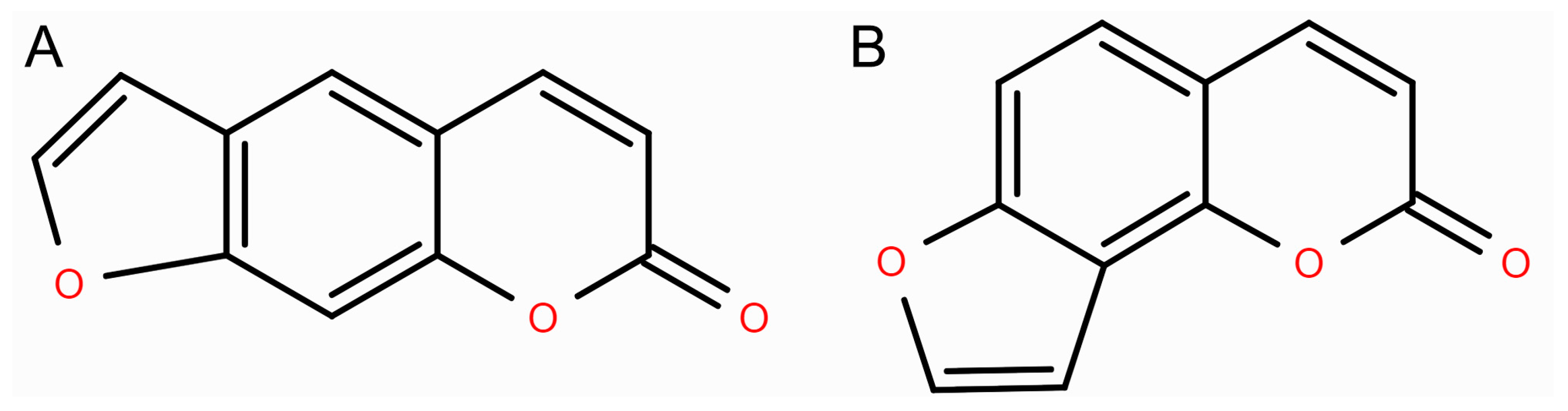
Figure 1. Chemical structures of linear (A) and angular (B) furanocoumarins.
Futurepharmacol 06 00036 g001
Figure 2. Schematic representation of xanthotoxin biosynthesis.
Futurepharmacol 06 00036 g002
Figure 3. General mechanisms of the anticancer effect of xanthotoxin.
Futurepharmacol 06 00036 g003
Disclaimer/Publisher’s Note: The statements, opinions and data contained in all publications are solely those of the individual author(s) and contributor(s) and not of MDPI and/or the editor(s). MDPI and/or the editor(s) disclaim responsibility for any injury to people or property resulting from any ideas, methods, instructions or products referred to in the content.