Article

Interaction of Albacarcin V and Related Polyketides with the Actin-Binding Protein EPLIN: A Molecular Docking Study


by
Gérard Vergoten
1 and
Christian Bailly
2,*
1
Institut de Chimie Pharmaceutique Albert Lespagnol (ICPAL), Faculté de Pharmacie, University of Lille, Inserm, INFINITE—U1286, F-59000 Lille, France
2
CHU de Lille, U1366 Inserm, UMR9020 CNRS, CRCLille—Cancer Research Center of Lille, University of Lille, F-59000 Lille, France
*
Author to whom correspondence should be addressed.
Future Pharmacol. 2026, 6(2), 20; https://doi.org/10.3390/futurepharmacol6020020
Submission received: 23 February 2026 / Revised: 9 March 2026 / Accepted: 23 March 2026 / Published: 1 April 2026

Abstract

Background/Objectives. The actin-binding protein EPLIN (epithelial protein lost in neoplasm), also known as LIMA1, contributes to the maintenance of cytoskeleton structure and dynamic. This protein, which interacts with multiple partners to regulate cell adhesion and migration, has been implicated in the progression of solid tumors and in tumor metastasis. Consequently, small molecules binding to EPLIN are actively searched. EPLIN has been characterized as a molecular target for the antitumor antibiotic albacarcin V which affects the cytoskeletal structure and induces cell growth arrest. Methods. We have modeled the binding of albacarcin and naturally occurring derivatives to EPLIN conformers, in order to locate the drug-binding site and to identify additional EPLIN binders. Nineteen compounds were studied, including albacarcins V (vinyl) and M (methyl), five gilvocarcins, four ravidomycins, two chrysomycins, and six related products (including polycarcin and fucomycin). Results. The modeling analysis confirmed the capacity of albacarcin V to bind to EPLIN and identified a few better binders. In particular, ravidomycin V bearing a dimethylamino sugar unit were identified as the best binders in the series, along with the two related anticancer natural products FE35A-B. Structure-binding relationships are discussed. The drug-binding site has been localized near the central residue Asn34 in the conformationally constrained domain between the two zinc-binding regions. Conclusions. This study provides guidance to the design of EPLIN inhibitors based on the ravidomycin core structure.

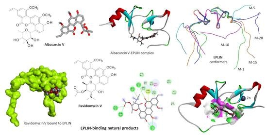
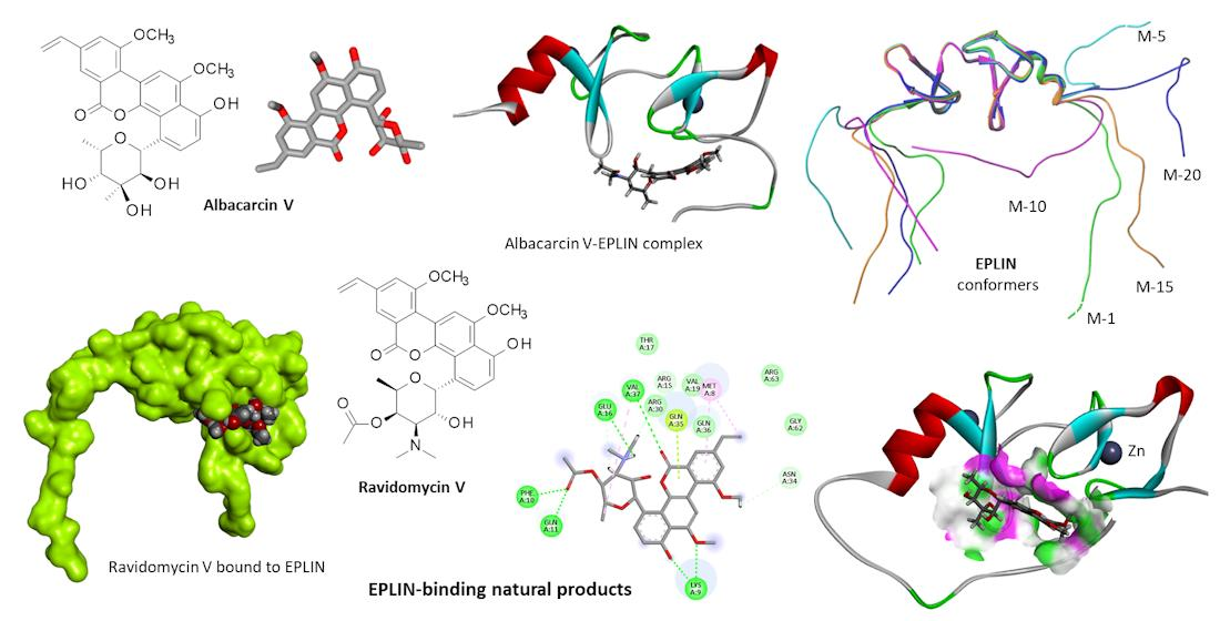
Graphical Abstract

1. Introduction

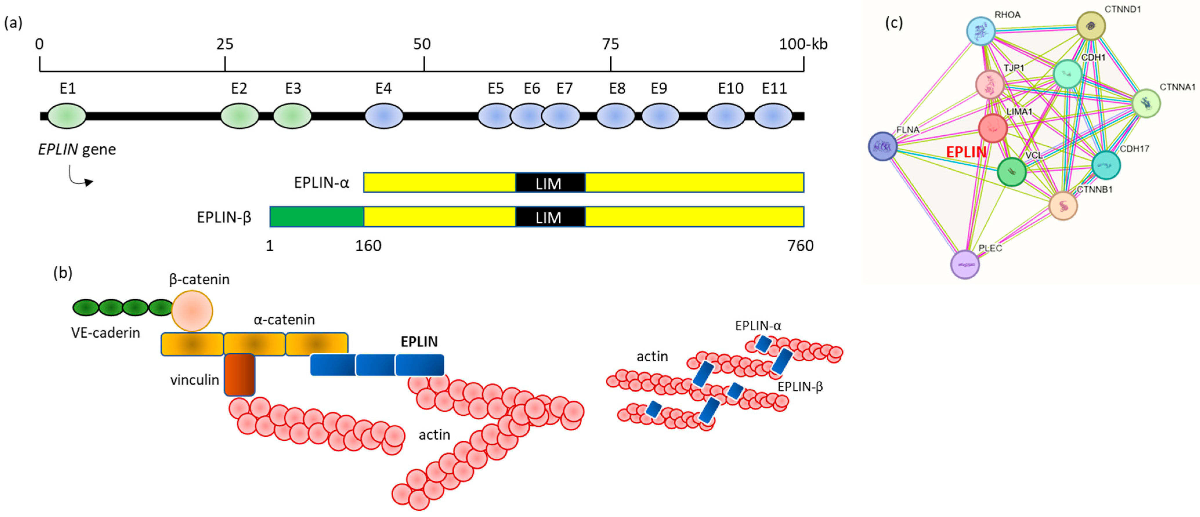
The protein EPLIN (epithelial protein lost in neoplasm), also known as LIMA1 (Lim domain and actin-binding protein 1), is a key regulator of cytoskeleton organization and dynamics. As an actin-binding protein, EPLIN plays major roles in cell growth, cell motility, cell junction remodeling, and cell metabolism. There are two isoforms α and β comprising 600 and 759 amino acids, respectively, generated by alternative splicing from the EPLIN gene (Figure 1a). EPLIN-α/β differentially control actin dynamics in endothelial cells [1,2]. These cytoskeletal proteins localize to filamentous actin to participate in actin-remodeling and in transendothelial migration of different cells, notably leucocytes [3,4]. A study with the mouse protein revealed that mEPLIN-α is largely expressed in embryonic tissue and adult lung and spleen, whereas mEPLIN-β is preferentially expressed in kidney, testis, lung and liver [5]. In humans, EPLIN-β can be detected in epithelial cells of oral mucosa, prostate and mammary glands [6]. EPLIN-β has been identified as a regulator of cell migration [7]. EPLIN-α contributes to the regulation of the dynamic of branched actin filaments [3] (Figure 1b).
At the biochemical level, EPLIN is characterized by the presence of a single centrally located LIM (lin-11, isl-1, and mec-3) domain which mediates protein–protein interactions. The protein can associate with diverse partners, such as the LIM-only domain protein PINCH-1 to regulate integrin-mediated adhesion of keratinocytes, for example [8]. EPLIN interacts with ornithine decarboxylase antizyme 1 (Az1) to modulate cellular migration [7]. The protein can bind also to α-catenin in endothelial and epithelial cells so as to tether the cadherin–catenin complex to the actin cytoskeleton [9]. Other EPLIN-interacting proteins have been identified or predicted (notably using the STRING database), such as the cytoskeleton/cell adhesion protein supervillin, a subunit of the vacuolar H+-ATPase, the actin regulator YWHAH, and a few others [10] (Figure 1c). Via its protein partners, EPLIN plays roles in cell migration, cytoskeletal dynamics, regulation of cell cycle regulation, gene expression, angiogenesis, and lipid metabolism [11].
Figure 1. (a) The genomic and protein structures of human EPLIN isoforms (adapted from [10], Elsevier, 2017). (b) Schematic of the EPLIN-α/β-catenin–cadherin complex linked to the cytoskeleton via branched and linear actin fibers (representation adapted in part from [3], Springer Nature, 2025 and [9], Elsevier, 2012). (c) Interaction of EPLIN (LIMA1) with protein partners (created with String v.12.0, String Consortium 2023, accessed on 18 February 2026, https://string-db.org/). CDH1, Cadherin-1; CDH17, Cadherin-17; CTNNA1, Catenin alpha-1; CTNNB1, Catenin beta-1; CTNND1, Catenin delta-1; FLNA, Filamin-A; PLEC, plectin; RHOA, GTPase RhoA; TJP1, tight junction protein ZO-1; VCL, vinculin.
Futurepharmacol 06 00020 g001
Recent studies have highlighted the key roles of EPLIN in the development and progression of solid tumors. Dysregulation of EPLIN is implicated in tumor growth, invasion, metastasis, and treatment resistance [10]. The altered expression of EPLIN in cancer cells leads to alterations of cytoskeletal dynamics, and hence to aberrant cell motility and invasiveness properties of tumor cells [12]. The protein is now considered as a putative tumor suppressor in colorectal tumors [13]. EPLIN plays an essential role in (i) prostate cancer, as a negative regulator of EMT (epithelial-mesenchymal transition) [14,15,16], (ii) gastric cancer, as a positive response element to chemotherapy [17], (iii) breast cancer, to control cell motility [18], (iv) pancreatic cancer, with a role in tumor growth and drug resistance [19], (v) ovarian cancer, as an inhibitor of cancer cell growth and migration [20], (vi) liver cancers (cholangiocarcinoma) [21], and other solid tumors, including pediatric tumors such as neuroblastoma and medulloblastoma [22]. EPLIN participates in cancer metastasis and resistance [12,23]. For these reasons, EPLIN is now considered as a therapeutic target in oncology and the discovery and design of small molecules targeting EPLIN is viewed as an option to combat cancers.
Recently, the small molecule 4-formylcolchicine (also designated FLIX 5 or NSC328403, Figure 2) targeting EPLIN has been shown to interact with an α-helix portion of the protein [22]. Even more recently, the antibiotic albacarcin V (FLIX 3, NSC354844, virenomycin V) was also identified as an EPLIN-targeting compound endowed with potent antitumor activities (Figure 2). The natural product showed marked activities in drug-resistant, triple-negative breast cancer (TNBC) and ovarian cancer. EPLIN was identified as its primary target, using a cellular thermal shift assay (CETSA) and other biochemical and cellular approaches [24]. These studies demonstrated that EPLIN is a druggable target and confirm its therapeutic value [13].
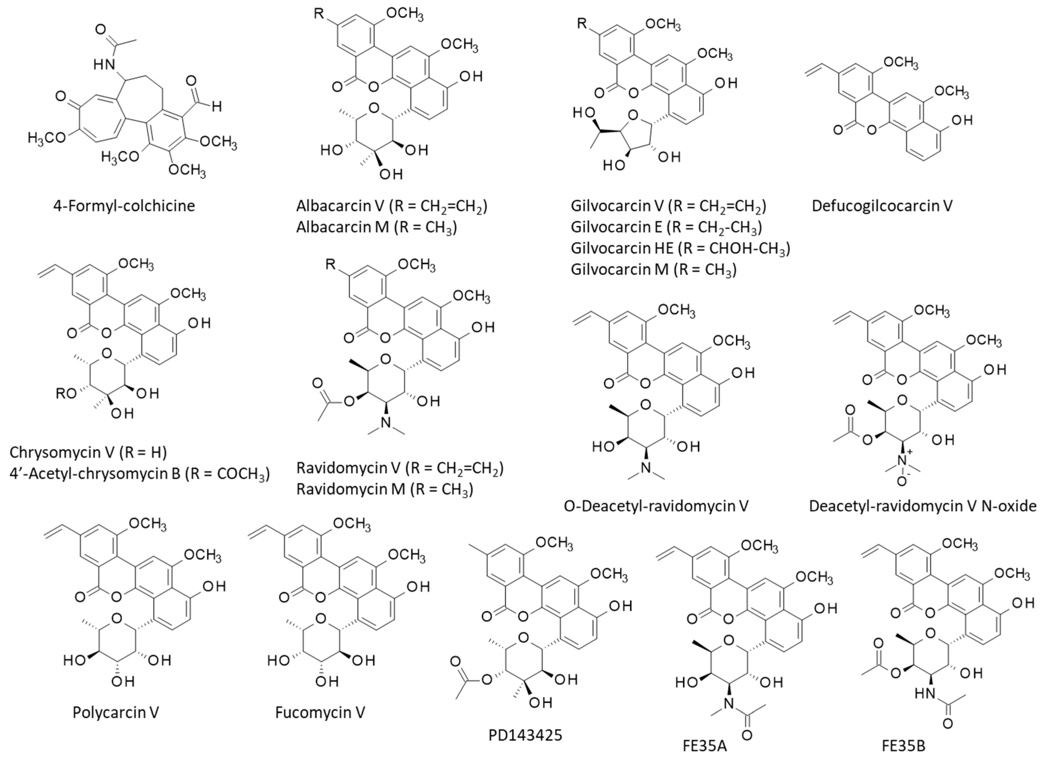
These considerations prompted us to search for other small molecules susceptible to bind to EPLIN. For this purpose, a molecular modeling analysis of small molecules binding to EPLIN was set up starting from the NMR-based structure of the LIM domain of human EPLIN (PDB: 2D8Y) and using albacarcin V as a lead model. On the one hand, the protein structure was analyzed to define the potential binding site close to the zinc finger sequence motif with a conserved distribution of cysteine and histidine residues. On the other hand, a library of 18 naturally occurring albacarcin derivatives was built. All compounds bear a close structural analogy with albacarcin V (Figure 2). They were selected to define structure-binding relationships. The study identifies a few natural products susceptible to form stable complexes with EPLIN, with an affinity comparable or superior to that of albacarcin V. Preliminary structure-binding relationships have been defined. The study shall guide the design of novel albacarcin-based EPLIN inhibitors.

2. Materials and Methods

2.1. Molecular Structures and Software

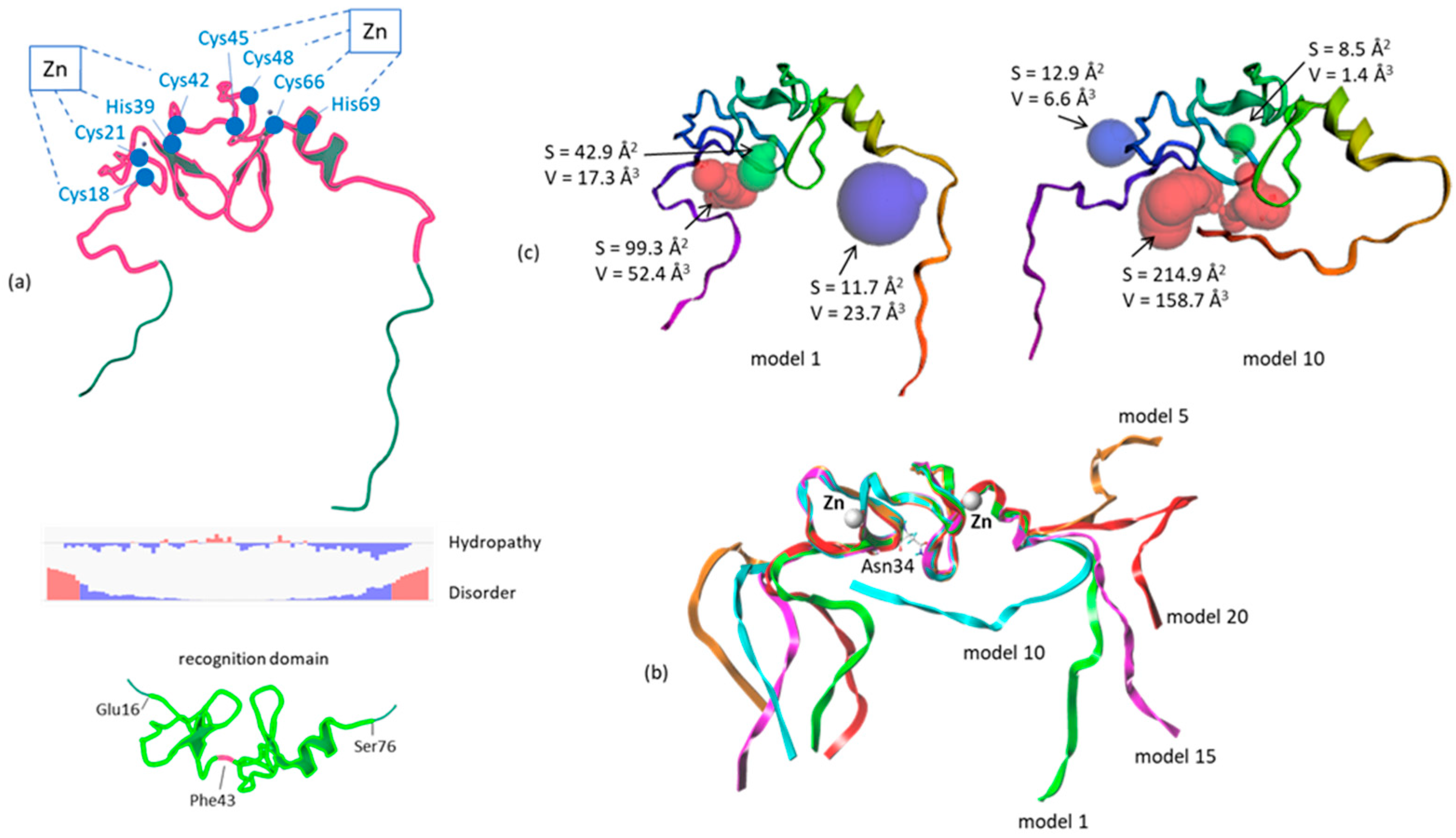
A solution structure of the LIM domain of human EPLIN is available from the protein data bank. It is an NMR-based structure of a single chain protein (91 amino acids) comprising a well-organized central portion (positions 9–82) sandwiched between two short disordered, flexible sequences (1–8 and 83–91) (PDB access code 2D8Y). The short protein includes two zinc-binding sites. The first one implicates residues Cys18-Cys21-His39 and Cys42, the second one residues Cys45-Cys48-Cys66-His69. They delimit to loops on both sides of the central residue Phe43, as illustrated in Figure 3a. The central portion of 71 aa (Glu16-Ser76) is well-organized with two small hydrophilic zones. The GOLD software (v5.3, Cambridge Crystallographic Data Centre, Cambridge, UK) was used for the docking analyses and the BOSS software (v4.9) was used to perform Monte Carlo conformational searches [25]. The compound structures, obtained from the PubChem database or created from the original publications, were represented graphically and analyzed using the Biovia 2020 discovery studio visualizer, from Dassault Systèmes (San Diego, CA, USA). The drug-binding site was identified with the freely accessible web server CASTpFold (Computed Atlas of Surface Topography of proteins) at https://cfold.bme.uic.edu/castpfold/, accessed on 19 December 2025 [26]. The UCSF Chimera molecular modeling package (v1.15) was used for visualization (http://www.cgl.ucsf.edu/chimera/), accessed on 23 January 2026.

2.2. Molecular Docking Analysis

EPLIN structure 2D8Y was analyzed with CASTpFold to locate potential drug-binding pockets. The available NMR-based protein structure includes 20 conformations which differ from the orientation of the above-mentioned flexible sequences. The central portion (Glu16-Ser76) remains practically identical in all models. A representation of five overlapped conformers (models 1, 5, 10, 15, 20) is shown in Figure 3b. Two representative models (1 and 10) were chosen for the docking analysis and identification of the binding pocket using CASTpFold (Figure 3c). It is a convenient and efficient software to analyze the 3D protein geometry, to localize ligand-binding sites and to identify essential amino acid residues involved in binding interactions in combination with the modeling software [27,28]. The best binding site was identified around residue Asn34 (ΔE = −74.0 kcal/mol), compared to the adjacent positions Glu20, Gln11, Ala58, Ser59, Gly62, and Ser76 (ΔE = −55.6, −58.7, −52.8, −53.3, −59.4, and −62.6 kcal/mol, respectively) (Figure 3c). The binding site was thus centered on the Cα of Asn34 of EPLIN. The side chains of amino acids Val19, Leu31, Asn34, Gln36, Phe38, Phe43, Leu54, Tyr57, Ile64, and Tyr65 were rendered fully flexible during the docking process. The docking grid centered on Asn34. The correct binding mode for each ligand was determined after selection of >100 poses, all valid from the energy analysis. The fitness scoring function was used to rank and to select the best poses. The Piecewise Linear Potential (PLP) fitness values were used to select the 6 best binding poses from which the binding energies were then calculated. The scoring function is incorporated into GOLD [29]. The empirical potential energy of the interaction (ΔE(interaction) = E(complex) − [E(protein) + E(ligand)]) was calculated for each compound. The SPASIBA spectroscopic method provides refined empirical MM force field parameters necessary to calculate the binding energies [30,31]. This specific force field for Monte Carlo (MC) simulations achieved the same level of convergence as Molecular Dynamics (MD), with less computation time [32]. Free energies of hydration (ΔG) were calculated using the Molecular Mechanics/Generalized Born Surface Area (MM/GBSA) procedure [33]. The same procedure was used for all natural products.

3. Results

3.1. Compounds Selection

The study was initiated with the two ligands for which binding to EPLIN has been validated experimentally: albacarcin V and 4-formyl-colchicine (Figure 2). Better results were obtained with the antibiotic albacarcin than with the tropolone alkaloid (see below). Therefore, the albacarcin (virenomycin) series was investigated further.
Albacarcin V (vinyl) and its methyl analogue (albacarcin M), produced by Streptomyces albaduncus, were compared. Then, we searched for structural analogues among natural products and identified sixteen compounds, including five gilvocarcin derivatives, four ravidomycin derivatives, two chrysomycin derivatives, and five other related products. The selection included polycarcin V, which is a gilvocarcin-type polyketide produced by Streptomyces polyformus and considered a DNA-damaging agent [34]. Fucomycin V has a D-fucopyranose unit instead of a D-ravidosamine in ravidomycin [35]. It was selected together with the anticancer natural products FE35A and FE35B from Streptomyces rochei [36]. Finally, we added the related product PD143425, described in the PubChem (CID: 163021450). Altogether, 19 compounds were analyzed for their capacity to bind to EPLIN (Figure 2). Most of them belong to the ravidomycin/gilvocarcin/chrysomycin group in the large class of angucycline-type aromatic polyketides [37,38].

3.2. Molecular Modeling Analysis

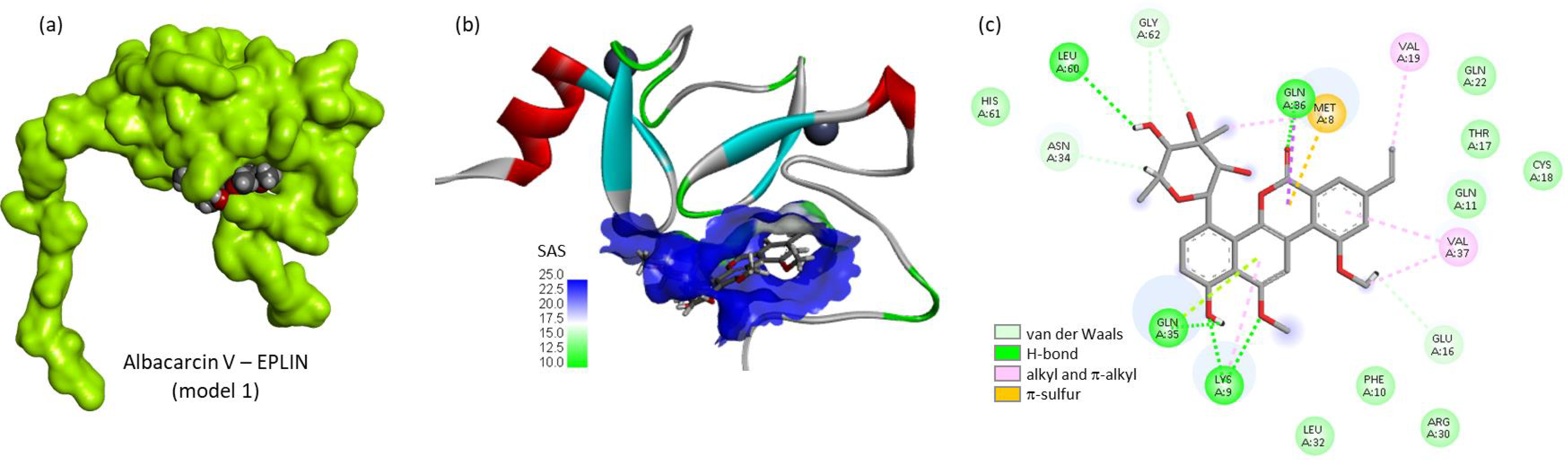
For each compound, docking against conformers 1 and 10 of the EPLIN protein was performed and the top scoring poses were selected. These two models were chosen because the orientation of the flexible side chains differs markedly between these two models and in model 10, the Ct side chain comes close to the Zn-bound structured region (Figure 3b). Table 1 reports the empirical energy of interaction (ΔE) and free energy of hydration (ΔG) measured for each compound using the two protein conformers. The reference product albacarcin V was found to form stable complexes with model 1 of EPLIN (Figure 4). Both the tetracyclic portion and the carbohydrate residue participate in the protein interaction. The drug positions itself below the two protein loops with a β-turn organization in a narrow region well accessible to the solvent. For this compound, the flexible (disordered) peptide portions on both sides of the central EPLIN recognition motif seem to influence significantly the binding process. Four H-bonds with residues Lys9, Gln35, Leu 60, and Gln86 contribute to stabilize the drug−protein complexes, in addition to a π-sulfur interaction (Met8) and several van der Waals contacts (Figure 4c). Both the tetracyclic chromophore and the appended glycosyl residue are implicated in the protein-binding process. In contrast, no major difference was observed with 4-formylcolchicine which binds equally well to EPLIN conformers 1 and 10. Based on these observations, the two models were used to evaluate binding of all the other ligands to EPLIN.
The docking results are relatively complex to interpret because the extent of binding is influenced by both the ligand structure and protein conformation. With model 10, binding energies (ΔE) vary from −42.30 kcal/mol (least favored ligand) to −84.00 kcal/mol (most favored ligand), representing a considerable amplitude of energy for a short series of ligands. All designed models have been analyzed and compared. Some interesting structure-binding information can be deduced from these calculations:
(i)
The drug glycosyl moiety plays a significant role in the protein interaction. The gilvocarcin V aglycone (defucogilvocarcin) binds much less efficiently to EPLIN compared to gilvocarcin V. The 4-fucofuranosyl moiety of gilvocarcin V contributes to the interaction with the protein. In all cases, the glycosyl residue attached to the tetracyclic unit is engaged in one or two H-bonds with the protein.
(ii)
The nature of the glycosyl residues influences significantly protein binding. In this respect, it is interesting to compare binding of the three compounds gilvocarcin V, chrysomycin V and polycarcin V, which only differ by their glycosyl residue. They share the same 6H-benzo[d]naphtho [1,2-b]pyran-6-one core but a distinct C-glycosyl residue. Gilvocarcin has a furanosyl residue with an E conformation [39]. Polycarcin presents an α-L-rhamnopyranosyl moiety instead of the D-fucofuranose of gilvocarcin. The methyl-pentose unit of polycarcin is replaced with a 3,5-dimethylpentose in the chrysomycins [40]. The best results were obtained with chrysomycin V (also known as chrysomycin A). This observation opens the door to the testing of analogues, such as 4′-acetylated chrysomycin A/B derivatives which have shown high cytotoxicity toward cancer cells [41].
(iii)
At first sight, an acetyl group on the glycosyl residue does not seem to be a detrimental element for EPLIN binding. Compound PD143425, which is a 4′-acetyl derivative of albacarcin M, showed a good binding to the protein model 1, a little better compared to albacarcin M.
(iv)
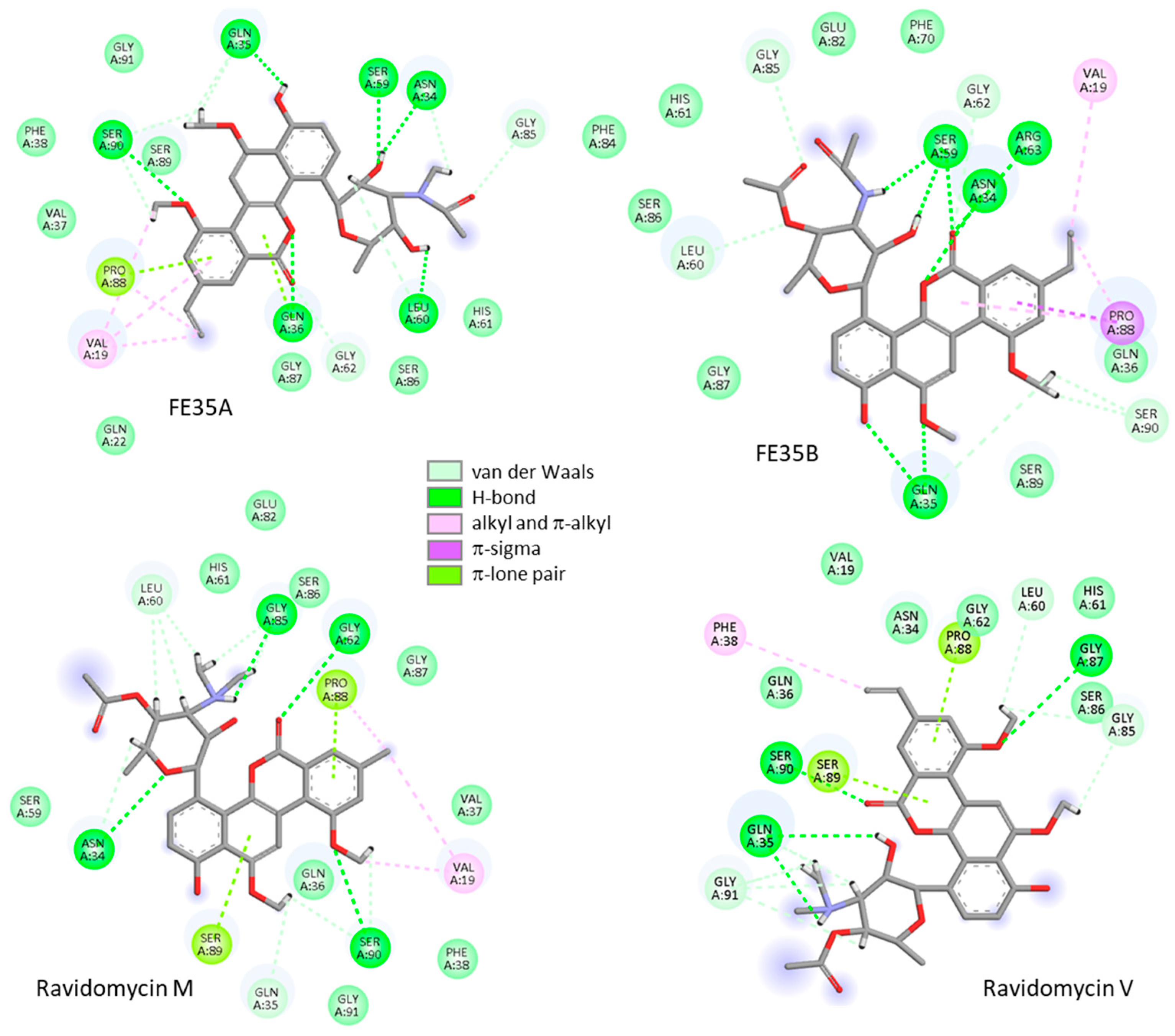
The replacement of the vinyl group on the antibiotics with a methyl group (V vs. M series) is often, but not always, unfavorable to protein interaction. With model 1, albacarcin V was found to be a better binder compared to albacarcin M, and the same trend was observed with ravidomycin V vs. M. But the reverse situation was found with model 10. In fact, the comparison of gilvocarcins V (vinyl), M (methyl), E (ethyl), HE (hydroxyethyl) suggests that the substituent at this position is not a major determinant to the protein interaction. However, it can help to have an ethyl or vinyl group at this position. The vinyl group of FE35A-B is implicated in an alkyl interaction with residue Val19 and the same interaction was observed in the case of albacarcin V, but no such interaction occurred in the case of albacarcin M (Figure 5).
(v)
The comparison of deacetyl-ravidomycin V and its N-oxide derivative suggests that the incorporation of a NO group on glycosyl residue can contribute to the protein interaction. With a single N-oxide compound in this series, no definitive conclusion can be drawn at present. However, the fact that compounds FE35A-B also present good binding characteristics reinforces this observation. FE35A, with an amino-acetyl group on the sugar residue, binds equally well to models 1 and 10 of EPLIN.
(vi)
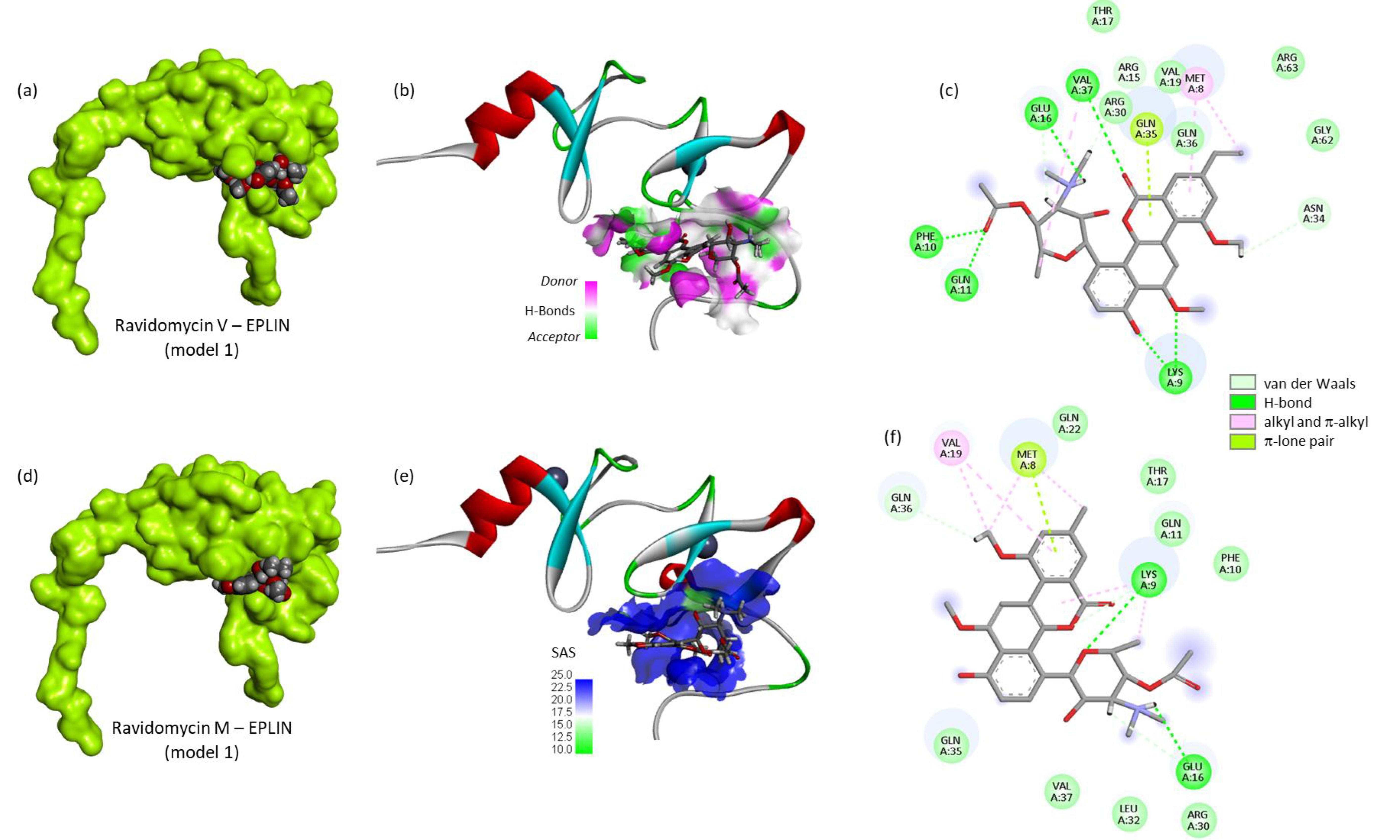
The presence of an amino sugar residue, as in the ravidomycin series, is a favorable element for drug binding to EPLIN (Figure 6). A significantly distinct binding behavior was observed with ravidomycin V vs. M with model 1 but globally the two compounds gave good results with the two protein conformers. The ravidomycin V-EPLIN model 1 recapitulates well the key binding elements: (a) a contribution of the vinyl unit (alkyl interaction with Met8), (b) importance of the O-acetyl on the sugar residue (two adjacent H-bonds to Ph10 and Gln11), (c) major role of the dimethylamino sugar residue (H-bond to Glu16), (d) contribution of the phenol-OH (H-bond to Val37). Altogether, these multiple interactions confer a high stability to the ravidomycin V-EPLIN complex.
We have extended our investigation with the ravidomycin V/M pair to compare their binding to models 1-5-10-15-20 (Table 2). The comparison of the five protein conformers reveals the influence of the two disordered regions flanking the central structured motif delimited by the Zn-binding units (Figure 3b). The analysis indicated the drug-binding process is not uniquely dependent on the central structured-motif; the flanking sequences participate in the drug interaction process. Ravidomycin V showed an equally good binding to EPLIN models 1-10-20 whereas binding to models 5-15 was less favored. About the same trend was observed with ravidomycin M, although the vinyl compound is a better binder than the methyl equivalent. The ravidosamine unit seems to contribute very positively to the protein interaction. Similarly, it is interesting to underline compound FE35A which binds equally well to EPLIN models 1 and 10, as observed with ravidomycin V. These two compounds can form stable complexes with EPLIN and their protein interaction is less influenced by the orientation of the flexible portion of the protein compared to the natural products studied (Figure 5, Table 1). Altogether, the comparison of the different ligands suggests that an amino sugar unit represents a favorable element for binding to EPLIN. The aminoglycosyl unit reinforces the stability of the EPLIN−drug complex.
Two conclusions emerge from our modeling analysis. From a protein perspective, four amino acid residues are predominantly implicated in drug interactions: Asn34, Gln35, Ser59 and Ser90. They are involved in H-bond interactions with many of the tested compounds, essentially with the (amino)glycosyl residue of the drugs. It would be interesting to design and test protein mutants (e.g., Gln/Thr, Ser/Gly) to better appreciate their specific contribution to the drug interaction. From a drug perspective, the in silico study agrees well with the experimental data which have evidenced binding of albacarcin V to EPLIN. In addition, the computational analysis points to the related product ravidomycin V as a potentially stronger EPLIN binder.

4. Discussion

Over the past ten years, Epithelial Protein Lost In Neoplasm or EPLIN (LIMA1) has emerged as a major regulator of cytoskeletal organization and dynamics. Its important roles in cell growth, motility and metabolism have been well characterized. The protein is largely expressed in malignant tumors and its roles in oncogenesis and tumor invasion are better and better understood. EPLIN is now considered a potential therapeutic target in oncology [23,42]. Recent studies have underlined the roles of EPLIN in tumorigenesis, metastasis and drug resistance [43,44,45]. Beyond cancer, EPLIN plays a role in lipid metabolism and cholesterol absorption [46,47,48]. As a consequence, EPLIN is now viewed as a new drug target to treat cancer and liver diseases [49]. A few peptides and small molecules have been identified recently, such as conotoxin peptides and the 4-formylcolchicine [22,50]. Very few EPLIN ligands have been identified thus far. The most promising compound is arguably the C-glycoside albacarcin V which has demonstrated a strong binding capacity to EPLIN [24]. This natural product caught our attention for at least two reasons. On the one hand, a good set of derivatives can be found in nature, notably most of the analogues used in this study. On the other hand, chemical approaches have been proposed to elaborate synthetic derivatives [51,52,53,54,55]. Recently, the total (gram-scale) syntheses of polycarcin V, gilvocarcin V, chrysomycin A and over 30 synthetic derivatives have been reported. Analogues were designed primarily as antituberculosis agents, but the proposed chemical platform can be exploited to design EPLIN binders [56].
Our docking study provides important information to guide future drug design. It is in a good agreement with the experimental data showing that albacarcin V can bind to EPLIN [24]. The modeling indicates the position of the drug-binding site in the protein, located in the conformationally constrained region of the β-sheets between the two zinc-binding regions. The central residue Asn34 is implicated in the drug-binding process. This central residue seems to play a major role in the drug–protein interaction. It will be necessary to validate this observation experimentally. In addition, the docking suggests new protein binders in the albacarcin series, notably the derivatives ravidomycin V/M and natural analogues FE35A-B. The best ligand in the series is ravidomycin V which can bind well to different conformers of EPLIN. This antitumor antibiotic, first discovered >20 years ago from Streptomyces ravidus, has shown marked antitumor activities in vivo, when administered to tumor-bearing rats [57]. It is an interesting active molecule and a useful model compound because both its biosynthesis and chemical synthesis have been described [58,59,60]. Chemically-modified analogues have been proposed in the past [61] and synthetic analogues have been recently designed [35]. Derivatives of the ravidosamine moiety of the natural product, which is well implicated in the binding to EPLIN, are also chemically accessible [62]. Therefore, a range of ravidomycin-type products could be accessed to investigate further binding to EPLIN and the biological consequences of the drug–protein interaction.
Ravidomycin and the two analogues FE35A-B display antitumor properties [36]. Their mechanism of action is not totally clear at present. Inhibition of DNA synthesis and catalytic inhibition of DNA-topoisomerase II were initially evidenced [63,64]. Ravidomycin and gilvocarcin V act as DNA damaging agents [65,66]. Chrysomycin A is also a topoisomerase II inhibitor [67]. Upon photoactivation, gilvocarcin V has been shown to selectively induce DNA cross-linking of phosphorylated histone H3 and the heat shock protein GRP78 [68]. This latter chaperone is a regulator of endoplasmic reticulum (ER) functions and an attractive anticancer drug target [69,70]. Beyond an action at the nuclear level, these compounds have been found to act on cell motility, reducing the invasion and migration capacities of tumor cells [71,72]. They exhibit DNA-independent activities. For example, deacetyl-ravidomycin M has been shown to inhibit CD23 expression in U937 cells [73,74] 4′-hydroxy-gilvocarcin V was shown to target the EGFR/Neu pathway in lung cancer [75], and chrysomycin A was found to modulate PI3K-Akt and Wnt signaling pathways in neuroglioma cells [76]. This latter compound, which is a bona fide EPLIN binder, exerts multiple activities, notably regulating enzymes implicated in oxidative stress [72]. These types of benzonaphthopyranone glycosides are multitargeted compounds [77]. Many aspects of the mechanism of action of these compounds remain to be discovered, understood and improved.

5. Conclusions

The present in silico analysis (i) confirms the good capacity of albacarcin V to bind to EPLIN (LIMA1), (ii) identifies the central zinc-binding subdomain as the potential binding site, (iii) suggests that several albacarcin analogues, notably ravidomycin V, can form very stable complexes with EPLIN, and (iv) provides structure-binding information to guide the design of novel EPLIN binders. This information shall help the identification of other natural products and the rational design of small molecules targeting EPLIN to tackle cancer. The next step will be to validate experimentally the capacity of ravidomycin V to bind to EPLIN and to investigate the cellular consequences of the EPLIN-drug binding. Cells overexpressing the cytoskeleton-associated protein can be used to investigate drug effects on cell growth, migration and invasiveness. Complementary biophysical and cellular methods can be employed to demonstrate the capacity of albacarcin V to interact with EPLIN and to regulate its functions. The present study offers interesting perspectives for this antitumor antibiotic and its analogues.

Author Contributions

Investigation, G.V. and C.B.; visualization, G.V. and C.B.; software, G.V.; methodology, G.V.; conceptualization, C.B.; writing—original draft, C.B.; writing—review and editing, C.B. All authors have read and agreed to the published version of the manuscript.

Funding

This research received no external funding.

Institutional Review Board Statement

Not applicable.

Informed Consent Statement

Not applicable.

Data Availability Statement

No new data were created or analysed in this study. Data sharing is not applicable to this article.

Conflicts of Interest

The authors declare no conflicts of interest.

Abbreviations

The following abbreviations are used in this manuscript:
EPLINepithelial protein lost in neoplasm
LIMlin-11, isl-1, and mec-3
LIMA1Lim domain and actin-binding protein 1
MM/GBSAMolecular Mechanics/Generalized Born Surface Area

References

  1. Taha, M.; Aldirawi, M.; März, S.; Seebach, J.; Odenthal-Schnittler, M.; Bondareva, O.; Bojovic, V.; Schmandra, T.; Wirth, B.; Mietkowska, M.; et al. EPLIN-α and -β Isoforms Modulate Endothelial Cell Dynamics through a Spatiotemporally Differentiated Interaction with Actin. Cell Rep. 2019, 29, 1010–1026. [Google Scholar] [CrossRef] [PubMed]
  2. Chen, S.; Maul, R.S.; Kim, H.R.; Chang, D.D. Characterization of the human EPLIN (Epithelial Protein Lost in Neoplasm) gene reveals distinct promoters for the two EPLIN isoforms. Gene 2000, 248, 69–76. [Google Scholar] [CrossRef]
  3. Aldirawi, M.; Ghanbari, P.; Mietkowska, M.; März, S.; Odenthal-Schnittler, M.; Franz, J.; Wegner, J.; Currie, S.; Kipcke, J.P.; Taha, M.; et al. A specific role for endothelial EPLIN-isoform-regulated actin dynamics in neutrophil transmigration. Sci. Rep. 2025, 15, 15698. [Google Scholar] [CrossRef]
  4. Song, Y.; Maul, R.S.; Gerbin, C.S.; Chang, D.D. Inhibition of anchorage-independent growth of transformed NIH3T3 cells by epithelial protein lost in neoplasm (EPLIN) requires localization of EPLIN to actin cytoskeleton. Mol. Biol. Cell 2002, 13, 1408–1416. [Google Scholar] [CrossRef]
  5. Maul, R.S.; Sachi Gerbin, C.; Chang, D.D. Characterization of mouse epithelial protein lost in neoplasm (EPLIN) and comparison of mammalian and zebrafish EPLIN. Gene 2001, 262, 155–160. [Google Scholar] [CrossRef]
  6. Maul, R.S.; Chang, D.D. EPLIN, epithelial protein lost in neoplasm. Oncogene 1999, 18, 7838–7841. [Google Scholar] [CrossRef]
  7. Li, D.; Neo, S.P.; Gunaratne, J.; Sabapathy, K. EPLIN-β is a novel substrate of ornithine decarboxylase antizyme 1 and mediates cellular migration. J. Cell Sci. 2023, 136, jcs260427. [Google Scholar] [CrossRef]
  8. Karaköse, E.; Geiger, T.; Flynn, K.; Lorenz-Baath, K.; Zent, R.; Mann, M.; Fässler, R. The focal adhesion protein PINCH-1 associates with EPLIN at integrin adhesion sites. J. Cell Sci. 2015, 128, 1023–1033. [Google Scholar] [CrossRef]
  9. Chervin-Pétinot, A.; Courçon, M.; Almagro, S.; Nicolas, A.; Grichine, A.; Grunwald, D.; Prandini, M.H.; Huber, P.; Gulino-Debrac, D. Epithelial protein lost in neoplasm (EPLIN) interacts with α-catenin and actin filaments in endothelial cells and stabilizes vascular capillary network in vitro. J. Biol. Chem. 2012, 287, 7556–7572. [Google Scholar] [CrossRef] [PubMed]
  10. Wu, D. Epithelial protein lost in neoplasm (EPLIN): Beyond a tumor suppressor. Genes Dis. 2017, 4, 100–107. [Google Scholar] [CrossRef] [PubMed]
  11. Zeng, J.; Jiang, W.G.; Sanders, A.J. Epithelial Protein Lost in Neoplasm, EPLIN, the Cellular and Molecular Prospects in Cancers. Biomolecules 2021, 11, 1038. [Google Scholar] [CrossRef] [PubMed]
  12. Lindell, E.; Zhang, X. Exploring the Enigma: The Role of the Epithelial Protein Lost in Neoplasm in Normal Physiology and Cancer Pathogenesis. Int. J. Mol. Sci. 2024, 25, 4970. [Google Scholar] [CrossRef]
  13. Zeng, J.; Sanders, A.J.; Ye, L.; Hargest, R.; Ruge, F.; Jiang, W.G. EPLIN, a Putative Tumour Suppressor in Colorectal Cancer, Implications in Drug Resistance. Int. J. Mol. Sci. 2022, 23, 15232. [Google Scholar] [CrossRef]
  14. Wu, D.; Osunkoya, A.O.; Kucuk, O. Epithelial protein lost in neoplasm (EPLIN) and prostate cancer: Lessons learned from the ARCaP model. Am. J. Clin. Exp. Urol. 2021, 9, 264–276. [Google Scholar]
  15. Collins, R.J.; Morgan, L.D.; Owen, S.; Ruge, F.; Jiang, W.G.; Sanders, A.J. Mechanistic insights of epithelial protein lost in neoplasm in prostate cancer metastasis. Int. J. Cancer. 2018, 143, 2537–2550. [Google Scholar] [CrossRef]
  16. Sanders, A.J.; Martin, T.A.; Ye, L.; Mason, M.D.; Jiang, W.G. EPLIN is a negative regulator of prostate cancer growth and invasion. J. Urol. 2011, 186, 295–301. [Google Scholar] [CrossRef] [PubMed]
  17. Gong, W.; Zeng, J.; Ji, J.; Jia, Y.; Jia, S.; Sanders, A.J.; Jiang, W.G. EPLIN Expression in Gastric Cancer and Impact on Prognosis and Chemoresistance. Biomolecules 2021, 11, 547. [Google Scholar] [CrossRef] [PubMed]
  18. Jäntti, N.Z.; Moreno-Layseca, P.; Chastney, M.R.; Dibus, M.; Conway, J.R.W.; Leppänen, V.M.; Hamidi, H.; Eylmann, K.; Oliveira-Ferrer, L.; Veltel, S.; et al. EPLINα controls integrin recycling from Rab21 endosomes to drive breast cancer cell migration. Dev. Cell 2025, 60, 3018–3033. [Google Scholar] [CrossRef]
  19. Zeng, J.; Wang, C.; Ruge, F.; Ji, E.K.; Martin, T.A.; Sanders, A.J.; Jia, S.; Hao, C.; Jiang, W.G. EPLIN, a prospective oncogenic molecule with contribution to growth, migration and drug resistance in pancreatic cancer. Sci. Rep. 2024, 14, 30850. [Google Scholar] [CrossRef]
  20. Liu, R.; Martin, T.A.; Jordan, N.J.; Ruge, F.; Ye, L.; Jiang, W.G. Epithelial protein lost in neoplasm-α (EPLIN-α) is a potential prognostic marker for the progression of epithelial ovarian cancer. Int. J. Oncol. 2016, 48, 2488–2496. [Google Scholar] [CrossRef]
  21. Obulkasim, H.; Adili, A.; Liu, Y.; Duan, S. Expression and molecular insights of lima1 in cholangiocarcinoma. Cell Adh. Migr. 2024, 18, 4–17. [Google Scholar] [CrossRef]
  22. Lindell, E.; Guo, J.; Zhao, M.; Rameika, N.; Lu, X.; Wacker, T.; Zhong, L.; Bergström, T.; Svanberg, S.; Chowdhury, A.I.; et al. Identification of a small molecule targeting EPLIN as a novel strategy for the treatment of pediatric neuroblastoma and medulloblastoma. Cell Death Dis. 2025, 16, 554. [Google Scholar] [CrossRef]
  23. Collins, R.J.; Jiang, W.G.; Hargest, R.; Mason, M.D.; Sanders, A.J. EPLIN: A fundamental actin regulator in cancer metastasis? Cancer Metastasis Rev. 2015, 34, 753–764. [Google Scholar] [CrossRef]
  24. Krahulcová, L.; Lindell, E.; Lu, X.; Conejeros Monsalve, J.J.; Haraldsson, M.; Li, Z.; Chen, X.; Aune, G.; Zhao, M.; Tong, L.; et al. Albacarcin V adds EPLIN as a novel and promising target for the treatment of female cancers and pediatric medulloblastoma. Biochem. Pharmacol. 2026, 244, 117625. [Google Scholar] [CrossRef]
  25. Jorgensen, W.L.; Tirado-Rives, J. Molecular modeling of organic and biomolecular systems using BOSS and MCPRO. J. Comput. Chem. 2005, 26, 1689–1700. [Google Scholar] [CrossRef]
  26. Ye, B.; Tian, W.; Wang, B.; Liang, J. CASTpFold: Computed Atlas of Surface Topography of the universe of protein Folds. Nucleic Acids Res. 2024, 52, W194–W199. [Google Scholar] [CrossRef]
  27. Zhao, J.; Cao, Y.; Zhang, L. Exploring the computational methods for protein-ligand binding site prediction. Comput. Struct. Biotechnol. J. 2020, 18, 417–426. [Google Scholar] [CrossRef] [PubMed]
  28. Huang, B.; Schroeder, M. LIGSITEcsc: Predicting ligand binding sites using the Connolly surface and degree of conservation. BMC Struct. Biol. 2006, 6, 19. [Google Scholar] [CrossRef] [PubMed]
  29. Jones, G.; Willett, P.; Glen, R.C.; Leach, A.R.; Taylor, R. Development and validation of a genetic algorithm for flexible docking. J. Mol. Biol. 1997, 267, 727–748. [Google Scholar] [CrossRef] [PubMed]
  30. Lagant, P.; Nolde, D.; Stote, R.; Vergoten, G.; Karplus, M. Increasing normal modes analysis accuracy: The SPASIBA spectroscopic force field introduced into the CHARMM program. J. Phys. Chem. A 2004, 108, 4019–4029. [Google Scholar] [CrossRef]
  31. Vergoten, G.; Mazur, I.; Lagant, P.; Michalski, J.C.; Zanetta, J.P. The SPASIBA force field as an essential tool for studying the structure and dynamics of saccharides. Biochimie 2003, 85, 65–73. [Google Scholar] [CrossRef]
  32. Jorgensen, W.L.; Tirado-Rives, J. Monte Carlo versus Molecular Dynamics for conformational sampling. J. Phys. Chem. 1996, 100, 14508–14513. [Google Scholar] [CrossRef]
  33. Meziane-Tani, M.; Lagant, P.; Semmoud, A.; Vergoten, G. The SPASIBA force field for chondroitin sulfate: Vibrational analysis of D-glucuronic and N-acetyl-D-galactosamine 4-sulfate sodium salts. J. Phys. Chem. A 2006, 110, 11359–11370. [Google Scholar] [CrossRef]
  34. Yue, Z.; Wu, F.; Guo, F.; Park, J.; Wang, J.; Zhang, L.; Liao, D.; Li, W.; Schärer, O.D.; Lei, X. Polycarcin V induces DNA-damage response and enables the profiling of DNA-binding proteins. Natl. Sci. Rev. 2022, 9, nwac046. [Google Scholar] [CrossRef]
  35. Park, K.J.; Maier, S.; Zhang, C.; Dixon, S.A.H.; Rusch, D.B.; Pupo, M.T.; Angus, S.P.; Gerdt, J.P. Ravidomycin Analogs from Streptomyces sp. Exhibit Altered Antimicrobial and Cytotoxic Selectivity. J. Nat. Prod. 2023, 86, 1968–1979. [Google Scholar] [CrossRef] [PubMed]
  36. Yamashita, N.; Shin-ya, K.; Furihata, K.; Hayakawa, Y.; Seto, H. New ravidomycin analogues, FE35A and FE35B, apoptosis inducers produced by Streptomyces rochei. J. Antibiot. 1998, 51, 1105–1108. [Google Scholar] [CrossRef] [PubMed][Green Version]
  37. Liu, H.S.; Chen, H.R.; Huang, S.S.; Li, Z.H.; Wang, C.Y.; Zhang, H. Bioactive Angucyclines/Angucyclinones Discovered from 1965 to 2023. Mar Drugs 2025, 23, 25. [Google Scholar] [CrossRef]
  38. Xu, X.; Huang, X.; Xu, W. Marine actinomycetes-derived angucyclines and angucyclinones with biosynthesis and activity--past 10 years (2014–2023). Eur. J. Med. Chem. 2025, 283, 117161. [Google Scholar] [CrossRef] [PubMed]
  39. Hirayama, N.; Takahashi, K.; Shirahata, K.; Ohashi, Y.; Sasada, Y. Crystal and molecular structure of antibiotic gilvocarcin M. Bull. Chem. Soc. Jpn. 1981, 54, 1338–1342. [Google Scholar] [CrossRef]
  40. Weiss, U.; Yoshihira, K.; Highet, R.J.; White, R.J.; Wei, T.T. The chemistry of the antibiotics chrysomycin A and B. Antitumor activity of chrysomycin A. J. Antibiot. 1982, 35, 1194–1201. [Google Scholar] [CrossRef]
  41. Wada, S.I.; Sawa, R.; Iwanami, F.; Nagayoshi, M.; Kubota, Y.; Iijima, K.; Hayashi, C.; Shibuya, Y.; Hatano, M.; Igarashi, M.; et al. Structures and biological activities of novel 4′-acetylated analogs of chrysomycins A and B. J. Antibiot. 2017, 70, 1078–1082, Corrigendum in J. Antibiot. 2017, 70, 1150.. [Google Scholar] [CrossRef]
  42. Wang, X.; Zhang, C.; Song, H.; Yuan, J.; Zhang, X.; Yuan, Y.; Zhang, L.; He, J. Characterization of LIMA1 and its emerging roles and potential therapeutic prospects in cancers. Front. Oncol. 2023, 13, 1115943. [Google Scholar] [CrossRef] [PubMed]
  43. Zhang, C.; Wang, X.; Song, H.; Yuan, J.; Zhang, X.; Yuan, Y.; Wang, Z.; Lei, Z.; He, J. M6A modification-mediated LIMA1 promotes the progression of hepatocellular carcinoma through the wnt-βcatenin/Hippo pathway. Cell Biol. Toxicol. 2024, 41, 9. [Google Scholar] [CrossRef]
  44. Lv, Z.; Zhao, S.; Wu, H. LIMA1 inhibits cisplatin resistance and malignant biological behavior of bladder cancer cells by suppressing the Wnt/β-catenin pathway. BMC Med. Genom. 2025, 18, 78. [Google Scholar]
  45. Chen, Y.; Wang, Z.; Sun, Y.; Li, X.; Wang, Y.; Liu, S. Inhibition of Breast Cancer Bone Metastasis by LRP5-Overexpressing Osteocytes via the LIMA1/MYO5B Signaling Axis. Int. J. Mol. Sci. 2026, 27, 777. [Google Scholar] [CrossRef]
  46. Le May, C.; Ducheix, S.; Cariou, B.; Rimbert, A. From Genetic Findings to new Intestinal Molecular Targets in Lipid Metabolism. Curr. Atheroscler. Rep. 2025, 27, 26. [Google Scholar] [CrossRef]
  47. Liu, Z.; Fan, K.; Abudukeremu, A.; Gao, M.; Tan, X.; Mao, X.; Li, X.; Ma, W.; Ma, X.; Li, C.; et al. LIMA1 links the E3 ubiquitin ligase RNF40 to lipid metabolism. Cell Death Discov. 2024, 10, 298. [Google Scholar] [CrossRef]
  48. Li, S.; Yang, F.; Cheng, F.; Zhu, L.; Yan, Y. Lipotoxic hepatocyte derived LIMA1 enriched small extracellular vesicles promote hepatic stellate cells activation via inhibiting mitophagy. Cell. Mol. Biol. Lett. 2024, 29, 82. [Google Scholar] [CrossRef] [PubMed]
  49. Yang, F.; Chen, Y.; Zheng, G.; Gu, K.; Fan, L.; Li, T.; Zhu, L.; Yan, Y. LIMA1 O-GlcNAcylation Promotes Hepatic Lipid Deposition through Inducing Β-catenin-Regulated FASn Expression in Metabolic Dysfunction-Associated Steatotic Liver Disease. Adv. Sci. 2025, 12, e2415941. [Google Scholar] [CrossRef] [PubMed]
  50. Luna-Nophal, A.; Díaz-Castillo, F.; Izquierdo-Sánchez, V.; Velázquez-Fernández, J.B.; Orozco-Morales, M.; Lara-Mejía, L.; Bernáldez-Sarabia, J.; Sánchez-Campos, N.; Arrieta, O.; Díaz-Chávez, J.; et al. Preclinical Efficacy and Proteomic Prediction of Molecular Targets for s-cal14.1b and s-cal14.2b Conotoxins with Antitumor Capacity in Xenografts of Malignant Pleural Mesothelioma. Mar. Drugs 2025, 23, 32. [Google Scholar] [CrossRef]
  51. Liu, W.; Hong, B.; Wang, J.; Lei, X. New Strategies in the Efficient Total Syntheses of Polycyclic Natural Products. Acc. Chem. Res. 2020, 53, 2569–2586. [Google Scholar] [CrossRef]
  52. James, C.A.; Snieckus, V. Combined directed remote metalation− transition metal catalyzed cross coupling strategies: The total synthesis of the aglycones of the gilvocarcins V, M, and E and arnottin I. J. Org. Chem. 2009, 74, 4080–4093. [Google Scholar] [CrossRef]
  53. Macdonald, S.J.; McKenzie, T.C.; Hassen, W.D. Synthesis of the aglycones of the ravidomycin family of antibiotics. J. Chem. Soc. Chem. Commun. 1987, 20, 1528–1530. [Google Scholar] [CrossRef]
  54. Jung, M.E.; Jung, Y.H. Total synthesis of the aglycone of the 8-methyl benzonaphthopyrone antibiotics, gilvocarcin M, virenomycin M, and albacarcin M. Tetrahedron lett. 1988, 29, 2517–2520. [Google Scholar] [CrossRef]
  55. Hart, D.J.; Merriman, G.H. A new synthesis of defucogilvocarcin M. Tetrahedron lett. 1989, 30, 5093–5096. [Google Scholar] [CrossRef]
  56. Wu, F.; Zhang, J.; Song, F.; Wang, S.; Guo, H.; Wei, Q.; Dai, H.; Chen, X.; Xia, X.; Liu, X.; et al. Chrysomycin A Derivatives for the Treatment of Multi-Drug-Resistant Tuberculosis. ACS Cent. Sci. 2020, 6, 928–938. [Google Scholar] [CrossRef] [PubMed]
  57. Sehgal, S.N.; Czerkawski, H.; Kudelski, A.; Pandev, K.; Saucier, R.; Vézina, C. Ravidomycin (AY-25, 545), a new antitumor antibiotic. J. Antibiot. 1983, 36, 355–361. [Google Scholar] [CrossRef]
  58. Kharel, M.K.; Nybo, S.E.; Shepherd, M.D.; Rohr, J. Cloning and characterization of the ravidomycin and chrysomycin biosynthetic gene clusters. Chembiochem 2010, 11, 523–532. [Google Scholar] [CrossRef] [PubMed]
  59. Kharel, M.K.; Pahari, P.; Lian, H.; Rohr, J. Enzymatic total synthesis of rabelomycin, an angucycline group antibiotic. Org. Lett. 2010, 12, 2814–2817. [Google Scholar] [CrossRef]
  60. Futagami, S.; Ohashi, Y.; Imura, K.; Hosoya, T.; Ohmori, K.; Matsumoto, T.; Suzuki, K. Total synthesis of ravidomycin: Revision of absolute and relative stereochemistry. Tetrahedron Lett. 2000, 41, 1063–1067. [Google Scholar] [CrossRef]
  61. Rakhit, S.; Eng, C.; Baker, H.; Singh, K. Chemical modification of ravidomycin and evaluation of biological activities of its derivatives. J. Antibiot. 1983, 36, 1490–1494. [Google Scholar] [CrossRef]
  62. Hsu, D.S.; Matsumoto, T.; Suzuki, K. Efficient synthetic route to ravidosamine derivatives. Synlett 2005, 5, 801–804. [Google Scholar] [CrossRef]
  63. Lorico, A.; Long, B.H. Biochemical characterisation of elsamicin and other coumarin-related antitumour agents as potent inhibitors of human topoisomerase II. Eur. J. Cancer 1993, 29, 1985–1991. [Google Scholar] [CrossRef]
  64. Singh, K. Studies on the mechanism of action of ravidomycin (AY-25,545). J. Antibiot. 1984, 37, 71–73. [Google Scholar] [CrossRef][Green Version]
  65. Greenstein, M.; Monji, T.; Yeung, R.; Maiese, W.M.; White, R.J. Light-dependent activity of the antitumor antibiotics ravidomycin and desacetylravidomycin. Antimicrob. Agents Chemother. 1986, 29, 861–866. [Google Scholar] [CrossRef] [PubMed]
  66. Wei, T.T.; Byrne, K.M.; Warnick-Pickle, D.; Greenstein, M. Studies on the mechanism of action of gilvocarcin V and chrysomycin A. J. Antibiot. 1982, 35, 545–548. [Google Scholar] [CrossRef]
  67. Zhang, J.; Liu, P.; Chen, J.; Yao, D.; Liu, Q.; Zhang, J.; Zhang, H.W.; Leung, E.L.; Yao, X.J.; Liu, L. Upgrade of chrysomycin A as a novel topoisomerase II inhibitor to curb KRAS-mutant lung adenocarcinoma progression. Pharmacol. Res. 2023, 187, 106565. [Google Scholar] [CrossRef] [PubMed]
  68. Matsumoto, A.; Hanawalt, P.C. Histone H3 and heat shock protein GRP78 are selectively cross-linked to DNA by photoactivated gilvocarcin V in human fibroblasts. Cancer Res. 2000, 60, 3921–3926. [Google Scholar] [PubMed]
  69. Yang, Y.; Li, W.; Zhao, Y.; Sun, M.; Xing, F.; Yang, J.; Zhou, Y. GRP78 in Glioma Progression and Therapy: Implications for Targeted Approaches. Biomedicines 2025, 13, 382. [Google Scholar] [CrossRef]
  70. Bailly, C.; Waring, M.J. Pharmacological effectors of GRP78 chaperone in cancers. Biochem. Pharmacol. 2019, 163, 269–278. [Google Scholar] [CrossRef]
  71. Liu, D.N.; Liu, M.; Zhang, S.S.; Shang, Y.F.; Song, F.H.; Zhang, H.W.; Du, G.H.; Wang, Y.H. Chrysomycin A Inhibits the Proliferation, Migration and Invasion of U251 and U87-MG Glioblastoma Cells to Exert Its Anti-Cancer Effects. Molecules 2022, 27, 6148. [Google Scholar] [CrossRef] [PubMed]
  72. Liu, D.N.; Zhang, W.F.; Feng, W.D.; Xu, S.; Feng, D.H.; Song, F.H.; Zhang, H.W.; Fang, L.H.; Du, G.H.; Wang, Y.H. Chrysomycin A Reshapes Metabolism and Increases Oxidative Stress to Hinder Glioblastoma Progression. Mar. Drugs 2024, 22, 391. [Google Scholar] [CrossRef] [PubMed]
  73. Arai, M.; Tomoda, H.; Matsumoto, A.; Takahashi, Y.; Woodruff, B.H.; Ishiguro, N.; Kobayashi, S.; Omura, S. Deacetylravidomycin M, a new inhibitor of IL-4 signal transduction, produced by Streptomyces sp. WK-6326. I. Taxonomy, fermentation, isolation and biological activities. J. Antibiot. 2001, 54, 554–561. [Google Scholar] [CrossRef] [PubMed]
  74. Arai, M.; Tomoda, H.; Tabata, N.; Ishiguro, N.; Kobayashi, S.; Omura, S. Deacetylravidomycin M, a new inhibitor of IL-4 signal transduction, produced by Streptomyces sp. WK-6326. II. Structure elucidation. J. Antibiot. 2001, 54, 562–566. [Google Scholar] [CrossRef][Green Version]
  75. Srinivasan, S.; Kharel, M.; Rohr, J.; Chendil, D. A novel EGFR/Neu targeting anticancer drug 4′-hydroxygilvocarcin V. Cancer Res. 2007, 67, 4847. [Google Scholar]
  76. Liu, D.N.; Liu, M.; Zhang, S.S.; Shang, Y.F.; Zhang, W.F.; Song, F.H.; Zhang, H.W.; Du, G.H.; Wang, Y.H. Chrysomycin A Regulates Proliferation and Apoptosis of Neuroglioma Cells via the Akt/GSK-3β Signaling Pathway In Vivo and In Vitro. Mar. Drugs 2023, 21, 329. [Google Scholar] [CrossRef]
  77. Jia, J.; Zheng, M.; Zhang, C.; Li, B.; Lu, C.; Bai, Y.; Tong, Q.; Hang, X.; Ge, Y.; Zeng, L.; et al. Killing of Staphylococcus aureus persisters by a multitarget natural product chrysomycin A. Sci. Adv. 2023, 9, eadg5995. [Google Scholar] [CrossRef]
Figure 2. Structures of the studied molecules.
Futurepharmacol 06 00020 g002
Figure 3. (a) Representation of EPLIN with the two zinc-binding sites (PDB: 2D8Y). The ordered portion is in pink. The two flanking disordered regions are in green. (b) Superimposed models of EPLIN conformations 1, 5, 10, 15, 20 to show the structured central region and the two flexible regions on each side. (c) Drug-binding zones within EPLIN models 1 and 10 identified with CASTp 3.0. The surface (S) and volumes (V) of the different sites are indicated.
Futurepharmacol 06 00020 g003
Figure 4. Binding of albacarcin V to EPLIN. (a) The compound binds to the conformationally constrained region of EPLIN (in green). (b) Details of the solvent accessible surface (SAS). (c) Binding map contacts (color code indicated).
Futurepharmacol 06 00020 g004
Figure 5. Binding map contacts for compounds FE35A-B and ravidomycin V/M bound to EPLIN (model 10).
Futurepharmacol 06 00020 g005
Figure 6. Models for the binding of ravidomycin V/M bound to EPLIN (model 1). (a,d) Ravidomycin V or M bound to EPLIN (in green). (b,e) Detailed views of a stick model of albacarcin V/M binding to EPLIN with the hydrogen bond donor/acceptor groups, or the solvent accessible surface (SAS). (c,f) Binding maps for ravidomycin V and M (color code indicated).
Futurepharmacol 06 00020 g006
Table 1. Measurements of ΔE and ΔG (kcal/mol.) for the binding of albacarcins to EPLIN models 1 and 10 (as represented in Figure 3b).
Compounds (CID) *EPLIN—Model 1EPLIN—Model 10
ΔEΔGΔEΔG
Albacarcin V (122815)−76.40−41.30−53.20−46.20
Albacarcin M (174343)−66.20−24.30−68.00−46.20
Gilvocarcin V (11027418)−72.80−25.40−69.10−25.40
Gilvocarcin M (10917833)−70.00−31.40−70.00−49.30
Gilvocarcin E (133570)−75.40−46.10−58.70−39.40
Gilvocarcin HE (71477114)−70.70−44.50−62.80−38.00
Defucogilvocarcin V (133386)−55.00−31.10−42.30−39.70
Chrysomycin V (21120197)−78.70−37.00−64.90−34.60
4′-Acetylchrysomycin B (139589471)−68.25−34.10−60.90−47.60
Ravidomycin V (102515418)−82.80−34.50−83.90−30.50
Ravidomycin M (102515079)−72.85−42.65−84.00−32.70
O-Deacetyl-ravidomycin V (90476999)−61.50−41.60−65.20−37.10
Deacetyl-ravidomycin V N-oxide (133983)−78.70−42.80−63.00−44.60
Polycarcin V (25111944)−68.15−44.50−67.40−51.35
Fucomycin V [35]−57.80−46.80−61.20−23.30
PD143425 (163021450)−71.50−44.10−64.90−42.80
FE35A−73.25−43.80−73.90−44.50
FE35B−78.50−30.70−68.00−32.30
4-Formyl-colchicine (100064)−64.20−42.44−62.90−38.50
* CID, compound identity numbers from the PubChem databank.
Table 2. Calculated ΔE and ΔG energies (kcal/mol.) for the interaction of ravidomycin V and M with five conformations of EPLIN (models 1, 5, 10, 15, 20 represented in Figure 3b).
CompoundsModel 1Model 5Model 10Model 15Model 20
ΔEΔGΔEΔGΔEΔGΔEΔGΔEΔG
Ravidomycin V−82.80−34.50−66.90−29.10−83.90−30.50−55.75−39.60−82.50−34.80
Ravidomycin M−72.85−42.65−49.40−25.70−84.00−32.70−56.80−27.40−69.00−20.50
Disclaimer/Publisher’s Note: The statements, opinions and data contained in all publications are solely those of the individual author(s) and contributor(s) and not of MDPI and/or the editor(s). MDPI and/or the editor(s) disclaim responsibility for any injury to people or property resulting from any ideas, methods, instructions or products referred to in the content.