Article

Synergistic Antinociceptive Effects of Ketorolac and Ascorbic Acid in a Formalin-Induced Pain Model

by
Josué Vidal Espinosa-Juárez
1,
Erika Florecita Hoover-Lazo
1,
Sergio de Jesús Rubio-Trujillo
1,
Citlaly Natali de la Torre-Sosa
1,
Nereida Violeta Vega-Cabrera
2,
Josselin Carolina Corzo-Gómez
1,
Refugio Cruz-Trujillo
1 and
Osmar Antonio Jaramillo-Morales
2,*
1
Escuela de Ciencias Químicas, Universidad Autónoma de Chiapas, Ocozocoautla de Espinosa 29140, Chiapas, Mexico
2
División de Ciencias de la Vida, Departamento de Enfermería y Obstetricia, Campus Irapuato-Salamanca, Universidad de Guanajuato, Irapuato 36500, Guanajuato, Mexico
*
Author to whom correspondence should be addressed.
Future Pharmacol. 2025, 5(2), 15; https://doi.org/10.3390/futurepharmacol5020015
Submission received: 12 March 2025 / Revised: 1 April 2025 / Accepted: 3 April 2025 / Published: 4 April 2025
(This article belongs to the Special Issue Novel Therapeutic Approach to Inflammation and Pain)

Abstract

Pain is a widespread global issue and one of the most common disabling conditions in daily life. A wide range of medications are available to reduce or eliminate pain, with nonsteroidal anti-inflammatory drugs (NSAIDs) being among those most commonly used. Additionally, new analgesic approaches, such as antioxidants (Ascorbic Acid), have been explored for their potential to relieve acute pain after surgery, cancer-related pain, and chronic pain not related to cancer with fewer adverse effects. Furthermore, the use of pharmacological combinations is an alternative treatment strategy to obtain a higher efficacy using lower drug concentrations, at which side effects are minimal. Background/Objectives: The aim of this study was to evaluate the pharmacological synergism of ketorolac and ascorbic acid in an inflammatory pain model. Methods: The individual and combined effects of ketorolac and ascorbic acid were evaluated in a formalin-induced pain model in mice. Four experimental groups were established: control (vehicle), ketorolac (KET), ascorbic acid (AA), and combination (KET/AA). Results: The combination of ketorolac and ascorbic acid produced a greater antinociceptive effect compared to the vehicle and individual treatments in the formalin model. Notably, even the lowest dose of the combination (KET 6.26/AA 3.21 µg/paw) exhibited a stronger effect than the maximum doses of each individual treatment KET (100 µg/paw) and AA (100 µg/paw). The effective concentration that produced 30% of antinociception (EC30) for the tested treatments were determined, and an isobologram analysis confirmed the presence of a synergistic interaction in these combinations. Conclusions: These findings suggest that the combination of ketorolac and ascorbic acid produces a synergistic antinociceptive effect in the formalin-induced pain model. The enhanced efficacy of the combination indicates a potential therapeutic advantage in pain management by reducing the required dosage of each compound while maintaining or improving analgesic effects.

1. Introduction

Pain is an unpleasant sensory and emotional experience, generally associated with real or potential tissue damage [1]. This phenomenon is common in daily life, and although we prefer to avoid it, it can manifest suddenly and temporarily or persist for prolonged periods. In either case, pain can become a debilitating condition, limiting daily activities and significantly affecting the quality of life. From acute injuries to chronic diseases and neoplasms, pain affects millions of people worldwide, highlighting the importance of proper management. Some studies [2,3,4] have identified the most common types of pain, including lumbar pain, sciatica, headaches, and neuropathic pain associated with chronic degenerative diseases. Additionally, other types of musculoskeletal pain, especially in the joints, are also common. The aforementioned conditions exert a direct influence on the quotidian existence of the individuals impacted, markedly diminishing their overall quality of life. Pain often begins as nociceptive, but if not properly managed, it may evolve into other types of pain. This progression underscores the need to investigate the molecular mechanisms involved and to develop effective treatments to mitigate or eliminate this discomfort.
In this context, non-steroidal anti-inflammatory drugs (NSAIDs), including both non-selective and selective inhibitors of cyclooxygenase 2 (COX-2), are among the most widely prescribed drugs worldwide [5]. These drugs are commonly used to treat fever, pain, and inflammation in conditions such as rheumatoid arthritis and osteoarthritis. However, their action on arachidonic acid-derived prostanoids and the inhibition of COX can have both beneficial and adverse effects. Prostaglandin H2 (PGH2), the product of COX activity, is converted into biologically active prostanoids that play a crucial role in inflammation and pain [6]. A widely used NSAID, ketorolac, is employed for the relief of nociceptive pain, primarily of inflammatory origin, and has been shown to be highly effective. Its mechanism of action involves inhibiting COX, thereby interfering with prostanoid synthesis. However, prolonged use can lead to adverse effects, particularly in the gastric mucosa, due to the inhibition of COX-1, which is responsible for protecting the gastric mucosa by synthesizing protective prostanoids [5].
Given the potential to combine different drugs to enhance analgesic effects and reduce the adverse effects of prolonged treatments, several studies have explored drug synergy. For instance, it has been found that ketorolac exhibits synergistic effects when combined with resveratrol [7] or B vitamins [8,9].
Additionally, some molecules, such as ascorbic acid (vitamin C), have demonstrated analgesic effects by stabilizing and reducing reactive oxygen species, decreasing levels of pro-inflammatory markers such as C-reactive protein and pro-inflammatory cytokines, e.g., tumor necrosis factor, interferon, and interleukin, and protecting cells and tissues from oxidative damage. Moreover, ascorbic acid is involved in collagen synthesis, promoting the recovery of damaged tissues [10]. AA either as monotherapy or in combination with other antioxidants has been shown to temper the need for painkillers after surgery. The analgesic benefits have also been demonstrated in the contexts of acute pain after surgery, cancer-related pain, and chronic pain not related to cancer [11,12,13]; for example, it has been shown that using doses of 2 g per day orally or 50 mg/kg/day intravenously of AA decreases the incidence of chronic pain syndromes [14]. In patients with osteoarthritis, a decrease in pain has been found using lower doses of 1 g/day for two weeks and in rheumatoid arthritis, using infusions used twice a week [15]. Recent research has shown a positive impact on the reduction in cancer-related pain, decreasing the requirement for analgesics and improving the quality of life of patients supplemented with high doses of AA (4 g/day). In addition, there is a significant decrease in pain secondary to bone metastasis and in episodes of breakthrough pain, using doses of 5 g intravenously every week or 500 to 4000 milligrams orally [16]. Also, a meta-analysis concluded that antioxidant supplementation with a combination of vitamins C and E was associated with a higher proportion of patients with endometriosis reporting a reduction in chronic pelvic pain [17].
Under these circumstances, as limited data are available on the effectiveness of the pharmacological antinociceptive interaction of vitamin C plus NSAID, the aim of this study was to assess whether the combination of ketorolac and ascorbic acid could enhance the antinociceptive effect in a formalin-induced pain model, considering the potential synergy between both compounds and their ability to provide a more effective treatment.

2. Materials and Methods

2.1. Animals

Seventy-eight male CD1 mice, weighing between 25 and 30 g, were used. The animals were obtained from the School of Chemical Sciences at the Autonomous University of Chiapas. They were housed in polycarbonate cages at room temperature (24 ± 2 °C) with a 12 h light/dark cycle and had ad libitum access to food before treatment. Six mice were used in each experimental group. The duration of the experiment was kept as short as possible, always ensuring that the number of animals used was the minimum necessary. Each animal was used for a single experiment and was euthanized immediately by cervical dislocation, afterward, following ethical guidelines for pain research as outlined in the Official Mexican Standard NOM-062-ZOO-1999 (Technical Specifications for the Production, Care, and Use of Laboratory Animals) [18] and adhering to ethical standards for animal experimentation [19]. The experimental procedures were approved by the research committee of the Autonomous University of Chiapas (Approval date: 19 April 2021; protocol number: 03/ECQ/RPR/247/20)

2.2. Drugs

Formaldehyde (37%) was purchased from J.T. Baker (Easton, Pennsylvania, USA). Ketorolac (AMSA Laboratories, Mexico City, Mexico) and ascorbic acid (Cayman Chemical, Ann Arbor, Michigan, USA) were used. The drugs were dissolved in an isotonic saline solution (0.9% NaCl, PiSA Laboratories, Mexico City, Mexico). All substances were freshly prepared for each use and administered subcutaneously (s.c.) into the right hind paw of the mouse with a 30-gauge needle in a 20 µL volume.

2.3. Pain Model

The formalin-induced pain model was used [20], which involves the subdermal administration of 20 µL of 2% formaldehyde into the dorsal area of the right hind limb using 1 mL syringes with a 30 G × 13 mm needle. After administration, the behavior was observed by counting the number of flinches of the affected limb for 1 min, every 5 min, over a period of 45 min. For this, the animals were placed inside polycarbonate cylinders, and 15 × 15 cm mirrors were used, positioned perpendicularly, to ensure complete visibility during the experiment. The observations were divided into two phases: the first phase covered the behavior evaluated from 0 to 10 min, and the second phase from 10 to 45 min.
The time course curve for each drug concentration was constructed by plotting the number of paw flinches induced by formalin as a function of time. The area under the curve (AUC) for the formalin phases was calculated using the trapezoidal method [21].

2.4. Experimental Groups

Two sets comprising 12 experimental groups (6 mice per group) were used (ketorolac, ascorbic acid, and ketorolac + ascorbic acid), to characterize the concentration–response curve of the individual drugs and the combination, as well as a control group that received a volume of vehicle (saline solution).
The first set (8 groups) received ketorolac. The drug was administered subcutaneously 15 min before the formalin injection at the following logarithmic concentrations: 10, 31.6, 56.2, and 100 µg/paw. Ascorbic acid was administered at concentrations of 10, 31.6, and 100 µg/paw, also 15 min prior to the formalin and vehicle application.
Once the concentration–effect curves for each individual drug were obtained, the effective concentration was found to be 30 (EC30), according to the lineal logarithmic analysis. Based on these results, the EC30 for the ketorolac–ascorbic acid combination were evaluated in the second set (4 groups). Four different doses of KET and ascorbic acid, AA, were administered in the combination (Table 1).
Subsequently, the EC30 experimental for the combination was calculated and compared with the theoretically obtained EC30 to determine the interaction type of the combination, using the isobologram as described by Talladira [22]. All concentrations of the individual drugs and the combination were administered in a 20 µL volume.

2.5. Statistical Analysis

Data on antinociceptive effects are presented as the mean ± standard error of the mean for each group. To analyze the antinociceptive effects at the different treatment time points, a two-way analysis of variance (ANOVA) was performed, followed by a Tukey post hoc test. Results from the isobolographic analysis were compared using a Student’s t-test. All analyses were conducted using GraphPad Prism 6.0 software (SPSS Inc., Chicago, IL, USA).

3. Results

The formalin-induced nociception model (2%) resulted in a biphasic paw flinching behavior. The first phase began immediately after the formalin injection and gradually decreased over the first 10 min. The second phase started around 15 min and lasted until approximately 45 min. The maximum average number of paw flinches observed during phase 2 was 10.38 ± 3.13. The area under the curve (AUC) for the vehicle group revealed that the highest nociceptive activity occurred during phase 2 (Figure 1a), with an average of 271.87 ± 34.91 Area Units (AU), while phase 1 showed an average of 110.31 ± 20.37 AU.

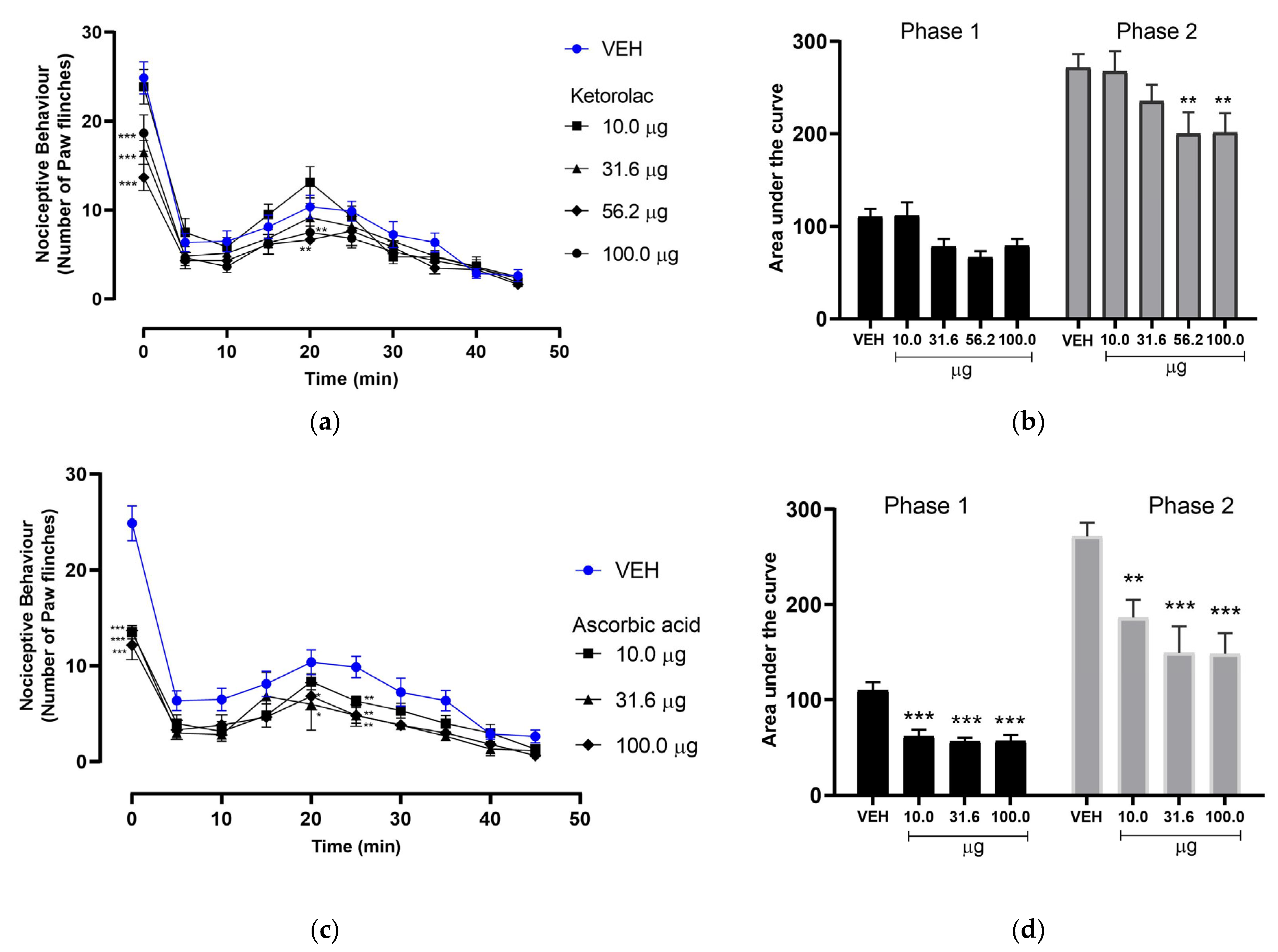
3.1. Effect of Ketorolac on Nociceptive Behavior in the Formalin-Induced Pain Model

KET was administered in doses of 10.0, 31.6, 56.2, and 100.0 µg/paw to the dorsal region of the right hind limb 15 min prior to formalin injection. The temporal course of paw flinches showed an increase in flinches starting at 15 min, which decreased at 30 min and ceased at 45 min. As observed in the temporal course of KET (Figure 1a), doses of 31.6, 56.2, and 100.0 µg/paw resulted in a decrease in the number of flinches, although these reductions were not statistically significant compared to the vehicle group.
A global analysis of the effects, based on the area under the curve (AUC), revealed a slight antinociceptive effect during phase 1, with the higher doses showing AUC values below those of the vehicle group (Figure 1b). However, no statistically significant differences were observed in this phase. In contrast, a significant difference was found during phase 2 when administering 56.2 and 100.0 µg/paw of KET (200 ± 57.00 AU and 201.25 ± 21.35 AU, respectively), compared to the vehicle.

3.2. Effect of Ascorbic Acid on Nociceptive Behavior in the Formalin-Induced Pain Model

The AA was administered in doses of 10.0, 31.6, and 100.0 µg/paw to the dorsal region of the right hind limb 15 min prior to formalin injection. In phase 1, nociceptive activity was highest during the first minute and persisted until the 5 and 10 min marks, after which there was a drastic decrease. Following this, the number of paw flinches began to increase again starting at 15 min, peaking at 25 min, and gradually decreasing until ceasing at 45 min (Figure 1c). The temporal course of AA showed that all doses (10.0, 31.6, and 100.0 µg/paw) caused a considerable decrease in the number of flinches compared to the vehicle group, indicating an antinociceptive effect.
A global analysis of the results revealed that the AUC for all doses of AA (10.0, 31.6, and 100.0 µg/paw) showed a significant antinociceptive effect during phase 1, with the AUC values for the doses falling below that of the vehicle, with averages of 61.66 ± 17.44, 56.25 ± 9.18, and 56.66 ± 16.02 AU, respectively, and statistically significant differences. Additionally, during phase 2, statistically significant differences were also found compared to the vehicle, with average AUC values of 186.25 ± 46.19, 149.58 ± 67.70, and 148.33 ± 52.95 AU for each dose, respectively (Figure 1d). This indicates that the antinociceptive effect was observed in both phases of the experiment.

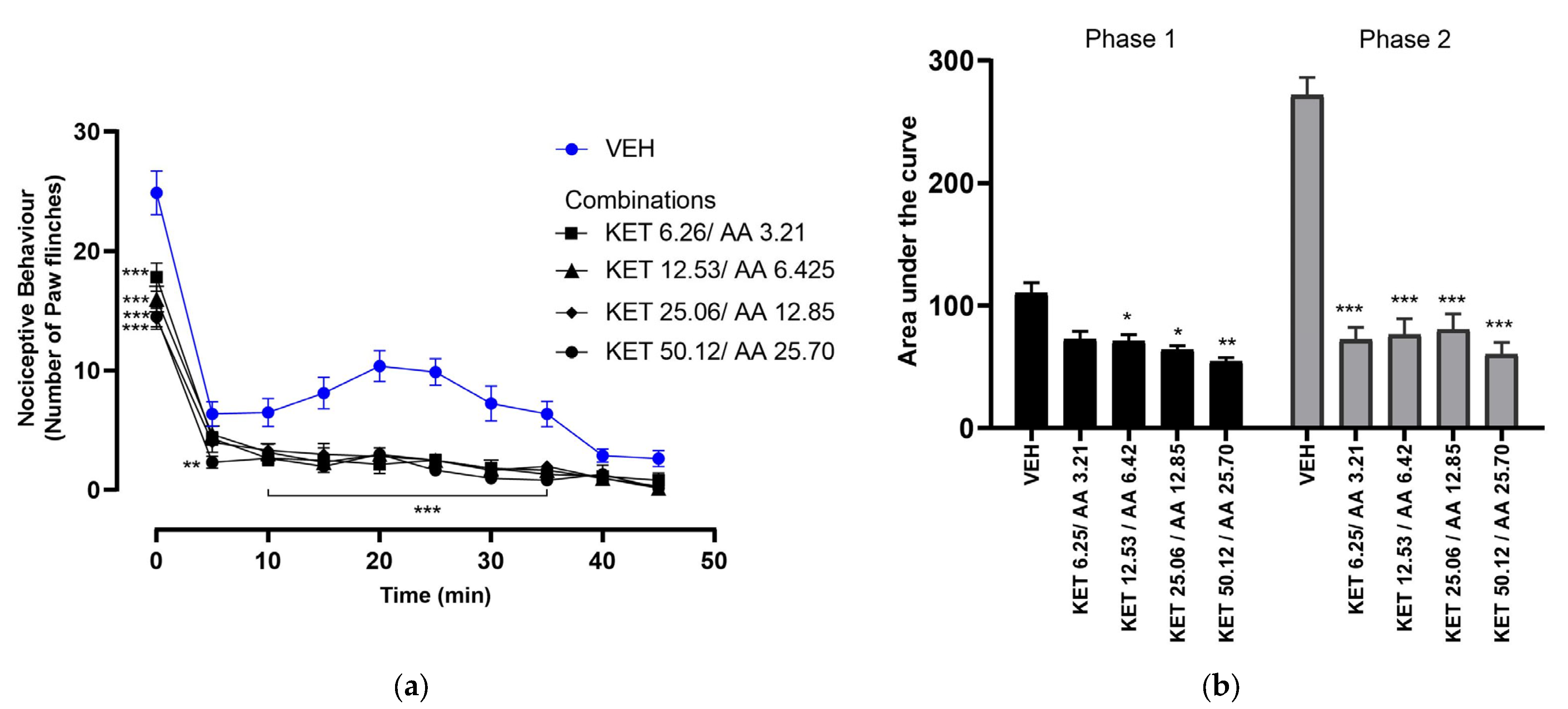
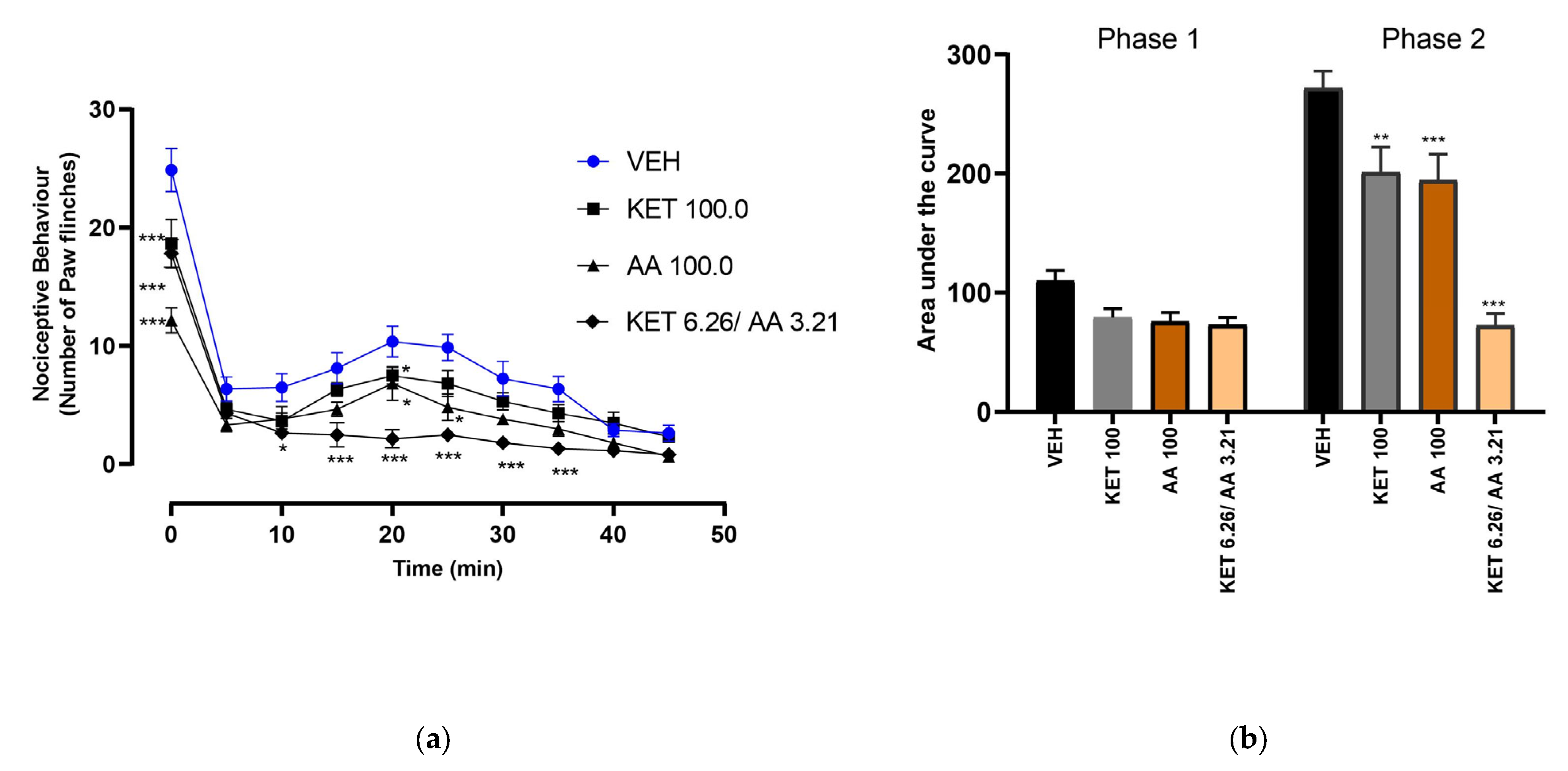
3.3. Dose–Response of Ketorolac and Ascorbic Acid Combinations

As with the individual treatments, these combinations were applied to the dorsal area of the right hind paw 15 min before the formalin test. The temporal course showed a gradual decrease in the number of paws shakes from minute 5, which persisted until the end of the evaluation (Figure 2a).
The AUC values for the combinations KET 6.26/AA 3.21, KET 12.53/AA 6.26, KET 25.06/AA 12.85, and KET 50.12/AA 25.7 in phase 1 were 72.9 ± 15.11, 71.25 ± 12.62, 64.16 ± 7.35, and 54.58 ± 7.48 UA, respectively. In phase 2, the AUC values were 72.5 ± 23.87, 76.25 ± 32.20, 80.41 ± 31.67, and 60.41 ± 23.09 UA, respectively (Figure 2b). Except for the combination KET 6.26/AA 3.21, which did not show a significant effect in phase 1, all other combinations exhibited statistically significant differences in both phases. In comparison to the vehicle group, the administration of KET and AA combinations produced a noticeable antinociceptive effect in both phases. The effect was more prominent during phase 2, when all combinations showed statistically significant differences.
We contrasted the effects of the combination that used the least amounts of the active principles with the individual doses that had the strongest antinociceptive effects in order to better understand these findings. The temporal analysis revealed that the KET 6.26/AA 3.21 combination resulted in the lowest number of paw flinches when compared to the vehicle (Figure 3a).
The AUC analysis indicated that the KET 6.26/AA 3.21 combination exhibited a greater antinociceptive effect than the 100 µg/paw doses of KET and AA (Figure 3b), as well as the vehicle, in both phases of the formalin test. Furthermore, all treatments showed statistically significant differences during phase 2 compared to the vehicle. In phase 1, the average AUC values were 79.16 ± 17.16 UA for KET 100 µg/paw, 72.91 ± 15.11 UA for AA 100 µg/paw, and 56.66 ± 16.66 UA for the KET 6.26/AA 3.21 combination. In phase 2, the average AUC values were 201.25 ± 51.35 UA for KET 100 µg/paw, 148.33 ± 52.95 UA for AA 100 µg/paw, and 72.50 ± 23.87 UA for the KET 6.26/AA 3.21 combination.
The combination KET 6.26/AA 3.21 exhibited the most pronounced antinociceptive effect in both phase 1 and phase 2 compared to the vehicle and the highest individual doses of KET and AA.

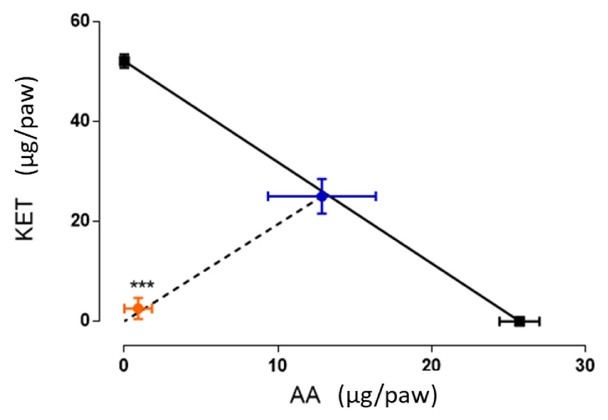
3.4. Isobolographic Analysis of the Antinociceptive Interaction Between the Combination of Ketorolac and Ascorbic Acid

An isobologram was constructed based on the EC30 values of KET and AA on fixed proportions (1:1), graphically representing the pharmacological interaction between these two compounds (Figure 4). The X-axis represents the EC30 of AA (25.7 ± 1.34 µg/paw), while the Y-axis represents the EC30 of KET (52.12 ± 1.30 µg/paw). The midpoint of the isobole indicates the theoretical additive dose calculated from the EC30 values of the individual treatments. The experimental EC30 for the combination fell well below the isobole, indicating a synergistic interaction. The resulting experimental EC30 was 2.6 ± 2.1 µg/paw, suggesting that the combination of these treatments potentiates their individual effects, thereby reducing the required dose to achieve antinociceptive efficacy.

4. Discussion

This study evaluated the combination of an antioxidant molecule, AA, and a NSAIDs, KET, in a formalin-induced pain model. The results confirmed the biphasic nature of the nociceptive response, with a neurogenic phase (0–10 min) followed by an inflammatory phase (15–45 min), consistent with the previous studies [23,24].
The individual administration of ketorolac produced dose-dependent antinociceptive effects, in agreement with the previous reports demonstrating the analgesic effect of this drug in the same model using systemic ketorolac and local tramadol [25,26]. Its primary mechanism of action involves COX-2 inhibition, leading to reduced synthesis of inflammatory prostanoids [27,28,29]. Evidence suggests that ketorolac exhibits synergistic effects in nociceptive pain models in rats when combined with tizanidine [30], methyleugenol [31], and B-complex vitamins [8,32]. Furthermore, ketorolac has shown enhanced analgesic effects when combined with other substances, such as tapentadol in a trigeminal pain model in mice [33]. Additional studies have reported a synergistic interaction when ketorolac is co-administered with drugs as tramadol [34].
Ascorbic acid, widely recognized for its antioxidant properties, also exhibited antinociceptive effects. Its action is attributed to its anti-inflammatory and antinociceptive properties, resulting from its ability to scavenge reactive oxygen species (ROS), reduce pro-inflammatory cytokine production, and modulate glutamatergic ionotropic receptors involved in pain signaling; additionally, ascorbic acid plays a key role as a cofactor in the synthesis of neurotransmitters implicated in pain processing [35,36,37,38]. Previous research has shown that its co-administration with other antioxidants or analgesics enhances the analgesic response [12,36,39,40]. Furthermore, studies have reported decreased ascorbic acid or vitamin C levels in hospitalized patients, elderly individuals, and patients with fractures or cancer [41,42], suggesting a correlation between vitamin C deficiency and increased pain perception.
This study provides the first evidence of the combined use of ketorolac and ascorbic acid in a formalin-induced pain model. The tested combinations demonstrated superior antinociceptive effects compared to individual treatments, suggesting an enhancement of the analgesic effect. Isobolographic analysis confirmed a synergistic interaction, indicating that the combination reduces the required dose for achieving significant analgesia. This finding aligns with previous research on pharmacological synergy between NSAIDs and antioxidants or vitamins [12,26,39].
From a methodological perspective, the evaluation of pharmacological interactions relies on dose–response strategies, with isobolographic analysis being the reference method due to its ease of interpretation and robustness. The observed synergy between ketorolac and ascorbic acid suggests that their complementary mechanisms contribute to pain relief. While ketorolac inhibits COX-2 and prostanoid synthesis, ascorbic acid reduces oxidative stress and inflammation by modulating cytokines and ROS, potentially acting together to enhance pain relief [30,32,36].
Future research could explore its application in other pain models, mechanisms of action, adverse effects and clinical studies to validate its efficacy in humans.

5. Conclusions

The combination of KET with AA represents a promising strategy to enhance the analgesic effects of NSAIDs, potentially reducing the required doses, supporting the potential use of this interaction in the treatment of inflammatory pain.

Author Contributions

Conceptualization, J.V.E.-J. and O.A.J.-M.; methodology, E.F.H.-L., S.d.J.R.-T., C.N.d.l.T.-S. and J.V.E.-J.; software, E.F.H.-L. and S.d.J.R.-T.; validation, J.V.E.-J., J.C.C.-G., R.C.-T. and C.N.d.l.T.-S.; formal analysis, J.V.E.-J., E.F.H.-L., S.d.J.R.-T., N.V.V.-C. and O.A.J.-M.; investigation, E.F.H.-L. and S.d.J.R.-T.; resources, J.V.E.-J., O.A.J.-M. and J.C.C.-G.; data curation, E.F.H.-L., S.d.J.R.-T. and J.V.E.-J.; writing—original draft preparation, E.F.H.-L., S.d.J.R.-T. and J.V.E.-J.; writing—review and editing, E.F.H.-L., S.d.J.R.-T., O.A.J.-M., C.N.d.l.T.-S., R.C.-T. and J.V.E.-J.; visualization, J.V.E.-J. and O.A.J.-M.; supervision, J.V.E.-J. and O.A.J.-M.; project administration, J.V.E.-J. and O.A.J.-M. All authors have read and agreed to the published version of the manuscript.

Funding

This research received no external funding.

Institutional Review Board Statement

The experimental protocol was approved by the Research Committee of the Autonomous University of Chiapas (Approval date: 19 April 2021; protocol number: 03/ECQ/RPR/247/20). The study adhered to ethical guidelines for pain research, following the Official Mexican Standard NOM-062-ZOO-1999 (Technical Specifications for the Production, Care, and Use of Laboratory Animals) [14], and complied with ethical standards for animal experimentation [15].

Informed Consent Statement

Not applicable.

Data Availability Statement

The data are contained within the article.

Acknowledgments

Hoover-Lazo and Rubio-Trujillo express their gratitude to the Institute of Science, Technology, and Innovation of the State of Chiapas, Mexico, for the scholarship granted to support the completion of this undergraduate thesis.

Conflicts of Interest

The authors declare no conflicts of interest.

References

  1. Raja, S.N.; Carr, D.B.; Cohen, M.; Finnerup, N.B.; Flor, H.; Gibson, S.; Keefe, F.J.; Mogil, J.S.; Ringkamp, M.; Sluka, K.A.; et al. The Revised International Association for the Study of Pain Definition of Pain: Concepts, Challenges, and Compromises. Pain 2020, 161, 1976–1982. [Google Scholar] [CrossRef] [PubMed]
  2. Wise, E.; Khanna, P.P. The Impact of Gout Guidelines. Curr. Opin. Rheumatol. 2015, 27, 225–230. [Google Scholar] [CrossRef] [PubMed]
  3. Bokermann, J.; König, H.H.; Hajek, A. Pain: Its Prevalence and Correlates among the Oldest Old. Aging Clin. Exp. Res. 2024, 36, 2. [Google Scholar] [CrossRef] [PubMed]
  4. Henschke, N.; Kamper, S.J.; Maher, C.G. The Epidemiology and Economic Consequences of Pain. Mayo Clin. Proc. 2015, 90, 139–147. [Google Scholar] [CrossRef]
  5. Atkinson, T.J.; Fudin, J. Nonsteroidal Antiinflammatory Drugs for Acute and Chronic Pain. Phys. Med. Rehabil. Clin. N. Am. 2020, 31, 219–231. [Google Scholar] [CrossRef]
  6. Bacchi, S.; Palumbo, P.; Sponta, A.; Coppolino, M.F. Clinical Pharmacology of Non-Steroidal Anti-Inflammatory Drugs: A Review. Antiinflamm. Antiallergy Agents Med. Chem. 2012, 11, 52–64. [Google Scholar] [CrossRef]
  7. Rojas-Aguilar, F.A.; Briones-Aranda, A.; Jaramillo-Morales, O.A.; Romero-Nava, R.; Esquinca-Avilés, H.A.; Espinosa-Juárez, J.V. The Additive Antinociceptive Effect of Resveratrol and Ketorolac in the Formalin Test in Mice. Pharmaceuticals 2023, 16, 1078. [Google Scholar] [CrossRef]
  8. Beltrán-Montoya, J.J.; Herrerias-Canedo, T.; Arzola-Paniagua, A.; Vadillo-Ortega, F.; Dueñas-Garcia, O.F.; Rico-Olvera, H. A Randomized, Clinical Trial of Ketorolac Tromethamine vs Ketorolac Trometamine plus Complex B Vitamins for Cesarean Delivery Analgesia. Saudi J. Anaesth. 2012, 6, 207–212. [Google Scholar] [CrossRef]
  9. Rahman, M. Vitamin B12 and Ketorolac on Pain and Inflammation in Long Evans Rats. J. Pain 2017, 18, S3–S4. [Google Scholar] [CrossRef]
  10. Bai, A.; Abdullah, F.; Kumar, J.; Lal, A.; Abbas, M.; Sandesh, R.; Naz, S.; Shahid, S.; Anees, F.; Memon, S. The Role of Vitamin C in Reducing Pain Associated With Diabetic Neuropathy. Cureus 2021, 13, e15895. [Google Scholar] [CrossRef]
  11. Ayatollahi, V.; Dehghanpour Farashah, S.; Behdad, S.; Vaziribozorg, S.; Rabbani Anari, M. Effect of Intravenous Vitamin C on Postoperative Pain in Uvulopalatopharyngoplasty with Tonsillectomy. Clin. Otolaryngol. 2017, 42, 139–143. [Google Scholar] [CrossRef] [PubMed]
  12. Lu, R.; Kallenborn-Gerhardt, W.; Geisslinger, G.; Schmidtko, A. Additive Antinociceptive Effects of a Combination of Vitamin C and Vitamin E after Peripheral Nerve Injury. PLoS ONE 2011, 6, e29240. [Google Scholar] [CrossRef]
  13. Mikirova, N.; Casciari, J.; Rogers, A.; Taylor, P. Effect of High-Dose Intravenous Vitamin C on Inflammation in Cancer Patients. J. Transl. Med. 2012, 10, 189. [Google Scholar] [CrossRef] [PubMed]
  14. Hung, K.C.; Lin, Y.T.; Chen, K.H.; Wang, L.K.; Chen, J.Y.; Chang, Y.J.; Wu, S.C.; Chiang, M.H.; Sun, C.K. The Effect of Perioperative Vitamin C on Postoperative Analgesic Consumption: A Meta-Analysis of Randomized Controlled Trials. Nutrients 2020, 12, 3109. [Google Scholar] [CrossRef]
  15. Carr, A.C.; Vissers, M.C.M.; Vissers, M.C.M.; Cook, J.S.; Cook, J.S. Parenteral Vitamin C Relieves Chronic Fatigue and Pain in a Patient with Rheumatoid Arthritis and Mononeuritis Multiplex Secondary to CNS Vasculitis. Case Rep. Clin. Pathol. 2015, 2, 57. [Google Scholar] [CrossRef]
  16. Klimant, E.; Wright, H.; Rubin, D.; Seely, D.; Markman, M. Intravenous Vitamin C in the Supportive Care of Cancer Patients: A Review and Rational Approach. Curr. Oncol. 2018, 25, 139–148. [Google Scholar] [CrossRef]
  17. Bayu, P.; Wibisono, J.J. Vitamin C and E Antioxidant Supplementation May Significantly Reduce Pain Symptoms in Endometriosis: A Systematic Review and Meta-Analysis of Randomized Controlled Trials. PLoS ONE 2024, 19, e0301867. [Google Scholar] [CrossRef]
  18. NOM-062-ZOO-1999; Especificaciones Técnicas Para La Producción, Cuidado y Uso de Los Animales de Laboratorio. Diario Oficial de la Feeración: Mexico City, Mexico, 2001. Available online: https://www.dof.gob.mx/nota_detalle.php?codigo=762506&fecha=22/08/2001#gsc.tab=0 (accessed on 31 March 2025).
  19. Zimmermann, M. Ethical Guidelines for Investigations of Experimental Pain in Conscious Animals. Pain 1983, 16, 109–110. [Google Scholar] [CrossRef]
  20. Dubuisson, D.; Dennis, S.G. The Formalin Test: A Quantitative Study of the Analgesic Effects of Morphine, Meperidine, and Brain Stem Stimulation in Rats and Cats. Pain 1977, 4, 161–174. [Google Scholar] [CrossRef]
  21. Gibaldi, M. Estimation of Area under the Curve. In Biopharmaceutics and Clinical Pharmacokinetics; Routledge: London, UK, 1991; pp. 377–378. [Google Scholar]
  22. Tallarida, R.J. The Interaction Index: A Measure of Drug Synergism. Pain 2002, 98, 163–168. [Google Scholar] [CrossRef]
  23. Tjølsen, A.; Berge, O.G.; Hunskaar, S.; Rosland, J.H.; Hole, K. The Formalin Test: An Evaluation of the Method. Pain 1992, 51, 5–17. [Google Scholar] [CrossRef] [PubMed]
  24. Lopes, D.M.; Cater, H.L.; Thakur, M.; Wells, S.; McMahon, S.B. A Refinement to the Formalin Test in Mice. F1000Research 2019, 8, 891. [Google Scholar] [CrossRef] [PubMed]
  25. Malmberg, A.B.; Yaksh, T. Pharmacology of the Spinal Action of Ketorolac, Morphine, ST-91, U50488H, and L-PIA on the Formalin Test and an Isobolographic Analysis of the NSAID Interaction. Anesthesiology 1993, 79, 270–281. [Google Scholar] [CrossRef] [PubMed]
  26. Del Carmen Martínez-Martínez, M.; Parra-Flores, L.I.; del Carmen Baeza-Flores, G.; Torres-López, J.E. Isobolographic Analysis of Antinociceptive Effect of Ketorolac, Indomethacin, and Paracetamol after Simultaneous Peripheral Local and Systemic Administration. Behav. Pharmacol. 2022, 33, 15–22. [Google Scholar] [CrossRef]
  27. Mroszczak, E.; Combs, D.; Chaplin, M.; Tsina, I.; Tarnowski, T.; Rocha, C.; Tam, Y.; Boyd, A.; Young, J.; Depass, L. Chiral Kinetics and Dynamics of Ketorolac. J. Clin. Pharmacol. 1996, 36, 521–539. [Google Scholar]
  28. Vadivelu, N.; Gowda, A.M.; Urman, R.D.; Jolly, S.; Kodumudi, V.; Maria, M.; Taylor, R.; Pergolizzi, J.V. Ketorolac Tromethamine—Routes and Clinical Implications. Pain Pract. 2015, 15, 175–193. [Google Scholar]
  29. Viegas, A.; Manso, J.; Corvo, M.C.; Marques, M.M.B.; Cabrita, E.J. Binding of Ibuprofen, Ketorolac, and Diclofenac to COX-1 and COX-2 Studied by Saturation Transfer Difference NMR. J. Med. Chem. 2011, 54, 8555–8562. [Google Scholar] [CrossRef]
  30. Rodríguez-Silverio, J.; Sánchez-Mendoza, M.E.; Arrieta-Valencia, J.; Rocha-Gonzalez, H.I.; Flores-Murrieta, F.J. Tizanidine Increases Antinociceptive Effect and Prevents Gastric Damage Induced by Ketorolac in the Rat. Drug Dev. Res. 2013, 74, 38–42. [Google Scholar] [CrossRef]
  31. Rocha-González, H.I.; Sánchez-Mendoza, M.E.; Cruz-Antonio, L.; Flores-Murrieta, F.J.; Cornelio-Huerta, X.I.; Arrieta, J. Antinociceptive Interaction and Pharmacokinetics of the Combination Treatments of Methyleugenol Plus Diclofenac or Ketorolac. Molecules 2020, 25, 5106. [Google Scholar] [CrossRef]
  32. Medina-Santillán, R.; Reyes-García, G.; Rocha-González, H.I.; Granados-Soto, V. B Vitamins Increase the Analgesic Effect of Ketorolac in the Formalin Test in the Rat—PubMed. Proc. West. Pharmacol. Soc. 2004, 47, 95–99. [Google Scholar]
  33. Barreras-Espinoza, I.; Soto-Zambrano, J.A.; Serafín-Higuera, N.; Zapata-Morales, R.; Alonso-Castro, Á.; Bologna-Molina, R.; Granados-Soto, V.; Isiordia-Espinoza, M.A. The Antinociceptive Effect of a Tapentadol-Ketorolac Combination in a Mouse Model of Trigeminal Pain Is Mediated by Opioid Receptors and ATP-Sensitive K+ Channels. Drug Dev. Res. 2017, 78, 63–70. [Google Scholar] [CrossRef] [PubMed]
  34. López-Muñoz, F.J.; Díaz-Reval, M.I.; Terrón, J.A.; Déciga Campos, M. Analysis of the Analgesic Interactions between Ketorolac and Tramadol during Arthritic Nociception in Rat. Eur. J. Pharmacol. 2004, 484, 157–165. [Google Scholar] [CrossRef] [PubMed]
  35. Zylinska, L.; Lisek, M.; Guo, F.; Boczek, T. Vitamin C Modes of Action in Calcium-Involved Signaling in the Brain. Antioxidants 2023, 12, 231. [Google Scholar] [CrossRef]
  36. Gęgotek, A.; Skrzydlewska, E. Antioxidative and Anti-Inflammatory Activity of Ascorbic Acid. Antioxidants 2022, 11, 1993. [Google Scholar] [CrossRef]
  37. Rosa, K.A.; Gadotti, V.M.; Rosa, A.O.; Rodrigues, A.L.S.; Calixto, J.B.; Santos, A.R.S. Evidence for the Involvement of Glutamatergic System in the Antinociceptive Effect of Ascorbic Acid. Neurosci. Lett. 2005, 381, 185–188. [Google Scholar] [CrossRef]
  38. Goecks, C.S.B.; Horst, A.; Moraes, M.S.; Scheid, T.; Kolberg, C.; Belló-Klein, A.; Partata, W.A. Assessment of Oxidative Parameters in Rat Spinal Cord After Chronic Constriction of the Sciatic Nerve. Neurochem. Res. 2012, 37, 1952–1958. [Google Scholar] [CrossRef]
  39. Riffel, A.P.K.; de Souza, J.A.; Santos, M.d.C.Q.; Horst, A.; Scheid, T.; Kolberg, C.; Belló-Klein, A.; Partata, W.A. Systemic Administration of Vitamins C and E Attenuates Nociception Induced by Chronic Constriction Injury of the Sciatic Nerve in Rats. Brain Res. Bull. 2016, 121, 169–177. [Google Scholar] [CrossRef]
  40. Park, J.M.; Kim, C.K.; Lee, H.C.; Jung, H.; Choi, K.U.; Hong, S.W.; Lim, D.G.; Baek, W.Y.; Kwak, K.H. Antiallodynic Effects of Vitamin C and Vitamin E in Chronic Post-Ischemia Pain Rat Model. Korean J. Anesthesiol. 2013, 65, 442–448. [Google Scholar] [CrossRef]
  41. Mayland, C.R.; Bennett, M.I.; Allan, K. Vitamin C Deficiency in Cancer Patients. Palliat. Med. 2005, 19, 17–20. [Google Scholar] [CrossRef]
  42. Hill-Mündel, K.; Schlegl, J.; Biesalski, H.K.; Ehnert, S.; Schröter, S.; Bahrs, C.; Nohr, D.; Nüssler, A.K.; Ihle, C. Preoperative Ascorbic Acid Levels in Proximal Femur Fracture Patients Have No Postoperative Clinical Impact, While Ascorbic Acid Levels upon Discharge Have a Major Effect on Postoperative Outcome. J. Clin. Med. 2019, 9, 66. [Google Scholar] [CrossRef]
Figure 1. Antinociceptive effect of ketorolac and ascorbic acid administered subcutaneously in the dorsal area of the right hind limb in the formalin model: (a) temporal course of the administration of 10.0, 31.6, 56.2, and 100.0 µg/paw of ketorolac; (b) area under the curve obtained in phases 1 and 2 of the formalin model after the administration of 10.0, 31.6, 56.2, and 100.0 µg/paw of KET; (c) temporal course of the administration of 10.0, 31.6, and 100.0 µg/paw of ascorbic acid; (d) area under the curve obtained in phases 1 and 2 of the formalin model after the administration of 10.0, 31.6, and 100.0 µg/paw of AA. Data are expressed as mean ± standard error of the mean (n = 6 per group); * p < 0.05, ** p < 0.01, *** p < 0.001 vs. Vehicle (Tukey post hoc test after two-way ANOVA).
Futurepharmacol 05 00015 g001
Figure 2. Antinociceptive effect of the combination of KET and AA administered subcutaneously in the dorsal area of the right hind limb in the formalin model. (a) Temporal course of the administration of KET and AA combinations at doses of 9.47, 18.95, 37.91, and 75.82 µg/paw; (b) area under the curve (AUC) obtained in phases 1 and 2 of the formalin model after the administration of the different combinations. Data are expressed as mean ± standard error of the mean (n = 6 per group); * p < 0.05, ** p < 0.01, *** p < 0.001 vs. Vehicle (Tukey post hoc test after two-way ANOVA).
Futurepharmacol 05 00015 g002
Figure 3. Antinociceptive effect of the maximum doses of each treatment administered subcutaneously in the dorsal area of the right hind limb in the formalin model. (a) Temporal course of the administration of 100.00 µg/paw of KET, 100.00 µg/paw of AA, and 9.47 µg/paw of the KET and AA combination, respectively. (b) AUC obtained in phases 1 and 2 of the formalin model after the administration of 100.00 µg/paw of KET, 100.00 µg/paw of AA, and 9.47 µg/paw of the KET and AA combination, respectively. Data are expressed as mean ± standard error of the mean (n = 6 per group); * p < 0.05, ** p < 0.01, *** p < 0.001 vs. KET 100; * p < 0.05, ** p < 0.01, *** p < 0.001 vs. AA 100 (Tukey post hoc test after two-way ANOVA).
Futurepharmacol 05 00015 g003
Figure 4. Isobologram of the pharmacological interaction between ketorolac and ascorbic acid administered subcutaneously in a formalin model. The horizontal and vertical bars represent the standard error of the mean (SEM), and the oblique middle line connecting the X and Y axes is the theoretical additivity line. The midpoint of this line represents the theoretical additive point based on the EC30 values (blue color). Experimental points below the line indicate a synergistic potentiation (orange color). Data are presented as mean ± SEM (n = 6), *** p < 0.001 (Student’s t-test).
Futurepharmacol 05 00015 g004
Table 1. Doses of ketorolac and ascorbic acid evaluated in combination, at a 1:1 ratio.
CombinationDose (µg/paw)
KetorolacAscorbic AcidTotal
EC30 + EC306.263.21 9.48
(EC30 + EC30)/212.53 6.42 18.96
(EC30 + EC30)/425.06 12.85 37.91
(EC30 + EC30)/850.12 25.70 75.82
Disclaimer/Publisher’s Note: The statements, opinions and data contained in all publications are solely those of the individual author(s) and contributor(s) and not of MDPI and/or the editor(s). MDPI and/or the editor(s) disclaim responsibility for any injury to people or property resulting from any ideas, methods, instructions or products referred to in the content.

