Review

Nanotechnology-Driven Therapeutic Innovations in Neurodegenerative Disorders: A Focus on Alzheimer’s and Parkinson’s Disease

by
Antea Krsek
1,† and
Lara Baticic
2,*,†
1
Faculty of Medicine, University of Rijeka, HR-51000 Rijeka, Croatia
2
Department of Medical Chemistry, Biochemistry and Clinical Chemistry, Faculty of Medicine, University of Rijeka, HR-51000 Rijeka, Croatia
*
Author to whom correspondence should be addressed.
These authors contributed equally to this work.
Future Pharmacol. 2024, 4(2), 352-379; https://doi.org/10.3390/futurepharmacol4020020
Submission received: 28 February 2024 / Revised: 19 April 2024 / Accepted: 28 April 2024 / Published: 30 April 2024
(This article belongs to the Special Issue Feature Papers in Future Pharmacology 2024)

Abstract

:
Neurodegenerative disorders entail a progressive loss of neurons in cerebral and peripheral tissues, coupled with the aggregation of proteins exhibiting altered physicochemical properties. Crucial to these conditions is the gradual degradation of the central nervous system, manifesting as impairments in mobility, aberrant behaviors, and cognitive deficits. Mechanisms such as proteotoxic stress, neuroinflammation, oxidative stress, and programmed cell death contribute to the ongoing dysfunction and demise of neurons. Presently, neurodegenerative diseases lack definitive cures, and available therapies primarily offer palliative relief. The integration of nanotechnology into medical practices has significantly augmented both treatment efficacy and diagnostic capabilities. Nanoparticles, capable of traversing the blood–brain barrier, hold considerable potential for diagnosing and treating brain pathologies. By combining gene therapy with nanotechnology, the therapeutic effectiveness against neurodegenerative diseases can be substantially enhanced. Recent advancements in nano-biomaterial-based methodologies have fortified existing approaches to neural stem cell (NSC) differentiation therapies. NSC-targeting technologies offer a promising, potentially safe method for treating neurodegenerative diseases. This review endeavors to summarize current insights and perspectives on nanotechnology-driven therapeutic innovations in neurodegenerative disorders, with a particular emphasis on Alzheimer’s and Parkinson’s disease.

1. Introduction

Neurodegenerative illnesses are conditions marked by a gradual loss of neurons in the brain and other peripheral organs, along with the accumulation of proteins exhibiting changed physicochemical characteristics [1]. The main characteristic of neurodegenerative diseases is defined by a gradual deterioration of the central nervous system (CNS), which results in mobility problems, behavioral abnormalities, and cognitive deficits [2]. Proteotoxic stress, neuroinflammation, oxidative stress, and programmed cell death all cause progressive neuronal malfunction and death, representing interconnected processes that contribute to the progressive malfunction and death of neurons, often observed in neurodegenerative diseases, such as Alzheimer’s (AD), Parkinson’s (PD), and Huntington’s disease (HD) [2,3]. Proteotoxic stress is caused by an imbalance between the production of misfolded or aggregated proteins and the cell’s capacity to properly fold, refold, or degrade these proteins. This accumulation of misfolded proteins impairs cellular defense mechanisms and contributes to neuronal dysfunction and death. Disruptions in the ubiquitin-proteasomal and lysosomal/autophagosomal systems are also noted [1,2]. Although most neurodegenerative illnesses are defined by anomalies in proteins, clinical symptoms frequently appear later, and patients may experience numerous disease processes at the same time [3].
Neuroinflammation is an inflammatory response within the central nervous system (CNS), induced as a response to various stimuli, such as infection, injury, or the accumulation of abnormal proteins. Acute neuroinflammation plays a beneficial role in tissue repair, but chronic or excessive neuroinflammation can lead to neuronal damage and exacerbate neurodegenerative processes [2,3]. During these molecular processes, a plethora of different inflammatory mediators are being generated, including reactive oxygen species (ROS), that lead to oxidative stress. Neurons are particularly vulnerable to oxidative damage due to their high metabolic activity and relatively low levels of antioxidant defenses. Oxidative stress can damage crucial macromolecules, such as nucleic acids, proteins, and lipids, disrupt cellular function, and contribute to neuronal degeneration. Moreover, programmed cell death, or apoptosis, as a highly regulated process that eliminates damaged cells, can also contribute to neurodegenerative disease progression since dysregulation of apoptotic pathways can lead to excessive neuronal death [1,2,3,4] (Figure 1).
Indeed, the interplay between proteotoxic stress, neuroinflammation, oxidative stress, and programmed cell death creates a vicious cycle of neuronal dysfunction and death in neurodegenerative diseases, highlighting the complex and multifactorial nature of these disorders. Therapeutic strategies aiming at targeting these causal connections may contribute to the development of new modalities focusing on slowing disease progression and preserving neuronal function [2,3,4].
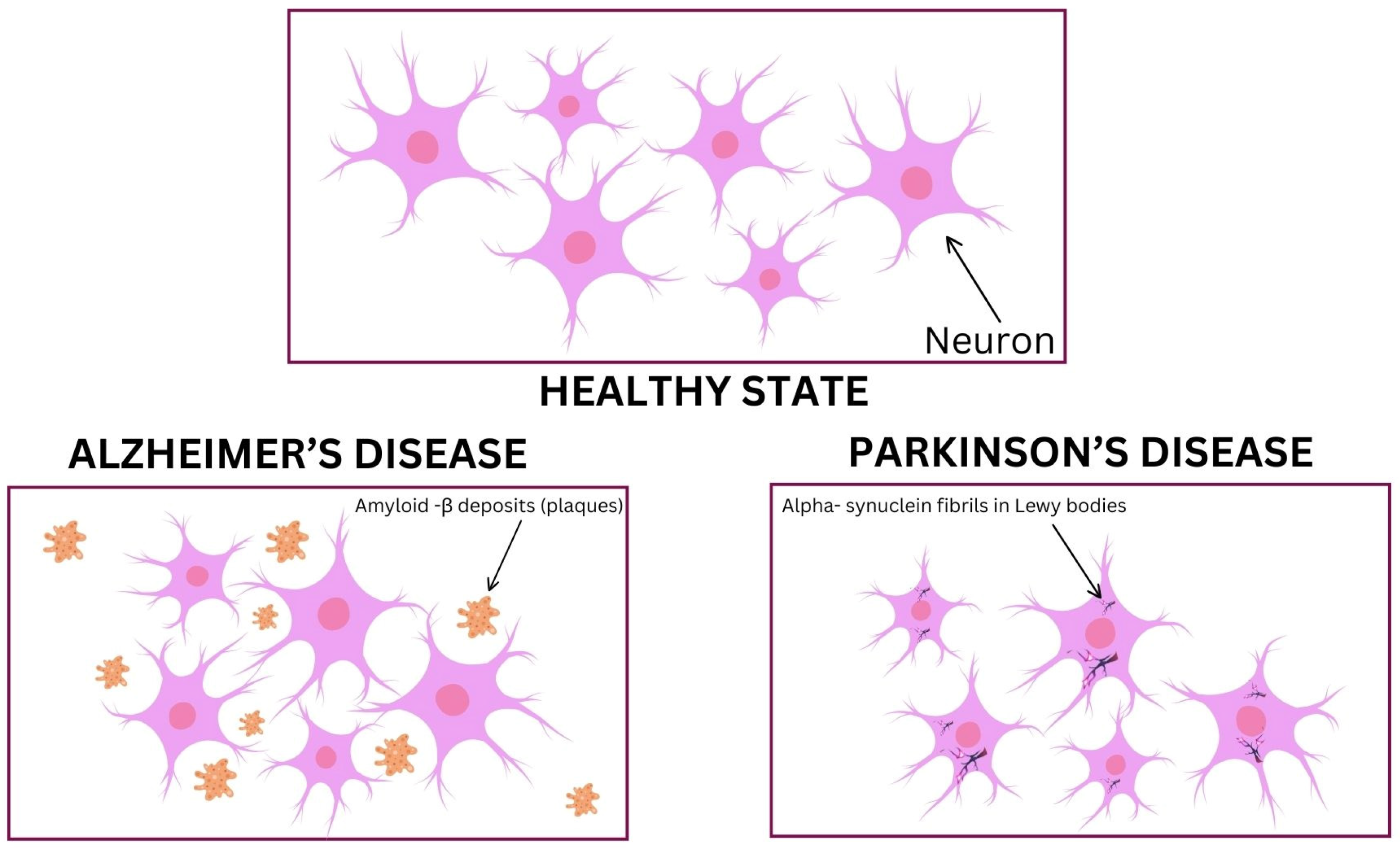
However, the absence of biomarkers presents diagnostic problems unless genetic alterations are implicated [4]. This places a burden on the public health sector. Immune-mediated illnesses, ischemia, neurodegeneration, and infections can all result from the activation of the immune system, even if it may also help with regeneration and repair [5]. A wide range of medical problems that impact the brain are included in the term “brain diseases and disorders”, including infections, tumors, and neurological disorders. As of 2019, they accounted for roughly 349 million disability-adjusted life-years (DALYs) and nearly 10 million deaths, making them one of the leading causes of disabilities and mortality [6]. According to Ghosh and Higgins, the term “brain diseases” frequently denotes medical conditions that are highly transmissible and typically caused by external agents, such as viruses or bacteria [7]. Conversely, “brain disorders” refer to non-communicable diseases that are often hereditary and stem from abnormalities in structure or function caused by birth defects or genetic aberrations [8]. Some of the main neurodegenerative disorders—amyotrophic lateral sclerosis (ALS), Parkinson’s disease (PD), Alzheimer’s disease (AD), and Huntington’s disease (HD)—are intimately associated with aging, genetics, and compromised immunity (Figure 2) [5].
Additionally, neurological dysfunction causes cognitive, motor, and behavioral deficits that are similar to many brain illnesses and disorders. Due to brain complexity and sensitivity, treating these diseases may be very challenging [9,10]. The capacity of nanoparticle (NP)-based therapeutics to cross the blood–brain barrier (BBB) has made them attractive therapeutic options for brain illnesses and disorders in recent years. Their special qualities include small size, selectivity, low toxicity, biodegradability, and solubility [11,12]. In the field of nanomedicine, which combines nanotechnology with medicine, pharmacologically active compounds are delivered to diseased areas, including the brain, using nanoscale particles [5]. Active substances are encapsulated within or on the surface of polymeric NPs, which are commonly measured in the range of 1 to 1000 nm. Although they have different structural morphologies, this word encompasses both nanospheres and nano-capsules [13]. Numerous classes of NPs have been created, each with unique physical and chemical characteristics, such as metal and metal oxides, fullerenes, liposomal, polymeric, solid-lipid, and polylactide-co-glycoside (PLGA) NPs [14,15]. Applications of nanoparticles are found in many domains, but in medicine, they show great promise for the detection and management of deep-seated illnesses, including brain tumors, metastatic malignancies, and neurodegenerative diseases [16,17].
A promising route for the development of new treatments for CNS illnesses is the combination of gene therapy with nanomedicine [13,14,15]. Beyond nanotechnology, several other therapeutic approaches are being explored to treat neurological diseases, most importantly gene therapy and optogenetics, stem cell therapy, immunotherapy, neuromodulation, and as in other pathologies, drug repurposing and drug discovery. Gene therapy involves the delivery of genetic material into cells in order to modify abnormal genes or to provide therapeutic benefits. In the context of neurological diseases, gene therapy holds promise for conditions with known genetic causes, such as HD, spinal muscular atrophy, and certain forms of PD [18]. Optogenetics combines genetic and optical techniques to control the activity of specific neurons with light-sensitive proteins. Stem cell therapy aims to replace damaged or dysfunctional cells in the nervous system with healthy cells derived from stem cells that show potential for treating conditions such as PD, AD, spinal cord injury, and stroke. Immunotherapy approaches harness the body’s immune system to target pathological processes in neurological diseases and hold promise for conditions such as AD, multiple sclerosis, and certain types of neuropathies. Neuromodulation techniques, such as deep-brain stimulation, transcranial magnetic stimulation, and transcranial direct current stimulation, can modulate neuronal activity and are intensively investigated for treating conditions such as PD, epilepsy, chronic pain, and other neurological pathologies. Finally, high-throughput screening, computational modeling, and other innovative approaches are accelerating drug discovery efforts that may contribute to the development of new pharmaceutical strategies to treat these diseases [13,14,15,16,17,18].
Neurodegenerative diseases currently have no known cure, and the therapies that are offered are primarily palliative. Western medicine uses antipsychotic drugs for psychiatric and behavioral problems associated with dementia in AD and dopaminergic therapy for movement difficulties in PD. There are several medications available to treat the symptoms of AD and PD but still, even with some clinical alleviation, medication treatment frequently becomes inadequate and inefficient as the illness progresses [5]. Because the blood–brain barrier (BBB) prevents effective drug transport to the brain, establishing appropriate drug concentrations within the central nervous system (CNS) is a significant difficulty in drug therapy [19].

2. Applications of Nanotechnology in Neurological Diseases and Disorders

2.1. Molecular Imaging (MI)

MI is a significant field in biomedical research that studies disease or physiological processes at the molecular level and visualizes the spatial distribution of pathologic changes in illnesses that are not yet identified (NDDs), which has shown promise. Research has demonstrated that it can enhance the accuracy of diagnosis and provide guidance for therapeutic measures. These imaging techniques provide a simple way to detect, describe, and measure certain bodily activity with remarkable sensitivity and accuracy [20,21]. The range of methods includes bioluminescence imaging, magnetic resonance imaging, positron emission tomography, ultrasound, X-ray radiography, magnetic resonance imaging, and single-photon emission computed tomography. MI techniques have been shown useful for characterizing and analyzing a wide range of brain-related illnesses, including infections, brain tumors, and neurological diseases [22,23]. The use of contrast chemicals, or probes, improves the accuracy of MI. These probes fall into two categories: targetable probes, which attach to certain targets, and activatable probes, which, when in contact with particular indications on their targets, provide an audible signal. Studies show that because oligopeptide nanoparticles may fluoresce when exposed to the low pH of the tumor microenvironment, they may be useful as activatable probes. Studies also show that targetable probes may be transported across the blood–brain barrier via polysorbate 80-coated poly(butyl cyanoacrylate) dextran polymeric nanoparticles, which makes it easier to see Aβ plaques in Alzheimer’s disease models [11]. Additionally, recent studies show that by interacting with highly expressed class A scavenger receptors, sulfated dextran-coated iron oxide nanoparticles might improve the bioimaging of microglia-induced brain inflammation [24]. Furthermore, fluorescence imaging has made use of rare-earth-doped nanoparticles, which bind to integrin α Vβ3 and release short-wave infrared light [25].
Recent advancements in structural and functional imaging, however, have made it possible to measure and evaluate the pathophysiological pathways underlying neurodegeneration in vivo. Molecular neuroimaging techniques, such as PET and SPECT imaging, are made possible in this case by the use of ad hoc radioligands, which allow for the in vivo evaluation of many pathological pathways. Some of these ligands—such as tau and amyloid—are used as targets for treatments aimed at changing the illness or as diagnostic tools to determine the specific abnormal protein aggregation that results in neurodegeneration.
Molecular imaging also makes it feasible to evaluate novel ligands, change brain glucose metabolism (using 18F-FDG PET), change the neurotransmitter receptor and transporter density (e.g., dopamine and serotonin), and assess brain inflammation (e.g., microglia activation, using 11C-PK11195). Based on a growing amount of evidence, these techniques, particularly early in the course of the illness, can provide invaluable diagnostic and prognostic value in neurodegeneration, and in certain cases, even provide a clear description of the illness. Testing is underway for additional PET ligands that investigate different targets, including 11C-UCB-J, which evaluates synaptic density and has great potential for understanding neurodegenerative illnesses [26].
The extent and quantification of a number of the recognized underlying pathologic characteristics in these illnesses may be achieved using molecular imaging biomarkers, improving the identification of risk factors and the characterization of the disease. These might affect the specific phenotype in an antagonistic, potentiating, synergistic, or independent manner [20,21,22]. Signs of neurodegeneration, such as brain glucose hypometabolism, and disease indicators from molecular imaging, such as tau and β-amyloid imaging, are already included in the new diagnostic criteria. Moreover, molecular imaging research has shown that the underlying pathologic traits—such as the deposition of Aβ in AD and the loss of nigrostriatal terminals in Parkinson’s disease—develop over decades and precede the clinical phenotype [25,26]. Novel approaches for designing clinical trials aimed at delaying or preventing the onset of dementia and cognitive impairment have been impacted by the development of biomarker-based tools for diagnosing brain illness in AD [21,26]. By defining the disease solely by the clinical syndromal presentation, it will be impossible to choose individuals who are a good fit for therapy (i.e., exhibiting the therapeutic target or neuropathology), as the use of these biomarkers has shown. The most reliable way to enable at-risk people to begin treatment early before chronic, irreversible brain damage occurs may be through the use of multimodal imaging. These imaging markers may thus become more important for patient selection and for monitoring novel therapy approaches [20,21,22,26]. Gold standards for diagnosis of neurodegenerative diseases involve clinical assessment, which may include cognitive testing, neurological examination, and often postmortem examination for definitive confirmation. However, these methods are often limited in their ability to accurately diagnose diseases in their early stages or to differentiate between different types of neurodegenerative diseases [20]. Comparative studies evaluating biomarkers and diagnostic approaches for neurodegenerative diseases in relation to the gold standard of diagnosis have been conducted to evaluate the sensitivity, specificity, accuracy, and predictive value of different biomarkers and diagnostic approaches in relation to the gold standard. However, further research is needed to evaluate their significance since they may not always fully replace the gold standard of diagnosis [20,21,22,25,26].

2.1.1. Innovations in Protein Aggregation Imaging for Neurodegenerative Disorders

A few protein aggregates, such as TDP-43, tau, α-synuclein, and β-amyloid, are suggestive of neurodegenerative illnesses. Traditionally, postmortem histology has been the gold standard for diagnosis; however, new advancements in molecular imaging have allowed for the in vivo identification of these aggregates, resulting in an earlier and more accurate diagnosis. PET and SPECT imaging techniques can detect β-amyloid and tau illnesses, but further research is needed to develop tracers that target protein aggregation diseases selectively [27]. Since unique protein pathologies may underlie different neurodegenerative illnesses, high-affinity tracers for particular accumulation types are necessary. Currently, efforts are being made to produce PET tracers for α-synuclein aggregation, which might significantly improve in vivo histopathology-based medical diagnosis [28]. However, challenges, such as poor target density and selectivity issues, impede progress and highlight the need for advanced development methods [29]. TDP-43 is a histopathologic feature of some forms of frontotemporal dementia, amyotrophic lateral sclerosis, and limbic-predominant, age-related TDP-43 encephalopathy. There are no suitable PET tracers for TDP-43 at the moment. It will be intriguing to observe if TDP-43 or alternative possibilities for neurodegenerative illness tracers materialize [30].

2.1.2. PET Imaging and Nanoparticles’ Role in Advancing Molecular Imaging in Brain Diseases

In cases where there is uncertainty about a patient’s diagnosis of neurodegenerative disease, PET imaging is usually employed as an extra diagnostic method. In these situations, the diagnosis is often made using clinical assessments to identify specific symptoms, and CT or MRI scans are performed to rule out other potential medical conditions.
Despite the syndromal approach, neurodegenerative illnesses have not seen any significant progress in the creation of medications that either cure or modify the disease over time, even if the latter are identified by distinct clinical characteristics. The first disease to employ a cutting-edge diagnostic strategy was AD. Characterizing AD by assessing biomarkers for neurodegeneration, tau and amyloid, is widely used in research and drug development [31]. However, although amyloid plaques and tau tangles are classical hallmarks of AD pathology, emerging research suggests that neuroinflammation and cellular responses play crucial roles in disease initiation and progression. In the early stages of AD, before significant neuronal damage occurs, there is often activation of microglia, as well as infiltration of peripheral immune cells, such as polymorphonuclear leukocytes (PMNs) and activation of astrocytes. These inflammatory responses are thought to contribute to neuronal dysfunction and cognitive decline. Focusing on the early stages of AD pathology, particularly inflammation, microglia activation, PMN infiltration, and astrocyte activation, is essential for developing effective therapeutic strategies. Proteomic analyses, such as those highlighted in a recent study [], provide valuable insights into the molecular mechanisms underlying early-stage AD and offer promising strategies for further investigation and intervention in order to delay or prevent the progression of AD and preserve cognitive function [29,31]. With aducanumab’s recent FDA clearance, AD sufferers now have a medication alternative that decreases the levels of β-amyloid [32]. Anticipating more drugs that target β-amyloid and successfully treat tau, α-synuclein, and other pathogenic aggregates to hit the market, it is expected that neurodegenerative illnesses will go from being classified as syndromes to being constructed physiologically. For etiological therapy choices, biomarker-based diagnosis is going to be required, and PET imaging may be crucial [30].

2.2. Detection of Biomarkers

A biomarker is defined as a molecule or signal that may be detected and is directly linked to a certain state or illness. Disease treatment depends on a biomarker’s ability to discriminate between healthy and unwell people and to accurately identify the stage of an illness [1,5]. Many biomarkers have been found in brain illnesses and disorders; however, the lack of appropriate detection methods frequently makes it difficult to use these biomarkers. Nanoparticles (NPs) have become useful instruments for the very effective identification of important biomarkers of brain illnesses and disorders [5,11,13]. Using NPs for detecting biomarkers in situ (in the brain) offers several advantages compared to detecting them in cerebrospinal fluid (CSF) or blood, which mainly refers to the invasiveness of the diagnostic procedures. Invasive procedures for obtaining CSF or blood samples carry important risks and discomfort for patients. In situ detection using nanoparticles may offer a less invasive alternative, reducing patient discomfort and improving compliance with diagnostic protocols. In addition, the enhanced sensitivity and specificity of in situ detection using NPs should be mentioned, where increased sensitivity can enable the detection of biomarkers at lower concentrations than traditional methods, enhancing the diagnostic accuracy. Detecting biomarkers in situ using NPs minimizes the need for sample collection and processing, thus reducing the risk of sample contamination and degradation [11,12,13].
Scientific knowledge indicates that elevated concentrations of ubiquitin-C-terminal hydrolase-L1 (UCH-L1) have been found in the plasma of TBI patients compared to healthy persons, indicating that UCH-L1 may serve as a biomarker for the disorder [33,34]. According to a recent study, a unique technique using gold nanoparticles’ (Au NPs’) surface plasmon resonance may quickly and precisely identify the UCH-L1 biomarker in patients suffering from TBI, with a full range of sensitivity and specificity [35]. Furthermore, there is a substantial correlation between Aβ levels and dementia and associated disorders [36,37]. According to studies, Aβ plaques in Alzheimer’s disease-modeling mice may be safely and successfully detected using modified magnetic nanoparticles [38]. Ferrández-Cabada and Ramos-Gómez have shown that raised cholesterol levels are a crucial sign of Alzheimer’s disease (AD), and elevated cholesterol levels may be efficiently detected using anti-cholesterol antibody-bound magnetic nanoparticles. Additionally, current studies suggest that AD biomarkers, such as tau proteins, inflammatory cytokines, and Aβ, may be detected by fluorescent nanoparticles. All of these results point to the possibility that nanoparticles provide a quick and effective way to uncover biomarkers for illnesses and conditions of the brain [39,40].

2.3. Drug Delivery

Treating brain illnesses and diseases still primarily involves the issue of transporting possible medications past the blood–brain barrier (BBB). To effectively reach their objectives in the brain, these medications must cross the BBB without inflicting any appreciable short- or long-term harm. Drug transportation across the blood–brain barrier is impeded by variables such as size and hydrogen bond count, but also other parameters, such as hydrophilicity, lipophilicity, charge, membrane transporters, and excretion (efflux) pumps. Considering these factors is crucial for the development of drugs targeting the central nervous system, as they determine the ability of a drug to reach its target in therapeutic concentrations while minimizing off-target effects and systemic toxicity [41,42].
Because of their capacity to pass across the BBB and serve as possible medication carriers, nanoparticles, or NPs, have attracted a lot of interest recently. NPs can carry medications that target the brain and have improved BBB-penetrating capacities [43,44,45]. Furthermore, gold nanoparticles (Au NPs) have shown promise in passing across the BBB to transport antibodies by binding to transferrin receptors; however, this is contingent upon the valency and affinity of the coated antibody [46]. In a mouse model of Alzheimer’s disease (AD), intranasal administration of huperzine A increased its bioavailability and retention time using lactoferrin-conjugated, N-trimethylated, chitosan-modified poly(lactic-co-glycolic acid) (PLGA) [47]. Taken together, recent scientific knowledge implies that NPs can efficiently and effectively transport different medications over the BBB. However, previously mentioned limitations have important implications in the choice and design of appropriate NPs. By considering these factors, NPs with optimized properties for crossing the BBB and delivering therapeutics to the CNS can be designed, potentially improving the treatment of neurodegenerative diseases, brain tumors, and other CNS disorders.

3. Nanoparticles in Neurodegenerative Diseases

3.1. Alzheimer’s Disease

Alzheimer’s disease (AD) is a common, irreversible kind of dementia characterized by progressive neuronal degradation that occurs with aging and results in impaired cognitive performance, gradual deterioration in memory, as well as other neuropathological symptoms (Figure 2). AD plays a significant role in between 60 and 80 percent of dementia patients [48]. It has to be emphasized that the majority of AD cases are of the late-onset type, particularly among individuals aged 65 and older, as opposed to the less common early-onset form that is associated with specific genetic mutations that are discussed further in this review. Late-onset AD is believed to have a multifactorial etiology involving a complex interplay of genetic, environmental, and lifestyle factors [48,49]. Understanding these multifaceted contributors is essential for developing effective approaches for prevention and treatment strategies for the majority of AD cases.
AD was ranked seventh in terms of causes of mortality in 2020 and 2021, the year COVID-19 joined the top 10 causes of death. AD is still the sixth most common cause of mortality in the United States among people over 65. Reports of fatalities from AD climbed by more than 145% between 2000 and 2019, whereas mortality from stroke, heart disease, and HIV declined [49]. Autosomal-dominant types of AD are linked to mutations in genes, including amyloid precursor protein (APP), presenilin 1 (PSEN1), and presenilin 2 (PSEN2), while sporadic AD is connected to apolipoprotein E (ApoE). The cleavage of amyloid-beta peptide (Aβ) by β-secretase and γ-secretase complexes is a crucial step and a possible target for therapy in AD pathogenesis [50]. Other strategies are being considered that target APP-cleaving enzymes, such as legumain (δ-secretase), rhomboid-like protein-4 (RHBDL4), caspases, meprin-β, and membrane-type matrix metalloproteinases (MT-MMPs/η-secretases) [50,51].
The buildup of Tau proteins is one of the pathogenic characteristics associated with the development of neurodegenerative diseases, such as AD [52,53]. According to recent studies, protein-capped iron oxide and cadmium sulfide nanoparticles efficiently prevent Tau proteins from polymerizing and fibrillating, with respective inhibition rates of 49% and 63% [54].
As the illness progresses, intracellular neurofibrillary tangles and extracellular β-amyloid plaques accumulate, causing neurotoxicity and synapse loss, which are another hallmark of AD [55,56]. PSEN1 gene mutations are the primary cause of familial AD (FAD) and encode presenilin-1 (PS1), a part of the γ-secretase complex that cleaves APP [57]. This mechanism is dysregulated, which results in the production of Aβ40 and Aβ42. The latter is more likely to aggregate and cause amyloid plaque development and neurodegeneration, which are hallmarks of FAD [58]. PSEN1 mutations disrupt memory, learning, and neuronal survival, which adds to dementia and neurodegeneration in FAD [57]. They also impair normal presenilin activity. The synthesis of Aβ, hyperphosphorylation of Tau proteins, and the subsequent development of AD are all influenced by the serine/threonine kinase GSK-3 [59]. Research indicates that GSK-3 regulates neuronal activity by working with histone deacetylase (HDAC) proteins [60,61]. As a result, GSK-3 and HDAC inhibitors have demonstrated effectiveness in reducing AD [62]. PSEN2 mutations are associated with many neurodegenerative disorders, including late-onset AD (LOAD) and early-onset AD (EOAD), notwithstanding their rarity [63]. Though not as much as PSEN1, PSEN2 mutations also play a role in Aβ42 accumulation and neurodegeneration [64,65]. The potential targets for treatment methods, such as gene therapy and nanomedicine, include PSEN1 and PSEN2.
Solid-lipid nanoparticles (SL NPs) loaded with the HDAC inhibitor nicotinamide dramatically attenuate the cognitive impairment linked to AD by lowering Tau protein phosphorylation in a rat model [66]. Similarly, administering vitamin D-binding protein-loaded PLGA NPs to 5XFAD mice mitigates cognitive impairments by blocking the binding and accumulation of Aβ [67]. According to Dos Santos Tramontin et al., Au NPs have also been shown to have cytoprotective benefits in AD rat models by boosting antioxidant status and anti-inflammatory responses [68]. According to Moore et al., surface-coated Au NPs have also shown decreased Aβ aggregation, and the effectiveness of this reduction is dependent on the surface chemistry and NP diameter [69]. In AD models, negative-surface-potential Au NPs have been demonstrated to significantly lessen Aβ fibrillization and related neurotoxicity [70,71]. According to recent research, smaller Au NPs can prevent Aβ fibrillization better than greater ones [72]. Genetic polymorphisms in the ApoE gene enhance vulnerability to AD. ApoE is essential for lipid transport and the healing of brain damage [73]. The ε4 allele of ApoE affects several processes, including Aβ control, glucose metabolism, mitochondrial function, and neuroinflammation, and is linked to an increased risk of AD and age-related cognitive decline. ApoE mutations impair lipid transport, worsen Aβ buildup, and worsen neuronal dysfunction and neurodegeneration [74].
In summary, surface-coated Au NPs, particularly those with negative surface potential and smaller diameters, have shown promise in reducing Aβ aggregation and related neurotoxicity in AD models. Understanding the interactions between NP and biological systems, as well as genetic factors influencing disease susceptibility, can provide insights into the development of novel therapeutic strategies for AD and other neurodegenerative diseases.

3.1.1. Nanoparticle-Based Therapy: Aducanumab

The therapy with Aducanumab works as a monoclonal antibody that binds to amyloid aggregates, potentially decreasing β-amyloid plaques in AD patients and restoring neurological function [75]. Anti-amyloid drug therapy aims to disrupt plaque formation, a key process in Alzheimer’s disease progression. Aducanumab, the only FDA-approved drug for this purpose, selectively binds to amyloid aggregates, distinguishing it from other Aβ immunotherapies. While Aβ aggregates are neurotoxic, monomeric Aβ has beneficial effects. Aducanumab’s unique binding interactions with amyloid enable shallow and compact binding, minimizing interactions with monomers [63,76]. Researchers are exploring its binding mechanisms further to enhance selectivity, potentially improving efficacy with smaller doses [77,78].
In a preclinical investigation, the effectiveness of Aducanumab in reducing Aβ plaques in mice was evaluated using a placebo-controlled trial. Aducanumab, a chimeric analogue, was administered to mice with overexpressed APP genes. While chronic therapy had no significant benefits, acute treatment significantly reduced the number of plaques (~48%, p < 0.0001). On the other hand, acute therapy did not stop the development of new plaques. Furthermore, aucasamine malabsorption may rectify intracellular calcium imbalances resulting from neurodegenerative diseases. According to research, people with AD may benefit from chronic therapy, as it was shown to enhance calcium permeability through NMDA receptors (p < 0.05) and restore SERCA pump regulation (p < 0.001) [79,80].
Aducanumab’s phase I clinical trials comprised a 53-person, randomized, double-blind, placebo-controlled, dose-escalation study. They were administered a placebo or escalating doses (0.3 to 60 mg/kg). With typical side effects such as headache and diarrhea, Aducanumab demonstrated excellent tolerance up to 30 mg/kg. High-dose patients experienced the most severe side effect, Amyloid-Related Imaging Abnormalities-Effusion (ARIA-E), which went away without any issues. Linear and dose-dependent pharmacokinetics were observed.
Aducanumab then entered a phase Ib clinical study called Prime, which included 165 patients with moderate AD or MCI. For a whole year, they were administered monthly dosages of 1, 3, 6, or 10 mg/kg. At dosages of 3 and 10 mg/kg, Aducanumab dramatically decreased Aβ plaques, enhanced cognitive function, and retarded the development of the MMSE. ARIA-E was more likely, nevertheless, particularly at larger dosages. Aducanumab has shown potential in treating cognitive problems linked to Aβ plaque development, notwithstanding the risk [5,81]. Monitoring for ARIA-E is an important aspect of clinical trials and treatment protocols involving amyloid-targeting therapies for AD disease and other neurodegenerative disorders characterized by amyloid pathology.
Beginning in September 2015, Aducanumab’s impact on AD was evaluated in two phase III trials: Engage and Emerge [82]. For a period of eighteen months, patients with moderate AD or MCI were administered Aducanumab at low or high dosages every four weeks. In both studies, high-dose Aducanumab significantly decreased the Aβ plaque size when compared to placebo, and lower reductions were seen with low doses. In phase III studies, only Aducanumab significantly reduced Aβ PET SUVR among monoclonal antibodies. According to an analysis of the Emerge trial, people with mild AD may benefit from high-dose Aducanumab since it may delay cognitive decline and enhance daily living skills.
Aducanumab may be able to help people with AD regain their neurological function by lowering Aß plaques and regaining neuronal calcium permeability, according to preclinical research and clinical trials [83,84]. Nonetheless, disparities in the outcomes of post hoc analyses have raised questions in the scientific and medical communities. According to certain research, Aducanumab’s advantages could only be statistically significant rather than clinically meaningful [85,86,87].
However, other research contends that the FDA’s decision to reject Aducanumab was based on a limited viewpoint that ignored other factors [88]. The discrepancy between the Emerge and Engage trials may have been due to confounding variables, but the observed incongruency has highlighted the need for more trials to boost confidence [85]. All cortical brain areas studied have shown Aducanumab to dramatically reduce amyloid, despite ongoing doubts about its therapeutic usefulness [89,90]. To sum up, Aducanumab has given hope to people striving to provide patients a safe and effective therapy choice for the management of AD.

3.1.2. Lacenemab

In a phase III trial spanning 18 months, the effectiveness of lecanemab was examined in persons ages 50 to 90 who had early Alzheimer’s disease, as determined by PET or cerebrospinal fluid tests for the presence of amyloid. Intravenous lecanemab (10 mg/kg every two weeks) or a placebo was administered to participants at random. The shift in the eighteen-month Clinical Dementia Rating–Sum of Boxes (CDR-SB) score was the main outcome. Changes in ADAS-cog14, ADCOMS, ADCS-MCI-ADL, and PET amyloid load were among the secondary objectives [91].
Lecanemab showed improvement in secondary clinical endpoints and a decrease in the Clinical Dementia Rating–Sum of Boxes (CDR-SB) score at 18 months when compared to placebo in this phase 3 study. Lecanemab exhibits selectivity for hazardous pathologic amyloid species and targets soluble aggregated species of Aβ. The trial’s CDR-SB score was higher than the prospectively set goal. An exploratory study revealed that lecanemab was numerically superior to placebo in terms of postponing the onset of dementia’s subsequent stages [92].
The frequency of amyloid-related imaging abnormalities (ARIA) was noted: ARIA-E was often low and asymptomatic, peaking in the first three months and decreasing in the fourth. The trial’s eighteen-month length and possible bias from ARIA events are among its drawbacks. Sensitivity studies, however, confirmed the primary endpoint results [93]. Long-term extension trials at different phases of Alzheimer’s disease are ongoing [94].
Overall, despite some side effects, lecanemab had encouraging effects in lowering brain amyloid levels and delaying the cognitive and functional deterioration associated with early Alzheimer’s disease. To properly evaluate its effectiveness and safety, further studies are required. However, it has to be emphasized that the side effects of the latest drugs for AD are serious and include brain bleeding, swelling, and death, which has to be evaluated in detail to evaluate the risk–benefit ratio of implementing those therapeutical options.

3.2. Parkinson’s Disease

Parkinson’s disease (PD; Figure 2) is one of the most common neurodegenerative conditions, which mostly affects those over 50. According to data from 2016, there were approximately 6.1 million people with PD, which is a 2.4-fold increase from 1990. Parkinson’s disease shows a discrete and well-defined set of damaged neurons, unlike illnesses such as ALS, leukodystrophies, and lipid storage disorders. The availability of rodent and primate models and a thorough understanding of dopamine neuron survival, function, and development have aided research on Parkinson’s disease [5,95]. The main cause of Parkinson’s disease is neuronal damage in the substantia nigra (SN) area, a brain region critical for motor control and affected in PD. In addition, hypoxia, or inadequate oxygen supply, has been found to be implicated in PD pathogenesis and has been proposed as a potential risk factor for PD, particularly concerning the SN. Hypoxia can lead to cellular stress, mitochondrial dysfunction, and oxidative damage, contributing to neuronal degeneration and the onset of PD symptoms. Studies have shown that hypoxia-inducible factors (HIFs), which regulate cellular responses to hypoxia, may play a role in PD pathophysiology by modulating gene expression and cellular survival mechanisms [5,95,96,97,98,99].
Lewy bodies (LBs) occur in the SN as a result of the disease’s loss of dopaminergic neurons, and symptoms can be both motor and non-motor [95]. This damage results in dystrophy of projection fibers to the corpus striatum and lower neurotransmitter levels, which in turn cause motor dysfunction and other non-motor symptoms. These neurons play a key role in motor control and dopamine production in a large region of the forebrain. When there is a considerable reduction in dopamine levels and a notable loss of dopamine neurons in the substantia nigra pars compacta (SNpc), symptoms usually appear. In addition to emotional symptoms, including apathy, anxiety, and despair, early signs include tremors, bradykinesia, stiffness, and gait problems [96]. LBs have α-synuclein clumps in them, and that leads to an increase in neuronal loss and stress sensitivity, advancing the illness [97,98,99].
Emerging evidence suggests that α-synuclein may also interact with components of the cytoskeleton, including actin proteins. Actin is a major structural protein involved in maintaining cell shape, cell motility, and intracellular transport. Dysregulation of the actin cytoskeleton has been implicated in PD pathogenesis, and α-synuclein has been shown to modulate actin dynamics, potentially contributing to synaptic dysfunction and neurodegeneration [95,99]. According to Lee et al., Chatterjee et al., and Oliveira et al., α-synuclein also contributes to inflammation and the activation of apoptosis [100,101,102]. Based on recent studies, there may be a diagnostic signal for PD in the form of significantly higher levels of α-synuclein in the plasma and serum of people with the illness than in healthy persons [103]. In addition, studies have suggested that α-syn expression may be influenced by hypoxia and HIF-1α signaling. Under hypoxic conditions, increased HIF-1α activity has been associated with elevated levels of α-syn expression in neuronal cells. It is hypothesized that α-synuclein accumulation may represent a neuronal response to hypoxia and cellular stress. Under conditions of reduced oxygen availability, neurons may upregulate α-synuclein expression as part of a protective response to maintain cellular homeostasis and mitigate oxidative damage. However, prolonged or excessive α-synuclein accumulation may overwhelm cellular defense mechanisms and contribute to neurotoxicity and neuronal dysfunction observed in PD [101,102,103].
NPs, as promising therapeutic strategies for neurodegenerative diseases, can be engineered to cross the BBB and deliver therapeutic agents directly to the brain, enhancing drug efficacy and minimizing off-target effects. NPs can encapsulate and deliver therapeutic drugs, such as dopamine replacement therapies or neuroprotective agents, to specific regions of the brain affected by PD. In addition, NPs can be utilized as vectors for delivering genetic material, such as siRNA or CRISPR/Cas9, to modulate the expression of genes associated with PD pathology, including LRRK2. Moreover, NPs can be designed to deliver neurotrophic factors or promote neural regeneration, offering potential therapeutic benefits for PD by preserving dopaminergic neurons and enhancing neuronal repair mechanisms [104,105,106,107,108,109,110]. Treating motor dysfunction in PD mouse models with α-synuclein, short-hairpin, RNA-loaded magnetic iron oxide (IO) NPs coated with oleic acid counteracts α-synuclein-mediated upregulation of the apoptotic markers, Bcl-2-associated X protein and p53, while increasing B-cell lymphoma 2 expression [104]. Likewise, polymeric NPs loaded with microRNA-124 show promise in correcting motor impairments and reducing Parkinson’s disease (PD) symptoms [105]. Reactive oxygen species levels in PD animal models have been demonstrated to decrease in response to cerulean nanoparticles [106]. In addition, treatment with iron chelation NPs modified with the HIV-1-transactivating transcriptor and zwitterionic poly(2-methacryloyloxyethyl phosphorylcholine) prolongs blood circulation and improves in vivo longevity, effectively reversing PD symptoms more effectively than individual treatments [107]. According to further research, treating PD mice models produced by alkaline reserpine with Au NPs greatly reduces behavioral abnormalities, boosts antioxidant status, and increases neuronal survival [108]. Additionally, compared to pure levodopa, the main medication used to treat Parkinson’s disease (PD), therapy with nano-dopamine medications in PD-induced animal models shows improvements in motor deficits with little toxicity [109]. Metformin-loaded polydopamine nanoparticles (NPs) demonstrate anti-inflammatory, anti-apoptotic, and antioxidative characteristics by inhibiting the histone-lysine N-methyltransferase enzyme, also referred to as the enhancer of zeste homolog 2, which in turn targets the proteolytic breakdown of phosphorylated serine 129 of the α-synuclein protein [110]. Many other NPs and nanodrugs have been shown to have significant potential in the treatment of Parkinson’s disease (PD) through the modulation of oxidative stress and inflammation. These include naringenin nano-emulsions loaded with vitamin E, selegiline chitosan NPs, NPs co-modified with borneol and lactoferrin, resveratrol NPs, and cerium NPs [111,112,113,114,115]. In conclusion, by controlling oxidative stress, apoptosis, inflammation, α-synuclein activities, and downstream effects on both motor and non-motor dysfunctions, NPs and nanodrugs show great potential in the treatment of Parkinson’s disease.
The course of Parkinson’s disease is significantly influenced by both hereditary and environmental variables, with a somewhat stronger impact from hereditary causes. The first genetic mutation was found in the SNCA gene, which codes for α-synuclein on chromosome 4 [116]. Subsequent genomic analysis identified SNCA triplications or duplications, indicating that elevated α-synuclein expression may cause PD and toxicity. Leucine-Rich Repeat Kinase 2 (LRRK2), DJ-1, ubiquitin carboxyl-terminal esterase L1 (UCHL 1), phosphatase and tensin homolog (PTEN), and Parkin are other genes linked to Parkinson’s disease. The LRRK2 gene mutations, namely the Gly2019Ser mutation, are the most frequently occurring monogenic mutations seen in both sporadic and familial Parkinson’s disease patients around the globe. Because kinase inhibitors may be used to boost LRRK2 kinase activity, this mutation presents a viable target for gene therapy [117].
PARK7 is another noteworthy gene, which codes for the protein DJ-1, which is involved in oxidative stress defense. Due to the association between DJ-1 mutations and early-onset familial Parkinson’s disease, gene therapy targeting the upregulation of DJ-1 levels in order to preserve dopaminergic neurons may be possible. Raising DJ-1 levels have been shown to have neuroprotective benefits in studies using rat models of Parkinson’s disease. Preclinical research has also demonstrated the potential of methods such as the use of recombinant fused TAT cells to avoid the blood–brain barrier and lessen the harm caused by toxins [118].
According to research on Lewy bodies in PD patients’ nerve cells, mutations in genes linked to the ubiquitin-proteasome system (UPS) have been linked to Parkinson’s disease (PD). To be more precise, one known cause is mutations in the UCHL1 gene [119]. Instability in free ubiquitin levels, motor ataxia, and axonal degeneration are caused by decreased expression of that gene. Preferred expression in the peripheral nervous system, UCHL1, a protein with 223 amino acids that makes up 1–2% of human brain protein, is found there. Mutations in the UCH-L1S18Y gene, on the other hand, have unique antioxidant protective properties that may lower the chance of Parkinson’s disease development. Consequently, for individuals with UCHL1-related Parkinson’s disease (PD), gene therapy and/or nanomedicine techniques show potential.
Known for its ability to reduce tumors, phosphatase and tensin homolog (PTEN) acts as a dual-specificity phosphatase that has the ability to phosphorylate both lipids and proteins, controlling the PI3K/AKT pathway. The activation of the proteolytic cascade for apoptosis by overexpression of PTEN results in reduced levels of cell survival kinase AKT, neuronal damage, and eventual death. On the other hand, elevated AKT has therapeutic promise for reducing brain damage by lowering oxidative stress and cell death.
Furthermore, PTEN-induced kinase 1 (PINK1) counteracts the apoptotic consequences of overexpressed PTEN in neuronal injury by avoiding oxidative DNA damage, reducing mitochondrial oxidative stress, inducing autophagy, and retaining mitochondrial function [2,5,8].
Early-onset PD, both sporadic and familial, is greatly influenced by the PARK2 gene, which is linked to the most prevalent autosomal recessive juvenile form of the disease. Important cellular processes, including apoptosis and mitochondrial quality control, are regulated by Parkin, an E3 ubiquitin ligase that is encoded by PARK2. Mitophagy results from the ubiquitination of mitochondrial proteins caused by PINK1 kinase when Parkin activity is lost. PD motor symptoms and dopaminergic neuron loss are brought on by the accumulation of malfunctioning mitochondria, which causes oxidative stress [120].
As a result, by carrying therapeutic genes to certain brain areas, NPs may make it easier to shut down overexpressed genes or express under-expressed genes, such as Parkin. Even though it is complicated, knowing the genetics of PD is essential to developing successful genetic-level therapies. Considering the genetic diversity among people, therapeutic gene techniques in personalized medicine show potential for minimizing adverse effects.

3.3. Hungtington’s Disease

Impaired motor, cognitive, and mental functions are hallmarks of Huntington’s disease (HD), a progressive neurological illness with autosomal-dominant etiology. A huntingtin gene mutation leads to the condition by extending polyglutamate repeats in exon-1 and causing functional abnormalities in its downstream protein that are mediated via post-translation; in summary, to malfunctioning neurons and, ultimately, neuronal death [121,122]. High rates of tryptophan metabolism, inflammation, oxidative stress, excitotoxicity, and gene dysregulation have all been linked to the advancement of HD, as evidenced by studies conducted on patients and animals [11].
Significantly lower levels of Se, an important metal that guards against cytotoxicity and redox imbalance, have been found in the brain autopsies of HD patients [123]. In contrast, new research indicates that HD patients’ blood samples had higher Se, iron, and chromium levels than those of healthy people [124]. Caenorhabditis worms treated with modest doses of Se NPs reverse neurological disorders by improving the oxidative state and decreasing huntingtin protein aggregation, suggesting possible therapeutic effects for HD [125]. Similar to this, TiO2 NPs show the capacity to catalyze the oxidation of methionine on the mutant huntingtin protein’s N-terminal domain, hence inhibiting the aggregation of proteins [126]. Thymoquinone-loaded solid-lipid NPs reduce the nuclear translocation of phosphorylated nuclear factor κB in a rat model, decrease the generation of inflammatory markers, and increase the activity of the ATPase enzyme [127].
In Neuro 2A and PC12 cellular models, encapsulating peptide-based polyglutamate aggregation inhibitors into PLGA NPs amplifies their protective effects, and in a Drosophila model of HD, it exhibits biocompatibility [128]. According to Debnath et al., poly(trehalose) NPs effectively impede the advancement of Huntington’s disease by reducing the build-up of mutant huntingtin protein in both neuronal cells and rodent models [129]
The animal model of HD has been shown to exhibit modifications in the metabolism of cholesterol, particularly in relation to the levels of 24S-hydroxycholesterol, an important cholesterol metabolite generated by hydroxylation, catalyzed by cholesterol-24 hydrolase [130]. Increasing this enzyme is essential for treating HD because it makes it easier for mutant huntingtin aggregates to be cleared by autophagy and proteasomal processes [131]. In HD mice, treatment with polymeric NPs modified by glycopeptide and loaded with cholesterol corrects behavioral and cognitive abnormalities [132]. Passoni et al. demonstrated the efficacy of liposomal NPs in delivering cholesterol in an HD mouse model using nose-to-brain delivery analysis, hence validating their potential for HD therapy [133].
One of the common symptoms of HD is known as chorea. The term chorea refers to the aberrant movement that results from these symptoms in the patient [134]. Even though the biochemical and genetic elements of HD are well understood, the present treatment strategies only target the symptoms of HD instead of the underlying illness. Until now, tetrabenazine is thought to be the most useful drug for treating cognitive impairment in people with HD, with benzodiazepines and neuroleptics following closely behind [135]. Along with the typical chorea symptoms, patients with HD frequently develop neurological symptoms, such as sadness and psychosis. The treatment of chorea brought on by Huntington’s disease involves the use of a wide range of neuroleptic drugs. Some HD patients who have seizures can get medication with common anticonvulsants, such as valproic acid. Parkinsonism is a disease that can occur in HD patients and is commonly treated with levodopa to lessen symptoms [136]. Drugs cannot currently be used to treat the mutant huntingtin protein; nevertheless, research is being carried out on potential disease-modifying therapeutics. Recombinant adeno-associated viruses (rAAVs) that carry RNA interference (RNAi) treatments have been employed in the HD sickness model to mitigate disease pathology [137]. The introduction of cells that produce ciliary neurotrophic factors into the brain has been linked to improvements in HD. Treatments to stop the progression of HD and eliminate the mutant huntingtin protein may be able to be delivered to the central nervous system (CNS) through the use of cutting-edge siRNA delivery methods. By focusing on important pathways implicated in the development of the illness, the potential neuroprotective function of NPs and their potential for therapy for HD and other neurodegenerative diseases are discussed in the next section.

4. Nanoparticles and Nanomedicine

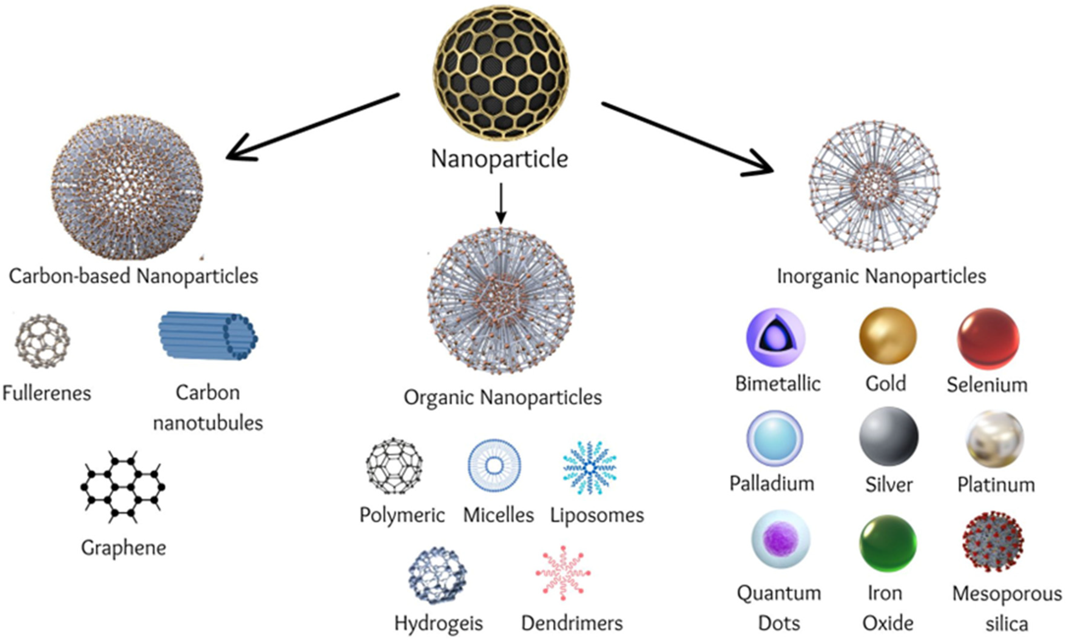
Benefiting from their beneficial traits, such as size, shape, and surface characteristics, the utilization of nanoscale particles in medicine, especially as carriers for medicines, holds tremendous promise in treating a wide range of disorders (Figure 3) [138]. Deliberate personalization is made possible by nanotechnology, which gives one command over their properties [63]. The versatility of nanoparticles (NPs) enables the attachment of various biomolecules, hence promoting the safe and effective transport of pharmacologically active substances, such as medicines or genes. Nanoparticles (NPs) whose diameters range from 1 to 100 nm are able to pass through important physiological barriers, such as those found in the circulation, lungs, liver, gastrointestinal tract, tumor vasculature, mucosal membranes, and the blood–brain barrier [139,140,141].
For this, many kinds of nanoparticles (NPs) have been used, each with unique properties, as therapeutic, diagnostic, or theranostic instruments. Target-specific nanomedicine has expanded its possibilities with the ability to attach medicines and therapeutic nucleic acids to nanoparticles. NPs are used in packaging, biotechnology, electronics, cosmetics, and other industries outside of medicine. NPs can be generically classified as inorganic, organic, or carbon-based. The physicochemical properties of nanoparticles (NPs), which differ between NPs, determine their biocompatibility. NPs may be modified to improve their binding affinities with the gene or medicine they carry, as well as to encourage cell-specific absorption, by using polymers and targeting ligands [142]. Noble metals with advantageous physiochemical, biological, and optical properties, such as gold (Au), silver (Ag), platinum (Pt), and palladium (Pd), are widely used [140,143]. Au NPs’ adjustable physicochemical characteristics make them appropriate for use in clinical settings, where they have demonstrated promise in treating a range of illnesses, including AIDS, syphilis, smallpox, cancer, and skin ulcers [144,145]. Ag NPs are helpful in the pre-treatment of wound infections because they have antibacterial and antiviral characteristics [146]. Nevertheless, their inclination to congregate and grow in bulk presents difficulties in their unaltered state [147]. Dental and electrical devices utilize Pd more frequently [143,148]. Quercetin-modified bimetallic Au-Pd NPs have been investigated as possible autophagy-inducing agents in Alzheimer’s disease [149]. Pt, which is well known for its antioxidant qualities and function in anticancer medications, such as oxaliplatin and cisplatin, may also be neurotoxic [140,150].
When taken in supplement form, selenium (Se) is known to lower the risk of a number of illnesses, including type 2 diabetes, osteoarthritis, cardiovascular disease, and neurological disorders, such as Alzheimer’s disease [151]. Benefits of Se NPs include enhanced bioavailability, biocompatibility, and biodegradability in vivo, as well as the antioxidant and anticancer qualities of Se with reduced cytotoxicity [152,153]. These NPs are becoming more widely known because of their possible synergistic effects with therapeutic genes or medications.
Because of their porous architectures, which provide greater surface areas for therapeutic cargo, mesoporous silica nanoparticles (MSNs) are being exploited more and more as nano-delivery vehicles [154,155]. Due to their porosity, MSNs may possibly enhance biological activity by delivering therapeutic genes and medications together [156]. Quercetin-encapsulated silica nanoparticles have demonstrated potential in combating copper-induced oxidative stress, which is known to occur in neurodegenerative disorders [157]. Because of their advantageous qualities, including minimal cytotoxicity, stability, and magnetism, iron oxides—also known as magnetic nanoparticles, or MNPs—have been the subject of much research in the field of nanomedicine. This is because these traits make them appropriate for use in applications such as magnetic hyperthermia and magnetic resonance imaging (MRI). Magnetofection, using an external magnetic field, may be used to target MNPs, increasing their usefulness for delivery systems [158,159,160]. However, unaltered MNPs can cluster and have a tendency to be hydrophobic, which limits their effectiveness in vivo [161].
Although quantum dots (QDs) have special optical qualities, their composition—which frequently includes metals, such as zinc and cadmium—can make them hazardous. Coated QDs or modified core-shell QDs can reduce this toxicity [156]. Without internal or external functionalization, carbon nanotubes—single- and multi-walled—are immunogenic, insoluble, cytotoxic, and hydrophobic, notwithstanding their ease of entry into cells [162]. Because polymeric delivery methods may bind anionic compounds, such as nucleic acids, they are preferred. This is especially true of those containing cationic polymers. Additionally, these polymers need to be stable in vivo, biocompatible, and biodegradable [156]. For this function, dendrimers—which contain several cationic groups—have gained popularity and are utilized to stabilize metallic NPs, such as Au NPs [163,164]. FDA-approved poly(lactic-co-glycolic acid) has demonstrated potential for medication delivery when paired with Au, and PEGylated variants of this polymer are being studied for AD [165,166]. When it comes to lipid-based NPs, liposomes are commonly utilized to carry bioactive chemicals, and they have demonstrated promising outcomes in animal models of AD [167,168].
Between them, inorganic nanoparticles (NPs) have an edge against organic ones in terms of stability, size, production, functionalization, and theranostic potential. In nanomedicine, all of the above-described NPs have demonstrated promise and may one day be used to treat neurological conditions, including AD and PD. However, in order for these nano-systems to function well, the NPs’ specified qualities need to be carefully considered and given priority [169].

4.1. Nanopharmaceuticals Currently on the Market and Clinical Trials in Progress

The fact that the US Food and Drug Administration has authorized over 250 nanodrugs is evidence of the efficacy of NPs in clinical studies. Notable medications among them are Plegridy (PEGylated interferon β-1a), DepoCyt (liposomal cytarabine), Invega Sustenna (paliperidone palmitate), and Doxil (liposomal doxorubicin HCL injection), which are prescribed for the treatment of MS, schizophrenia, multiple myeloma, and lymphomatous meningitis, respectively [170]. Over 33% of available medications are liposomal formulations, making them the most common type of nanodrugs on the market. It is worth mentioning that some of these formulations have been used extensively in clinical settings for cancer treatment [171].
The possible uses of NPs in medical contexts have also been investigated in a number of clinical studies. According to results from Maier-Hauff et al., individuals with glioblastoma who received magnetic nanoparticle treatment in addition to less radiation had an overall better prognosis than those who were treated with traditional therapy [172]. Furthermore, it has been demonstrated that NPs lessen the toxicity linked to traditional medications [173]. Notably, magnetic nanoparticles have shown a 70% capacity to distribute and transport temozolomide, a chemotherapeutic agent, into cerebral tumor areas in dogs [174].
Promising outcomes in the management of migraines have been observed in recent research, wherein the administration of curcumin NPs and omega-3 fatty acids significantly decreased inflammation by inhibiting the expression of inflammatory markers, such as TNF-α, intercellular adhesive molecule 1, and cyclooxygenase-2/inducible nitric oxide synthase [175,176,177]. All things considered, these results highlight the tremendous promise of NPs in the treatment of brain disorders through improved medication delivery, synergistic benefits, and reduced drug toxicity.
The use of new nanomedicines in scientific research has garnered significant attention as a potentially effective strategy for managing neurodegenerative disorders (NDs). These nanomaterial-based formulations have shown remarkable therapeutic performance, including enhanced brain delivery with specific effects. These nanoformulations can minimize brain damage, promote neuroprotection, improve behavioral outcomes in non-verbal dementia patients, and limit Aβ aggregation, according to preclinical research. There is not much research investigating the application of nanoparticles in the treatment of neurological illnesses. Regarding the treatment of transthyretin-mediated amyloidosis, a noteworthy clinical trial examined the use of a lipid nanoparticle-based formulation. The experiment produced encouraging results, and the regulatory body later approved the product for commercial distribution. Phase I research using CRISPR/Cas9 gene technology and lipid nanoparticles for therapeutic medication delivery is one of the ongoing clinical trials [178].
Just a small number of clinical trials examining medications, such as therapeutic antibodies and secretase inhibitors in Alzheimer’s disease (AD), have been finished, with the majority being discontinued [179]. Surprisingly, since 2003, there has been a global shortage of new medication research for AD [180]. The NIH library was searched recently, and only two pieces of research about the transport of nanoparticles (NPs) were found, indicating this tendency. The research, entitled “Safety, tolerability, and efficacy assessment of intranasal nanoparticles of APH-1105, a novel alpha-secretase modulator for mild to moderate cognitive impairment due to Alzheimer’s disease”, was scheduled to begin in 2023. Phase II of the trial, titled “A Phase 2, pilot open-label, sequential group, investigator-blinded study of magnetic resonance spectroscopy (31P-MRS) to evaluate the effects of CNM-Au8 for the bioenergetic enhancement of the impaired neuronal redox state in Parkinson’s disease,” commenced in December 2019 and was scheduled to end in July 2021 [181]. In this study, gold nanocrystals were used. Although gold nanocrystals have been approved lately for the treatment of multiple sclerosis, further information on this continuing investigation is highly anticipated. If these trials are successful, it might greatly progress the use of NPs in future studies [182].
The objective of the research, “Evaluation of the impact of CNM-Au8 on altered neuronal redox state in Parkinson’s disease using 31-MRS imaging”, is also used to determine how gold nanocrystals affect people with Parkinson’s disease (PD), looking at their pharmacokinetic and pharmacodynamic characteristics, as well as their safety [183].
Although nanotechnology has been around for a while, its first uses have just lately surfaced. Recent research has indicated a great deal of interest in the use of nanostructures to package various drugs or biomolecules to influence CNS inflammation, prevent microbial infections, and influence neuronal regeneration. Taken together, these results provide a crucial basis for further investigations focused on enhancing pharmacokinetics, decreasing systemic drug toxicity, and identifying illnesses in people at an early stage. These developments are desperately required since there is a serious risk that the frequency of neurological illnesses may rise [184].

4.2. Challenges of Nanoparticles

When it comes to using nanoparticles as therapeutic delivery systems for neurodegenerative illnesses, there are a lot of obstacles to overcome. In addition to the blood–brain barrier (BBB), which is the main barrier to successful treatments, worries regarding neurotoxicity from nano-delivery methods also surface, which brings up safety concerns. The most common way that this neurotoxicity shows up is as oxidative stress, which is greatly impacted by the shape, size, surface area, solubility, concentration, length of time, and mode of delivery of the nanotherapeutic. Even though the human body needs some metals for particular functions, the buildup and aggregation of metal nanoparticles may be a cause for concern. Iron nanoparticles have been revealed to be significantly harmful in studies employing the PC12 cell neuronal model, while reactive oxygen species have been discovered to be produced by manganese and copper nanoparticles. Neural stem cells exposed to zinc oxide NPs underwent apoptosis, while Sprague Dawley rats exposed to oral silver NP treatment had toxicity and accumulation in the kidney, liver, and brain [5]. When iron oxide nanoparticles were administered in animal models, the mice developed oxidative stress, neurodegeneration, neuronal apoptosis that was reliant on the cell cycle, and neurobehavioral damage [185,186,187].
Notwithstanding these difficulties, NPs are promising candidates for use in nanomedicine due to their previously noted physicochemical characteristics. NP formulations must include biocompatible, biodegradable, and readily excreted components from the system in order to address some of these issues [188]. Furthermore, the kind of NP used typically determines the reported toxicities, and surface functionalization provides a technique to reduce negative effects and interactions. As such, there is no one-size-fits-all method for selecting a NP and using it. Since many metals are necessary for bodily functions and many metal carriers are non-metals, it is important to evaluate the benefits and drawbacks of each. As such, the concentration that is employed will be essential to preserving homeostatic balance. Since cell-specific targeting is necessary to address damaged or altered genes while maintaining the integrity of normally functioning genes and cells, targeted methods will be crucial in the treatment of AD and PD. Nonetheless, it is clear that further research on NPs is required for developing CNS therapies. Because there is currently a dearth of knowledge on NP neurotoxicity, it is imperative that further in vitro and in vivo research be conducted in order to lay the groundwork for future studies. Developing an optimal NP formulation may be made easier for nanomedicine by using new technology, especially in silico investigations, computer and mathematical modeling, and improved bioinformatics understanding [5].

5. Future Perspectives

Since neurological diseases (NDs) include complicated physiological components and sophisticated networks, current treatment techniques have not been able to successfully stop the progression of NDs. There is a disconnect between research results and their use in clinical trials, as seen by the insufficient therapeutic outcomes that persist despite significant research efforts. Many unique approaches to administering medications to the central nervous system (CNS) have been investigated in a significant quantity of scientific studies over the past few decades, and these approaches have the potential to be used in promising medical applications. The disorders persist in progress even with the availability of drugs that purport to reduce or eliminate NDs’ symptoms. However, problems still exist when it comes to transporting therapeutic and imaging substances beyond the blood–brain barrier (BBB), underscoring the fundamental obstacles in therapy.
In an effort to find more potent forms of therapy, a variety of delivery systems, including pharmaceuticals, biomolecules (including proteins, peptides, and monoclonal antibodies), and phytoconstituents, have been investigated for the administration of therapeutic agents targeting NDs. Furthermore, a method for treating NDs is known as gene therapy, and this approach requires knowledge of the particular ND types and the genes associated with their development. Using neural stem cells (NSCs) and creating therapeutic treatments that can alter NSC differentiation to affect neurogenesis is another possible strategy.
Moreover, NPs have the ability to cross extracellular and intracellular obstacles that obstruct effective gene transport. Combining gene therapy and nanomedicine has the potential to cure a number of illnesses, including monogenic disorders. While minimizing damage to healthy cells, a thorough knowledge of the genetic abnormalities implicated is crucial to developing effective and tailored therapy regimens [189].
Such a study establishes the foundation for tactics that might enhance the lives of millions of people globally and minimize illnesses connected to brain injury, even though an immediate medical solution may not be achievable. The fields of neurobiology and nanomedicine may be able to combine to provide novel therapies for different NDs. However, toxicity-related issues in nanomedicine need to be resolved by using biocompatible nanocarriers. Furthermore, the high expense of treating NDs with nanotechnology makes it necessary to find affordable methods that also increase the safety and clinical effectiveness [178,190].
Scientific research in the field of neurodegenerative diseases has made significant signs of progress in recent years, but there are still several gaps that need to be addressed, which mainly include challenges in their early detection and diagnosis, where the development of new reliable biomarkers and imaging techniques for early detection is essential [5,20,21,22,23,24,25,26,27,28,29,30]. Likewise, it is necessary to elucidate the underlying mechanisms of disease progression to identify potential targets for intervention. In this context, it is also essential to understand the causal connection between genetic predisposition and environmental factors since there is a critical need to develop disease-modifying therapies that can slow or stop disease progression [2,8,40,69]. Moreover, inflammatory processes in the brain are increasingly recognized as a contributing factor in neurodegenerative diseases, and neuroinflammation is a potential therapeutic target that should further be investigated [101,102].
Scientific research regarding non-pharmacological interventions, including cognitive training, physical exercise, dietary adjustments, and lifestyle modifications, are increasingly recognized as promising supplementary approaches for managing neurodegenerative diseases. Nevertheless, further rigorous research is necessary to confirm their effectiveness and determine the most effective ways to implement them. Finally, implementing precision medicine approaches that take into account individual genetic profiles and clinical characteristics could lead to more targeted and effective treatments. Addressing these challenges in research requires interdisciplinary collaboration, innovative methodologies, and sustained funding support to accelerate progress in understanding and treating neurodegenerative diseases [1,2].
Nanomedicine is an emerging promising therapeutical approach in neurodegenerative disease but, nevertheless, the lack of standardized analytical methods for characterizing NPs is challenging for researchers, developers, and government regulatory agencies. Accurately assessing critical quality attributes, such as particle size distribution, surface chemistry, and stability, is essential for ensuring the quality and performance of nanoparticle-based therapies, since inconsistencies in measurement techniques and reporting standards can hinder the comparability of data across studies and regulatory submissions. While nanoparticle therapeutics hold great promise for revolutionizing drug delivery and treatment strategies for neurodegenerative diseases, the complexity of their development and regulatory approval process is very demanding [191]. Addressing these challenges requires collaborative efforts among researchers in different scientific fields, industry stakeholders, and regulatory agencies to establish clear guidelines, standardized protocols, and reliable analytical methods for evaluating the safety and efficacy of nanoparticle-based therapeutical approaches to neurodegenerative diseases [192,193].
Numerous scientific studies indicate a consistent annual rise in the development of medications based on nanotechnology [192,193,194,195]. Pharmaceutical nanomedicine products exert a significant influence on the worldwide pharmaceutical market and healthcare infrastructure [196]. Since 1995, roughly 80 nanomedicine products have secured marketing authorization from both the US Food and Drug Administration (FDA) and the European Medicines Agency (EMA), with double this number undergoing clinical trials [192]. The FDA and EMA have, to some extent, different procedures for approving nanomedicines, leading to differences in timelines and requirements. One crucial difference is the approach to non-clinical biodistribution studies, which are mandatory in the EU but not necessarily required by the FDA. This can result in additional time and resources being spent on fulfilling EU-specific requirements. An example of a nanomedicine that faced differences in approval processes between the FDA and EMA is liposomal doxorubicin (Doxil/Caelyx). While the FDA approved Doxil for multiple indications, including ovarian cancer and AIDS-related Kaposi’s sarcoma, in the late 1990s, it took several more years for the EMA to grant approval for similar indications [192]. Abraxane, a nanoparticle albumin-bound formulation of paclitaxel, is another example of differences in the regulatory requirements and timelines between the two agencies that contributed to this discrepancy in approval timings. While the FDA approved Abraxane for the treatment of breast cancer in 2005, EMA approval was obtained several years later, in 2008. These delays were partly due to differences in requirements for clinical trial data and non-clinical studies between the two agencies [191,192,193,194,195].
Halwani, in his comprehensive review, recently summarized all globally marketed nanomedicines approved by the FDA and MDA, including all important details, such as active ingredients, type of nanotechnology, and indications for certain pathologies’ treatment [192]. Currently, a plethora of new nanoparticle-based therapeutical approaches are being evaluated by the FDA, EMA, and other agencies but, in general, there is a lack of standards in the evaluation of these compounds due to their unique properties as therapeutics, which makes this evaluation even more challenging [192,197]. The EMA and FDA released reflection papers and preliminary guidelines for the industry to outline their current perspectives on assessing nanotherapeutics [196]. A systematic method for assessing similarity, starting from quality considerations, such as critical quality attributes (CQA) and evaluation of nanoscale properties, progressing to non-clinical biodistribution analysis (mandatory in the EU but not in the US), and ultimately to clinical evaluation, is essential [196]. However, there is still an absence of a clearly defined or unified approval process for nanotherapeutics, leading to potential disparities in approval outcomes. Advancement requires the establishment of a science-driven forum involving stakeholders and field experts. However, an agenda has been established, focusing on CQA assessment, dissemination of scientific and clinical research, consensus building on nomenclature and labeling, and regulatory measures concerning substandard complex drug products [198]. The need for greater harmonization in approval procedures is essential to expedite access to these innovative therapies.
The exchange of instructions and experiences between the European Medicines Agency (EMA) and the US Food and Drug Administration (FDA) can significantly benefit the commercialization of nanomedicines in the context of research, regulatory, and approval processes’ review. Sharing best practices and expertise in analytical methods and characterization techniques for nanomedicines can enhance the quality and consistency of data submitted in regulatory applications. This can bring great benefit to the scientific community and can help in the development and harmonization of guidance documents and regulatory frameworks specifically tailored for nanomedicines, benefiting patients affected by neurodegenerative and other diseases and, ultimately, improving general public health [192].

6. Conclusions

A promising strategy for addressing enduring issues in conventional medicine is emerging—nanomedicine. Combining gene therapy with nanotechnology offers potential for improved therapeutic outcomes, raising hope for eradicating neuronal-damage-related illnesses, such as Alzheimer’s disease (AD), Parkinson’s disease (PD), and Huntington’s disease (HD). Despite numerous pharmacological options, many struggle to penetrate the blood–brain barrier (BBB) for meaningful clinical results. Brain-targeting strategies with nano-biomaterials offer alternatives, allowing modulation of neural pathways for desired therapeutic effects.
Gene therapy and nanoparticle-based treatments hold promise for treating neurological diseases, but they also face significant limitations and challenges, such as delivery challenges, where the major obstacle for both gene therapy and nanoparticle-based treatments is effectively delivering therapeutic agents to the target cells or tissues within the CNS, as well as bypassing or overcoming the BBB. In addition, the activation of the immune response during those treatments may lead to inflammation, tissue damage, and clearance of the therapeutic agent. Both gene therapy and nanoparticle-based treatments may unintentionally affect non-target cells or tissues, leading to off-target effects. Achieving sustained therapeutic effects over the long term remains a challenge for both gene therapy and nanoparticle-based treatments. Factors such as degradation of therapeutic agents, immune responses, and loss of transgene expression or nanoparticle functionality over time can limit the durability of treatment outcomes. Developing strategies to enhance the long-term efficacy and stability of these therapies is crucial for their clinical success. Overcoming these obstacles is crucial to achieve the effectiveness of gene therapy and nanoparticle-based treatments in the treatment of neurodegenerative diseases.
Nanotechnology in medicine has enhanced the treatment efficacy and the diagnosis of illnesses. Nanoparticles, capable of crossing the BBB, hold promise for diagnosing and treating challenging brain conditions. Combining gene therapy with nanotechnology could enhance effectiveness in treating neurodegenerative diseases. Recent advancements in nano-biomaterial-based techniques have strengthened existing neural stem cell (NSC) differentiation treatments. NSC-targeting technologies offer a promising, potentially safe method for treating neurodegenerative diseases.
Thorough testing for toxicity and stability is crucial for the diverse range of nanoparticles available. Further investigation into nanoparticle toxicology and bioaccumulation in clinical settings is essential for ensuring safety and effectiveness. Prioritizing therapeutic nanoparticles with low toxicity is crucial for optimal results. Improved formulations, including specific antibodies, can enhance selectivity to target specific biomarkers, offering promise for treating brain illnesses and disorders with nanotechnology.
Finally, lifestyle interventions, including diet, exercise, cognitive stimulation, and social engagement, can impact brain health and may complement other therapeutic approaches for neurological diseases. Rehabilitation strategies, such as physical therapy, occupational therapy, speech therapy, and cognitive rehabilitation, aim to improve function and quality of life for individuals living with neurological conditions. These approaches, along with nanotechnology, represent a multifaceted and evolving landscape in the quest to develop effective treatments for neurological diseases. Integration of these diverse strategies and interdisciplinary collaboration will be crucial for advancing therapeutic options and improving outcomes for patients affected by neurodegenerative diseases.

Author Contributions

Conceptualization, A.K. and L.B.; writing—original draft preparation, A.K.; writing—review and editing, L.B.; supervision, L.B.; project administration, L.B.; funding acquisition, L.B. All authors have read and agreed to the published version of the manuscript.

Funding

This research received no external funding.

Institutional Review Board Statement

Not applicable.

Informed Consent Statement

Not applicable.

Data Availability Statement

No new data were created or analyzed in this study. Data sharing is not applicable to this article.

Conflicts of Interest

The authors declare no conflicts of interest.

References

  1. Kovacs, G.G. Concepts and classification of neurodegenerative diseases. Handb. Clin. Neurol. 2017, 145, 301–307. [Google Scholar] [CrossRef] [PubMed]
  2. Dugger, B.N.; Dickson, D.W. Pathology of Neurodegenerative Diseases. Cold Spring Harb. Perspect. Biol. 2017, 9, a028035. [Google Scholar] [CrossRef] [PubMed]
  3. Uchikado, H.; DelleDonne, A.; Ahmed, Z.; Dickson, D.W. Lewy bodies in progressive supranuclear palsy represent an independent disease process. J. Neuropathol. Exp. Neurol. 2006, 65, 387–395. [Google Scholar] [CrossRef] [PubMed]
  4. Ghasemi, M.; Brown, R.H.J. Genetics of Amyotrophic Lateral Sclerosis. Cold Spring Harb. Perspect. Med. 2018, 8, a024125. [Google Scholar] [CrossRef] [PubMed]
  5. Jagaran, K.; Singh, M. Nanomedicine for Neurodegenerative Disorders: Focus on Alzheimer’s and Parkinson’s Diseases. Int. J. Mol. Sci. 2021, 22, 9082. [Google Scholar] [CrossRef] [PubMed] [PubMed Central]
  6. Ding, C.; Wu, Y.; Chen, X.; Chen, Y.; Wu, Z.; Lin, Z.; Kang, D.; Fang, W.; Chen, F. Global, regional, and national burden and attributable risk factors of neurological disorders: The Global Burden of Disease study 1990–2019. Front. Public Health 2022, 10, 952161. [Google Scholar] [CrossRef] [PubMed] [PubMed Central]
  7. Ghosh, P.; Higgins, D.E. Listeria monocytogenes Infection of the Brain. J. Vis. Exp. 2018, 140, e58723. [Google Scholar] [CrossRef] [PubMed] [PubMed Central]
  8. Borsche, M.; König, I.R.; Delcambre, S.; Petrucci, S.; Balck, A.; Brüggemann, N.; Zimprich, A.; Wasner, K.; Pereira, S.L.; Avenali, M.; et al. Mitochondrial damage-associated inflammation highlights biomarkers in PRKN/PINK1 parkinsonism. Brain 2020, 143, 3041–3051. [Google Scholar] [CrossRef] [PubMed] [PubMed Central]
  9. Baccarini, C.I.; Simon, M.W.; Brandon, D.; Christensen, S.; Jordanov, E.; Dhingra, M.S. Safety and Immunogenicity of a Quadrivalent Meningococcal Conjugate Vaccine in Healthy Meningococcal-Naïve Children 2–9 Years of Age: A Phase III, Randomized Study. Pediatr. Infect. Dis. J. 2020, 39, 955–960. [Google Scholar] [CrossRef] [PubMed] [PubMed Central]
  10. Pardridge, W.M. Drug transport across the blood-brain barrier. J. Cereb. Blood Flow. Metab. 2012, 32, 1959–1972. [Google Scholar] [CrossRef] [PubMed] [PubMed Central]
  11. Ngowi, E.E.; Wang, Y.Z.; Qian, L.; Helmy, Y.A.S.H.; Anyomi, B.; Li, T.; Zheng, M.; Jiang, E.S.; Duan, S.F.; Wei, J.S.; et al. The Application of Nanotechnology for the Diagnosis and Treatment of Brain Diseases and Disorders. Front. Bioeng. Biotechnol. 2021, 9, 629832. [Google Scholar] [CrossRef] [PubMed] [PubMed Central]
  12. Rabanel, J.M.; Piec, P.A.; Landri, S.; Patten, S.A.; Ramassamy, C. Transport of PEGylated-PLA nanoparticles across a blood brain barrier model, entry into neuronal cells and in vivo brain bioavailability. J. Control Release 2020, 328, 679–695. [Google Scholar] [CrossRef] [PubMed]
  13. Zielińska, A.; Carreiró, F.; Oliveira, A.M.; Neves, A.; Pires, B.; Venkatesh, D.N.; Durazzo, A.; Lucarini, M.; Eder, P.; Silva, A.M.; et al. Polymeric Nanoparticles: Production, Characterization, Toxicology and Ecotoxicology. Molecules 2020, 25, 3731. [Google Scholar] [CrossRef] [PubMed] [PubMed Central]
  14. Rasouli, M.R.; Tabrizian, M. An ultra-rapid acoustic micromixer for synthesis of organic nanoparticles. Lab. Chip. 2019, 19, 3316–3325. [Google Scholar] [CrossRef] [PubMed]
  15. Matsuno, J.; Kanamaru, T.; Arai, K.; Tanaka, R.; Lee, J.H.; Takahashi, R.; Sakurai, K.; Fujii, S. Synthesis and characterization of nanoemulsion-mediated core crosslinked nanoparticles, and in vivo pharmacokinetics depending on the structural characteristics. J. Control Release 2020, 324, 405–412. [Google Scholar] [CrossRef] [PubMed]
  16. Zhang, M.; Viennois, E.; Prasad, M.; Zhang, Y.; Wang, L.; Zhang, Z.; Han, M.K.; Xiao, B.; Xu, C.; Srinivasan, S.; et al. Edible ginger-derived nanoparticles: A novel therapeutic approach for the prevention and treatment of inflammatory bowel disease and colitis-associated cancer. Biomaterials 2016, 101, 321–340. [Google Scholar] [CrossRef] [PubMed] [PubMed Central]
  17. Li, Y.; Hao, L.; Liu, F.; Yin, L.; Yan, S.; Zhao, H.; Ding, X.; Guo, Y.; Cao, Y.; Li, P.; et al. Cell penetrating peptide-modified nanoparticles for tumor targeted imaging and synergistic effect of sonodynamic/HIFU therapy. Int. J. Nanomed. 2019, 14, 5875–5894, Erratum in Int. J. Nanomed. 2019, 14, 6867. [Google Scholar] [CrossRef] [PubMed]
  18. Elbaz, A.; Dufouil, C.; Alperovitch, A. Interaction between genes and environment in neurodegenerative diseases. C. R. Biol. 2007, 330, 318–328. [Google Scholar] [CrossRef] [PubMed]
  19. Chiara, T.; Origlia, N.; Mattu, C.; Accorroni, A.; Chiono, V. Current Limitations in the Treatment of Parkinson’s and Alzheimer’s Diseases: State-of-the-Art and Future Perspective of Polymeric Carriers. Curr. Med. Chem. 2018, 25, 5755–5771. [Google Scholar] [CrossRef] [PubMed]
  20. Weissleder, R.; Mahmood, U. Molecular imaging. Radiology 2001, 219, 316–333. [Google Scholar] [CrossRef] [PubMed]
  21. Loftus, J.R.; Puri, S.; Meyers, S.P. Multimodality imaging of neurodegenerative disorders with a focus on multiparametric magnetic resonance and molecular imaging. Insights Imaging 2023, 14, 8. [Google Scholar] [CrossRef] [PubMed]
  22. Aldossary, N.M.; Kotb, M.A.; Kamal, A.M. Predictive value of early MRI findings on neurocognitive and psychiatric outcomes in patients with severe traumatic brain injury. J. Affect. Disord. 2019, 243, 1–7. [Google Scholar] [CrossRef] [PubMed]
  23. Bocan, T.M.; Stafford, R.G.; Brown, J.L.; Akuoku Frimpong, J.; Basuli, F.; Hollidge, B.S.; Zhang, X.; Raju, N.; Swenson, R.E.; Smith, D.R. Characterization of Brain Inflammation, Apoptosis, Hypoxia, Blood-Brain Barrier Integrity and Metabolism in Venezuelan Equine Encephalitis Virus (VEEV TC-83) Exposed Mice by In Vivo Positron Emission Tomography Imaging. Viruses 2019, 11, 1052. [Google Scholar] [CrossRef] [PubMed] [PubMed Central]
  24. Tang, T.; Valenzuela, A.; Petit, F.; Chow, S.; Leung, K.; Gorin, F.; Louie, A.Y.; Dhenain, M. In Vivo MRI of Functionalized Iron Oxide Nanoparticles for Brain Inflammation. Contrast Media Mol. Imaging 2018, 2018, 3476476. [Google Scholar] [CrossRef] [PubMed] [PubMed Central]
  25. Naczynski, D.J.; Stafford, J.H.; Türkcan, S.; Jenkins, C.; Koh, A.L.; Sun, C.; Xing, L. Rare-Earth-Doped Nanoparticles for Short-Wave Infrared Fluorescence Bioimaging and Molecular Targeting of αVβ3-Expressing Tumors. Mol. Imaging 2018, 17, 1536012118799131. [Google Scholar] [CrossRef] [PubMed] [PubMed Central]
  26. Schaller, B.J. Strategies for molecular imaging dementia and neurodegenerative diseases. Neuropsychiatr. Dis. Treat 2008, 4, 585–612. [Google Scholar] [CrossRef] [PubMed] [PubMed Central]
  27. Barthel, H.; Villemagne, V.L.; Drzezga, A. Future Directions in Molecular Imaging of Neurodegenerative Disorders. J. Nucl. Med. 2022, 63 (Suppl. S1), 68S–74S. [Google Scholar] [CrossRef] [PubMed]
  28. Valotassiou, V.; Malamitsi, J.; Papatriantafyllou, J.; Dardiotis, E.; Tsougos, I.; Psimadas, D.; Alexiou, S.; Hadjigeorgiou, G.; Georgoulias, P. SPECT and PET imaging in Alzheimer’s disease. Ann. Nucl. Med. 2018, 32, 583–593. [Google Scholar] [CrossRef] [PubMed]
  29. Gustavsson, T.; Syvänen, S.; O’Callaghan, P.; Sehlin, D. SPECT imaging of distribution and retention of a brain-penetrating bispecific amyloid-β antibody in a mouse model of Alzheimer’s disease. Transl. Neurodegener. 2020, 9, 37. [Google Scholar] [CrossRef] [PubMed]
  30. Korat, Š.; Bidesi, N.S.R.; Bonanno, F.; Di Nanni, A.; Hoàng, A.N.N.; Herfert, K.; Maurer, A.; Battisti, U.M.; Bowden, G.D.; Thonon, D.; et al. Alpha-synuclein PET tracer development: An overview about current efforts. Pharmaceuticals 2021, 14, 847. [Google Scholar] [CrossRef] [PubMed]
  31. Guo, Y.; You, J.; Zhang, Y.; Liu, W.-S.; Huang, Y.-Y.; Zhang, Y.-R.; Zhang, W.; Dong, Q.; Feng, J.-F.; Cheng, W.; et al. Plasma proteomic profiles predict future dementia in healthy adults. Nat. Aging 2024, 4, 247–260. [Google Scholar] [CrossRef] [PubMed]
  32. Garibotto, V.; Albert, N.L.; Barthel, H.; van Berckel, B.; Boellaard, R.; Brendel, M.; Cecchin, D.; Ekmekcioglu, O.; van de Giessen, E.; Guedj, E.; et al. EANM Neuroimaging Committee. The approval of a disease-modifying treatment for Alzheimer’s disease: Impact and consequences for the nuclear medicine community. Eur. J. Nucl. Med. Mol. Imaging 2021, 48, 3033–3036. [Google Scholar] [CrossRef] [PubMed]
  33. Kobeissy, F.; Arja, R.D.; Munoz, J.C.; Shear, D.A.; Gilsdorf, J.; Zhu, J.; Yadikar, H.; Haskins, W.; Tyndall, J.A.; Wang, K.K. The game changer: UCH-L1 and GFAP-based blood test as the first marketed in vitro diagnostic test for mild traumatic brain injury. Expert. Rev. Mol. Diagn. 2024, 24, 67–77. [Google Scholar] [CrossRef] [PubMed]
  34. Posti, J.P.; Takala, R.S.; Runtti, H.; Newcombe, V.F.; Outtrim, J.; Katila, A.J.; Frantzén, J.; Ala-Seppälä, H.; Coles, J.P.; Hossain, M.I.; et al. The Levels of Glial Fibrillary Acidic Protein and Ubiquitin C-Terminal Hydrolase-L1 During the First Week After a Traumatic Brain Injury: Correlations With Clinical and Imaging Findings. Neurosurgery 2016, 79, 456–464. [Google Scholar] [CrossRef] [PubMed]
  35. Singh, G.P.; Nigam, R.; Tomar, G.S.; Monisha, M.; Bhoi, S.K.; S, A.; Sengar, K.; Akula, D.; Panta, P.; Anindya, R. Early and rapid detection of UCHL1 in the serum of brain-trauma patients: A novel gold nanoparticle-based method for diagnosing the severity of brain injury. Analyst 2018, 143, 3366–3373. [Google Scholar] [CrossRef] [PubMed]
  36. van Steenoven, I.; van der Flier, W.M.; Scheltens, P.; Teunissen, C.E.; Lemstra, A.W. Amyloid-β peptides in cerebrospinal fluid of patients with dementia with Lewy bodies. Alzheimers Res. Ther. 2019, 11, 83. [Google Scholar] [CrossRef] [PubMed] [PubMed Central]
  37. de Oliveira, F.F.; Miraldo, M.C.; de Castro-Neto, E.F.; de Almeida, S.S.; Matas, S.L.A.; Bertolucci, P.H.F.; Naffah-Mazzacoratti, M.D.G. Differential associations of clinical features with cerebrospinal fluid biomarkers in dementia with Lewy bodies and Alzheimer’s disease. Aging Clin. Exp. Res. 2023, 35, 1741–1752. [Google Scholar] [CrossRef] [PubMed]
  38. Zeng, J.; Wu, J.; Li, M.; Wang, P. A Novel Magnetic Nanoparticle for Early Detection of Amyloid Plaques in Alzheimer’s Disease. Arch. Med. Res. 2018, 49, 282–285. [Google Scholar] [CrossRef] [PubMed]
  39. Fernández-Cabada, T.; Ramos-Gómez, M. A Novel Contrast Agent Based on Magnetic Nanoparticles for Cholesterol Detection as Alzheimer’s Disease Biomarker. Nanoscale Res. Lett. 2019, 14, 36. [Google Scholar] [CrossRef] [PubMed] [PubMed Central]
  40. Sun, L.; Liu, D.; Fu, D.; Yue, T.; Scharre, D.; Zhang, L. Fluorescent peptide nanoparticles to detect amyloid-beta aggregation in cerebrospinal fluid and serum for Alzheimer’s disease diagnosis and progression monitoring. Chem. Eng. J. 2021, 405, 126733. [Google Scholar] [CrossRef]
  41. Pardridge, W.M.; Mietus, L.J. Transport of steroid hormones through the rat blood-brain barrier. Primary role of albumin-bound hormone. J. Clin. Investig. 1979, 64, 145–154. [Google Scholar] [CrossRef] [PubMed] [PubMed Central]
  42. Harilal, S.; Jose, J.; Parambi, D.G.T.; Kumar, R.; Unnikrishnan, M.K.; Uddin, M.S.; Mathew, G.E.; Pratap, R.; Marathakam, A.; Mathew, B. Revisiting the blood-brain barrier: A hard nut to crack in the transportation of drug molecules. Brain Res. Bull. 2020, 160, 121–140. [Google Scholar] [CrossRef] [PubMed]
  43. He, H.; Yao, J.; Zhang, Y.; Chen, Y.; Wang, K.; Lee, R.J.; Yu, B.; Zhang, X. Solid lipid nanoparticles as a drug delivery system to across the blood-brain barrier. Biochem. Biophys. Res. Commun. 2019, 519, 385–390. [Google Scholar] [CrossRef] [PubMed]
  44. Sadegh Malvajerd, S.; Azadi, A.; Izadi, Z.; Kurd, M.; Dara, T.; Dibaei, M.; Sharif Zadeh, M.; Akbari Javar, H.; Hamidi, M. Brain Delivery of Curcumin Using Solid Lipid Nanoparticles and Nanostructured Lipid Carriers: Preparation, Optimization, and Pharmacokinetic Evaluation. ACS Chem. Neurosci. 2019, 10, 728–739. [Google Scholar] [CrossRef] [PubMed]
  45. Akbari, J.; Saeedi, M.; Ahmadi, F.; Hashemi, S.M.H.; Babaei, A.; Yaddollahi, S.; Rostamkalaei, S.S.; Asare-Addo, K.; Nokhodchi, A. Solid lipid nanoparticles and nanostructured lipid carriers: A review of the methods of manufacture and routes of administration. Pharm. Dev. Technol. 2022, 27, 525–544. [Google Scholar] [CrossRef] [PubMed]
  46. Johnsen, K.B.; Bak, M.; Melander, F.; Thomsen, M.S.; Burkhart, A.; Kempen, P.J.; Andresen, T.L.; Moos, T. Modulating the antibody density changes the uptake and transport at the blood-brain barrier of both transferrin receptor-targeted gold nanoparticles and liposomal cargo. J. Control Release 2019, 295, 237–249. [Google Scholar] [CrossRef] [PubMed]
  47. Meng, Q.; Wang, A.; Hua, H.; Jiang, Y.; Wang, Y.; Mu, H.; Wu, Z.; Sun, K. Intranasal delivery of Huperzine A to the brain using lactoferrin-conjugated N-trimethylated chitosan surface-modified PLGA nanoparticles for treatment of Alzheimer’s disease. Int. J. Nanomed. 2018, 13, 705–718. [Google Scholar] [CrossRef] [PubMed] [PubMed Central]
  48. Alzheimer’s Disease Facts and Figures. Alzheimers Dementia. 2020. Available online: https://alz-journals.onlinelibrary.wiley.com/doi/full/10.1002/alz.12068 (accessed on 18 February 2023).
  49. 2023 Alzheimer’s disease Facts and Figures. Alzheimers Dement. 2023, 19, 1598–1695. [CrossRef] [PubMed]
  50. García-González, L.; Pilat, D.; Baranger, K.; Rivera, S. Emerging Alternative Proteinases in APP Metabolism and Alzheimer’s Disease Pathogenesis: A Focus on MT1-MMP and MT5-MMP. Front. Aging Neurosci. 2019, 11, 244. [Google Scholar] [CrossRef]
  51. Agatonovic-Kustrina, S.; Kettle, C.; Morton, D.W. A molecular approach in drug development for Alzheimer’s disease. Biomed. Pharmacother. 2018, 106, 553–565. [Google Scholar] [CrossRef]
  52. Nam, E.; Lee, Y.B.; Moon, C.; Chang, K.A. Serum Tau Proteins as Potential Biomarkers for the Assessment of Alzheimer’s Disease Progression. Int. J. Mol. Sci. 2020, 21, 5007. [Google Scholar] [CrossRef] [PubMed] [PubMed Central]
  53. Tagai, K.; Ono, M.; Kubota, M.; Kitamura, S.; Takahata, K.; Seki, C.; Takado, Y.; Shinotoh, H.; Sano, Y.; Yamamoto, Y.; et al. High-Contrast In Vivo Imaging of Tau Pathologies in Alzheimer’s and Non-Alzheimer’s Disease Tauopathies. Neuron 2021, 109, 42–58.e8. [Google Scholar] [CrossRef] [PubMed]
  54. Sonawane, S.K.; Ahmad, A.; Chinnathambi, S. Protein-Capped Metal Nanoparticles Inhibit Tau Aggregation in Alzheimer’s Disease. ACS Omega 2019, 4, 12833–12840. [Google Scholar] [CrossRef] [PubMed] [PubMed Central]
  55. Ciccone, L.; Shi, C.; di Lorenzo, D.; Van Baelen, A.C.; Tonali, N. The Positive Side of the Alzheimer’s Disease Amyloid Cross-Interactions: The Case of the Aβ 1-42 Peptide with Tau, TTR, CysC, and ApoA1. Molecules 2020, 25, 2439. [Google Scholar] [CrossRef] [PubMed] [PubMed Central]
  56. Sanabria-Castro, A.; Alvarado-Echeverria, I.; Monge-Bonilla, C. Molecular pathogenesis of Alzheimer’s disease: An update. Ann. Neurosci. 2017, 24, 46–54. [Google Scholar] [CrossRef] [PubMed]
  57. Kelleher, R.J.; Shen, J. Presenilin-1 mutations and Alzheimer’s disease. Proc. Natl. Acad. Sci. USA 2017, 114, 629–631. [Google Scholar] [CrossRef] [PubMed]
  58. Sun, L.; Zhou, R.; Yang, G.; Shi, Y. Analysis of 138 pathogenic mutations in presenilin-1 on the in vitro production of Aβ42 and Aβ40 peptides by γ-secretase. Proc. Natl. Acad. Sci. USA 2017, 114, E476–E485. [Google Scholar] [CrossRef] [PubMed]
  59. Sayas, C.L.; Ávila, J. GSK-3 and Tau: A Key Duet in Alzheimer’s Disease. Cells 2021, 10, 721. [Google Scholar] [CrossRef] [PubMed] [PubMed Central]
  60. Bardai, F.H.; Price, V.; Zaayman, M.; Wang, L.; D’Mello, S.R. Histone deacetylase-1 (HDAC1) is a molecular switch between neuronal survival and death. J. Biol. Chem. 2012, 287, 35444–35453. [Google Scholar] [CrossRef] [PubMed] [PubMed Central]
  61. Wen, W.; Hu, J.; Wang, C.; Yang, R.; Zhang, Y.; Huang, B.; Qiao, T.; Wang, J.; Chen, X. Re-exploration of tetrahydro-β-carboline scaffold: Discovery of selective histone deacetylase 6 inhibitors with neurite outgrowth-promoting and neuroprotective activities. Bioorg Med. Chem. Lett. 2024, 102, 129670. [Google Scholar] [CrossRef] [PubMed]
  62. Soares Romeiro, L.A.; da Costa Nunes, J.L.; de Oliveira Miranda, C.; Simões Heyn Roth Cardoso, G.; de Oliveira, A.S.; Gandini, A.; Kobrlova, T.; Soukup, O.; Rossi, M.; Senger, J.; et al. Novel Sustainable-by-Design HDAC Inhibitors for the Treatment of Alzheimer’s Disease. ACS Med. Chem. Lett. 2019, 10, 671–676. [Google Scholar] [CrossRef] [PubMed] [PubMed Central]
  63. Chen, W.; Ouyang, J.; Yi, X.; Xu, Y.; Niu, C.; Zhang, W.; Wang, L.; Sheng, J.; Deng, L.; Liu, Y.N.; et al. Black Phosphorus Nanosheets as a Neuroprotective Nanomedicine for Neurodegenerative Disorder Therapy. Adv. Mater. 2018, 30, 1703458. [Google Scholar] [CrossRef] [PubMed]
  64. Yang, Y.; Bagyinszky, E.; An, S.S.A. Presenilin-1 (PSEN1) Mutations: Clinical Phenotypes beyond Alzheimer’s Disease. Int. J. Mol. Sci. 2023, 24, 8417. [Google Scholar] [CrossRef] [PubMed] [PubMed Central]
  65. Sun, Y.; Islam, S.; Michikawa, M.; Zou, K. Presenilin: A Multi-Functional Molecule in the Pathogenesis of Alzheimer’s Disease and Other Neurodegenerative Diseases. Int. J. Mol. Sci. 2024, 25, 1757. [Google Scholar] [CrossRef] [PubMed] [PubMed Central]
  66. Vakilinezhad, M.A.; Amini, A.; Akbari Javar, H.; Baha’addini Beigi Zarandi, B.F.; Montaseri, H.; Dinarvand, R. Nicotinamide loaded functionalized solid lipid nanoparticles improves cognition in Alzheimer’s disease animal model by reducing Tau hyperphosphorylation. Daru 2018, 26, 165–177. [Google Scholar] [CrossRef] [PubMed] [PubMed Central]
  67. Jeon, S.G.; Cha, M.Y.; Kim, J.I.; Hwang, T.W.; Kim, K.A.; Kim, T.H.; Song, K.C.; Kim, J.J.; Moon, M. Vitamin D-binding protein-loaded PLGA nanoparticles suppress Alzheimer’s disease-related pathology in 5XFAD mice. Nanomedicine 2019, 297–307. [Google Scholar] [CrossRef] [PubMed]
  68. Dos Santos Tramontin, N.; da Silva, S.; Arruda, R.; Ugioni, K.S.; Canteiro, P.B.; de Bem Silveira, G.; Mendes, C.; Silveira, P.C.L.; Muller, A.P. Gold Nanoparticles Treatment Reverses Brain Damage in Alzheimer’s Disease Model. Mol. Neurobiol. 2020, 57, 926–936. [Google Scholar] [CrossRef] [PubMed]
  69. Moore, K.A.; Pate, K.M.; Soto-Ortega, D.D.; Lohse, S.; van der Munnik, N.; Lim, M.; Jackson, K.S.; Lyles, V.D.; Jones, L.; Glassgow, N.; et al. Influence of gold nanoparticle surface chemistry and diameter upon Alzheimer’s disease amyloid-β protein aggregation. J. Biol. Eng. 2017, 11, 5. [Google Scholar] [CrossRef] [PubMed] [PubMed Central]
  70. Chen, J.; Ma, Q.; Li, M.; Chao, D.; Huang, L.; Wu, W.; Fang, Y.; Dong, S. Glucose-oxidase like catalytic mechanism of noble metal nanozymes. Nat. Commun. 2021, 12, 3375. [Google Scholar] [CrossRef] [PubMed] [PubMed Central]
  71. Liao, Y.H.; Chang, Y.J.; Yoshiike, Y.; Chang, Y.C.; Chen, Y.R. Negatively charged gold nanoparticles inhibit Alzheimer’s amyloid-β fibrillization, induce fibril dissociation, and mitigate neurotoxicity. Small 2012, 8, 3631–3639. [Google Scholar] [CrossRef] [PubMed]
  72. Gao, G.; Zhang, M.; Gong, D.; Chen, R.; Hu, X.; Sun, T. The size-effect of gold nanoparticles and nanoclusters in the inhibition of amyloid-β fibrillation. Nanoscale 2017, 9, 4107–4113. [Google Scholar] [CrossRef] [PubMed]
  73. Yamazaki, Y.; Zhao, N.; Caulfield, T.R.; Liu, C.C.; Bu, G. Apolipoprotein E and Alzheimer disease: Pathobiology and targeting strategies. Nat. Rev. Neurol. 2019, 15, 501–518. [Google Scholar] [CrossRef] [PubMed] [PubMed Central]
  74. Chartier-Harlin, M.C.; Parfitt, M.; Legrain, S.; Pérez-Tur, J.; Brousseau, T.; Evans, A.; Berr, C.; Vidal, O.; Roques, P.; Gourlet, V. Apolipoprotein E, epsilon 4 allele as a major risk factor for sporadic early and late-onset forms of Alzheimer’s disease: Analysis of the 19q13.2 chromosomal region. Hum. Mol. Genet. 1994, 3, 569–574. [Google Scholar] [CrossRef] [PubMed]
  75. Haddad, H.W.; Malone, G.W.; Comardelle, N.J.; Degueure, A.E.; Kaye, A.M.; Kaye, A.D. Aducanumab, a Novel Anti-Amyloid Monoclonal Antibody, for the Treatment of Alzheimer’s Disease: A Comprehensive Review. Health Psychol. Res. 2022, 10, 31925. [Google Scholar] [CrossRef] [PubMed] [PubMed Central]
  76. Ogino, M.; Ichimura, M.; Nakano, N.; Minami, A.; Kitagishi, Y.; Matsuda, S. Roles of PTEN with DNA Repair in Parkinson’s Disease. Int. J. Mol. Sci. 2016, 17, 954. [Google Scholar] [CrossRef] [PubMed] [PubMed Central]
  77. Inoshita, T.; Shiba-Fukushima, K.; Meng, H.; Hattori, N.; Imai, Y. Monitoring Mitochondrial Changes by Alteration of the PINK1-Parkin Signaling in Drosophila. Methods Mol. Biol. 2018, 1759, 47–57. [Google Scholar] [CrossRef] [PubMed]
  78. Devireddy, S.; Liu, A.; Lampe, T.; Hollenbeck, P.J. The Organization of Mitochondrial Quality Control and Life Cycle in the Nervous System in Vivo in the Absence of PINK1. J. Neurosci. 2015, 35, 9391–9401. [Google Scholar] [CrossRef] [PubMed]
  79. Charan, R.A.; LaVoie, M.J. Pathologic and therapeutic implications for the cell biology of Parkin. Mol. Cell. Neurosci. 2015, 66, 62–71. [Google Scholar] [CrossRef] [PubMed]
  80. Hattori, N.; Mizuno, Y. Twenty years since the discovery of the parkin gene. J. Neural Transm. 2017, 124, 1037–1054. [Google Scholar] [CrossRef]
  81. Jacoupy, M.; Hamon-Keromen, E.; Ordureau, A.; Erpapazoglou, Z.; Coge, F.; Corvol, J.-C.; Nosjean, O.; la Cour, C.M.; Millan, M.J.; Boutin, J.A.; et al. The PINK1 kinase-driven ubiquitin ligase Parkin promotes mitochondrial protein import through the presequence pathway in living cells. Sci. Rep. 2019, 9, 11829. [Google Scholar] [CrossRef]
  82. Tian Hui Kwan, A.; Arfaie, S.; Therriault, J.; Rosa-Neto, P.; Gauthier, S. Lessons Learnt from the Second Generation of Anti-Amyloid Monoclonal Antibodies Clinical Trials. Dement. Geriatr. Cogn. Disord. 2020, 49, 334–348. [Google Scholar] [CrossRef] [PubMed]
  83. Kastanenka, K.V.; Bussiere, T.; Shakerdge, N.; Qian, F.; Weinreb, P.H.; Rhodes, K.; Bacskai, B.J. Immunotherapy with Aducanumab Restores Calcium Homeostasis in Tg2576 Mice. J. Neurosci. 2016, 36, 12549–12558. [Google Scholar] [CrossRef]
  84. Gamage, K.K.; Kumar, S. Aducanumab Therapy Ameliorates Calcium Overload in a Mouse Model of Alzheimer’s Disease. J. Neurosci. Off. J. Soc. Neurosci. 2017, 37, 4430–4432. [Google Scholar] [CrossRef]
  85. Kuller, L.H.; Lopez, O.L. ENGAGE and EMERGE: Truth and consequences? Alzheimers Dement. 2021, 17, 692–695. [Google Scholar] [CrossRef]
  86. Alexander, G.C.; Emerson, S.; Kesselheim, A.S. Evaluation of Aducanumab for Alzheimer Disease: Scientific Evidence and Regulatory Review Involving Efficacy, Safety, and Futility. JAMA 2021, 325, 1717–1718. [Google Scholar] [CrossRef]
  87. Andrews, J.S.; Desai, U.; Kirson, N.Y.; Zichlin, M.L.; Ball, D.E.; Matthews, B.R. Disease severity and minimal clinically important differences in clinical outcome assessments for Alzheimer’s disease clinical trials. Alzheimers Dement. N. Y. N. 2019, 5, 354–363. [Google Scholar] [CrossRef] [PubMed]
  88. Cummings, J.; Aisen, P.; Lemere, C.; Atri, A.; Sabbagh, M.; Salloway, S. Aducanumab produced a clinically meaningful benefit in association with amyloid lowering. Alzheimers Res. Ther. 2021, 13, 98. [Google Scholar] [CrossRef]
  89. Schneider, L. A resurrection of aducanumab for Alzheimer’s disease. Lancet Neurol. 2020, 19, 111–112. [Google Scholar] [CrossRef]
  90. Sevigny, J.; Chiao, P.; Bussière, T.; Weinreb, P.H.; Williams, L.; Maier, M.; Dunstan, R.; Salloway, S.; Chen, T.; Ling, Y.; et al. The antibody aducanumab reduces Aβ plaques in Alzheimer’s disease. Nature 2016, 537, 50–56. [Google Scholar] [CrossRef] [PubMed]
  91. Van Dyck, C.H.; Swanson, C.J.; Aisen, P.S.; Bateman, R.J.; Chen, C.; Gee, M.; Kanekiyo, M.; Li, D.; Reyderman, L.; Cohen, S.; et al. Lecanemab in early Alzheimer’s disease. N. Engl. J. Med. 2023, 388, 9–21. [Google Scholar] [CrossRef]
  92. Tucker, S.; Möller, C.; Tegerstedt, K.; Lord, A.; Laudon, H.; Sjödahl, J.; Söderberg, L.; Spens, E.; Sahlin, C.; Waara, E.R.; et al. The murine version of BAN2401 (mAb158) selectively reduces amyloid-β protofibrils in brain and cerebrospinal fluid of tg-ArcSwe mice. J. Alzheimers Dis. 2015, 43, 575–588. [Google Scholar] [CrossRef] [PubMed]
  93. Fleisher, A.S.; Chen, K.; Liu, X.; Roontiva, A.; Thiyyagura, P.; Ayutyanont, N.; Joshi, A.D.; Clark, C.M.; Mintun, M.A.; Pontecorvo, M.J.; et al. Using positron emission tomography and florbetapir F18 to image cortical amyloid in patients with mild cognitive impairment or dementia due to Alzheimer disease. Arch. Neurol. 2011, 68, 1404–1411. [Google Scholar] [CrossRef] [PubMed]
  94. Mintun, M.A.; Lo, A.C.; Duggan Evans, C.; Wessels, A.M.; Ardayfio, P.A.; Andersen, S.W.; Shcherbinin, S.; Sparks, J.; Sims, J.R.; Brys, M.; et al. Donanemab in Early Alzheimer’s Disease. N. Engl. J. Med. 2021, 384, 1691–1704. [Google Scholar] [CrossRef] [PubMed]
  95. Forno, L.S. Neuropathology of Parkinson’s disease. J. Neuropathol. Exp. Neurol. 1996, 55, 259–272. [Google Scholar] [CrossRef] [PubMed]
  96. Han, J.W.; Ahn, Y.D.; Kim, W.; Shin, C.M.; Jeong, S.J.; Song, Y.S.; Bae, Y.J.; Kim, J. Psychiatric Manifestation in Patients with Parkinson’s Disease. J. Korean Med. Sci. 2018, 33, 300. [Google Scholar] [CrossRef] [PubMed]
  97. Spillantini, M.G.; Schmidt, M.L.; Lee, V.M.; Trojanowski, J.Q.; Jakes, R.; Goedert, M. Alpha-synuclein in Lewy bodies. Nature 1997, 388, 839–840. [Google Scholar] [CrossRef] [PubMed]
  98. Spillantini, M.G.; Crowther, R.A.; Jakes, R.; Hasegawa, M.; Goedert, M. alpha-Synuclein in filamentous inclusions of Lewy bodies from Parkinson’s disease and dementia with lewy bodies. Proc. Natl. Acad. Sci. USA 1998, 95, 6469–6473. [Google Scholar] [CrossRef] [PubMed]
  99. Cooper, J.F.; Spielbauer, K.K.; Senchuk, M.M.; Nadarajan, S.; Colaiácovo, M.P.; Van Raamsdonk, J.M. α-synuclein expression from a single copy transgene increases sensitivity to stress and accelerates neuronal loss in genetic models of Parkinson’s disease. Exp. Neurol. 2018, 310, 58–69. [Google Scholar] [CrossRef] [PubMed]
  100. Lee, F.J.; Liu, F.; Pristupa, Z.B.; Niznik, H.B. Direct binding and functional coupling of alpha-synuclein to the dopamine transporters accelerate dopamine-induced apoptosis. FASEB J. 2001, 15, 916–926. [Google Scholar] [CrossRef]
  101. Chatterjee, K.; Roy, A.; Banerjee, R.; Choudhury, S.; Mondal, B.; Halder, S.; Basu, P.; Shubham, S.; Dey, S.; Kumar, H. Inflammasome and α-synuclein in Parkinson’s disease: A cross-sectional study. J. Neuroimmunol. 2020, 338, 577089. [Google Scholar] [CrossRef]
  102. Oliveira, L.M.; Falomir-Lockhart, L.J.; Botelho, M.G.; Lin, K.H.; Wales, P.; Koch, J.C.; Gerhardt, E.; Taschenberger, H.; Outeiro, T.F.; Lingor, P.; et al. Elevated α-synuclein caused by SNCA gene triplication impairs neuronal differentiation and maturation in Parkinson’s patient-derived induced pluripotent stem cells. Cell Death Dis. 2015, 6, e1994. [Google Scholar] [CrossRef] [PubMed]
  103. Chang, C.W.; Yang, S.Y.; Yang, C.C.; Chang, C.W.; Wu, Y.R. Plasma and serum Alpha-Synuclein as a biomarker of diagnosis in patients with Parkinson’s disease. Front. Neurol. 2020, 10, 1388. [Google Scholar] [CrossRef]
  104. Niu, S.; Zhang, L.K.; Zhang, L.; Zhuang, S.; Zhan, X.; Chen, W.Y.; Du, S.; Yin, L.; You, R.; Li, C.H.; et al. Inhibition by multifunctional magnetic nanoparticles loaded with alpha-synuclein RNAi plasmid in a Parkinson’s disease model. Theranostics 2017, 7, 344–356. [Google Scholar] [CrossRef] [PubMed]
  105. Saraiva, C.; Paiva, J.; Santos, T.; Ferreira, L.; Bernardino, L. MicroRNA-124 loaded nanoparticles enhance brain repair in Parkinson’s disease. J. Control. Release 2016, 235, 291–305. [Google Scholar] [CrossRef] [PubMed]
  106. Kwon, H.J.; Kim, D.; Seo, K.; Kim, Y.G.; Han, S.I.; Kang, T.; Soh, M.; Hyeon, T. Ceria nanoparticle systems for selective scavenging of mitochondrial, intracellular, and extracellular reactive oxygen species in Parkinson’s disease. Angew. Chem. 2018, 57, 9408–9412. [Google Scholar] [CrossRef] [PubMed]
  107. Wang, N.; Jin, X.; Guo, D.; Tong, G.; Zhu, X. Iron chelation nanoparticles with delayed saturation as an effective therapy for Parkinson disease. Biomacromolecules 2017, 18, 461–474. [Google Scholar] [CrossRef] [PubMed]
  108. da Silva Córneo, E.; de Bem Silveira, G.; Scussel, R.; Correa, M.; da Silva Abel, J.; Luiz, G.P.; Feuser, P.E.; Silveira, P.C.L.; Machado-de-Ávila, R.A. Effects of gold nanoparticles administration through behavioral and oxidative parameters in animal model of Parkinson’s disease. Coll. Surf. B Biointerf. 2020, 196, 111302. [Google Scholar] [CrossRef] [PubMed]
  109. Vong, L.B.; Sato, Y.; Chonpathompikunlert, P.; Tanasawet, S.; Hutamekalin, P.; Nagasaki, Y. Self-assembled polydopamine nanoparticles improve treatment in Parkinson’s disease model mice and suppress dopamine-induced dyskinesia. Acta Biomater. 2020, 109, 220–228. [Google Scholar] [CrossRef] [PubMed]
  110. Sardoiwala, M.N.; Srivastava, A.K.; Kaundal, B.; Karmakar, S.; Choudhury, S.R. Recuperative effect of metformin loaded polydopamine nanoformulation promoting EZH2 mediated proteasomal degradation of phospho-α-synuclein in Parkinson’s disease model. Nanomedicine 2020, 24, 102088. [Google Scholar] [CrossRef]
  111. Gaba, B.; Khan, T.; Haider, M.F.; Alam, T.; Baboota, S.; Parvez, S.; Ali, J. Vitamin E loaded, naringenin nanoemulsion via intranasal delivery for the management of oxidative stress in a 6-OHDA Parkinson’s disease model. BioMed. Res. Int. 2019, 2019, 2382563. [Google Scholar] [CrossRef]
  112. Sridhar, V.; Gaud, R.; Bajaj, A.; Wairkar, S. Pharmacokinetics and pharmacodynamics of intranasally administered selegiline nanoparticles with improved brain delivery in Parkinson’s disease. Nanomedicine 2018, 14, 2609–2618. [Google Scholar] [CrossRef] [PubMed]
  113. Tang, S.; Wang, A.; Yan, X.; Chu, L.; Yang, X.; Song, Y.; Sun, K.; Yu, X.; Liu, R.; Wu, Z.; et al. Brain-targeted intranasal delivery of dopamine with borneol and lactoferrin co-modified nanoparticles for treating Parkinson’s disease. Drug Deliv. 2019, 26, 700–707. [Google Scholar] [CrossRef]
  114. Palle, S.; Neerati, P. Improved neuroprotective effect of resveratrol nanoparticles as evinced by abrogation of rotenone-induced behavioral deficits and oxidative and mitochondrial dysfunctions in rat model of Parkinson’s disease. Naunyn Schmiedebergs Arch. Pharmacol. 2018, 391, 445–453. [Google Scholar] [CrossRef] [PubMed]
  115. Hegazy, M.A.; Maklad, H.M.; Samy, D.M.; Abdelmonsif, D.A.; El Sabaa, B.M.; Elnozahy, F.Y. Cerium oxide nanoparticles could ameliorate behavioral and neurochemical impairments in 6-hydroxydopamine induced Parkinson’s disease in rats. Neurochem. Int. 2017, 10, 8361–8371. [Google Scholar] [CrossRef] [PubMed]
  116. Nussbaum, R.L.; Polymeropoulos, M.H. Genetics of Parkinson’s disease. Hum. Mol. Genet. 1997, 6, 1687–1691. [Google Scholar] [CrossRef]
  117. Tolosa, E.; Vila, M.; Klein, C.; Rascol, O. LRRK2 in Parkinson disease: Challenges of clinical trials. Nat. Rev. Neurol. 2020, 16, 97–107. [Google Scholar] [CrossRef] [PubMed]
  118. Batelli, S.; Invernizzi, R.W.; Negro, A.; Calcagno, E.; Rodilossi, S.; Forloni, G.; Albani, D. The Parkinson’s disease-related protein DJ-1 protects dopaminergic neurons in vivo and cultured cells from alpha-synuclein and 6-hydroxydopamine toxicity. Neurodegener. Dis. 2015, 15, 13–23. [Google Scholar] [CrossRef]
  119. Lowe, J.; McDermott, H.; Landon, M.; Mayer, R.J.; Wilkinson, K.D. Ubiquitin carboxyl-terminal hydrolase (PGP 9.5) is selectively present in ubiquitinatedinclusion bodies characteristic of human neurodegenerative diseases. J. Pathol. 1990, 161, 153–160. [Google Scholar] [CrossRef]
  120. Zhu, M.; Cortese, G.P.; Waites, C.L. Parkinson’s disease-linked Parkin mutations impair glutamatergic signaling in hippocampal neurons. BMC Biol. 2018, 16, 100. [Google Scholar] [CrossRef]
  121. Langbehn, D.R.; Brinkman, R.R.; Falush, D.; Paulsen, J.S.; Hayden, M.R.; International Huntington’s Disease Collaborative Group. A new model for prediction of the age of onset and penetrance for Huntington’s disease based on CAG length. Clin. Genet. 2004, 65, 267–277. [Google Scholar] [CrossRef]
  122. Walker, F.O. Huntington’s Disease. Lancet 2007, 369, 218–228. [Google Scholar] [CrossRef]
  123. Lu, Z.; Marks, E.; Chen, J.; Moline, J.; Barrows, L.; Raisbeck, M.; Volitakis, I.; Cherny, R.A.; Chopra, V.; Bush, A.I.; et al. Altered selenium status in Huntington’s disease: Neuroprotection by selenite in the N171-82Q mouse model. Neurobiol. Dis. 2014, 71, 34–42. [Google Scholar] [CrossRef] [PubMed]
  124. Squadrone, S.; Brizio, P.; Abete, M.C.; Brusco, A. Trace elements profile in the blood of Huntington’ disease patients. J. Trace Elem. Med. Biol. 2020, 57, 18–20. [Google Scholar] [CrossRef]
  125. Cong, W.; Bai, R.; Li, Y.F.; Wang, L.; Chen, C. Selenium nanoparticles as an efficient nanomedicine for the therapy of Huntington’s disease. ACS Appl. Mater. Interf. 2019, 11, 34725–34735. [Google Scholar] [CrossRef] [PubMed]
  126. Ceccon, A.; Tugarinov, V.; Clore, G.M. TiO2 nanoparticles catalyze oxidation of huntingtin Exon 1-derived peptides impeding aggregation: A quantitative NMR study of binding and kinetics. J. Am. Chem. Soc. 2019, 141, 94–97. [Google Scholar] [CrossRef]
  127. Ramachandran, S.; Thangarajan, S. Thymoquinone loaded solid lipid nanoparticles counteracts 3-Nitropropionic acid induced motor impairments and neuroinflammation in rat model of Huntington’s disease. Metab. Brain Dis. 2018, 33, 1459–1470. [Google Scholar] [CrossRef]
  128. Joshi, A.S.; Singh, V.; Gahane, A.; Thakur, A.K. Biodegradable nanoparticles containing mechanism based peptide inhibitors reduce polyglutamine aggregation in cell models and alleviate motor symptoms in a Drosophila model of Huntington’s disease. ACS Chem. Neurosci. 2019, 10, 1603–1614. [Google Scholar] [CrossRef]
  129. Debnath, K.; Pradhan, N.; Singh, B.K.; Jana, N.R.; Jana, N.R. Poly(trehalose) nanoparticles prevent amyloid aggregation and suppress polyglutamine aggregation in a huntington’s disease model mouse. ACS Appl. Mater. Interf. 2017, 9, 24126–24139. [Google Scholar] [CrossRef] [PubMed]
  130. Leoni, V.; Long, J.D.; Mills, J.A.; Di Donato, S.; Paulsen, J.S.; Predict-Hd study group. Plasma 24S-hydroxycholesterol correlation with markers of Huntington disease progression. Neurobiol. Dis. 2013, 55, 37–43. [Google Scholar] [CrossRef]
  131. Kacher, R.; Lamazière, A.; Heck, N.; Kappes, V.; Mounier, C.; Despres, G.; Dembitskaya, Y.; Perrin, E.; Christaller, W.; Sasidharan Nair, S.; et al. CYP46A1 gene therapy deciphers the role of brain cholesterol metabolism in Huntington’s disease. Brain 2019, 142, 2432–2450. [Google Scholar] [CrossRef]
  132. Valenza, M.; Chen, J.Y.; Di Paolo, E.; Ruozi, B.; Belletti, D.; Ferrari Bardile, C.; Leoni, V.; Caccia, C.; Brilli, E.; Di Donato, S. Cholesterol-loaded nanoparticles ameliorate synaptic and cognitive function in Huntington’s disease mice. EMBO Mol. Med. 2015, 7, 1547–1564. [Google Scholar] [CrossRef]
  133. Passoni, A.; Favagrossa, M.; Colombo, L.; Bagnati, R.; Gobbi, M.; Diomede, L.; Birolini, G.; Di Paolo, E.; Valenza, M.; Cattaneo, E.; et al. Efficacy of cholesterol nose-to-brain delivery for brain targeting in Huntington’s disease. ACS Chem. Neurosci. 2020, 11, 367–372. [Google Scholar] [CrossRef] [PubMed]
  134. Imarisio, S.; Carmichael, J.; Korolchuk, V.; Chen, C.-W.; Saiki, S.; Rose, C.; Krishna, G.; Davies, J.E.; Ttofi, E.; Underwood, B.R.; et al. Huntington’s Disease: From pathology and genetics to potential therapies. Biochem. J. 2008, 412, 191–209. [Google Scholar] [CrossRef]
  135. Eje, O.; Licitra, F.; Underwood, B.R.; Rubinsztein, D.C. Huntington Disease: Mechanism of Pathogenesis and recent developments in its therapeutic strategies: A review. J. Chem. Rev. 2023, 5, 129–142. [Google Scholar]
  136. Adam, O.R.; Jankovic, J. Symptomatic treatment of Huntington Disease. Neurotherapeutics 2008, 5, 181–197. [Google Scholar] [CrossRef]
  137. Machida, Y.; Okada, T.; Kurosawa, M.; Oyama, F.; Ozawa, K.; Nukina, N. rAAV-mediated shRNA ameliorated neuropathology in Huntington Disease model mouse. Biochem. Biophys. Res. Commun. 2006, 343, 190–197. [Google Scholar] [CrossRef] [PubMed]
  138. Maney, V.; Singh, M. The synergism of Platinum-Gold bimetallic nanoconjugates enhance 5-Fluorouracil delivery in vitro. Pharmaceutics 2019, 11, 439. [Google Scholar] [CrossRef]
  139. Von Roemeling, C.; Jiang, W.; Chan, C.K.; Weissman, I.L.; Kim, B.Y. Breaking down the barriers to precision cancer nanomedicine. Trends Biotechnol. 2017, 35, 159–171. [Google Scholar] [CrossRef]
  140. Maney, V.; Singh, M. An in vitro assessment of Chitosan/Bimetallic PtAu nanocomposites as delivery vehicles for Doxorubicin. Nanomedicine 2017, 12, 2625–2640. [Google Scholar] [CrossRef]
  141. Venkatas, J.; Singh, M. Nanomedicine-mediated optimization of Immuno-therapeutic approaches in Cervical cancer. Nanomedicine 2021, 16, 1311–1328. [Google Scholar] [CrossRef] [PubMed]
  142. Aderibigbe, B.A. Metal-based nanoparticles for the treatment of infectious diseases. Molecules 2017, 22, 1370. [Google Scholar] [CrossRef]
  143. Yaqoob, S.B.; Adnan, R.; Khan, A.M.R.; Rashid, M. Gold, Silver, and Palladium Nanoparticles: A Chemical Tool for Biomedical Applications. Front. Chem. 2020, 8, 376. [Google Scholar] [CrossRef]
  144. Oladimeji, O.; Akinyelu, A.; Singh, M. Co-polymer Functionalised Gold Nanoparticles show efficient Mitochondrial Targeted Drug Delivery in Cervical Carcinoma Cells. J. Biomed. Nanotechnol. 2020, 16, 853–866. [Google Scholar] [CrossRef] [PubMed]
  145. Chitra, M.A. Rapid detection of staphylococcus aureus genomic dna using peptide nucleic acid and gold nanoparticles. Proc. Natl. Acad. Sci. USA 2018, 88, 803–811. [Google Scholar] [CrossRef]
  146. Chaloupka, K.; Malam, Y.; Seifalian, A.M. Nanosilver as a new generation of nanoproduct in biomedical applications. Trends Biotechnol. 2010, 28, 580–588. [Google Scholar] [CrossRef] [PubMed]
  147. Gounden, S.; Daniels, A.; Singh, M. Chitosan-modified Silver Nanoparticles Enhance Cisplatin activity in Breast Cancer Cells. Biointerface Res. Appl. Chem. 2021, 11, 10572–10584. [Google Scholar] [CrossRef]
  148. Pattadar, D.K.; Sharma, J.N.; Mainali, B.P.; Zamborini, F.P. Anodic stripping electrochemical analysis of metal nanoparticles. Curr. Opin. Electrochem. 2019, 13, 147–156. [Google Scholar] [CrossRef]
  149. Liu, Y.; Zhou, H.; Yin, T.; Gong, Y.; Yuan, G.; Chen, L.; Liu, J. Quercetin-modified gold-palladium nanoparticles as a potential autophagy inducerfor the treatment of Alzheimer’s disease. J. Colloid. Interface Sci. 2019, 552, 388–400. [Google Scholar] [CrossRef]
  150. Kanat, O.; Ertas, H.; Caner, B. Platinum-induced neurotoxicity: A review of possible mechanisms. World, J. Clin. Oncol. 2017, 8, 329–335. [Google Scholar] [CrossRef] [PubMed]
  151. Chaudhary, S.; Umar, A.; Mehta, S. Selenium nanomaterials: An overview of recent developments in synthesis, properties and potential applications. Prog. Mater. Sci. 2016, 83, 270–329. [Google Scholar] [CrossRef]
  152. Maiyo, F.; Singh, M. Folate-Targeted mRNA Delivery Using Chitosan Functionalized Selenium Nanoparticles: Potential in Cancer Immunotherapy. Pharmaceuticals 2019, 12, 164. [Google Scholar] [CrossRef]
  153. Singh, D.; Singh, M. Hepatocellular-Targeted mRNA Delivery using functionalized Selenium Nanoparticles in vitro. Pharmaceutics 2021, 13, 298. [Google Scholar] [CrossRef]
  154. Moodley, T.; Singh, M. Sterically Stabilized Polymeric Mesoporous Silica Nanoparticles Improve Doxorubicin Efficiency: Tailored Cancer Therapy. Molecules 2020, 25, 742. [Google Scholar] [CrossRef] [PubMed]
  155. Doadrio, A.L.; Sánchez-Montero, J.M.; Doadrio, J.C.; Salinas, A.J.; Vallet-Regí, M. Mesoporous silica nanoparticles as a new carrier methodology in the controlled release of the active components in a polypill. Eur. J. Pharm. Sci. 2017, 97, 1–8. [Google Scholar] [CrossRef] [PubMed]
  156. Padayachee, J.; Daniels, A.N.; Balgobind, A.; Ariatti, M.; Singh, M. HER-2/neu and MYC gene silencing in breast cancer: Therapeutic potential and advancement in non-viral nanocarrier systems. Nanomedicine 2020, 15, 1437–1452. [Google Scholar] [CrossRef]
  157. Nday, C.M.; Halevas, E.; Jackson, G.E.; Salifoglou, A. Quercetin encapsulation in modified silica nanoparticles: Potential use against Cu(II)-induced oxidative stress in neurodegeneration. J. Inorg. Biochem. 2015, 145, 51–64. [Google Scholar] [CrossRef] [PubMed]
  158. Almaki, J.H.; Nasiri, R.; Idris, A.; Majid, F.A.A.; Salouti, M.; Wong, T.S.; Dabagh, S.; Marvibaigi, M.; Amini, N. Synthesis, characterization and in vitro evaluation of exquisite targeting SPIONs–PEG–HER in HER2+ human breast cancer cells. Nanotechnology 2016, 27, 105601. [Google Scholar] [CrossRef] [PubMed]
  159. Mngadi, S.; Mokhosi, S.; Singh, M.; Mdlalose, W.B. Chitosan-functionalized Mg0.5Co0.5Fe2O4 magnetic nanoparticles enhance delivery of 5-fluorouracil in vitro. Coatings 2020, 10, 446. [Google Scholar] [CrossRef]
  160. Ramnandan, D.; Mokhosi, S.; Daniels, A.; Singh, M. Chitosan, Polyethylene glycol and Polyvinyl alcohol modified MgFe2O4 ferrite magnetic nanoparticles in Doxorubicin delivery: A comparative study in vitro. Molecules 2021, 26, 3893. [Google Scholar] [CrossRef]
  161. Ansari, M.O.; Ahmad, M.F.; Shadab, G.G.H.A.; Siddique, H.R. Superparamagnetic iron oxide nanoparticles based cancer theranostics: A double edge sword to fight against cancer. J. Drug Deliv. Sci. Technol. 2018, 45, 177–183. [Google Scholar] [CrossRef]
  162. Sanginario, A.; Miccoli, B.; Demarchi, D. Carbon Nanotubes as an Effective Opportunity for Cancer Diagnosis and Treatment. Biosensors 2017, 7, 9. [Google Scholar] [CrossRef] [PubMed]
  163. Mbatha, L.S.; Maiyo, F.; Daniels, A.; Singh, M. Dendrimer-coated Gold Nanoparticles for Efficient Folate-Targeted mRNA Delivery in vitro. Pharmaceutics 2021, 13, 900. [Google Scholar] [CrossRef] [PubMed]
  164. Mbatha, L.S.; Maiyo, F.C.; Singh, M. Dendrimer Functionalized Folate-Targeted Gold Nanoparticles for Luciferase Gene Silencing in vitro: A Proof of Principle Study. Acta Pharm. 2019, 69, 49–61. [Google Scholar] [CrossRef] [PubMed]
  165. Akinyelu, A.; Oladimeji, O.; Singh, M. Lactobionic Acid-Chitosan Functionalized Gold Coated Poly (lactide-co-glycolide) Nanoparticles for Hepatocyte Targeted Gene Delivery. Adv. Nat. Sci. Nanosci. Nanotechnol. 2020, 11, 045017. [Google Scholar] [CrossRef]
  166. Sánchez-López, E.; Ettcheto, M.; Egea, M.A.; Espina, M.; Cano, A.; Calpena, A.C.; Camins, A.; Carmona, N.; Silva, A.M.; Souto, E.B.; et al. Memantine loaded PLGA PEGylated nanoparticles for Alzheimer’s disease: In vitro and in vivo characterization. J. Nanobiotechnol. 2018, 16, 32. [Google Scholar] [CrossRef] [PubMed]
  167. Li, W.; Zhou, Y.; Zhao, N.; Hao, B.; Wang, X.; Kong, P. Pharmacokinetic behavior and efficiency of acetylcholinesterase inhibition in rat brain after intranasal administration of galanthamine hydrobromide loaded flexible liposomes. Environ. Toxicol. Pharmacol. 2012, 34, 272–279. [Google Scholar] [CrossRef] [PubMed]
  168. Tong-un, T.; Wannanon, P.; Wattanathorn, J.; Phachonpai, W. Cognitive-enhancing and antioxidant activities of quercetin liposomes in animal model of Alzheimer’s disease. J. Biol. Sci. 2010, 10, 84–91. [Google Scholar] [CrossRef]
  169. Kassem, L.M.; Ibrahim, N.A.; Farhana, S.A. Nanoparticle Therapy Is a Promising Approach in the Management and Prevention of Many Diseases: Does It Help in Curing Alzheimer Disease? J. Nanotechnol. 2020, 2020, 8147080. [Google Scholar] [CrossRef]
  170. Ventola, C.L. Progress in nanomedicine: Approved and investigational nanodrugs. Pharm. Ther. 2017, 42, 742–755. [Google Scholar]
  171. D’Mello, S.R.; Cruz, C.N.; Chen, M.L.; Kapoor, M.; Lee, S.L.; Tyner, K.M. The evolving landscape of drug products containing nanomaterials in the United States. Nat. Nanotechnol. 2017, 12, 523–529. [Google Scholar] [CrossRef] [PubMed]
  172. Maier-Hauff, K.; Ulrich, F.; Nestler, D.; Niehoff, H.; Wust, P.; Thiesen, B.; Orawa, H.; Budach, V.; Jordan, A. Efficacy and safety of intratumoral thermotherapy using magnetic iron-oxide nanoparticles combined with external beam radiotherapy on patients with recurrent glioblastoma multiforme. J. Neurooncol. 2011, 103, 317–324. [Google Scholar] [CrossRef] [PubMed] [PubMed Central]
  173. Eckes, J.; Schmah, O.; Siebers, J.W.; Groh, U.; Zschiedrich, S.; Rautenberg, B.; Hasenburg, A.; Jansen, M.; Hug, M.J.; Winkler, K.; et al. Kinetic targeting of pegylated liposomal doxorubicin: A new approach to reduce toxicity during chemotherapy (CARL-trial). BMC Cancer 2011, 11, 337. [Google Scholar] [CrossRef] [PubMed] [PubMed Central]
  174. Young, J.S.; Bernal, G.; Polster, S.P.; Nunez, L.; Larsen, G.F.; Mansour, N.; Podell, M.; Yamini, B. Convection-Enhanced Delivery of Polymeric Nanoparticles Encapsulating Chemotherapy in Canines with Spontaneous Supratentorial Tumors. World Neurosurg. 2018, 117, e698–e704. [Google Scholar] [CrossRef] [PubMed] [PubMed Central]
  175. Abdolahi, M.; Tafakhori, A.; Togha, M.; Okhovat, A.A.; Siassi, F.; Eshraghian, M.R.; Sedighiyan, M.; Djalali, M.; Mohammadzadeh Honarvar, N.; Djalali, M. The synergistic effects of ω-3 fatty acids and nano-curcumin supplementation on tumor necrosis factor (TNF)-α gene expression and serum level in migraine patients. Immunogenetics 2017, 69, 371–378. [Google Scholar] [CrossRef] [PubMed]
  176. Abdolahi, M.; Jafarieh, A.; Sarraf, P.; Sedighiyan, M.; Yousefi, A.; Tafakhori, A.; Abdollahi, H.; Salehinia, F.; Djalali, M. The Neuromodulatory Effects of ω-3 Fatty Acids and Nano-Curcumin on the COX-2/ iNOS Network in Migraines: A Clinical Trial Study from Gene Expression to Clinical Symptoms. Endocr. Metab. Immune Disord. Drug Targets 2019, 19, 874–884. [Google Scholar] [CrossRef] [PubMed]
  177. Soveyd, N.; Abdolahi, M.; Djalali, M.; Hatami, M.; Tafakhori, A.; Sarraf, P.; Honarvar, N.M. The Combined Effects of ω -3 Fatty Acids and Nano-Curcumin Supplementation on Intercellular Adhesion Molecule-1 (ICAM-1) Gene Expression and Serum Levels in Migraine Patients. CNS Neurol. Disord. Drug Targets 2018, 16, 1120–1126. [Google Scholar] [CrossRef] [PubMed]
  178. Nayab, D.E.; Din, F.U.; Ali, H.; Kausar, W.A.; Urooj, S.; Zafar, M.; Khan, I.; Shabbir, K.; Khan, G.M. Nano biomaterials based strategies for enhanced brain targeting in the treatment of neurodegenerative diseases: An up-to-date perspective. J. Nanobiotechnol. 2023, 21, 477. [Google Scholar] [CrossRef] [PubMed] [PubMed Central]
  179. Pardridge, W.M. Treatment of Alzheimer’s disease and Blood-Brain barrier drug delivery. Pharmaceuticals 2020, 13, 394. [Google Scholar] [CrossRef] [PubMed]
  180. Sun, A.; Benet, L.Z. Late-Stage Failures of Monoclonal Antibody Drugs: A Retrospective Case Study Analysis. Pharmacology 2020, 105, 145–163. [Google Scholar] [CrossRef] [PubMed]
  181. ClinicalTrials.gov. Available online: https://clinicaltrials.gov/ct2/results?cond=alzheimers+and+parkinsons (accessed on 15 August 2021).
  182. Clene Awaits US Patent Covering Gold Nanocrystals’ Use in Treating MS. Available online: https://multiplesclerosisnewstoday.com/news-posts/2021/01/20/clene-awaits-us-patent-covering-gold-nanocrystal-use-ms-treatment/ (accessed on 15 August 2021).
  183. Eltanameli, B.; Sneed, K.; Pathak, Y. Nanomedicine and Nano Formulations for neurodegenerative Diseases. Biomedical. J. Sci. Tech. Res. 2022, 42, 33387–33396. [Google Scholar]
  184. Feng, L.; Wang, H.; Xue, X. Recent progress of nanomedicine in the treatment of central Nervous System Diseases. Adv. Ther. 2020, 3, 1900159. [Google Scholar] [CrossRef]
  185. Yarjanli, Z.; Kamran, G.; Abolghasem, E.; Soheila, R.; Ali, Z. Iron oxide nanoparticles may damage to the neural tissue throughiron accumulation, oxidative stress, and protein aggregation. BMC Neurosci. 2017, 18, 51–67. [Google Scholar] [CrossRef] [PubMed]
  186. Manickam, V.; Dhakshinamoorthy, V.; Perumal, E. Iron oxide nanoparticles induces cell cycle-Dependent neuronal apoptosis in mice. J. Mol. Neurosci. 2018, 64, 352–362. [Google Scholar] [CrossRef]
  187. Dhakshinamoorthy, V.; Manickam, V.; Perumal, E. Neurobehavioural toxicity of iron oxide nanoparticles in mice. Neurotox. Res. 2017, 32, 187–203. [Google Scholar] [CrossRef]
  188. Tereanu, D.M.; Chircov, C.; Grumezescu, A.M.; Volceanov, A.; Teleanu, R.I. Impact of Nanoparticles on Brain Health: An Up to Date Overview. J. Clin. Med. 2018, 7, 490. [Google Scholar] [CrossRef] [PubMed]
  189. Batool, S.; Sohail, S.; Din, F.U.; Alamri, A.H.; Alqahtani, A.S.; Alshahrani, M.A.; Alshehri, M.A.; Choi, H.G. A detailed insight of the Tumor targeting using nanocarrier drug delivery system. Drug Deliv. 2023, 30, 2183815. [Google Scholar] [CrossRef]
  190. Wei, M.; Yang, Z.; Li, S.; Le, W. Nanotherapeutic and Stem Cell Therapeutic Strategies in Neurodegenerative Diseases: A Promising Therapeutic Approach. Int. J. Nanomed. 2023, 18, 611–626. [Google Scholar] [CrossRef] [PubMed] [PubMed Central]
  191. Dri, D.A.; Rinaldi, F.; Carafa, M.; Marianecci, C. Nanomedicines and nanocarriers in clinical trials: Surfing through regulatory requirements and physico-chemical critical quality attributes. Drug Deliv. Transl. Res. 2023, 13, 757–769. [Google Scholar] [CrossRef] [PubMed] [PubMed Central]
  192. Halwani, A.A. Development of Pharmaceutical Nanomedicines: From the Bench to the Market. Pharmaceutics 2022, 14, 106. [Google Scholar] [CrossRef] [PubMed] [PubMed Central]
  193. Farjadian, F.; Ghasemi, A.; Gohari, O.; Roointan, A.; Karimi, M.; Hamblin, M.R. Nanopharmaceuticals and nanomedicines currently on the market: Challenges and opportunities. Nanomedicine 2019, 14, 93–126. [Google Scholar] [CrossRef]
  194. Choi, Y.H.; Han, H.-K. Nanomedicines: Current status and future perspectives in aspect of drug delivery and pharmacokinetics. J. Pharm. Investig. 2018, 48, 43–60. [Google Scholar] [CrossRef] [PubMed]
  195. Gadekar, V.; Borade, Y.; Kannaujia, S.; Rajpoot, K.; Anup, N.; Tambe, V.; Kalia, K.; Tekade, R.K. Nanomedicines accessible in the market for clinical interventions. J Control Release 2021, 330, 372–397. [Google Scholar] [CrossRef] [PubMed]
  196. Mühlebach, S. Regulatory challenges of nanomedicines and their follow-on versions: A generic or similar approach? Adv. Drug Deliv. Rev. 2018, 131, 122–131. [Google Scholar] [CrossRef] [PubMed]
  197. Gao, S.; Xu, B.; Sun, J.; Zhang, Z. Nanotechnological advances in cancer: Therapy a comprehensive review of carbon nanotube applications. Front. Bioeng. Biotechnol. 2024, 12, 1351787. [Google Scholar] [CrossRef]
  198. Hussaarts, L.; Mühlebach, S.; Shah, V.P.; McNeil, S.; Borchard, G.; Flühmann, B.; Weinstein, V.; Neervannan, S.; Griffiths, E.; Jiang, W.; et al. Equivalence of complex drug products: Advances in and challenges for current regulatory frameworks. Ann. N. Y. Acad. Sci. 2017, 1407, 39–49. [Google Scholar] [CrossRef]
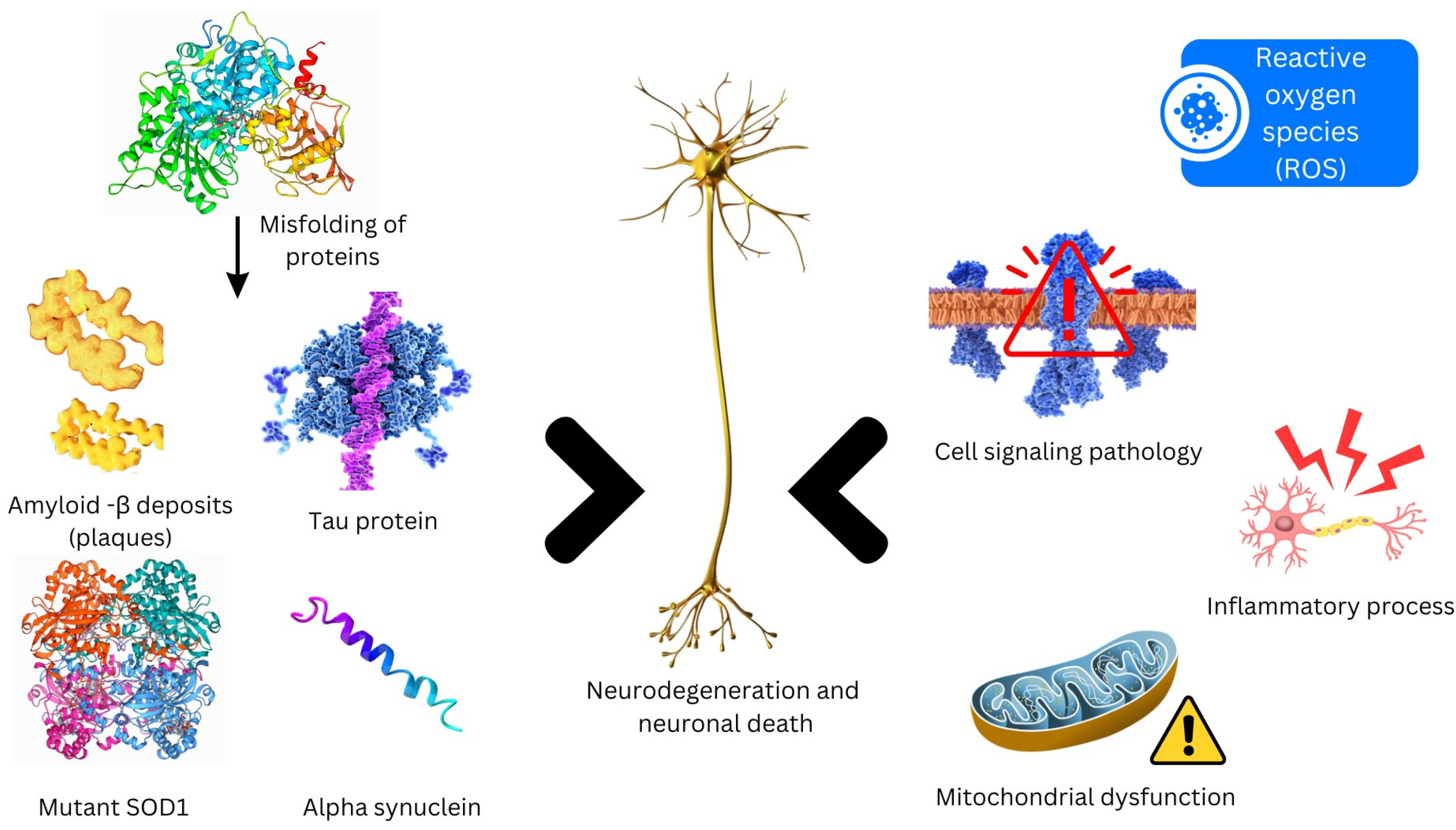
Figure 1. Molecular mechanisms resulting in neurodegeneration.
Futurepharmacol 04 00020 g001
Figure 2. Amyloid plaques observed in Alzheimer’s disease, and α-synuclein aggregates found within neocortical neurons in Parkinson’s disease. Adapted from [5], MDPI, 2021.
Futurepharmacol 04 00020 g002
Figure 3. A wide range of nanoparticle categories are available, encompassing those frequently utilized in the field of nanomedicine.
Futurepharmacol 04 00020 g003
Disclaimer/Publisher’s Note: The statements, opinions and data contained in all publications are solely those of the individual author(s) and contributor(s) and not of MDPI and/or the editor(s). MDPI and/or the editor(s) disclaim responsibility for any injury to people or property resulting from any ideas, methods, instructions or products referred to in the content.