Article

Comparative Study of the Effects of Curcuminoids and Tetrahydrocurcuminoids on Melanogenesis: Role of the Methoxy Groups

1
Department of Biochemistry and Cell Biology, Stony Brook University, Stony Brook, NY 11794-5215, USA
2
Department of Biomedical Engineering, Stony Brook University, Stony Brook, NY 11794-5281, USA
Future Pharmacol. 2024, 4(1), 256-278; https://doi.org/10.3390/futurepharmacol4010016
Submission received: 4 December 2023 / Revised: 29 February 2024 / Accepted: 29 February 2024 / Published: 8 March 2024

Abstract

:
Curcuminoids are naturally occurring yellow-colored compounds that, when hydrogenated to remove their conjugated double bond, become colorless and are referred to as tetrahydrocurcuminoids. Curcuminoids consist of pure curcumin (PC) in major amounts and demethoxycurcumin (DC) and bisdemethoxycurcumin (BDC) in minor amounts. Tetrahydrocurcuminoids similarly consist mainly of tetrahydrocurcumin (THC), along with minor amounts of tetrahydrodemethoxycurcumin (THDC) and tetrahydrobisdemethoxycurcumin (THBDC). Previous studies have shown the inhibitory effects of PC, DC, and BDC on melanin production, but there are contradictory findings about THC. In addition, there are currently no reports on the effects of THDC and THBDC on melanogenesis. Our previous report described that, in contrast to PC, which suppressed melanin production, THC stimulated melanin production in B16F10 and MNT-1 cells; this effect was ascribed to the loss of the conjugated heptadiene moiety of PC. However, whether this finding can be generalized to the two curcumin derivatives (DC and BDC), such that THDC and THBDC might also stimulate melanogenesis, has not been addressed. Herein, a comparative study of six curcumin derivatives (PC, DC, BDC, THC, THDC, and THBDC) was undertaken to identify their effects on melanogenesis with the goal of elucidating the structure–activity relationships (SARs) focused on assessing the two regions of the parent curcumins’ structure: (i) the hydrogenation of the two double bonds bridging the phenyl rings to the β-diketone moiety, and (ii) the effect of the ortho-methoxy substituent (-OCH3) on the two phenyl rings. To determine the direct effects of the six compounds, antioxidant activity and tyrosinase activity were assessed in cell-free systems before cellular experiments utilizing the B16F10 mouse melanoma cells, MNT-1 human melanoma cells, and primary cells. Evaluations were made on cytotoxicity, melanin concentration, and cellular tyrosinase activity. The results showed that BDC inhibited melanogenesis in B16F10 and MNT-1 cells. However, it was ineffective in primary human melanocytes, while THBDC continued to exhibit anti-melanogenic capacity in normal human melanocytes. Moreover, these findings provide a novel perspective into the role of the methoxy groups of PC on the biological effects of melanogenesis and also confirm that the removal of the conjugated double bonds abolishes the anti-melanogenic capacity of PC and DC only, but not BDC, as THBDC maintained anti-melanogenic activity that was greater than BDC. However, the outcome is contingent upon the specific kind of cell involved. To the best of our knowledge, this work presents novel findings indicating that the anti-melanogenic capacity of the colored BDC is not only intact but enhanced after its hydrogenation as observed in THBDC. The findings show potential for using colorless THBDC as a pharmacological candidate to diminish the increased pigmentation characteristic of skin hyperpigmentation disorders. Future pharmacological therapeutics that incorporate pure THBDC or THBDC-enriched extracts, which retain both a colorless appearance and potent anti-melanogenic activity, can be applied to compounds for anti-melanoma therapeutics where the demand for nontoxic novel molecules is desired for established efficacies.

Graphical Abstract

1. Introduction

Melasma, café-au-lait macules, solar lentigo, post-inflammatory hyperpigmentation, ephelides, lichen planus pigmentosus, and periorbital melanosis are some common hyperpigmentation skin disorders [1,2]. These occur due to overstimulation of melanogenesis, a biochemical process that involves the synthesis and distribution of the pigment melanin. Typically, this is carried out by melanocytes within lysosome-related organelles, known as melanosomes, contained within the melanocyte cytoplasm. Tyrosinase, a key enzyme, exerts a pivotal regulatory function in the process of melanin pigment biosynthesis. The process of melanin synthesis commences with the oxidation of tyrosine, resulting in the formation of dopaquinone (DQ), facilitated by the enzyme tyrosinase [3]. Tyrosinase inhibitors possess clinical utility in managing dermatological conditions characterized by melanin overproduction and hold significance in cosmetics and clinical fields due to their ability to diminish excessive pigmentation [4,5,6,7]. Numerous extracts derived from plants or phytochemicals have shown potential anti-melanogenic properties, as supported by in vitro cell culture experiments or 3D human-skin-tissue models. As an example, the production of melanin in B16F10 mouse melanoma cells was diminished using Korean red ginseng extract through the inhibition of cellular tyrosinase activity [8]. A separate study also demonstrated that the primary bioactive compound, ginsenoside F1, derived from the Panax ginseng plant, exhibited inhibitory effects on melanin export in coculture and skin tissue models [9].
Curcuma longa, more commonly known as turmeric, is classified as a member of the Zingiberaceae family and the Curcuma genus. The crop is grown in tropical and subtropical climates all over the globe, but it is widespread in the nations of Southeast Asia and the Indian subcontinent [10,11]. Turmeric is well recognized as a golden spice that offers several health advantages and primarily consists of a curcuminoid class of compounds. The curcuminoid content in turmeric rhizome varies depending on the type, often ranging from around 3% to 8% by weight [11]. Curcuminoids refer to a combination of pure curcumin (PC), demethoxycurcumin (DC), and bisdemethoxycurcumin (BDC). The composition of turmeric extracts primarily consists of PC, which accounts for a significant proportion ranging from 75% to 81%. Subsequently, DC is present in concentrations of 15% to 19%, while BDC is found in lower quantities, ranging from 2.2% to 6.5% [12]. The curcuminoids have comparable chemical structures, with the exception of the varying number of methoxy groups on the two phenyl moieties. PC contains a methoxy group on both of its phenyl moieties, whereas DC only has a methoxy group on one of its phenyl moieties, and BDC does not have any methoxy groups (Figure 1A). Curcuminoids possess diverse pharmacological properties, including but not limited to anti-inflammatory, anti-angiogenic, antioxidant, anticancer, and immunomodulating actions [13]. Curcumin, the principal constituent of curcuminoids, exhibits remarkable therapeutic potential owing to its pleiotropic characteristics, which facilitate its efficacious targeting of a multitude of molecules implicated in various pathological states [14,15,16]. PC was shown to exert anti-inflammatory and antioxidant properties, as was extensively documented previously [17,18]. On the contrary, it has been observed that DC and BDC exhibit enhanced efficacy in inhibiting the proliferation of neoplastic cells compared to PC [19]. BDC was shown to demonstrate a higher level of biological activity compared to other curcuminoids [20]. Moreover, BDC had the most potent antibacterial action, while curcumin showed the least efficacy [21].
The conjugated unsaturated system is responsible for giving curcuminoids their characteristic yellowish coloration. The reduction of the double bonds in vivo, through the metabolism of curcumin, or through a chemical hydrogenation reaction employing catalysts, results in tetrahydrocurcumin (THC), a color-free compound [22,23]. Tetrahydrodemethoxycurcumin (THDC) and Tetrahydrobisdemethoxycurcumin (THBDC) are the other two colorless tetrahydrocurcuminoids that result from the chemical reduction of purified DC and BDC, respectively [23]. The chemical structures of THC, THDC, and THBDC are shown in Figure 1B. According to a prior study [24], there is a correlation between the number of methoxy groups in curcumin metabolites and their inhibitory effects on human colon cancer cells. Specifically, THBDC exhibited the most pronounced inhibitory effects on cellular migration in colon cancer cells compared to THDC and THC. Multiple studies have shown that curcumin has limited stability in aqueous buffer at a physiological pH [25,26,27,28]. Moreover, curcumin exhibited lower stability than BDC in a physiological medium [29]. Another study showed that curcumin exhibited lower stability compared to THC in a culture medium, with a 4.37-fold lower half-life [27].
Several earlier studies [30,31,32,33] have established the anti-melanogenic efficacy of Curcuma longa extracts in vivo and in vitro. For example, the oral administration of turmeric extracts significantly decreased the size of melanin granules in the epidermis of hairless mice subjected to prolonged UVB radiation exposure [30]. A nine-week application of a cream containing turmeric extract in Asian women significantly diminished hyperpigmented spots on the face [33]. Another study reported the inhibitory effects of Curcuma longa extract on mushroom tyrosinase activity [31]. In a separate study [32], a partially purified Curcuma longa extract showed potent inhibition of cellular tyrosinase activity and melanin production in hormone-stimulated B16F10 cells. Noteworthily, other studies that reported the anti-melanogenic effect of curcumin in B16F10 mouse melanoma cells [34] or primary human melanocytes [35] utilized a curcuminoids mixture containing 65–75% curcumin rather than purified curcumin. Additionally, they employed different experimental conditions, such as the use of α-melanocyte-stimulating hormone (MSH). Our earlier studies [36,37,38] were the first to report on the anti-melanogenic effects of purified curcumin (99% purity) on in vitro melanogenesis using B16F10 cells, although the purified curcumin compound failed to exhibit any discernible anti-melanogenic activity in MNT-1 human cells or primary human melanocytes. Recently, a study [39] established that purified curcuminoid compounds, PC, DC, and BDC, suppressed melanogenesis, with the effects of BDC being the most potent without toxicity, as shown in B16F10 cells and zebrafish in vivo models. As the authors [39] did not provide the purity of the PC compound in their study, which was procured from Sigma-Aldrich, it is likely that it was also not of high purity.
The effects of tetrahydrocurcuminoids on melanogenesis have yielded inconclusive findings. For instance, commercially available products such as Tetrahydrocurcuminoids® CG (comprising 95% Tetrahydrocurcuminoids) and Sabiwhite® (comprising 96% THC), developed by Sabinsa Corporation (East Windsor, NJ, USA), are marketed as agents for skin lightening that inhibit melanogenesis [40,41]. Nevertheless, it has been observed that the application of an undisclosed composition of a tetrahydrocurcuminoid cream in a commercial setting resulted in an augmentation of melanogenesis when exposed to UVB radiation in vivo. Additionally, tetrahydrocurcuminoids have been identified as efficacious agents for inducing self-tanning [42]. Our previous study demonstrated that purified THC stimulated melanogenesis in B16F10 and MNT-1 cells [37]. Conversely, another study employing low or high concentrations of THC in hormone-stimulated B16F10 cells revealed inhibitory effects on melanogenesis [43,44].
No research has been conducted so far on the effects of the isolated compounds THDC and THBDC on melanogenesis utilizing B16F10 mouse cells. Moreover, the effects of THDC, THBDC, DC, and BDC on melanogenesis in human melanocytes has yet to be explored. Evaluating the biological efficacy of naturally occurring compounds in human cells is an essential step in the process of determining whether the compound has the potential to be used as a pharmacological therapeutic for inhibiting skin hyperpigmentation. This is because the biological activity of a molecule tested in an in vitro setting consisting of mouse cells may differ from the biological activity displayed in a human cell culture system. This phenomenon has been observed in the case of naturally occurring compounds, including gomisin N [45], citric acid [46], and quercetin [47,48]. The author’s previous study showed that, in contrast to PC, THC stimulated melanin synthesis in both B16F10 and MNT-1 cells [37]; this effect was ascribed to the crucial role of the double bonds since THC differs from PC due to the absence of conjugated double bonds of the heptadiene moiety. However, to test if this result can be generalized to the other derivatives of curcumin (DC and BDC), such that THDC and THBDC might also stimulate melanogenesis, the three purified curcuminoids (PC, DC, and BDC) and the three purified tetrahydrocurcuminoids (THC, THDC, and THBDC) were evaluated to identify their effects on melanogenesis and the role of structure–activity relationships (SARs) in eliciting the biological effects. PC and THC were included as reference controls to enable comparison to the other two curcuminoids and their respective tetrahydrocurcuminoids. The study of SARs was focused on assessing the two regions of the parent curcumin’s structure: (i) the hydrogenation of the two double bonds bridging the phenyl rings to the β-diketone moiety, and (ii) the impact of the ortho-methoxy substituent (-OCH3) on the two phenyl rings. The search for novel anti-melanogenic compounds necessitates a thorough SAR study to identify the key regions of the chemical structure of a candidate compound and to provide a foundation for the design of novel pharmacological derivatives. The implications of these findings are of significant importance in the advancement of curcumin-based therapeutics and underscore the value of using in vitro cell cultures in the process of discovering future pharmacological agents. The outcomes of this study possess potential utility in the development of improved pigmentation inhibitors, which may be regarded as promising pharmacological entities in the advancement of therapeutic strategies for the mitigation or management of hyperpigmentation disorders arising from excessive melanin synthesis.

2. Materials and Methods

2.1. Materials

Pure Curcumin (PC; 99% purity) was acquired from Selleck Chemicals (Houston, TX, USA). Bisdemethoxycurcumin (BDMC; >98% purity) and Demethoxycurcumin (DMC; >99% purity) were purchased from MedChem Express LLC (Monmouth Junction, NJ, USA). Tetrahydrocurcumin (THC; >95% purity) was obtained from Chemscene Inc. (NJ, USA). Tetrahydrodemethoxycurcumin (THDC; 95% purity) and Tetrahydrobisdemethoxycurcumin (THBDC; 95% purity) were purchased from MuseChem (Fairfield, NJ, USA). A 20 mM stock solution of the six compounds was prepared using 100% dimethyl sulfoxide (DMSO) solvent. This solution was then aliquoted and stored at −20 °C until use. MTS cytotoxicity assay (CellTiter 96® Aqueous One Solution Reagent) was purchased from Promega Corp. (Madison, WI, USA). Mushroom tyrosinase, L-DOPA, and kojic acid (KA) were purchased from Sigma-Aldrich (St. Louis, MO, USA). The 2, 2-diphenyl-1-picrylhydrazyl (DPPH) radical reagent were procured from Molecular Probes Inc. (Eugene, OR, USA).

2.2. Mushroom Tyrosinase Activity

The evaluation of the direct effects of the six compounds on tyrosinase enzyme activity was conducted with mushroom tyrosinase enzyme in conjunction with L-DOPA substrate. This evaluation was performed utilizing the methodology outlined in our previous publications [38,49].

2.3. DPPH Radical Scavenging Assay

The antioxidant activity of all the compounds was evaluated using the DPPH radical solution, following the methods described in our earlier publication [50]. The DPPH scavenging activity was reported as a percentage of the untreated control group.

2.4. Cell Culture

B16F10 mouse melanoma cells (CRL-6475™) were procured from the American Type Culture Collection (ATCC; Manassas, VA, USA), and HaCaT cells were obtained from AddexBio (San Diego, CA, USA). Both sets of cells were maintained using Dulbecco’s modified eagles’ medium (DMEM) containing 10% heat-inactivated fetal bovine serum (HI-FBS) and 1% antibiotics (penicillin-streptomycin). MNT-1 human melanoma cells were graciously provided by Dr. Michael Marks (University of Pennsylvania, Philadelphia, PA, USA) and were cultured using DMEM supplemented with 18% HI-FBS, 10% AIM-V medium, 1% antibiotics, and 1% minimum essential medium (MEM). Primary human melanocytes from darkly pigmented donor (HEMn-DP cells; Cascade Biologics™, C2025C, Portland, OR, USA) were acquired through the vendor Thermo Fisher Scientific, Inc. (Waltham, MA, USA), and cultivated using medium 254 supplemented with 1% human melanocyte growth supplement and antibiotic mixture.

2.5. MTS Cell Viability Assay

A total of 5 × 10³ B16F10 cells were seeded in each well of a 96-well plate. After 24 h, the test compounds were added at concentrations ranging from 5 to 40 µM. To generate the working concentrations of 5, 10, 20, and 40 µM utilized in the experiments, working stock solutions were prepared using 100% DMSO at 250× concentrations of 1.25, 2.5, 5, and 10 mM. These stock solutions were then diluted (at a dilution factor of 250) using the culture medium to maintain a constant final DMSO concentration of 0.4%, which does not cause cytotoxicity. Consequently, 0.4% DMSO was used to treat the control group in each experiment. The cultures were maintained for three days.
For the experimentation involving other cells, MNT-1 cells and HaCaT cells were cultivated in 96-well plates at a density of 12 × 103 and 7 × 10³ cells per well, respectively, for a duration of 24 h. Following this, the culture medium was substituted with the test chemicals, and the cultures were sustained for a period of 5 days, during which the compounds were renewed on the third day of culture. In the case of HEMn-DP cells, a quantity of 10 × 103 cells was cultivated in a 96-well plate for a duration of 24 h. Subsequently, the test compounds were added, and the cultures were sustained for a period of 6 days, with one instance of compound renewal over this period. At the end of treatments with compounds, the viability of different cells was estimated, which involved incubation of cells with a mixture of culture medium and MTS reagent for 60–90 min at 37 °C. The spectrophotometric measurement of the absorbance of aliquots at a wavelength of 490 nm was performed using a Versamax™ microplate reader (Molecular Devices, San Jose, CA, USA).

2.6. Estimation of Cellular Melanin Content

The melanin contents in B16F10 cells after 72 h treatment with the test compounds were evaluated according to the method described in our previous study [37]. For the measurement of melanin content in MNT-1 cells, 1.2 × 10⁵ cells/well were seeded in a 12-well plate, and after 24 h, the compounds were added at different concentrations, and cultures were incubated for 5 days. Subsequently, melanin contents were determined based on a methodology similar to that used in B16F10 cells.
HEMn-DP cells (2 × 10⁵ cells/well in 3 mL medium) were cultured in six-well culture plates for three days. After this, the medium was replaced with compounds containing fresh medium, and cultures were kept for a duration of 6 days (with one compound renewal in between). At the end of treatments, cells were harvested and solubilized in 125 µL of hot NaOH, and aliquots of lysates were transferred to a 96-well plate. The relative melanin content was determined according to the method described above.

2.7. Intracellular Tyrosinase Activity

In 24-well tissue culture plates, B16F10 cells were grown at a density of 2 × 10⁴ cells per well. After a period of 24 h, the medium was changed, test compounds were added, and the plates were cultured for a further three days. After the completion of the treatments, the cells were detached, and the cell pellets were rinsed in PBS, lysed with lysis buffer for 20 min while being placed on ice, and centrifuged afterward. After that, 25 µL of lysates were aliquoted onto a 96-well microplate, and then 75 µL of a solution containing 3 mM L-DOPA was added. After that, the absorbance was determined using a microplate reader and measured in kinetic mode at 475 nm for ten minutes. The linear range of the slope of velocities of inhibition was used to determine the percentage of tyrosinase activity.
MNT-1 cells (6 × 10⁴ cells/well) were seeded in a 24-well plate, and after 24 h of culturing, the test compounds were added to the cultures and maintained for five days. At the end of treatments, cells were lysed and processed, similar to the method described above, with the exception that the activity was measured for a period of 30 min.

2.8. Statistical Analysis

All the analyses were conducted using GraphPad Prism statistical software (version 10.0), and data are reported as mean ± SD. One-way analysis of variance (ANOVA) with Dunnett’s post hoc test was run in all experiments except the melanin content experiments in HEMn-DP cells, where a Student’ t-test was used. The differences were deemed statistically significant at a significance level of p < 0.05. The following symbols indicated different degrees of significance: * p < 0.05 vs. control, ** p < 0.01 vs. control, $ p < 0.001 vs. control, and # p < 0.0001 vs. control.

3. Results

3.1. Effect of Compounds on Mushroom Tyrosinase Activity and Antioxidant Activity in a Cell-Free System

Results show that DC has a somewhat greater effect on the inhibition of mushroom tyrosinase activity compared to PC, while BDC has a weak inhibitory effect, which is less than that of PC or DC (Figure 2A). Next, both THC and THDC lost the tyrosinase activity inhibitory effects of PC and DC, respectively, while in the case of THBDC, the inhibitory activity was enhanced at a concentration of 40 µM as compared to BDC at 40 µM (Figure 2A). A superior inhibition of 32.90% was obtained for THBDC (40 µM) as compared to an inhibition of 19.93% by BDC at the same concentration. Interestingly, at the concentration of 20 µM, BDC and THBDC inhibited the activity by 13.54% and 15.75%, which was nearly identical (Figure 2A).
Collectively, these results indicate that the loss of both methoxy groups of PC will negatively impact its mushroom tyrosinase-inhibitory activity, while the loss of one methoxy group seems to have no impact or might enhance the activity to some degree. On the contrary, the chemical reduction of PC will lead to complete abrogation of the inhibitory activity, which, however, can be rescued with the addition of two methoxy groups (resulting in THBDC) at the highest concentration of 40 µM.
The DPPH antioxidant assay findings revealed that DC had a higher antioxidant activity than PC, while BDC had no antioxidant activity (Figure 2B). THC showed greater antioxidant activity than parent PC. THDC lost the antioxidant activity of parent compound DC to some degree at concentrations ≥ 10 µM, while THBDC lacked antioxidant activity altogether (Figure 2B). The mean IC50 values of the DPPH assay of PC, DC, and THC were determined to be 31.78 µM, 24.97 µM, and 14.54 µM, respectively.
Altogether, these results reveal that the hydrogenation of the heptadiene moiety of PC enhances its antioxidant activity. However, successive removal of one and two methoxy groups of THC leads to a diminution of the antioxidant activity with no antioxidant activity for bisdemethoxy compounds, irrespective of the hydrogenation.

3.2. Effect of Compounds on Viability and Melanin Production in B16F10 Cells

The MTS assay results show that PC and DC were markedly cytotoxic in a similar manner at higher concentrations of 20 and 40 µM; PC significantly lowered viability to 10.35% and 13.30%, while DC lowered viability to 9.18% and 10.74% at 20 and 40 µM, respectively (Figure 3A). At the same time, the other four compounds did not exert any cytotoxicity over the 5–40 µM concentration range (Figure 3A). Based on cytotoxicity data, PC and DC were selected only at concentrations of 5 and 10 µM, while the other four compounds were selected at 5–40 µM for the determination of melanin contents.
The visual observation of pellets of B16F10 cells after treatment with all six compounds showed visibly lighter color (compared to untreated control) over more comprehensive concentration ranges, specifically for BDC and THBDC, while PC and DC showed lighter color only at the highest concentration of 10 µM (Figure 3B). Moreover, THDC did not show any major change, although THC-treated cells showed the opposite effect and pellets appeared darker at the highest concentration (Figure 3B). KA, a known skin lightener, employed as a positive control (at 500 µM concentration) showed a lighter pellet (Figure 3B).
The qualitative observations were corroborated using the semi-quantitative method of determination of relative cellular melanin contents. PC significantly suppressed melanin content by 28.25% at 10 µM, while THC showed no change at 10 µM, but instead stimulated melanin content significantly by 20.50% at 40 µM (Figure 3C). DC significantly suppressed melanin content by 23.27% at 10 µM, while THDC showed no significant changes at any concentration (Figure 3D). Lastly, treatment with BDC led to significant suppressions of 29.05%, 40.61%, and 65.18% at 10, 20, and 40 µM concentrations, respectively (Figure 3E). Interestingly, most of the suppressive capacity of BDC on melanogenesis was retained in its hydrogenated counterpart, THBDC, which showed significant suppressions of 26.02%, 31.82%, and 30.70% at 10, 20, and 40 µM concentrations, respectively, although the greatest suppression could not be achieved (Figure 3E).
Collectively, these findings suggest that at the concentration of 10 µM, PC, DC, and BDC exhibit similar capacities to suppress cellular melanin, which is lost after the hydrogenation of PC and DC; however, the removal of both methoxy groups of THC rescues this capacity as seen in THBDC at 10 µM, thus indicative of the key requirement of the removal of both methoxy groups.

3.3. Effect of Compounds on Tyrosinase Activity in B16F10 cells

PC at 5 and 10 µM significantly suppressed B16F10 cell tyrosinase activity by 12.46% and 11.06%, respectively (Figure 4A), while the hydrogenated compound THC had no effect. DC and THDC compounds had no discernible impact on tyrosinase activity across all concentrations (Figure 4B). Lastly, BDC significantly suppressed tyrosinase activity by 37.77% at 40 µM, with no change at lower concentrations. In comparison, THBDC suppressed tyrosinase activity to a similar degree, with a significant inhibition of 31.21% at 40 µM (Figure 4C).
Overall, these results indicate that the tyrosinase-inhibitory capacity of PC is lost either after the removal of one or both methoxy groups or by its hydrogenation. However, tyrosinase-inhibitory capacity is enhanced in the case of the removal of both methoxy groups at higher concentrations (as in BDC), and this inhibitory capacity is maintained despite the corresponding hydrogenation (as in THBDC).

3.4. Effect of Compounds on Viability and Melanin Production in MNT-1 Cells

PC displayed significant cytotoxicity to MNT-1 cells at the highest concentration of 40 µM, with viabilities lowered to 21.56%, while DC showed greater cytotoxicity with a significant decline in viability to 12.64% at 40 µM (Figure 5A). At the same time, the other four compounds were nontoxic over the entire concentration range (Figure 5A).
PC did not inhibit cellular melanin levels, although a trend for their increase was seen, while THC significantly enhanced melanin levels over a wide concentration range (Figure 5B). Next, DC and THDC showed a disparate effect, where DC at 10 µM significantly increased melanin levels by 19.48%, while THDC significantly increased melanin levels by 27.05% at 5 µM, while, at all other concentrations, none of the two compounds exerted any effect (Figure 5C). Treatment with BDC resulted in the significant suppression of melanin content by 15.89% at 40 µM, with no change at lower concentrations (Figure 5D). Interestingly, treatment with THBDC resulted in suppression of melanin content at multiple concentrations, with significant decreases by 22.44%, 21.25%, and 24.91% at 10, 20, and 40 µM, respectively (Figure 5D).
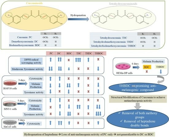
Together, these results show that the hydrogenation of PC leads to a stimulatory effect on melanogenesis, as seen in THC. Moreover, the removal of both methoxy groups is necessary to achieve a melanogenesis-inhibitory effect, as BDC and THBDC, both of which lack the two methoxy groups, were the only compounds to exert an anti-melanogenic effect. Moreover, it can also be observed that the hydrogenated compound THC, which stimulates melanogenesis, can turn into a melanogenesis inhibitor if both methoxy groups are eliminated (as in THBDC).

3.5. Effect of Compounds on MNT-1 Cell Morphology

MNT-1 cells treated with THBDC (20 and 40 µM) appeared visually lighter with lesser melanin pigment compared to the control and other groups, as observed microscopically (Figure 6). These data corroborate the spectrophotometric results of MNT-1 cells where BDC and TBDC showed anti-melanogenic capacity, while the other four compounds showed no inhibitory effects.

3.6. Effect of Compounds on Tyrosinase Activity in MNT-1 Cells

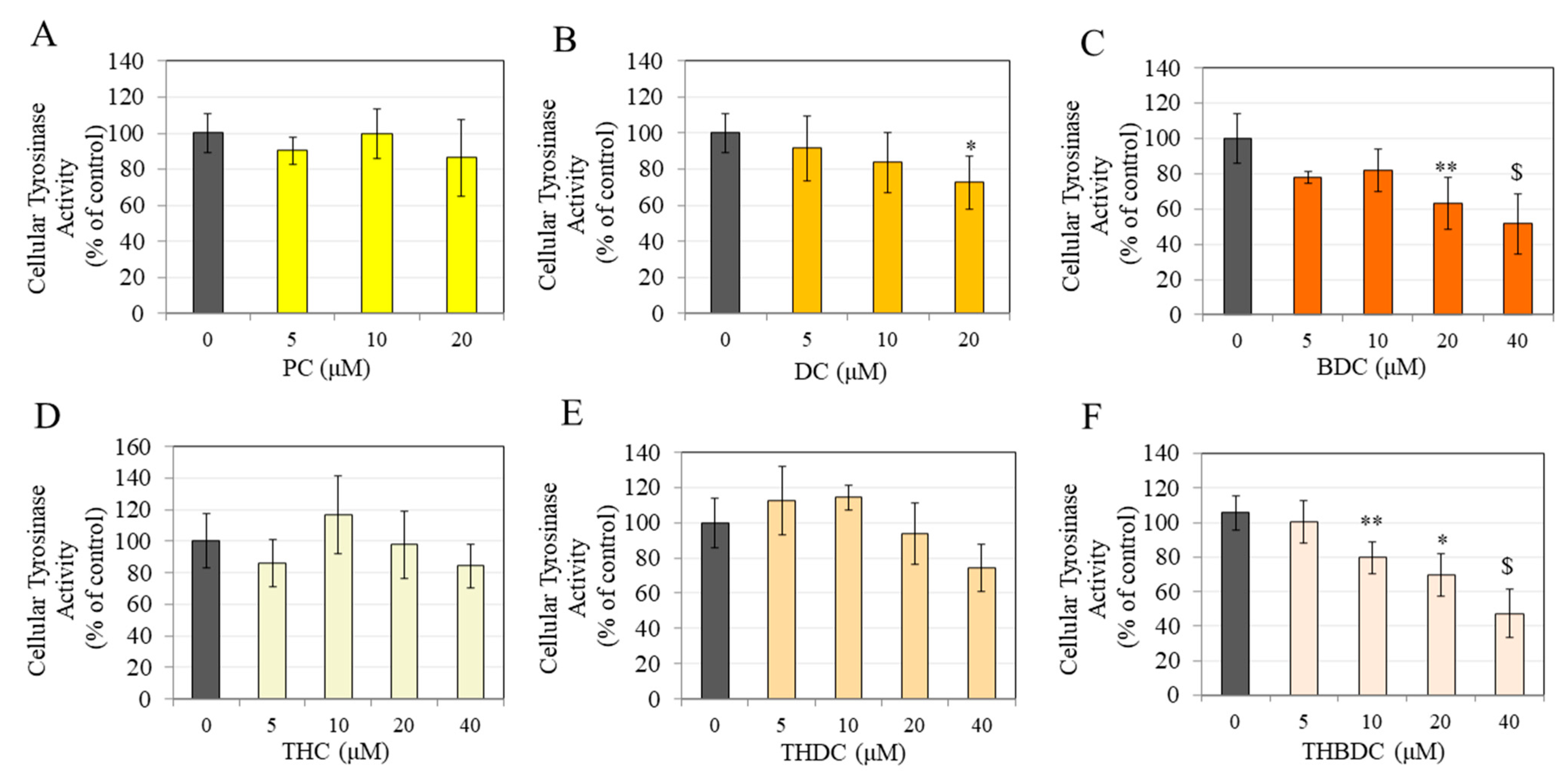
PC did not alter tyrosinase activity at any concentration in MNT-1 cells (Figure 7A), while DC suppressed the activity by 27.31% at 20 µM (Figure 7B). At the same time, BDC significantly inhibited the activity of tyrosinase by 36.88% and 48.30% at 20 and 40 µM, respectively (Figure 7C). THC and THDC had no significant effects on the tyrosinase activity (Figure 7D,E), while THBDC treatment significantly inhibited tyrosinase activity by 26.06%, 36.01%, and 58.44%, at 10, 20, and 40 µM, respectively (Figure 7F). Overall, the results indicate a potential correlation between the anti-melanogenic effects of BDC and THBDC, which may be attributed, at least partially, to the inhibition of tyrosinase enzyme activity.

3.7. Effect of Compounds on HaCaT Cell Viability

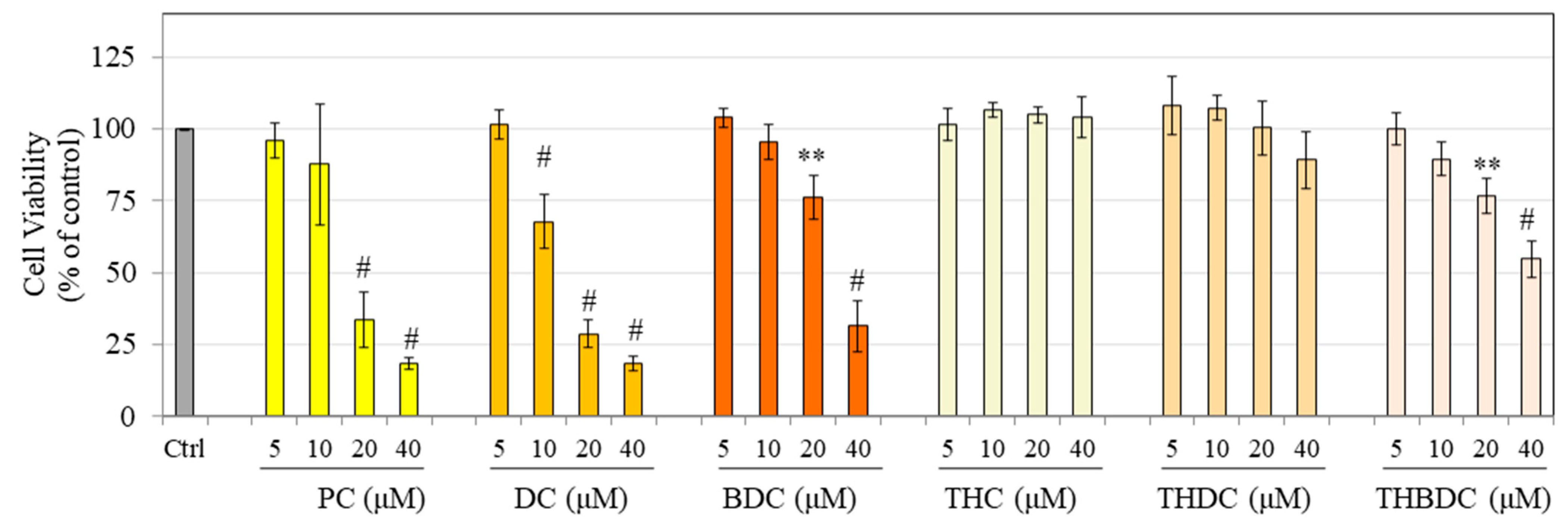
Results showed that in HaCaT cells, treatment with PC at concentrations of 20 and 40 µM resulted in pronounced cytotoxic effects, which, after the removal of one methoxy group (as in DC), did not change the cytotoxic effect, but with successive removal of the second methoxy group (as in BDC), abolished the cytotoxicity by some amounts. DC was, however, also cytotoxic at 10 µM, unlike PC and BDC (Figure 8). Both THC and THDC showed no cytotoxicity at any concentration. However, THBDC showed significant cytotoxicity; viability was lowered by 23.16% and 45.24% at 20 and 40 µM, respectively (Figure 8).
Together, these data suggest that the hydrogenation of PC and DC abolished their cytotoxicity to HaCaT cells. However, hydrogenation of BDC did not abolish the cytotoxicity, which remained high at 20 µM and were still significantly elevated compared to the untreated control, even at 40 µM.

3.8. Effect of Compounds on HEMn-DP Cells

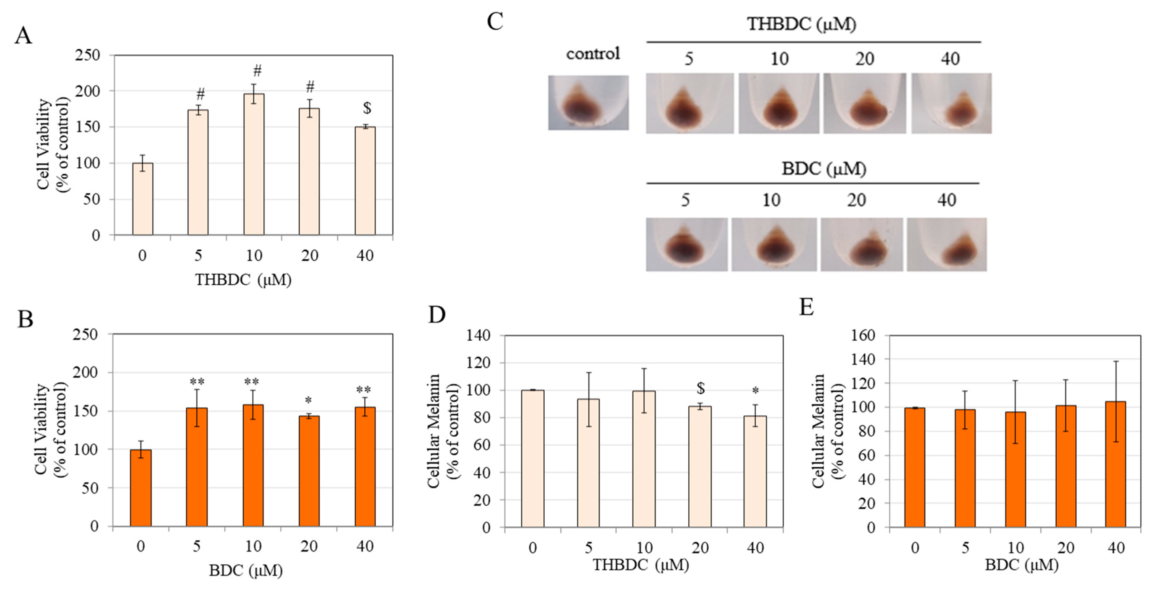
Due to their remarkable impact on diminishing melanogenesis, the selection for subsequent experiments on primary human melanocytes (HEMn-DP cells) was limited exclusively to BDC and THBDC. The results of the MTS assay in HEMn-DP cells showed that the compounds THBDC and BDC did not compromise HEMn-DP cell viability; instead, a sharp increase was seen at all concentrations (Figure 9A,B).
Based on visual observations, THBDC seemed to diminish melanin production within cells at higher concentrations, as the pellet color was lighter than the control (Figure 9C). Estimation of relative melanin contents confirmed that THBDC significantly diminished melanin production in cells; a significant reduction of 12.06%, and 18.68% was obtained at 20 and 40 µM, respectively (Figure 9D), while BDC did not seem to inhibit melanin production (Figure 9E).

4. Discussion

The findings of this study demonstrate that the number of methoxy groups and the conjugated double bonds in the structure of curcumin exert a critical influence on the anti-melanogenic capacity that is cell-type-specific. The anti-melanogenic and anti-tyrosinase activities of BDC were retained or enhanced for THBDC, which is a novel finding. Furthermore, the anti-melanogenic capacity of DC in B16F10 cells was abolished when it was hydrogenated to THDC. In contrast, in MNT-1 human cells, DC showed the opposite effect of stimulating melanogenesis that was retained in THDC at a lower concentration. Hence, our earlier discovery [37] that the removal of the conjugated double bond of PC results in the loss of PC’s anti-tyrosinase and anti-melanogenic activity cannot be generalized to other curcumin derivatives (DC and BDC) and is specific to PC. Notably, this finding corroborates the findings of our recently published study [51], where TH-CMC2.24 (obtained using hydrogenation of CMC2.24, a novel BDC analog) was demonstrated to be a more robust inhibitor of melanogenesis as compared to CMC2.24 in primary human melanocytes. In addition, TH-CMC2.24 exhibited nontoxicity in HaCaT cells over a wide concentration range up to 36 µg/mL (84 µM), while THBDC in the current study was cytotoxic to HaCaT cells at concentrations ≥20 µM. Hence, the novel results of this study provide a solid basis for future design and pursuit of THBDC-based novel analogs as pharmaceuticals for the treatment of skin hyperpigmentation disorders.
Our results indicate that the IC50 value for antioxidant activity of THC is 2.19-fold lower than PC, which is in line with a prior study [52] that also showed similar IC50 values, with THC exhibiting a 1.85-fold lower IC50 compared to PC, which aligns with the current results. BD and THBDC showed no antioxidant activity, while THDC only showed activity at 40 µM. The ranking order of potency of the six compounds in antioxidant activity is THC >> DC > PC > THDC > BD = THBDC. As shown previously, the presence of an ortho-methoxy group in PC confers increased stability of the phenoxy radical [53] and has been shown to be essential in imparting antioxidant characteristics to this molecule [54]. The ortho-methoxy phenol group has the capacity to form an intramolecular hydrogen bond with the phenolic hydrogen, hence facilitating the hydrogen atom abstraction from ortho-methoxy phenols [55,56]. The significance of the hydrogenation of the two double bonds that are conjugated to the β-diketones has been proposed as a crucial factor for THC’s antioxidant activity, alongside the presence of the phenolic hydroxyl group within its structure [23,57]. According to the mean DPPH assay IC50 values of PC (31.78 µM), DC (24.97 µM), THC (14.54 µM), and THDC (>40 µM), it can be confirmed that the hydrogenation of the heptadiene moiety of PC enhances its radical scavenging activity, which is consistent with our prior study [37]. Conversely, DC exhibited a contrasting trend, as the hydrogenation of its heptadiene moiety diminished its radical scavenging activity. These results are in contrast with an earlier study [58] where the DPPH IC50 values for curcumin, DC, THC, and THDC were reported to be 11.8 µM, 16.8 µM, 9.8 µM, and 14.2 µM, respectively, indicating that the antioxidant activities of both curcumin and DC were enhanced through hydrogenation. However, these discrepancies might be related to the utilization of a low-purity curcumin instead of a high-purity PC compound in the study above. Additionally, the concentration used in the DPPH assay differed from the current experimental design. Intriguingly, in the same study [58], the authors also reported that the radical scavenging activity of octahydrocurcumin (IC50: 11.4 µM), a compound obtained by successively hydrogenating THC, was diminished after the removal of one methoxy group, with octahydrodemethoxycurcumin showing considerably lower DPPH activity (IC50: 33.1 µM). Based on the findings, it is evident that the removal of one methoxy group from the hydrogenated derivative has a detrimental effect on its ability to scavenge radicals. This aligns with the results obtained from comparing THC and THDC in this study, where a similar removal of one methoxy group from THC significantly reduced its radical scavenging activity.
Several earlier studies have shown a correlation between the antioxidant and anti-tyrosinase activity of plant extracts or bioactive compounds [59,60,61,62]. The current study also found a direct relation between these two activities for PC and DC in the cell-free system but not in cell cultures based on species (mouse or human). Furthermore, there was no correlation between these two activities for BDC, THC, THDC, and THBDC. This is because THC and THDC did not inhibit tyrosinase enzyme activity under both cell-free and cellular conditions despite exhibiting antioxidant capacity, and despite THC being a more potent antioxidant than THDC. In the case of THBDC, tyrosinase activity (both cell-free and cellular) was inhibited in the absence of antioxidant activity. In our previous study [63], a hydrogenated standardized extract of curcuminoids (Curowhite™) that stimulated melanin production showed remarkable antioxidant activity in a DPPH assay, yet the ROS levels in MNT-1 cell cultures were not altered. Moreover, the cell-free mushroom tyrosinase activity was unchanged, although cellular tyrosinase activity was inhibited. Hence, there was no direct correlation between the antioxidant activity and tyrosinase-inhibitory activity of the Curowhite formulation. These inconsistent findings can be attributed to the fact that the antioxidant activity determined using the DPPH assay may not accurately predict the effect in a biological system, owing to factors such as the permeability of cell membranes and the differential transport of antioxidants between the lipid and aqueous phases of cells [64,65]. Moreover, the compounds’ ability to act as an antioxidant and also suppress the formation of tyrosinase-catalyzed dopaquinone formation (in the tyrosinase activity assay) is contingent upon multiple factors, including the source of enzyme, the presence of cells, and the compounds’ ability to cross the cell membrane and reach melanosomes.
A previous study [66] reported that a 24 h treatment with BDC displayed significant cytotoxicity to B16F10 cells at 10 and 25 µM concentrations. Another study [39] that examined PC, DC, and BDC over a 72 h duration reported that B16F10 cell viability after BDC treatment was significantly lower at 20 and 40 µM. Moreover, the viability of cells treated with PC and DC was 71.87% and 76.74% at 20 µM, while they were approximately 40–50% at 40 µM for both compounds [39]. The results of these studies are in contrast to the findings of the current study, where a 72 h treatment with BDC did not lower cellular viability across the full range of concentrations tested (5–40 µM). Additionally, in B16F10 cells, both PC and DC exerted significant cytotoxicity that was much higher at 20 and 40 µM than in the previous study [39]. Nevertheless, the current results of PC and DC exhibiting higher cytotoxicity than BDC are consistent with other studies [39,67]. The results of increased MTS viability in HEMn-DP cells after a 6-day treatment with THBDC are reminiscent of the results of our prior study, where a THBDC derivative, namely, TH-CMC2.24, was examined [51].
The findings of no change in the activity of mushroom tyrosinase catalyzed by L-DOPA substrate following THC treatment, as well as the findings that THC was non-cytotoxic and increased the production of melanin in both B16F10 and MNT cells (without affecting the activity of cellular tyrosinase), are in line with our earlier study [37]. The data on the non-cytotoxicity of PC at 5–10 µM and significant cytotoxicity at 20 µM in B16F10 cells are also consistent with our previous studies [37,38]. Our results of a significant reduction in melanin production at 10 µM PC accompanied by suppression of cellular tyrosinase activity in B16F10 cells are also similar to our previous result [38]. Moreover, in our earlier study [37], PC at 5 µM did not exhibit any effects on the melanin levels or tyrosinase activity of MNT-1 cells, which is similar to the current study, although this study examined PC across higher concentrations (5–40 µM).
Both PC and DC had an equivalent anti-melanogenic effect in B16F10 cells, while only PC demonstrated the ability to inhibit cellular tyrosinase activity. Furthermore, it was seen that in MNT-1 cells, treatment with PC did not yield any significant effects on melanogenesis. However, treatment with DC at a concentration of 10 µM resulted in the stimulation of melanogenesis, although no changes were observed at a concentration of 20 µM. Additionally, it was found that the activity of cellular tyrosinase remained unaltered at a concentration of 10 µM but was suppressed at 20 µM. The cause of the inconsistent response of DC on melanogenesis remains unknown; although, a previous study [39] similarly observed contradictory impacts of DC on melanin synthesis. The authors of the study [39] observed that in B16F10 cells that were stimulated with MSH, treatment with DC increased cellular melanin but decreased tyrosinase activity at 10 µM, while it suppressed melanin in a zebrafish embryo in vivo model, where it modestly decreased melanin levels at 5 µM concentration. Interestingly, the results of our study also show an opposite effect of DC in B16F10 and MNT-1 cells, as DC decreased intracellular melanin at 10 µM in B16F10 cells but increased intracellular melanin in MNT-1 cells. In both cells, DC continued to have no significant effect on cellular tyrosinase activity. Part of these results contrasts with a previous study [39] that showed increased intracellular melanin but decreased tyrosinase activity in B16F10 cells that were treated with 10 µM DC. Despite this, the results of increased intracellular melanin due to DC in MNT-1 cells partially agree with the study above. In the current study, the responses of compounds were examined under basal conditions (unstimulated) in B16F10 cells; the results of decreased melanin production with DC contrast to the results of the prior study [39] where DC enhanced melanin production in MSH-stimulated B16F10 cells. Part of this divergent effect may be ascribed to the increased interaction of DC with MSH via the formation of two hydrogen bonds, as demonstrated in an in silico study before [68]. Hence, in the presence of MSH, DC might exhibit a different biological effect. Moreover, it is not unusual to see a contrasting outcome in relation to this phenomenon, since there have been documented instances of compounds exhibiting contradictory findings. As an example, it was noted that the use of grape extract did not affect melanin levels under basal conditions but increased melanogenesis under MSH stimulation [68]. Another compound, retinoic acid, enhanced melanin production in basal conditions but decreased it in MSH-stimulated conditions [69]. Interestingly, the compound THC also increased melanin production in basal conditions [37] but decreased it in MSH stimulation [43].
The results of a lack of any inhibition of tyrosinase activity by BDC in B16F10 cells at concentrations of 10 and 20 µM at which melanin content was decreased agree with a prior study [39] that also showed that BDC (10 µM) decreased intracellular melanin in hormone-stimulated B16F10 cells in the absence of any change in cellular tyrosinase activity. However, another study [66] reported a decrease in the tyrosinase activity and melanin content of B16F10 cells after BDC treatment; the authors ascribed the decreases to a BD-induced decline in cell viability, leading to the conclusion that BDC might not be an effective anti-melanogenic compound. However, this discrepancy might be linked to the use of BDC compound with lesser purity, but the authors did not furnish information regarding the percentage purity of BDC. Notwithstanding these reports, the potent depigmentation effects of BDC in the absence of toxicity were confirmed in hormone-stimulated B16F10 cells and in an in vivo model of zebrafish in a recently published study [39]. In a previous study [70], the results of the molecular docking investigations using PC, DC, and BDC in conjunction with a bacterial tyrosinase revealed that all three compounds had binding capabilities towards tyrosinase and another melanogenic enzyme, namely, tyrosinase-related protein-1 (TRP-1). Notably, it was shown that their binding mechanisms closely resembled those of kojic acid. Among all the ligands, it was shown that BDC possessed the highest affinity for TRP-1. Curcumin (low-purity grade) was shown to decrease tyrosinase protein levels in human melanocytes [35]. In our previous study [36], BDC and DC derivatives showed disparate effects on tyrosinase protein and activity, although both compounds inhibited melanogenesis in human melanocytes. Specifically, the BDC derivative (CMC2.24) did not attenuate tyrosinase protein levels but inhibited cellular tyrosinase activity, while the DC derivative (CMC2.5) attenuated tyrosinase protein levels without altering cellular tyrosinase activity. A limitation of the current study is that only tyrosinase activities in cell-free and cell cultures were measured, while tyrosinase protein levels were not examined. The examination of these and other melanogenic proteins necessitates further studies.
The results of higher anti-melanogenic activity of BDC than PC, DC, and THC, with a differential efficacy of BDC in mouse and human cells, are reminiscent of the results of a previous study [71] that demonstrated that BDC emerged as the most potent inhibitor of the activity of the 3β-hydroxysteroid dehydrogenase enzyme compared to THC, PC, and DC, with a differential efficacy across human and rat species. Notably, the two other compounds, THBDC and THDC, were not examined in their study; hence, it remains unknown if THBDC may have been superior to BDC in their assays. LogP values are a measure of the lipophilicity of a drug and are useful in SAR studies [72]. In the previous study [71], it was also shown that the higher inhibitory effects of BDC were correlated to its lower logP values and, thus, lower lipophilicity, leading the authors to suggest that removing both methoxy groups is expected to increase potency. Our findings of the higher capacity of BDC or THBDC (both demethoxylated) inhibiting tyrosinase activity and melanogenesis can be attributed to the removal of both of their methoxy groups, which enables the hydroxyl groups to form hydrogen bonds more easily with the tyrosinase enzyme, thereby enhancing their inhibitory potency. Previous reports have elucidated the intermolecular interactions and binding modes of tyrosinase inhibitors inside the active site of the human tyrosinase protein using computational methods such as in silico modeling and molecular docking analysis [73,74]. As the current study focused on the effects of the compounds in different cell culture models in vitro, lipophilicity evaluation and docking analysis to further elucidate the SAR results of the compounds THBDC, THDC, or THC were not undertaken, as they were beyond the scope of the study. THBDC is hypothesized to exhibit the lowest binding energy and strongest binding affinity towards the human tyrosinase protein compared to BDC, THDC, and THC. The rigorous evaluation of this hypothesis through molecular docking and 3D-QSAR studies is fitting for a follow-up study that would significantly contribute to our knowledge in this area.
PC and its five derivatives (DC, BDC, THC, THDC, and THBDC) were meticulously chosen due to their prevalence and easy accessibility as curcuminoids without any additional substituents. This selection aligns perfectly with our two-fold objective. The synthesis of additional PC derivatives and their inclusion in the SAR studies were beyond the scope of this study. Nevertheless, our previous study [51] already reported on newly synthesized hydrogenated BDC derivatives (referred to as partial and fully hydrogenated TH-CMC2.24) that lack both methoxy groups but have an additional phenylaminocarbonyl substituent in the central carbon of the β-diketone. These TH-CMC2.24 derivatives were very potent anti-melanogenic candidates with no cytotoxicity to keratinocytes (which contrasts with the results of the current study on THBDC that show some keratinocyte cytotoxicity). Based on the results of our prior study and the current study, it is evident that the basic scaffold of THBDC plays a crucial role in its anti-melanogenic activity. By making chemical modifications to introduce various substituents, the biological properties of THBDC can be potentially enhanced while decreasing its cytotoxicity, as seen in TH-CMC2.24. Recently, a library of 23 THC derivatives was synthesized, and a 3D-QSAR study was conducted to explore the anti-inflammatory activity of the derivatives [75]. To further enhance our understanding, it would be valuable to conduct additional studies focusing on the rational design of novel THBDC derivatives with different structural characteristics and patterns of substituents.
In a previous investigation [76], the ex vivo skin permeation of curcumin and BDC was assessed, revealing that BDC demonstrated remarkable permeation and retention capabilities within the skin, surpassing those of curcumin. The authors attributed the absence of methoxy groups in BDC to its enhanced dermal tissue penetration capacity. Compared to curcumin and DC, BDC has higher bioavailability [77] and enhanced stability in the physiological medium, which is primarily attributed to the absence of methoxy groups [29,78]. These beneficial properties of BDC also inspired the development of BDC-enriched turmeric extracts. The typical concentration of BDC (2–3%) in the regular turmeric extracts was enriched to 30–35% [12]. Other studies [77,79,80] have reported a much higher enrichment rate of 70–75% and demonstrated that these BDC-enriched extracts exhibited superior biological activity compared to conventional turmeric extracts. Based on the results of the current study, it is evident that BD has considerable potential in both mouse and human melanoma cells while having no discernible impact on normal human melanocytes. Consequently, BD emerges as a potential candidate for use as an adjuvant in melanoma treatment by targeting melanin production. This is because several therapeutic approaches for melanoma focus on the depigmentation of melanomas with the objective of enhancing their sensitivity to subsequent treatments, such as chemotherapy [81] or radiotherapy [82]. Future research might evaluate whether the melanogenic response differs between BD-enriched turmeric extract and pure BD. It would be interesting to see whether the former exhibits improved efficacy with negligible toxicity.
An interesting finding from this study is that BDC and its hydrogenated derivative THBDC displayed no cytotoxicity across the entire concentration range of 5–40 µM that was examined in melanocytes (B16F10 cells, MNT-1 cells, and HEMn-DP cells), but they both induced cytotoxicity to HaCaT keratinocytes. The explanation for this effect is unclear, although BD might be selectively cytotoxic to unpigmented cells such as HaCaT cells. Alternatively, the endogenous glutathione (GSH) levels that might differ between the cells might account for the cytotoxic effects; all these hypotheses warrant future investigations. A limitation of this study is the use of HaCaT cells, which have a non-tumorigenic and immortalized nature and exhibit molecular modifications such as anomalous constitutive NF-κB activity, which is in contrast to the behavior of normal human epidermal keratinocytes (NHEKs) [83]. It has been recommended to exercise caution while employing HaCaT cells as a surrogate for primary keratinocytes in the investigation of UV-induced cell signaling pathways [84]. Previous studies [85,86] have shown that HaCaT cells are more vulnerable than NHEKs when exposed to different chemicals, such as hydrogen peroxide and silver hydrogel dressing, resulting in cytotoxic effects. This sensitivity was not found in NHEKs. Although the results with HaCaT cells show that the compound THBDC induced cytotoxicity at 20 and 40 µM (the concentrations that showed efficacy at suppressing melanin pigmentation in primary human melanocytes), it is necessary to conduct further viability assessment of THBDC utilizing normal human keratinocytes.
Owing to a dearth of biological studies on the THBDC compound, its drawbacks or potential adverse effects remain unknown at this time. Nevertheless, the results of THBDC with HaCaT cells showed some level of cytotoxicity by THBDC at concentrations of 20 and 40 µM at which it suppressed melanin production in primary human melanocytes. Hence, this may be a disadvantage, although it will be necessary to conduct the analysis using the more physiological primary human keratinocytes which might be more resistant to such cytotoxicity. Furthermore, due to their increased resistance to cytotoxic effects compared to 2D cultures, employing 3D cultures for cytotoxicity testing is more likely to provide a more accurate representation of the actual physiological responses to compounds, in contrast to current models that rely on 2D cultures [87]. In addition, it will be essential to explore in future studies if THBDC-based formulation is safe for topical use without causing skin sensitization. BDC has shown beneficial effects with higher solubility than curcumin [77,78], and THC has shown better solubility, stability, and bioavailability than its parent compound, curcumin [27,88,89]. Thus, it is anticipated that THBDC, a hydrogenated derivative of BDC, may demonstrate higher stability and bioavailability than BDC. However, owing to the lack of studies on the use of oral or topical delivery systems incorporating THBDC, there is a need to conduct additional investigations to evaluate and characterize a suitable formulation. Interestingly, several studies have reported on various topical delivery vehicles, including lecithin-based nanoemulsions [90], lipidic nanoparticles [91,92], or transferosomes [93] for the preparation of THC formulations, which can serve as a point of reference.
Nontoxic concentrations of PC (5–10 µM) and THC (5–25 µM) did not elicit any effects on melanin production in HEMn-DP cells after a 2-day treatment, as shown in our previous study [36]. Hence, in the current study, PC or THC were not included in experiments on HEMn-DP cells. Additionally, the HEMn-DP cellular melanin levels were not compared following treatment with DC or THDC in conjunction with BD- and THBDC-treated cells, as the primary focus was to assess the melanogenic impact of THBDC in comparison to BDC within these primary melanocytes as they showed better efficacy in earlier experiments. Moreover, the mechanisms involved in the diminution of melanogenesis by THBDC in HEMn-DP cells were not explored as they were beyond the scope; hence, further studies are warranted to elucidate the in-depth mechanisms. As this study focused on the in vitro evaluation of the effect of compounds on melanogenesis, THBDC’s anti-melanogenic efficacy in an animal model or clinical trial was not assessed. It is worth noting that a previous study documented the anti-melanogenic effectiveness of oral curcumin extract in a mouse model exposed to UVB radiation [30]. Similarly, another study has demonstrated the ability of a topical cream containing turmeric extract to inhibit hyperpigmentation in Asian volunteers [33]. Therefore, the examination of the efficacy of either a topical or orally administered THBDC compound in eliciting an anti-melanogenic effect in vivo using similar models is a necessary prerequisite before considering this compound as a potential candidate for skin hyperpigmentation treatment. Due to some limitations, it is not currently feasible to conduct pre-clinical studies with animals or in vivo clinical studies with human subjects to examine the efficacy of the THBDC compound. Melanoderm™ (Mattek Corporation, Ashland, MA, USA), a human skin tissue equivalent consisting of melanocytes and keratinocytes, is a frequently used alternative to animal studies for screening potential anti-melanogenic compounds [94,95]. Interestingly, the human melanocytes employed in these tissue models are identical to the primary human melanocytes, HEMn-DP cells, used in our study. Therefore, as the experiments with HEMn-DP cells already demonstrated the anti-melanogenic capacity of THBDC, it provides a strong basis for future investigation, starting with evaluating this compound on a Melanoderm model and eventually conducting human clinical studies. Moreover, to plan further in vivo experiments, it will be essential to conduct experiments that involve the preparation, physicochemical characterization, and optimization of a suitable delivery formulation capable of facilitating the permeation of THBDC through the human skin to melanocytes.
The incorporation of curcuminoids in dermal formulations poses a significant challenge due to the vibrant yellow–orange coloration exhibited by these compounds [96]. This characteristic not only presents technical difficulties but also diminishes consumer appeal. The absence of color exhibited by THBDC, in contrast to its precursor BDC, confers additional benefits in its application within cosmetic and pharmaceutical compositions. Consistent with the findings of this study, THBDC demonstrated comparable or enhanced anti-melanogenic efficacy compared to BDC when assessed in human melanoma cells and human melanocytes. These data could form the basis for future investigations into developing innovative pharmacological compounds to improve skin hyperpigmentation. Moreover, while the in vitro results of this study have indeed established the skin-lightening potential of these natural compounds, it is imperative to conduct additional research into the in vivo anti-melanogenic capacity of these natural compounds, particularly in the context of skin tissue equivalent models or clinical trials, to ascertain their suitability for incorporation into hyperpigmentation-diminishing molecules.

5. Conclusions

The investigation of the SARs and the analysis of the function of the specific substituent in inducing biological activity can potentially contribute to the enhanced development of novel skin pigmentation inhibitors with improved efficacy. The findings presented herein establish a fundamental framework elucidating the novel depigmentation properties of THBDC, a hydrogenated derivative of BDC, a constituent of turmeric extract. These findings offer a compelling impetus for future research on the development and pharmacological assessment of skin formulations incorporating these bioactive curcuminoids, particularly THBDC. Furthermore, these results propose its potential utility as an efficacious agent in mitigating melanin overproduction in human melanocytes, although additional investigations are warranted to elucidate the underlying mechanisms governing the inhibitory impact of THBDC on in vivo human hyperpigmentation.

Funding

This work was partially supported by unrestricted funds through the Stony Brook Foundation.

Institutional Review Board Statement

Not applicable.

Informed Consent Statement

Not applicable.

Data Availability Statement

The data used in this study are available from the corresponding author, upon reasonable request.

Acknowledgments

The provision of MNT-1 cells from Michael Marks (University of Pennsylvania) and the use of laboratory resources under Sanford Simon (Department of Biochemistry and Cell Biology, Stony Brook University) are acknowledged. The author would also like to acknowledge Lorne Golub and Stephen Walker at the Department of Oral Biology and Pathology, Stony Brook University, for their support.

Conflicts of Interest

The author declares no conflict of interest.

References

  1. Wang, R.F.; Ko, D.; Friedman, B.J.; Lim, H.W.; Mohammad, T.F. Disorders of hyperpigmentation. Part I. Pathogenesis and clinical features of common pigmentary disorders. J. Am. Acad. Dermatol. 2023, 88, 271–288. [Google Scholar] [CrossRef] [PubMed]
  2. Sarkar, R.; Choubey, V.; Devadasan, S. Hyperpigmentary skin disorders. In Atlas of Dermatology, Dermatopathology and Venereology: Inflammatory Dermatoses; Springer: Berlin/Heidelberg, Germany, 2022; pp. 223–241. [Google Scholar]
  3. Rzepka, Z.; Buszman, E.; Beberok, A.; Wrześniok, D. From tyrosine to melanin: Signaling pathways and factors regulating melanogenesis. Adv. Hyg. Exp. Med. 2016, 70, 695–708. [Google Scholar] [CrossRef]
  4. Parvez, S.; Kang, M.; Chung, H.; Bae, H. Naturally occurring tyrosinase inhibitors: Mechanism and applications in skin health, cosmetics and agriculture industries. Phytother. Res. Int. J. Devoted Pharmacol. Toxicol. Eval. Nat. Prod. Deriv. 2007, 21, 805–816. [Google Scholar] [CrossRef] [PubMed]
  5. Masum, M.N.; Yamauchi, K.; Mitsunaga, T. Tyrosinase inhibitors from natural and synthetic sources as skin-lightening agents. Rev. Agric. Sci. 2019, 7, 41–58. [Google Scholar] [CrossRef]
  6. Chang, T.-S. Natural melanogenesis inhibitors acting through the down-regulation of tyrosinase activity. Materials 2012, 5, 1661–1685. [Google Scholar] [CrossRef]
  7. Smit, N.; Vicanova, J.; Pavel, S. The hunt for natural skin whitening agents. Int. J. Mol. Sci. 2009, 10, 5326–5349. [Google Scholar] [CrossRef]
  8. Saba, E.; Kim, S.-H.; Lee, Y.Y.; Park, C.-K.; Oh, J.-W.; Kim, T.-H.; Kim, H.-K.; Roh, S.-S.; Rhee, M.H. Korean Red Ginseng extract ameliorates melanogenesis in humans and induces antiphotoaging effects in ultraviolet B–irradiated hairless mice. J. Ginseng Res. 2020, 44, 496–505. [Google Scholar] [CrossRef]
  9. Lee, C.-S.; Nam, G.; Bae, I.-H.; Park, J. Whitening efficacy of ginsenoside F1 through inhibition of melanin transfer in cocultured human melanocytes–keratinocytes and three-dimensional human skin equivalent. J. Ginseng Res. 2019, 43, 300. [Google Scholar] [CrossRef]
  10. Dosoky, N.S.; Setzer, W.N. Chemical composition and biological activities of essential oils of Curcuma species. Nutrients 2018, 10, 1196. [Google Scholar] [CrossRef]
  11. Karthik Varma, A.C.; Jude, S.; Varghese, B.A.; Kuttappan, S.; Amalraj, A. Curcuma longa. In Herbs, Spices and Their Roles in Nutraceuticals and Functional Foods; Elsevier: Amsterdam, The Netherlands, 2023; pp. 15–30. [Google Scholar]
  12. Majeed, M.; Bani, S.; Pandey, A.; Ibrahim, A.M.; Thazhathidath, S. Assessment of Safety Profile of Activated Curcumin C3 Complex (AC 3®), Enriched Extract of Bisdemethoxycurcumin from the Rhizomes of Curcuma longa. J. Toxicol. 2023, 2023, 3729399. [Google Scholar] [CrossRef]
  13. Fadus, M.C.; Lau, C.; Bikhchandani, J.; Lynch, H.T. Curcumin: An age-old anti-inflammatory and anti-neoplastic agent. J. Tradit. Complement. Med. 2017, 7, 339–346. [Google Scholar] [CrossRef]
  14. Mir, R.H.; Mir, P.A.; Shah, A.J.; Banday, N.; Sabreen, S.; Maqbool, M.; Jan, R.; Shafi, N.; Masoodi, M.H. Curcumin as a privileged scaffold molecule for various biological targets in drug development. Stud. Nat. Prod. Chem. 2022, 73, 405–434. [Google Scholar]
  15. Hatamipour, M.; Johnston, T.P.; Sahebkar, A. One molecule, many targets and numerous effects: The pleiotropy of curcumin lies in its chemical structure. Curr. Pharm. Des. 2018, 24, 2129–2136. [Google Scholar] [CrossRef]
  16. Singh, P.; Bhooshan Pandey, K.; Ibrahim Rizvi, S. Curcumin: The yellow molecule with pleiotropic biological effects. Lett. Drug Des. Discov. 2016, 13, 170–177. [Google Scholar] [CrossRef]
  17. Somparn, P.; Phisalaphong, C.; Nakornchai, S.; Unchern, S.; Morales, N.P. Comparative antioxidant activities of curcumin and its demethoxy and hydrogenated derivatives. Biol. Pharm. Bull. 2007, 30, 74–78. [Google Scholar] [CrossRef]
  18. Kim, J.E.; Kim, A.R.; Chung, H.Y.; Han, S.Y.; Kim, B.S.; Choi, J.S. In vitro peroxynitrite scavenging activity of diarylheptanoids from Curcuma longa. Phytother. Res. 2003, 17, 481–484. [Google Scholar] [CrossRef]
  19. Yodkeeree, S.; Chaiwangyen, W.; Garbisa, S.; Limtrakul, P. Curcumin, demethoxycurcumin and bisdemethoxycurcumin differentially inhibit cancer cell invasion through the down-regulation of MMPs and uPA. J. Nutr. Biochem. 2009, 20, 87–95. [Google Scholar] [CrossRef] [PubMed]
  20. Sandur, S.K.; Pandey, M.K.; Sung, B.; Ahn, K.S.; Murakami, A.; Sethi, G.; Limtrakul, P.; Badmaev, V.; Aggarwal, B.B. Curcumin, demethoxycurcumin, bisdemethoxycurcumin, tetrahydrocurcumin and turmerones differentially regulate anti-inflammatory and anti-proliferative responses through a ROS-independent mechanism. Carcinogenesis 2007, 28, 1765–1773. [Google Scholar] [CrossRef] [PubMed]
  21. Hung, S.-J.; Hong, Y.-A.; Lin, K.-Y.; Hua, Y.-W.; Kuo, C.-J.; Hu, A.; Shih, T.-L.; Chen, H.-P. Efficient photodynamic killing of Gram-positive bacteria by synthetic curcuminoids. Int. J. Mol. Sci. 2020, 21, 9024. [Google Scholar] [CrossRef] [PubMed]
  22. Kumar Ray, U.; Nowduri, A.; Babu Korupolu, R.; Boju, S.; Kumar, S.; TSSSundaram, D.; Sekhara Rao Nethinti, C. Controlled Catalytic Reduction in Synthesising Pure Tetrahydrocurcumin. Asian J. Chem. Sci. 2022, 11, 46–53. [Google Scholar]
  23. Osawa, T.; Sugiyama, Y.; Inayoshi, M.; Kawakishi, S. Antioxidative activity of tetrahydrocurcuminoids. Biosci. Biotechnol. Biochem. 1995, 59, 1609–1612. [Google Scholar] [CrossRef] [PubMed]
  24. Cheng, A.C.; Lee, M.F.; Li, C.T.; Chang, K.H.; Pan, M.H. Effects of curcumin metabolites on human colon adenocarcinoma cells. FASEB J. 2016, 30, 691.14. [Google Scholar] [CrossRef]
  25. Nimiya, Y.; Wang, W.; Du, Z.; Sukamtoh, E.; Zhu, J.; Decker, E.; Zhang, G. Redox modulation of curcumin stability: Redox active antioxidants increase chemical stability of curcumin. Mol. Nutr. Food Res. 2016, 60, 487–494. [Google Scholar] [CrossRef] [PubMed]
  26. Wang, Y.-J.; Pan, M.-H.; Cheng, A.-L.; Lin, L.-I.; Ho, Y.-S.; Hsieh, C.-Y.; Lin, J.-K. Stability of curcumin in buffer solutions and characterization of its degradation products. J. Pharm. Biomed. Anal. 1997, 15, 1867–1876. [Google Scholar] [CrossRef] [PubMed]
  27. Saradhi, U.V.; Ling, Y.; Wang, J.; Chiu, M.; Schwartz, E.B.; Fuchs, J.R.; Chan, K.K.; Liu, Z. A liquid chromatography–tandem mass spectrometric method for quantification of curcuminoids in cell medium and mouse plasma. J. Chromatogr. B 2010, 878, 3045–3051. [Google Scholar] [CrossRef] [PubMed]
  28. Kharat, M.; Du, Z.; Zhang, G.; McClements, D.J. Physical and chemical stability of curcumin in aqueous solutions and emulsions: Impact of pH, temperature, and molecular environment. J. Agric. Food Chem. 2017, 65, 1525–1532. [Google Scholar] [CrossRef] [PubMed]
  29. Gordon, O.N.; Luis, P.B.; Ashley, R.E.; Osheroff, N.; Schneider, C. Oxidative transformation of demethoxy-and bisdemethoxycurcumin: Products, mechanism of formation, and poisoning of human topoisomerase IIα. Chem. Res. Toxicol. 2015, 28, 989–996. [Google Scholar] [CrossRef]
  30. Sumiyoshi, M.; Kimura, Y. Effects of a turmeric extract (Curcuma longa) on chronic ultraviolet B irradiation-induced skin damage in melanin-possessing hairless mice. Phytomedicine 2009, 16, 1137–1143. [Google Scholar] [CrossRef]
  31. Firmansyah, D.; Sumiwi, S.A.; Saptarini, N.M.; Levita, J. Curcuma longa extract inhibits the activity of mushroom tyrosinase and the growth of murine skin cancer B16F10 cells. J. Herbmed Pharmacol. 2022, 12, 153–158. [Google Scholar] [CrossRef]
  32. Jang, J.Y.; Lee, J.H.; Jeong, S.Y.; Chung, K.T.; Choi, Y.H.; Choi, B.T. Partially purified Curcuma longa inhibits alpha-melanocyte-stimulating hormone-stimulated melanogenesis through extracellular signal-regulated kinase or Akt activation-mediated signalling in B16F10 cells. Exp. Dermatol. 2009, 18, 689–694. [Google Scholar] [CrossRef] [PubMed]
  33. Swanson, C.; Deng, D.; Robinson, L.; Raleigh, P. Topical turmeric extract in a moisturizing cream formula reduces the appearance of facial spots and fine lines and wrinkles on human facial skin. J. Am. Acad. Dermatol. 2010, 62, 01591–01596. [Google Scholar]
  34. Lee, J.-H.; Jang, J.-Y.; Park, C.; Kim, B.-W.; Choi, Y.-H.; Choi, B.-T. Curcumin suppresses α-melanocyte stimulating hormone-stimulated melanogenesis in B16F10 cells. Int. J. Mol. Med. 2010, 26, 101–106. [Google Scholar] [PubMed]
  35. Tu, C.X.; Lin, M.; Lu, S.S.; Qi, X.Y.; Zhang, R.X.; Zhang, Y.Y. Curcumin inhibits melanogenesis in human melanocytes. Phytother. Res. 2012, 26, 174–179. [Google Scholar] [CrossRef] [PubMed]
  36. Goenka, S.; Simon, S.R. Novel Chemically Modified Curcumin (CMC) analogs exhibit anti-melanogenic activity in primary human melanocytes. Int. J. Mol. Sci. 2021, 22, 6043. [Google Scholar] [CrossRef] [PubMed]
  37. Goenka, S.; Simon, S.R. Comparative study of curcumin and its hydrogenated metabolites, tetrahydrocurcumin, hexahydrocurcumin, and octahydrocurcumin, on melanogenesis in B16F10 and MNT-1 cells. Cosmetics 2021, 8, 4. [Google Scholar] [CrossRef]
  38. Goenka, S.; Johnson, F.; Simon, S.R. Novel chemically modified curcumin (CMC) derivatives inhibit tyrosinase activity and melanin synthesis in B16f10 mouse melanoma cells. Biomolecules 2021, 11, 674. [Google Scholar] [CrossRef]
  39. Jeon, H.-J.; Kim, K.; Kim, C.; Lee, S.-E. Antimelanogenic Effects of Curcumin and Its Dimethoxy Derivatives: Mechanistic Investigation Using B16F10 Melanoma Cells and Zebrafish (Danio rerio) Embryos. Foods 2023, 12, 926. [Google Scholar] [CrossRef]
  40. Majeed, M.; Pineda, R.; Chan, G.; Gabriel, T.; Dayrit, J.; Pelayo, C.; Prakash, L. A randomized, doubleblind, placebo-controlled, comparative study the safety and efficacy of 0.25% tetrahydrocurcumin (turmeric) cream as depigment agent against 4% hydroquinone cream. HPC Today 2010, 3, 44–46. [Google Scholar]
  41. Majeed, M.; Badmaev, V. Cross-Regulin Composition of Tumeric-Derived Tetrahydrocurcuminoids for Skin Lightening and Protection Against UVB Rays. U.S. Patent 6,653,327, 25 November 2003. [Google Scholar]
  42. Candau, D. Self-Tanning Composition Containing a Tetrahydrocurcuminoid and a Self-Tanning Agent. U.S. Patent 6,875,426, 5 April 2005. [Google Scholar]
  43. Ku, B.; Kim, D.; Choi, E.-M. Tetrahydrocurcumin Inhibits α-MSH-induced Melanogenesis via GSK3β Activation in B16F10 Melanoma Cells. Toxicol. Environ. Health Sci. 2019, 11, 210–218. [Google Scholar] [CrossRef]
  44. Trivedi, M.K.; Gangwar, M.; Mondal, S.C.; Jana, S. Protective effects of tetrahydrocurcumin (THC) on fibroblast and melanoma cell lines in vitro: It’s implication for wound healing. J. Food Sci. Technol. 2017, 54, 1137–1145. [Google Scholar] [CrossRef] [PubMed]
  45. Chae, J.K.; Subedi, L.; Jeong, M.; Park, Y.U.; Kim, C.Y.; Kim, H.; Kim, S.Y. Gomisin N inhibits melanogenesis through regulating the PI3K/Akt and MAPK/ERK signaling pathways in melanocytes. Int. J. Mol. Sci. 2017, 18, 471. [Google Scholar] [CrossRef]
  46. Zhou, S.; Sakamoto, K. Citric acid promoted melanin synthesis in B16F10 mouse melanoma cells, but inhibited it in human epidermal melanocytes and HMV-II melanoma cells via the GSK3β/β-catenin signaling pathway. PLoS ONE 2020, 15, e0243565. [Google Scholar] [CrossRef]
  47. Nagata, H.; Takekoshi, S.; Takeyama, R.; Homma, T.; Yoshiyuki Osamura, R. Quercetin enhances melanogenesis by increasing the activity and synthesis of tyrosinase in human melanoma cells and in normal human melanocytes. Pigment Cell Res. 2004, 17, 66–73. [Google Scholar] [CrossRef]
  48. Yang, Y.M.; Son, Y.O.; Lee, S.A.; Jeon, Y.M.; Lee, J.C. Quercetin inhibits α-MSH-stimulated melanogenesis in B16F10 melanoma cells. Phytother. Res. 2011, 25, 1166–1173. [Google Scholar] [CrossRef]
  49. Goenka, S. In Vitro Evaluation of Dental Resin Monomers, Triethylene Glycol Dimethacrylate (TEGDMA), and 2-Hydroxyethyl Methacrylate (HEMA) in Primary Human Melanocytes: A Pilot Study. Oral 2023, 3, 353–371. [Google Scholar] [CrossRef]
  50. Goenka, S.; Nagabhushanam, K.; Majeed, M.; Simon, S.R. Calebin-A, a Curcuminoid analog inhibits α-MSH-induced melanogenesis in B16F10 mouse melanoma cells. Cosmetics 2019, 6, 51. [Google Scholar] [CrossRef]
  51. Goenka, S. Novel Hydrogenated Derivatives of Chemically Modified Curcumin CMC2. 24 Are Potent Inhibitors of Melanogenesis in an In Vitro Model: Influence of Degree of Hydrogenation. Life 2023, 13, 1373. [Google Scholar] [CrossRef] [PubMed]
  52. Morales, N.P.; Sirijaroonwong, S.; Yamanont, P.; Phisalaphong, C. Electron paramagnetic resonance study of the free radical scavenging capacity of curcumin and its demethoxy and hydrogenated derivatives. Biol. Pharm. Bull. 2015, 38, 1478–1483. [Google Scholar] [CrossRef] [PubMed]
  53. Barclay, L.R.C.; Vinqvist, M.R.; Mukai, K.; Goto, H.; Hashimoto, Y.; Tokunaga, A.; Uno, H. On the antioxidant mechanism of curcumin: Classical methods are needed to determine antioxidant mechanism and activity. Org. Lett. 2000, 2, 2841–2843. [Google Scholar] [CrossRef] [PubMed]
  54. Sreejayan, N.; Rao, M. Free radical scavenging activity of curcuminoids. Arzneim.-Forsch. 1996, 46, 169–171. [Google Scholar]
  55. Foti, M.; Ruberto, G. Kinetic solvent effects on phenolic antioxidants determined by spectrophotometric measurements. J. Agric. Food Chem. 2001, 49, 342–348. [Google Scholar] [CrossRef]
  56. de Heer, M.I.; Mulder, P.; Korth, H.-G.; Ingold, K.U.; Lusztyk, J. Hydrogen atom abstraction kinetics from intramolecularly hydrogen bonded ubiquinol-0 and other (poly) methoxy phenols. J. Am. Chem. Soc. 2000, 122, 2355–2360. [Google Scholar] [CrossRef]
  57. Sugiyama, Y.; Kawakishi, S.; Osawa, T. Involvement of the β-diketone moiety in the antioxidative mechanism of tetrahydrocurcumin. Biochem. Pharmacol. 1996, 52, 519–525. [Google Scholar] [CrossRef] [PubMed]
  58. Girst, G.; Ötvös, S.B.; Fülöp, F.; Balogh, G.T.; Hunyadi, A. Pharmacokinetics-driven evaluation of the antioxidant activity of curcuminoids and their major reduced metabolites—A medicinal chemistry approach. Molecules 2021, 26, 3542. [Google Scholar] [CrossRef] [PubMed]
  59. Hassan, W.N.A.W.; Zulkifli, R.M.; Ahmad, F.; Yunus, M.A.C. Antioxidant and tyrosinase inhibition activities of Eurycoma longifolia and Swietenia macrophylla. J. Appl. Pharm. Sci. 2015, 5, 006–010. [Google Scholar] [CrossRef]
  60. Mazlan, N.A.; Mediani, A.; Abas, F.; Ahmad, S.; Shaari, K.; Khamis, S.; Lajis, N. Antioxidant, antityrosinase, anticholinesterase, and nitric oxide inhibition activities of three Malaysian Macaranga species. Sci. World J. 2013, 2013, 312741. [Google Scholar] [CrossRef] [PubMed]
  61. Kim, Y.J.; Kang, K.S.; Yokozawa, T. The anti-melanogenic effect of pycnogenol by its anti-oxidative actions. Food Chem. Toxicol. 2008, 46, 2466–2471. [Google Scholar] [CrossRef] [PubMed]
  62. Chan, E.W.C.; Lim, Y.Y.; Wong, L.; Lianto, F.S.; Wong, S.; Lim, K.; Joe, C.; Lim, T. Antioxidant and tyrosinase inhibition properties of leaves and rhizomes of ginger species. Food Chem. 2008, 109, 477–483. [Google Scholar] [CrossRef]
  63. Goenka, S. Effects of a Standardized Hydrogenated Extract of Curcumin (CurowhiteTM) on Melanogenesis: A Pilot Study. Nutraceuticals 2023, 3, 421–437. [Google Scholar] [CrossRef]
  64. Liu, R.H.; Finley, J. Potential cell culture models for antioxidant research. J. Agric. Food Chem. 2005, 53, 4311–4314. [Google Scholar] [CrossRef]
  65. Frankel, E.N.; Meyer, A.S. The problems of using one-dimensional methods to evaluate multifunctional food and biological antioxidants. J. Sci. Food Agric. 2000, 80, 1925–1941. [Google Scholar] [CrossRef]
  66. Lo, C.-Y.; Liu, P.-L.; Lin, L.-C.; Chen, Y.-T.; Hseu, Y.-C.; Wen, Z.-H.; Wang, H.-M. Antimelanoma and antityrosinase from Alpinia galangal constituents. Sci. World J. 2013, 2013, 186505. [Google Scholar] [CrossRef] [PubMed]
  67. Tima, S.; Ichikawa, H.; Ampasavate, C.; Okonogi, S.; Anuchapreeda, S. Inhibitory effect of turmeric curcuminoids on FLT3 expression and cell cycle arrest in the FLT3-overexpressing EoL-1 leukemic cell line. J. Nat. Prod. 2014, 77, 948–954. [Google Scholar] [CrossRef] [PubMed]
  68. Zhou, S.; Riadh, D.; Sakamoto, K. Grape extract promoted α-msh-induced melanogenesis in b16f10 melanoma cells, which was inverse to resveratrol. Molecules 2021, 26, 5959. [Google Scholar] [CrossRef] [PubMed]
  69. Sato, K.; Morita, M.; Ichikawa, C.; Takahashi, H.; Toriyama, M. Depigmenting mechanisms of all-trans retinoic acid and retinol on B16 melanoma cells. Biosci. Biotechnol. Biochem. 2008, 72, 2589–2597. [Google Scholar] [CrossRef] [PubMed]
  70. Firmansyah, D.; Sumiwi, S.; Saptarini, N.; Levita, J. Molecular interaction of curcumin, demethoxycurcumin, bisdemethoxycurcumin, and turmerone of Curcuma longa with tyrosinase and tyrosinase-related protein-1. Rasayan J. Chem. 2021, 14, 2298–2303. [Google Scholar] [CrossRef]
  71. Qiao, X.; Ye, L.; Lu, J.; Pan, C.; Fei, Q.; Zhu, Y.; Li, H.; Lin, H.; Ge, R.-S.; Wang, Y. Curcumin analogues exert potent inhibition on human and rat gonadal 3β-hydroxysteroid dehydrogenases as potential therapeutic agents: Structure-activity relationship and in silico docking. J. Enzym. Inhib. Med. Chem. 2023, 38, 2205052. [Google Scholar] [CrossRef] [PubMed]
  72. Sprous, D.; Palmer, R.; Swanson, J.; Lawless, M. QSAR in the pharmaceutical research setting: QSAR models for broad, large problems. Curr. Top. Med. Chem. 2010, 10, 619–637. [Google Scholar] [CrossRef]
  73. Shehzadi, S.A.; Saeed, A.; Perveen, F.; Channar, P.A.; Arshad, I.; Abbas, Q.; Kalsoom, S.; Yousaf, S.; Simpson, J. Identification of two novel thiazolidin-2-imines as tyrosinase inhibitors: Synthesis, crystal structure, molecular docking and DFT studies. Heliyon 2022, 8, e10098. [Google Scholar] [CrossRef]
  74. Patil, S.; Sistla, S.; Jadhav, J. Interaction of small molecules with human tyrosinase: A surface plasmon resonance and molecular docking study. Int. J. Biol. Macromol. 2016, 92, 1123–1129. [Google Scholar] [CrossRef]
  75. González, Y.; Mojica-Flores, R.; Moreno-Labrador, D.; Pecchio, M.; Rao, K.J.; Ahumedo-Monterrosa, M.; Fernández, P.L.; Larionov, O.V.; Lakey-Beitia, J. Tetrahydrocurcumin Derivatives Enhanced the Anti-Inflammatory Activity of Curcumin: Synthesis, Biological Evaluation, and Structure–Activity Relationship Analysis. Molecules 2023, 28, 7787. [Google Scholar] [CrossRef]
  76. Alvarado, H.-L.; Limón, D.; Calpena-Campmany, A.-C.; Mallandrich, M.; Rodríguez-Cid, L.; Aliaga-Alcalde, N.; González-Campo, A.; Pérez-García, L. Intrinsic Permeation and Anti-Inflammatory Evaluation of Curcumin, Bisdemethoxycurcumin and Bisdemethylcurcumin by a Validated HPLC-UV Method. Int. J. Mol. Sci. 2023, 24, 6640. [Google Scholar] [CrossRef]
  77. Sudeep, V.H.; Gouthamchandra, K.; Chandrappa, S.; Naveen, P.; Reethi, B.; Venkatakrishna, K.; Shyamprasad, K. In vitro gastrointestinal digestion of a bisdemethoxycurcumin-rich Curcuma longa extract and its oral bioavailability in rats. Bull. Natl. Res. Cent. 2021, 45, 84. [Google Scholar] [CrossRef]
  78. Jain, R. Biological activities of bisdesmethoxycurcumin. J. Nat. Sci. Med. 2020, 3, 219. [Google Scholar] [CrossRef]
  79. Gouthamchandra, K.; Sudeep, H.V.; Chandrappa, S.; Raj, A.; Naveen, P.; Shyamaprasad, K. Efficacy of a Standardized Turmeric Extract Comprised of 70% Bisdemothoxy-Curcumin (REVERC3) Against LPS-Induced Inflammation in RAW264. 7 Cells and Carrageenan-Induced Paw Edema. J. Inflamm. Res. 2021, 14, 859. [Google Scholar] [CrossRef] [PubMed]
  80. Hv, S.; Raj, A.; Gouthamchandra, K.; Chandrappa, S.; Shyamprasad, K. Kinetics and computational analysis of cholinesterase inhibition by REVERC3, a bisdemethoxycurcumin-rich Curcuma longa extract: Relevance to the treatment of Alzheimer’s disease. SAGE Open Med. 2020, 8, 2050312120973499. [Google Scholar] [CrossRef] [PubMed]
  81. Slominski, A.; Zbytek, B.; Slominski, R. Inhibitors of melanogenesis increase toxicity of cyclophosphamide and lymphocytes against melanoma cells. Int. J. Cancer 2009, 124, 1470–1477. [Google Scholar] [CrossRef]
  82. Brożyna, A.A.; VanMiddlesworth, L.; Slominski, A.T. Inhibition of melanogenesis as a radiation sensitizer for melanoma therapy. Int. J. Cancer 2008, 123, 1448–1456. [Google Scholar] [CrossRef] [PubMed]
  83. Lewis, D.A.; Hengeltraub, S.F.; Gao, F.C.; Leivant, M.A.; Spandau, D.F. Aberrant NF-κB activity in HaCaT cells alters their response to UVB signaling. J. Investig. Dermatol. 2006, 126, 1885–1892. [Google Scholar] [CrossRef] [PubMed]
  84. Muthusamy, V.; Piva, T.J. A comparative study of UV-induced cell signalling pathways in human keratinocyte-derived cell lines. Arch. Dermatol. Res. 2013, 305, 817–833. [Google Scholar] [CrossRef] [PubMed]
  85. Boonkaew, B.; Kempf, M.; Kimble, R.; Cuttle, L. Cytotoxicity testing of silver-containing burn treatments using primary and immortal skin cells. Burns 2014, 40, 1562–1569. [Google Scholar] [CrossRef]
  86. Liu, L.; Xie, H.; Chen, X.; Shi, W.; Xiao, X.; Lei, D.; Li, J. Differential response of normal human epidermal keratinocytes and HaCaT cells to hydrogen peroxide-induced oxidative stress. Clin. Exp. Dermatol. 2012, 37, 772–780. [Google Scholar] [CrossRef]
  87. Sun, T.; Jackson, S.; Haycock, J.W.; MacNeil, S. Culture of skin cells in 3D rather than 2D improves their ability to survive exposure to cytotoxic agents. J. Biotechnol. 2006, 122, 372–381. [Google Scholar] [CrossRef]
  88. Aggarwal, B.B.; Deb, L.; Prasad, S. Curcumin differs from tetrahydrocurcumin for molecular targets, signaling pathways and cellular responses. Molecules 2014, 20, 185–205. [Google Scholar] [CrossRef]
  89. Zhu, L.; Xue, Y.; Feng, J.; Wang, Y.; Lu, Y.; Chen, X. Tetrahydrocurcumin as a stable and highly active curcumin derivative: A review of synthesis, bioconversion, detection and application. Food Biosci. 2023, 53, 102591. [Google Scholar] [CrossRef]
  90. Tang, X.; Dong, Q.; Li, J.; Li, F.; Michniak-Kohn, B.B.; Zhao, D.; Ho, C.-T.; Huang, Q. Anti-melanogenic mechanism of tetrahydrocurcumin and enhancing its topical delivery efficacy using a lecithin-based nanoemulsion. Pharmaceutics 2021, 13, 1185. [Google Scholar] [CrossRef] [PubMed]
  91. Saini, K.; Arora, C.; Saini, M.; Sharma, S.; Chitkara, D.; Kakkar, V. Preclinical safety of tetrahydrocurcumin loaded lipidic nanoparticles incorporated into tacrolimus ointment: In vitro and in vivo evaluation. Food Chem. Toxicol. 2022, 167, 113260. [Google Scholar] [CrossRef]
  92. Kakkar, V.; Kaur, I.P.; Kaur, A.P.; Saini, K.; Singh, K.K. Topical delivery of tetrahydrocurcumin lipid nanoparticles effectively inhibits skin inflammation: In vitro and in vivo study. Drug Dev. Ind. Pharm. 2018, 44, 1701–1712. [Google Scholar] [CrossRef] [PubMed]
  93. Kanshide, A.; Peram, M.R.; Chandrasekhar, N.; Jamadar, A.; Kumbar, V.; Kugaji, M. Formulation, Optimization, and Antioxidant Evaluation of Tetrahydrocurcumin-Loaded Ultradeformable Nanovesicular Cream. J. Pharm. Innov. 2022, 18, 980–998. [Google Scholar] [CrossRef]
  94. Lee, J.H.; Lee, E.-S.; Bae, I.-H.; Hwang, J.-A.; Kim, S.-H.; Kim, D.-Y.; Park, N.-H.; Rho, H.S.; Kim, Y.J.; Oh, S.-G. Antimelanogenic efficacy of melasolv (3,4,5-trimethoxycinnamate thymol ester) in melanocytes and three-dimensional human skin equivalent. Ski. Pharmacol. Physiol. 2017, 30, 190–196. [Google Scholar] [CrossRef] [PubMed]
  95. Kolbe, L.; Mann, T.; Gerwat, W.; Batzer, J.; Ahlheit, S.; Scherner, C.; Wenck, H.; Stäb, F. 4-n-butylresorcinol, a highly effective tyrosinase inhibitor for the topical treatment of hyperpigmentation. J. Eur. Acad. Dermatol. Venereol. 2013, 27, 19–23. [Google Scholar] [CrossRef] [PubMed]
  96. Arct, J.; Ratz-Łyko, A.; Mieloch, M.; Witulska, M. Evaluation of skin colouring properties of curcuma longa extract. Indian J. Pharm. Sci. 2014, 76, 374. [Google Scholar] [PubMed]
Figure 1. Chemical structures of (A) curcuminoids PC, DC, and BDC, and (B) hydrogenated curcuminoids THC, THDC, and THBDC.
Futurepharmacol 04 00016 g001
Figure 2. (A) Tyrosinase activity and (B) antioxidant activity of the six compounds measured in a cell-free system using standard assays. All data are the mean ± SD of triplicate determinations (n = 3). * p < 0.05, ** p < 0.01, $ p < 0.001, and # p < 0.0001 versus control.
Futurepharmacol 04 00016 g002
Figure 3. (A) B16F10 cell viability estimated using MTS assay of all six compounds. (B) Photographs of pellets of B16F10 cells after treatment with different compounds shown from a representative experiment. Melanin contents of (C) PC and THC; (D) DC and THDC; and (E) BDC and THBDC. KA positive control used at 0.5 mM. Data for (A) are mean ± SD of three independent experiments (n = 3), while data for (C,D) are mean ± SD of values combined from two independent experiments (n = 4). * p < 0.05, ** p < 0.01, and # p < 0.0001 versus control.
Futurepharmacol 04 00016 g003
Figure 4. Tyrosinase activity of B16F10 cells estimated after a 3-day treatment with the compounds (A) PC and THC; (B) DC and THDC; and (C) BDC and THBDC at varying concentrations. All data are mean ± SD of values combined from two independent experiments (n = 4). * p < 0.05 versus control.
Futurepharmacol 04 00016 g004
Figure 5. (A) MNT-1 cell viability estimated using MTS assay after a 5-day treatment with the six compounds. Effects of (B) PC and THC; (C) DC and THDC; and (D) BDC and THBDC on intracellular melanin contents of MNT-1 cells. All data are mean ± SD of three independent experiments (n = 3). * p < 0.05, ** p < 0.01, $ p < 0.001, and # p < 0.0001 versus control.
Futurepharmacol 04 00016 g005
Figure 6. Representative photographs of MNT-1 cells of untreated (Ctrl) group and after treatment with compounds PC, DC, and BDC at 20 µM, and compounds THC, THDC, and THBDC at 20 and 40 µM.
Futurepharmacol 04 00016 g006
Figure 7. MNT-1 cell tyrosinase activity estimated spectrophotometrically after a 5-day incubation with (A) PC; (B) DC; (C) BDC; (D) THC; (E) THDC; and (F) THBDC. Data for (A,B) are mean ± SD of values combined from three independent experiments (n = 6), while all other data are mean ± SD of values combined from two independent experiments (n = 4). * p < 0.05, ** p < 0.01, and $ p < 0.001 versus control.
Futurepharmacol 04 00016 g007
Figure 8. Viability of HaCaT cells after a 5-day treatment with the six compounds. Data are mean ± SD of at least three independent experiments (n = 3). ** p < 0.01, and # p < 0.0001 versus control.
Futurepharmacol 04 00016 g008
Figure 9. Viability of HEMn-DP cells after a 6-day treatment with (A) THBDC and (B) BDC; data are average of triplicate determinations (n = 3) for (A,B). (C) Photos of cell pellets from a representative experiment; relative melanin contents of HEMn-DP cells after treatment with (D) THBDC and (E) BDC. Data are average of three and two independent experiments for (D,E), respectively. * p < 0.05, ** p < 0.01, $ p < 0.001, and # p < 0.0001 versus control.
Futurepharmacol 04 00016 g009
Disclaimer/Publisher’s Note: The statements, opinions and data contained in all publications are solely those of the individual author(s) and contributor(s) and not of MDPI and/or the editor(s). MDPI and/or the editor(s) disclaim responsibility for any injury to people or property resulting from any ideas, methods, instructions or products referred to in the content.