Abstract
Many cells in the human body strongly react on decreased oxygen concentrations, generally defined as hypoxia. Therefore, inducing hypoxia in vitro is essential for research. Classically, hypoxia is induced using a hypoxia chamber, but alternative methods exist that do not require special equipment. Here, we compared three different methods to induce hypoxia without a hypoxia chamber: the chemical stabilization of HIF-1α by CoCl2, the decrease in pericellular oxygen concentrations by increased media height, and the consumption of oxygen by an enzymatic system. Hypoxia induction was further analyzed within three different cell culture systems: 2D (adherent) osteoprogenitor cells, monocytic (suspension) cells, and in a 3D in vitro fracture hematoma model. The different methods were analyzed within the scope of fracture healing regarding inflammation and differentiation. We could show that all three induction methods were feasible for hypoxia induction within adherent cells. Increased media heights did not stimulate a hypoxic response within suspension cells and in the 3D system. Chemical stabilization of HIF-1α showed limitations when looking at the expression of cytokines in osteoprogenitors and monocytes. Enzymatic reduction of oxygen proofed to be most effective within all three systems inducing inflammation and differentiation.
1. Introduction
During recent years, cell culture in medical and basic research has evolved towards more complex systems aiming at a more accurate simulation of the in vivo situation. Aspects of the development are the transition from 2D to 3D environments, the inclusion of different cell types within one system, or the reconstitution of interactions between organs in vitro for example in chip systems [1]. The availability of oxygen in vivo must be taken into consideration for different tissues as well, and adjusted in different cell culture systems accordingly because the oxygen tension has a crucial influence on cellular behavior in vivo [2]. The lack of oxygen, known as hypoxia, is not only detrimental (e.g., in cancers [3]) but may also be beneficial for health. For example in bone, low oxygen is important in stem cell niches, where hypoxia favors stem cell maintenance and proliferation [4]. Hypoxia is also one of the main drivers of early fracture healing. Due to fracturing of the bone, blood vessels rupture and the oxygen supply is capped from the surrounding soft tissue resulting in local hypoxia. Moreover, a fracture hematoma is formed within the fracture gap [5,6]. The hematoma acts as scaffold for the recruitment of first inflammatory and immune cells and later osteoprogenitor cells [7]. Cells are attracted by the secretion of a specific cytokine and growth factor profile, which is pro-inflammatory during early stages (e.g., TNF-α, IL-1β, IL-6, CCL2) and later switches to an anti-inflammatory environment due to maturation of the hematoma (IL-10, TGF-β, VEGFA, BMP-2/4) favoring differentiation of osteoprogenitors and revascularization [8,9].
In conventional cell culture incubators, pericellular oxygen concentrations reach approximately 18.6% (141 mmHg) due to the humidified atmosphere and the enrichment with 5% CO2, which does not correspond to the physiological tissue oxygen levels in vivo (physioxia) [10]. Oxygen levels within specific tissues vary drastically and are dependent on the oxygen supply due to vascularization and in vivo diffusion rates [11]. It is proposed that physioxia varies between 2 and 9% (3 to 70 mmHg) in healthy tissue. Therefore, it must be mentioned that although culturing cells in room air is still the gold standard and often considered as normoxic control, this environment is hyperoxic for most cell types. Considering the cellular sensitivity to oxygen, obtained results may be considerably biased.
Within bone, partial oxygen pressure (pO2) is dependent on the location and ranges between 50 mmHg in the periosteum to 13 mmHg in extravascular bone marrow [12]. Per exact definition, hypoxic environments are characterized by a reduction in oxygen tension compared to physiological conditions. Nevertheless, in literature levels below 2% oxygen are often generally defined as hypoxic [13,14].
Cells sense low oxygen concentrations by hypoxia-inducible factors (HIFs) with HIF-1α being the best-studied and most important one. Whereas HIF-1α is constitutively expressed in various cell types, the well-known HIF-2α has a distinct other expression pattern and is mainly expressed during embryonic development and in adult vascular endothelial cells [15,16]. Under normoxia, HIF-1α is hydroxylated by prolyl hydroxylase domain proteins (PHDs), which mark it for ubiquitinylation by Von-Hippel-Lindau E3 Ligase (VHL) and subsequent proteasomal degradation [17]. Because PHDs require molecular oxygen, hypoxic conditions prevent HIF-1α from being hydroxylated. HIF-1α forms a nuclear transcription complex with constitutively expressed HIF-1β and various other co-factors, which can enhance the expression of genes at hypoxia response elements (HRE) [17,18]. Hypoxia responsive genes are for instance involved in proliferation (IGF), angiogenesis (VEGF), erythropoiesis (EPO), anaerobic glucose metabolism (PGK1), or inflammation (TNF-α, NF-κB) [19,20].
In vitro, oxygen tensions can be modulated, and hypoxia can be induced via different methods. The most straight forward one is the usage of hypoxia incubators or chambers filled with a gas mixture with a defined amount of oxygen (for hypoxia~1–2% O2). However, this method requires special equipment, e.g., a hypoxia incubator and a special gas mixture that needs to be stored appropriately in a gas cabinet. Therefore, alternative methods to induce hypoxia have been developed: (i) Pericellular oxygen concentrations in conventional cell culture are determined by the cellular oxygen consumption-ability and the oxygen diffusion rate which is determined by the height of the applied medium [9]. Depending on the oxygen consumption, the pO2 can therefore be reduced by raising the culture medium height [21]. (ii) Oxygen levels can as well be reduced enzymatically. A system described by Mueller et al., uses glucose oxidase (GOX) to consume oxygen within the culture medium. Via the addition of catalase (CAT), the produced H2O2 can be converted to H2O, thereby consuming 1/2 O2 per cycle [22]. (iii) Very popular are also hypoxia mimetics like cobalt chloride (CoCl2) or desferrioxamine, which do not affect oxygen tension but chemically stabilize HIF-1α. Hypoxia mimetics are mainly classified into iron chelators, iron competitors, and 2 oxoglutarate (2OG) analogs. By competing with iron ions, CoCl2 can inhibit the PHDs responsible for HIF-1α hydroxylation, and therefore enable its stabilization [23]. The different methods for induction of hypoxia in vitro are summarized in Figure 1.
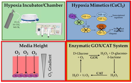
Figure 1.
Methods for hypoxia induction in vitro. Only hypoxia mimetics, media height and GOX/CAT system were tested within the study (red border). With CoCl2: Cobalt chloride, PHD: Prolyl hydroxylase domain proteins, HIF-1: Hypoxia-inducible factor 1, VHL: Von Hippel–Lindau E3 Ligase, GOX: Glucose oxidase, CAT: Catalase.
In this study, we aim to compare hypoxia induction methods which do not require additional equipment, therefore without using the hypoxia chamber, in 2D monolayer of osteoprogenitor cells and monocytic suspension culture as well as in the 3D co-culture approach of an in vitro fracture hematoma model. Successful hypoxia induction will be monitored within the scope of fracture healing. Besides monitoring HIF-1α protein levels and pericellular oxygen concentrations, chemokine and cytokine production and differentiation will be used as readouts for successful hypoxia induction. By comparing different methods we want to highlight challenges, limitations and applicability of the tested methods also in relation to different cell types and culture system.
2. Materials and Methods
2.1. Ethics Statement
All experiments were performed in accordance to the declaration of Helsinki. Experiments with primary human cells (osteoblasts and blood) were ethically approved by the ethics committee of the medical faculty of the University of Tuebingen (ethical vote for osteoblasts: 536/2016BO2 approved 03.08.2016, for blood: 844/2020BO2 approved 21 October 2020).
2.2. Culture of Osteogenic Cells as Representatives for 2D Culture
Human immortalized bone marrow derived mesenchymal stem cell (hB-MSCs) line SCP-1 were obtained from Prof. Schieker [24]. Cells were cultured in Minimal Essential Medium Alpha (MEM-alpha) supplemented with 5% fetal bovine serum (FCS).
The osteogenic progenitor cell line SaOS-2 (DSMZ # ACC 243) was cultured in RPMI 1640 Medium with 5% FCS.
Primary human osteoblasts (hOBs) were isolated and expanded as previously described [25]. After Collagenase digestion hOBs were expanded in culture medium (DMEM, 5% FCS, 1% Penicillin/Streptomycin (P/S), 50 µM L-ascorbate-2-phosphate, 50 µM β-glycerol-phosphate) until passages 2 or 3.
Cells were cultivated at 37 °C, 5% CO2, humidified atmosphere. Medium was changed every 3–4 days. Cells were subcultured when confluent [26]. Cells were regularly tested negative for mycoplasma (MycoAlertTM Mycoplasma Detection Kit, LT07-118, Lonza, Basel, Switzerland).
2.3. Culture of THP-1 Cells as Representatives for Suspension Culture
The human monocytic suspension cell line THP-1 (DSMZ # ACC 16) was cultured in RPMI 1640 Medium with 5% FCS (37 °C, 5% CO2, humidified atmosphere). Medium was changed every 3–4 days. Cell density was kept between 0.2–1 × 10⁶ cells/mL. Cells were regularly tested negative for mycoplasma (MycoAlertTM Mycoplasma Detection Kit, LT07-118, Lonza, Basel, Switzerland).
2.4. Preparation and Culture of Human In Vitro Fracture Hematoma Models
In vitro fracture hematomas were prepared following a protocol previously published [27]. Briefly, as representative for hB-MSCs served SCP-1 cells, derived from a female donor [24]. Whole blood was drawn from healthy volunteers. For sex-specific PCR, only blood of male donors was used. Briefly, freshly collected human whole blood was mixed in a 1:1 ratio (60 µL each) with a 1 × 10⁶ SCP-1 cells/mL suspension containing 10 mM CaCl2 in non-adherent 96-well plates. Coagulation was induced by incubation for 1 h at 37 °C, 5% CO2, humidified atmosphere. Afterwards, clots were transferred to new plates and cultured up to 96 h in MEM-alpha supplemented with 5% FCS and 1% P/S (37 °C, 5% CO2, humidified atmosphere). If in vitro hematomas were cultivated for longer than 48 h, medium was partially changed (25 µL) daily to maintain secreted factors, thereby also refreshing the respective stimuli.
2.5. Hypoxia Induction
2.5.1. Cobalt Chloride (CoCl2)
For chemical hypoxia induction, usual growth medium was supplemented with 0.1 M (adherent cells) or 0.4 M (in vitro hematomas) CoCl2 (Sigma-Aldrich/Merck, Darmstadt, Germany) [28].
2.5.2. Glucose Oxidase (GOX)/Catalase (CAT)
Enzymatic induction of hypoxia was introduced by combination of the two enzymes glucose oxidase and catalase as previously described [22]. According to the original publication and our own optimization, GOX (G6125-10KU, Sigma, St. Louis, MO, USA,) was diluted 1:10,000 (w/v) and CAT (C30-100MG (23100 U/mg) Sigma, St. Louis, MO, USA) was diluted 1:5000 (v/v) in the respective culture media.
2.5.3. Medium Height
For 2D monolayer cultures media heights were adjusted according to Camp et al. [21]. Media heights used, associated volumes and predicted oxygen concentrations are listed in Table 1. Experiments with in vitro fracture hematomas and for establishment of medium height were performed in 96-well plates, experiments for RNA isolation in six-well plates.

Table 1.
Media heights, associated volumes in 96-well plates and 6-well plates, and the predicted pO2 [21].
Due to their intrinsic height, increased media height for the in vitro hematomas was only analyzed for 300 µL medium which is comparable to the 250 µL used in the 2D cultures.
2.6. Assessment of Changes in Oxygen Concentrations by Means of Hypoxia IT Dye
ImageIT dyes were used as fluorescent probes for detection of low oxygen tensions. The dyes only develop fluorescence when oxygen concentrations decrease below 5%. Signal intensity thereby increases with decreasing oxygen levels. Oxygen concentrations below 2% can be generally considered as hypoxic [13]. The non-accumulating Image IT red hypoxia reagent (Thermo Scientific, Waltham, MA, USA) was directly added into the medium of adherent cells in a final concentration of 10 µM.
Suspension cells and 3D cultured cells were loaded with 1 mM of the accumulating ImageIT (Thermo Scientific, Waltham, MA, USA) green hypoxia reagent for 30 min before inducing hypoxic conditions. In both cases changes in fluorescence were detected with a FLUOstar Omega Plate Reader (BMG Labtech, Ortenberg, Germany). Additionally, after 2 h microscopy images were taken using a fluorescence microscope (EVOS FL, life technologies, Darmstadt, Germany) if not indicated differently.
2.7. Life-Dead Staining
Cells were stained with 0.1% Calcein-AM and 0.03% propidium iodide in culture medium for 20 min. Afterwards images were taken in 10× magnification in GFP and RFP channel using the EVOS FL fluorescence microscope.
2.8. Resazurin Conversion Assay
Mitochondrial activity was assessed by resazurin conversion. Briefly, 100 µL of a 0.0025% resazurin solution was added to cells/in vitro hematomas. After, incubation at 37°C resorufin was quantified via its fluorescence (λex = 544 nm and λem = 590–10 nm) with the FLUOstar Omega Plate Reader.
2.9. Alkaline Phosphatase (ALP) Activity Assay
For determining alkaline phosphatase activity, osteogenic cells and in vitro hematomas were washed with PBS and transferred to new 96-well plates before incubating with 200 µL ALP substrate solution (1 mg/mL pNPP, 50 mM glycine, 1 mM MgCl2, 100 mM TRIS; pH 10.5) at 37 °C for 1 h. After incubation working solution was briefly centrifuged to remove impurities. Formation of pNP was determined photometrically (λ = 405 nm) using the FLUOstar Omega plate reader from 75 µL working solution.
2.10. Semiquantitative Reverse-Trasncription (RT) PCR
RNA was isolated by phenol/chloroform extraction and quantified by spectrophotometry. In vitro fracture hematomas were pooled (n ≥ 12) and homogenized using homogenization pestles (SLG, Gauting, Germany) prior to the isolation. Up to 2.5 µg RNA was transcribed into cDNA using a First Strand cDNA Synthesis Kit (Thermo Scientific, Waltham, MA, USA). Semi quantitative PCRs were performed using Red HS master mix (Biozym, Hessisch Oldendorf, Germany). Cycling conditions for each primer set were optimized to range in the logarithmic phase of amplification. Used primers and cycling conditions are listed in Table 2. 18s, HPRT or GAPDH were used as internal references. PCR products were separated on 1.8% Agarose gels with 0.0007% ethidium bromide and visualized using an INTAS GelDoc system (INTAS, Göttingen, Germany). Densiometric analysis was performed using the ImageJ gel analysis tool.

Table 2.
Primer specificities for semi-quantitative RT-PCR.
2.11. Western Blot
Protein lysates for Western blot analysis were obtained by lysis of cells in ice-cold radio immunoprecipitation assay buffer (RIPA) with protease and phosphatase inhibitors. After quantification with micro Lowry, 30 µg of protein was applied on 12% sodium dodecyl sulfate polyacrylamide gels (SDS-PAGES) and separated by size. Proteins were blotted onto nitrocellulose membranes by tank blotting. Membranes were blocked with 5% bovine serum albumin (BSA) and incubated with primary antibody (HIF-1α: BD Bioschcienes 610958, HPRT: Santa Cruz Biotechnology, sc-376938; β-actin: Cell Signaling Technology, 4970) followed by HRP conjugated secondary antibody (Anti-Mouse IgG HRP-linked Antibody: 7076, Cell Signaling Technology, Anti-Rabbit IgG HRP-linked Antibody: Santa Cruz Biotechnology, 7074). Signals were detected by chemiluminescence using an ECL substrate solution and were detected with a charge-couple device camera (INTAS, Göttingen; Germany). Densitometric analysis was performed using the ImageJ (Version 1.52, NIH, Bethesda, MD, USA) analysis tool.
2.12. Enzyme Linked Immunosorbent Assay (ELISA)
Secretion of cytokines IL-1β, TNF-α and CCL2 was quantified in undiluted culture supernatants using mini ABTS-ELISA kits (Peprotech, Hamburg, Germany) following the manufacturer’s instructions.
2.13. Statistics
Results are represented either as bar or as line diagrams showing mean ± SEM of at least three independent experiments performed in triplicates. Statistics were made using Graph Pad Prism 8 (San Diego, CA, USA). Data were compared using non-parametric Kruskal–Wallis tests following Dunns’ multiple comparison or two way ANOVAs following Sidak’s multiple comparison tests if not indicated differently. Significance levels for all tests were defined as * p < 0.05, ** p < 0.01, *** p < 0.001, **** p < 0.0001.
3. Results
3.1. Confirmation of Hypoxia Induction in Osteogenic Cells
Accumulation of HIF-1α is the main cellular response to hypoxic conditions [29]. To prove induction of hypoxia osteogenic cells were treated with the three different hypoxia induction methods: increased medium height, enzymatic consumption of oxygen and CoCl2. For the induction with medium height different medium heights were tested ranging from 2 mm (Ctrl) to 10 mm. As a readout for the induction of hypoxia cells were stained with the Image-IT™ hypoxia reagent which reversibly reacts to low oxygen concentrations. Here, medium heights up to 10 mm were used which is as low as 5 mmHg. Results are shown in Figure 2. The hypoxia dye (Figure 2a) showed strong fluorescence at 5.4 mm medium height. Before, only very low fluorescence could be observed. At higher medium height the cell number started to decrease which resulted in lower fluorescence. The GOX/CAT system also strongly induced fluorescence of the hypoxia reagent. After 2 h of hypoxia induction both CoCl2 and GOX/CAT strongly induced HIF-1α protein levels (Figure 2b), showing a strong cellular reaction to hypoxia. The three different tested medium heights did not lead to an increase in HIF-1α protein levels.
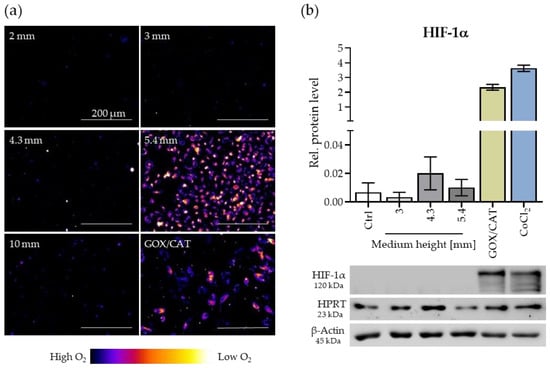
Figure 2.
Effect of different hypoxia induction methods in osteogenic cells. SaOS-2 cells were cultivated in common aerobic conditions without additional stimulus (Ctrl), with increased medium height, the enzymatic GOX/CAT system, and 0.1 mM CoCl2. (a) Depiction of oxygen levels by Image-IT Hypoxia reagent after 3 h incubation, scale bar 200 µm. Images are pseudo-colored with Fire using the ImageJ software (see legend). CoCl2 does not reduce oxygen level. (b) HIF-1α protein level after 2 h of hypoxia induction with an exemplary image of one blot. Data are presented as mean ± SEM (N = 3, n = 3).
3.2. Effect of Different Hypoxia Induction Methods on Chemokine Release in Osteogenic Cells
Hypoxia is strongly present in the fracture gap. There hypoxia induces chemokine release by osteogenic cells for attraction of immune cells. To test the functional outcome of the different hypoxia induction methods osteogenic cells were treated with hypoxic conditions for 24 h and the gene expression of relevant cytokines and chemokines was analyzed (Figure 3). For the test of increased medium height 5.4 mm (MH) was chosen as it showed increased fluorescence with the hypoxia reagent but did not reduce viability of the cells (Figure 2a).
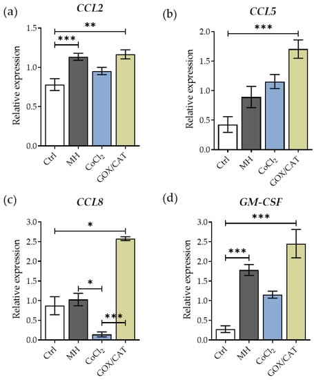
Figure 3.
Comparison of chemokine release from osteogenic cells by different hypoxia induction methods. Osteogenic cells were cultivated in common aerobic culture conditions without additional stimulus (Ctrl), with increased medium height (MH, 5.4 mm), 0.1 mM CoCl2 and the enzymatic GOX/CAT system. Gene expression of cytokines (a) CCL2, (b) CCL5, (c) CCL8 and (d) GM-CSF was analyzed after 24 h (N ≥ 3, n = 2). Statistics were made using non-parametric Kruskal–Wallis Tests with * p < 0.05, ** p < 0.01, *** p < 0.001. Data are shown as mean ± SEM.
The GOX/CAT system induced cytokine expression of all analyzed cytokines (CCL2, CCL5, CCL8, and GM-CSF, Figure 3a–d). CCL2 was also induced by medium height and CoCl2, nevertheless the increase was not significantly with CoCl2. CCL5 expression is significantly induced by the enzymatic system but not significantly by CoCl2 or medium height. CCL8, however, is only induced by the enzymatic system and reduced by CoCl2. GM-CSF is induced by medium height, by CoCl2 and by the enzymatic system but not significantly by CoCl2. CoCl2 does not lead to a significant expression induction of any cytokine, it just shows a trend of induction.
3.3. Induction of Hypoxia in Suspension Cells
THP-1 cells were used for testing the three alternative hypoxia induction methods with suspension cells. Changes in pO2 were followed by staining with the irreversible accumulating Green Image-IT ™ hypoxia reagent and gene expression of hypoxia-inducible genes was analyzed by RT-PCR. Results are displayed in Figure 4.
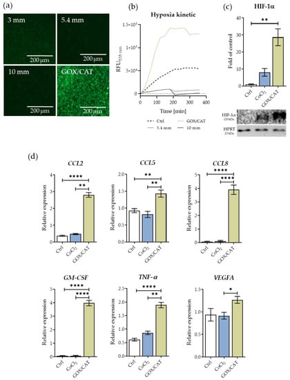
Figure 4.
Hypoxia induction in THP-1 suspension cells by different hypoxia induction methods. Cells were cultured in common aerobic culture conditions without additional stimulus (Ctrl), with 0.1 mM CoCl2 or the GOX/CAT system. (a+b) Analysis of pO2 in response to different media heights and GOX/CAT system with green Image-IT™ hypoxia reagent. (a) Microscopy images were taken after 6 h. Scale bar 200 µm. (b) Accumulation of dye was followed by kinetic measurement over 360 min (N = 3, n = 3). (c) HIF-1α protein level after 2 h of hypoxia induction with exemplary image of one blot (N = 2, n = 2). (d) Expression of cytokines CCL2, CCL5, CCL8, GM-CSF, TNF-α and growth factor VEGFA after 24 h (N = 3, n = 4). Statistics were made using non-parametric Kruskal–Wallis Tests with * p < 0.05, ** p < 0.01, **** p < 0.0001. Data are shown as mean ± SEM.
Microscopy images after 6 h showed development of fluorescence with the GOX/CAT system whereas no fluorescence could be observed in cells treated with the different medium heights. The corresponding time course fluorescence measurement showed an increase in fluorescence over a time of 6 h for GOX/CAT but no change in fluorescence for the different medium heights (Figure 4b).
GOX/CAT and CoCl2 both lead to an increase in HIF-1α protein levels compared to the control after 2 h of stimulation but GOX/CAT showed a more pronounced increase.
Treatment with the enzymatic system led to very pronounced increase of expression of the analyzed cytokines CCL2, TNF-α, CCL5, CCL8, GM-CSF, and also the growth factor VEGFA after 24 h whereas chemically stabilization of HIF-1α by CoCl2 could not increase the expression of the analyzed targets to that extend. Only pro-inflammatory cytokines TNFα and CCL2 showed an increasing trend due to stimulation with CoCl2.
3.4. Induction of Hypoxia in In Vitro Fracture Hematomas
To evaluate the different hypoxia induction methods also in an appropriate 3D system, in vitro fracture hematomas were chosen. Hypoxia is one of the main drivers of early fracture healing and results mainly from rupturing of the surrounding blood vessels and the resulting separation from blood supply. Hypoxia induction of the three methods was evaluated by means of expression profiles of HIF-1α regulated genes VEGFA and RUNX2, which are the key transcription factors during early fracture healing. As can be seen in Figure 5, chemical stabilization of HIF-1α as well as enzymatic hypoxia induction significantly induced expression of VEGFA and RUNX2. Media height stimulated hematomas did not show any increase in expression of HIF-1α regulated genes, but a significant increase in mitochondrial activity after 48 h in comparison to all other stimulations which indicated increased proliferation of SCP-1 cells (Supplementary Figure S1). Interestingly, control hematomas also showed an increase in expression of hypoxia regulated genes. Nevertheless, the effect was less prominent and the expression seemed delayed by about 24 h (Supplementary Figure S1a).
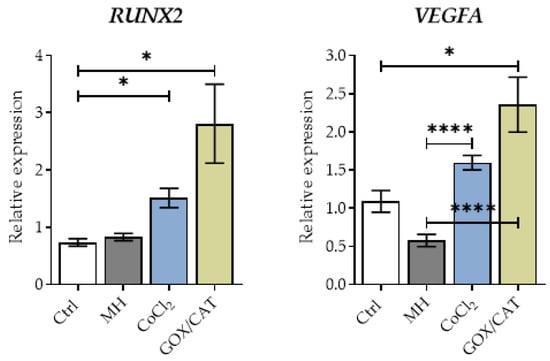
Figure 5.
Comparison of different hypoxia induction methods in in vitro fracture hematomas. In vitro hematomas were cultivated in common aerobic culture conditions without additional stimulus (Ctrl), with increased medium height (MH), 0.4 mM CoCl2 and the enzymatic GOX/CAT system. Gene expression was analyzed for targets VEGFA and RUNX2 after 24 h (N = 3, n ≥ 3). Statistics were made using non-parametric Kruskal–Wallis Tests with * p < 0.05, **** p < 0.0001. Data are shown as mean ± SEM.
Based on the previously shown expression profiles (Figure 5), for analysis of functional effects of hypoxia on the in vitro fracture hematomas solely the enzymatic system was chosen. Clots cultivated in “normoxia” and hypoxia were compared also over the course of 96 h incubation time. Hypoxia significantly reduced mitochondrial activity as can be seen in Figure 6b. This could be attributed to reduced amounts of SCP-1 cells, as can be seen in the life dead staining images (Figure 6a). Figure 6d showed secretion of proinflammatory cytokines TNF-α and IL-1β during earlier time points and generally higher levels of CCL2 by the enzymatic system. It further induced osteogenic differentiation monitored by ALP activity (Figure 6c).
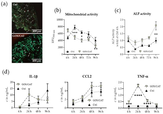
Figure 6.
Functional effects of enzymatically induced hypoxia (GOX/CAT) in in vitro hematomas in comparison to common aerobic culture conditions without additional stimulus (Ctrl). (a) Live–dead staining images from control and GOX/CAT stimulated hematomas after 96 h of incubation in 10x magnification. Arrows are indicating dead cells (red). (b) mitochondrial activity determined by resazurin conversion (N = 4, n = 3). (c) Alkaline phosphatase (ALP) activity (N = 3, n = 3). (d) Secretion of proinflammatory cytokines IL-1β, CCL2, and TNF-α (N = 4, n = 3). Statistics were made using non-parametric Sidak’s multiple comparison tests with * p < 0.05, ** p < 0.01, *** p < 0.001, **** p < 0.0001. Significance indicators in (b,c) and (d) show significances between GOX/CAT and non-stimulated samples at the same time point. Data are shown as mean ± SEM.
All in all, stimulation with the enzymatic system led to the development of the characteristic phases of healing, starting with an inflammatory phase followed by a differentiation phase and seems therefore an appropriate system.
4. Discussion
In this work, we compared the induction of hypoxia using three different easy and simple systems in osteogenic (adherent) cells, monocytic (suspension) cells, and in vitro fracture hematomas (3D system).
Hypoxia is a strong biological regulator being involved in several cellular processes and diverse pathogenic processes [30,31,32,33]. In bone, hypoxic conditions are important to maintain stem cell features in bone marrow [34]. Furthermore, during fracture, hypoxia is one of the main characteristics of the fracture hematoma, driving the inflammatory response and thus the healing process [35].
For in vitro research, the standard procedure to induce hypoxia is the reduction of oxygen in the culture environment (1–5% compared to 18%), which subsequently leads to a reduction in the pericellular oxygen tension. However, the usage of hypoxia chambers or incubators has several disadvantages and pitfalls. One major limitation is, that the environmental oxygen concentration not necessarily corresponds to the oxygen concentration at the cellular level [36]. Thus, we here describe three alternative methods to induce or mimic hypoxia without reducing environmental oxygen concentrations with the additional major advantage that they do not require special equipment. An increase of the medium height reduces the partial oxygen pressure at the cellular site, initially driven by the cells’ oxygen consumption [21]. CoCl2 has long been known as a strong inducer of HIF-1α in cellular systems mimicking the cellular response to hypoxia to a certain extent. The glucose oxidase/catalase system reduces the oxygen in cultures by its enzymatic consumption [22]. Table 3 summarizes the advantages and disadvantages of the three alternative hypoxia induction systems and in comparison to induction with a hypoxia chamber.

Table 3.
Overview of different in vitro hypoxia induction methods.
CoCl2 and the enzymatic system induced stabilization of HIF-1α protein in our cell culture systems. In contrast, the increased media heights failed to induce HIF-1α in osteogenic cells within 2 h, did not show an effect in monocytic cells and on the in vitro fracture hematomas. Because the cellular response to hypoxia is known to depend on the drop in oxygen not on hypoxia per se, the rather slow decrease of pericellular oxygen concentrations may not promote a strong HIF-1α response. Potentially, increased medium heights may therefore be used to adjust pericellular oxygen tension in cell cultures to prevent hyperoxia present when cultured in standard culture conditions (room air).
Chemical induction of hypoxia with CoCl2 led to a stabilization of HIF-1α protein in osteogenic cells, and to a lesser extent also in monocytic cells. However, it did not significantly induce cytokine expression. This highlights the necessity for “real hypoxia”. It has to be proposed that a higher concentration of CoCl2 may be necessary in monocytic suspension cells [37], which is in line with our observations in the 3D culture system. Our data show that reactions to hypoxia in osteogenic cells (2D culture), monocytic cells (suspension culture) and in vitro fracture hematomas (3D culture) were most successful using the enzymatic system. From in vitro and in vivo experiments well known hypoxia related induction of chemokines in osteogenic [38,39] and monocytic [40] cells was seen. Functional responses in in vitro fracture hematomas (VEGFA, RUNX2) were also similar to results obtained in an equine fracture hematoma model using an hypoxia chamber [27] as well as patient and in vivo data [41,42]. Additionally, all cytokines were stronger induced by GOX/CAT and in the osteogenic cells also by the increased media heights, than when just stabilizing HIF-1α by CoCl2.
Introducing chemical and enzymatic hypoxia inducers can affect cellular functions apart from inducing hypoxia. For instance, cobalt chloride has been shown to induce cellular apoptosis in an hypoxia independent manner when supplemented in higher doses [37,43]. When using the enzymatic system, ratios between catalase and glucose oxidase have to be established properly. Misbalance between the two enzymes can lead to an excess H2O2 production which, if not intentionally chosen, further stimulates inflammatory conditions [44]. When osteoprogenitors were stimulated with H2O2 alone, we did not observe any effects on the expression of various cytokines, indicating that the effect of the enzymatic system in our experiment was mainly related to the resulting hypoxia and that the carefully selected enzymatic system did not produce excessive ROS (Supp. Figure S2). Because the enzymes loose functionality over time, to maintain the hypoxic environment, medium has to be changed on a regular basis [22].
The induction of VEGFA in the control conditions in the in vitro fracture hematomas shows that hypoxia is already internally induced in the 3D structure as already found in other 3D structures [45]. Uneven distribution of oxygen is a general problem of non-vascularized 3D cultures, that also occurs when working with hypoxia chambers [46]. Thus, for 3D structures, chemical inducers or the enzymatic system may be the better choice than increasing the medium height or reducing environmental oxygen.
Promising results in hypoxia induction can also be achieved by combining two hypoxia induction methods. In a similar setup to our 3D model, Pfeiffenberger et al. used a hypoxic environment (1% O2) in combination with the alternative chemical prolylhydroxylase inhibitor DFO (desferrioxamine) to promote the hypoxic response as it was not strong enough with either the hypoxic environment or the chemical inducer alone [47]. In our set-up, the enzymatic system alone was sufficient for a pronounced hypoxic response of the cells.
Similarly, elevated medium heights alone are not sufficient to induce strong hypoxia, they can be preferably used to support other hypoxia induction methods. A combination of the increased medium height with reduced environmental oxygen enhanced the reduction in pericellular oxygen levels [48]. Alternatively, a combination of medium height and CoCl2 also effectively induces chemokine expression in osteogenic cells (Supp. Figure S2) thus allowing for induction of “real hypoxia” in combination with a strong HIF-1α response.
Another factor to consider when selecting a method for hypoxia induction is the possibility of intermittent hypoxia (preconditioning/hypoxia with reoxygenation phases). Intermittent hypoxia was shown to be more effective in inducing inflammation in monocytic cells and preconditioning to moderate hypoxia improves tissue regeneration processes and tolerance to stronger hypoxic conditions [49,50]. In principle, the induction of intermittent hypoxia is possible with all methods. In hypoxia chambers and with increased medium heights intermittent hypoxia has to be tightly controlled by oxygen measurements as the change in pericellular oxygen concentrations is rather slow. For all other induction methods (partial) medium changes are needed which allow a fast onset of hypoxia, where the enzymatic system has the advantage of an onset of hypoxia in less than two minutes [51].
When looking at the feasibility of the different induction methods for usage in animal models, reduction of environmental oxygen and chemical inducers are the common methods. The chemical inducers have the advantage that they can be applied locally thus enabling usage, e.g., in a fracture hematoma where they showed strong positive effects on angiogenesis induction, thus promotion of fracture healing [52]. To our knowledge the enzymatic system was not yet tested in animal models.
In comparison to the widely used hypoxia chambers all here tested methods are much cheaper and easier to use (Table 3). The enzymatic system leads to a similar induction of the cellular hypoxia response whereas CoCl2 just resembles some hypoxia features and the increase of medium height induces more a physiological oxygen concentration than a real hypoxia. When considering some basic limitations of the three here used systems they provide a good alternative to the hypoxia chamber.
5. Conclusions
In this work, we compared three different hypoxia induction methods for cell culture systems in regard to the hypoxic conditions in the beginning of bone fracture healing. The enzymatic system with glucose oxidase and catalase induced strong hypoxic cellular responses in 2D cultures (osteogenic cells), suspension cultures (monocytic cells), and in 3D culture (in vitro fracture hematoma). CoCl2 strongly increased HIF-1α protein levels in osteogenic cells but failed to induce significant expression of chemokines. In suspension cultures, CoCl2 induced a HIF-1α response but did not lead to cytokine expression. In the in vitro fracture hematomas, CoCl2 induced expression of VEGFA but to a much lesser extent than the enzymatic system. The induction of hypoxia by medium height was successful in osteogenic cells were it induced chemokine expression but was not sufficient in suspension cells and in the in vitro fracture hematomas. In conclusion, for all three cell culture systems the enzymatic system showed the strongest hypoxic response and provided an easy set-up.
Supplementary Materials
The following are available online at https://www.mdpi.com/article/10.3390/oxygen1010006/s1, Figure S1: 48 h measurements of in vitro fracture hematomas (mitochondrial activity, RUNX2 and VEGFA expression), Figure S2: Cytokine expression of osteogenic cells in response to H2O2, Figure S3: Chemokine expression and release of osteogenic cells by combination of medium height and CoCl2 to induce hypoxia.
Author Contributions
Conceptualization, S.E.; methodology, C.L., H.R., and B.B.; software, A.K.N. and T.H.; validation, C.L. and H.R.; formal analysis, C.L., H.R., and B.B.; investigation, C.L., H.R., and B.B.; resources, S.E. and A.K.N.; writing—original draft preparation, H.R., C.L., and S.E.; writing—review and editing, all authors; supervision, A.K.N.; project administration, S.E.; funding acquisition, S.E. All authors have read and agreed to the published version of the manuscript.
Funding
S.E. received funding from the German Research Foundation (DFG EH 471/2). C.L. receives support from the “Studienstiftung des deutschen Volkes”.
Institutional Review Board Statement
The study was conducted according to the guidelines of the Declaration of Helsinki, and approved by the Ethics Committee of the University Clinic Tübingen (536/2016BO2 approved 3 August 2016; 844/2020BO2 approved 21 October 2020), regulating donor recruitment for the isolation of osteoprogenitor cells and the isolation of blood for the in vitro hematomas, which serve as reference material in this study. For the work with the cell lines no ethical approval was required.
Informed Consent Statement
Informed consent was obtained from all subjects providing osteoprogenitor cells or blood for this study.
Data Availability Statement
The datasets generated during and/or analyzed during the current study are available from the corresponding author on reasonable request.
Acknowledgments
We would like to thank Leonie Fecht and Henrike Held for support of the experiments. Parts of this work have been performed by Caren Linnemann and Helen Rinderknecht as part of their thesis. Figure 1 was partially made using BioRender.com (accessed on 20 August 2021).
Conflicts of Interest
The authors declare no conflict of interest.
Abbreviations
| ALP | Alkaline Phosphatase |
| BMP | Bone morphogenetic protein |
| CAT | Catalase |
| CCL | (C-C motif) ligand |
| CoCl2 | Cobalt chloride |
| Ctrl | Control |
| DFO | Desferrioxamine |
| DMOG | Dimethyloxalylglycine |
| EPO | Erythropoietin |
| GAPDH | Glyceraldehyde 3-phosphate dehydrogenase |
| GOX | Glucose oxidase |
| HIF | Hypoxia inducible factor |
| hOB | Human osteoblast |
| HPRT | Hypoxanthine-guanine phosphoribosyltransferase |
| HRE | Hypoxia responsive elements |
| IGF | Insulin-like growth factor |
| IL | Interleukin |
| MH | Increased medium height |
| MSC | Mesenchymal stem cell |
| PHD | Prolyl hydroxylase |
| RUNX2 | Runt-related transcription factor 2 |
| TNF | Tumor necrosis factor |
| VEGF | Vascular endothelial growth factor |
| VHL | Von-Hippel-Lindau E3 Ligase |
References
- Corrò, C.; Novellasdemunt, L.; Li, V.S.W. A brief history of organoids. Am. J. Physiol.-Cell Physiol. 2020, 319, C151–C165. [Google Scholar] [CrossRef]
- Carreau, A.; El Hafny-Rahbi, B.; Matejuk, A.; Grillon, C.; Kieda, C. Why is the partial oxygen pressure of human tissues a crucial parameter? Small molecules and hypoxia. J. Cell. Mol. Med. 2011, 15, 1239–1253. [Google Scholar] [CrossRef] [Green Version]
- Harris, A.L. Hypoxia—A key regulatory factor in tumour growth. Nat. Rev. Cancer 2002, 2, 38–47. [Google Scholar] [CrossRef]
- Nombela-Arrieta, C.; Pivarnik, G.; Winkel, B.; Canty, K.J.; Harley, B.; Mahoney, J.E.; Park, S.-Y.; Lu, J.; Protopopov, A.; Silberstein, L.E. Quantitative imaging of haematopoietic stem and progenitor cell localization and hypoxic status in the bone marrow microenvironment. Nature 2013, 15, 533–543. [Google Scholar] [CrossRef]
- Andrew, J.G.; Andrew, S.; Freemont, A.; Marsh, D. Inflammatory cells in normal human fracture healing. Acta Orthop. 1994, 65, 462–466. [Google Scholar] [CrossRef] [PubMed] [Green Version]
- Claes, L.; Recknagel, S.; Ignatius, A. Fracture healing under healthy and inflammatory conditions. Nat. Rev. Rheumatol. 2012, 8, 133–143. [Google Scholar] [CrossRef]
- Marsell, R.; Einhorn, T.A. The biology of fracture healing. Injury 2011, 42, 551–555. [Google Scholar] [CrossRef] [PubMed] [Green Version]
- Maruyama, M.; Rhee, C.; Utsunomiya, T.; Zhang, N.; Ueno, M.; Yao, Z.; Goodman, S.B. Modulation of the Inflammatory Response and Bone Healing. Front. Endocrinol. 2020, 11, 386. [Google Scholar] [CrossRef] [PubMed]
- Croes, M.; Oner, F.C.; Kruyt, M.C.; Blokhuis, T.J.; Bastian, O.; Dhert, W.; Alblas, J. Proinflammatory Mediators Enhance the Osteogenesis of Human Mesenchymal Stem Cells after Lineage Commitment. PLoS ONE 2015, 10, e0132781. [Google Scholar] [CrossRef]
- Wenger, R.; Kurtcuoglu, V.; Scholz, C.; Marti, H.; Hoogewijs, D. Frequently asked questions in hypoxia research. Hypoxia 2015, 3, 35–43. [Google Scholar] [CrossRef] [Green Version]
- Jones, D.P. Intracellular diffusion gradients of o2 and atp. Am. J. Physiol.-Cell Physiol. 1986, 250, C663–C675. [Google Scholar] [CrossRef]
- Spencer, J.A.; Ferraro, F.; Roussakis, E.; Klein, A.; Wu, J.; Runnels, J.M.; Zaher, W.; Mortensen, L.; Alt, C.; Turcotte, R.; et al. Direct measurement of local oxygen concentration in the bone marrow of live animals. Nature 2014, 508, 269–273. [Google Scholar] [CrossRef] [PubMed] [Green Version]
- Bertout, J.A.; Patel, S.A.; Simon, M.C. The impact of O2 availability on human cancer. Nat. Rev. Cancer 2008, 8, 967–975. [Google Scholar] [CrossRef] [PubMed] [Green Version]
- Ast, T.; Mootha, V.K. Oxygen and mammalian cell culture: Are we repeating the experiment of Dr. Ox? Nat. Metab. 2019, 1, 858–860. [Google Scholar] [CrossRef] [PubMed]
- Flamme, I.; Fröhlich, T.; von Reutern, M.; Kappel, A.; Damert, A.; Risau, W. HRF, a putative basic helix-loop-helix-PAS-domain transcription factor is closely related to hypoxia-inducible factor-1α and developmentally expressed in blood vessels. Mech. Dev. 1997, 63, 51–60. [Google Scholar] [CrossRef]
- Wiesener, M.S.; Jürgensen, J.S.; Rosenberger, C.; Scholze, C.K.; Hörstrup, J.H.; Warnecke, C.; Mandriota, S.; Bechmann, I.; Frei, U.A.; Pugh, C.W.; et al. Widespread hypoxia-inducible expression of hif-2alpha in distinct cell populations of different organs. FASEB J. 2003, 17, 271–273. [Google Scholar] [CrossRef] [Green Version]
- Maxwell, P.; Wiesener, M.S.; Chang, G.-W.; Clifford, S.C.; Vaux, E.C.; Cockman, M.; Wykoff, C.C.; Pugh, C.; Maher, E.; Ratcliffe, P. The tumour suppressor protein VHL targets hypoxia-inducible factors for oxygen-dependent proteolysis. Nature 1999, 399, 271–275. [Google Scholar] [CrossRef]
- Gossage, L.; Eisen, T.; Maher, E.R. VHL, the story of a tumour suppressor gene. Nat. Rev. Cancer 2014, 15, 55–64. [Google Scholar] [CrossRef]
- Sarada, S.; Himadri, P.; Mishra, C.; Geetali, P.; Ram, M.S.; Ilavazhagan, G. Role of Oxidative Stress and NFkB in Hypoxia-Induced Pulmonary Edema. Exp. Biol. Med. 2008, 233, 1088–1098. [Google Scholar] [CrossRef]
- Semenza, G.L. HIF-1 and mechanisms of hypoxia sensing. Curr. Opin. Cell Biol. 2001, 13, 167–171. [Google Scholar] [CrossRef]
- Camp, J.P.; Capitano, A.T. Induction of Zone-Like Liver Function Gradients in HepG2 Cells by Varying Culture Medium Height. Biotechnol. Prog. 2007, 23, 1485–1491. [Google Scholar] [CrossRef]
- Mueller, S.; Millonig, G.; Waite, G. The GOX/CAT system: A novel enzymatic method to independently control hydrogen peroxide and hypoxia in cell culture. Adv. Med Sci. 2009, 54, 121–135. [Google Scholar] [CrossRef] [Green Version]
- Epstein, A.C.; Gleadle, J.M.; McNeill, L.A.; Hewitson, K.S.; O’Rourke, J.; Mole, D.R.; Mukherji, M.; Metzen, E.; Wilson, M.I.; Dhanda, A.; et al. C. elegans EGL-9 and Mammalian Homologs Define a Family of Dioxygenases that Regulate HIF by Prolyl Hydroxylation. Cell 2001, 107, 43–54. [Google Scholar] [CrossRef] [Green Version]
- Böcker, W.; Yin, Z.; Drosse, I.; Haasters, F.; Rossmann, O.; Wierer, M.; Popov, C.; Locher, M.; Mutschler, W.; Docheva, D.; et al. Introducing a single-cell-derived human mesenchymal stem cell line expressing hTERT after lentiviral gene transfer. J. Cell. Mol. Med. 2008, 12, 1347–1359. [Google Scholar] [CrossRef] [PubMed] [Green Version]
- Ehnert, S.; Fentz, A.-K.; Schreiner, A.; Birk, J.; Wilbrand, B.; Ziegler, P.; Reumann, M.K.; Wang, H.; Falldorf, K.; Nussler, A.K. Extremely low frequency pulsed electromagnetic fields cause antioxidative defense mechanisms in human osteoblasts via induction of •O2− and H2O2. Sci. Rep. 2017, 7, 14544. [Google Scholar] [CrossRef] [PubMed] [Green Version]
- Aspera-Werz, R.H.; Ehnert, S.; Heid, D.; Zhu, S.; Chen, T.; Braun, B.; Sreekumar, V.; Arnscheidt, C.; Nussler, A.K. Nicotine and Cotinine Inhibit Catalase and Glutathione Reductase Activity Contributing to the Impaired Osteogenesis of SCP-1 Cells Exposed to Cigarette Smoke. Oxidative Med. Cell. Longev. 2018, 2018, 1–13. [Google Scholar] [CrossRef] [Green Version]
- Pfeiffenberger, M.; Bartsch, J.; Hoff, P.; Ponomarev, I.; Barnewitz, D.; Thöne-Reineke, C.; Buttgereit, F.; Gaber, T.; Lang, A. Hypoxia and mesenchymal stromal cells as key drivers of initial fracture healing in an equine in vitro fracture hematoma model. PLoS ONE 2019, 14, e0214276. [Google Scholar] [CrossRef] [PubMed] [Green Version]
- Al Okail, M.S. Cobalt chloride, a chemical inducer of hypoxia-inducible factor-1α in u251 human glioblastoma cell line. J. Saudi Chem. Soc. 2010, 14, 197–201. [Google Scholar] [CrossRef] [Green Version]
- Maes, C.; Carmeliet, G.; Schipani, E. Hypoxia-driven pathways in bone development, regeneration and disease. Nat. Rev. Rheumatol. 2012, 8, 358–366. [Google Scholar] [CrossRef] [Green Version]
- Brusevold, I.J.; Husvik, C.; Schreurs, O.; Schenck, K.; Bryne, M.; Søland, T.M. Induction of invasion in an organotypic oral cancer model by CoCl2, a hypoxia mimetic. Eur. J. Oral Sci. 2010, 118, 168–176. [Google Scholar] [CrossRef]
- Ryan, S.; Taylor, C.; McNicholas, W.T. Selective Activation of Inflammatory Pathways by Intermittent Hypoxia in Obstructive Sleep Apnea Syndrome. Circulation 2005, 112, 2660–2667. [Google Scholar] [CrossRef] [PubMed] [Green Version]
- Sethi, K.; Rao, K.; Bolton, D.M.; Patel, O.; Ischia, J.J. Targeting hif-1α to prevent renal ischemia-reperfusion injury: Does it work? Int. J. Cell Biol. 2018, 2018, 9852791. [Google Scholar] [CrossRef] [Green Version]
- Halberg, N.; Khan, T.; Trujillo, M.E.; Wernstedt-Asterholm, I.; Attie, A.D.; Sherwani, S.; Wang, Z.V.; Landskroner-Eiger, S.; Dineen, S.; Magalang, U.J.; et al. Hypoxia-Inducible Factor 1α Induces Fibrosis and Insulin Resistance in White Adipose Tissue. Mol. Cell. Biol. 2009, 29, 4467–4483. [Google Scholar] [CrossRef] [Green Version]
- Jang, Y.-Y.; Sharkis, S.J. A low level of reactive oxygen species selects for primitive hematopoietic stem cells that may reside in the low-oxygenic niche. Blood 2007, 110, 3056–3063. [Google Scholar] [CrossRef] [PubMed] [Green Version]
- Kolar, P.; Gaber, T.; Perka, C.; Duda, G.N.; Buttgereit, F. Human Early Fracture Hematoma Is Characterized by Inflammation and Hypoxia. Clin. Orthop. Relat. Res. 2011, 469, 3118–3126. [Google Scholar] [CrossRef] [Green Version]
- Pavlacky, J.; Polak, J. Technical Feasibility and Physiological Relevance of Hypoxic Cell Culture Models. Front. Endocrinol. 2020, 11, 57. [Google Scholar] [CrossRef] [PubMed] [Green Version]
- Guo, M.; Song, L.-P.; Jiang, Y.; Liu, W.; Yu, Y.; Chen, G.-Q. Hypoxia-mimetic agents desferrioxamine and cobalt chloride induce leukemic cell apoptosis through different hypoxia-inducible factor-1α independent mechanisms. Apoptosis 2006, 11, 67–77. [Google Scholar] [CrossRef]
- Ishikawa, M.; Ito, H.; Kitaori, T.; Murata, K.; Shibuya, H.; Furu, M.; Yoshitomi, H.; Fujii, T.; Yamamoto, K.; Matsuda, S. MCP/CCR2 Signaling Is Essential for Recruitment of Mesenchymal Progenitor Cells during the Early Phase of Fracture Healing. PLoS ONE 2014, 9, e104954. [Google Scholar] [CrossRef] [Green Version]
- Kim, D.S.; Lee, M.W.; Ko, Y.J.; Park, H.J.; Park, Y.J.; Kim, D.-I.; Jung, H.L.; Sung, K.W.; Koo, H.H.; Yoo, K.H. Application of human mesenchymal stem cells cultured in different oxygen concentrations for treatment of graft-versus-host disease in mice. Biomed. Res. 2016, 37, 311–317. [Google Scholar] [CrossRef] [Green Version]
- Sierra-Filardi, E.; Nieto, C.; Domínguez-Soto, Á.; Barroso, R.; Sánchez-Mateos, P.; Puig-Kroger, A.; López-Bravo, M.; Joven, J.; Ardavín, C.; Fernandez, J.L.R.; et al. CCL2 Shapes Macrophage Polarization by GM-CSF and M-CSF: Identification of CCL2/CCR2-Dependent Gene Expression Profile. J. Immunol. 2014, 192, 3858–3867. [Google Scholar] [CrossRef] [Green Version]
- Hoff, P.; Gaber, T.; Strehl, C.; Jakstadt, M.; Hoff, H.; Schmidt-Bleek, K.; Lang, A.; Röhner, E.; Huscher, D.; Matziolis, G.; et al. A Pronounced Inflammatory Activity Characterizes the Early Fracture Healing Phase in Immunologically Restricted Patients. Int. J. Mol. Sci. 2017, 18, 583. [Google Scholar] [CrossRef] [PubMed]
- Hoff, P.; Gaber, T.; Strehl, C.; Schmidt-Bleek, K.; Lang, A.; Huscher, D.; Burmester, G.R.; Schmidmaier, G.; Perka, C.; Duda, G.N.; et al. Immunological characterization of the early human fracture hematoma. Immunol. Res. 2016, 64, 1195–1206. [Google Scholar] [CrossRef]
- Muñoz-Sánchez, J.; Chánez-Cárdenas, M.E. The use of cobalt chloride as a chemical hypoxia model. J. Appl. Toxicol. 2018, 39, 556–570. [Google Scholar] [CrossRef] [PubMed]
- Owegi, H.; Egot-Lemaire, S.; Waite, L.R.; Waite, G.N. Macrophage activity in response to steady-state oxygen and hydrogen peroxide concentration—Biomed 2010. Biomed. Sci. Instrum. 2010, 46, 57–62. [Google Scholar] [PubMed]
- Qin, Y.; Greene, V.R.; Chattopadhyay, C.; Ekmekcioglu, S.; Liu, C.; Grimm, E.A. Abstract 2936: Induction of hypoxia in 3D human melanoma spheroids leads to c-Met activation and resistance to Vemurafenib. Cancer Res. 2013, 73, 2936. [Google Scholar] [CrossRef]
- Funamoto, K.; Zervantonakis, I.; Liu, Y.; Ochs, C.J.; Kim, C.; Kamm, R.D. A novel microfluidic platform for high-resolution imaging of a three-dimensional cell culture under a controlled hypoxic environment. Lab Chip 2012, 12, 4855–4863. [Google Scholar] [CrossRef] [PubMed] [Green Version]
- Pfeiffenberger, M.; Hoff, P.; Thöne-Reineke, C.; Buttgereit, F.; Lang, A.; Gaber, T. The in vitro human fracture hematoma model—A tool for preclinical drug testing. Altex 2020, 37, 561–578. [Google Scholar] [CrossRef]
- Oze, H.; Hirao, M.; Ebina, K.; Shi, K.; Kawato, Y.; Kaneshiro, S.; Yoshikawa, H.; Hashimoto, J. Impact of medium volume and oxygen concentration in the incubator on pericellular oxygen concentration and differentiation of murine chondrogenic cell culture. Vitr. Cell. Dev. Biol.-Anim. 2012, 48, 123–130. [Google Scholar] [CrossRef] [PubMed]
- Chuang, L.-P.; Chen, N.-H.; Lin, S.-W.; Chang, Y.-L.; Liao, H.-R.; Lin, Y.-S.; Chao, I.-J.; Lin, Y.; Pang, J.-H. Increased C-C Chemokine Receptor 2 Gene Expression in Monocytes of Severe Obstructive Sleep Apnea Patients and under Intermittent Hypoxia. PLoS ONE 2014, 9, e113304. [Google Scholar] [CrossRef] [PubMed]
- Rybnikova, E.; Samoilov, M. Current insights into the molecular mechanisms of hypoxic pre- and postconditioning using hypobaric hypoxia. Front. Neurosci. 2015, 9, 388. [Google Scholar] [CrossRef] [Green Version]
- Baumann, R.P.; Penketh, P.G.; Seow, H.A.; Shyam, K.; Sartorelli, A.C. Generation of Oxygen Deficiency in Cell Culture Using a Two-Enzyme System to Evaluate Agents Targeting Hypoxic Tumor Cells. Radiat. Res. 2008, 170, 651–660. [Google Scholar] [CrossRef] [PubMed] [Green Version]
- Huang, J.; Liu, L.; Feng, M.; An, S.; Zhou, M.; Li, Z.; Qi, J.; Shen, H. Effect of CoCl2 on fracture repair in a rat model of bone fracture. Mol. Med. Rep. 2015, 12, 5951–5956. [Google Scholar] [CrossRef] [PubMed] [Green Version]
- Pressley, M.; Gallaher, J.A.; Brown, J.S.; Tomaszewski, M.R.; Borad, P.; Damaghi, M.; Gillies, R.J.; Whelan, C.J. Cycling hypoxia selects for constitutive HIF stabilization. Sci. Rep. 2021, 11, 5777. [Google Scholar] [CrossRef] [PubMed]
Publisher’s Note: MDPI stays neutral with regard to jurisdictional claims in published maps and institutional affiliations. |
© 2021 by the authors. Licensee MDPI, Basel, Switzerland. This article is an open access article distributed under the terms and conditions of the Creative Commons Attribution (CC BY) license (https://creativecommons.org/licenses/by/4.0/).