Review

What Applied Physical Chemistry Can Contribute to Understanding Cancer: Toward the Next Generation of Breakthroughs

Department of Ophthalmology and Visual Sciences, University of Michigan Medical School, 1000 Wall Street, Ann Arbor, MI 48105, USA
*
Author to whom correspondence should be addressed.
AppliedChem 2023, 3(3), 378-399; https://doi.org/10.3390/appliedchem3030024
Submission received: 16 June 2023 / Revised: 13 July 2023 / Accepted: 13 July 2023 / Published: 19 July 2023

Abstract

:
Living systems, whether healthy or diseased, must obey the laws of chemistry. The purpose of this review is to identify the interpretive limits of cellular biochemistry using, largely, the tools of physical chemistry. We illustrate this approach using two major concepts in cancer: carcinogenicity and cancer recurrences. Cells optimize the chemical performance of enzymes and pathways during cancer recurrences. Biology has been primarily concerned with the analysis of high affinity interactions, such as ligand–receptor interactions. Collective weak interactions (such as van der Waals forces) are also important in determining biosystem behaviors, although they are infrequently considered in biology. For example, activity coefficients determine the effective concentrations of biomolecules. The in vivo performance of enzymes also depends upon intracellular conditions such as high protein concentrations and multiple regulatory factors. Phase separations within membranes (two dimensions) and nucleoli (three dimensions) are a fundamental regulatory factor within cells, as phase separations can alter reactant concentrations, local dielectric constants, and other factors. Enzyme agglomeration also affects the performance of biochemical pathways. Although there are many examples of these phenomena, we focus on the key steps of cancer: carcinogenicity and the biochemical mechanism of cancer recurrences. We conjecture that oxidative damage to histones contributes to carcinogenicity, which is followed by nucleolar phase separations and subsequent DNA damage that, in turn, contributes to the redistribution of enzymes mediating metabolic changes in recurrent breast cancer.

1. Introduction

In the 1950s, Watson and Crick, who were specialists in virology and physics, respectively, deduced the structure of DNA based upon the x-ray crystallography of DNA samples. Their work led to the “central dogma of molecular biology,” which laid the foundation for the field of molecular biology. This work illustrates the power of interdisciplinary research. The broad direction of life science research was summarized by Crick: “the ultimate aim of the modern movement in biology is in fact to explain all biology in terms of physics and chemistry [1].” This approach has been enormously successful and should continue [2]. Yet, due to the organization of scientific administration (grant committees, promotion and tenure committees, etc.) [3], research remains predominantly within silos; and these silos can become divorced from other fields, including physical chemistry and cancer research.
Cells must obey the laws of physical chemistry, but is anyone checking? Not really. For example, the medical literature is filled with articles about the oxidant superoxide, but superoxide anions are rarely an oxidant. Superoxide oxidizes certain Fe-S containing proteins such as succinate dehydrogenase and NADH (reduced nicotinamide adenine dinucleotide)-ubiquinone oxidoreductase [4,5]. Its protonated form, the hydroperoxide radical, is a dangerous oxidant, but only at low pH values [5] (pKa = 4.88). As membrane surfaces are negatively charged, proton concentrations are higher and therefore, hydroperoxide radicals become more abundant [6]. As hydroperoxide radicals are uncharged and enter cell membranes [6], they likely contribute to the cell damage attributed to superoxide anions. Hydroperoxide radicals promote the lipid peroxidation pathway. Superoxide anions are in general better reductants than oxidants [7]. Although superoxide is not an oxidant, it interacts with other species to form downstream oxidative radicals. Superoxide reactivity is an example of research performed in a silo. On the other hand, research has shown that most Nobel Prizes in physiology or medicine go to scientists performing interdisciplinary research [8]. For example, the 2012 Nobel Prize in Chemistry was awarded to Lefkowitz and Kobika, two biologists. Impactful work defies silos.
The aim of this review is to present applications of established physical–chemical knowledge in new biological settings, to bridge the silos, with the goal of gaining a better understanding of the biogenesis and detection of cancer and to encourage new research in the quantitative chemistry of cancer.

2. Re-Visiting a Few Basic Chemical Principles in a Cellular Context

Enzymatic reactions are typically studied in vitro, or outside an organism, using monodisperse, highly diluted near ideal solutions in isotropic and well-defined environments. However, living cells are just the opposite—highly anisotropic structures that often display compartmentalization at several levels. The reactants contained in these compartments are far from equilibrium and ideality. Whether or not in vitro findings have substantial relevance to living matter is uncertain. As a rule, it is best to be cautious and to carefully consider the limitations of experimental findings. In this section, we will apply basic concepts in physical chemistry to cells, especially cancer cells. Some of these concepts are summarized in Table 1, then applied in Section 3 and Section 4.

2.1. Concentration

One of the most useful concepts in in vitro biochemistry is biomolecule concentration. It is also one of the most difficult concepts to properly apply in cellular biochemistry. Let us consider the question of intracellular adenosine triphosphate (ATP) concentrations; these ideas are generally applicable to other molecules. Typically, one would use in vitro cells to measure ATP levels, which increases O2 availability for respiration (thus, perturbing the parameter being measured). In such a test, cells are first disrupted using detergents, organic solvents, or physical means. The test will not measure the ATP concentration that can participate in biology; it measures the total amount of ATP in a cell because the cells were dissolved. The total concentration is believed to be about 1 to 10 mM [9,10,11,12] with much of the ATP bound to proteins, such as actin. The free or effective concentration of ATP is not the same as the total amount of ATP. A recent study suggests that the ATP concentration in rapidly growing cancer cell lines after extraction is ~1 mM [13]. Another issue in the assessment of ATP concentrations within cells is the spatial heterogeneity of ATP production [13,14,15]. For example, using genetically encoded constructs, Imamura et al. [14] have shown that the ATP level in the nucleus is considerably higher than in the cytosol which is higher than that within mitochondria. Thus, it is possible that previous estimates of ATP at ~5 mM in the nucleus [11] with lower concentrations in the cytoplasm were correct. This raises the question: “what is the origin of this nuclear ATP?” As all glycolytic enzymes can be found, at least transiently, in the nucleus [16,17], nuclear glycolysis seems possible; but it is not likely because there is no proof that nuclear glycolytic enzymes make ATP. One known source of ATP production in nuclei is the conversion of poly(ADP-ribose) to ADP-ribose, which, in the presence of pyrophosphate, forms ATP catalyzed by pyrophosphatase NUDIX5 [18]. NUDIX5-derived ATP can be used in chromatin remodeling [19] and, very likely, to support other functions. NUDIX5 appears to be important in cancer because enhanced expression is associated with poor prognosis whereas suppression of NUDIX5 reduces proliferation and invasiveness of carcinoma cells [19,20]. It seems likely that ATP and NUIX5 play multiple roles in cells.
To understand biochemical reactions in living cells, it is important to know the effective concentration of solutes. In the case of ATP, we obtain an estimate of the effective concentration by measuring the free concentration. The ATP molecules bound to actin are essentially in a solid phase. However, this only corrects for tightly bound ATP, and the concentration must also be corrected for other ATP interactions. This correction requires knowledge of the activity coefficient. Unfortunately, the correction for low affinity interactions is difficult. Not only is it difficult to measure biochemical activity coefficients in vitro, but the application of activity coefficients is difficult because solute molecules interact with themselves and other molecules to decrease the effective solute concentration. Although progress has been made in biomolecule activity coefficients [21], estimates for the intracellular environment have not yet been made. It might be useful to develop means of estimating the impact of these interactions on enzyme reactions in vivo (within living organisms), which could support computational modeling of biochemical reactions in vivo.
ATP is an amphipathic molecule, with both hydrophilic (triphosphate) and hydrophobic (adenosine) regions. A recent study by Patel et al. [22] provides strong evidence that ATP acts as a hydrotrope or hydrotrope-“like” material. Hydrotropes are amphipathic compounds that solubilize hydrophobic molecules in aqueous media. A concentration of ~1 mM ATP is necessary to support in vitro and in vivo hydrotrope activity. ATP at this concentration inhibits the aggregation of proteins (generally positively charged and relatively disordered) and in some cases, dissolves filamentous protein aggregates. ATP also binds to small compounds [13]. A similar molecule, guanosine triphosphate (GTP), also possesses hydrotropic activity. The high nuclear ATP concentrations may be necessary to promote the solubilization of proteins and chromatin within nuclei. Nuclear ATP appears to be uniformly distributed in cancer cells [13]. As ~100 μM ATP is sufficient for ATP-driven enzymology, its role as a hydrotrope provides a function for higher concentrations of nuclear ATP.

2.2. Intracellular Conditions: Concentration, Solvation, and Proximity

Intracellular conditions bear little resemblance to extracellular conditions. The intracellular environment is a slightly alkaline KCl solution, while the extracellular environment is primarily a NaCl solution. Cells possess multiple redox couples to manage intracellular chemistry [23,24]. Unfortunately, it is difficult to properly monitor all these parameters simultaneously.
The protein concentrations within cells are very high and can be near their solubility limit or in a supersaturated solution. For example, in erythrocytes, hemoglobin is found at a concentration of 330 g/L [25,26], a concentration that would form a supersaturated solution in vitro. Intracellular protein concentrations vary from 300 to 400 g/L—corresponding to nominally saturated or supersaturated conditions in vitro [27,28]. Under certain conditions, hemoglobin type S and C polymerize in vivo. In the case of hemoglobin S, the formation of polymers leads to the sickle-shaped erythrocytes of sickle cell anemia. Indeed, supersaturating conditions causing proteins to aggregate may be an important pathway in the biogenesis of diseases [27,28]. Having said that, not all agglomerations (a clump of cohesive substances) inhibit cell functions. For example, the activity of some enzymes is increased by agglomeration (see Section 4).
To begin to understand the complex intracellular environment, several approaches have been developed to provide access to the cell’s interior. Methods used in these in situ, or in their original locations, experiments include cells treated with organic solvents such as toluene, electroporation, and semi-intact or permeabilized cells (e.g., streptolysin O) [29,30,31,32]. These samples are used to assess enzyme reactions under conditions similar to those of living cells in comparison to in vitro conditions. When the activities of several enzymes were tested, the Km values were unchanged, decreased, or increased. For glycolytic enzymes, many Km values remain unchanged or increased in magnitude; however, the Km values of phosphofructokinase (PFK) and glyceraldehyde 3-phosphate dehydrogenase (GAPDH) were increased in permeabilized cells [30], suggesting that at this rate-controlling step of glycolysis, these enzymes have a lower affinity for substrate.
PFK will be used as an example of in vitro and in situ differences in enzyme activity. PFK is an allosteric highly regulated rate-limiting step of glycolysis [33]. PFK’s activity is influenced by many factors including PFK concentration, fructose 6-phosphate (F6P) concentration, assembly state (monomer, dimer, tetramer, cluster, agglomeration), binding to other structures such as membranes and the cytoskeleton, environmental conditions (temperature, pH, and redox conditions, and allosteric factors including ATP, citrate, fructose 2,6-bisphosphate (F2,6BP), fructose 1,6-bisphosphate (F1,6BP, etc.) [29,30,31,32,33,34,35,36,37]. As an allosteric enzyme, PFK does not follow Michalis–Menton kinetics and therefore, Km values do not apply. Instead, S0.5 values are used. The in vitro and in situ S0.5 values of PFK are 0.2 and 0.4 mM [29]. The S0.5 value for F6P is 22 μM, which is near the intracellular concentration of F6P. Interestingly, the allosteric properties of PFK are reduced in situ and at high PFK concentrations [29,30,31,32,33,34,35,36,37]. It would seem the in vivo regulatory potential of allosteric modifiers may be less than thought based upon their in vitro biochemistry. The specificity constant (kcat/Km) (sometimes referred to as the enzyme efficiency) for F6P is 0.24 × 10⁸ M−1s−1, which is close to that of diffusion-limited reactions (108–109 M−1s−1) [37].
The rate-controlling event of diffusion-limited reactions is the arrival of the substrate at the enzyme; thus, the enzyme’s reaction is faster than the transport of the substrate to the enzyme [37]. Although uncommon in biochemistry, a few examples of diffusion-limited enzymes are acetylcholine esterase, superoxide dismutase, fumarase, triosephosphate isomerase, and carbonic anhydrase. To provide optimal enzyme activity, diffusion-limited enzymes are uniformly distributed throughout cells. If these enzymes were clustered together, they would locally deplete their substrate, thus reducing the product formation rate. On the other hand, most enzymes are reaction-controlled, with specificity constants of about 105 M−1s−1. Although the performance of individual enzymes cannot be accelerated by orders of magnitude, enzyme agglomeration can increase a biochemical pathway’s product output by orders of magnitude [38,39].
When numerous consecutive enzymes of a pathway become clustered, the properties of the pathway may differ from that of individual components [40]. A leading example of such a system is the purinosome, which synthesizes the purine precursor inosine monophosphate from phosphoribosyl pyrophosphate, ATP, formate, and other substrates. Purinosomes form in tumor cells during hypoxic conditions [41]. Purinosomes are particularly useful because they assemble, disassemble, and traffic to mitochondria on an easily accessible laboratory timescale. Fluorescence microscopy studies have co-localized purinosome subunits with one another and showed that their clustering/de-clustering is regulated by the number of purines and other molecules in the culture medium [42]. Purinosomes co-localize with and translocate along microtubules at about 55 nm/sec. [43], but their motion is constrained near mitochondria [44]. The purinosome’s enzymes have specificity constants of ~105 M−1s−1 in vitro. Using mass spectrometry imaging, Pareek et al. [45] have directly shown the presence of subcellular regions low in substrates and high in synthetic intermediates produced by purinosome when cells are grown in the absence of exogenous purines. These hotspots of substrate depletion are spatially coincident with purinosomes in cells [45], thus suggesting that diffusion is a limiting factor in this pathway. This seems surprising because their specificity constant is in the order of 105 M−1s−1. Thus, the purified enzymes in vitro behave like reaction limited enzymes, but the in vivo pathway displays characteristics of a diffusion limited activity. There is no paradox because the specificity constant is measured using purified monodisperse enzymes in vitro. Purinosomes only assemble within cells and have not been reconstituted in vitro; these structures are large enough to contain dozens or hundreds of copies of each enzyme within this pathway. Clustering enhances the reaction rate by almost an order of magnitude—thus, the catalytic performance of the pathway is influenced by the supramolecular structure of its component enzymes [46]. Purinosome catalytic activity may also be influenced by their ability to move along microtubules to enhance their proximity with mitochondria, which are also associated with microtubules. This is anticipated to increase the availability of mitochondrial metabolites, including formate, for synthetic reactions of purinosomes [46]. Proximity relationships are important in purine synthesis at multiple levels.

2.3. Phase Separations in Two and Three Dimensions

Compartmentalization of intracellular chemical reactions is a crucial strategy of cells. Cells often separate distinct functions into separate membrane-bound organelles [47]. This is a good strategy to separate oxidative and reductive chemistry as well as anabolic and catabolic biological pathways into different intracellular containers to prevent intracellular chemistry from operating at cross-purposes. For example, proteins are synthesized in the endoplasmic reticulum and cytoplasm, but digested within lysosomes. ATP is primarily synthesized by mitochondria using potential energy stored in the form of a proton gradient. This standard model of cell biology has been revised in the past few years by the introduction of the role of phase separations into cellular health and disease. The intracellular phase changes take place in two-dimensional cell membranes and in three-dimensional liquid–liquid phase separations (LLPS) of cell structures such as nucleoli.
There is a rich literature concerning lipid phase separations in model lipid membranes [48,49,50,51]. Lipid–lipid interactions, and consequent line tension at the perimeter of phase separations, contribute to these structures, which vary greatly in size. Specializations or “domains” in cell membranes take many forms, including lateral phase separations. Cell membrane domains are created in many ways including protein-to-protein interactions, attachment of an external agent, linkage to the cytoskeleton, and membrane curvature. Proving the existence of cell membrane domains in general, and lipid phase separations specifically, is very challenging. The emerging consensus is that cell plasma membranes and intracellular vacuole membranes can exhibit lipid phase separations, some of which are quite compelling. Plasma membrane phase separations are typically ~20 nm in size whereas vacuolar membrane phase separations are ~200 nm in size. Membrane phase separations may form liquid-ordered and liquid-disordered phases. Membrane lipid phase separations have been observed in bacteria, amoeba, and higher eukaryotes [52,53,54,55,56,57,58,59]. Evidence for the existence of lipid phase separations in vacuole membranes comes from fluorescence microscopy and freeze fracture electron microscopy [60]. The study of lipid phase separations in plasma membranes is difficult because the phase separations are small, their existence is transitory, and plasma membranes are quite heterogeneous. Using B cell receptors as a model system, lateral phase separations have been demonstrated using Förster resonance energy transfer (FRET) [57] and super-resolution fluorescence microscopy coupled with cross-correlation data analysis [59]. Specifically, it was found that the B cell receptor traffics to liquid ordered regions of plasma membranes, which constitute a small percentage of the total surface area. These lateral phase separations are important because they recruit Lyn kinase to the lipid ordered region and exclude phosphatases, such as CD45, both of which heighten signaling. Thus, the lipid phase separation influences the physiological behavior of the signaling apparatus [57,58,59].
In three dimensions, LLPS form membrane-free organelle-like structures, which are also known as biological condensates. Nucleoli are a type of LLPS observed in eukaryotic cells. In some cases, LLPS may be specific for a certain cell type. A few of the types of LLPS found within cells include nucleoli, microtubule organizing centers/centrosomes, stress granules, Cajal bodies, P bodies, and others [60,61,62]. The formation of three-dimensional LLPS can be promoted by highly repetitive regions. Several types of physical interactions such as protein–protein, RNA–protein interactions, RNA–RNA interactions, poly-ADP-ribosylation, hydrophobic, electrostatic, π–π orbital interactions, and multivalent cation–π interactions [63,64] participate in these phase separations. LLPS exhibit high protein concentrations but can be dissolved by dilution. These unique phases are known to sequester/desequester certain biomolecules, such as PFK [65]. They also provide an environment where molecular motion is reduced and protein concentrations are high, which are believed to facilitate certain chemical reactions.
Nucleoli participate in ribosome production including rDNA synthesis, maturation of rRNA, assembly of ribosome subunits, and transport to the cytoplasm. As new ribosomes are essential in producing proteins to support cell division, the nucleolus is essential in cell proliferation. Having said that, most proteins in nucleoli are not related to ribosome synthesis, which suggests additional functions for nucleoli. In cancer, the nucleolar size increases dramatically, which is due, at least in part, to the burden of ribosome production. Indeed, large nucleoli (and/or an increase in the number of nucleoli) indicate the presence of tumor cells, but this does not prove that the cells are aggressive and will become invasive or metastatic. Another important function of nucleoli is to repair DNA, which will be discussed in Section 4.
LLPS have several distinct catalytic advantages, which have been tested in vitro [66]. DEAD (asp-glu-ala-asp) box proteins contain the DEAD amino acid sequence and can be found in nuclei where they function as RNA helicases by unwinding short RNA duplexes. The nucleolar DEAD-box protein Ddx4 spontaneously forms LLPS droplets in vitro and in living cells [67]. A feature of these LLPS is that their dielectric constant is ~45, which is about the same as the solvent DMSO (dimethylsulfoxide); this alone could have substantial effects on chemical reaction rates [68]. Short double strand DNA is spontaneously accumulated and then separated within these droplets, with no supply of energy. LLPS can also be created using chimeric proteins composed of the low complexity region of the DEAD-box proteins and the enzyme adenylate kinase [69]. These molecules spontaneously form LLPS with relative protein concentrations increased ~50–150-fold and a 5-fold increase in catalytic activity at a dielectric constant of ~32. Thus, it is possible to create mini-reactors to construct molecules in vitro or in vivo.

2.4. Redox Conditions

The intracellular redox status is important in normal cell functions (such as signaling, transcriptional regulation, and post-transcriptional changes), cell damage by oxidative or reductive stress, cell growth and proliferation, and enzyme kinetics [23,24]. Intracellular redox conditions are often assessed using the glutathione couple GSSG/GSH, NAD+/NADH, and/or NADP+/NADPH. Other biologically relevant redox couples include flavin adenine dinucleotide (FAD)/FADH2, flavin mononucleotide (FMN)/FMNH2, and thioredoxin reductase (Trx(SH2)/TrxSS) [23]. Experiments typically measure the ratio of oxidized and reduced glutathione and the total glutathione concentration.
Cells may be damaged by oxidative conditions, as outlined in the next section. Superoxide anions, per se, are not a significant factor in oxidative cell damage, as they react with Fe-S centers of certain proteins such as succinate dehydrogenase and NADH-ubiquinone oxidoreductase [45] but rather make a better reducing agent [7]. However, downstream products of superoxide anions such as those of the Haber-Weiss reaction, lead to the production of hydroxyl radicals. Hydroxyl radicals initiate the lipid peroxidation pathway and its products (e.g., 4-hydroxy-2-nonenal; Figure 1) to cause extensive cell damage. The protonated form of superoxide anions, hydroperoxide radicals (HOO•), is reactive, and could contribute to intramembrane reactions.
The importance of understanding redox conditions in complicated cellular environments is underscored by human studies. One might think that antioxidants would be useful in protecting against the potential damage of oxidizing agents in vivo (as illustrated in Section 3). Adding antioxidants will reduce the oxidative burden on cells. However, in a clinical trial assessing antioxidant use by smokers (who may have pre-invasive lung cancer lesions), the cancer risk of patients is significantly increased due to antioxidant intake [70]. A decrease in oxidative burden is thought to allow an increase in the level of intracellular reducing agents. This may perturb intracellular signaling, such as the redox-sensitive RhoA (GDP → GTP) reaction, to cause indolent cancer lesions to become aggressive. The clinical trial was terminated early, but it does teach that manipulating redox levels must be carefully performed.
The redox potentials of chemotherapeutic agents are another important area to consider. Aggressive cancer cells display the Warburg Effect, which is also known as aerobic glycolysis [36]. During aerobic glycolysis, the activity of the glycolytic pathway is greatly increased while mitochondrial activity only accounts for ~50% of ATP production. The increased production of NADH unbalances the NAD+/NADH redox couple. Low levels of NAD+ cause reductive stress and could lead to tumor cell death. Although oxidative anti-cancer drugs are widely used, they may provide a mechanism for the most aggressive tumor cells to avoid reductive stress, and therefore hasten cancer recurrences. This might happen because certain anti-cancer oxidative drugs accept electrons from NADPH and glutathione [71]. Therefore, we have proposed that oxidative therapy should be coupled with subsequent reductive tumor therapy to address the potential ability of oxidative agents to protect a subset of dangerous recurrent tumor cells, especially in cancer patients suffering metastatic disease [72].

3. Carcinogenicity

Nucleosomes participate in packaging long DNA molecules within living cells. DNA has a high negative charge and is stored by wrapping it around positively charged histones. These structures are quite stable [73]. However, these primary interactions between histones and DNA only neutralize about 50% of DNA’s negative charges. To undergo the next level of compaction, the remaining negative charges must be neutralized. Other agents such as linker histones, positive counterions, etc., are responsible for neutralizing these remaining charges, which are required to further condense DNA [73]. Tumor cells can be recognized by their large nucleoli [74], which suggest a reduction in DNA compaction. Altered histone management in cancer is suggested by the fact that nucleosomes are observed in the circulatory systems of patients [75].
Carcinogenesis is about how cancers come to be. Genetic contributions to cancer may be inherited or acquired during one’s lifetime. Chemical processes, especially oxidation, are believed to participate in carcinogenesis. The oxidation of cellular lipids leads to breakdown products such as 4-hydroxynonenal (4-HNE) and 4-oxononenal (4-ONE). To better understand these events, we examined tissue samples from patients diagnosed with the pre-invasive disease ductal carcinoma in situ (DCIS) of the breast. 4-HNE, a product of the lipid peroxidation pathway, is genotoxic and causes DNA fragmentation and mutations. It is a reactive amphipathic breakdown product of ω-6 fatty acids in cell membranes, where it can locally accumulate to high levels. 4-HNE forms stable adducts with proteins, which occur via Schiff base formation or Michael addition [76,77] (Figure 1).
Untreated tissue samples were observed using conventional fluorescence microscopy to find the chemical adducts formed by deleterious oxidative reactions (Schanen and Petty, 2023, unpublished). Human tissue sections were labeled with antibodies that recognize the histone H2A.X and 4-HNE adducts. In addition to being a marker for the cell nucleus, H2A.X was chosen because this molecule is associated with DNA repair. Figure 2 shows that the nucleus, as defined by the presence of the H2A.X, is a primary target of 4-HNE. This is true for patients who will and will not experience a subsequent recurrence. The preferential labeling of the nucleus suggests that the spatial distribution of 4-HNE adducts may be due to additional factors determining the location of adduct formation.
Several reasons may account for the rather selective labeling of the nucleus with anti-4-HNE adduct antibodies (Figure 2C,D). 4-HNE (and related molecules) are generated from ω-6 fatty acids in cell membrane lipids, which are then increased in number by the lipid peroxidation pathway. 4-HNE resides largely in membranes with the carbonyl group near the lipid headgroups [78]. Due to the limited solubility of 4-HNE in the aqueous cytosol and the presence of aldehyde dehydrogenase and aldose reductase in the cytoplasm, the cytoplasm is minimally labeled with 4-HNE adducts. 4-HNE is much more soluble in nuclei. The high concentrations of ATP in nuclei [9,10,11,12,13] may act as a hydrotrope [22] to solubilize 4-HNE; ATP has been previously shown to interact with small molecules [14]. The high nuclear concentration of ATP may stabilize proteins [79] and nucleic acids. Moreover, the dielectric constant of nucleoli-like LLPS is 45, about the same as DMSO in which 4-HNE is highly soluble (50 mg/mL). The biological effect of these reactions is illustrated in Figure 3. Under normal circumstances, the negatively charged DNA polymers bind to histones (Figure 3C,E). However, in the case of 4-one damage, 4-one and other molecules such as 4-HNE react with histones [80], decreasing the positive charges expressed on the histone. This is expected to have many effects on targets including [81] sterically hindering the binding of epigenetic markers and DNA packing (Figure 3D,F). As the DNA is no longer well-sequestered by histones, it becomes more accessible to chemical and environmental damage. If the damage is serious, a tumor may become aggressive. If these changes, through a complicated and poorly understood set of reactions, ignite the rapid uptake and metabolism of glucose, the cells are destined to become aggressive, and the patients may recur after surgery.

4. Chemical Engineering Meets Cancer Biology: Enzyme Agglomeration in Cancer Recurrences

Enzyme aggregates or clusters are often encountered in cellular biochemistry, and studies of monodisperse enzymes in dilute solution may be irrelevant to human disease. The aggregates range from dimers to agglomerations of many thousands of enzymes. For example, PFK may be expressed as a dimer, but only becomes fully functional as a tetramer due to allosteric interactions among the subunits [82]. It can form higher order structures in homologous or heterologous fashions.
Metabolons are multienzyme complexes wherein the product of one enzyme in a pathway is a substrate of another nearby enzyme within the metabolon. The metabolons may be membrane-associated (e.g., glycolytic metabolons) or not (e.g., purinosomes). Additional examples of metabolons are the respiratory protein complexes (e.g., NADH dehydrogenase) and Kreb’s TCA (tricarboxylic acid cycle) enzymes of mitochondria [83,84]. In some cases, metabolons are membrane associated, and in others they are not.
Glycolytic enzymes may be attached to the inner side of plasma membranes. For example, in human erythrocytes, the integral membrane protein band 3, an ion transporter, binds to GAPDH, aldolase, pyruvate kinase (PK), PFK, and lactate dehydrogenase (LDH) via specific glycolytic enzyme binding sequences Asp6-Asp-Tyr-Glu-Asp10 and Glu19-Glu-Tyr-Glu-Asp23 [85]. Not surprisingly, phosphorylation of Tyr8 or Tyr21 blocks the binding of glycolytic enzymes to band 3, which is associated with deoxygenated erythrocytes. Moreover, other peripheral membrane proteins and cytoskeletal proteins display glycolytic enzyme binding sequences and bind to glycolytic enzymes. In erythrocytes, these glycolytic enzymes also bind to spectrin, ankyrin, actin, p55, and protein 4.2 [85]. In yeast, actin microfilaments stabilize glycolytic metabolons [86]. In other cell types, another cytoskeletal component, microtubules, and monomeric tubulin bind to GAPDH, aldolase, PK, and LDH with a KD of ~1–5 μM. In addition, PFK, phosphoglucose isomerase, and phosphoglycerate kinase bind to microtubules. Moreover, caveolin-1, a cytoskeletal structure that mediates the formation of caveolea, possesses a binding site for PFK and aldolase. Total glycolytic enzyme binding is increased in breast cancer cells due to the overexpression of caveolin-1 [87]. Returning specifically to the erythrocyte, layers of glycolytic enzymes are formed at the inner surface of the plasma membrane (composed of glycolytic enzyme-binding components) and the adjacent cytoskeletal layers (spectrin, actin, ankyrin)—thus creating a thin container lined with catalytic enzymes. This region can retain ATP at concentrations higher than that of the bulk cytoplasm, thus forming a membrane proximal compartment to power membrane transport systems of the erythrocyte [88].
As the metabolism undergoes dramatic changes to support the proliferation and aggressive behavior of tumor cells, we sought to better understand the regulatory steps of metabolism in cancer. Computational analysis of glycolysis has shown that its key rate-limiting steps are glucose transport, hexokinase action, PFK catalysis, and lactic acid export from cells [89]. Glucose uptake via phospho-Ser-226-GLUT1 and PFK1 (PFKL) steps were found to be of especial interest in retrospective case-control clinical trials, as they serve as a prognostic test for cancer recurrences [90,91,92,93]. In recurrent cancer cells, these two biomarkers underwent trafficking to the plasma membrane (and elsewhere), presumably to enhance glucose uptake. However, the normally expressed forms in non-recurrent cancer, glucose transporter type 1 (GLUT1), and phosphofructokinase type P (PFKP) could be found in tumor cells of samples from women who did and did not subsequently develop a recurrent cancer. As protein phosphorylation and enzyme isoform switching alter metabolic pathway performance in normal and cancer cells [94,95], we focused on phospho-Ser-GLUT1 and PFKL isoforms that greatly enhanced performance (Table 1). The Vmax of GLUT1 is increased 5-fold by phosphorylation of the serine residue at position 226 [96]. When phospho-Ser226-GLUT1 was localized in DCIS samples of women who did not subsequently experience a recurrence, this biomarker was found at or near the nucleus (Figure 4A). In contrast, when tissue samples from patients who experienced a recurrence were studied, phospho-Ser226-GLUT1 was found in the vicinity of the plasma membrane (Figure 4B). This contrasts with the non-phosphorylated form of GLUT1, which did not undergo any remarkable changes when recurrent and non-recurrent forms of DCIS were compared. It seems likely that phospho-Ser226-GLUT1 moved from the cell center to the cell periphery to promote the uptake of external glucose in cells from recurrent patients.
PFKP (PFK, platelet type) and PFKM (PFK, muscle type) are feedback inhibited by the PFK type 1 product F1,6BP. On the other hand, PFKL is feedback activated by F1,6BP, which auto-activates enzyme performance. When samples from women who did not have a subsequent cancer recurrence were tested using anti-PFKL antibodies, PFKL was found in the nucleoli of tumor cells (Figure 4C), where it co-localizes with histone H2A.X [91]. On the other hand, when samples from recurrent women were tested, PFKL was generally found at the periphery of ductal epithelial cells (Figure 4D). In contrast, PFKP and PFKM displayed no remarkable changes in cellular localization when non-recurrent and recurrent cells were compared [91]. Importantly, the disappearance of PFKL from nucleoli parallels a nucleolar phase change that has been observed and reconstituted [65], which suggests that the nucleolar phase change is one factor accompanying the activation of aerobic glycolysis in aggressive cancer. Thus, during cancer recurrences, tumor cells mobilize the higher performing transporter phospho-Ser226-GLUT1 and the enzyme PFKL to the plasma membrane where phospho-Ser226-GLUT1 increases net glucose flux while PFKL can be accelerated to high levels to remove its substrate from the cytoplasm to maintain a large glucose gradient (and flux) in tumor cells.
Immunofluorescence micrographs of the two proteins described above, phospho-Ser226-GLUT1 and PFKL (Figure 4), are sufficient to accurately predict the outcomes (recurrences or non-recurrences) of DCIS patients [92,93]. To establish prognoses at a high level of accuracy, we employed computer vision. Pathologists and computer vision programs do the same thing: they evaluate tissue patterns to draw inferences about the underlying tissue chemistry. Pathologists observe several variables in micrographs, such as color and size, whereas computer vision analyzes many millions of variables to find several dozens of mathematical variables that predict recurrences and non-recurrences. Over the past few decades, pathologists have been asked to do more, including predicting disease outcomes, and this is now possible using machine tools to assist pathologists [93].
In addition to phospho-Ser226-GLUT1 and PFKL, the perimembrane region of ductal epithelial cells of women who will experience a cancer recurrence are known as metabolic platforms because they also contain: 6-phosphofructo-2-kinase/fructose-2,6-biphosphatase 4 (PFKFB4), glutamate cysteine ligase catalytic domain (GCLC), glutathione synthetase (GS), pentose phosphate pathway (PPP) components including glucose 6-phosphate dehydrogenase (G6PD), transketolase (TKT), and transketolase-like protein 1 (TKTLP1), RhoA and RhoA with bound GTP, and other enzymes [90]. The presence of these many enzymes, additional signaling proteins, and especially the cytoskeletal components (microtubules, microfilaments, caveolea), contribute to forming metabolic platforms. Moreover, since glycolytic enzymes can interact with other glycolytic enzymes in a homologous or heterologous fashion, such interactions may stabilize these structures. Metabolic platforms may have a large effect on associated biochemical pathways by forming a unique metabolic compartment at the cell surface where substrates enter cells. The kinetic benefit of metabolons and other enzyme oligomers is that they can form a pathway or channel between consecutive steps of a chemical pathway. Each metabolic platform has very many copies of each of many enzymes, thus forming a large organelle-like structure about the periphery of cells. The unique supramolecular structure of metabolic platforms is analogous to metabolons, but at a much larger scale. As mentioned above, large peripheral membrane structures have been described for erythrocytes [88]. In metabolic platforms, the concentration of enzyme active sites is very high while the solvent content is very low, making reactions more likely. Thus, solutes percolate through the voids and channels within metabolic platforms. As noted by Castellani et al. [39], such clusters can accelerate a two-step reaction by 8-fold and a three-step reaction by 110-fold. A potential limitation of this strategy is that substrate depletion zones may form near the structure. Two strategies to counteract this are: putting the enzymes at the periphery of the cell where their peripheral location will give metabolic platforms direct access to incoming substrates, allowing recurrent tumor cells to survive under harsh conditions, and to direct cell metabolism to serve the needs to the recurrent tumor cells.
Recently, enzyme agglomeration has been deployed in chemical engineering to produce products more efficiently in larger amounts [97,98,99,100,101,102,103,104]; the primary advantages are a relative abundance of active sites to substrates and the channeling effects. Microdroplets can be genetically engineered using chimeric enzymes containing enzyme activities of interest and DEAD domains to create nucleoli-like regions to produce products [66]. In this case, the solvent’s dielectric constant and local concentrations (i.e., solute solubility) are key features.
A glance at Figure 4 reveals that glycolytic elements change position in samples from women who subsequently experience a cancer recurrence. This happens because the machinery described above is needed near the plasma membrane to manage the high flux of glucose necessary to support aerobic glycolysis. Therefore, mathematic relationships of biomarkers within human tissues were extracted from patient micrographs using training datasets containing micrographs from recurrent and non-recurrent patients. Training datasets were immunofluorescence micrographs of patient samples stained with anti-phospho-Ser226-GLUT1 and PFKL. Therefore, one could perform binary classifications of patients with unknown outcomes [38,72,90,91,92,93]. For example, the computer was trained with images such as those of Figure 4. In the images of phospho-Ser-GLUT1, the computer was trained on the peripheral distribution of this antigen in epithelial cells. Other locations of this antigen were also learned by the computer including blood vessels, cancer-associated fibroblasts, myoepithelial cells in combination with appropriate weighting factors to arrive at a predicted outcome. In this way, a patient’s outcome could be predicted many years in advance at high accuracy for patients held out of the training dataset [93]. The software identified recurrent cases with no false negatives, non-recurrent women, and false positives due to the efficacy of mastectomies [93]. As blood vessels of African American women have elasticities differing from other races [93], it is very important to balance the training datasets properly and to perform tests on a large holdout dataset including many African American women. As these computer findings underscore, the entire tissue, not just tumor cells, participates in cancer recurrence predictions—and presumably, physiological recurrences. Importantly, 20% of micrographic fields (an average among patients) could lead to a recurrent cancer. This supports the idea that tumors are heterogeneous, possessing both cells that could and could not lead to a recurrence [93]. The machine analysis of DCIS patients will permit improved DCIS patient management. This approach could also be used as a companion diagnostic to choose therapeutic antibodies by inspecting the binding of various therapeutic antibodies to a specific recurrent DCIS patient’s tissue, which may permit the elimination of disease at the in situ stage.

5. Discussion

We began this review with a call for the increased application of physical chemistry in cancer biology. To avoid writing in silos, fundamental chemical principles and cancer-related biology were reviewed simultaneously. The point is to integrate, insofar as possible, the theoretical and experimental rigor of physical chemistry with breast cancer studies.
To illustrate the importance of applying physical chemistry to cancer biology, we present two fundamental problems in cancer biology: carcinogenesis and cancer recurrences. Consider breast tissue at a steady state in homeostasis within its organismal environment. Every cell is exposed to many millions of potentially harmful oxidative reactions every day. If the repair efficacy is 99.9%, re a vast number of damaged DNA molecules exist. If an oxidative reaction damages junk DNA, which accounts for a considerable portion of human DNA, no problem emerges because this DNA encodes nothing. We surmise that junk DNA has a function—to function as a decoy target. If a mutation occurs in an unimportant region of a gene product, no change in outcome will be observed. However, if a key part of a sensitive protein, such as a tumor suppressor gene, is damaged, that change will make a substantial difference in patient outcome. In Figure 3, we illustrate one key site of damage, histones. 4-HNE and similar aldehydes are generated by the attack of radicals on ω-6 fatty acids in cell membranes. However, 4-HNE is poorly soluble in the aqueous cytosol. Any 4-HNE molecules in the cytoplasm can be destroyed by aldehyde dehydrogenase or by aldose reductase activity (Figure 3). 4-HNE molecules that find their way to the nuclear envelope will spontaneously partition into nucleoli due to their dielectric constant (~45). High ATP concentrations may also contribute to solubilizing 4-HNE in aqueous regions of the nucleoplasm. Reactions of 4-HNE with histones alter the surface charge of histones by blocking positive charges at their surfaces, thus reducing electrostatic interactions with DNA. This modification also sterically hinders the association of DNA with histones. Consequently, DNA is no longer safely ensconced at nucleosomes. The unwound DNA makes a better target for damage. If a tumor suppressor gene is mutated to a non-functional state, tumor aggression may not be restrained.
Tumor cells are recognized by changes in their nuclear staining with H&E dyes. The enlarged nuclei appear to correlate with changes in DNA compaction, ribosome production, nucleosomes in the peripheral blood of patients, PFKL trafficking in a subset of recurrent cells, and as this region enlarges, there will likely be changes in chemical reactivities, nucleolar LLPS, and local dielectric constants. These ideas are consistent with recent interest in the role of nucleoli in cancer [105,106,107]. Changes in nucleolar phase likely contribute to PFKL release and to DNA de-compaction and consequent mutations. These changes may also desequester additional enzymes to re-wire cell metabolism, thus promoting recurrences.
The re-wiring of the cell metabolism increases glucose uptake by tumor cells. As described above, phospho-Ser226-GLUT1 departs from the nuclear area to accumulate at the cell periphery, while PFKL is desequestered from nucleoli before trafficking to the region of the plasma membrane. Both steps accelerate glucose transport by moving glucose or accelerating its metabolism to maintain the glucose gradient. Other rate-limiting steps of glycolysis, including hexokinase II and the monocarboxylate transporter type 4, undergo changes in number and/or location to maintain glucose internalization and lactic acid removal. The collection of the glycolytic, hexose monophosphate shunt, and glutathione synthesis machinery at the plasma membrane form metabolic platforms (Figure 5). Due to enhanced catalytic ability of such agglomerations, metabolic platforms likely contribute to metabolic alterations in aggressive cancer cells. From a basic science standpoint, these steps account for the biochemical mechanism of the Warburg Effect. From a clinical standpoint, computer vision analysis of phospho-Ser226-GLUT1 and PFKL are sufficient to diagnose a DCIS patient that has a lesion capable of recurring and requires clinical intervention.
As the scientific community has characterized most of the high-affinity interactions, perhaps it is time to move on to the lower-affinity interactions. Chemists may avoid weak interactions because they are more difficult to measure. Biologists, with broad brushstrokes, do weak interactions the indignity of calling them non-specific, and discarding their potential relevance. However, are weak interactions important? Of course, they are. Although van der Waals forces are weak, they collectively participate in membrane–membrane adhesion, protein stabilization, and the hydrophobic effect, among others. Moreover, as intracellular proteins are near or above their reported solubility, small changes in conditions can have a large effect on solubility. A single point mutation in hemoglobin creates polymer-like structures in erythrocytes of patients with sickle cell anemia. As intracellular proteins are near the cusp of their solubility, small changes in conditions cause protein agglomerations leading to neuromuscular disease [27,108]. LLPS also effect neurodegeneration [108]. Certain proteins spontaneously come out of solution—no ligand-to-receptor interactions are required. In the case of metabolic platforms, several mechanisms including diffusion and active transport on the cytoskeleton can explain their accumulation at the plasma membrane. Although the factors stabilizing metabolic platforms are unknown, the saturated to super-saturated cytosolic protein concentration, the overexpression of glycolytic enzymes in cancer, and the affinity of these enzymes for one another may contribute to metabolic platform stabilization.
We have previously discussed several drugs that interact with elements of the mechanistic steps of cancer recurrence mentioned above, and therefore, they might contribute to the effects of the drugs [92]. These include:
  • Taxol—dissociates PFK from the plasma membrane [109],
  • Colchicine and other alkaloids—disrupt intracellular trafficking by inhibiting microtubule functions [110],
  • Local anesthetics—disrupt microfilaments, and thereby destabilize metabolons [111,112,113],
  • Prenylation inhibitors—disrupt membrane association of certain signaling proteins [114,115],
  • KU55933—blocks GLUT1 translocation to the plasma membrane [116],
This suggests that the pathway described above may be a useful drug target.
Although many drugs act via high affinity lock-and-key interactions, some drugs of unknown mechanism may act by influencing weaker interactions. For example, weak interactions (Kd ~10−5 to 10−3 M) combined with network analysis have proven to be a useful approach in conventional drug design [117]. Metabolons and metabolic platforms seem to be excellent candidates for this approach, as there are multiple sites on multiple different proteins contributing to these structures. The Asp-Asp-Tyr-Glu-Asp sequence is an example of relevant blocking peptide. As agglomeration is expected to greatly increase pathway performance, these targets may show clinical efficacy (see bullet points above). Interfering with these interactions may influence pathway performance, as mentioned above. In another form of cancer therapy, hyperthermia, tissue temperature is raised to as high as 113° F. As low affinity biochemical interactions exhibit ΔG0 = ~−5 kcal/mol, hyperthermia could contribute to reducing low-affinity protein–protein interactions. These low-affinity interactions may include the reduction in the stability of metabolic platforms.
We suggest additional lines of future research. One important strategy is to stabilize the nucleoli. If nucleoli could be stabilized, it might be possible to maintain their internal dielectric constant and reactivity profile and block desequestration of metabolic enzymes that precede the Warburg Effect. Although additional physical parameters of tumor cell nuclei are needed to understand the stabilization, there is already some movement in terms of developing lead compounds in nucleolar stabilization. In this way, it may be possible to enhance PFK detention within nucleoli. The BRAT (TRIM-NHL protein Brain tumor) tumor suppressor gene that is known as tripartite motif-containing protein 3 (TRIM3) in humans [118] enhances longevity by reducing fibrillarin production, thereby promoting compaction [119]. As PFK nucleolar detention should be augmented by stabilizing nucleoli, enhancing the brat tumor suppressor pathway may be a route to increase PFKL detention and thereby block the Warburg Effect.
There are no computational simulations that correctly model the metabolic changes occurring during the formation of recurrent cancer, which is not surprising because the parameterization is so poor. For example, the enzyme isoforms and therefore, their kinetic rate constants, equilibrium constants, sizes, homophilic and heterophilic interactions, etc., are all wrong or missing. These errors are compounded by the fact that glycolysis, glutathione synthesis, and NADPH formation are largely taking place near the cell periphery (especially in breast cancer metastases). Another problem is that we do not know the effective concentrations of metabolites because we do not know their activity coefficients. Other parameters, such as allosteric abilities, must be adjusted to match their in situ or in vivo conditions. Such data will likely contribute to a robust computational model that replicates the in vivo behavior of aggressive tumor cell metabolism, which is likely to teach us new aspects of aerobic glycolysis. Such a computational model would allow us to simulate the behavior of one or more drugs on system properties, which could reveal new drugs to specifically interfere with breast cancer recurrences.

Author Contributions

Conceptualization, H.R.P.; supervision, H.R.P.; writing, H.R.P.; resources, H.R.P.; investigation, P.S.; figure preparation, P.S.; data curation, P.S. All authors have read and agreed to the published version of the manuscript.

Funding

This work was supported by the Michigan Economic Development Fund and the Mildred E. Swanson Foundation.

Acknowledgments

I thank the St. Louis Breast Cancer Registry of the Washington University School of Medicine for supplying many of the samples discussed above. I also thank the research assistants in my laboratory for contributing to the studies from my group discussed above including Ali Kraft and Richard Cheung.

Conflicts of Interest

The author is a co-founder of Predictoma, Inc. which produces prognostic software for medical specialists, and due to holding equity, has a potential financial conflict of interest. The sponsors had no role in the design, execution, interpretations, or writing of the study.

References

  1. Crick, F. Of Molecules and Men; Washington University Press: Seattle, WA, USA, 1966; p. 10. [Google Scholar]
  2. Ledford, H. How to solve the world’s biggest problems. Nature 2015, 525, 308–311. [Google Scholar] [CrossRef] [PubMed] [Green Version]
  3. Stone, F. Deconstructing silos and supporting collaboration. Employ. Relat. Today 2009, 31, 11–18. [Google Scholar] [CrossRef]
  4. Andrés, C.M.C.; Pérez de la Lastra, J.M.; Andrés Juan, C.; Plou, F.J.; Pérez-Lebeña, E. Superoxide anion chemistry-its role at the core of the innate immunity. Int. J. Mol. Sci. 2023, 24, 1841. [Google Scholar] [CrossRef] [PubMed]
  5. Missirlis, F.; Hu, J.; Kirby, K.; Hilliker, A.J.; Rouault, T.A.; Phillips, J.P. Compartment-specific protection of iron-sulfur proteins by superoxide dismutase. J. Biol. Chem. 2003, 278, 47365–47369. [Google Scholar] [CrossRef] [Green Version]
  6. Aikens, J.; Dix, T.A. Perhydroxyl radical (HOO·) initiated lipid peroxidation. The role of fatty acid hydroperoxides. J. Biol. Chem. 1991, 266, 15091–15098. [Google Scholar] [CrossRef]
  7. Bharara, M.S.; Atwood, D.A. Oxygen: Inorganic chemistry. In Encyclopedia of Inorganic Chemistry; King, R.B., Crabtree, R.H., Lukehart, C.M., Atwood, D.A., Scott, R.A., Eds.; Wiley Online Library: Hoboken, NJ, USA, 2006. [Google Scholar] [CrossRef]
  8. Li, D.; Wang, Y.; Liu, Z.-P. Academic background of Nobel prize laureates reveals the importance of multidisciplinary education in medicine. Soc. Sci. Hum. Open 2021, 3, 100114. [Google Scholar] [CrossRef]
  9. Traut, T.W. Physiological concentrations of purines and pyrimidines. Mol. Cell. Biochem. 1994, 140, 1–22. [Google Scholar] [CrossRef]
  10. Betel, I. The endogenous substrate for nuclear oxidative phosphorylation. Arch. Biochem. Biophys. 1969, 134, 271–274. [Google Scholar] [CrossRef]
  11. Miller, D.S.; Horowitz, S.B. Intracellular compartmentalization of adenosine triphosphate. J. Biol. Chem. 1986, 261, 13911–13915. [Google Scholar] [CrossRef]
  12. Zimmerman, J.J.; von Saint André-von Arnim, A.; McLaughlin, J. Chapter 74—Cellular Respiration. In Pediatric Critical Care, 4th ed.; Fuhrman, B.P., Zimmerman, J.J., Eds.; Mosby: Maryland Heights, MO, USA, 2011; pp. 1058–1072. [Google Scholar]
  13. Liu, L.; Liang, X.; Li, Z.; Zhang, M.; Gao, M. Detection of ATP in cancer cells with a label-free fluorescent aptasensor. Nanomedicine 2022, 17, 765–774. [Google Scholar] [CrossRef]
  14. Imamura, H.; Nhat, K.P.; Togawa, H.; Saito, K.; Iino, R.; Kato-Yamada, Y.; Nagai, T.; Noji, H. Visualization of ATP levels inside single living cells with fluorescence resonance energy transfer-based genetically encoded indicators. Proc. Natl. Acad. Sci. USA 2009, 106, 15651–15656. [Google Scholar] [CrossRef] [PubMed]
  15. Tamima, U.; Sarkar, S.; Islam, M.R.; Shil, A.; Kim, K.H.; Reo, Y.J.; Jun, Y.W.; Banna, H.; Lee, S.; Ahn, K.H. A small-molecule fluorescence probe for nuclear ATP. Angew. Chem. Int. Ed. 2023, 62, e202300580. [Google Scholar] [CrossRef] [PubMed]
  16. Boukouris, A.E.; Zervopoulos, S.D.; Michelakis, E.D. Metabolic enzymes moonlighting in the nucleus: Metabolic regulation of gene transcription. Trends Biochem. Sci. 2016, 41, 712–730. [Google Scholar] [CrossRef] [PubMed]
  17. Huangyang, P.; Simon, M.C. Hidden features: Exploring the non-canonical functions of metabolic enzymes. Dis. Model. Mech. 2018, 11, dmm033365. [Google Scholar] [CrossRef] [Green Version]
  18. Wright, R.H.; Lioutas, A.; Le Dily, F.; Soronellas, D.; Pohl, A.; Bonet, J.; Nacht, A.S.; Samino, S.; Font-Mateu, J.; Vicent, G.P.; et al. ADP-ribose-derived nuclear ATP synthesis by NUDIX5 is required for chromatin remodeling. Science 2016, 352, 1221–1225. [Google Scholar] [CrossRef]
  19. Zhang, H.; Zhang, L.Q.; Yang, C.C.; Li, J.; Tian, X.Y.; Li, D.N.; Cui, J.; Cai, J.P. The high expression of NUDT5 indicates poor prognosis of breast cancer by modulating AKT/Cyclin D signaling. PLoS ONE 2021, 16, e0245876. [Google Scholar] [CrossRef]
  20. Tan, D.; Zhang, Y. Silencing of Nudix type 5 represses proliferation and invasion and enhances chemosensitivity of gastric carcinoma cells by affecting the AKT/GSK-3β/β-catenin pathway. Toxicol. Appl. Pharmacol. 2022, 441, 115968. [Google Scholar] [CrossRef]
  21. Held, C.; Cameretti, L.F.; Sadowski, G. Measuring and modeling activity coefficients in aqueous amino acid solutions. Ind. Eng. Chem. Res. 2011, 50, 131–141. [Google Scholar] [CrossRef]
  22. Patel, A.; Malinovska, L.; Saha, S.; Wang, J.; Alberti, S.; Krishnan, Y.; Hyman, A.A. ATP as a biological hydrotrope. Science 2017, 356, 753–756. [Google Scholar] [CrossRef]
  23. Bilan, D.S.; Shokhina, A.G.; Lukyanov, S.A.; Belousov, V.V. Main cellular redox couples. Russ. J. Bioorganic Chem. 2015, 41, 341–356. [Google Scholar] [CrossRef]
  24. Martinovich, G.G.; Cherenkevich, S.N.; Sauer, H. Intracellular redox state: Towards quantitative description. Eur. Biophys. J. 2005, 34, 937–942. [Google Scholar] [CrossRef] [PubMed]
  25. Aprelev, A.; Weng, W.; Zakharov, M.; Rotter, M.; Yosmanovich, D.; Kwong, S.; Briehl, R.W.; Ferrone, F.A. Metastable polymerization of sickle hemoglobin in droplets. J. Mol. Biol. 2007, 369, 1170–1174. [Google Scholar] [CrossRef] [PubMed] [Green Version]
  26. Canterino, J.E.; Galkin, O.; Vekilov, P.G.; Hirsch, R.E. Phase separation and crystallization of hemoglobin C in transgenic mouse and human erythrocytes. Biophys. J. 2008, 95, 4025–4033. [Google Scholar] [CrossRef] [PubMed] [Green Version]
  27. Ciryam, P.; Kundra, R.; Morimoto, R.I.; Dobson, C.M.; Vendruscolo, M. Supersaturation is a major driving force for protein aggregation in neurodegenerative diseases. Trends Pharmacol. Sci. 2015, 36, 72–77. [Google Scholar] [CrossRef] [Green Version]
  28. Ellis, R.J. Macromolecular crowding: An important but neglected aspect of the intracellular environment. Curr. Opin. Struct. Biol. 2001, 11, 114–119. [Google Scholar] [CrossRef]
  29. Bañuelos, M.; Gancedo, C. In situ study of the glycolytic pathway in Saccharomyces cerevisiae. Arch. Microbiol. 1978, 117, 197–201. [Google Scholar] [CrossRef] [Green Version]
  30. Aragón, J.J.; Felíu, J.E.; Frenkel, R.A.; Sols, A. Permeabilization of animal cells for kinetic studies of intracellular enzymes: In situ behavior of the glycolytic enzymes of erythrocytes. Proc. Natl. Acad. Sci. USA 1980, 77, 6324–6328. [Google Scholar] [CrossRef]
  31. Clegg, J.S.; Jackson, S.A. Glycolysis in permeabilized L-929 cells. Biochem. J. 1988, 255, 335–344. [Google Scholar]
  32. Walev, I.; Bhakdi, S.C.; Hofmann, F.; Djonder, N.; Valeva, A.; Aktories, K.; Bhakdi, S. Delivery of proteins into living cells by reversible membrane permeabilization with streptolysin-O. Proc. Natl. Acad. Sci. USA 2001, 98, 3185–3190. [Google Scholar] [CrossRef]
  33. Dunaway, G.A. A review of animal phosphofructokinase isozymes with an emphasis on their physiological role. Mol. Cell. Biochem. 1983, 52, 75–91. [Google Scholar] [CrossRef]
  34. Srivastava, D.K.; Bernhard, S.A. Biophysical chemistry of metabolic reaction sequences in concentrated enzyme solution and in the cell. Annu. Rev. Biophys. Biophys. Chem. 1987, 16, 175–204. [Google Scholar] [CrossRef] [PubMed]
  35. Aragón, J.J.; Sols, A. Regulation of enzyme activity in the cell: Effect of enzyme concentration. FASEB J. 1991, 5, 2945–2950. [Google Scholar] [CrossRef] [PubMed]
  36. Boscá, L.; Aragón, J.J.; Sols, A. Modulation of muscle phosphofructokinase at physiological concentration of enzyme. J. Biol. Chem. 1985, 260, 2100–2107. [Google Scholar] [CrossRef] [PubMed]
  37. Martínez-Costa, O.H.; Estévez, A.M.; Sánchez, V.; Aragón, J.J. Purification and properties of phosphofructokinase from Dictyostelium discoideum. Eur. J. Biochem. 1994, 226, 1007–1017. [Google Scholar] [CrossRef]
  38. Petty, H.R. Enzyme trafficking and co-clustering precede and accurately predict human breast cancer recurrences: An interdisciplinary review. Am. J. Physiol. Cell Physiol. 2022, 322, C991–C1010. [Google Scholar] [CrossRef]
  39. Castellana, M.; Wilson, M.Z.; Xu, Y.; Joshi, P.; Cristea, I.M.; Rabinowitz, J.D.; Gitai, Z.; Wingreen, N.S. Enzyme clustering accelerates processing of intermediates through metabolic channeling. Nat. Biotechnol. 2014, 32, 1011–1018. [Google Scholar] [CrossRef] [PubMed] [Green Version]
  40. Bar-Even, A.; Noor, E.; Savir, Y.; Liebermeister, W.; Davidi, D.; Tawfik, D.S.; Milo, R. The moderately efficient enzyme: Evolutionary and physicochemical trends shaping enzyme parameters. Biochemistry 2011, 50, 4402–4410. [Google Scholar] [CrossRef]
  41. Doigneaux, C.; Pedley, A.M.; Mistry, I.N.; Papayova, M.; Benkovic, S.J.; Tavassoli, A. Hypoxia drives the assembly of the multienzyme purinosome complex. J. Biol. Chem. 2020, 295, 9551–9566. [Google Scholar] [CrossRef]
  42. An, S.; Kumar, R.; Sheets, E.D.; Benkovic, S.J. Reversible compartmentalization of de novo purine biosynthetic complexes in living cells. Science 2008, 320, 103–106. [Google Scholar] [CrossRef]
  43. French, J.B.; Jones, S.A.; Deng, H.; Pedley, A.M.; Kim, D.; Chan, C.Y.; Hu, H.; Pugh, R.J.; Zhao, H.; Zhang, Y.; et al. Spatial colocalization and functional link of purinosomes with mitochondria. Science 2016, 351, 733–737. [Google Scholar] [CrossRef] [Green Version]
  44. Chan, C.Y.; Pedley, A.M.; Kim, D.; Xia, C.; Zhuang, X.; Benkovic, S.J. Microtubule-directed transport of purine metabolons drives their cytosolic transit to mitochondria. Proc. Natl Acad. Sci. USA 2018, 115, 13009–13014. [Google Scholar] [CrossRef] [PubMed] [Green Version]
  45. Pareek, V.; Tian, H.; Winograd, N.; Benkovic, S.J. Metabolomics and mass spectrometry imaging reveal channeled de novo purine synthesis in cells. Science 2020, 368, 283–290. [Google Scholar] [CrossRef] [PubMed]
  46. Pedley, A.M.; Benkovic, S.J. A new view into the regulation of purine metabolism: The Purinosome. Trends Biochem. Sci. 2017, 42, 141–154. [Google Scholar] [CrossRef] [PubMed] [Green Version]
  47. Bar-Peled, L.; Kory, N. Principles and functions of metabolic compartmentalization. Nat. Metab. 2022, 4, 1232–1244. [Google Scholar] [CrossRef] [PubMed]
  48. Haluska, C.K.; Baptista, M.S.; Fernandes, A.U.; Schroder, A.P.; Marques, C.M.; Itri, R. Photo-activated phase separation in giant vesicles made from different lipid mixtures. Biochim. Biophys. Acta 2012, 1818, 666–672. [Google Scholar] [CrossRef]
  49. Baoukina, S.; Tieleman, D.P. Computer simulations of phase separation in lipid bilayers and monolayers. Methods Membr. Lipids 2015, 1232, 307–322. [Google Scholar]
  50. Williams, W.P. Cold-induced lipid phase transitions. Trans. R. Soc. Lond. B Biol. Sci. 1990, 326, 555–567. [Google Scholar]
  51. Sofińska, K.; Lupa, D.; Chachaj-Brekiesz, A.; Czaja, M.; Kobierski, J.; Seweryn, S.; Skirlińska-Nosek, K.; Szymonski, M.; Wilkosz, N.; Wnętrzak, A.; et al. Revealing local molecular distribution, orientation, phase separation, and formation of domains in artificial lipid layers: Towards comprehensive characterization of biological membranes. Adv. Colloid Interface Sci. 2022, 301, 102614. [Google Scholar] [CrossRef]
  52. Gohrbandt, M.; Lipski, A.; Grimshaw, J.W.; Buttress, J.A.; Baig, Z.; Herkenhoff, B.; Walter, S.; Kurre, R.; Deckers-Hebestreit, G.; Strahl, H. Low membrane fluidity triggers lipid phase separation and protein segregation in living bacteria. EMBO J. 2022, 41, e109800. [Google Scholar] [CrossRef]
  53. Leveille, C.L.; Cornell, C.E.; Merz, A.J.; Keller, S.L. Yeast cells actively tune their membranes to phase separate at temperatures that scale with growth temperatures. Proc. Natl. Acad. Sci. USA 2022, 119, e2116007119. [Google Scholar] [CrossRef]
  54. Xue, S.; Zhou, F.; Zhao, T.; Zhao, H.; Wang, X.; Chen, L.; Li, J.P.; Luo, S.Z. Phase separation on cell surface facilitates bFGF signal transduction with heparan sulphate. Nat. Commun. 2022, 13, 1112. [Google Scholar] [CrossRef] [PubMed]
  55. Rayermann, S.P.; Rayermann, G.E.; Cornell, C.E.; Merz, A.J.; Keller, S.L. Hallmarks of reversible separation of living, unperturbed cell membranes into two liquid phases. Biophys. J. 2017, 113, 2425–2432. [Google Scholar] [CrossRef] [PubMed] [Green Version]
  56. Toulmay, A.; Prinz, W.A. Direct imaging reveals stable, micrometer-scale lipid domains that segregate proteins in live cells. J. Cell Biol. 2013, 202, 35–44. [Google Scholar] [CrossRef] [Green Version]
  57. Sohn, H.W.; Tolar, P.; Jin, T.; Pierce, S.K. Fluorescence resonance energy transfer in living cells reveals dynamic membrane changes in the initiation of B cell signaling. Proc. Natl. Acad. Sci. USA 2006, 103, 8143–8148. [Google Scholar] [CrossRef] [PubMed]
  58. Bálint, Š.; Dustin, M.L. Localizing order to boost signaling. Elife 2017, 6, e25375. [Google Scholar] [CrossRef]
  59. Stone, M.B.; Shelby, S.A.; Núñez, M.F.; Wisser, K.; Veatch, S.L. Protein sorting by lipid phase-like domains supports emergent signaling function in B lymphocyte plasma membranes. Elife 2017, 6, e19891. [Google Scholar] [CrossRef]
  60. Tong, X.; Tang, R.; Xu, J.; Wang, W.; Zhao, Y.; Yu, X.; Shi, S. Liquid-liquid phase separation in tumor biology. Signal Transduct. Target. Ther. 2022, 7, 221. [Google Scholar] [CrossRef]
  61. Wang, B.; Zhang, L.; Dai, T.; Qin, Z.; Lu, H.; Zhang, L.; Zhou, F. Liquid-liquid phase separation in human health and diseases. Signal Transduct. Target. Ther. 2021, 6, 290. [Google Scholar] [CrossRef]
  62. Wright, R.H.G.; Le Dily, F.; Beato, M. ATP, Mg2+, Nuclear phase separation, and genome accessibility. Trends Biochem. Sci. 2019, 44, 565–574. [Google Scholar] [CrossRef] [Green Version]
  63. Das, S.; Lin, Y.H.; Vernon, R.M.; Forman-Kay, J.D.; Chan, H.S. Comparative roles of charge, π, and hydrophobic interactions in sequence-dependent phase separation of intrinsically disordered proteins. Proc. Natl. Acad. Sci. USA 2020, 117, 28795–28805. [Google Scholar] [CrossRef]
  64. Vernon, R.M.; Chong, P.A.; Tsang, B.; Kim, T.H.; Bah, A.; Farber, P.; Lin, H.; Forman-Kay, J.D. Pi-Pi contacts are an overlooked protein feature relevant to phase separation. Elife 2018, 7, e31486. [Google Scholar] [CrossRef] [PubMed]
  65. Jang, S.; Xuan, Z.; Lagoy, R.C.; Jawerth, L.M.; Gonzalez, I.J.; Singh, M.; Prashad, S.; Kim, H.S.; Patel, A.; Albrecht, D.R.; et al. Phosphofructokinase relocalizes into subcellular compartments with liquid-like properties in vivo. Biophys. J. 2021, 120, 1170–1186. [Google Scholar] [CrossRef] [PubMed]
  66. Zhang, Y.; Narlikar, G.J.; Kutateladze, T.G. Enzymatic reactions inside biological condensates. J. Mol. Biol. 2021, 433, 166624. [Google Scholar] [CrossRef] [PubMed]
  67. Nott, T.J.; Petsalaki, E.; Farber, P.; Jervis, D.; Fussner, E.; Plochowietz, A.; Craggs, T.D.; Bazett-Jones, D.P.; Pawson, T.; Forman-Kay, J.D.; et al. Phase transition of a disordered nuage protein generates environmentally responsive membraneless organelles. Mol. Cell 2015, 57, 936–947. [Google Scholar] [CrossRef] [Green Version]
  68. Banani, S.F.; Lee, H.O.; Hyman, A.A.; Rosen, M.K. Biomolecular condensates: Organizers of cellular biochemistry. Nat. Rev. Mol. Cell Biol. 2017, 18, 285–298. [Google Scholar] [CrossRef]
  69. Kuffner, A.M.; Prodan, M.; Zuccanni, R.; Capasso Palmiero, U.; Faltova, L.; Arosio, P. Acceleration of an enzymatic reaction in liquid phase separated compartments based on intrinsically disordered protein domains. ChemSystemsChem 2020, 2, e200001. [Google Scholar] [CrossRef] [Green Version]
  70. Alpha-Tocopherol, Beta Carotene Cancer Prevention Study Group. The effect of vitamin E and beta carotene on the incidence of lung cancer and other cancers in male smokers. N. Engl. J. Med. 1994, 330, 1029–1033. [Google Scholar] [CrossRef]
  71. Wondrak, G.T. Redox-directed cancer therapeutics: Molecular mechanisms and opportunities. Antioxid. Redox Signal. 2009, 11, 3013–3069. [Google Scholar] [CrossRef] [Green Version]
  72. Petty, H.R. Prognostic evaluation of ductal carcinoma in situ lesions using monoclonal antibodies and machine learning. In Handbook of Cancer and Immunology; Rezaei, N., Ed.; Springer-Nature: London, UK, 2022. [Google Scholar]
  73. Onufriev, A.V.; Schiessel, H. The nucleosome: From structure to function through physics. Curr. Opin. Struct. Biol. 2019, 56, 119–130. [Google Scholar] [CrossRef]
  74. Montanaro, L.; Treré, D.; Derenzini, M. Nucleolus, ribosomes, and cancer. Am. J. Pathol. 2008, 173, 301–310. [Google Scholar] [CrossRef] [Green Version]
  75. McAnena, P.; Brown, J.A.; Kerin, M.J. Circulating nucleosomes and nucleosome modifications as biomarkers in cancer. Cancers 2017, 9, 5. [Google Scholar] [CrossRef] [PubMed] [Green Version]
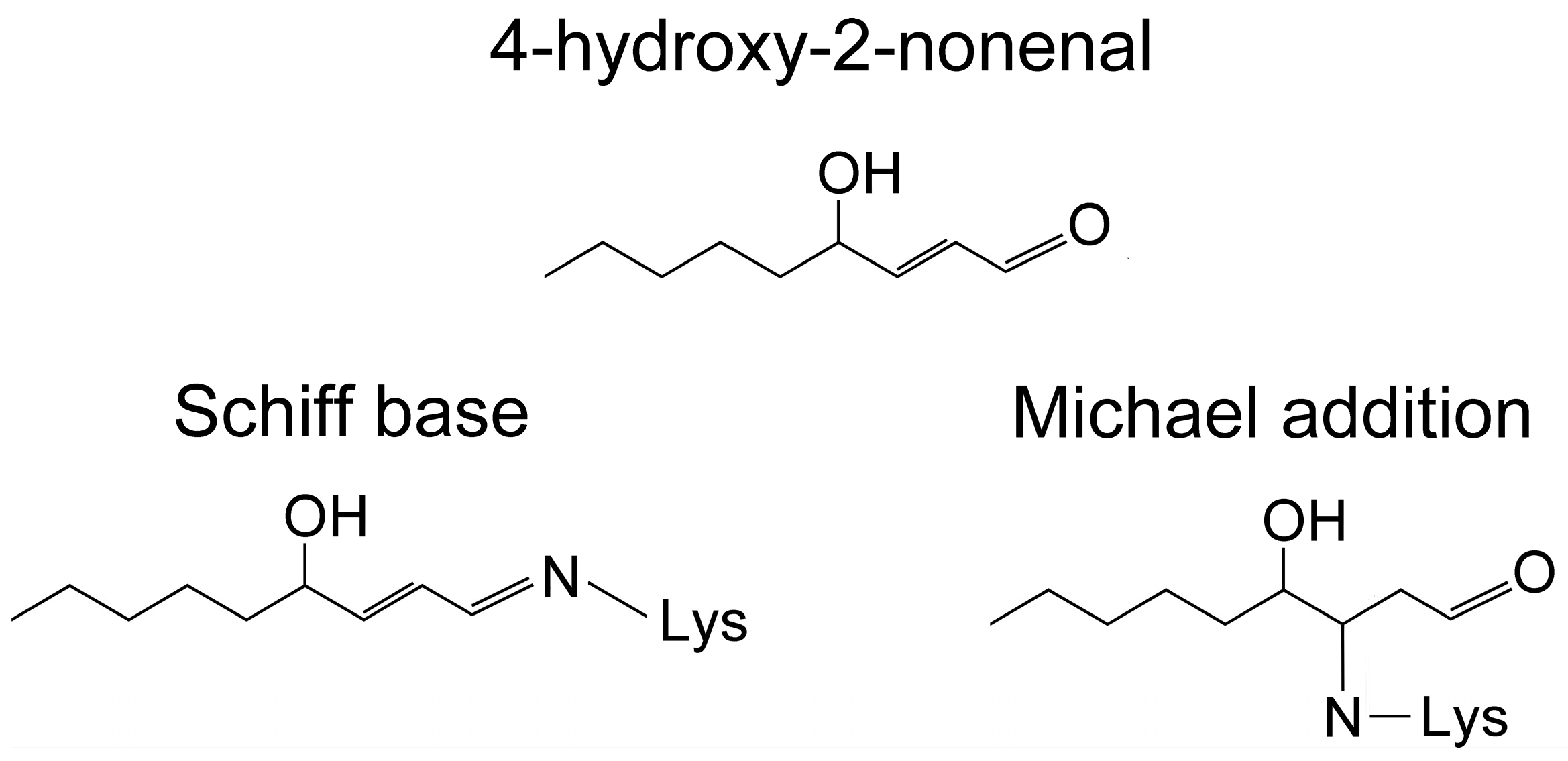
  76. Sayre, L.M.; Lin, D.; Yuan, Q.; Zhu, X.; Tang, X. Protein adducts generated from products of lipid oxidation: Focus on HNE and one. Drug Metab. Rev. 2006, 38, 651–675. [Google Scholar] [CrossRef] [PubMed]
  77. Rauniyar, N.; Prokai, L. Detection and identification of 4-hydroxy-2-nonenal Schiff-base adducts along with products of Michael addition using data-dependent neutral loss-driven MS3 acquisition: Method evaluation through an in vitro study on cytochrome c oxidase modifications. Proteomics 2009, 9, 5188–5193. [Google Scholar] [CrossRef] [PubMed] [Green Version]
  78. Vazdar, M.; Jurkiewicz, P.; Hof, M.; Jungwirth, P.; Cwiklik, L. Behavior of 4-hydroxynonenal in phospholipid membranes. J. Phys. Chem. B 2012, 116, 6411–6415. [Google Scholar] [CrossRef]
  79. Ou, X.; Lao, Y.; Xu, J.; Wutthinitikornkit, Y.; Shi, R.; Chen, X.; Li, J. ATP can efficiently stabilize protein through a unique mechanism. JACS Au 2021, 1, 1766–1777. [Google Scholar] [CrossRef]
  80. Galligan, J.J.; Rose, K.L.; Beavers, W.N.; Hill, S.; Tallman, K.A.; Tansey, W.P.; Marnett, L.J. Stable histone adduction by 4-oxo-2-nonenal: A potential link between oxidative stress and epigenetics. J. Am. Chem. Soc. 2014, 136, 11864–11866. [Google Scholar] [CrossRef] [Green Version]
  81. Galligan, J.J.; Marnett, L.J. Histone adduction and its functional impact on epigenetics. Chem. Res. Toxicol. 2017, 30, 376–387. [Google Scholar] [CrossRef] [Green Version]
  82. Schirmer, T.; Evans, P.R. Structural basis of the allosteric behaviour of phosphofructokinase. Nature 1990, 343, 140–145. [Google Scholar] [CrossRef]
  83. Robinson, J.B., Jr.; Inman, L.; Sumegi, B.; Srere, P.A. Further characterization of the Krebs tricarboxylic acid cycle metabolon. J. Biol. Chem. 1987, 262, 1786–1790. [Google Scholar] [CrossRef]
  84. Islam, M.M.; Nautiyal, M.; Wynn, R.M.; Mobley, J.A.; Chuang, D.T.; Hutson, S.M. Branched-chain amino acid metabolon: Interaction of glutamate dehydrogenase with the mitochondrial branched-chain aminotransferase (BCATm). J. Biol. Chem. 2010, 285, 265–276. [Google Scholar] [CrossRef] [Green Version]
  85. Chu, H.; Low, P.S. Mapping of glycolytic enzyme-binding sites on human erythrocyte band 3. Biochem. J. 2006, 400, 143–151. [Google Scholar] [CrossRef] [PubMed]
  86. Araiza-Olivera, D.; Chiquete-Felix, N.; Rosas-Lemus, M.; Sampedro, J.G.; Peña, A.; Mujica, A.; Uribe-Carvajal, S. A glycolytic metabolon in Saccharomyces cerevisiae is stabilized by F-actin. FEBS J. 2013, 280, 3887–3905. [Google Scholar] [CrossRef] [PubMed]
  87. Raikar, L.S.; Vallejo, J.; Lloyd, P.G.; Hardin, C.D. Overexpression of caveolin-1 results in increased plasma membrane targeting of glycolytic enzymes: The structural basis for a membrane associated metabolic compartment. J. Cell. Biochem. 2006, 98, 861–871. [Google Scholar] [CrossRef] [PubMed]
  88. Chu, H.; Puchulu-Campanella, E.; Galan, J.A.; Tao, W.A.; Low, P.S.; Hoffman, J.F. Identification of cytoskeletal elements enclosing the ATP pools that fuel human red blood cell membrane cation pumps. Proc. Natl. Acad. Sci. USA 2012, 109, 12794–12799. [Google Scholar] [CrossRef] [PubMed]
  89. Tanner, L.B.; Goglia, A.G.; Wei, M.H.; Sehgal, T.; Parsons, L.R.; Park, J.O.; White, E.; Toettcher, J.E.; Rabinowitz, J.D. Four key steps control glycolytic flux in mammalian cells. Cell Syst. 2018, 7, 49–62. [Google Scholar] [CrossRef]
  90. Kraft, A.M.; Petty, H.R. Spatial locations of certain enzymes and transporters within preinvasive ductal epithelial cells predict human breast cancer recurrences. Am. J. Physiol. Cell Physiol. 2020, 319, C910–C921. [Google Scholar] [CrossRef]
  91. Cheung, R.A.; Kraft, A.M.; Petty, H.R. Relocation of phosphofructokinases within epithelial cells is a novel event preceding breast cancer recurrence that accurately predicts patient outcomes. Am. J. Physiol. Cell Physiol. 2021, 321, C654–C670. [Google Scholar] [CrossRef]
  92. Petty, H.R. Using machine vision of glycolytic elements to predict breast cancer recurrences: Design and implementation. Metabolites 2023, 3, 41. [Google Scholar] [CrossRef]
  93. Saatchi, Y.; Schanen, P.; Cheung, R.A.; Petty, H.R. Computer vision identifies recurrent and non-recurrent ductal carcinoma in situ lesions with special emphasis on African American women. Am. J. Pathol. 2023, in press. [Google Scholar] [CrossRef]
  94. Tomaiuolo, M.; Bertram, R.; Houle, D. Enzyme isoforms may increase phenotypic robustness. Evolution 2008, 62, 2884–2893. [Google Scholar] [CrossRef] [Green Version]
  95. Kahraman, A.; Karakulak, T.; Szklarczyk, D.; von Mering, C. Pathogenic impact of transcript isoform switching in 1,209 cancer samples covering 27 cancer types using an isoform-specific interaction network. Sci. Rep. 2020, 10, 14453. [Google Scholar] [CrossRef] [PubMed]
  96. Lee, E.E.; Ma, J.; Sacharidou, A.; Mi, W.; Salato, V.K.; Nguyen, N.; Jiang, Y.; Pascual, J.M.; North, P.E.; Shaul, P.W.; et al. A protein kinase C phosphorylation motif in GLUT1 affects glucose transport and is mutated in GLUT1 deficiency syndrome. Mol. Cell 2015, 58, 845–853. [Google Scholar] [CrossRef] [PubMed] [Green Version]
  97. Kondrat, S.; Karuss, U.; von Lieres, E. Enzyme co-localization: Mechanisms and benefits. Curr. Res. Chem. Biol. 2022, 2, 100031. [Google Scholar] [CrossRef]
  98. Giunta, G.; Tostevin, F.; Tănase-Nicola, S.; Gerland, U. Optimal spatial allocation of enzymes as an investment problem. Commun. Phys. 2022, 5, 319. [Google Scholar] [CrossRef]
  99. Hinzpeter, F.; Tostevin, F.; Buchner, A.; Gerland, U. Trade-offs and design principles in the spatial organization of catalytic particles. Nat. Phys. 2022, 18, 203–211. [Google Scholar] [CrossRef]
  100. Fink, T.; Stevović, B.; Verwaal, R.; Roubos, J.A.; Gaber, R.; Benčina, M.; Jerala, R.; Gradišar, H. Metabolic enzyme clustering by coiled coils improves the biosynthesis of resveratrol and mevalonate. AMB Express 2020, 10, 97. [Google Scholar] [CrossRef]
  101. Wheeldon, I.; Minteer, S.D.; Banta, S.; Barton, S.C.; Atanassov, P.; Sigman, M. Substrate channelling as an approach to cascade reactions. Nat. Chem. 2016, 8, 299–309. [Google Scholar] [CrossRef]
  102. Schmidt, S.; Castiglione, K.; Kourist, R. Overcoming the incompatibility challenge in chemoenzymatic and multi-catalytic cascade reactions. Chemistry 2018, 24, 1755–1768. [Google Scholar] [CrossRef]
  103. Qiu, X.Y.; Xie, S.S.; Min, L.; Wu, X.M.; Zhu, L.Y.; Zhu, L. Spatial organization of enzymes to enhance synthetic pathways in microbial chassis: A systematic review. Microb. Cell Factories 2018, 17, 120. [Google Scholar] [CrossRef] [Green Version]
  104. Gopich, I.V. Cluster channeling in cascade reactions. J. Phys. Chem. B 2021, 125, 2061–2073. [Google Scholar] [CrossRef]
  105. Stępiński, D. The nucleolus, an ally, and an enemy of cancer cells. Histochem. Cell Biol. 2018, 150, 607–629. [Google Scholar] [CrossRef] [PubMed]
  106. Helpap, B. Nucleolar grading of breast cancer. Comparative studies on frequency and localization of nucleoli and histology, stage, hormonal receptor status and lectin histochemistry. Virchows Arch. A Pathol. Anat. Histopathol. 1989, 415, 501–508. [Google Scholar] [CrossRef] [PubMed]
  107. Carotenuto, P.; Pecoraro, A.; Palma, G.; Russo, G.; Russo, A. Therapeutic approaches targeting nucleolus in cancer. Cells 2019, 8, 1090. [Google Scholar] [CrossRef] [PubMed] [Green Version]
  108. Spannl, S.; Tereshchenko, M.; Mastromarco, G.J.; Ihn, S.J.; Lee, H.O. Biomolecular condensates in neurodegeneration and cancer. Traffic 2019, 20, 890–911. [Google Scholar] [CrossRef]
  109. Glass-Marmor, L.; Beitner, R. Taxol (paclitaxel) induces a detachment of phosphofructokinase from cytoskeleton of melanoma cells and decreases the levels of glucose 1,6-bisphosphate, fructose 1,6-bisphosphate and ATP. Eur. J. Pharmacol. 1999, 370, 195–199. [Google Scholar] [CrossRef]
  110. Dhyani, P.; Quispe, C.; Sharma, E.; Bahukhandi, A.; Sati, P.; Attri, D.C.; Szopa, A.; Sharifi-Rad, J.; Docea, O.; Mardare, I.; et al. Anticancer potential of alkaloids: A key emphasis to colchicine, vinblastine, vincristine, vindesine, vinorelbine and vincamine. Cancer Cell Int. 2022, 22, 206. [Google Scholar] [CrossRef]
  111. Schwartz, D.; Beitner, R. Detachment of the glycolytic enzymes, phosphofructokinase, and aldolase, from cytoskeleton of melanoma cells, induced by local anesthetics. Mol. Genet. Metab. 2000, 69, 159–164. [Google Scholar] [CrossRef]
  112. Grandhi, R.K.; Perona, B. Mechanisms of action by which local anesthetics reduce cancer recurrence, a systematic review. Pain Med. 2020, 21, 401–414. [Google Scholar] [CrossRef]
  113. Tejeda-Muñoz, N.; Mei, K.C.; Sheladiya, P.; Monka, J. Targeting membrane trafficking as a strategy for cancer treatment. Vaccines 2022, 10, 790. [Google Scholar] [CrossRef]
  114. Allal, C.; Favre, G.; Couderc, B.; Salicio, S.; Sixou, S.; Hamilton, A.D.; Sebti, S.M.; Lajoie-Mazenc, I.; Pradines, A. RhoA prenylation is required for promotion of cell growth and transformation and cytoskeleton organization but not for induction of serum response element transcription. J. Biol. Chem. 2000, 275, 31001–31008. [Google Scholar] [CrossRef] [Green Version]
  115. Wang, M.; Casey, P.J. Protein prenylation: Unique fats make their mark on biology. Nat. Rev. Mol. Cell Biol. 2016, 17, 110–122. [Google Scholar] [CrossRef] [PubMed]
  116. Harris, B.R.E.; Zhang, Y.; Tao, J.; Shen, R.; Zhao, X.; Cleary, M.P.; Wang, T.; Yang, D.Q. ATM inhibitor KU-55933 induces apoptosis and inhibits motility by blocking GLUT1-mediated glucose uptake in aggressive cancer cells with sustained activation of Akt. FASEB J. 2021, 35, e21264. [Google Scholar] [CrossRef] [PubMed]
  117. Csermely, P.; Agoston, V.; Pongor, S. The efficiency of multi-target drugs: The network approach might help drug design. Trends Pharmacol. Sci. 2005, 26, 178–182. [Google Scholar] [CrossRef] [PubMed] [Green Version]
  118. Chen, G.; Kong, J.; Tucker-Burden, C.; Anand, M.; Rong, Y.; Rahman, F.; Moreno, C.S.; Van Meir, E.G.; Hadjipanayis, C.G.; Brat, D.J. Human Brat ortholog TRIM3 is a tumor suppressor that regulates asymmetric cell division in glioblastoma. Cancer Res. 2014, 74, 4536–4548. [Google Scholar] [CrossRef] [PubMed] [Green Version]
  119. Tiku, V.; Jain, C.; Raz, Y.; Nakamura, S.; Heestand, B.; Liu, W.; Späth, M.; Suchiman, H.E.D.; Müller, R.U.; Slagboom, P.E.; et al. Small nucleoli are a cellular hallmark of longevity. Nat. Commun. 2017, 8, 16083. [Google Scholar] [CrossRef] [Green Version]
Figure 1. Structures of 4-hydroxy 2-nonenal (4-HNE) and its reaction products with lysine (Lys) residues of proteins. 4-HNE reacts with Lys via Schiff base formation and Michael addition. Both products may rearrange to stabilize the linkage.
Appliedchem 03 00024 g001
Figure 2. Representative fluorescence micrographs of histone H2A.X (A,B) and 4-HNE-labeled human DCIS tissue sections. Samples from patients who did not experience a cancer recurrence (A,C) and who did experience a cancer recurrence (B,D) are shown. For each patient, (A,C) and (B,D), both biomarkers were found in the nuclei of ductal epithelial cells. This indicates that 4-HNE and H2A.X are present in the nuclei of both recurrent and non-recurrent DCIS tissues. Thus, co-incident labeling of both histones and 4-HNE could be an early step in the formation of indolent and aggressive DCIS lesions. (Bar = 200 μm).
Appliedchem 03 00024 g002
Figure 3. Potential contributions of 4-HNE to histone and DNA damage. This figure focuses upon 4-HNE damage to histones, although 4-HNE also directly reacts with DNA. Panels (A,B) illustrate an undamaged cell and a cell damaged by exposure to 4-HNE, respectively. Both undamaged and damaged cells have enzymes (aldehyde dehydrogenase, ALDH2, and aldose reductases) that limit the cytoplasmic damage due to aldehydes. In damaged cells, 4-HNE is present, primarily in lipid membranes. However, 4-HNE is much more soluble in nucleoli because high concentrations of ATP in nuclei act as a hydrotrope to solubilize 4-HNE, and the dielectric constant of nucleoli is 45, about the same as DMSO in which 4-HNE is highly soluble (50 mg/mL). In undamaged cells, positively charged amino acids electrostatically interact with DNA, as illustrated by panel (C). However, in damaged cells, the positive charged histone binding sites are blocked due to the formation of stable 4-HNE adducts (panel (D)). In undamaged cells, DNA wraps around histones (E). DNA storage is less effective in damaged cells, leading to DNA strands that are much more susceptible to further damage, including that of additional 4-HNE molecules (F).
Appliedchem 03 00024 g003
Figure 4. Spatial properties of glycolytic components in human DCIS lesions. Immunofluorescence micrographs of DCIS samples from women who did not exhibit cancer recurrences (A,C) and women who did report a cancer recurrence (B,D). Sections of tissue were labeled with antibodies directed against phospho-Ser226-GLUT1 (A,B) and anti-PFKL (C,D). Samples were labeled with secondary antibodies to visualize these spatial patterns. For patients who did not experience a recurrence, phospho-Ser226-GLUT1 and PFKL were found near the cell center, which appears to be the region of the nucleus and nucleolus. These spatial patterns of glycolytic elements changed for women who reported a recurrence. For these women, phospho-Ser226-GLUT1 and PFKL could be found near the cell periphery (B,D). Presumably, the phospho-Ser226-GLUT1 and PFKL accumulated at the periphery to mediate the internalization and metabolism of glucose, respectively. Many micrographs like these were used to train computers to recognize recurrent and non-recurrent lesions. These results underscore that the location, not simply the amount of biomarker, is important in assessing the recurrent or non-recurrent status of a patient. (Bar = 200 μm).
Appliedchem 03 00024 g004
Figure 5. An illustration of peripheral enzyme agglomerations or metabolic platforms in recurrent breast cancer cells. On the left side is shown a biological plasma membrane in black and white. Mechanisms of membrane association are illustrated along the left-hand side including, from top to bottom: prenylation, membrane metabolons, and caveolae, including caveolin-1-associated enzymes. To the center-right is shown a component of the cytoskeleton, such as a microtubule with bound PFKL. A broad variety of enzymes are illustrated in this figure by the broad variety of colors. This corresponds to the metabolic platforms shown in Figure 4D. This includes enzymes of glycolysis, the hexose monophosphate shunt, and the glutathione biosynthesis pathway in various colors. For clarity, this figure underestimates the intracellular crowding of the cytoplasm.
Appliedchem 03 00024 g005
Table 1. Physical and Chemical Influences on Enzyme and Pathway Performance in Cancer 1.
Influence TypeSelected Types and Examples 2
Local EnvironmentpHPFK
Redox ConditionOxidative, Reductive
Ionic ConditionPotassium, Calcium
Dielectric ConstantActive sites, LLPS
ElectrostaticNucleosomes and other binding events
ChemicalConcentrationActivity coefficient, effective concentration
Isoform switchingPFK
PhosphorylationSignaling, enzyme, and transport molecules
SolubilityProteins, metabolites
Low affinity binding sitesAsp-Asp-Thy-Glu-Asp sequences
High affinity binding sitesLigands, cofactors, ATP, GTP, Allosteric factors, ubiquitination, prenylation, etc.
Emergent BehaviorsHydrotropesATP, GTP, and other amphipathic molecules
Phase separationsTwo and three dimensional
FeedbackPositive and negative
TranslocationMicrofilaments and microtubules
Assembly stateMonomer, oligomer, mixed oligomers, clusters, metabolons, metabolic platforms
1 Chemical performance and clinical outcomes depend upon complex combinations of these several factors. 2 These are representative, not exhaustive.
Disclaimer/Publisher’s Note: The statements, opinions and data contained in all publications are solely those of the individual author(s) and contributor(s) and not of MDPI and/or the editor(s). MDPI and/or the editor(s) disclaim responsibility for any injury to people or property resulting from any ideas, methods, instructions or products referred to in the content.

