1. Introduction
Wound care is a critical, yet often scientifically overlooked aspect of recreational aquaculture, particularly in sport angling and ornamental Koi rearing, where topical treatments have been left to evolve over decades with little to no regulation, largely taking inspiration from the salmonid industry, but also essentially through hearsay amongst hobbyists. Whilst the salmonid sector is improving its environmental credentials, the recreational fish care market has not kept pace. Yet, effective wound care and infection management is essential in sport and pet fish due to the scope of pathogenic threats and environmental stressors associated with fish cultivation.
Angling is the largest participation sport in the UK, with around four million anglers, and is worth £1.7 billion to the UK economy, with carp fishing representing the highest grossing angling sector [
1]. Another estimated four million people keep fish as pets in the UK, where the ornamental trades economic worth is approximately £1 billion, with £666 million in UK sales during 2022–2023 [
2]. European markets are approximately double this size in both disciplines and USA and Asian markets are expanding exponentially. Indeed, China produces 2.9 million tons of Common Carp for household consumption annually [
3], demonstrating the potential for transferable technologies in international carp farming markets. On the ornamental side, a Kohaku Koi, named ‘S-Legend’, sold for
$1.8 million USD in 2018, only to die months later from poor health, confirming the immediate need for innovation in carp health biotech solutions. In terms of economic contribution, the UK cyprinid market is larger than the UK salmon market, illustrating the significant market need for fish care products, such as those addressing topical wound care. However, currently no published data exists to quantify the efficacy or safety of the products currently mass-marketed for wound treatment in cyprinids for recreational markets like angling and Koi rearing. The majority of commercial carp-care products carry no ingredient or hazard information on their packaging. Aside from the Veterinary Medicine Department’s ‘small animal exemption license’, which does not apply to wild fish, there is no monitoring or regulation of the growing use of historically toxic compounds with poor safety and environmental profiles in recreational aquaculture, or their disposal/discharge into natural aquatic environments and waterways. Many products have been adopted and marketed by hobbyists, pet owners and fishery managers with no empirical evidence to support common claims of ‘cleaning’, ‘protecting’ and ‘healing’ wounds. This has led to an ‘off-label’ usage culture, and a lack of rational design in the industry. Here we present the first quantitative study on the efficacy of the market leaders sold and used in recreational angling and Koi keeping applications.
Catching, handling, transportation, poor environmental management and predation are some of the risk factors for valuable stock suffering superficial—as well as more chronic—surface injuries/sores. These wounds then create entry points for pathogen colonization, leading to infections that can severely impact fish growth and survival [
4,
5] as well as appearance and thus commercial value. Consequently, the application of effective, safe and environmentally sound fish healthcare products is fundamental to reducing mortality rates, improving overall fish welfare, and maintaining sustainable aquaculture practices.
Aquatic environments present unique challenges for both wound healing and disease management; once broken, the skin is exposed to waterborne pathogens including bacteria, fungi and parasites. Diseases such as fin rot, tail rot, and ulcerative infections are commonly caused by opportunistic bacterial species such as
Aeromonas spp.,
Pseudomonas spp., and
Vibrio spp. [
6,
7,
8]. Once an infection has colonized a wound site, subsequent recovery is dependent upon environmental stressors, host immune response capacity, and the efficacy of externally applied therapeutic interventions [
9].If left untreated, infections can spread rapidly through direct contact with other fish or contaminated vegetation/substratum as well as via direct routes through water, often resulting in mass mortality events, significant economic losses and damage to reputation in commercial fisheries, with associated ecological imbalances that can spread to adjoining natural water courses.
Despite a growing acknowledgement of the importance of fish healthcare, current therapeutic interventions are largely based on historically used chemical compounds which often introduce secondary health risks and undocumented environmental damage [
10]. Many chemical-based treatments include formalin, a solution of formaldehyde in water shown to be highly toxic to aquatic life at high concentrations [
11]. Malachite green is widely used in ornamental fish keeping and recreational angling for control of fungal pathogens and wound infections, despite being banned for use in food chain applications in many countries due to its carcinogenic and mutagenic effects [
12]. When these substances are applied in aquaculture settings, they also do not remain confined to the treated fish, but disperse into the surrounding water, exposing the user to their toxicity. The accumulation of chemical residues in water bodies can have long-term environmental consequences, affecting non-target organisms, disrupting aquatic food chains, contaminating water sources and also endangering the hobbyist or fishery worker who comes into contact with such chemicals on a regular basis [
13].
Antibiotics including oxytetracycline, florfenicol, and sulphonamides have been used in excess to control aquatic pathogens, contributing to the emergence of antimicrobial resistance (AMR). Antibiotic residues that enter the environment through water discharge can also promote the development of antibiotic-resistant bacteria, which pose a significant threat to both aquatic and human health [
14]. These resistant bacteria can spread through the water column, affecting wild fish populations and eventually entering the human food chain [
15]. However, antibiotics are prescription only medicines (POMs) and are not readily available to hobbyists and recreational aqua culturists. Combined with the Fish Vet Society listing just twenty-five veterinarians willing to treat fish in the UK, the adoption of off-label and unproven remedies or toxic legacy compounds is simply a reaction to market need to protect fishery stocks. However, it is essential that the scientific community should monitor, test and update market direction regularly to ascertain ‘best practice’ standards for the development of new products in this area, as well as to develop novel model test-bed systems for establishing efficacy benchmarks against which new products can be evaluated.
Limited testing of commercially available topical fish healthcare products restricts consumer understanding of their true efficacy and environmental implications, and combined with little/no information on formulation being supplied at point of sale, fish farmers and hobbyists are kept blind to both the potential dangers of the products in current usage and also as to whether the products they rely on actually have any scientifically proven basis for use in their desired application. Therefore, in this study we have quantified the antimicrobial, antifungal, cellular toxicity and wet-adhesion properties of several market-leading topical carp-care products using the well-established cyprinid fibroblast cell line, ZF4, to study toxicity responses, combined with a pseudo-ex vivo wet-adhesion carp skin assay, to investigate wet-adhesion properties in commercial wound sealant formulations.
2. Materials and Methods
2.1. Compounds Tested
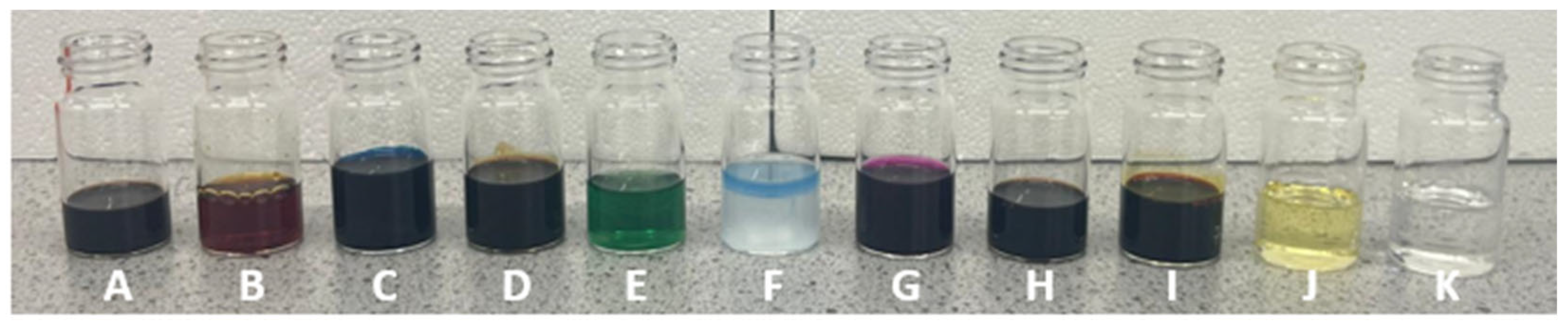
A panel of market leading products for topical carp care in recreational angling and Koi care was identified for testing using the search engine (Google) terms: ‘Carp care products’ and ‘topical carp spray’ (accessed June 2024). The Institute of Fisheries Management provided advisory guidance on the most prevalent formulation compositions so as to ensure at least one product was included from all known major ingredient categories, summarized in
Figure 1 and
Table 1, below. Propolis is a widely accepted standout market leader for topical wound-care applications in recreational aquaculture markets, with many different product options from different manufacturers available. For this reason, we included four separate manufacturer’s propolis-based products for comparison. Interestingly, significant variability between propolis product coloration and viscosity was observed (
Figure 1A,B,D,H), likely indicating different concentrations or sources of propolis. Initially, resources to test ten different formulations were established; this was increased to eleven to include an additional essential oil-derived formulationfrom our list of top hits identified during preliminary compound screening (
Figure 1J,K). We have anonymized all product identities used in this study to protect commercial reputations and improve industry understanding, open dialogue and raise standards of fish care through rational design, rather than expose poor practice.
2.2. Bacterial MIC Testing
The pathogen panels assembled for testing here were devised with expert oversight from the Institute of Fisheries Management (IFM) and Lincs Fish Health (LFH), constituting the main bacterial and fungal pathogens of carp in the UK. Aeromonas hydrophila (ATCC, [CDC 359-60, IAM 12460, NCIB 9240, NCMB 86, NCTC 8049, RH 250]), Aeromonas salmonicida (ATCC, NCMB 1102 [19291, 20]) and Pseudomonas fluorescens (ATCC, NCTC 10,038 [28/5, CCEB 546, DSM 50090, NCIB 9046, NCPPB 1964, PJ239, R. Hugh 818, R.Y. Stanier 192, Biotype A]) strains were purchased from the ATCC, and streaked onto Mueller Hinton agar plates (Fisher Scientific, Loughborough LE11 5RG, UK) for viable colony selection. Colonies were picked using a sterile cell scraper (Fisher Scientific, Loughborough LE11 5RG, UK) and inoculated into Mueller Hinton broth (Fisher Scientific, Loughborugh LE11 5RG, UK). These samples were then incubated for 24 h at 37 °C until bacterial growth was established. The optical density of the sample was measured at 0.8 OD, and the inoculum diluted to 0.5 McFarland standard.
Inoculated samples were diluted to achieve 5 × 10⁵ CFU/mL when inoculated into Mueller Hinton Broth containing serially diluted compounds of interest at the relevant concentrations across 96-well round-bottom plates (Fisher Scientific, Loughborough LE11 5RG, UK). Serially diluted PBS served as the negative control and Penicillin/Streptomycin mix (Fisher Scientific, Loughborugh LE11 5RG, UK; 20 U/mL final) as the positive control at a concentration of 5 × 10⁵ CFU/mL. Inoculated samples (200 µL total volume) were incubated at 37 °C without shaking for 24 h. MICs were determined here under standard laboratory conditions as above; pathogen susceptibility times may differ under environmentally relevant temperatures encountered in seasonal carp aquaculture where temperature fluctuations exist. MICs were recorded visually by eye, with correlative quantification using Presto Blue (PB)—20 μL PB (Fisher Scientific, UK) was added to each well and the plate was re-incubated at 37 °C for 10 min. Fluorescence was recorded at 550/590 excitation/emission wavelengths on a BMG Labtech FLUOstar Optima 96-well plate reader (BMG Labtech, Aylesbury, UK) and the data was collected and analyzed using BMG Optima, Excel and GraphPad Prism (V10) software. MICs were only recorded where fold-changes relative to PBS controls were recorded below 0.5.
It should be noted here that more representative growth conditions could be obtained through conducting MIC assays at temperatures more equivalent to UK average water temperatures; however, growth rates would be so much reduced so as to make these assays impractical, and the small potential differences that might be incurred were deemed inconsequential here.
2.3. Fungal MIC Testing
Saprolegnia parasitica (ATCC, TP 41) bead stocks were purchased from the ATCC, and inoculated into Potato Dextrose Broth (Fisher Scientific, Loughborough LE11 5RG, UK). These samples were then incubated at room temperature (RT, 25 °C) without shaking for 48 h, until fungal growth was established via visual assessment. Optical densities of samples were measured, and the inoculum was diluted to a McFarland standard of 0.5. Using a sterile L-shaped cell spreader (Fisher Scientific, Loughborough LE11 5RG, UK), 200 µL inoculum was spread evenly across Potato Dextrose agar plates and dried at RT for 30 min. Compounds of interest at the relevant concentrations were serially diluted across 24-well, flat-bottom plates (Corning, Fisher Scientific, Loughborough LE11 5RG, UK.) Sterile Potato Dextrose Agar plugs were used as a negative control and untreated mycelium plugs served as a positive control. Mycelium plugs were taken using a flame sterilized 5 mm metal cork borer. One mycelium plug was placed into each well of the plate and incubated at RT without shaking for 48 h. Plates were imaged from above using a handheld mobile device, and images imported into ImageJ (Fiji, x86 64 UK) for analysis. Fungal growth was quantified using the ‘Freehand Tool’, selecting ‘area’ in the ‘analyse—measurements’ tab. Growth areas were exported to Excel, where fold-changes relative to positive controls (unchecked fungal growth) were calculated. The data was then imported to GraphPad Prism (v10.6) for graphical representation and statistical analysis using Two-Way ANOVA with post hoc Tukey test as recommended in the software.
2.4. In Vitro ZF4 Cyprinid Fibroblast Cell Toxicity Testing
Cyprinid fibroblast cells, ZF4 were obtained from the American Tissue Culture Collection (ATCC) and cultured in a humidified 28 °C environment with 5% CO2 in T150 flat bottom vented flasks (Corning, Fisher Scientific, Loughborough LE11 5RG, UK). Cells were passaged at 50% confluency using TrypLE Express (Fisher Scientific, Loughborough LE11 5RG, UK) and plated in 96-well plates (Corning, Fisher Scientific, Loughborough LE11 5RG, UK) for experiments. Growth medium consisted of 10% v/v heat inactivated Fetal Bovine Serum (FBS, Fisher Scientific, Loughborough LE11 5RG, UK) with 200 mM L-Glutamine (L-Glut, Fisher Scientific, Loughborough LE11 5RG, UK). Treatment Media (TM), used for toxicity assays consisted of 2% FBS with 200 mM L-Glut. Cells were plated at 60% confluency in 96-well plates using a multichannel pipette and cultured in 100 μL growth media for 48 h to achieve 100% confluency. Cells were then washed once with 100 μL 1× Dulbecco’s Phosphate-Buffered Saline (DPBS, Fisher Scientific, Loughborough LE11 5RG, UK) and placed in 100 μL TM for 24 h prior to treatment with the applied compounds. Serial dilutions of all compounds tested were added at 10 mg/mL (or 10% v/v if not water soluble) for a period of 24 h, with DPBS serial dilution serving as negative control. Following treatment, 10 μL Presto Blue was added to each well using a multichannel pipette, and the plate was left to incubate at 28 °C for 10 min prior to reading excitation/emission wavelengths at 550/590 nm on a 96-well plate reader (FLUOstar Optima, BMG Labtech, Ayelsbury HP19 8JR, UK). Results were exported to Microsoft Excel and subsequently to GraphPad Prism (V10) for analysis.
2.5. Apoptosis/ROS Induction Assays Using ZF4 In Vitro
ZF4 cells were cultured as per toxicity testing, above. Cells were plated at 70% confluency in 8-well chamber slides (Ibidi, 82166 Gräfelfing, Germany) in growth media consisting of 10% v/v heat inactivated Fetal Bovine Serum (FBS, Fisher Scientific, Loughborough LE11 5RG, UK) with 200 mM L-Glutamine (L-Glut, Fisher Scientific, Loughborough LE11 5RG, UK). After 24 h cells formed monolayers and media was changed to Treatment Media (TM), consisting of 2% FBS with 200 mM L-Glut for another 24 h, prior to treatments being applied for a further 6 h. CellEvent Caspase 3/7 (Invitrogen, Paisley PA4 9RF, UK) and ROS Brite 670 (Santa Cruz, Bristol BS32 4UA, UK) were applied for 30 min according to manufacturer’s instructions, nuclei were counterstained using Hoechst (1 µg/mL), and live cells were imaged in chamber slides using a Zeiss LSM 910 inverted confocal microscope using Zen 3.1 software (Zeiss) under 5% humidified CO2 atmosphere at 28 °C as per ZF4 culture conditions. Images were analyzed using Fiji (ImageJ), selecting ‘Default’ colour thresholding setting, followed by particle analysis of stained area size or nuclear count per image to give staining area per nucleus. Experiments were performed in triplicate, using separate chamber slides.
2.6. Wet-Adhesion Testing
Empty glass slides were labelled and weighed. 100 µL of chilled distilled water (10 °C) was pipetted onto the bottom third of the slide (3 cm) and the weight was recorded. 0.06 g of the sample was weighed directly onto the slide and distributed evenly across the lower 3 cm area. Wet-adhesion chambers (made from empty pipette boxes) were filled with 50 mL of the chilled distilled water (10 °C). Slides were then submerged from 0 to 48 h with beakers kept in a temperature-controlled incubator. At relevant timepoints, slides were removed and drained dry on blue roll until excess water had drained. Slide weight was recorded at each timepoint to confirm sample retention to a minimum of 0.01 g of sample remaining, or no remnants could not be seen under a light microscope at 200× magnification.
Pseudo-ex vivo assays were performed using juvenile (3–4-year-old) common carp skin, obtained as freshly as possible from a local supplier (24–48 h previously). Briefly, carp were stored whole at 4 °C until collected, professionally filleted, and the skin removed with the scales intact. A sharpened 20 mm leather punch was used to create uniform skin discs from the mid-flank of the fish, which were adhered scale-side up, to glass slides using 30 µL waterproof superglue (Gorilla Glue). Slides were weighed +/− glue and product at the start of the assay, and assays were carried out as per the above method for uncoated slides, incubating +/− 0.06 g commercial products for the indicated time points and recording weights at each stage.
2.7. pH Testing
All measurements were taken using a handheld digital pH meter (Hanna Instruments HI-9810432 HALO2 Wireless). Prior to testing, three-point calibration with pH 4.00, 7.00 and 10.00 buffer solutions were performed according to manufacturer’s instructions. In between measurements, the electrode was rinsed with deionized water and gently blotted dry. Measurements were repeated three times to confirm experimental accuracy; no deviation was observed.
2.8. Statistical Analysis
All data was exported to Microsoft Excel and GraphPad Prism (V10) for analysis, graphical layout and statistical analysis. One way Analysis of Variance (ANOVA) was performed to determine significance between data sets, using post hoc Tukey’s test with p < 0.05 used to indicate significance. For replicates, n = 3–5 was employed throughout experimental replicates.
4. Discussion
The introduction and licensing of novel therapeutics in the aquaculture sector is rare in comparison with other veterinary disciplines. Coupled to the lack of veterinary professionals offering specialist diagnostic and treatment options for fish in the UK, the current medicinal landscape for recreational aquaculture has evolved through attempts to emulate pet fish markets, which itself seems to have evolved through emulating the salmon industry. What is missing in the recreational context is regulation. Hence compounds such as Malachite green can be purchased from pet shops under Schedule 6 Small Animal Exemption and subsequently used off-label in open-water systems, with limited regulatory oversight owing to the broader resource pressures faced by regulatory agencies. The pet fish and recreational angling markets in the UK deliver more to the UK economy in financial terms than the UK salmon industry, yet zero evidence exists in the literature assessing the efficacy, tolerability, toxicity or environmental impact of recreational fish health products, which have been in widespread usage since the late ‘80s/early ‘90s. There are two–four million anglers in the UK (approx. 3–6% of the population), and twice that number keep fish as pets in tanks or garden ponds, representing a significant and important area of the UK economy and of public interest. Those figures increase by a factor of ten moving into Europe, where our neighbours share similar passions for angling and aquaculture, and many cold-water fish species besides salmonids are prized and seasonal delicacies. Carp in particular are widely eaten, caught and kept across the UK and Europe, and have exploded in popularity in the past three decades due to their longevity (50–60 years), hard fighting qualities, robust and resilient nature, their keen and adaptable social intelligence and their unique and distinctive external character and beauty—qualities appreciated and highly valued in angling, fish keeping and fish farming, with large specimens fetching upwards of £100/Lb, and more in the Koi industry. Despite their enormous popularity and financial value, the recreational ‘fish care’ sector surprisingly offers no scientific evidence, or obvious rational design at all, and off-label usage inspired by other markets is widespread.
Licensed medications have been developed for specific parasitic infections in pet aquarium fish, with good evidence to support efficacy, such as Fluke-Solve® or Lice-Solve®, but for general antibiotics, antimycotics, and wound sealants, the majority of products on the market lack any real threshold standards for evaluation or indeed any empirical testing throughout all strands of recreational aquaculture. This is perhaps understandable, as antibiotic treatments for recreational fish species such as carp are very difficult to acquire due to the extremely low number of practicing fish vets in the UK, leaving stock owners forced into off-label choices. Compounds such as Malachite Green, Potassium Permanganate, Betadine and Formalin, amongst others, are now in wide-spread usage for treating topical wounds in carp. But importantly, no compound has to date been approved for use in wild fish, i.e., beyond that stipulated by the VMD Schedule 6 Small Animal Exemption that only covers pet fish in small-scale closed environments (tanks and small garden ponds).
Our aim here was to establish the first empirical data set for commercially available topical ‘carp care’ products being marketed in the UK for angling and Koi keeping, both to provide efficacy testing data for those products and to produce an experimental testing standard that could be applied to future therapeutics development and testing for carp-based aquaculture. We hope this will initiate the refinement and replacement of historically used and environmentally damaging compounds such as Malachite green, which we have actually shown here for the first time are actually counter-productive to wound health in their application to cyprinids through damage to the fish’s own cells and skin, potentially delaying wound healing, and most certainly causing wider environmental/ecological damage at the concentrations currently marketed.
Current topical wound sealants for carp are entirely based on the use of biocides, under the assumption that preventing infection is the best route to wound healing. The idea that biocides can actually be detrimental to wound healing has completely been missed, and somewhat alarmingly, many products tested here had little to no activity against carp pathogens, whilst displaying high levels of toxicity to cyprinid skin cells. Wound healing is a balance—the ideal commercial formulation would have capacity to be both anti-bacterial/antifungal and pro-regenerative to the wound bed. We have shown here that non-toxic, environmentally friendly compounds with such properties do exist, but more research is required to extrapolate these findings into anything commercially viable. We have focused here on assessing the efficacy of such products, rather than their human safety profiles, as the move to natural products that we suggest would negate the need for such concern in this area if MHRA/EMA/FDA-approved materials could be shown to be effective against cyprinid pathogens.
This study has assessed the antimicrobial and antifungal efficacy, alongside the cytotoxicity, of a panel of commercially available topical wound treatments, using two known natural products for comparison. Opportunistic aquatic bacteria A. salmonicida, A. hydrophila and P. fluorescens, alongside pathogenic oomycote S. parasitica, were used to assess antimicrobial activity, cellular toxicity and wet adhesion. Significant differences in efficacies between the test compounds were revealed, indicating a need for standardization testing within the aquatic healthcare industry and more scientifically based rationale for development of new fish health products in recreational areas of aquaculture.
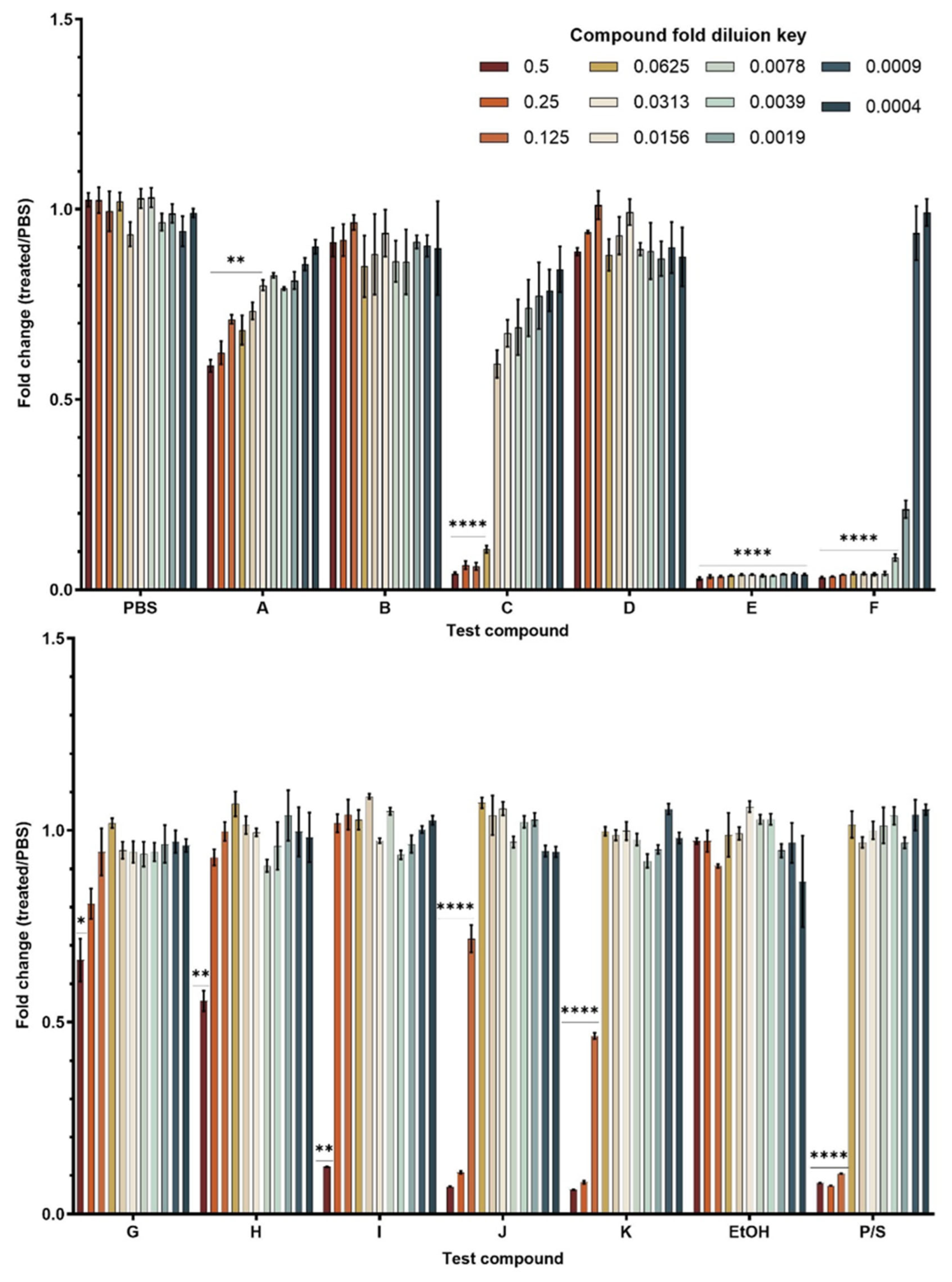
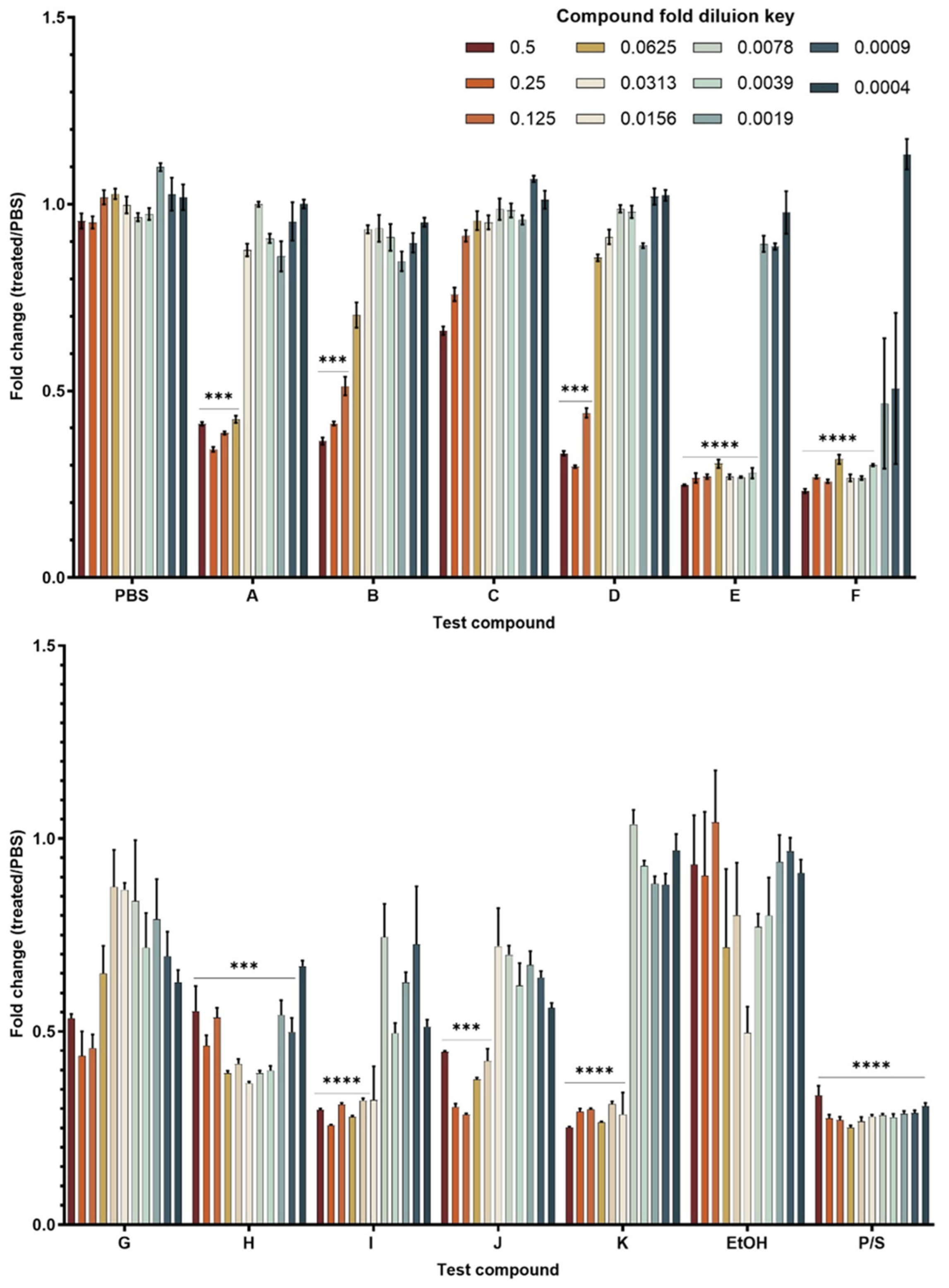
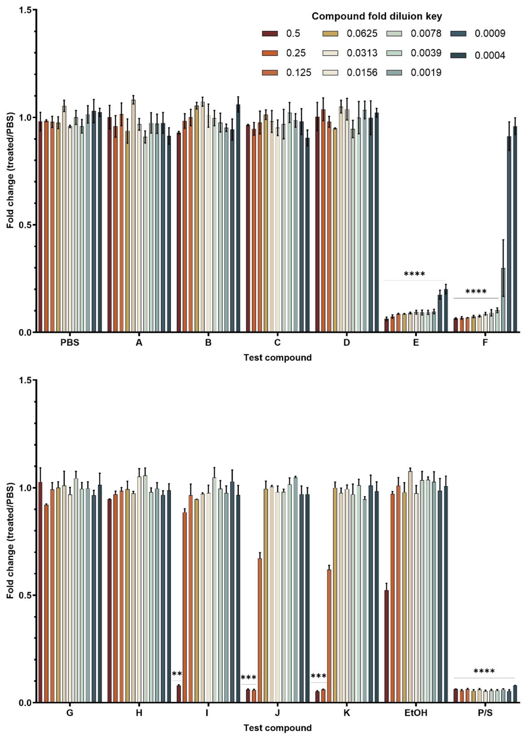
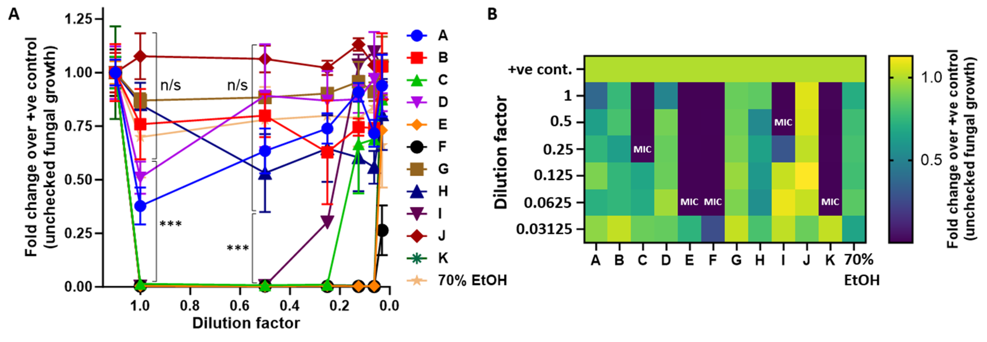
Table one provides a visual summary of the products tested for antimicrobial, antifungal, cyprinid toxicity and pH, which is an often-overlooked characteristic of recreational veterinary medical preparations, particularly in aquaculture. Remarkably, none of the products tested actually met the criteria required for a topical carp-care product as defined here. Products A, B, D G and H failed to reduce bacterial or fungal viability, suggesting a misalignment between commercial availability and clinical utilization. Although compounds E and F were effective against both the bacteria and fungi tested here, the concentration of active ingredient within these preparations retained activity at 1:5000 (a 12-fold dilution) for bacteria and 1:2500 for fungi. In fact, some bacterial MICs remained not determined in our assays. This is astounding given that one compound was malachite-green-based, a compound with significant ecological and environmental connotations, and one which is readily available and widely used in angling and fish keeping. It would be likely that other, non-pathogenic microorganisms and even invertebrate species might be affected by such inordinate overdosing. Taken together with the ease with which litres of Malachite green can be obtained over the counter, the scale of the underlying problem becomes a concern. Compounds E and F also displayed pH values of 4.7 and 4.2, respectively indicating markedly acidic formulations. Further investigation into products E and F using a cellular viability assay showed 100% ZF4 cell killing, highlighting the concentrations required to achieve their bactericidal effect would result in non-specific cytotoxic effects, most certainly damaging to the fish.
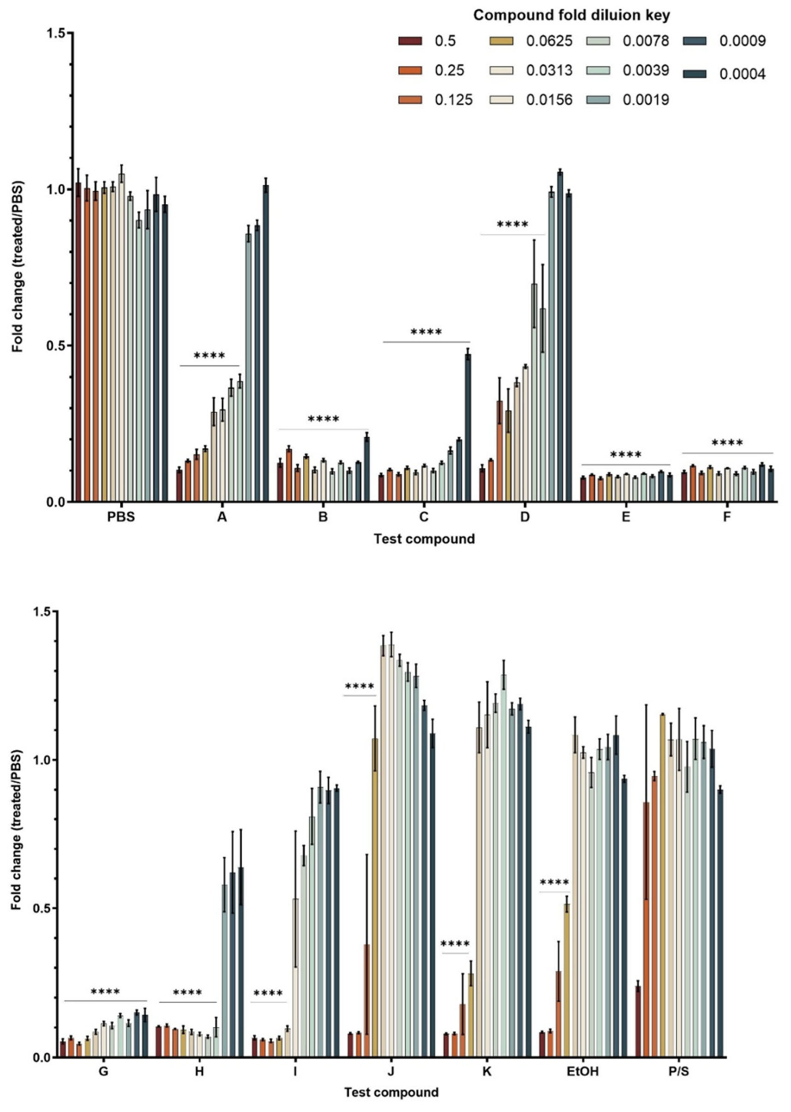
The consistent inhibitory effect across the MIC testing of compound I, a povidone-iodine formulation was as expected, performing well across all pathogen tests. However, compound I also displayed significant cellular toxicity in ZF4, more so than the natural compounds identified (H and J) and did not display the natural compound response of increased ZF4 fibroblast proliferation at lower concentrations (
Figure 4). Histopathological studies in common carp indicate that waterborne povidone-iodine concentrations as low as 5 ppm resulted in destruction of the primary and secondary lamellae, alongside lesions and bleeding of the gills [
19]. Additionally, significant up-regulation of apoptosis-related genes (Cyt-c, Caspase-3, Bok) in gill tissues have been recently documented in cases of sub-lethal concentration testing at 1.89 mg/L, remaining even after a 7-day recovery period in clean, untreated water [
20]. Iodine, a heavy metal, has also long been known more broadly as a poison in aquatic environments, demonstrating toxicity in invertebrates and other aquatic life, rendering its usage in aquaculture systems inappropriate and potentially damaging [
21]. In combination with a wet-adhesive delivery vector, iodine preparations may have some use in aquaculture, but their inherent toxicity and broader environmental damage in aquatic environments makes them a secondary option to less toxic natural compounds, into which more research should be carried out.
Natural products are an underexplored area in aquaculture research and here we have shown that two plant-derived compounds (J and K) identified during preliminary compound selection displayed significant promise for future investigation, compound K particularly displayed bacterial and fungal cell killing, notably with only marginal toxicity in ZF4 cells. This offers the potential, given the right dosing, for a compound to act as a biocide against bacterial/fungal pathogens for a defined and tailorable period of time, through controlled dose and dissolution kinetics, or via controlled adhesion times. Of note, compounds J and K both actually induced ZF4 cell proliferation when diluted to only a low degree. This was an unexpected result, and the mechanism for this remains unknown to us at this time. However, from a commercial perspective, it offers the attractive potential for developing dual-usage technologies, for wound sealing and regeneration. With the right dissolution, or release kinetics, a formulation could in theory be designed to administer bacterial/fungal cell killing on first contact, followed by induction of skin cell proliferation and wound healing as the same active component dissolves or is released into the aquatic environment from its delivery vector. Recent research surrounding the use of commercial phytochemicals as alternative antibacterial treatments against Aeromonas species highlights the potential of essential oils such as Thymus vulgaris, Eugenia caryophyllus and Melaleuca alternifolia using an MIC assay [
22]. The efficacy of Eugenia caryophyllus was illustrated by MIC values ranging from 0.01 to 0.38 mg/mL across all Aeromonas species tested, showing significant bactericidal ability and potential to be used synergistically with other phytochemicals to greater effect [
22], evidencing the potential for natural products to play more of a role in cleaning up the ‘medicinal’ aspects of recreational aquaculture.
Further research should be undertaken to evaluate the potential of other natural compounds as a means of replacing the older, more toxic formulations still being employed in topical fish health. Compounds J and K found here, demonstrate that such compounds may exist; we do not disclose these here only because more research is required before such compounds enter first-use in any commercial discipline; but the demonstration of the potential of natural compounds in this context is encouraging.
A fundamental limitation shared by all topical aquatic wound-care treatments is limited wet adhesion, requiring wounds to be manually dried—risking contamination—and the majority being applied as liquids—often overspilling into eyes, gills and mouths—where they risk causing off-target damage. Such liquid and hand-gel style preparations offer a limited residence time on the skin surface, largely due to the conditions of the aquatic environment and the dynamic nature of the mucosal layer. Fish skin is covered by a continuously secreted mucin barrier that is both rapidly renewed and readily sloughed off, often resulting in dilution or wash-off of topically applied treatment agents upon return to the water [
23]. Additionally, veterinary guidelines consistently reiterate that topical agents should be kept away from the eyes and gills, owing to the extreme sensitivity of these tissues [
19]. Although this challenge is widely acknowledged within aquaculture, there has been little research into alternative treatments with greater adherence to the mucosal layer, without damage to the fish’s own tissues through either the adhesive itself, or the indirect application of toxic treatments. Further research is also required in this area to develop natural biocide compounds with better safety and environmental profiles, but also to combine these with effective, long-lasting and appropriate adhesive delivery vectors for sustained or controlled release, which could also potentially be adapted for ease of vaccination or spawning induction, etc.
Compounds A, B, D and H were commercially available propolis-based topical treatment solutions, marketed for the treatment of ulcers and open wounds. However, as propolis is insoluble in water, the suspected solvent used in these commercial preparations is ethanol. Whilst oil-based and water-based propolis extraction processes have been studied, at present the only widely applicable method for this process is ethanol extraction [
24]. Fish skin health depends heavily on the integrity of the mucin-rich mucus barrier, both to prevent infection as well as to induce healing after wounding/infection, and there is extensive veterinary guidance against aggressive contact for risk of damaging this protective layer [
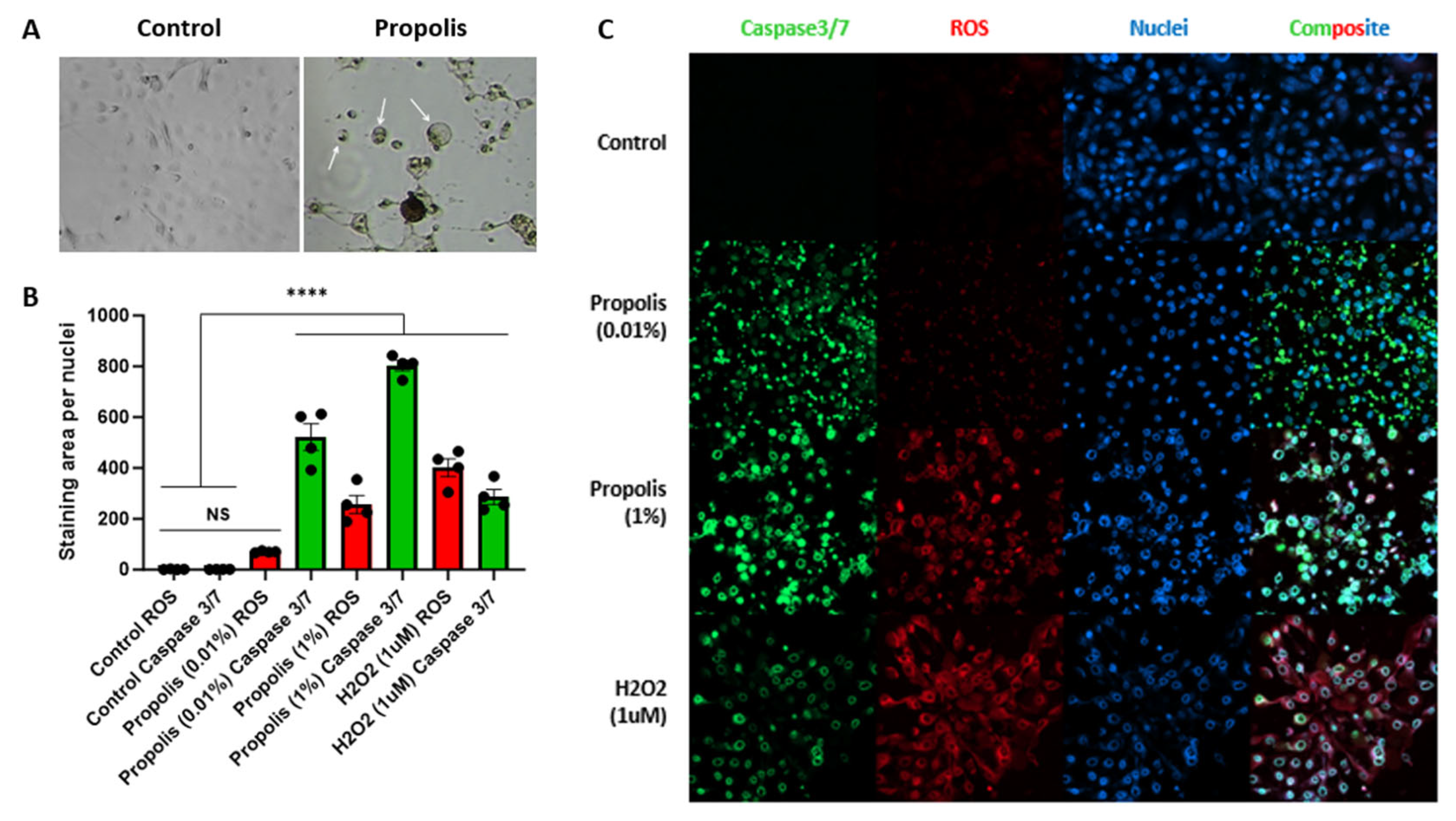
25]. Thus, although perhaps being the most extensively used carp-care preparation in current commercial preparations, propolis is in fact completely unsuitable for use in wound treatment in carp. Compounding the mucosal removal concern, our data here has shown that propolis formulations were ineffective against pathogens, yet even dilute preparations were toxic to cyprinid cells, inducing apoptotic cell death cascades and cell lysis (
Figure 7). Indeed, our data is in agreement with the body of pre-existing literature on the anticancer potential of propolis extracts in ethanol acting through apoptosis induction in a variety of cancer cell models, and using propolis from various sources [
26,
27,
28]. This apoptosis-inducing effect is not confined to cancer cells, however, with control or ‘normal’ cells lines also having been shown to display increased sensitivity over cancer cells [
29].
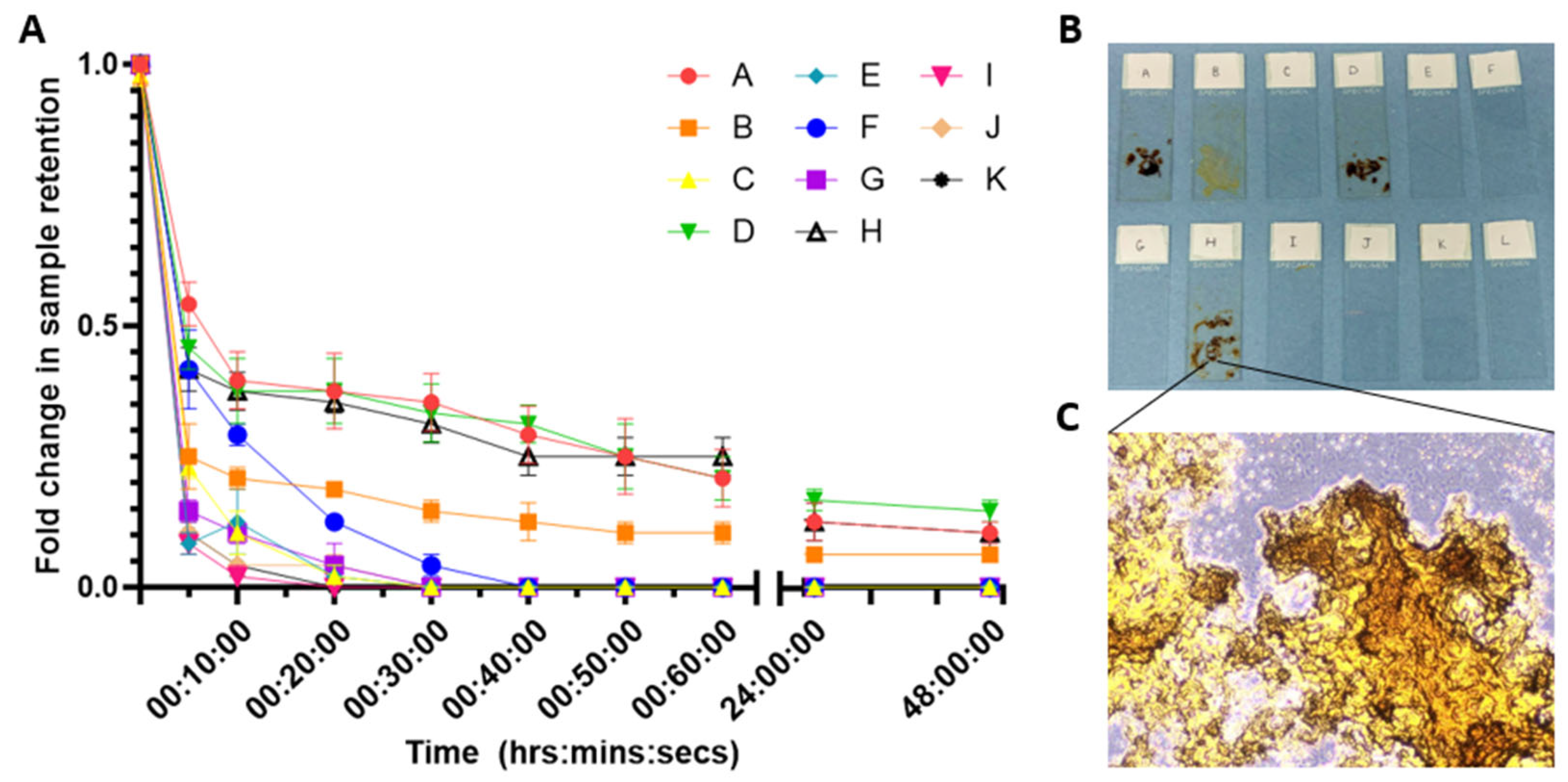
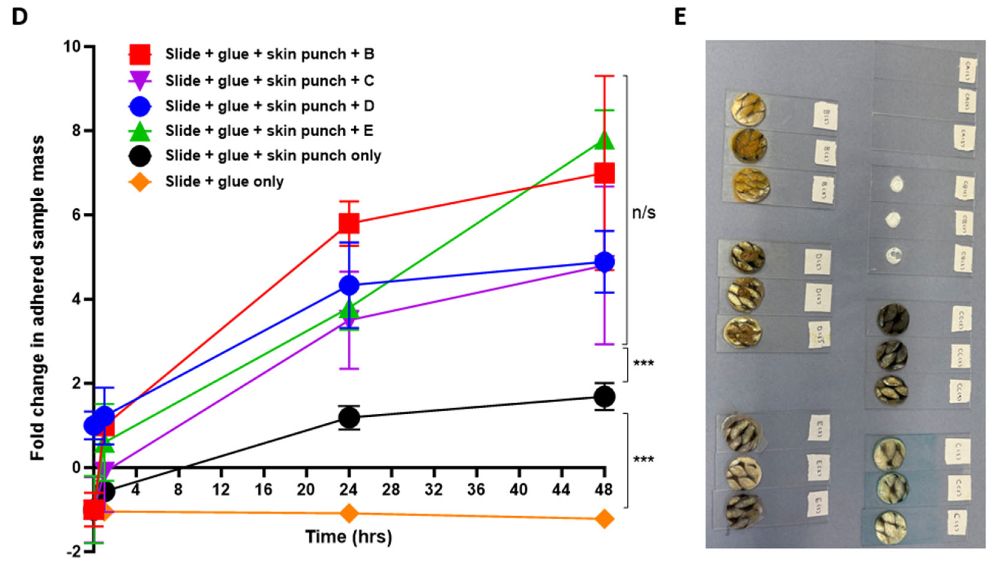
Our pseudo-ex vivo wet-adhesion model using intact carp skin punches raised a surprising and unexpected additional finding—those formulations tested here actually displayed a significant permeabilizing effect on intact carp skin (
Figure 8D). Ethanol has been shown to permeabilize fish skin before [
25], and is indeed well-known and widely used for this purpose in other areas such as for penetration enhancement in human cosmetics. Yet, this result was completely unexpected in this context; indeed, the finding raises multiple concerns: Firstly, that these products are not efficient wet adhesives, hence this permeabilization effect would leave the skin damaged and then unsealed once the applied products wash/fall off. Linked to this, the products that are liquids—and not even gel/wax based—are widely known to run off during application, coating other, undamaged areas of the fish; EtOH-based products applied in this manner would certainly have the effect of removing the essential mucin-based protective mucosal layer on the skin’s surface through solvent-induced precipitation, but also may induce skin permeabilization over a much wider area. Secondly, the skin is the major defensive barrier of the fish, as it is in humans. Once permeabilized, skin becomes an entry point for opportunistic pathogens—not only the bacteria and fungi studied here, but potentially also more infectious agents such as viruses, which are far more adept at waterborne transmission. Koi Herpes Virus (KHV) is of particular concern here. Elucidating the full implication of this finding is beyond the scope of this work, however, and pathogen transmission through carp skin permeabilized in this manner requires histological verification, but the rationale is clear. The possibility does also exist that other, less-refined and possibly more damaging solvents might also be contributing to this effect, but without proper product labelling this has not been fully verified in this study; we assume solvent effects are due to the use of EtOH here, since EtOH is certainly the most widely used solvent in commercially available propolis formulations, which is likely equivalent to other commercial preparations.
Interestingly and perhaps surprisingly, the propolis-based products tested in this study conferred no antibacterial or antifungal activity in any of the microorganisms tested here. This is in contrast to previously documented antibacterial and antifungal effects of ethanol propolis extracts in other studies, using raw propolis [
30,
31,
32]. These effects are highly variable within the literature, and interpretation of propolis action is compounded by the multiple sources and differing compositions of this complex and highly variable material. Differing efficacies have been observed even within the same regional preparations of propolis extracts against different microorganisms [
33], making any true comparison to the literature impossible with no standardized propolis source or extraction procedure. We are unaware of any other study that has examined the effect of propolis extracts on the cyprinid pathogens used in this study, so our data may be a genuine reflection of a lack of propolis toxicity against these species. Alternatively, those commercial products used here may not be pure enough or stable enough to confer the true effects of propolis on these species. A comparison using raw propolis extract would be required to determine this, but this is outside the scope of this study, and is also difficult where ethanol can no longer be used as carrier solvent.
From a regulatory perspective, propolis is listed in the VMD Schedule 6 Exemptions from licensing authorization as a treatment of pet fish kept in exclusively closed-water conditions, but there is no approval for its use as a topical treatment in either open-water conditions, or for food-fish [
33]. This also applies to all currently marketed carp-care/wound-care treatments for carp, which are without exception all products that have authorization for use in closed tanks/aquariums, but not for wild/open-water fish. The VMD, of course, have higher priorities than carp care in angling markets, hence the practice of ‘off-label’ carp care has developed unchecked, lacking proper controls or standards. Anglers and fish farmers/keepers of course only have the best intentions, want the best for their fish, and would certainly never knowingly cause harm, but better dialogue is required between science and the trade if things are to improve.
Together with the data we present here showing that currently marketed propolis-based formulations have no significant antibacterial or antifungal effects on the most common carp pathogens (
Figure 1,
Figure 2,
Figure 3 and
Figure 4), it is curious how propolis usage in cyprinids has arisen. Interestingly, to date, propolis has no regulatory approvals pertaining to any health or medical market usage for human or animal application that we are aware of, questioning why the perception exists that it should be beneficial to fish. More probably, the perceived visual change from liquid to wax on contact with water is the real commercial draw for marketing propolis wound-care formulations for application to fish—albeit, as we have shown here, without any actual health or indeed adhesion benefits. Conversely, propolis formulations appear from our own data to be toxic to fish cells, but not to their pathogens, which is somewhat counterintuitive for a cyprinid wound sealant preparation. We also found propolis to be ineffective as a wet adhesive, its structure progressively deteriorating almost immediately on contact with water, leaving incomplete surface coatings that do not confer efficient wound sealing (
Figure 8).
5. Conclusions
This study provides the first quantitative assessment of commercially available topical carp-care formulations in 30+ years of such products being marketed for recreational aquaculture and angling use in the UK and Europe. The findings demonstrate substantial variability in antimicrobial and antifungal efficacy between products, with several formulations exhibiting significant cyprinid cellular toxicity, poor submerged wet adhesion and limited pathogen inhibition at non-toxic concentrations. Commercial propolis formulations were additionally shown to induce apoptotic and oxidative stress responses in cyprinid fibroblasts in vitro. In contrast, naturally derived bioactive formulations demonstrated improved antimicrobial efficacy alongside more favourable regenerative and safety profiles. Collectively, these findings highlight the urgent need for improved scientific benchmarking, formulation transparency and evidence-based development of safer and more effective topical wound-care products for the carp industry.
We highlight the widespread lack of activity against common carp pathogens, substantial variability in formulation safety and environmental compatibility, lack of wet adhesion, cellular toxicity to cyprinid cells, and significant permeabilizing effects on intact carp skin through inappropriate solvent application. We also provide evidence that natural plant-derived alternatives may exist and should be further explored to establish suitability for application in tropical fish treatment.
We hope this data will serve as a baseline testing framework for those wishing to bring novel carp wound sealants to market, and more importantly to engage and improve dialogue between the trade and academia, with a view to facilitating improvements in rational design, safety and efficacy of future carp care products.
Given the current crisis of UK waterways with unrelenting pressures from sewage, agricultural run offs and other industries, the development of safer and more environmentally conscious fish healthcare products may have significant value within recreational carp markets—particularly angling and Koi keeping, where the primary concerns are fish welfare and the preservation of our natural history.
With further research into safer and more effective alternative formulations, fishery managers, fish keepers, anglers and aquaculturalists more broadly would benefit from superior safety and environmental profiles, improving the health of the fish, the user and the environment, as well as the ecological and welfare impacts of these industries. Aquaculture and angling are hobbies often founded in childhood, and safer, more environmentally friendly carp care products that lead to healthier and longer-lived fish are essential to encourage a new, more environmentally and ethically aware generation to take up these most rewarding and enriching of pastimes.



 ,
,