Article

Gravitational Ischemia in the Brain: How Interfering with Its Release May Predispose to Either Alzheimer’s- or Parkinson’s-like Illness, Treatable with Hyperbaric Oxygen

by
J. Howard Jaster
1 and
Giulia Ottaviani
2,*
1
London Corporation, London SW7 1EW, UK
2
Anatomic Pathology, Lino Rossi Research Center, Department of Biomedical, Surgical and Dental Sciences, Università degli Studi di Milano, 20122 Milan, Italy
*
Author to whom correspondence should be addressed.
Physiologia 2023, 3(4), 510-521; https://doi.org/10.3390/physiologia3040037
Submission received: 1 August 2023 / Revised: 17 September 2023 / Accepted: 26 September 2023 / Published: 4 October 2023
(This article belongs to the Special Issue Feature Papers in Human Physiology–2nd Edition)

Abstract

:
The physiological mechanisms for releasing and resolving gravitational ischemia in the brain, and their susceptibility to malfunction, may play an important role in a variety of neurological illnesses. An astronaut on a space walk in a micro-gravity environment may be susceptible to neuro-ocular symptoms associated with unopposed gravity-resistance mechanisms for partially preventing gravitational ischemia in the brain, and for attenuating its impact—mechanisms which may be required for normal brain physiology on Earth. Astronauts on the International Space Station typically breathe a mixture of gasses similar in composition to what they breathed on Earth, following the 1967 death of three astronauts, including Ed White, by fire on the Apollo 1 spacecraft, which was carrying 100% oxygen. For the last decade, astronauts have been studied extensively by flight physicians regarding the commonly experienced symptoms of VIIP, or ‘visual impairment and intracranial pressure’ syndrome. In this paper, we compare VIIP syndrome to the neuro-ocular and Parkinson’s-like symptoms which occurred during and after the 1918–1919 influenza pandemic. The common denominator may be gravitational ischemia in the brain, and the mirror-imaging failed mechanisms for its release (in influenza) versus unopposed gravity-resistance mechanisms (in astronauts). Some research has suggested that astronauts may benefit from breathing oxygen concentrations somewhat higher than 20%, and under slightly elevated pressure. These may possibly prevent maladaptive mechanisms leading to Alzheimer’s- or Parkinson’s-like illness by compensating for impaired mechanisms for releasing and resolving gravitational ischemia in the brain.

1. Introduction

During the early 2020s, the concept of brain ischemia was both challenged and expanded. Historically, its essential parameters have been decreased blood flow, and decreased oxygen (O2) tension, in parenchymal brain tissue—occurring focally, regionally, or globally within the brain [1,2,3]. These were thought to be caused mostly by stenotic and occlusive (intra-vascular) cerebrovascular disease, and to constitute a strictly pathological process, resulting in strokes and in harbingers of strokes, called transient ischemic attacks (TIAs).
Today, some investigators question whether those same essential parameters of ischemia are caused mostly by compressive forces originating on the exterior of blood vessels, or at least exterior to their endothelial lining. Several have been identified, such as the cerebrovascular constrictive effects of decreased carbon dioxide (CO2) levels (as from hyper-ventilation) [1,2,3]. However, the most prominent and ubiquitous is probably gravity, which has led some to think of brain ischemia as primarily a process of normal physiology, and which is only occasionally pathological.
Gravity may act as a dimmer switch that turns many cerebral electrical circuits up and down during the course of a 24-h period — depending on body position and head tilt — or it may not, if gravity is only minimally present (Figure 1). What remains entirely unknown is which circuits benefit from that cyclical variation in intensity, and why [1,2,3]. However, medical intervention in the future will likely utilize them.
Alzheimer’s disease and Parkinson’s disease may occur, in part, as maladaptive responses to chronic cerebral ischemia, caused by unresolved gravitational ischemia in the brain [3]. These neurodegenerative diseases occur in an older age group, where other systemic illnesses often play a role. Acute heart failure, sleep apnea, or chronic asthma [2] may contribute to cerebral ischemia or hypoxia—each by its own mechanisms for reducing parenchymal oxygen tension. There are associations, as well, between infectious agents and Parkinson’s disease.

1.1. Neurodegenerative Diseases and Gravity in the Brain

In mid-2022, a neurological investigator reported on infections and changes in commensal bacteria in the pathogenesis of Parkinson’s disease [1]—a disease considered largely idiopathic. The report [1] focused on the observation that some patients have developed Parkinson’s disease after suffering from an infectious illness. Several different infectious agents were associated with Parkinson’s disease, and the time lag between the infection and Parkinson’s disease was variable. Concurrent to that report [1], and more recently, several other papers [2,3,4,5,6,7,8,9,10,11,12,13,14,15,16,17,18,19,20,21,22,23,24,25] may have some relevance.
Enclosed within the cranial vault, the brain is one of the least mobile and least accessible organs in the body. The external surfaces of the brain lie still against the relatively hard inside surfaces of the skull. The meninges and cerebrospinal fluid surrounding the brain may provide some cushioning, but do not mitigate the effects of gravity. In contrast, the heart and lungs are continuously in motion, and they are surrounded to a significant degree by soft tissues [2,3,4]. Gravitational ischemia in the brain is induced by the mass of one brain component acting upon another in the Earth’s gravitational field. In any given head position, the ‘upper’ part of the brain (relative to the Earth’s center of gravity) is sitting on the ‘lower’ part of the brain as a weight burden [2,3,4].
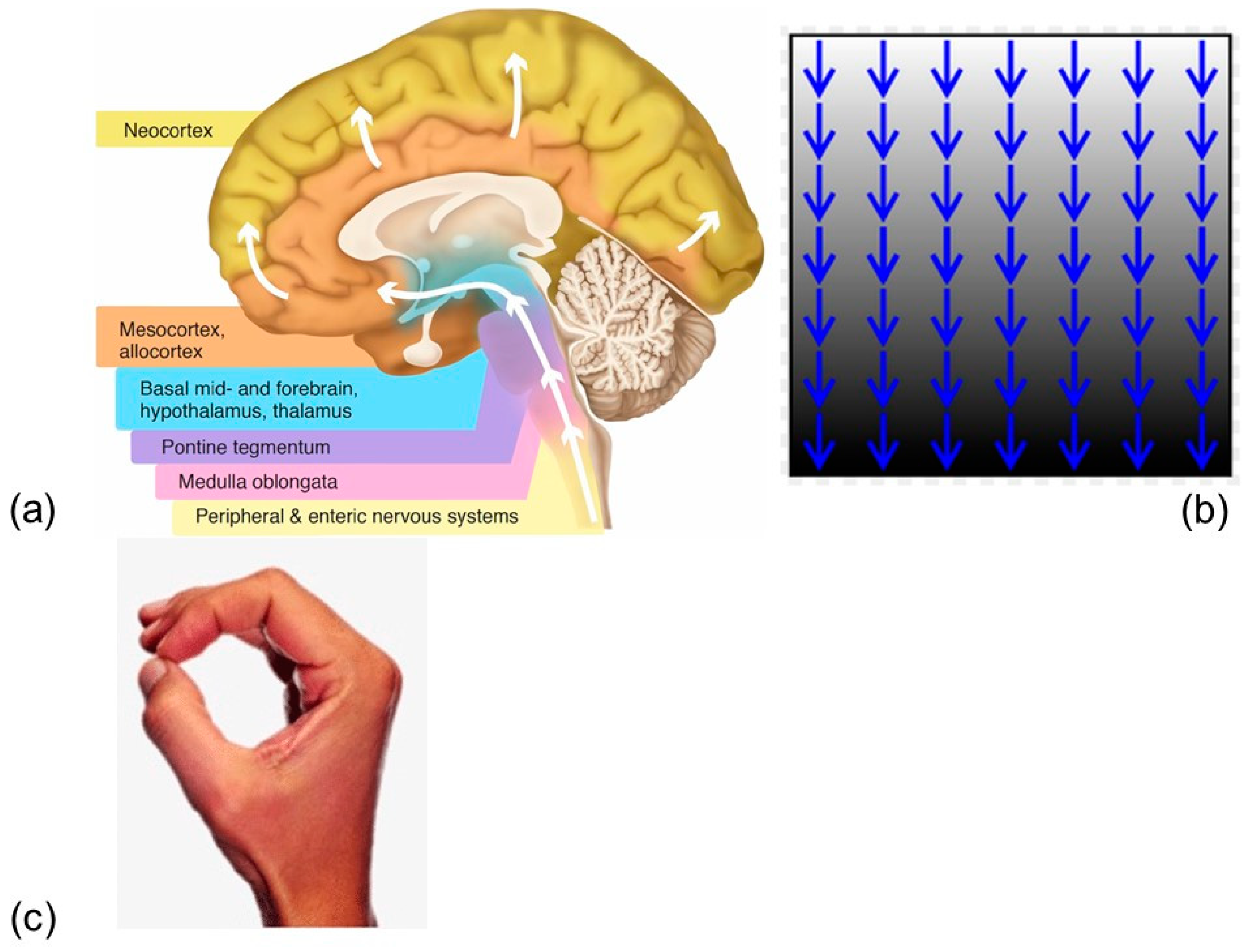
For most people, their head and body orientations are vertical or upright for most of the day and then approximately horizontal for 8 h at night during sleep. Ischemia, which may occur in the lower layers, associated with partial collapse of vascular walls under external pressure, can be reversed in its earlier stages. Sleep deprivation typically involves holding the head in an upright position for a longer period of time, and it exacerbates gravitational ischemia in the brain [2,3,4] (Figure 2a,b).
In healthy individuals, the horizontal supine body positioning associated with sleep helps to release (and resolve) gravitational ischemia and redistribute blood flow after a 16-hour period of vertical head positioning during the waking hours of the day. Restoration of blood flow by fully reopening capillary vascular beds follows repositioning, and is mediated by unloading (release) of ischemic regions [2,3].
Severe gravitational ischemia in the brain may potentially be largely preventable by frequently changing the head tilt, just as ischemic skin breakdown, bed sores, and decubitus ulcers are currently prevented by frequent passive changes in general body positioning by nursing personnel, focused on the effects of gravity [2,3].

1.1.1. Ischemia

‘Ischemia’ describes the focal (and sometimes global) tissue changes that occur when blood flow is transiently reduced, and the tissue has briefly incurred a significant lack of glucose and other nutrients as well as gas exchange. The ‘tissue’ may be an area of skin, muscle, brain, or almost any part of the body. Ischemia is a type of injury, with a variety of etiologies, both internal (intra-luminal) and external (extra-luminal) to the blood vessels supplying the region. It may be caused by prolonged continuous regional external pressure as from sitting in a chair—as the chair ‘pushes’ against the skin and muscle of the buttocks. In comparison, most transient ischemic attacks (TIA’s) are caused not by external pressure but by intravascular disease (plaques and clots inside of blood vessels) in the brain or neck, and TIA’s cause focal (and occasionally global) neurological symptoms which typically resolve after a minute or so. Slurred speech is focal, neurologically, and unconsciousness is global. Ischemia is a dynamic (non-static) injury process, most early stages of which are reversible—which correlates with a mild degree of injury, compared with injuries of lost blood flow that progress to more severe and permanent damage (infarction)—yet ischemia is nonetheless associated with significant intracellular changes.
The extra-vascular substrates of focal ischemia may be visually demonstrated by directly opposing the tip of the thumb to the tip of the index finger, which if pressed together typically reveal both nail beds blanching from pink toward white (Figure 2c). It is not the application of pressure per se, but rather its resultant disruption in blood flow which leads to ischemia. Gravity affects the entire bodies of those of us on the earth’s surface. Except for a runner in mid-flight over a hurdle, gravity holds us in direct opposition to the surface of the earth, or maybe to some piece of furniture acting as its surrogate, and except for a ballerina doing a pirouette, those bodily areas of opposition to the earth’s surface are mostly broad and multiple at any given time. Potential focal gravitational ischemic lesions are always present somewhere in our skin and underlying muscles, and in normal physiology we move our bodies around to ‘chase’ them from one area of skin to another.
A macro-analogy from obstetrics might be the mass effect of the uterus in pregnancy, intermittently obstructing blood return to the heart through the inferior vena cava based on its position, resulting in hypotension, and consistent with the term ‘gravid’.
An injury or metabolic insult to the nervous system may simultaneously or alternately produce paired signs or symptoms—one ‘positive’ (displaying aberrant hyperactivity) and the other ‘negative’ (displaying partial inactivity) in terms of the specific function of the involved neural tissue. Common pairs include tingling and numbness, tinnitus (ringing) and hearing loss, manic and depressive thoughts and behavior, scintillating scotomas, and blind spots. Dream images during sleep may be a ‘positive’ symptom of gravitational ischemia in the occipital (visual) cortex, which is typically on the ‘bottom’ of the brain during sleep, while vision during sleep is functionally depressed, a ‘negative’ symptom.
However, why is sleep needed if changing body position from vertical to horizontal, by itself, will release gravitational ischemia in the brain? Sleep is needed to induce (force) people (and many animals) to lie down. Cave men did not know that they needed to lie down so that gravitational ischemia could be released in their brains. Sleep has probably evolved to perform many other physiological electro-chemical functions. Additionally, and more pragmatically, it prevented a cave man from walking around in the dark after sunset and stumbling into a pride of lions.

1.1.2. Space Flight

Some of the possible contributions of gravitational ischemia in the brain to normal physiology may be inferred by observing what happens when gravity is removed. In 2017, in vivo neuroanatomy and neurophysiology were assessed by magnetic resonance images (MRI) and other modalities in 16 astronauts in the Space Shuttle Program [5]. This study was undertaken in part to assess a common medical problem among astronauts following longer-duration flights—then called VIIP, for ‘visual impairment and intracranial pressure syndrome’ by flight-physicians. Fast forward to 2023, when the condition had become known as SANS, for ‘spaceflight associated neuro-ocular syndrome’ [6]—the etiology remains unknown, but the features of illness seem to revolve around increased intracranial pressure.
Space flight may reveal the most immediate, prominent, and least integrated of the brain’s natural counter-forces to gravity—which find themselves suddenly unopposed. Like a collection of coiled springs, accustomed to maintaining resistance against gravity, these physiological processes may be the most available and noticeable when gravity is removed, or when infectious agents interact with them. They may utilize physiological processes which rely upon the continuously shifting electrolyte and metabolic gradients caused by gravity—which may act as a dimmer switch in brain physiology, with its many continuously rolling multi-directional effects, based on head tilting and general body position.
Conversely, another group of space-flight-microgravity investigators looked at brief head-down tilt in younger adults for executive function and found improvement [7]. Their 65 test subjects were not space-flight participants, and the short duration of their head-down tilt minimized confounding problems such as cerebral venous congestion.
However, other studies of overall brain elasticity and stiffness have revealed significant changes which often occur in old age [8]. Resulting, in part, from these changes may be a decreased effectiveness of horizontal positioning and other sleep-related mechanisms, such as glymphatics [9,10,11], which may normally help to release gravitational ischemia. These changes may invite other, even maladaptive, mechanisms to occur, altering cerebrovascular (and blood–brain barrier) permeability characteristics in an effort to facilitate the release of gravitational ischemia. Some infectious agents on some occasions may play a role comparable to that of altered elasticity or stiffness in the brain.
In older adults with primary sarcopenia, regionally decreased cerebral blood flow and hypoperfusion were described by other investigators [12] to occur in central autonomic ‘network hubs’, including the hypothalamus which influences muscle metabolism [3], and which are located near the base of the skull, a gravitationally dependent region (during vertical position). These were consistent with the occurrence of ischemia, though the investigators never mentioned gravitational ischemia in the brain, and they were associated with the occurrence and progression of Alzheimer’s disease [12]. This autonomic anatomical distribution is often affected in Parkinson’s disease (Figure 2). The primary metabolic effect is decreased oxygen tension in the involved parenchymal brain tissue. The autonomic networking effects of gravitational ischemia in the brain involving intestinal disease were mentioned in a recent general discussion of possible gravitational influences in irritable bowel syndrome (IBS) [13].
The study [12], which included 95 older adults, examined clinically and, with several imaging modalities, documented decreased cerebral blood flow in the insula, anterior cingulate cortex, subcallosal area, straight gyrus, hypothalamus, amygdala, and head of the caudate nucleus. This was strongly associated with two other major clinical findings—primary loss of muscle mass, and the onset and development of dementia.
These important findings [12] were not further characterized as to which of the three, if any, played a more etiological role, while the other two were largely resultant effects. However, regionally decreased blood flow in the brain would seem better positioned to cause the other two. Gravity could have played a role in establishing this anomalous blood flow pattern. The details of timing and location in the associations of sarcopenia, progressive dementia, and regionally decreased blood flow in the brain are supportive of an etiology which includes gravitational ischemia in the brain.

1.1.3. Infectious Agents (Influenza)

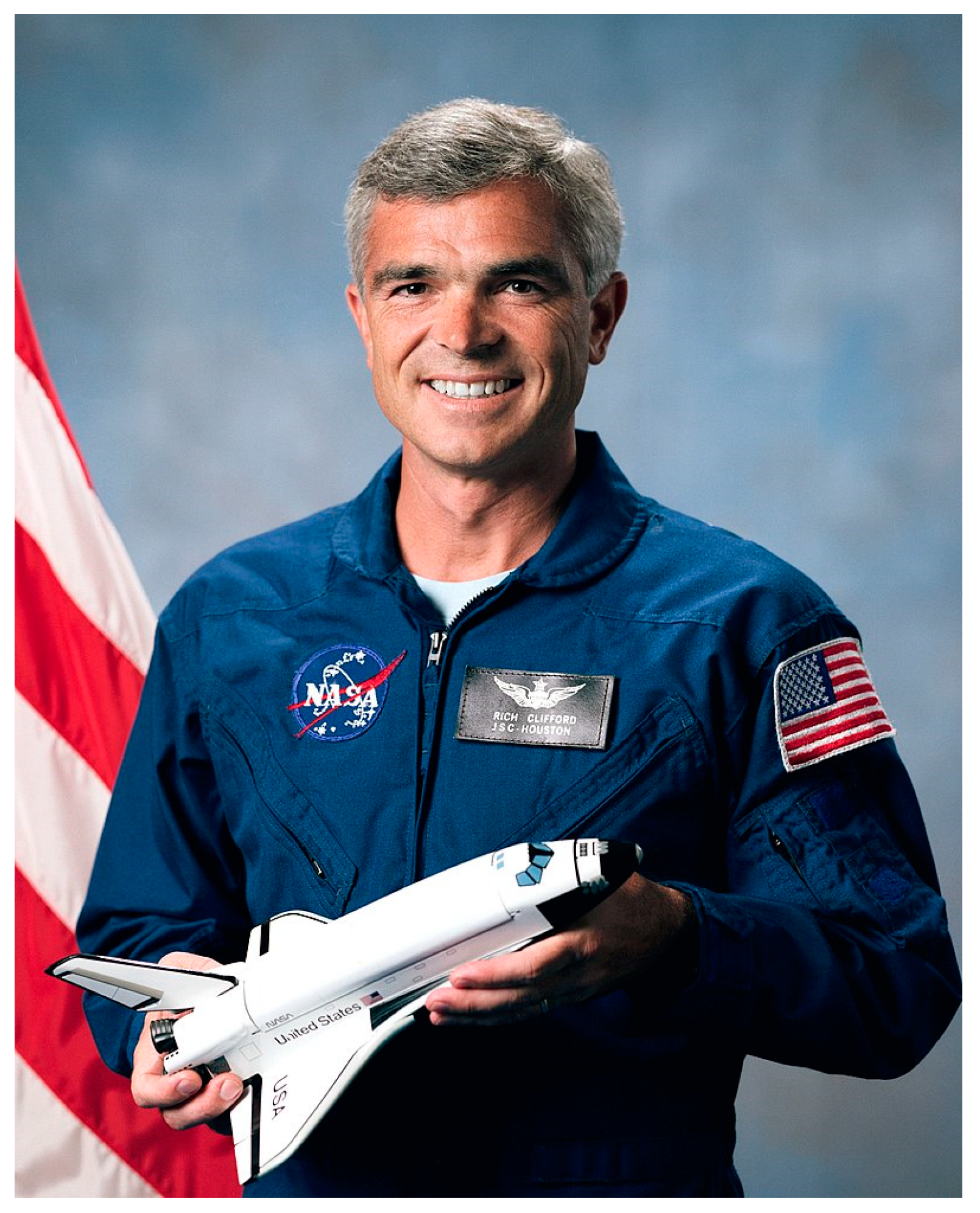
Historically, encephalitis lethargica, and post-encephalitic Parkinsonism, which occurred following the influenza pandemic of 1918–1919, and which affected mostly younger adults [1], may have disrupted the normal blood–brain barrier and glymphatic mechanisms, resulting in sleepiness, a condition which others have associated with increased gravitational ischemia in the brain. Studied for over a century, an etiological association between infectious diseases and Parkinson’s disease has never been confirmed. The ocular and visual phenomena which occurred in encephalitis lethargica were imperfectly similar to the ocular and visual phenomena associated with the reduced gravitational forces in space flight. Numerous earlier reports of short-duration (15-day) space flights have listed lethargy among the complaints of astronauts—but not necessarily associated with the ocular and visual disturbances, which have occurred mostly in long-duration (150-day) flights. One astronaut was confirmed to have developed Parkinson’s disease while participating in the Space Shuttle Program (Figure 3). Although this was only an anecdotal incident, the astronaut was relatively young (age 42) at the time of diagnosis, and the temporal association with space flight was strong.
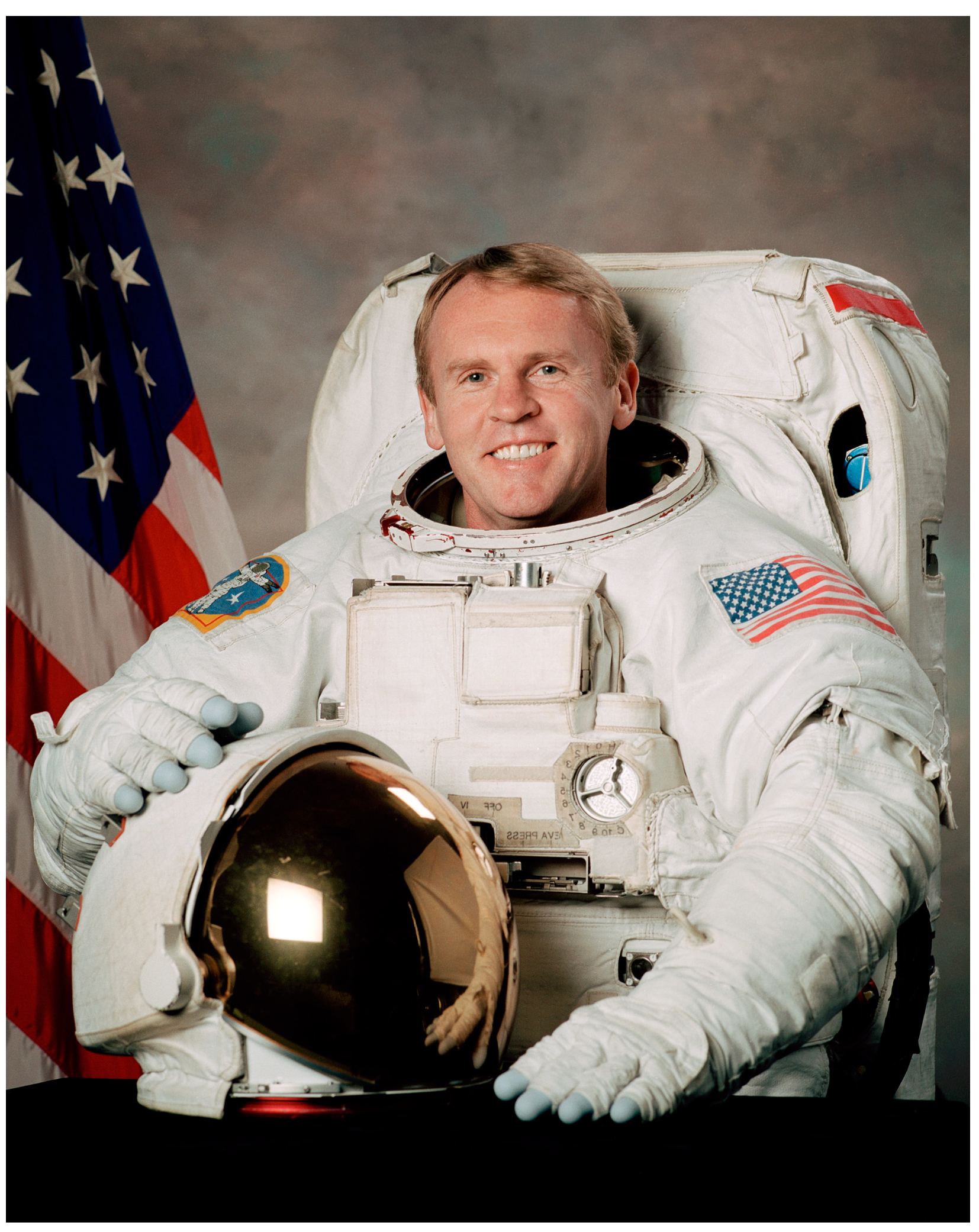
Two other astronauts developed Parkinson’s disease later in life, years after completion of their participation in spaceflight (Figure 4 and Figure 5). Taken together, these cases suggest an incidence of Parkinson’s disease much higher than in the general population, providing a mirror image of the rash of cases of post-encephalitic Parkinsonism a century earlier.
All of these observations taken together, involving post-encephalitic Parkinsonism and spaceflight, suggest a multi-factorial etiology for Parkinson’s symptoms, including genetics (family history) and toxins. At the same time, we may have only a fraction of the pertinent neurological information potentially available regarding these two groups.
One astronaut (Figure 5) had a significant high school athletic career, during which he could possibly have incurred head injuries, which may have predisposed him to neurodegenerative disease, and this type of historical information is not available to us.

1.2. Hyperbaric Oxygen

Hyperbaric oxygen therapy involves using pure oxygen at increased pressure (typically 2–3 atmospheres) leading to increased oxygen levels in the blood (hyperoxemia) and tissue (hyperoxia). The increased pressure and oxygen bioavailability potentially have a variety of applications [14,15]. It may also have antimicrobial, immune-modulatory, and angiogenic properties. Hyperbaric oxygen is associated, as well, with a variety of adverse effects, some of which can be mitigated through dosage and scheduling.
Regionally decreased oxygen tension in the brain, associated with hypoperfusion [4,12] and gravitational ischemia [2], may be a feature in some cases of Alzheimer’s disease, which has not been widely appreciated, and which may potentially be utilized for prevention and treatment. Gravitational ischemia may become more accentuated than in normal physiology, due to age-related changes in the rigidity and elasticity of the brain [8]. Oxygen therapy generally, administered in a variety of ways, may be beneficial in some cases of sporadically occurring Parkinson’s or Alzheimer’s disease, or in some spaceflight situations.
Alterations in glymphatic function and in neurovascular permeability, associated with cognitive decline in Alzheimer’s disease [9,10,11,12], may in part be maladaptive attempts to release and resolve gravitational ischemia in the brain, partly through deposition of beta-amyloid. Recent reports have suggested that oxygen therapy, including hyperbaric oxygen, may have a role in both prevention and treatment of some neurodegenerative diseases, consistent with ischemia sometimes playing a role in their etiology [14,15]. Lewy bodies and alpha-synuclein associated with Parkinson’s disease alter blood–brain barrier permeability in a manner which may immediately resolve gravitational ischemia in the brain, yet may lead to adverse consequences long term, suggesting a maladaptive mechanism [16,17].
Taken together, these reports [2,3,4,5,6,7,8,9,10,11,12,13,14,15,16,17] might suggest the possibility that some infectious agents mirror space flight in their capacity to alter neurophysiology by affecting the ability of the brain to dissipate gravitational ischemia or to convert it to useful purposes. They and others [18,19,20,21,22,23] imply, as well, that oxygen (including hyperbaric) may play a supplemental role in management and in therapy in some cases.

2. Results

Even more recently, medical investigators have written [24] about the impact of asthma on the brain—as evidenced by diffusion MRI, cerebrospinal fluid (CSF) biomarkers, and cognitive decline. Their retrospective study shows how early brain microstructural changes and cognitive decline may progress to dementia in patients with asthma [24]. At the same time, they acknowledge in their discussion that we do not yet have a mechanistic understanding of how asthma contributes deleteriously to brain health’ [24]. They mention as well that ‘asthma is known to affect the vasculature, and vascular dysfunction contributes to Alzheimer’s disease pathogenesis. Alternatively, hypoxia could mediate the influence of asthma on brain health. In reality, a combination of mechanisms likely give rise to the relationships reported here’ [24]. These observations may be consistent with recent reports describing gravitational ischemia in the brain.
A number of other studies have now reported that spaceflight alters human brain morphology [4,5,6,22,25]. One recently [25], in its introduction, reported that spaceflight induces an upward shift of the brain within the skull, resulting in cortical crowding and narrowing of the sulci at the top of the brain. These findings suggest that longer time in microgravity results in greater cortical crowding at the apex of the brain, and greater compensatory ventricular volume expansion following 6-month flights.
Mirror-Imaging Dynamics of Gravitational Ischemia in the Brain
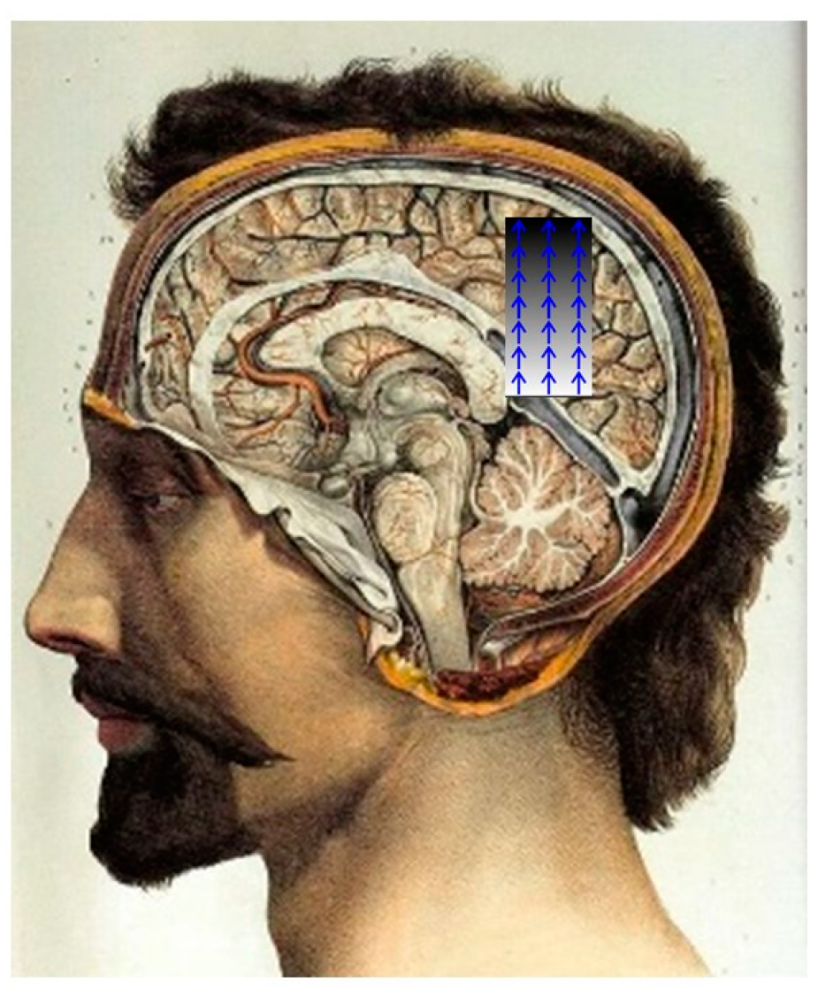
Normal gravity-resistance mechanisms in the brain likely involve cerebral arterial pressure, cerebral venous pressure, and CSF pressure, as well as the exhalation phase of the breathing cycle—all working in concert to push the brain ‘upward’ (against gravity), regardless of the body position and head tilt. In standing (vertical) position, net brain movement is toward the mid-sagittal suture of the skull (Figure 6). Furthermore, one consideration is the shelving structures in the brain which limit its movement. These include the falx cerebri, cerebellar tentorium, diaphragma sellae, and the base of the skull.
During spaceflight in a micro-gravity environment, these gravity-resistance forces are unopposed. They remain unopposed throughout spaceflight, and possible contribute to the brain structural changes observed by investigators [25]. They also likely contribute to VIIP clinical syndrome [26,27]. A detailed mechanism is unknown.
The mechanism for VIIP and Parkinson’s symptoms in astronauts is probably ischemia, at least in part [28,29]. It is the reverse of gravitational ischemia and is caused by the brain being pushed into the apex of the skull. There is no built-in physiological mechanism for this ischemia to be resolved or released during spaceflight.
All of this stands in contrast to what may have happened during the influenza pandemic of 1918–1919, when the release and resolution of gravitational ischemia in the brain was likely disrupted by processes related to the presence of the influenza virus. This was a failure of the mechanism to release and resolve gravitational ischemia in the brain.
Creating a three-dimensional picture of the degree of gravitational ischemia in the brain in different brain locations in a single position would itself be a project for a computational neuroscientist. Then, adding in different head positions and time increases its complexity.
Our collective understanding of gravitational ischemia in the brain is important because we have the tools to fix problems that require only re-positioning and oxygen.

3. Discussion

It has been suggested that the extra-vascularly derived compressive gravitational ischemia in the brain may have much more quantitative significance than does the stenotic /occlusive intra-vascularly derived occlusive cerebrovascular disease (causing strokes), and, as such, it may play a large role in normal brain physiology (sleep) and only occasionally be involved in pathophysiology (including amyloid deposition). Other recent observations regarding alterations of neuroanatomy and neurophysiology during and after space flight [22], together with related clinical symptoms, suggest that fluctuating cyclical regional gravitational ischemia in the brain is not only normal for humans, it may also be necessary.
What will be the direction of future research relating gravitational ischemia in the brain to Alzheimer’s- and Parkinson’s-like illness? There are several possibilities, and some of them may revolve around measuring oxygen tensions at different foci in different parts of the brain, as a reflection of relative blood flow and ischemia. These may utilize new technologies together with existing procedures. Tissue-integrating, bioresorbable life-time-based phosphorescent oxygen sensors [28] may possibly be a useful tool in this regard.
Another may be invasive epilepsy monitoring with a grid of subdural electrodes, which has been a standard diagnostic procedure for decades [29]. However, the addition of oxygen sensors into the grid at foci corresponding to the electrodes may yield information about a gradient of ischemia (possibly correlating with gravity). Furthermore, blood oxygen-level-dependent MRI is now available with high spatiotemporal resolution [30]—a potentially important feature, as oxygen levels vary over time during changes in head position. These may potentially be related specifically to Parkinson’s-like illness, using functional near-infrared spectroscopy (fNIRS) [31].

Author Contributions

Conceptualization, J.H.J.; methodology, G.O.; software, G.O.; validation, J.H.J. and G.O.; formal analysis, J.H.J.; investigation, J.H.J.; resources, J.H.J.; data curation, J.H.J.; writing—original draft preparation, G.O.; writing—review and editing, J.H.J.; visualization, G.O.; supervision, G.O.; project administration. All authors have read and agreed to the published version of the manuscript.

Funding

This research received no external funding.

Institutional Review Board Statement

Not applicable.

Informed Consent Statement

Not applicable.

Data Availability Statement

Not applicable.

Conflicts of Interest

The authors declare no conflict of interest.

References

  1. Shannon, K.M. Infections and Changes in Commensal Bacteria and the Pathogenesis of Parkinson’s Disease. J. Park. Dis. 2022, 12, S45–S51. [Google Scholar] [CrossRef] [PubMed]
  2. Jaster, J.H.; Ottaviani, G. Acute Heart Failure, 90-Day Mortality, and Gravitational Ischemia in the Brain. Diagnostics 2022, 12, 1473. [Google Scholar] [CrossRef]
  3. Jaster, J.H.; Ottaviani, G. Late Seizures Following Cerebral Venous Thrombosis-May Be a Maladaptive Attempt to Release Gravitational Ischemia in the Brain. Neurol. Sci. 2022, 43, 6573–6574. [Google Scholar] [CrossRef] [PubMed]
  4. Yokoyama, Y.; Yamada, Y.; Kosugi, K.; Yamada, M.; Narita, K.; Nakahara, T.; Fujiwara, H.; Toda, M.; Jinzaki, M. Effect of Gravity on Brain Structure as Indicated on Upright Computed Tomography. Sci. Rep. 2021, 11, 392. [Google Scholar] [CrossRef]
  5. Roberts, D.R.; Albrecht, M.H.; Collins, H.R.; Asemani, D.; Chatterjee, A.R.; Spampinato, M.V.; Zhu, X.; Chimowitz, M.I.; Antonucci, M.U. Effects of Spaceflight on Astronaut Brain Structure as Indicated on MRI. N. Engl. J. Med. 2017, 377, 1746–1753. [Google Scholar] [CrossRef]
  6. Ong, J.; Tarver, W.; Brunstetter, T.; Mader, T.H.; Gibson, C.R.; Mason, S.S.; Lee, A. Spaceflight Associated Neuro-Ocular Syndrome: Proposed Pathogenesis, Terrestrial Analogues, and Emerging Countermeasures. Br. J. Ophthalmol. 2023, 107, 895–900. [Google Scholar] [CrossRef] [PubMed]
  7. Mekari, S.; Murphy, R.J.L.; MacKinnon, A.R.S.; Hollohan, Q.; Macdougall, S.C.; Courish, M.K.; Kimmerly, D.S.; Neyedli, H.F. The Impact of a Short-Period Head-down Tilt on Executive Function in Younger Adults. Sci. Rep. 2022, 12, 20888. [Google Scholar] [CrossRef] [PubMed]
  8. Lv, H.; Kurt, M.; Zeng, N.; Ozkaya, E.; Marcuz, F.; Wu, L.; Laksari, K.; Camarillo, D.B.; Pauly, K.B.; Wang, Z.; et al. MR Elastography Frequency-Dependent and Independent Parameters Demonstrate Accelerated Decrease of Brain Stiffness in Elder Subjects. Eur. Radiol. 2020, 30, 6614–6623. [Google Scholar] [CrossRef]
  9. Yu, Y.; Zhang, N.; Xiang, B.; Ding, N.; Liu, J.; Huang, J.; Zhao, M.; Zhao, Y.; Wang, Y.; Ma, Z. In Vivo Characterization of Cerebrovascular Impairment Induced by Amyloid β Peptide Overload in Glymphatic Clearance System Using Swept-Source Optical Coherence Tomography. Neurophotonics 2023, 10, 015005. [Google Scholar] [CrossRef] [PubMed]
  10. Scott-Massey, A.; Boag, M.K.; Magnier, A.; Bispo, D.P.C.F.; Khoo, T.K.; Pountney, D.L. Glymphatic System Dysfunction and Sleep Disturbance May Contribute to the Pathogenesis and Progression of Parkinson’s Disease. Int. J. Mol. Sci. 2022, 23, 12928. [Google Scholar] [CrossRef]
  11. Hsu, J.-L.; Wei, Y.-C.; Toh, C.H.; Hsiao, I.-T.; Lin, K.-J.; Yen, T.-C.; Liao, M.-F.; Ro, L.-S. Magnetic Resonance Images Implicate That Glymphatic Alterations Mediate Cognitive Dysfunction in Alzheimer Disease. Ann. Neurol. 2023, 93, 164–174. [Google Scholar] [CrossRef]
  12. Demura, T.; Okuno, T.; Miwa, T.; Iritani, O.; Nakano, H.; Yamamoto, J.; Shiga, H.; Kodera, K.; Morimoto, C.; Demura, N.; et al. Sarcopenia and Decline in Appendicular Skeletal Muscle Mass Are Associated with Hypoperfusion in Key Hubs of Central Autonomic Network on 3DSRT in Older Adults with Progression of Normal Cognition to Alzheimer’s Disease. Geriatr. Gerontol. Int. 2023, 23, 16–24. [Google Scholar] [CrossRef] [PubMed]
  13. Brouillette, M. Gravity Could Be the Cause of Your IBS, According to This Doctor. Available online: https://www.popularmechanics.com/science/health/a42552346/gravity-may-cause-irritable-bowel-syndrome/ (accessed on 20 July 2023).
  14. Yang, C.; Yang, Q.; Xiang, Y.; Zeng, X.-R.; Xiao, J.; Le, W.-D. The Neuroprotective Effects of Oxygen Therapy in Alzheimer’s Disease: A Narrative Review. Neural Regen. Res. 2023, 18, 57–63. [Google Scholar] [CrossRef]
  15. Mensah-Kane, P.; Sumien, N. The Potential of Hyperbaric Oxygen as a Therapy for Neurodegenerative Diseases. GeroScience 2023, 45, 747–756. [Google Scholar] [CrossRef] [PubMed]
  16. Wong, Y.Y.; Wu, C.-Y.; Yu, D.; Kim, E.; Wong, M.; Elez, R.; Zebarth, J.; Ouk, M.; Tan, J.; Liao, J.; et al. Biofluid Markers of Blood-Brain Barrier Disruption and Neurodegeneration in Lewy Body Spectrum Diseases: A Systematic Review and Meta-Analysis. Park. Relat. Disord. 2022, 101, 119–128. [Google Scholar] [CrossRef] [PubMed]
  17. Hourfar, H.; Aliakbari, F.; Aqdam, S.R.; Nayeri, Z.; Bardania, H.; Otzen, D.E.; Morshedi, D. The Impact of α-Synuclein Aggregates on Blood-Brain Barrier Integrity in the Presence of Neurovascular Unit Cells. Int. J. Biol. Macromol. 2023, 229, 305–320. [Google Scholar] [CrossRef]
  18. Tsai, C.-Y.; Wu, S.-M.; Kuan, Y.-C.; Lin, Y.-T.; Hsu, C.-R.; Hsu, W.-H.; Liu, Y.-S.; Majumdar, A.; Stettler, M.; Yang, C.-M.; et al. Associations between Risk of Alzheimer’s Disease and Obstructive Sleep Apnea, Intermittent Hypoxia, and Arousal Responses: A Pilot Study. Front. Neurol. 2022, 13, 1038735. [Google Scholar] [CrossRef]
  19. Chen, J.; Zhang, F.; Zhao, L.; Cheng, C.; Zhong, R.; Dong, C.; Le, W. Hyperbaric Oxygen Ameliorates Cognitive Impairment in Patients with Alzheimer’s Disease and Amnestic Mild Cognitive Impairment. Alzheimer’s Dement. 2020, 6, e12030. [Google Scholar] [CrossRef] [PubMed]
  20. Ortega, M.A.; Fraile-Martinez, O.; García-Montero, C.; Callejón-Peláez, E.; Sáez, M.A.; Álvarez-Mon, M.A.; García-Honduvilla, N.; Monserrat, J.; Álvarez-Mon, M.; Bujan, J.; et al. A General Overview on the Hyperbaric Oxygen Therapy: Applications, Mechanisms and Translational Opportunities. Medicina (Kaunas) 2021, 57, 864. [Google Scholar] [CrossRef]
  21. Zhang, Y.; Zhou, Y.; Jia, Y.; Wang, T.; Meng, D. Adverse Effects of Hyperbaric Oxygen Therapy: A Systematic Review and Meta-Analysis. Front. Med. 2023, 10, 1160774. [Google Scholar] [CrossRef]
  22. Gupta, U.; Baig, S.; Majid, A.; Bell, S.M. The Neurology of Space Flight; How Does Space Flight Effect the Human Nervous System? Life Sci. Sp. Res. 2023, 36, 105–115. [Google Scholar] [CrossRef]
  23. Levine, K.S.; Leonard, H.L.; Blauwendraat, C.; Iwaki, H.; Johnson, N.; Bandres-Ciga, S.; Ferrucci, L.; Faghri, F.; Singleton, A.B.; Nalls, M.A. Virus Exposure and Neurodegenerative Disease Risk across National Biobanks. Neuron 2023, 111, 1086–1093. [Google Scholar] [CrossRef]
  24. Nair, A.K.; Van Hulle, C.A.; Bendlin, B.B.; Zetterberg, H.; Blennow, K.; Wild, N.; Kollmorgen, G.; Suridjan, I.; Busse, W.W.; Dean, D.C.; et al. Impact of Asthma on the Brain: Evidence from Diffusion MRI, CSF Biomarkers and Cognitive Decline. Brain Commun. 2023, 5, fcad180. [Google Scholar] [CrossRef]
  25. McGregor, H.R.; Hupfeld, K.E.; Pasternak, O.; Beltran, N.E.; De Dios, Y.E.; Bloomberg, J.J.; Wood, S.J.; Mulavara, A.P.; Riascos, R.F.; Reuter-Lorenz, P.A.; et al. Impacts of Spaceflight Experience on Human Brain Structure. Sci. Rep. 2023, 13, 7878. [Google Scholar] [CrossRef] [PubMed]
  26. Adelaide-Born Astronaut Reveals He Has Parkinson’s Disease. Available online: https://www.facebook.com/7NEWSAdelaide/videos/adelaide-born-astronaut-reveals-he-has-parkinsons-disease/878348489458878/ (accessed on 20 July 2023).
  27. SA Astronaut Andy Thomas Reveals Parkinson’s Diagnosis. Available online: https://newslettercollector.com/newsletter/morning-sa-astronaut-andy-thomas-revea-ls-parkinson-s-diagnosis/ (accessed on 20 July 2023).
  28. Presley, K.F.; Falcucci, T.; Shaidani, S.; Fitzpatrick, V.; Barry, J.; Ly, J.T.; Dalton, M.J.; Grusenmeyer, T.A.; Kaplan, D.L. Engineered Porosity for Tissue-Integrating, Bioresorbable Lifetime-Based Phosphorescent Oxygen Sensors. Biomaterials 2023, 301, 122286. [Google Scholar] [CrossRef] [PubMed]
  29. Matarrese, M.A.G.; Loppini, A.; Fabbri, L.; Tamilia, E.; Perry, M.S.; Madsen, J.R.; Bolton, J.; Stone, S.S.D.; Pearl, P.L.; Filippi, S.; et al. Spike Propagation Mapping Reveals Effective Connectivity and Predicts Surgical Outcome in Epilepsy. Brain 2023, 146, 3898–3912. [Google Scholar] [CrossRef] [PubMed]
  30. Kim, J.H.; Taylor, A.J.; Himmelbach, M.; Hagberg, G.E.; Scheffler, K.; Ress, D. Characterization of the Blood Oxygen Level Dependent Hemodynamic Response Function in Human Subcortical Regions with High Spatiotemporal Resolution. Front. Neurosci. 2022, 16, 1009295. [Google Scholar] [CrossRef]
  31. Sousani, M.; Rojas, R.F.; Preston, E.; Ghahramani, M. Towards a Multi-Modal Brain-Body Assessment in Parkinson’s Disease: A Systematic Review in FNIRS (February 2023). IEEE J. Biomed. Health Inform. 2023. online ahead of print. [Google Scholar] [CrossRef] [PubMed]
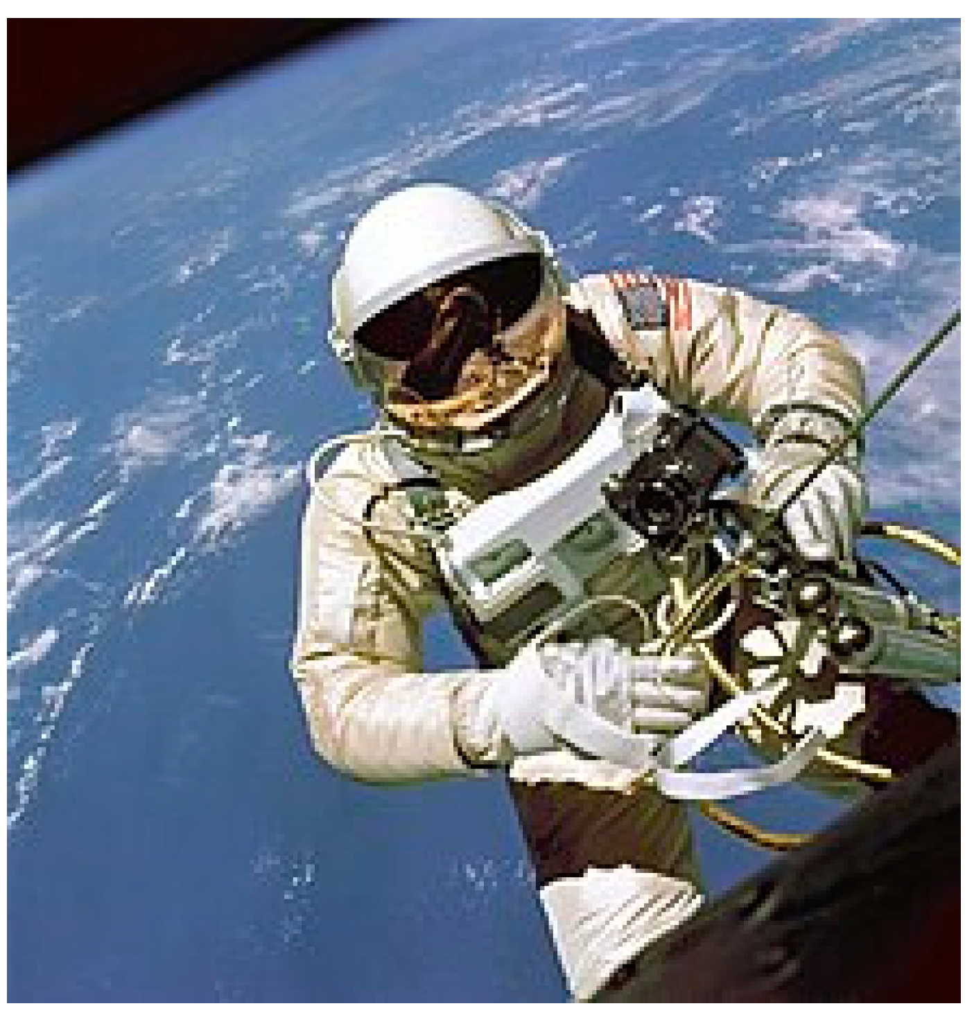
Figure 1. Astronaut Ed White makes the first American spacewalk during Gemini 4 in June 1965. Disclaimer for Figure 1: This account is based entirely on public records. The authors [JHJ and GO] had no personal acquaintance with the astronaut, and no access to his personal medical records. Although known to many in the general public, this case has not previously been discussed in the medical literature, to the best of our knowledge. Adapted from Wikipedia, with Creative Commons licensing. https://en.wikipedia.org/wiki/Ed_White_(astronaut)#/media/File:EdWhiteFirstAmericanSpacewalker.1965.ws.jpg (Accessed on 20 July 2023).
Physiologia 03 00037 g001
Figure 2. (a) Sequential layering in an upright brain, from the bottom up (white arrows), by Lewy bodies in Parkinson’s disease (adapted from Braak Staging), suggests a horizontal sedimentary stratification, appearing to be influenced by gravity over time. Adapted from Wikimedia Commons, with Creative Commons licensing. https://en.wikipedia.org/wiki/Braak_staging#/media/File:BraakStagingbyVisanjiEtAl.png. Accessed on 27 September 2023; (b) gravity: schematic stratification of biological tissue into pancaking layers under the influence of gravity. Lower layers incur progressively increasing weight burden from upper layers, and thus increasing compression of blood vessels and reduction of blood flow, possibly resulting in regional ischemia; (c) opposition of thumb to index finger.
Physiologia 03 00037 g002
Figure 3. Astronaut Clifford in 1990. Historical medical review of a known public figure—Astronaut, Michael R. ‘Rich’ Clifford (1952–2021). Michael Richard Clifford (1952–2021) was a United States Army officer and NASA astronaut. As a military officer, Clifford was assigned to the Johnson Space Center in July 1987. As a Space Shuttle Vehicle Integration engineer, his duties involved engineering liaison for launch and landing operations of the Space Shuttle Program. Clifford was selected to join NASA Astronaut Group 13 in July 1990. He also served in a variety of technical assignments. Clifford logged over 3400 h flying in a wide variety of fixed and rotary winged aircrafts. He logged six hours of spacewalk time over three Space Shuttle missions. He conducted a spacewalk while docked to an orbiting space station—the Russian space station Mir. His total time in space was 27 days. He was diagnosed with Parkinson’s disease in 1994 when he was 42 years old. He suspected that exposures to pesticides used in farms and other chemicals used on car engines when he was a youngster contributed to the early onset of his illness. After retirement from his space career in 1997, he became an advocate for Parkinson’s disease awareness and encouraged other patients of the disease to live life to its fullest. Film-maker Zach Jankovic, son of Dr. Joseph Jankovic who helped diagnose Clifford, made a documentary detailing Clifford’s experiences with the disease; the short film entitled The Astronaut’s Secret won the American Academy of Neurology Foundation’s 2012 annual ‘Neuro Film Festival’ competition and was released in 2014. The documentary did not mention gravitational ischemia in the brain. Clifford died from complications of Parkinson’s on 28 December 2021, at the age of 69. Disclaimer for Figure 3. This account is based entirely on public records. The authors [JHJ and GO] had no personal acquaintance with the Astronaut, and no access to his personal medical records. Although known to many in the general public, this case has not previously been discussed in the medical literature, to the best of our knowledge. Adapted from Wikipedia, with Creative Commons licensing. https://en.wikipedia.org/wiki/Michael_R._Clifford. Accessed on 20 July 2023.
Physiologia 03 00037 g003
Figure 4. Astronaut Andy Thomas. South Australian NASA astronaut Andy Thomas reveals Parkinson’s diagnosis. From Twitter:. As reported on Twitter by7NEWS.com.au #7NEWS on 5 September 2021, Adelaide-born astronaut Andy Thomas says he is working to minimize the consequences, after revealing he has Parkinson’s disease. One of Adelaide’s favorite sons says he suspected something was wrong for a long time before a visit to the doctor ‘that came as a relief’. Dr. Andrew ‘Andy’ Sydney Withiel Thomas, (born 18 December 1951) is an Australian and American aerospace engineer and a former NASA astronaut. He has dual nationality; he became a U.S. citizen in December 1986, hoping to gain entry to NASA’s astronaut program. He is married to fellow NASA astronaut Shannon Walker. Thomas was selected by NASA in March 1992 and reported to the Johnson Space Center in August 1992. In August 1993, following one year of training, he was appointed a member of the NASA Astronaut Corps and was qualified for an assignment as a mission specialist on Space Shuttle flight crews. Total time in space: 177 days. In early September 2021, astronaut Thomas revealed his recent diagnosis of Parkinson’s disease, through a few different media outlets [26,27]. Disclaimer for Figure 4: This account is based entirely on public records. The authors [JHJ and GO] had no personal acquaintance with the astronaut, and no access to his personal medical records. Although known to many in the general public, this case has not previously been discussed in the medical literature, to the best of our knowledge. Adapted from Wikipedia, with Creative Commons licensing.: Wikipedia: https://en.wikipedia.org/wiki/Andy_Thomas. Accessed on: 20 July 2023.
Physiologia 03 00037 g004
Figure 5. Astronaut Sam Durrance. Dr. Samuel Thornton Durrance (17 September 1943–5 May 2023) was an American scientist who flew aboard two NASA Space Shuttle missions as a payload specialist. Durrance was born 17 September 1943, in Tallahassee, Florida, but grew up in Tampa, Florida. He graduated from Plant High School in 1961, lettering in American football for three years and playing both defense and offense. He received a PhD degree in astro-geophysics at the University of Colorado at Boulder, 1980. Durrance was a principal research scientist in the Department of Physics and Astronomy at Johns Hopkins University, Baltimore, Maryland. He was a co-investigator for the Hopkins Ultraviolet Telescope, one of the instruments of the Astro Observatory. Durrance logged over 615 h in space as a payload specialist and member of the crew of Space Shuttle Columbia for the STS-35/Astro-1 and Space Shuttle Endeavour for the STS-67/Astro-2 missions. Total time in space: 25 days. Durrance was diagnosed with Alzheimer’s disease in 2021. He died at a hospice facility in Viera, Florida, on 5 May 2023, of complications from a fall, after battling dementia and Parkinson’s disease. He was 79. Disclaimer for Figure 5: This account is based entirely on public records. The authors [JHJ and GO] had no personal acquaintance with the Astronaut, and no access to his personal medical records. Although known to many in the general public, this case has not previously been discussed in the medical literature, to the best of our knowledge. Adapted from Wikipedia, with Creative Commons licensing: https://en.wikipedia.org/wiki/Samuel_T._Durrance. Accessed on 20 July 2023.
Physiologia 03 00037 g005
Figure 6. Schematic of astronaut’s brain while in spaceflight. Arrows show direction of net forces acting grossly on the brain within the skull, likely caused by natural anti-gravity mechanisms—rendered suddenly unopposed by gravity, during spaceflight. Mid-sagittal view of brain in relationship to eye, face, and neck. Vertical (upright) position. Adapted from Wikipedia, with Creative Commons licensing: https://en.wikipedia.org/wiki/Human_brain#/media/File:Human_brain.jpg. Accessed on: 20 July 2023.
Physiologia 03 00037 g006
Disclaimer/Publisher’s Note: The statements, opinions and data contained in all publications are solely those of the individual author(s) and contributor(s) and not of MDPI and/or the editor(s). MDPI and/or the editor(s) disclaim responsibility for any injury to people or property resulting from any ideas, methods, instructions or products referred to in the content.