
Occurrence and Ecological Significance of Microalgae Species Associated with Egg Clutches of Aquatic Animals
Abstract
1. Introduction
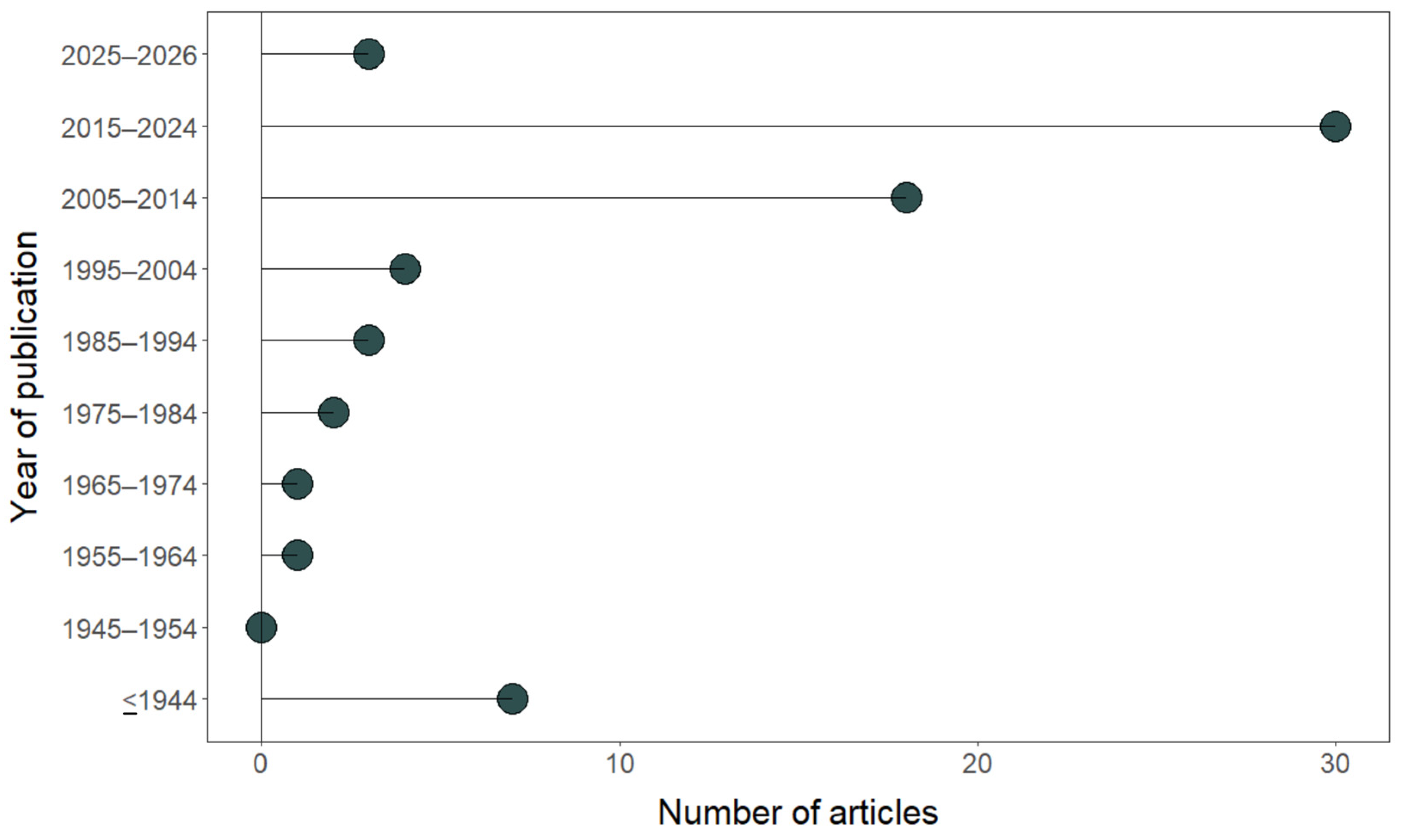
2. General Characteristics of the Occurrence of Microorganisms in Animal Egg Masses
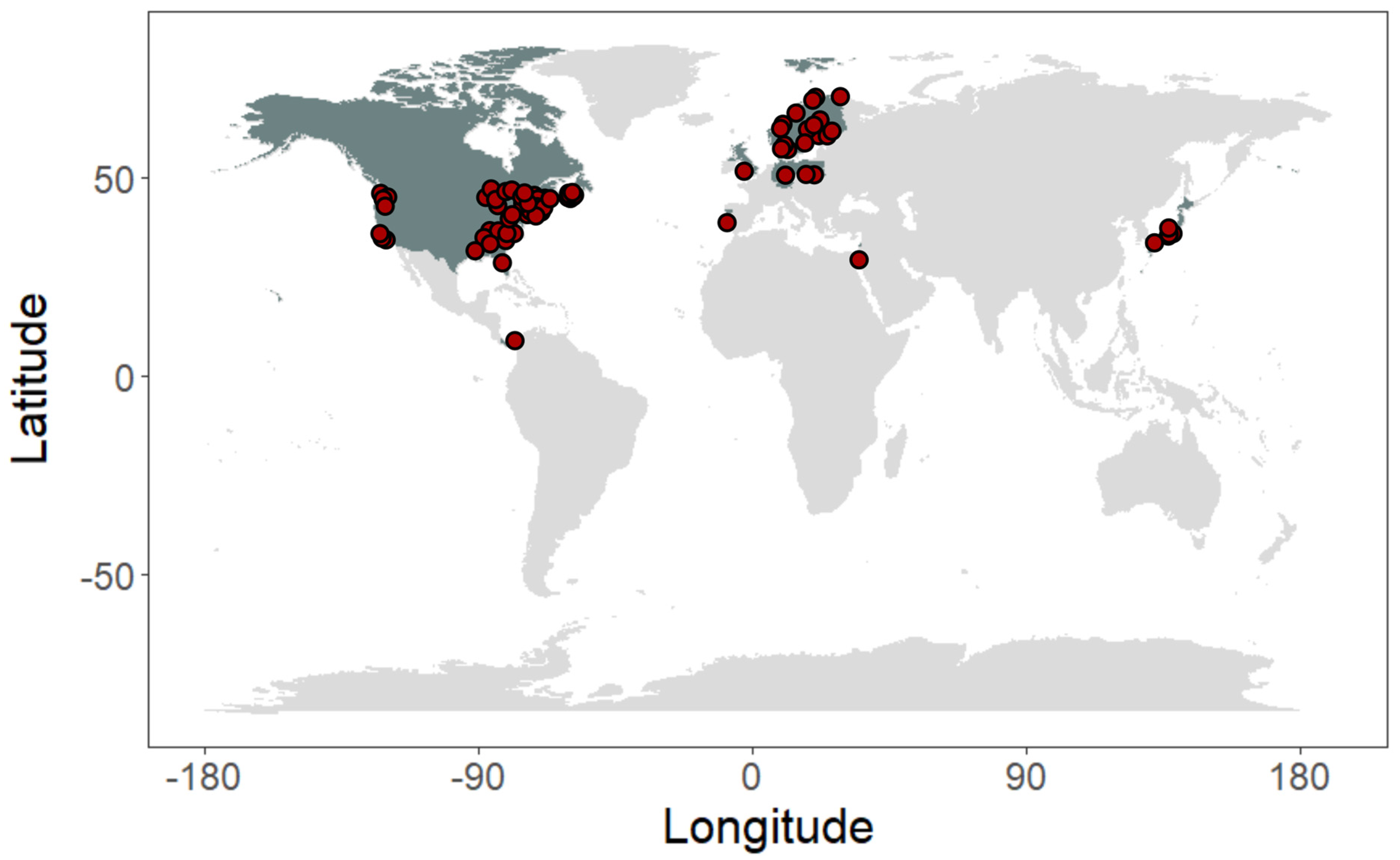
3. Study Sites and Methods

| Methods Used | Reference |
|---|---|
| Historical field observations; larval morphological comparison | [28] |
| Light microscopy; field observations | [12] |
| Experimental manipulation (light vs. dark incubation); developmental observations | [29] |
| Experimental manipulation; oxygen measurements | [23] |
| Laboratory culturing techniques and field research | [30] |
| Light microscopy; physiological and biochemical analyses | [16] |
| Light microscopy; fluorescence microscopy; long-term culture observations; monitoring of sexual cycle | [31] |
| Ultrastructural/histological analysis; electron microscopy; laboratory strain comparisons | [32] |
| Laboratory manipulation experiments; multi-population sampling; embryonic staging (Harrison stages); field collection and controlled rearing | [33] |
| Field observations; nest surveys; reproductive success analysis | [34] |
| Field observations; experimental analysis of egg masses | [4] |
| Laboratory culture; histological analysis; transmission electron microscopy (TEM) | [35] |
| Laboratory oxygen manipulation experiments; controlled fluctuating oxygen treatments; measurements of embryonic development, hatching time, developmental stage, and egg capsule oxygen conductance (GO2) | [36] |
| Laboratory light manipulation experiments; measurements of embryonic rotation, muscular contractions, hatching success, developmental stage, and hatching timing | [37] |
| Laboratory exposure experiments using environmentally relevant atrazine concentrations; assessment of algal persistence, embryonic survival, hatching success, and developmental stage | [38] |
| Laboratory experiments manipulating light intensity and oviposition substrate; measurements of embryonic development rate, hatchling size, and microphyte fouling; field observations | [39] |
| Fluorescence imaging (chlorophyll autofluorescence); algal 18S rDNA amplification; FISH; ultrastructural analysis; histology | [15] |
| Field measurements of oxygen gradients in natural breeding ponds; laboratory pH manipulation experiments; analyses of oxygen, ammonia, lactate, embryo mass, and developmental microenvironment | [40] |
| Field surveys; breeding site monitoring; mtDNA sequencing | [17] |
| Light microscopy, transmission electron microscopy (TEM), histology, ribotype (molecular) analysis | [41] |
| Field collection, algal population quantification, radiolabeled carbon (14C) tracer experiments, developmental and growth measurements | [42] |
| Laboratory culture of isolated algae, atrazine toxicity assays (96 h), chlorophyll fluorescence (PSII yield), optical density and growth rate measurements, recovery assays | [43] |
| Light microscopy, cell counts, immunocytochemistry, chlorophyll a quantification, pulse-amplitude modulated (PAM) fluorometry | [3] |
| Field collection, radiolabeled carbon (14C) tracer experiments, developmental stage analysis | [44] |
| Field sampling of amphibian egg masses, algal isolation and culture, DNA extraction, 18S rDNA sequencing, phylogenetic analyses | [14] |
| Isolation of algae from salamander egg masses, algal culturing, optimization of growth media and culture conditions, 18S rDNA sequencing, laboratory toxicity assay using Zn2+ | [45] |
| Egg capsule manipulation (capsulated vs. de-capsulated embryos), oxygen and ammonia measurements under light and dark conditions, microscopy of algal localization during embryogenesis | [20] |
| In vivo exposure of egg masses to graded atrazine concentrations (up to 338 mg/L), measurement of PSII yield, hatching success, developmental timing; laboratory algal culture and 96 h toxicity assays | [46] |
| Water sampling and filtration, PCR amplification, 18S rDNA sequencing, phylogenetic assignment within Oophila clade | [47] |
| Strain-based culture experiments under light/dark conditions, growth assays in different media (BBM ± glucose/galactose), physiological comparison of heterotrophic capability; taxonomic review and epitype designation using historical and molecular data | [48] |
| Dual RNA-seq (host + symbiont transcriptomics), comparison of intracellular vs. extracellular algal states, gene expression profiling of salamander cells, algal culturing and metabolic inference | [49] |
| Laboratory rearing of clear vs. white egg mass morphs under light (algae present) and dark (algae absent) conditions; field deployment across temperature and dissolved oxygen gradients; comparison of embryo development and hatching success | [50] |
| Male-removal field experiment combined with 16S rRNA gene amplicon sequencing comparing egg microbiomes with and without paternal attendance | [51] |
| Microscopy (morphological characterization); PCR-DGGE; 18S rRNA sequencing; phylogenetic analysis across populations | [18] |
| RNA-Seq transcriptome analysis comparing developmental stages before and after plastid acquisition; differential gene expression analyses | [52] |
| Field pairing of clear and white egg masses; measurement of algal density in egg capsules; larval morphometrics; swim performance trials; mesocosm predation/survival experiment; statistical comparison between morphs | [53] |
| Cross-species algal transplant experiments (swapping cultured symbionts between hosts); embryo co-culture assays; microscopy of tissue entry; comparison of cultured vs. endogenous algal strains; transcriptomic re-analysis of algal gene expression | [54] |
| Nuclear SSU rRNA phylogenetic analysis of algal isolates; mitochondrial phylogeography of host; herbicide toxicity assays (atrazine and 2,4-D); 96 h exposure and recovery growth experiments | [55] |
| Carbon fixation assays under light/dark conditions; separation of host vs. algal carbon fixation contributions; photosynthesis inhibition experiments; comparative metabolic tracing within egg capsules | [56] |
| 18S rRNA phylogenetic analysis; large-scale photobioreactor cultivation; growth rate and areal productivity measurements; biochemical composition profiling (proteins, lipids, carbohydrates, pigments, fatty acids) | [57] |
| Field sampling of egg capsules and pond water over two breeding seasons; nested sampling design; environmental DNA extraction; 18S rRNA amplicon sequencing; microbial community analysis | [19] |
| Field sampling of 149 egg masses across four breeding seasons; quantitative PCR (qPCR) using newly developed Oophila-specific primers; phylogenetic analysis; linear mixed-effects modeling | [58] |
| Egg masses reared under three light regimes (0 h, 14 h, and 24 h light) to manipulate algal photosynthesis; measurements of embryonic growth, survival, metabolic rate under hypoxia, larval growth over two weeks, metabolic rate, and starvation tolerance | [59] |
| DNA metabarcoding (multiple genetic markers); 18S rDNA sequencing; phylogenetic analyses of 18S rDNA and 18 nuclear genes (transcriptomes); comparison of microbial communities from egg clutches, pond water, sediment, and submerged leaves | [60] |
| Bacterial cultivation; 16S rRNA (V4/V5) amplicon sequencing; comparison of bacterial communities from individual egg capsules and surrounding pond water across two ponds and sampling periods | [61] |
| Deep-sea specimen collection (ROV); sex identification of juveniles; histology; light and electron microscopy; fluorescence in situ hybridization (FISH) to localize bacterial symbionts during oogenesis | [62] |
| Field surveys of breeding ponds (2018–2020); egg clutch searches; DNA barcoding; PCR-based mitotyping to distinguish R. dalmatina from R. temporaria and R. arvalis | [63] |
| Algal transformation/transfection; foreign DNA expression; antibiotic selection; GFP reporter assays; RT-PCR for transgene expression; fluorescence microscopy | [27] |
| Sampling of intracapsular fluid from individual egg capsules across embryonic developmental stages; 16S rRNA gene amplicon sequencing; bacterial diversity and community composition analyses | [64] |
| Phylotranscriptomics; RNA-seq; analysis of 76 single-copy nuclear protein-coding genes; inclusion of draft genomes; multi-locus phylogenetic reconstruction and comparison with previous clade frameworks | [65] |
| DNA extraction from historical herbarium/phycological material; PCR with Oophila-specific 18S rRNA primers; cloning and sequencing; phylogenetic analysis including modern reference sequences | [25] |
| Citizen science data (435 egg clutch observations); phylogenetic analysis of environmental and clutch-associated algae; fluorescence microscopy to test for intracellular invasion; comparative sequence analysis | [66] |
| High-throughput amplicon sequencing (PacBio and Illumina); 18S rRNA gene metabarcoding; ASV-based denoising of 16.37 M reads; haplotype network analysis; re-analysis of previous datasets; comparative culture sequencing | [26] |
| Field sampling of egg masses; algal isolation and culturing; morphological characterization; phylogenetic analysis of cultured isolates; comparative clade assignment | [21] |
4. Description of Associated Aquatic Animals and Microalgae
4.1. Characteristics and Description of Animal Species
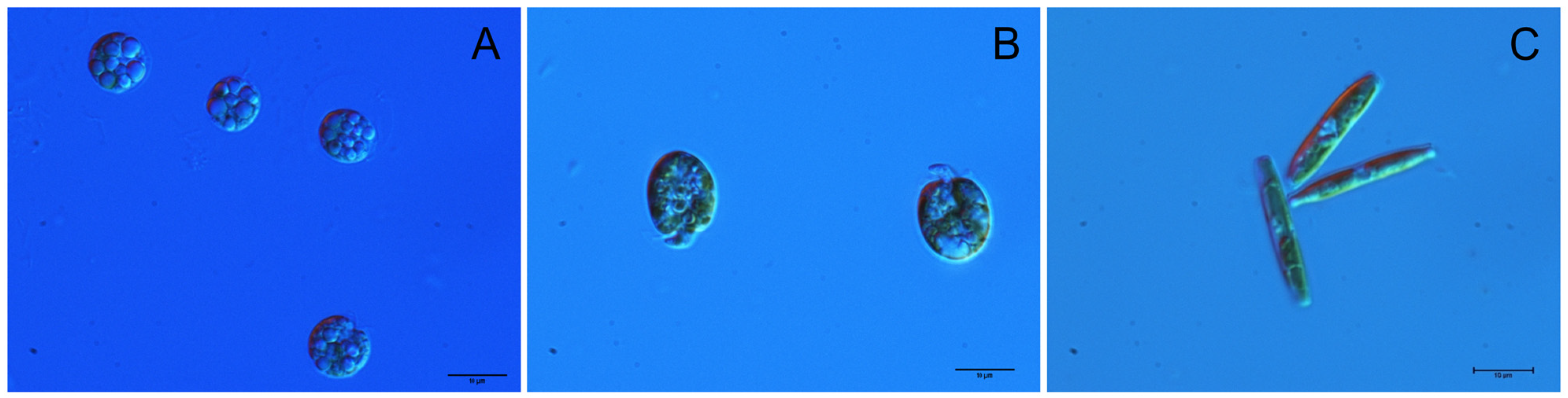
4.2. Characteristics and Composition of Microalgae
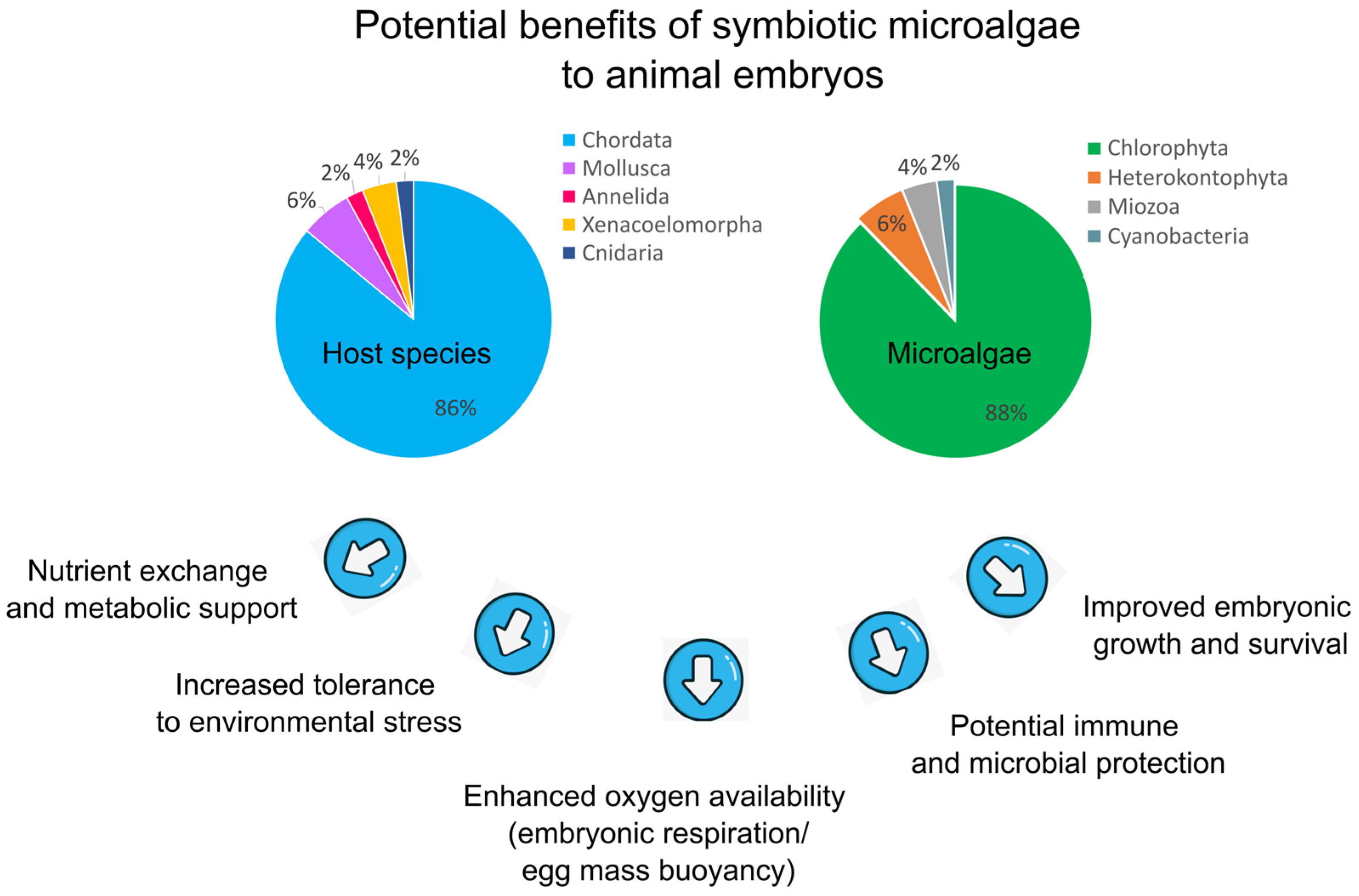
5. Benefits Resulting from the Symbiosis Between Microalgae and Aquatic Animals
6. Summary and Future Research Directions
Supplementary Materials
Author Contributions
Funding
Institutional Review Board Statement
Informed Consent Statement
Data Availability Statement
Conflicts of Interest
References
- Guiry, M.D.; Guiry, G.M.; Morrison, L.; Rindi, F.; Miranda, S.V.; Mathieson, A.C.; Parker, B.C.; Langangen, A.; John, D.M.; Bárbara, I.; et al. AlgaeBase: An on-line resource for algae. Cryptogam. Algol. 2014, 35, 105–115. [Google Scholar] [CrossRef]
- Shestakov, S.V.; Karbysheva, E.A. The origin and evolution of cyanobacteria. Biol. Bull. Rev. 2017, 7, 259–272. [Google Scholar] [CrossRef]
- Bishop, C.D.; Miller, A.G. Dynamics of the growth, life history transformation and photosynthetic capacity of Oophila amblystomatis (Chlorophyceae), a green algal symbiont associated with embryos of the northeastern yellow spotted salamander Ambystoma maculatum (Amphibia). Symbiosis 2014, 63, 47–57. [Google Scholar] [CrossRef]
- Peyton, K.A.; Hanisak, M.D.; Lin, J. Marine algal symbionts benefit benthic invertebrate embryos deposited in gelatinous egg masses. J. Exp. Mar. Biol. Ecol. 2004, 307, 139–164. [Google Scholar] [CrossRef]
- Reitan, K.I.; Rainuzzo, J.R.; Øie, G.; Olsen, Y. A review of the nutritional effects of algae in marine fish larvae. Aquaculture 1997, 155, 207–221. [Google Scholar] [CrossRef]
- Desnitskiy, A.G. A symbiosis of amphibian embryos and larvae with unicellular green algae. Russ. J. Herpetol. 2017, 24, 183–192. [Google Scholar] [CrossRef]
- Kerney, R.; Burns, J.; Kim, E. Investigating mechanisms of algal entry into salamander cells. In Algal and Cyanobacteria Symbioses; World Scientific: Singapore, 2017; pp. 209–239. [Google Scholar]
- Maruyama, S.; Kim, E. Evolution of photosynthetic eukaryotes; current opinion, perplexity, and a new perspective. In Symbiosis: Cellular, Molecular, Medical and Evolutionary Aspects; Springer International Publishing: Cham, Switzerland, 2020; pp. 337–351. [Google Scholar]
- Nyholm, S.V. In the beginning: Egg–microbe interactions and consequences for animal hosts. Philos. Trans. R. Soc. B Biol. Sci. 2020, 375, 20190593. [Google Scholar] [CrossRef] [PubMed]
- Yang, H.; Genot, B.; Duhamel, S.; Kerney, R.; Burns, J.A. Organismal and cellular interactions in vertebrate–alga symbioses. Biochem. Soc. Trans. 2022, 50, 609–620. [Google Scholar] [CrossRef] [PubMed]
- Orr, H. Note on the development of amphibians, chiefly concerning the central nervous system; with additional observations on the hypophysis, mouth, and the appendages and skeleton of the head. J. Cell Sci. 1888, 2, 295–324. [Google Scholar] [CrossRef]
- Gilbert, P.W. Observations on the eggs of Ambystoma maculatum with especial reference to the green algae found within the egg envelopes. Ecology 1942, 23, 215–227. [Google Scholar] [CrossRef]
- Lambert, F.D. An unattached zoosporic form of Coleochaete. Tufts Coll. Stud. Sci. Ser. 1910, 3, 62–68. [Google Scholar] [CrossRef]
- Kim, E.; Lin, Y.; Kerney, R.; Blumenberg, L.; Bishop, C. Phylogenetic analysis of algal symbionts associated with four North American amphibian egg masses. PLoS ONE 2014, 9, e108915. [Google Scholar] [CrossRef] [PubMed]
- Kerney, R.; Kim, E.; Hangarter, R.P.; Heiss, A.A.; Bishop, C.D.; Hall, B.K. Intracellular invasion of green algae in a salamander host. Proc. Natl. Acad. Sci. USA 2011, 108, 6497–6502. [Google Scholar] [CrossRef] [PubMed]
- Goff, L.J.; Stein, J.R. Ammonia: Basis for algal symbiosis in salamander egg masses. Life Sci. 1978, 22, 1463–1468. [Google Scholar] [CrossRef] [PubMed]
- Bonk, M.; Bury, S.; Hofman, S.; Szymura, J.M.; Pabijan, M. A reassessment of the northeastern distribution of Rana dalmatina (Bonaparte, 1840). Herpetol. Notes 2012, 5, 345–354. [Google Scholar]
- Muto, K.; Nishikawa, K.; Kamikawa, R.; Miyashita, H. Symbiotic green algae in eggs of Hynobius nigrescens, an amphibian endemic to Japan. Phycol. Res. 2017, 65, 171–174. [Google Scholar] [CrossRef]
- Jurga, E.; Graham, L.; Bishop, C.D. Oophila is monophyletic within a three-taxon eukaryotic microbiome in egg masses of the salamander Ambystoma maculatum. Symbiosis 2020, 81, 187–199. [Google Scholar] [CrossRef]
- Small, D.P.; Bennett, R.S.; Bishop, C.D. The roles of oxygen and ammonia in the symbiotic relationship between the spotted salamander Ambystoma maculatum and the green alga Oophila amblystomatis during embryonic development. Symbiosis 2014, 64, 1–10. [Google Scholar] [CrossRef]
- Genot, B.; Abedin, S.E.; Shinozaki, N.; Kerney, R.; Maruyama, S. Two distinct green algal symbionts cohabiting in the Japanese black salamander Hynobius nigrescens. bioRxiv 2026. [Google Scholar] [CrossRef]
- Pinder, A.W.; Friet, S.C. Oxygen transport in egg masses of the amphibians Rana sylvatica and Ambystoma maculatum: Convection, diffusion and oxygen production by algae. J. Exp. Biol. 1994, 197, 17–30. [Google Scholar] [CrossRef] [PubMed]
- Hutchison, V.H.; Hammen, C.S. Oxygen utilization in the symbiosis of embryos of the salamander, Ambystoma maculatum and the alga, Oophila amblystomatis. Biol. Bull. 1958, 115, 483–489. [Google Scholar] [CrossRef]
- Bachmann, M.D.; Carlton, R.G.; Burkholder, J.M.; Wetzel, R.G. Symbiosis between salamander eggs and green algae: Microelectrode measurements inside eggs demonstrate effect of photosynthesis on oxygen concentration. Can. J. Zool. 1986, 64, 1586–1588. [Google Scholar] [CrossRef]
- Bishop, C.D.; Garbary, D.J. Taxonomy and nomenclature of Oophila amblystomatis (Chlorophyceae, Chlamydomonadales). J. Phycol. 2024, 60, 380–386. [Google Scholar] [CrossRef] [PubMed]
- Wallace, S.E.; Burns, J.A.; Hale, R.E.; Kerney, R.R.; Mott, C.L.; Bishop, C.D. High partner specificity in an algal-salamander mutualism at continental scale. Front. Amphib. Reptile Sci. 2025, 3, 1609494. [Google Scholar] [CrossRef]
- Genot, B.; Burns, J.A. Transformation of the symbiotic alga Oophila amblystomatis: A new tool for animal-algae symbiosis studies. Symbiosis 2022, 87, 143–151. [Google Scholar] [CrossRef]
- Henry, W.V.; Twitty, V.C. Contributions to the life histories of Dicamptodon ensatus and Ambystoma gracile. Copeia 1940, 1940, 247–250. [Google Scholar] [CrossRef] [PubMed]
- Gilbert, P.W. The alga–egg relationship in Ambystoma maculatum, a case of symbiosis. Ecology 1944, 25, 366–369. [Google Scholar] [CrossRef]
- Dolyak, B.L. A Study of the Life Cycle of the Green Alga Oophila amblystomatis. Master’s Thesis, Southern Connecticut State University, New Haven, CT, USA, 1972. [Google Scholar]
- Thorington, G.; Berger, B.; Margulis, L. Transmission of symbionts through the sexual cycle of Hydra viridis. I. Observations on living organisms. Trans. Am. Microsc. Soc. 1979, 98, 401–413. [Google Scholar] [CrossRef]
- Campbell, R.D. Transmission of symbiotic algae through sexual reproduction in hydra: Movement of algae into the oocyte. Tissue Cell 1990, 22, 137–147. [Google Scholar] [CrossRef] [PubMed]
- Marco, A.; Blaustein, A.R. Symbiosis with green algae affects survival and growth of northwestern salamander embryos. J. Herpetol. 2000, 34, 617–621. [Google Scholar] [CrossRef] [PubMed][Green Version]
- Candolin, U. Effects of algae cover on egg acquisition in male three-spined stickleback. Behaviour 2004, 141, 1389–1399. [Google Scholar] [CrossRef]
- Barneah, O.; Brickner, I.; Hooge, M.; Weis, V.M.; Benayahu, Y. First evidence of maternal transmission of algal endosymbionts at an oocyte stage in a triploblastic host, with observations on reproduction in Waminoa brickneri (Acoelomorpha). Invertebr. Biol. 2007, 126, 113–119. [Google Scholar] [CrossRef]
- Valls, J.H.; Mills, N.E. Intermittent hypoxia in eggs of Ambystoma maculatum: Embryonic development and egg capsule conductance. J. Exp. Biol. 2007, 210, 2430–2435. [Google Scholar] [CrossRef] [PubMed]
- Tattersall, G.J.; Spiegelaar, N. Embryonic motility and hatching success of Ambystoma maculatum are influenced by a symbiotic alga. Can. J. Zool. 2008, 86, 1289–1298. [Google Scholar] [CrossRef]
- Olivier, H.M.; Moon, B.R. The effects of atrazine on spotted salamander embryos and their symbiotic alga. Ecotoxicology 2010, 19, 654–661. [Google Scholar] [PubMed]
- Fernandes, D.A.; Podolsky, R.D. Developmental consequences of association with a photosynthetic substrate for encapsulated embryos of an intertidal gastropod. J. Exp. Mar. Biol. Ecol. 2011, 407, 370–376. [Google Scholar] [CrossRef]
- Bianchini, K.; Tattersall, G.J.; Sashaw, J.; Porteus, C.S.; Wright, P.A. Acid water interferes with salamander–green algae symbiosis during early embryonic development. Physiol. Biochem. Zool. 2012, 85, 470–480. [Google Scholar] [CrossRef] [PubMed]
- Hikosaka-Katayama, T.; Koike, K.; Yamashita, H.; Hikosaka, A.; Koike, K. Mechanisms of maternal inheritance of dinoflagellate symbionts in the acoelomorph worm Waminoa litus. Zool. Sci. 2012, 29, 559–567. [Google Scholar] [CrossRef] [PubMed]
- Graham, E.R.; Fay, S.A.; Davey, A.; Sanders, R.W. Intracapsular algae provide fixed carbon to developing embryos of the salamander Ambystoma maculatum. J. Exp. Biol. 2013, 216, 452–459. [Google Scholar] [CrossRef] [PubMed]
- Baxter, L.; Brain, R.; Rodriguez-Gil, J.L.; Hosmer, A.; Solomon, K.; Hanson, M. Response of the green alga Oophila sp., a salamander endosymbiont, to a PSII-inhibitor under laboratory conditions. Environ. Toxicol. Chem. 2014, 33, 1858–1864. [Google Scholar] [CrossRef] [PubMed]
- Graham, E.R.; McKie-Krisberg, Z.M.; Sanders, R.W. Photosynthetic carbon from algal symbionts peaks during the latter stages of embryonic development in the salamander Ambystoma maculatum. BMC Res. Notes 2014, 7, 764. [Google Scholar] [CrossRef] [PubMed]
- Rodríguez-Gil, J.L.; Brain, R.; Baxter, L.; Ruffell, S.; McConkey, B.; Solomon, K.; Hanson, M. Optimization of culturing conditions for toxicity testing with the alga Oophila sp. (Chlorophyceae), an amphibian endosymbiont. Environ. Toxicol. Chem. 2014, 33, 2566–2575. [Google Scholar] [CrossRef] [PubMed]
- Baxter, L.; Brain, R.A.; Hosmer, A.J.; Nema, M.; Müller, K.M.; Solomon, K.R.; Hanson, M.L. Effects of atrazine on egg masses of the yellow-spotted salamander (Ambystoma maculatum) and its endosymbiotic alga (Oophila amblystomatis). Environ. Pollut. 2015, 206, 324–331. [Google Scholar] [CrossRef] [PubMed]
- Lin, Y.; Bishop, C.D. Identification of free-living Oophila amblystomatis (Chlorophyceae) from yellow spotted salamander and wood frog breeding habitat. Phycologia 2015, 54, 183–191. [Google Scholar] [CrossRef]
- Schultz, N. The Symbiotic Green Algae Oophila (Chlamydomonadales, Chlorophyceae): A Heterotrophic Growth Study and Taxonomic History. Master’s Thesis, University of Connecticut Graduate School, Storrs, CT, USA, 2016. [Google Scholar]
- Burns, J.A.; Zhang, H.; Hill, E.; Kim, E.; Kerney, R. Transcriptome analysis illuminates the nature of the intracellular interaction in a vertebrate-algal symbiosis. eLife 2017, 6, e22054. [Google Scholar] [CrossRef] [PubMed]
- Hale, R.E.; Kennedy, C.; Winkelman, D.; Brown, C. An advantage of clear over white egg mass morphs in metabolically demanding microhabitats suggests a role of symbiotic algae in the maintenance of a polymorphism in the spotted salamander (Ambystoma maculatum). Evol. Ecol. Res. 2017, 18, 637–650. [Google Scholar]
- Hughey, M.C.; Delia, J.; Belden, L.K. Diversity and stability of egg-bacterial assemblages: The role of paternal care in the glassfrog Hyalinobatrachium colymbiphyllum. Biotropica 2017, 49, 792–807. [Google Scholar] [CrossRef]
- Chan, C.X.; Vaysberg, P.; Price, D.C.; Pelletreau, K.N.; Rumpho, M.E.; Bhattacharya, D. Active host response to algal symbionts in the sea slug Elysia chlorotica. Mol. Biol. Evol. 2018, 35, 1706–1711. [Google Scholar] [CrossRef] [PubMed]
- D’Errico, M. The role of Oophila amblystomatis in the maintenance of egg mass color dimorphism in Ambystoma maculatum: Effects on larval morphology, performance, and survival. Capstone UNC Asheville J. Undergrad. Scholarsh. 2019, 32, 279–287. [Google Scholar]
- Kerney, R.R.; Leavitt, J.S.; Hill, E.M.; Zhang, H.; Kim, E.; Burns, J. Co-cultures of Oophila amblystomatis between Ambystoma maculatum and Ambystoma gracile hosts show host-symbiont fidelity. Symbiosis 2019, 78, 73–85. [Google Scholar] [CrossRef]
- Nema, M.; Hanson, M.L.; Müller, K.M. Phylogeny of the egg-loving green alga Oophila amblystomatis (Chlamydomonadales) and its response to the herbicides atrazine and 2,4-D. Symbiosis 2019, 77, 23–39. [Google Scholar] [CrossRef]
- Burns, J.A.; Kerney, R.; Duhamel, S. Heterotrophic carbon fixation in a salamander-alga symbiosis. Front. Microbiol. 2020, 11, 1815. [Google Scholar] [CrossRef] [PubMed]
- Correia, N.; Pereira, H.; Silva, J.T.; Santos, T.; Soares, M.; Sousa, C.B.; Silva, J. Isolation, identification and biotechnological applications of a novel, robust, free-living Chlorococcum (Oophila) amblystomatis strain isolated from a local pond. Appl. Sci. 2020, 10, 3040. [Google Scholar] [CrossRef]
- McDaniels, G.M.; McDaniels, K.T.; Murdock, C.A.; Rayburn, J.R.; Cline, G.R. The phenology of the symbiotic association between Ambystoma maculatum and unicellular algae (Oophila) using molecular techniques. J. Herpetol. 2020, 54, 485–491. [Google Scholar] [CrossRef] [PubMed]
- Small, D.P.; Bishop, C.D. Physiological benefits and latent effects of an algal-salamander symbiosis. Comp. Biochem. Physiol. Part A Mol. Integr. Physiol. 2020, 246, 110715. [Google Scholar] [CrossRef] [PubMed]
- Anslan, S.; Sachs, M.; Rancilhac, L.; Brinkmann, H.; Petersen, J.; Künzel, S.; Schwarz, A.; Arndt, H.; Kerney, R.; Vences, M. Diversity and substrate-specificity of green algae and other micro-eukaryotes colonizing amphibian clutches in Germany, revealed by DNA metabarcoding. Sci. Nat. 2021, 108, 29. [Google Scholar] [CrossRef] [PubMed]
- Bishop, C.; Jurga, E.; Graham, L. Patterns of bacterial diversity in embryonic capsules of the spotted salamander Ambystoma maculatum: An expanding view of a symbiosis. FEMS Microbiol. Ecol. 2021, 97, fiab128. [Google Scholar] [CrossRef] [PubMed]
- Igawa-Ueda, K.; Ikuta, T.; Tame, A.; Yamaguchi, K.; Shigenobu, S.; Hongo, Y.; Takaki, Y.; Fujikura, K.; Maruyama, T.; Yoshida, T. Symbiont transmission onto the cell surface of early oocytes in the deep-sea clam Phreagena okutanii. Zool. Sci. 2021, 38, 140–147. [Google Scholar] [CrossRef] [PubMed]
- Mołoniewicz, L.; Zając, B.; Stachyra, P.; Szymura, J.M.; Pabijan, M. Extension of the known northeastern range limits of the agile frog (Rana dalmatina) in southern Poland. Salamandra 2021, 57, 268–272. [Google Scholar]
- Burgess, W.L.; Bishop, C.D. Bacterial diversity in egg capsular fluid of the spotted salamander Ambystoma maculatum decreases with embryonic development. Microb. Ecol. 2023, 86, 1789–1798. [Google Scholar] [CrossRef] [PubMed]
- Vences, M.; Sachs, M.; Irisarri, I.; Bartels, F.; Eriksson, P.F.; Künzel, S.; Kurabayashi, A.; Laugen, A.T.; Vegso, Z.T.; Bishop, C.D.; et al. Phylotranscriptomic relationships of the Oophila clade of green algae associated to amphibian egg masses. Mol. Phylogenetics Evol. 2024, 200, 108165. [Google Scholar] [CrossRef] [PubMed]
- Eriksson, P.F.; Kerney, R.; Larson-Pollock, R.; Vickers, E.; Timenes Laugen, A. Evidence of Holarctic distribution: Common frog Rana temporaria eggs in Fennoscandia host photosymbiotic green algae (Oophila sp.). Oikos 2025, 2025, e10752. [Google Scholar] [CrossRef]
- Melo Clavijo, J.; Donath, A.; Serôdio, J.; Christa, G. Polymorphic adaptations in metazoans to establish and maintain photosymbioses. Biol. Rev. 2018, 93, 2006–2020. [Google Scholar] [CrossRef] [PubMed]
- Begemann, G. Ecology: Invasion of the body snatchers: Symbiotic algae enter cells of salamander host. Zebrafish 2011, 8, 99–100. [Google Scholar]
- Bishop, S.C. The salamanders of New York. N. Y. State Mus. Bull. 1941, 324, 51–81. [Google Scholar]
- Storer, T.I. A Synopsis of the Amphibia of California; University of California Press: Oakland, CA, USA, 1925; Volume 27, pp. 1–342. [Google Scholar]
- Kojima, A.; Hirose, E. Transfer of prokaryotic algal symbionts from a tropical ascidian (Lissoclinum punctatum) colony to its larvae. Zool. Sci. 2010, 27, 124–127. [Google Scholar] [CrossRef] [PubMed]
- Nilsson, J.; Andersson, J.; Karås, P.; Sandström, O. Recruitment failure and decreasing catches of perch (Perca fluviatilis L.) and pike (Esox lucius L.) in the coastal waters of southeast Sweden. Boreal Environ. Res. 2004, 9, 295. [Google Scholar]
- Brown, C. Differential fitness maintains polymorphism in Ambystoma maculatum. In Proceedings of the National Conference on Undergraduate Research (NCUR) 2016, University of North Carolina Asheville, Asheville, NC, USA, 7–9 April 2016; pp. 324–329. [Google Scholar]
- Lee, R.E. Phycology; Cambridge University Press: New York, NY, USA, 2008. [Google Scholar]
- Mihara, S.; Hase, E. Studies on the vegetative life cycle of Chlamydomonas reinhardi Dangeard in synchronous culture: Some characteristics of the cell cycle. Plant Cell Physiol. 1971, 12, 225–236. [Google Scholar] [CrossRef]
- Mills, N.E.; Ward, Z.A. Egg hypoxia decreases posthatching survival and delays metamorphosis in Ambystoma maculatum (Spotted Salamander). J. Herpetol. 2015, 49, 616–620. [Google Scholar] [CrossRef]


| Key Finding | Reference |
|---|---|
| Embryos of salamanders and frogs were collected for developmental observations; the exact identity of the Amblystoma species could not be confirmed | [11] |
| The authors propose that the larvae reported by Storer (1925) were likely misidentified and, based on morphological and developmental evidence, more plausibly belong to Ambystoma gracile rather than Dicamptodon ensatus. | [28,70] |
| The authors demonstrated that unicellular green algae colonize all three egg envelopes after oviposition and proposed a symbiotic association with developing A. maculatum embryos | [12] |
| The presence of the alga significantly reduced embryo mortality, accelerated embryonic development, increased larval size at hatching, and supported a mutualistic relationship between the alga and salamander embryos | [29] |
| The study demonstrated that photosynthetic oxygen produced by Oophila amblystomatis contributes to embryonic development, supporting a mutualistic exchange between the alga and salamander embryos | [23] |
| The authors proposed the life cycle of Oophila amblystomatis based on laboratory cultures and field observations, describing its developmental stages associated with salamander egg masses | [30] |
| The study demonstrated that symbiotic algae assimilate ammonia excreted by developing embryos, reducing nitrogen accumulation in the perivitelline fluid and promoting embryonic development | [16] |
| The study showed that algal and bacterial symbionts are released during sexual reproduction and transmitted extracellularly, allowing re-establishment of the symbiosis across generations; both symbionts were tracked throughout the sexual cycle of H. viridis under laboratory conditions | [31] |
| The study demonstrated that photosynthetic oxygen production by symbiotic algae exceeds the combined respiratory demand of embryos and algae, leading to rapid oxygen accumulation and even supersaturation within eggs under light conditions, despite hypoxic surrounding water | [24] |
| The study demonstrated a three-step process by which Chlorella symbionts migrate from endodermal digestive cells through extracellular spaces and are ultimately incorporated into developing oocytes via phagocytosis, establishing vertical transmission | [32] |
| The study showed that Ambystoma maculatum egg masses lack convective oxygen transport and rely on diffusion and algal photosynthesis, with O. amblystomatis producing sufficient oxygen to generate hyperoxic conditions in light and partially offset nocturnal hypoxia | [22] |
| The study showed that algal density within the egg perivitelline membrane influences embryonic development and survival in a population-dependent manner, indicating context-dependent effects of algal symbionts | [33] |
| The study showed that dense filamentous algal growth altered the breeding habitat of three-spined sticklebacks, increasing the density of breeding males and promoting a more even distribution of eggs among nests by influencing mating dynamics and sexual selection | [34] |
| Embryos grazed on diatoms within the egg masses, improving the fitness of newly emerged juveniles. Symbiotic algae increased egg mass buoyancy, potentially enhancing larval dispersal and reducing predation by benthic predators | [4] |
| The study provided the first definitive evidence of maternal transmission of dinoflagellate algal symbionts in a triploblastic organism by demonstrating the presence of symbionts within developing oocytes and embryos | [35] |
| The study demonstrated that naturally occurring diurnal oxygen fluctuations generated by O. amblystomatis influence embryonic development, with lower minimum oxygen levels delaying development and causing premature hatching | [36] |
| The study demonstrated that algal photosynthesis influences both embryonic development and behavior. Embryos associated with actively photosynthesizing algae hatched earlier and more synchronously, exhibited reduced ciliary rotation under oxygen-rich conditions, and decreased muscular activity under hypoxic conditions, indicating behavioral adaptations that optimize oxygen use during development | [37] |
| The study demonstrated that atrazine eliminated O. amblystomatis from salamander egg masses and significantly reduced embryonic survival and hatching success while delaying development | [38] |
| The study demonstrated that oviposition on the photosynthetic seagrass Zostera marina accelerated embryonic development under moderate and high light conditions, likely through increased oxygen production. Association with Z. marina limited microphyte growth in egg ribbons of H. vesicula | [39] |
| The study demonstrated that O. amblystomatis can invade embryonic salamander tissues and cells during development, with evidence of algal persistence from early embryonic stages and possible oviductal transmission between generations, indicating a more intimate, partly intracellular symbiosis than previously recognized | [15] |
| The study demonstrated that acidic water (pH 4.5) compromises the benefits of the Ambystoma–Oophila symbiosis by reducing oxygen availability, increasing ammonia and lactate accumulation, and inhibiting embryonic growth | [40] |
| The study documented the northeastern distribution of Rana dalmatina in Poland, identified new breeding localities, and proposed possible postglacial colonization routes based on mitochondrial DNA analyses | [17] |
| Demonstrated that accessory-follicle cells mediate the vertical transmission of two dinoflagellate symbionts into developing oocytes. The study also showed that W. litus maintains a specific Symbiodinium lineage independently of its coral host, indicating an active and highly regulated mechanism of symbiont inheritance | [41] |
| Demonstrated that higher intracapsular densities of O. amblystomatis accelerate salamander embryonic growth and development and provided the first direct evidence that photosynthetically fixed carbon is transferred from an algal symbiont to a vertebrate host | [42] |
| Demonstrated that isolated Oophila sp. is relatively tolerant to atrazine compared with standard algal test species and is unlikely to be inhibited at environmentally relevant atrazine concentrations | [43] |
| Demonstrated that O. amblystomatis undergoes a distinct life-history transition within salamander egg capsules, transforming from motile zoospores into non-motile capsule-associated cells, likely zygotes | [3] |
| Demonstrated that photosynthetically fixed carbon is transferred from O. amblystomatis to salamander embryos only during the second half of embryonic development | [44] |
| Demonstrated that amphibian egg-associated algae comprise a distinct Oophila clade with at least four phylogenetic subclades, showing partial host specificity and evidence that both host switching and co-speciation have shaped the evolution of amphibian–algal symbioses | [14] |
| Developed a reliable method for isolating and culturing Oophila sp. under near-standard laboratory conditions, providing a platform for ecotoxicological studies and demonstrating genetic variation among amphibian-associated algal symbionts | [45] |
| Demonstrated that algal photosynthesis supplies oxygen while algae consume embryo-derived ammonia, suggesting ammonia may limit algal growth | [20] |
| Atrazine reduced algal photosynthetic efficiency in a dose-dependent manner but did not significantly affect algal growth or salamander embryonic development. Both embryo development and algal populations showed high resilience and rapid recovery, indicating that the symbiosis is robust to environmentally relevant atrazine exposure | [46] |
| Demonstrated that pond water in amphibian breeding habitats contains Oophila lineages that match those found in egg masses, including host-associated subclades, supporting the hypothesis that environmental reservoirs serve as the source of symbiotic algae and suggesting possible host specialization | [47] |
| Demonstrated that genetically distinct Oophila strains vary in metabolic flexibility, including potential heterotrophic growth, suggesting strain-specific functional contributions to the symbiosis. Also clarified the taxonomic status of Oophila and proposed an epitype to stabilize future molecular and morphological identification | [48] |
| Showed that intracellular algae undergo strong metabolic reprogramming and stress responses, shifting toward fermentative metabolism, while potentially utilizing host-derived glutamine as a nitrogen source. Host salamander cells display moderated immune modulation rather than full immune activation, indicating a controlled interaction rather than pathogenic response | [49] |
| Clear egg mass morphs show higher embryo survival than white morphs under oxygen-stressed conditions because algae enhance oxygen availability; without algae (or under reduced light penetration), survival drops sharply, confirming algae-mediated protection against hypoxia in dense jelly masses | [50] |
| Egg clutches host a diverse and stable bacterial community; however, paternal care does not significantly shape or maintain egg bacterial assemblages, suggesting eggs acquire microbes through other environmental or maternal routes rather than direct male transmission | [51] |
| Egg capsules contain a consistent, likely unialgal symbiont forming a distinct subclade within the Oophila clade; this represents the first confirmed Oophila–amphibian association outside North America and suggests either vertical transmission or environmental acquisition depending on presence/absence in some egg masses | [18] |
| Acquisition of V. litorea plastids induces major changes in host gene expression, including upregulation of innate immune (MAMP-PPR) pathways, oxidative stress responses, DNA repair, and metabolic processes | [52] |
| No significant differences in algal density between clear and white egg masses, and no differences in larval performance, morphology, or survival; egg mass color morph effects are not mediated by algal density or embryo performance differences | [53] |
| Host identity strongly constrains algal tissue invasion: A. maculatum permits intracellular entry of both native and some foreign algae, while A. gracile resists invasion entirely. Intracapsular vs. cultured algae differ in gene expression, suggesting physiological shifts linked to life cycle state and host environment | [54] |
| Oophila amblystomatis is genetically paraphyletic and split into at least two major clades. The algae show relatively low sensitivity to atrazine and 2,4-D and recover fully after exposure, suggesting these herbicides are unlikely to directly disrupt the symbiont or indirectly affect host development via algal impairment | [55] |
| Salamander embryos themselves fix substantial inorganic carbon, especially in the dark, overwhelming detection of algal-to-host carbon transfer. Host and algal systems compete for inorganic carbon, making any direct photosynthate transfer from algae to embryo difficult to resolve under experimental conditions | [56] |
| Phylogenetically related to Oophila but proposed as Chlorococcum amblystomatis, supporting its potential as a high-value industrial microalgal strain rather than a strict amphibian symbiont | [57] |
| Egg capsules contained exclusively Oophila-clade green algae, supporting a highly specific symbiosis consistent with vertical transmission and/or competitive exclusion. Besides Oophila, only low-diversity chytrid fungi and cercozoan (allapsid) protists were consistently detected, indicating that salamander egg capsules are highly selective microbial habitats | [19] |
| The probability of detecting Oophila DNA and the amount of algal DNA within egg capsules increased with egg mass age, indicating progressive colonization during embryonic development. No significant differences among ponds, sites, or years were detected, and phylogenetic analysis confirmed that the Alabama algae belong to the established Oophila clades | [58] |
| Absence of algal photosynthesis (0 h light) reduced embryonic growth and survival, but larvae later showed compensatory growth. Continuous algal photosynthesis (24 h light) did not improve embryonic performance beyond normal light conditions and resulted in negative latent effects, including smaller larvae, reduced metabolic rates, and lower starvation tolerance. These findings suggest that the benefits of the A. maculatum–Oophila mutualism are optimized under intermediate, natural light conditions rather than maximal algal productivity | [59] |
| Frog egg clutches harbor distinct microbial communities compared with surrounding substrates. Oophila “Clade B” was found exclusively in egg clutches, supporting its specialization for amphibian eggs and suggesting it represents the “true” Oophila, whereas “Clade A” likely represents opportunistic Chlorococcum species | [60] |
| Egg capsules contained much lower bacterial diversity than pond water and harbored a consistent core microbiome dominated by members of Burkholderiales (especially Herbaspirillum) and Oligoflexales. These bacteria were repeatedly detected across ponds and sampling times, suggesting that the classic Oophila–Ambystoma association is likely multipartite, involving stable bacterial associates in addition to the algal symbiont | [61] |
| Symbiotic bacteria were localized on the outer surface of primary oocytes, but were absent from oogonia, indicating that maternal transmission occurs during oocyte development. The study provides new insight into the mechanism of vertical transmission of intracellular bacterial symbionts in deep-sea clams | [62] |
| Forty new breeding localities of R. dalmatina were discovered, extending the known northeastern range in Poland by approximately 100 km. The study identifies the Roztocze region as an important stronghold for this peripheral population and provides valuable information for its conservation | [63] |
| The authors developed a reliable method for transgene expression and presumed stable transformation of Oophila amblystomatis. Transformed algae exhibited heritable antibiotic resistance and GFP fluorescence, providing the first genetic manipulation toolkit for this symbiotic alga and enabling future functional studies of the unique Ambystoma–Oophila symbiosis | [27] |
| Bacterial diversity within egg capsules declined as embryos developed. Community composition was influenced by embryonic stage, pond, year, and their interactions, indicating that the Ambystoma–Oophila association is accompanied by a dynamic bacterial microbiome rather than being a simple bipartite symbiosis | [64] |
| Oophila is not monophyletic as previously assumed, and instead comprises multiple well-supported subclades associated with different amphibian hosts. Some amphibian-associated lineages are interspersed with free-living algae (Chlamydomonas, Chlorococcum), supporting a need for major taxonomic revision of Moewusinia and suggesting repeated or flexible origins of amphibian–alga associations across geography and host species | [65] |
| DNA from the original 1905 type-associated material clusters predominantly within the Oophila clade, confirming that Oophila amblystomatis is a valid, coherent lineage tied to historical type material. The results support retaining the species within Oophila and reject its transfer to Chlorococcum, resolving a key taxonomic debate | [25] |
| Oophila-like algae are present in R. temporaria egg clutches across Fennoscandia, representing multiple subclades. However, no evidence of intracellular algal invasion was detected in frog embryos | [66] |
| The symbiosis shows very high host–symbiont specificity, with no chlorophytes outside the Oophila clade detected. Genetic variation is dominated by two widespread intragenomic ASVs (A and B), whose relative abundance varies geographically (including with latitude). Evidence suggests these variants are intragenomic haplotypes capable of segregation, not distinct species-level lineages, refining understanding of global Oophila diversity | [26] |
| Egg masses consistently contain two distinct algal lineages, indicating multiple symbiotic Oophila-related taxa co-occurring in Japanese salamander eggs, including one lineage closely related to North American symbionts and another within a Chlorococcum-related group | [21] |
Disclaimer/Publisher’s Note: The statements, opinions and data contained in all publications are solely those of the individual author(s) and contributor(s) and not of MDPI and/or the editor(s). MDPI and/or the editor(s) disclaim responsibility for any injury to people or property resulting from any ideas, methods, instructions or products referred to in the content. |
© 2026 by the authors. Licensee MDPI, Basel, Switzerland. This article is an open access article distributed under the terms and conditions of the Creative Commons Attribution (CC BY) license.
Share and Cite
Pisarska, W.; Ochab, M.; Opalińska, Z.; Śliwińska, S. Occurrence and Ecological Significance of Microalgae Species Associated with Egg Clutches of Aquatic Animals. Phycology 2026, 6, 93. https://doi.org/10.3390/phycology6030093
Pisarska W, Ochab M, Opalińska Z, Śliwińska S. Occurrence and Ecological Significance of Microalgae Species Associated with Egg Clutches of Aquatic Animals. Phycology. 2026; 6(3):93. https://doi.org/10.3390/phycology6030093
Chicago/Turabian StylePisarska, Wiktoria, Marcelina Ochab, Zuzanna Opalińska, and Sylwia Śliwińska. 2026. "Occurrence and Ecological Significance of Microalgae Species Associated with Egg Clutches of Aquatic Animals" Phycology 6, no. 3: 93. https://doi.org/10.3390/phycology6030093
APA StylePisarska, W., Ochab, M., Opalińska, Z., & Śliwińska, S. (2026). Occurrence and Ecological Significance of Microalgae Species Associated with Egg Clutches of Aquatic Animals. Phycology, 6(3), 93. https://doi.org/10.3390/phycology6030093