Article

Commercial Arthrospira platensis Extract Modifies the Photophysiology of Cladocopium goreaui, Coral Endosymbiont Microalgae

by
Thibault Le Verge-Campion
1,*,
Thierry Jauffrais
1,
Luc Lefeuvre
2 and
Fanny Houlbrèque
1
1
ENTROPIE, IRD, Ifremer, Université de la Nouvelle-Calédonie, Univ La Réunion, CNRS, UMR 9220 ENTROPIE, 98800 Nouméa, New Caledonia
2
Laboratoires Dermatologiques d’Uriage, 92200 Neuilly-Sur-Seine, France
*
Author to whom correspondence should be addressed.
Phycology 2025, 5(3), 50; https://doi.org/10.3390/phycology5030050
Submission received: 22 April 2025 / Revised: 6 June 2025 / Accepted: 25 June 2025 / Published: 22 September 2025

Abstract

Arthrospira platensis extract is incorporated into sunscreen formulations for its beneficial and UV-protective properties on cultured human cells. However, its effects have not yet been assessed on non-target organisms such as endosymbiotic microalgae in coral tissue. To evaluate its effects, we investigated the photophysiology of the cultured dinoflagellate Cladocopium goreaui using PAM fluorometry (RLC, OJIP) after a 5-day exposure to different extract concentrations. Our results show that, through a hormetic effect, A. platensis enhances the performance index (Pi_Abs) at 0.018 mg L−1 by increasing the number of active reaction centers (RC/ABS) and improving electron transfer efficiency (φEo, ψEo) along the electron transport chain. Conversely, beyond 108.8 mg L−1, negative impacts appear on PSII, increasing the apparent antenna size (ABS/RC) and impairing the oxygen-evolving complex (K-peak), ultimately reducing the maximum relative electron transport rate (rETRm). This relative toxicity, obtained only for the highest concentrations, supports its potential incorporation into cosmetic formulations. This study contributes to improving the ecotoxicity assessment of cosmetic products on non-target organisms.

1. Introduction

Coral reefs are the most impressive biomineral production on the planet. The longevity and spatial extension of corals are largely due to the existence within their endodermal cells of symbiotic microalgae, belonging to the family Symbiodiniaceae [1] and capable of photosynthesis [2]. Extreme coral reef biodiversity, comparable to that of tropical forests, contributes to the economic development of more than a hundred countries that enjoy them along their coasts [3]. Nowadays, in addition to the impact of global warming, coral reefs are threatened by multiple stressors [4,5] including pharmaceutical pollution from direct bathing or indirect releases into the environment [6].
In recent years, more and more pharmaceutical and personal care products (PCPs) have been released into the environment [7]. The presence and persistence of their residues, with active biological action, contribute to create pan-global contamination [8]. Those residues can be detected in all areas of the globe, ranging from urban areas to estuaries, including polar zones [9], temperate zones [10], or tropical waters [11,12,13]. Among these PCPs, certain ultra-violet filters (UVFs) incorporated in sunscreen products are responsible for various harmful effects on corals, altering their metabolism [14] and physiology [15,16,17,18,19] and ultimately leading to coral bleaching [20], the expulsion of the symbiotic microalgae from coral gastrodermal cells. Under normal conditions, through photosynthesis, these Symbiodiniaceae provide the coral host with most of its energy requirements [21]. However, under stressful conditions, photosynthetic mechanisms are disrupted, leading to an excessive production of reactive oxygen species (ROS), toxic to the host [22], causes the Symbiodiniaceae to be expelled. Most studies on UVFs have, however, focused on their direct impact on corals [13,23] rather than on their symbiotic algae. However, Vuckovic et al. [24] have demonstrated that UVF metabolites can also have adverse effects on coral symbionts.
In order to develop environmentally safer sunscreen products containing UVFs, refining formulations is crucial to minimize their impact on organisms. Recent studies suggest that the addition of natural substances could help organisms cope with abiotic stress. Microalgal extracts, both marine or freshwater, are of interest due to their composition involving pigments (carotenoids, phycoerythrin, phycocyanin), nutrients, and antioxidants, as well as UV-absorbing compounds. Already used in pharmaceutical and agronomic industries, algae extracts demonstrate beneficial effects through their antioxidant and protective properties, as well as their role as biostimulants for cultures such as cereals and vineyards, increasing both plant quality and yield [25]. Commercialized as powders or aqueous extracts with the objective of being incorporated into commercial cosmetic formulations, these substances require evaluation of their impacts on model or non-target organisms.
To date, there have been very few studies on the impact of these substances on corals and particularly on symbionts, the most sensitive stage of coral physiology [26,27]. This study investigates the effects of an algal-based extract derived from the cyanobacterium Arthrospira platensis containing phycocyanin (PC), applied at different concentrations to the ex hospite dinoflagellate Cladocopium goreaui. Cladocopium is considered the most dominant genus of symbiotic dinoflagellates in the Indo-Pacific region [1,28] with C. goreaui being a representative symbiont in New Caledonia [28,29]. In addition, C. goreaui is well cultivated under laboratory conditions [30,31], making it a relevant model for ecotoxicological assessments [27,31,32]. More specifically, this study aims to assess the effects of this extract on the growth, photophysiological capacities, activity, and functioning of photosystems, as well as on the efficiency of the electron acceptor intersystem of C. goreaui. We focus in particular on the fluorescent transient analysis (OJIP), a fast, accurate, and non-invasive method for characterizing photosystem structure, as well as electron transfers within organisms exposed to abiotic stress [33,34].

2. Materials and Methods

2.1. Arthrospira Platensis Extract and Concentration Exposure

The extract tested in this study was purchased from GreenTech (SAS GREENSEA, Saint-Beauzire, France). This extract was composed of Arthrospira platensis (CAS n° 223751-80-2; estimated 1–10%) diluted in glycerin (CAS n° 56-81-5; estimated 75–90%) and water (CAS n° 7733-18-5; estimated 10–20%). The phycocyanin concentration was measured at 1.1 g L−1 according to the certificate of analyses provided by the manufacturer. A diluted stock solution of the complete commercial extract was prepared at 1.6 g L−1 in filtered seawater (FSW, 0.2 μm) [31] enriched with L1 media and stored in the dark at 4 °C.
Exposure concentrations were chosen based on three studies estimating the amount of total sunscreen released into seawater during a summer day [35,36,37]. Predicted environmental sunscreen concentration was obtained as follows: PECSunscreen = (N × B × SA × m)/V (1) with N: the number of people on the beach (here fixed at 3000), B: the percentage of people applying sunscreen before bathing (47%), SA: average number of sunscreen applications per person (2.4), m: the amount of sunscreen applied per person and per application (15 g pers−1), and V: the seawater turnover near the beach (5850 m3). These parameters were set according to Ficheux et al. [38] and Labille et al. [35]. PECExtract was obtained by multiplying PECSunscreen by the fraction of the extract (%Extract) in the used formulation: PECExtract = PECSunscreen × %Extract (2) (% extract = 0.2% [39]). Finally, PECExtract was estimated to 0.016 mg L−1 in seawater and corresponds to the lowest dose of the study (LD). To assess the potential effects of the substance at higher concentrations, three additional doses were selected: Medium Dose (MD), High Dose (HD), and Extreme Dose (ED), at 0.16 mg L−1, 1.6 mg L−1, and 1600 mg L−1, respectively. An additional experiment conducted using glycerol at equivalent concentrations (except ED) showed no significant impact on the growth or photophysiology (OJIP, RLC) of C. goreaui (Supplementary Materials Tables S1 and S2).

2.2. Microalgal Culture

Marine microalgae Cladocopium goreaui (SCF055.01-10, WT10) were purchased from the Australian Institute of Marine Sciences (AIMS, Townsville, Australia). Cultures were maintained in filtered seawater (0.2 μm, salinity 35) enriched with L1 media [40] at 26 °C in a thermostatic chamber. A day: night cycle was respected following a 14 h:10 h ratio, with an irradiance of 70 ± 10 μmol photons m−2 s−1. Culture renewal was operated every 3 weeks to maintain the microalgae in exponential growth [31,41].
Microalgae growth cultures were run in triplicates using 250 mL sterilized glass flasks filled with FSW enriched in L1 media. The necessary volume of the extract stock solution was added to the conical flasks to reach the exposure concentrations (LD, MD, HD and ED) with a final volume of 150 mL. The initial cell concentration in each flask was 3.5 · 105 cell mL−1. Flasks were sampled daily (2 mL) for 20 days to perform fluorometric assessments. During the experiment, flasks were randomized daily to ensure similar light conditions [31].

2.3. Physiological Measurements on Algae

2.3.1. Growth Analysis

Growth was followed using two complementary methods. Cell enumeration was performed using cell counts on a Nageotte hemocytometer under optical microscopy (Leica DM750, Leïca Microsystems, Wetzlar, Germany) while minimal fluorescence (F0) was measured by pulse amplitude modulation (PAM) fluorometry (AquaPen-C AP 110-C, Photon Systems Instruments, Dràsov, Czech Republic). To enhance the precision and repeatability of cell measurements, a proxy was created based on cell counts on a hemocytometer and F0 measurements (Supplementary Materials Figure S1). A Gompertz model was fitted to the data to assess the maximum growth rate (µmax in days−1), the maximum cell concentration (A in log(C − C0)), and the latency time (λ in days) [42,43]:
F(t) = A × exp(−exp (µmax × exp(1)/α × (λ − t) + 1)).

2.3.2. PAM Fluorometry

Photosynthetic parameters were measured using a PAM fluorometer (AquaPen-C AP 110-C, Photon Systems Instruments, Dràsov, Czech Republic) equipped with a blue light- emitting diode at 470 nm. All samples were first dark-adapted for 15 min to ensure oxidized (open) PSII reactional centers, then homogenized before measurements at room temperature. Rapid Light Curves (RLCs), allowing one to evaluate the light utilization at different intensities, were generated by illuminating samples with 7 increasing light steps (10, 20, 50, 100, 300, 500, and 1000 μmol photons m−2 s−1) [44]. The Jassby and Platt model [45] was used to fit the RLCs and estimate quantitative parameters: rETRm, alpha (α), and Ek. RLCs were also used to calculate the Stern–Volmer coefficient indicating the non-photochemical quenching induced by light curve (NPQind) [46].
To investigate more precisely the electron pathway within PSII, Fluorescence rise OJIP curves were recorded from 50 μs to 2 s after a 2100 μmol photons m−2 s−1 saturating pulse (corresponding to a 70% super pulse as recommended by the manufacturer) on the same dark-readapted and homogenized samples. Data were extracted and extrapolated according to Strasser et al. [33] to generate the parameters described in Appendix A.1 (Table A1). The OJIP transients were represented as the total fluorescence intensity (Ft) plotted on a logarithmic time scale from O-step (50 μs) to P-step (here considered as Fm) [33] to ensure a sufficient signal prior to analysis. Ft was double-normalized by F0 and Fm to plot the variable fluorescence (Vt) over time Vt = (Ft − F0)/(Fm − F0) to compare signals with different intensities. Vt can also be plotted between each biophysical step to further analyze a specific phase (OJ, OI, JI or IP) as W(YZ) = (Ft − FY)/(FZ − FY). Finally, each parameter was normalized with the control as ΔW(YZ)t = [W(YZ)Treated]t − [W(YZ)Control]t to reveal potential hidden treatment impacts [47]. Wk, corresponding to a specific time, is obtained at t300: (F300 − F0)/(Fj − F0) = Vk/Vj, and represented in function of Fv/Fm.

2.4. Statistical Analyses

Statistical analyses were performed thanks to RStudio software (R Core Team 2017). Data were tested for normality and homoskedasticity using Shapiro–Wilk and Levene tests, respectively. One way ANOVA or Kruskal–Wallis tests were conducted to identify a concentration effect on parameters, followed by post-hoc analysis with Tukey HSD or the Dunn test. All data are presented as mean ± SD with the significance level placed at α = 0.05.
Pi_Abs was plotted as a percentage response relative to the control (control = 100%) as a function of the concentration logarithm. A linear model was tested to fit the data, then adjusted by adding a quadratic term to the model to improve the adjustment. The lowest Akaike Information Criterion (AIC) was used to identify the best model, and residuals were graphically assessed using a QQ-plot.

3. Results

3.1. Growth Rates

Cladocopium goreaui exhibited similar growth parameters (A, μmax, and λ) for the control (CTL), low dose (LD), medium dose (MD), and high dose (HD) conditions (Table 1). In contrast, parameters for the extreme dose (ED) were reduced, with a 77% decrease in the maximum cell concentration (A) and a 45% decrease in the maximum growth rate (μmax) (p < 0.01) compared to the control condition.

3.2. Chlorophyll Fluorescence Signal After 5 Days Exposure

3.2.1. Rapid Light Curves

There was no difference in the maximum relative electron transport rate (rETRm) between CTL, LD, MD, and HD treatments (Figure 1 and Table 2). A 33% reduction in rETRm and a 22% reduction of Ik (the saturating light parameter) were observed in ED treatment. No difference in NPQ was observed in the non-photochemical quenching- (NPQ) induced parameter in all treatments (p > 0.05).

3.2.2. Rapid Chl a Fluorescence Transient Analyses

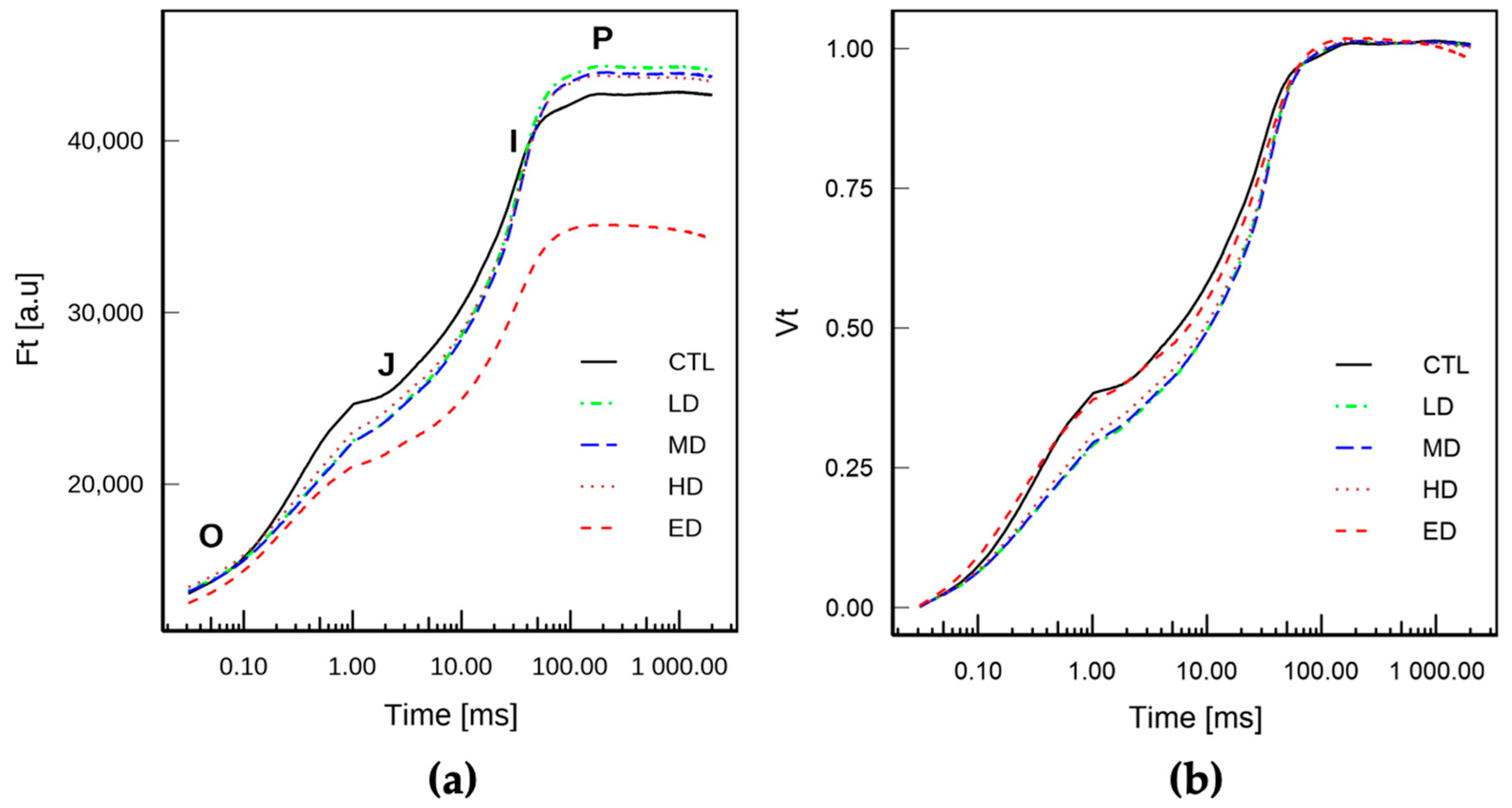
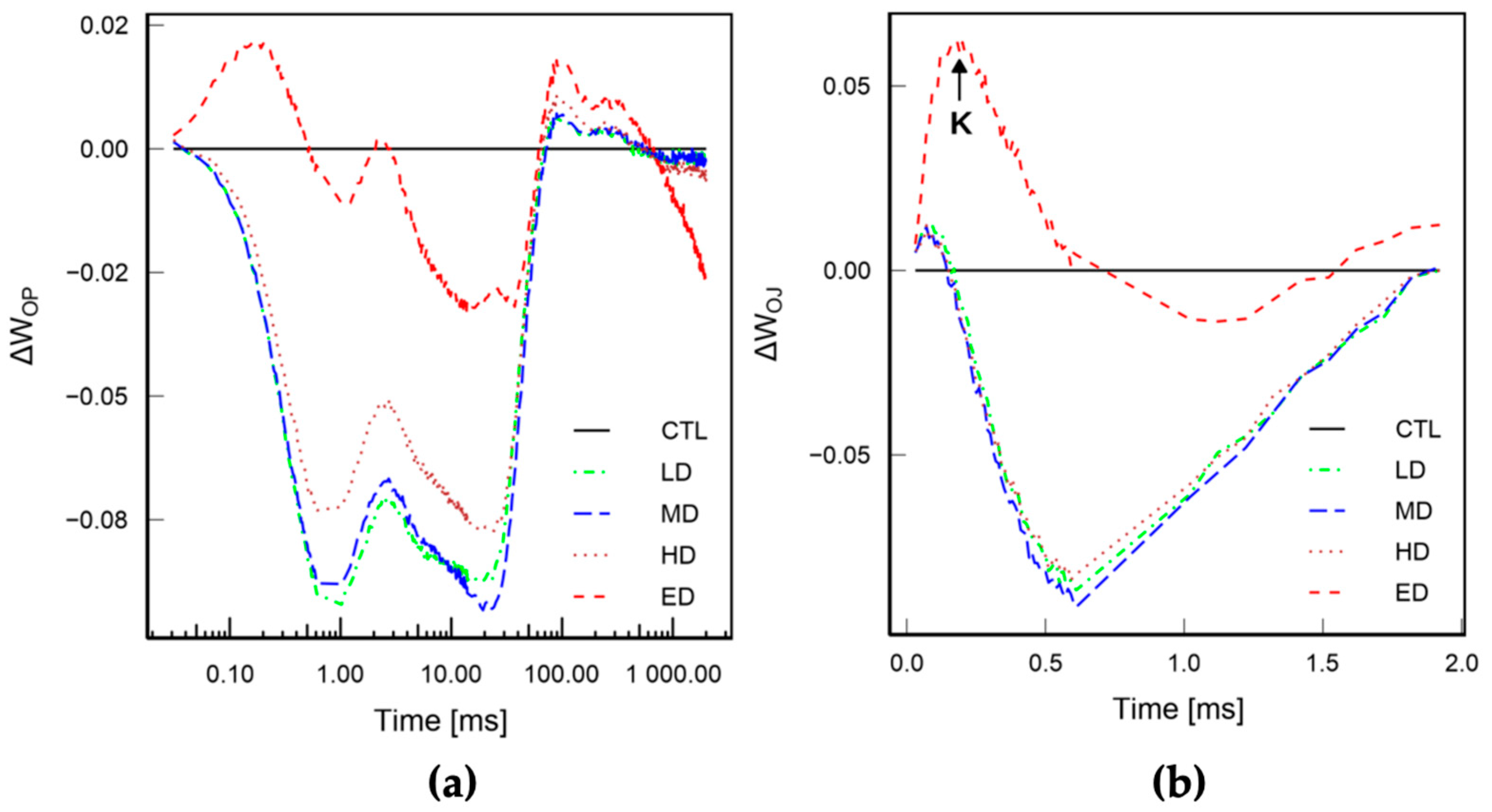
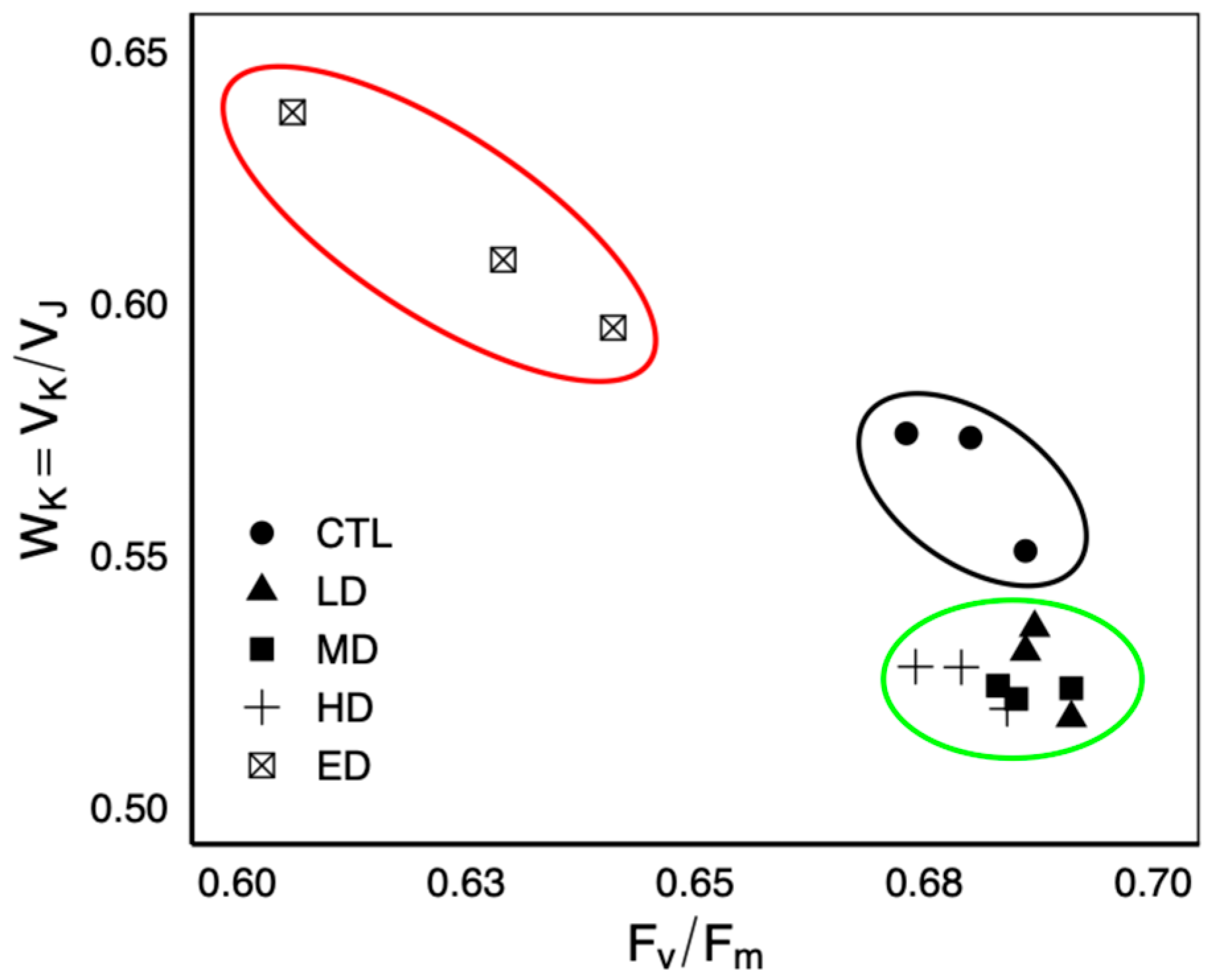
Chl a contained in C. goreaui exhibited a typical fluorescent transient shape in each treatment, going from O-step (F0) to P-step, including two intermediate steps: J-step and I-step (Figure 2a). The lowest signal intensity (Ft) was observed for the ED treatment (Figure 2a). The double normalized fluorescence (Vt = (Ft − F0)/(Fm − F0)) is presented in Figure 2b. Main variations were observed during the O-J phase (0 to 2 ms), with a slight reduction in Vj (corresponding to Vt at 2 ms) for the LD, MD, and HD treatments. A control normalized representation ΔWOP = WOP[Sample] − WOP[Control] allowed a better identification of Vt variations compared to the control condition (Figure 3a). ΔWOP amplitudes for LD, MD and HD were overall negatives during the O-J (0 to 2 ms) and J-I phases (2 to 30 ms) before becoming positives on the I-P-phase (30 ms to Fm). Only the ED treatment showed a positive amplitude during the O-J phase before returning to the control baseline at the J-step (Vj). To further the analysis of the O-J phase, a normalized representation against the control as ΔWOJ = WOJ[Sample] − WOJ[Control] (Figure 3b) revealed a clear positive new band (or peak) near 300 μs, characterized by a higher variable fluorescence in the ED treatment compared to control one (Vt[ED] > Vt[CTL] at 300 μs). This new band, generally hidden on a classic OJIP representation, corresponds to the “K-step” and gives an OKJIP shape to the fluorescence transient in the ED treatment, whereas other treatments revealed the classic OJIP shape. This K-peak amplitude was well defined for ED, as highlighted by the Vk/Vj ratio (Vk/Vj > 0.6), which differed notably from the other treatments (Vk/Vj < 0.6) (Figure 4).
When examining Vk/Vj ratio as a function of Fv/Fm, three distinct groups appeared, illustrated by colored ellipses (ellipses did not indicate any statistical significance) (Figure 4). The ED group was segregated from other treatments, including the control one, by a higher Vk/Vj ratio and a lower Fv/Fm. Interestingly, Vk/Vj ratios for LD, MD, and HD treatments were lower than those of the control one, forming two additional groups with negligible variations in Fv/Fm. In all treatments, Fv/Fm values were above 0.5, with ED values (0.62 ± 0.01) being lower than LD (0.68 ± 0.00, p = 0.012).

3.3. Parameters Extracted from JIP Transient

All parameters (n = 13) extracted from the OJIP analysis showed significant differences between treatments. However, statistical differences were not systematically observed between the control condition and treatments, but rather within treatments, especially between the two smallest concentrations (LD, MD), which displayed comparable values, and the highest one (ED), which statistically differed from them (Table 3).
The relative fluorescence at the J-step (Vj) for LD was significantly reduced by 20% compared to the control (p = 0.04) (Figure 2b and Table 3). Consequently, M0, the initial slope of the standardized transient (Vt) from the O-step to 300 µs, was also affected by this reduction in Vj, though the effect was not significant (Table 3, p = 0.054). Considering specific fluxes, ABS/RC, TR0/RC, and DI0/RC for MD were not significantly but rather slightly reduced by 10% compared to the control condition. ABS/RC, TR0/RC, ET0/RC, and DI0/RC for ED, however, increased by 29, 17, 7, and 35% (p < 0.05) compared to MD, finally exceeding the control values (Table 3).
In contrast, efficiencies followed an opposite trend with φP0, ψE0, and φE0 for ED being reduced by 8, 8, and 35%, respectively, compared to LD (p < 0.05). ψE0 and φE0 for LD were increased by 11 and 14% compared to the control, although they were not significant. φP0 exhibited the lowest variations, with a maximum reduction of 7% for ED compared to the control.
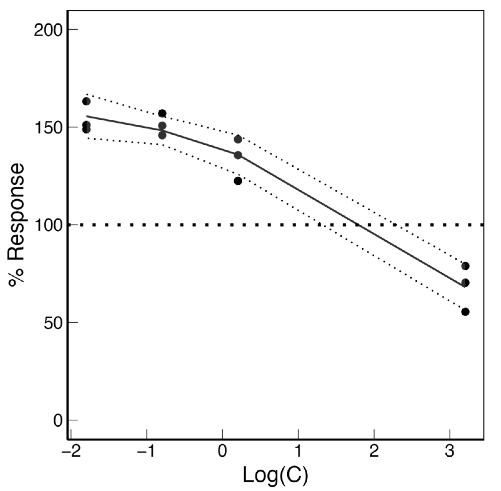
By analyzing the variation coefficient of the parameters, the strongest variations were observed for Pi_Abs (30%). To characterize the relationship between Pi_Abs and the concentration, data were first modeled using linear or quadratic models (Figure 5). Based on the Akaike Information Criterion (AIC), the quadratic model provided the best fit for Pi_Abs, resulting in a concave parabolic curve where higher values corresponding to the best response. LD and MD induced an over-response up to 150%, which decreased with the increasing concentration to reach a 16% reduction at the ED concentration (Figure 5). Analysis revealed a non-linear relationship between the response and the logarithmic concentration within the experimental range. The curve (best described by the equation y = −0.48x2 − 6.03x + 138.83) identified a maximum response at x = −6.28, corresponding to a real extract concentration of 0.018 mg L−1.

4. Discussion

4.1. Physiological Response of C. goreaui to the A. platensis Extract

Based on growth rate data, the Arthrospira platensis extract at environmental concentrations (LD and MD) did not exhibit any signs of toxicity on the dinoflagellate Cladocopium goreaui after 7 days in culture. Conversely, with a concentration up to 105 the environmental one, the reduction of the maximum growth rate and the maximum cell concentration indicated a strong impact on the growth physiology of C. goreaui [42,43].
Unlike the impacts of herbicides [31], benzophenone-3 [27], copper [48], or microplastics [32], for which a reduction in the energy used for primary photochemistry has been demonstrated, our study did not reveal substantial differences in Fv/Fm, indicating that the impacts were located beyond PSII. These impacts were detected using fast chlorophyll, a fluorescence induction (OJIP), applied to coral endosymbionts, and which provides more detailed insights into the dynamics of electron transport. The PAM fluorometric approach (OJIP, RLC) revealed a biphasic response of microalgae Performance Index (Pi_Abs) to the extract. Pi_Abs is generally used as stress indicator for photosynthetic organisms [33,49,50] or as an indicator of adaptation to specific environments [51,52]. This index is the product of three distinct processes: the RC-active density (RC/ABS), the primary photochemistry efficiency (φP0 also expressed as Fv/Fm), and the probability that a trapped electron goes beyond QA (ψEo) [34,53]. Consequently, a small variation, positive or negative, of one of these parameters influences the Pi_Abs response and enables comparisons between experimental and control conditions. Pi_Abs changes have been shown to reveal early responses to certain specific stress such as drought, high temperature, or metal stress before visible alterations occur [51,54,55].
In this study, the microalgae Pi_Abs biphasic response to the extract is characterized by (1) an increase in energy conservation from photons absorbed by PSII to the reduction of intersystem electron acceptors at low doses (LD and MD), reaching a maximum at 0.018 mg L−1, followed by (2) a decline in energy conservation to a “Zero Equivalent Point” (ZEP) at 108.8 mg L−1. Beyond this threshold, which also corresponds to the No-Observed-Adverse-Effect-Level (NOAEL), adverse photophysiological effects occur on the PSII, primarily due to an impairment of the Oxygen Evolving Complex (OEC). This biphasic relation aligns with the hormetic principle described by Calabrese and Blain [56] and follows an “inverted U-shape curve” with a maximum at 157%, characteristic of a classic hormetic stimulatory response [57]. In the present study, the NOAEL is calculated at 108.8 mg L−1, a concentration up to 104 times greater than those used in the cosmetic industry, suggesting that the extract toxicity only occurs at high doses up to 104 times greater than those used in the cosmetic industry [39].

4.2. Enhancement of the Photosynthetic Steps at Low Doses, Based on Absorption Basis

At low doses, the energy conservation is enhanced by two parameters of Pi_Abs: RC/ABS and ψEo. (1) The increase of RC/ABS indicates a higher RC-active density of the biochemical complex [33], accompanied by a reduction in specific energy fluxes ABS, TR0, ET0, and DI0 per active RC (i.e., QA-reducing RC). This suggests a reduction in the apparent antenna size indicating that the received energy is distributed among a greater number of active RCs [34,58]. Consequently, RCs receiving a lower amount of energy remain longer in their reduced state before being oxidized, thereby reducing their net closure rate (M0) [33]. Interestingly, these adaptations are usually found when organisms have to cope with high light and regulate their RC density to prevent PSII photophysiological damages [55]. Here, low doses of A. platensis extracts would increase the light absorption efficiency of C. goreaui. (2) Once trapped in the Electron Transport Chain (ETC), electrons are then transported through the intersystem to reach the final electron acceptor: ferredoxin. In our case, the yield of trapped electron transported beyond QA (ψE0) is increased compared to a condition without the extract, which facilitates the reduction of the downstream ETC entities: QB, PQ, b6/f, and PC. The electron transport yield (φE0) is also increased due to the combination of φP0 and ψE0, reflecting a higher quality of electron transport from the antenna to the intersystem of electron acceptors. Similarly, such improved electron transport efficiency is also found when photosynthetic organisms are exposed to substances with a protective role, such as antioxidants, allowing the quenching of oxygen reactive species (ROS) generated during the photosynthetic processes [58,59,60,61].
In this experiment, the increase in RC-active density and yield efficiencies at low doses could be attributed to the presence of micronutrients in the Arthrospira platensis extract. This cyanobacterium is known to be a source of trace elements such as manganese (Mn), iron (Fe), or copper (Cu) [62], which play important roles in photosynthesis or cellular repair mechanisms in algae [63,64]. In particular, Mn is involved in the OEC structure and also serves as a cofactor for the superoxide dismutase enzyme (SOD), which scavenges ROS [65]. In addition, A. platensis is also known to produce phycobiliproteins [66], specifically phycocyanin, which is concentrated at 1.1 g L−1 in the used extract, and may have an antioxidant effect on algae. In our case, given the hydrosoluble properties of phycocyanin, this pigment is unlikely to act directly as a protective antioxidant within the chloroplasts where photosynthesis takes place. However, its effects could be indirect. Although the precise mechanisms remain unclear, Xu et al. [67] suggested that exogenous phycocyanin enhanced the antioxidant enzymes activities in Arabidopsis thaliana. Similarly, Varia et al. [68] reported that lettuce grown on phycocyanin-rich soil exhibited a higher flavonoid content, which also contributes to the antioxidant response. These findings highlight the need for further mechanistic evaluation into the beneficial effects of such compounds for coral symbionts.

4.3. High-Dose Adverse Effects of Substances on Photophysiology

Conversely, at high doses beyond NOAEL, A. platensis extract induces adverse effects on C. goreaui photophysiology. Specifically, Pi_Abs at ED is reduced by a factor of two compared to the optimum (157 to 75% of the control condition) and by 30% compared to the control without extract. The apparent antenna size also undergoes modifications in the opposite way as low doses, as evidenced by the reduction in the fraction of active reaction centers (RC-active). With fewer active RCs, the absorption per RC increases, leading to higher specific fluxes (TR0 and ET0) and a saturation of oxidized RCs. As a result, the net closure rate of RCs increases, requiring them to dissipate excess energy (DI0/RC) to prevent photodamage by heat or fluorescence under 2100 μmol photons m−2 s−1. On the contrary, the absence of variation in non-photochemical quenching induced by RLC (NPQind) between treatments, together with the absence of photoinhibition, indicates that light intensity at 1000 μmol photons m−2 s−1 is not inhibitory to PSII [69].
While antenna modifications occur, primary photochemistry is slightly reduced, with φP0 decreasing to 0.63, indicating a small reduction in the fraction of energy transferred to the electron transport chain (ETC). A decrease in φP0 could result either from PSII impairment or from a slowdown in electron transport downstream in the ETC. In our case, φP0 remains relatively high, suggesting that PSII is in a good functional state.
Nevertheless, the presence of a WK/WJ ratio above 0.6 suggests an accumulation of reduced QA and highly fluorescent pheophytin (Pheo) caused by electron limitation on the PSII donor side [70]. Strasser et al. [33] demonstrated that this limitation results from an alteration of the Oxygen-Evolving Complex (OEC), which is unable to supply enough electrons to the excited P680 (P680+), hindering its return to the ground state (P680). This creates a bottleneck at Pheo, which can no longer transfer its electron to QA, and consequently reduces electron transfer beyond QA (from QA to QB, and ultimately to NADPH). The impairment of the OEC and the PSII modifications finally participate to prompt a decrease in the maximum electron transport rate (rETRm) in the ETC [71,72,73].

5. Conclusions

According to our results, different effects can be found following exposure to an A. platensis extract. At low doses, the extract (1) induces a reorganization of the apparent antenna size by increasing the fraction of RC-active as an adaptation to favorable environment and (2) supplies beneficial compounds that protect the ETC, enhancing the electron transport efficiency [63]. Conversely, at high dose, the extract (1) causes apparent antenna modifications to prevent photodamage, (2) alters the EOC due to an excess of ROS produced by excessive energy, (3) reduces the ETC performance, and consequently (4), limits growth of C. goreaui.
We show for the first time that a low concentration of A. platensis extract added to the culture medium of C. goreaui does not induce toxicity. On the contrary, this low dose enhances light absorption efficiency and electron transport, ultimately improving the algal Performance Index. Furthermore, this study assesses the ecotoxicological impact of an algal-based product at environmentally relevant concentrations, a key aspect given its inclusion in sunscreen formulations and the associated risks for coral reef ecosystems. Our study evaluated the exposure of the free-form of C. goreaui to different doses of extract. In view of these results, it is therefore essential to test these same doses on the symbiotic form of C. goreaui (inside the coral host) as well as on other symbiont genera, such as Durusdinium, which are also found in the eastern Pacific region [74]. Symbionts in culture exhibit a relatively similar physiological, morphological, and cellular cycle, while in hospite they display more heterogeneous cells according to their cellular cycle, potentially attenuating the effects of the commercial product.

Supplementary Materials

The following supporting information can be downloaded at: https://www.mdpi.com/article/10.3390/phycology5030050/s1, Table S1: Parameters extracted from Rapid Light Curves (RLC) modelized by Jassby and Platt model. rETRm: maximum relative electron transport rate; Ik: rETRm/α, saturating point; α: ability to use low light intensities. p-values were obtained following a Kruskal–Wallis test. Table S2: Chl a fluorescence parameters in Cladocopium goreaui exposed to different concentrations of an 80% glycerol solution, matrix of the Arthrospira platensis extract. Control (CTL), low dose (LD), medium dose (MD), high dose (HD). Values are mean ± SD (n = 3). p-values were obtained following a Kruskal–Wallis test. Figure S1: Pearson’s correlation analysis between cell concentration (C) in cell mL−1 and minimal fluorescence (F0).

Author Contributions

Conceptualization, T.L.V.-C., F.H., T.J. and L.L.; Methodology, T.L.V.-C., F.H., T.J. and L.L.; Validation, F.H., T.J. and L.L.; Formal Analysis, T.L.V.-C. and T.J.; Investigation, T.L.V.-C., F.H., T.J. and L.L.; Resources, F.H. and T.J.; Data Curation, T.L.V.-C., F.H. and T.J.; Writing—Original Draft Preparation, T.L.V.-C.; Writing—Review and Editing, T.L.V.-C., F.H., T.J. and L.L.; Visualization, T.L.V.-C., F.H. and T.J.; Supervision, F.H. and T.J.; Project Administration, F.H. and L.L.; Funding Acquisition, F.H. and L.L. All authors have read and agreed to the published version of the manuscript.

Funding

T.L.V.-C. was beneficiary of a PhD grant (N° 400116/00) by URIAGE dermatological laboratories. Part of this work was also funded by IRD (PIGMENT project).

Institutional Review Board Statement

Not applicable.

Informed Consent Statement

Not applicable.

Data Availability Statement

Data will be made available on request.

Acknowledgments

The authors would like to thank Noémie Coulombier from the Technopole of New Caledonia for her technical support and for welcoming T.L.V.-C. to the LEMA laboratory.

Conflicts of Interest

The authors declare no conflicts of interest.

Abbreviations

The following abbreviations are used in this manuscript:
PAMPulse Amplificated Fluorometry
OJIPJIP-test
RLCRapid Light Curve
ETCElectron Transport Chain
PSIIPhotosystem II
OECOxygen Evolving Complex
PCPPersonal Care Product
UVFUltra-Violet Filter
ROSReactive oxygen species
PCPhycocyanin
NPQNon-Photochemical Quenching
PARPhotosynthetically Active Radiation
NOAELNo Observed Adverse Effect Level

Appendix A

Appendix A.1

Table A1. Parameters, formulas and their definitions used in this study for the analysis of the Chl a fluorescence [34].
ParameterDefinitionFormulaMeaning
Basic fluorescence data measured or calculated from the JIP-test
F0Minimal fluorescence after dark adaptation F at 50 μsMinimum fluorescence when all PSII RCs are open
FmMaximal fluorescence during a saturating flash on dark adapted sampleF at P-step (peak)Maximal fluorescence when all PSII RCs are closed (can be attributed to Fp)
FmMaximum fluorescence in a light-adapted sample under a saturating flash
FtFluorescence at time t
FvVariable fluorescenceFt − F0
Fv/FmMaximum quantum yield for primary photochemistry(Fm − F0)/FmMaximum light utilization efficiency of PSII
VtRelative variable fluorescence at time t(Ft − F0)/(Fm − F0)Relative fluorescence double normalized on Fm and F0
M0Initial slope (in ms−1) of the fluorescent transcient normalized on the variable fluorescence (Vt)M0 = [(ΔF/Δt)0]/(Fm − F0)QA reduction rate
Biophysical parameters derived from the basic parameters by the JIP-test
Specific energy fluxes (per active RC: QA-reducing PSII reducing center)
ABS/RCAbsorption flux at the antenna per RCM0 × (1/VJ) × (1/ φP0)PSII apparent antenna size. RC/ABS the reciprocal corresponds to the fraction of active RC per antenna.
TR0/RCTrapped energy flux leading to QA reduction per active RCM0 × (1/ VJ) The rate of which an electron is trapped in RC resulting in reduction of QA to QA
ET0/RCElectron transport flux per active RCM0 × (1/VJ) × (1 − VJ)The rate by which an electron moves from QA to PQ
DI0/RCDissipation flux into heat per active RCABS/RC − TR0/RC
Quantum yields and efficiencies
φP0Maximum quantum yield for primary photochemistryFv/FmQuantum yield for primary chemistry
φE0Quantum yield for electron transport[1 − (F0/Fm)] × (1 − Vj)
ψE0 (1 − Vj)The probability that an electron moves further than QA
Performance index
Pi_AbsPerformance index for energy conservation[RC/ABS] × [ φP0/(1 − φP0)] × [ψE0/(1 − ψE0)]Performance index for energy conservation from a photon absorbed by PSII until the reduction of intersystem electron acceptors
RLC measured and extracted parameters
rETRRelative electron transport ratePAR × QY × 0.5 × 0.84
rETRmaxMaximum relative electron transport rate rETR levelling at a maximum light-saturated rate
αInitial RLC slope, maximal light use coefficient for PSII Ability to use low light intensities
IkLight saturating index [μmol photon m−2 s−1]Ik = rETRmaxAbility to use high light intensities
NPQindInduced Non Photochemical Quenching during RLC experiment(Fm − Ft)/(Ft)Ability to dissipate energy into heat as a protective mechanism. Stern-Volmer coefficient

References

  1. LaJeunesse, T.C.; Parkinson, J.E.; Gabrielson, P.W.; Jeong, H.J.; Reimer, J.D.; Voolstra, C.R.; Santos, S.R. Systematic Revision of Symbiodiniaceae Highlights the Antiquity and Diversity of Coral Endosymbionts. Curr. Biol. 2018, 28, 2570–2580.e6. [Google Scholar] [CrossRef] [PubMed]
  2. Muscatine, L. The Role of Symbiotic Algae in Carbon and Energy Flux in Reef Corals. Coral Reefs 1990, 25, 75. [Google Scholar]
  3. Burke, L.M. Reefs at Risk Revisited; World Resources Institute: Washington, DC, USA, 2011; ISBN 978-1-56973-762-0. [Google Scholar]
  4. Hughes, T.P.; Kerry, J.T.; Álvarez-Noriega, M.; Álvarez-Romero, J.G.; Anderson, K.D.; Baird, A.H.; Babcock, R.C.; Beger, M.; Bellwood, D.R.; Berkelmans, R.; et al. Global Warming and Recurrent Mass Bleaching of Corals. Nature 2017, 543, 373–377. [Google Scholar] [CrossRef]
  5. Hoegh-Guldberg, O.; Andréfouët, S.; Fabricius, K.E.; Diaz-Pulido, G.; Lough, J.; Marshall, P.A.; Pratchett, M.S. Vulnerability of Coral Reefs in the Tropical Pacific to Climate Change. In Vulnerability of Tropical Pacific Fisheries and Aquaculture to Climate Change; Secretariat of the Pacific Community: Noumea, New Caledonia, 2011; Volume 49. [Google Scholar]
  6. Sánchez-Quiles, D.; Blasco, J.; Tovar-Sánchez, A. Sunscreen Components Are a New Environmental Concern in Coastal Waters: An Overview. In The Handbook of Environmental Chemistry; Springer: Berlin/Heidelberg, Germany, 2020. [Google Scholar]
  7. Gaw, S.; Thomas, K.V.; Hutchinson, T.H. Sources, Impacts and Trends of Pharmaceuticals in the Marine and Coastal Environment. Phil. Trans. R. Soc. B 2014, 369, 20130572. [Google Scholar] [CrossRef]
  8. Watkins, Y.S.D.; Sallach, J.B. Investigating the Exposure and Impact of Chemical UV Filters on Coral Reef Ecosystems: Review and Research Gap Prioritization. Integr. Envir Assess. Manag. 2021, 17, 967–981. [Google Scholar] [CrossRef]
  9. D’Amico, M.; Kallenborn, R.; Scoto, F.; Gambaro, A.; Gallet, J.C.; Spolaor, A.; Vecchiato, M. Chemicals of Emerging Arctic Concern in North-Western Spitsbergen Snow: Distribution and Sources. Sci. Total Environ. 2024, 908, 168401. [Google Scholar] [CrossRef]
  10. Apel, C.; Joerss, H.; Ebinghaus, R. Environmental Occurrence and Hazard of Organic UV Stabilizers and UV Filters in the Sediment of European North and Baltic Seas. Chemosphere 2018, 212, 254–261. [Google Scholar] [CrossRef]
  11. Downs, C.A.; Bishop, E.; Diaz-Cruz, M.S.; Haghshenas, S.A.; Stien, D.; Rodrigues, A.M.S.; Woodley, C.M.; Sunyer-Caldú, A.; Doust, S.N.; Espero, W.; et al. Oxybenzone Contamination from Sunscreen Pollution and Its Ecological Threat to Hanauma Bay, Oahu, Hawaii, U.S.A. Chemosphere 2022, 291, 132880. [Google Scholar] [CrossRef] [PubMed]
  12. Mitchelmore, C.L.; He, K.; Gonsior, M.; Hain, E.; Heyes, A.; Clark, C.; Younger, R.; Schmitt-Kopplin, P.; Feerick, A.; Conway, A.; et al. Occurrence and Distribution of UV-Filters and Other Anthropogenic Contaminants in Coastal Surface Water, Sediment, and Coral Tissue from Hawaii. Sci. Total Environ. 2019, 670, 398–410. [Google Scholar] [CrossRef]
  13. Mitchelmore, C.L.; Burns, E.E.; Conway, A.; Heyes, A.; Davies, I.A. A Critical Review of Organic Ultraviolet Filter Exposure, Hazard, and Risk to Corals. Environ. Toxicol. Chem. 2021, 40, 967–988. [Google Scholar] [CrossRef]
  14. Stien, D.; Suzuki, M.; Rodrigues, A.M.S.; Yvin, M.; Clergeaud, F.; Thorel, E.; Lebaron, P. A Unique Approach to Monitor Stress in Coral Exposed to Emerging Pollutants. Sci. Rep. 2020, 10, 9601. [Google Scholar] [CrossRef]
  15. Downs, C.A.; Kramarsky-Winter, E.; Segal, R.; Fauth, J.; Knutson, S.; Bronstein, O.; Ciner, F.R.; Jeger, R.; Lichtenfeld, Y.; Woodley, C.M.; et al. Toxicopathological Effects of the Sunscreen UV Filter, Oxybenzone (Benzophenone-3), on Coral Planulae and Cultured Primary Cells and Its Environmental Contamination in Hawaii and the U.S. Virgin Islands. Arch. Environ. Contam. Toxicol. 2016, 70, 265–288. [Google Scholar] [CrossRef] [PubMed]
  16. Fel, J.-P.; Lacherez, C.; Bensetra, A.; Mezzache, S.; Béraud, E.; Léonard, M.; Allemand, D.; Ferrier-Pagès, C. Photochemical Response of the Scleractinian Coral Stylophora Pistillata to Some Sunscreen Ingredients. Coral Reefs 2019, 38, 109–122. [Google Scholar] [CrossRef]
  17. He, T.; Tsui, M.M.P.; Tan, C.J.; Ng, K.Y.; Guo, F.W.; Wang, L.H.; Chen, T.H.; Fan, T.Y.; Lam, P.K.S.; Murphy, M.B. Comparative Toxicities of Four Benzophenone Ultraviolet Filters to Two Life Stages of Two Coral Species. Sci. Total Environ. 2019, 651, 2391–2399. [Google Scholar] [CrossRef]
  18. He, T.; Tsui, M.M.P.; Tan, C.J.; Ma, C.Y.; Yiu, S.K.F.; Wang, L.H.; Chen, T.H.; Fan, T.Y.; Lam, P.K.S.; Murphy, M.B. Toxicological Effects of Two Organic Ultraviolet Filters and a Related Commercial Sunscreen Product in Adult Corals. Environ. Pollut. 2019, 245, 462–471. [Google Scholar] [CrossRef] [PubMed]
  19. Wijgerde, T.; van Ballegooijen, M.; Nijland, R.; van der Loos, L.; Kwadijk, C.; Osinga, R.; Murk, A.; Slijkerman, D. Adding Insult to Injury: Effects of Chronic Oxybenzone Exposure and Elevated Temperature on Two Reef-Building Corals. Sci. Total Environ. 2020, 733, 139030. [Google Scholar] [CrossRef]
  20. Danovaro, R.; Bongiorni, L.; Corinaldesi, C.; Giovannelli, D.; Damiani, E.; Astolfi, P.; Greci, L.; Pusceddu, A. Sunscreens Cause Coral Bleaching by Promoting Viral Infections. Environ. Health Perspect. 2008, 116, 441–447. [Google Scholar] [CrossRef]
  21. Muscatine, L.; Porter, J.W. Reef Corals: Mutualistic Symbioses Adapted to Nutrient-Poor Environments. BioScience 1977, 27, 454–460. [Google Scholar] [CrossRef]
  22. Downs, C.A.; Fauth, J.E.; Halas, J.C.; Dustan, P.; Bemiss, J.; Woodley, C.M. Oxidative Stress and Seasonal Coral Bleaching. Free Radic. Biol. Med. 2002, 33, 533–543. [Google Scholar] [CrossRef]
  23. Burns, E.E.; Davies, I.A. Coral Ecotoxicological Data Evaluation for the Environmental Safety Assessment of Ultraviolet Filters. Environ. Toxic. Chem. 2021, 40, 3441–3464. [Google Scholar] [CrossRef]
  24. Vuckovic, D.; Tinoco, A.I.; Ling, L.; Renicke, C.; Pringle, J.R.; Mitch, W.A. Conversion of Oxybenzone Sunscreen to Phototoxic Glucoside Conjugates by Sea Anemones and Corals. Science 2022, 376, 644–648. [Google Scholar] [CrossRef] [PubMed]
  25. Salvi, L.; Niccolai, A.; Cataldo, E.; Sbraci, S.; Paoli, F.; Storchi, P.; Rodolfi, L.; Tredici, M.R.; Mattii, G.B. Effects of Arthrospira Platensis Extract on Physiology and Berry Traits in Vitis Vinifera. Plants 2020, 9, 1805. [Google Scholar] [CrossRef]
  26. Moeller, M.; Pawlowski, S.; Petersen-Thiery, M.; Miller, I.B.; Nietzer, S.; Heisel-Sure, Y.; Kellermann, M.Y.; Schupp, P.J. Challenges in Current Coral Reef Protection—Possible Impacts of UV Filters Used in Sunscreens, a Critical Review. Front. Mar. Sci. 2021, 8, 665548. [Google Scholar] [CrossRef]
  27. Zhang, K.; Shen, Z.; Yang, W.; Guo, J.; Yan, Z.; Li, J.; Lin, J.; Cao, X.; Tang, J.; Liu, Z.; et al. Unraveling the Metabolic Effects of Benzophenone-3 on the Endosymbiotic Dinoflagellate Cladocopium Goreaui. Front. Microbiol. 2023, 13, 1116975. [Google Scholar] [CrossRef] [PubMed]
  28. Butler, C.C.; Turnham, K.E.; Lewis, A.M.; Nitschke, M.R.; Warner, M.E.; Kemp, D.W.; Hoegh-Guldberg, O.; Fitt, W.K.; Van Oppen, M.J.H.; LaJeunesse, T.C. Formal Recognition of Host-generalist Species of Dinoflagellate (Cladocopium, Symbiodiniaceae) Mutualistic with Indo-Pacific Reef Corals. J. Phycol. 2023, 59, 698–711. [Google Scholar] [CrossRef]
  29. Alessi, C.; Lemonnier, H.; Camp, E.F.; Wabete, N.; Payri, C.; Rodolfo Metalpa, R. Algal Symbiont Diversity in Acropora Muricata from the Extreme Reef of Bouraké Associated with Resistance to Coral Bleaching. PLoS ONE 2024, 19, e0296902. [Google Scholar] [CrossRef]
  30. Quigley, K.M.; Alvarez Roa, C.; Beltran, V.H.; Leggat, B.; Willis, B.L. Experimental Evolution of the Coral Algal Endosymbiont, Cladocopium goreaui: Lessons Learnt across a Decade of Stress Experiments to Enhance Coral Heat Tolerance. Restor. Ecol. 2021, 29, e13342. [Google Scholar] [CrossRef]
  31. Marzonie, M.; Flores, F.; Sadoun, N.; Thomas, M.C.; Valada-Mennuni, A.; Kaserzon, S.; Mueller, J.F.; Negri, A.P. Toxicity Thresholds of Nine Herbicides to Coral Symbionts (Symbiodiniaceae). Sci. Rep. 2021, 11, 21636. [Google Scholar] [CrossRef]
  32. Liang, J.; Niu, T.; Zhang, L.; Yang, Y.; Li, Z.; Liang, Z.; Yu, K.; Gong, S. Polystyrene Microplastics Exhibit Toxic Effects on the Widespread Coral Symbiotic Cladocopium Goreaui. Environ. Res. 2025, 268, 120750. [Google Scholar] [CrossRef]
  33. Strasser, R.J.; Tsimilli-Michael, M.; Srivastava, A. Analysis of the Chlorophyll a Fluorescence Transient. In Chlorophyll a Fluorescence; Papageorgiou, G.C., Govindjee, Eds.; Advances in Photosynthesis and Respiration; Springer: Dordrecht, The Netherlands, 2004; Volume 19, pp. 321–362. ISBN 978-1-4020-3217-2. [Google Scholar]
  34. Tsimilli-Michael, M. Special Issue in Honour of Prof. Reto J. Strasser—Revisiting JIP-Test: An Educative Review on Concepts, Assumptions, Approximations, Definitions and Terminology. Photosynthetica 2020, 58, 275–292. [Google Scholar] [CrossRef]
  35. Labille, J.; Slomberg, D.; Catalano, R.; Robert, S.; Apers-Tremelo, M.-L.; Boudenne, J.-L.; Manasfi, T.; Radakovitch, O. Assessing UV Filter Inputs into Beach Waters during Recreational Activity: A Field Study of Three French Mediterranean Beaches from Consumer Survey to Water Analysis. Sci. Total Environ. 2020, 706, 136010. [Google Scholar] [CrossRef] [PubMed]
  36. Poiger, T.; Buser, H.-R.; Balmer, M.E.; Bergqvist, P.-A.; Müller, M.D. Occurrence of UV Filter Compounds from Sunscreens in Surface Waters: Regional Mass Balance in Two Swiss Lakes. Chemosphere 2004, 55, 951–963. [Google Scholar] [CrossRef]
  37. Milinkovitch, T.; Vacher, L.; Le Béguec, M.; Petit, E.; Dubillot, E.; Grimmelpont, M.; Labille, J.; Tran, D.; Ravier, S.; Boudenne, J.-L.; et al. Sunscreen Use during Recreational Activities on a French Atlantic Beach: Release of UV Filters at Sea and Influence of Air Temperature. Environ. Sci. Pollut. Res. 2024, 31, 41046–41058. [Google Scholar] [CrossRef] [PubMed]
  38. Ficheux, A.S.; Chevillotte, G.; Wesolek, N.; Morisset, T.; Dornic, N.; Bernard, A.; Bertho, A.; Romanet, A.; Leroy, L.; Mercat, A.C.; et al. Consumption of Cosmetic Products by the French Population Second Part: Amount Data. Food Chem. Toxicol. 2016, 90, 130–141. [Google Scholar] [CrossRef] [PubMed]
  39. Joly, F.; Branka, J.-E.; Darnis, E.; Lefeuvre, L. Telomere Protective Effects of a Cyanobacteria Phycocyanin against Blue Light and UV Irradiations: A Skin Anti-Aging and Photo-Protective Agent. J. Cosmet. Dermatol. Sci. Appl. 2019, 09, 336–345. [Google Scholar] [CrossRef]
  40. Guillard, R.R.L.; Hargraves, P.E. Stichochrysis Immobilis Is a Diatom, Not a Chrysophyte. Phycologia 1993, 32, 234–236. [Google Scholar] [CrossRef]
  41. Chakravarti, L.J.; Beltran, V.H.; Van Oppen, M.J.H. Rapid Thermal Adaptation in Photosymbionts of Reef-building Corals. Glob. Change Biol. 2017, 23, 4675–4688. [Google Scholar] [CrossRef]
  42. Jauffrais, T.; Agogué, H.; Gemin, M.-P.; Beaugeard, L.; Martin-Jézéquel, V. Effect of Bacteria on Growth and Biochemical Composition of Two Benthic Diatoms Halamphora Coffeaeformis and Entomoneis Paludosa. J. Exp. Mar. Biol. Ecol. 2017, 495, 65–74. [Google Scholar] [CrossRef]
  43. Coulombier, N.; Blanchier, P.; Le Dean, L.; Barthelemy, V.; Lebouvier, N.; Jauffrais, T. The Effects of CO2-Induced Acidification on Tetraselmis Biomass Production, Photophysiology and Antioxidant Activity: A Comparison Using Batch and Continuous Culture. J. Biotechnol. 2021, 325, 312–324. [Google Scholar] [CrossRef]
  44. Sibat, M.; Mai, T.; Chomérat, N.; Bilien, G.; Lhaute, K.; Hess, P.; Séchet, V.; Jauffrais, T. Gambierdiscus Polynesiensis from New Caledonia (South West Pacific Ocean): Morpho-Molecular Characterization, Toxin Profile and Response to Light Intensity. Harmful Algae 2025, 145, 102859. [Google Scholar] [CrossRef]
  45. Jassby, A.D.; Platt, T. Mathematical Formulation of the Relationship between Photosynthesis and Light for Phytoplankton: Photosynthesis-Light Equation. Limnol. Oceanogr. 1976, 21, 540–547. [Google Scholar] [CrossRef]
  46. Ralph, P.J.; Gademann, R. Rapid Light Curves: A Powerful Tool to Assess Photosynthetic Activity. Aquat. Bot. 2005, 82, 222–237. [Google Scholar] [CrossRef]
  47. Oukarroum, A.; Madidi, S.E.; Schansker, G.; Strasser, R.J. Probing the Responses of Barley Cultivars (Hordeum vulgare L.) by Chlorophyll a Fluorescence OLKJIP under Drought Stress and Re-Watering. Environ. Exp. Bot. 2007, 60, 438–446. [Google Scholar] [CrossRef]
  48. Tang, J.; Cai, W.; Yan, Z.; Zhang, K.; Zhou, Z.; Zhao, J.; Lin, S. Interactive Effects of Acidification and Copper Exposure on the Reproduction and Metabolism of Coral Endosymbiont Cladocopium Goreaui. Mar. Pollut. Bull. 2022, 177, 113508. [Google Scholar] [CrossRef] [PubMed]
  49. Gan, T.; Yin, G.; Zhao, N.; Tan, X.; Wang, Y. A Sensitive Response Index Selection for Rapid Assessment of Heavy Metals Toxicity to the Photosynthesis of Chlorella Pyrenoidosa Based on Rapid Chlorophyll Fluorescence Induction Kinetics. Toxics 2023, 11, 468. [Google Scholar] [CrossRef] [PubMed]
  50. Pavlović, I.; Mlinarić, S.; Tarkowská, D.; Oklestkova, J.; Novák, O.; Lepeduš, H.; Bok, V.V.; Brkanac, S.R.; Strnad, M.; Salopek-Sondi, B. Early Brassica Crops Responses to Salinity Stress: A Comparative Analysis Between Chinese Cabbage, White Cabbage, and Kale. Front. Plant Sci. 2019, 10, 450. [Google Scholar] [CrossRef]
  51. Antunović Dunić, J.; Mlinarić, S.; Pavlović, I.; Lepeduš, H.; Salopek-Sondi, B. Comparative Analysis of Primary Photosynthetic Reactions Assessed by OJIP Kinetics in Three Brassica Crops after Drought and Recovery. Appl. Sci. 2023, 13, 3078. [Google Scholar] [CrossRef]
  52. Mlinarić, S.; Žuna Pfeiffer, T.; Krstin, L.; Špoljarić Maronić, D.; Ožura, M.; Stević, F.; Varga, M. Adaptation of Amorpha Fruticosa to Different Habitats Is Enabled by Photosynthetic Apparatus Plasticity. Photosynthetica 2021, 59, 137–147. [Google Scholar] [CrossRef]
  53. Strasser, R.J.; Srivastava, A.; Tsimilli-Michael, M. The Fluorescence Transient as a Tool to Characterize and Screen Photosynthetic Samples. Probing Photosynth. Mech. Regul. Adapt. 2000, 25, 445–483. [Google Scholar]
  54. Kumar, D.; Singh, H.; Raj, S.; Soni, V. Chlorophyll a Fluorescence Kinetics of Mung Bean (Vigna radiata L.) Grown under Artificial Continuous Light. Biochem. Biophys. Rep. 2020, 24, 100813. [Google Scholar] [CrossRef]
  55. Kalaji, H.M.; Carpentier, R.; Allakhverdiev, S.I.; Bosa, K. Fluorescence Parameters as Early Indicators of Light Stress in Barley. J. Photochem. Photobiol. B Biol. 2012, 112, 1–6. [Google Scholar] [CrossRef] [PubMed]
  56. Calabrese, E.; Blain, R. The Occurrence of Hormetic Dose Responses in the Toxicological Literature, the Hormesis Database: An Overview. Toxicol. Appl. Pharmacol. 2005, 202, 289–301. [Google Scholar] [CrossRef]
  57. Calabrese, E.J.; Blain, R.B. The Hormesis Database: The Occurrence of Hormetic Dose Responses in the Toxicological Literature. Regul. Toxicol. Pharmacol. 2011, 61, 73–81. [Google Scholar] [CrossRef] [PubMed]
  58. Wu, P.; Ma, Y.; Ahammed, G.J.; Hao, B.; Chen, J.; Wan, W.; Zhao, Y.; Cui, H.; Xu, W.; Cui, J.; et al. Insights into Melatonin-Induced Photosynthetic Electron Transport under Low-Temperature Stress in Cucumber. Front. Plant Sci. 2022, 13, 1029854. [Google Scholar] [CrossRef] [PubMed]
  59. Pospíšil, P. Production of Reactive Oxygen Species by Photosystem II. Biochim. Biophys. Acta (BBA)-Bioenerg. 2009, 1787, 1151–1160. [Google Scholar] [CrossRef]
  60. Chen, X.; Zhou, Y.; Cong, Y.; Zhu, P.; Xing, J.; Cui, J.; Xu, W.; Shi, Q.; Diao, M.; Liu, H. Ascorbic Acid-Induced Photosynthetic Adaptability of Processing Tomatoes to Salt Stress Probed by Fast OJIP Fluorescence Rise. Front. Plant Sci. 2021, 12, 594400. [Google Scholar] [CrossRef]
  61. Oukarroum, A.; Bussotti, F.; Goltsev, V.; Kalaji, H.M. Correlation between Reactive Oxygen Species Production and Photochemistry of Photosystems I and II in Lemna gibba L. Plants under Salt Stress. Environ. Exp. Bot. 2015, 109, 80–88. [Google Scholar] [CrossRef]
  62. Rahim, A.; Çakir, C.; Ozturk, M.; Şahin, B.; Soulaimani, A.; Sibaoueih, M.; Nasser, B.; Eddoha, R.; Essamadi, A.; El Amiri, B. Chemical Characterization and Nutritional Value of Spirulina Platensis Cultivated in Natural Conditions of Chichaoua Region (Morocco). S. Afr. J. Bot. 2021, 141, 235–242. [Google Scholar] [CrossRef]
  63. Smythers, A.L.; Crislip, J.R.; Slone, D.R.; Flinn, B.B.; Chaffins, J.E.; Camp, K.A.; McFeeley, E.W.; Kolling, D.R.J. Excess Manganese Increases Photosynthetic Activity via Enhanced Reducing Center and Antenna Plasticity in Chlorella Vulgaris. Sci. Rep. 2023, 13, 11301. [Google Scholar] [CrossRef]
  64. Singh, H.; Kumar, D.; Soni, V. Performance of Chlorophyll a Fluorescence Parameters in Lemna Minor under Heavy Metal Stress Induced by Various Concentration of Copper. Sci. Rep. 2022, 12, 10620. [Google Scholar] [CrossRef]
  65. Biscéré, T.; Ferrier-Pagès, C.; Gilbert, A.; Pichler, T.; Houlbrèque, F. Evidence for Mitigation of Coral Bleaching by Manganese. Sci. Rep. 2018, 8, 16789. [Google Scholar] [CrossRef]
  66. Fernandes, R.; Campos, J.; Serra, M.; Fidalgo, J.; Almeida, H.; Casas, A.; Toubarro, D.; Barros, A.I.R.N.A. Exploring the Benefits of Phycocyanin: From Spirulina Cultivation to Its Widespread Applications. Pharmaceuticals 2023, 16, 592. [Google Scholar] [CrossRef]
  67. Xu, Q.; Zhu, T.; Zhao, R.; Zhao, Y.; Duan, Y.; Liu, X.; Luan, G.; Hu, R.; Tang, S.; Ma, X.; et al. Arthrospira Promotes Plant Growth and Soil Properties under High Salinity Environments. Front. Plant Sci. 2023, 14, 1293958. [Google Scholar] [CrossRef] [PubMed]
  68. Varia, J. Biostimulation with Phycocyanin-Rich Spirulina Extract in Hydroponic Vertical Farming. Sci. Hortic. 2022, 299, 111042. [Google Scholar] [CrossRef]
  69. Mériot, V.; Roussel, A.; Brunet, N.; Chomerat, N.; Bilien, G.; Le Déan, L.; Berteaux-Lecellier, V.; Coulombier, N.; Lebouvier, N.; Jauffrais, T. Heterocapsa Cf. Bohaiensis (Dinoflagellate): Identification and Response to Nickel and Iron Stress Revealed through Chlorophyll a Fluorescence. Photosynthetica 2024, 62, 27. [Google Scholar] [CrossRef] [PubMed]
  70. Brestic, M.; Zivcak, M.; Kalaji, H.M.; Carpentier, R.; Allakhverdiev, S.I. Photosystem II Thermostability in Situ: Environmentally Induced Acclimation and Genotype-Specific Reactions in Triticum aestivum L. Plant Physiol. Biochem. 2012, 57, 93–105. [Google Scholar] [CrossRef]
  71. Shanker, A.K.; Amirineni, S.; Bhanu, D.; Yadav, S.K.; Jyothilakshmi, N.; Vanaja, M.; Singh, J.; Sarkar, B.; Maheswari, M.; Singh, V.K. High-Resolution Dissection of Photosystem II Electron Transport Reveals Differential Response to Water Deficit and Heat Stress in Isolation and Combination in Pearl Millet [Pennisetum glaucum (L.) R. Br.]. Front. Plant Sci. 2022, 13, 892676. [Google Scholar] [CrossRef]
  72. Stefanov, M.A.; Rashkov, G.D.; Yotsova, E.K.; Borisova, P.B.; Dobrikova, A.G.; Apostolova, E.L. Different Sensitivity Levels of the Photosynthetic Apparatus in Zea mays L. and Sorghum bicolor L. under Salt Stress. Plants 2021, 10, 1469. [Google Scholar] [CrossRef]
  73. Zhu, Y.; Lin, Q.; Yang, Y.; Xia, Y.; Cai, H.; Feng, X.; Gonçalves, R.J.; Guan, W. Ocean Acidification Enhances the Tolerance of Dinoflagellate Prorocentrum Donghaiense to Nanoplastic-Induced Oxidative Stress by Modulating Photosynthetic Performance. Front. Mar. Sci. 2024, 11, 1494930. [Google Scholar] [CrossRef]
  74. Tanvet, C.; Camp, E.F.; Sutton, J.; Houlbrèque, F.; Thouzeau, G.; Rodolfo-Metalpa, R. Corals Adapted to Extreme and Fluctuating Seawater pH Increase Calcification Rates and Have Unique Symbiont Communities. Ecol. Evol. 2023, 13, e10099. [Google Scholar] [CrossRef]
Figure 1. rETR of C. goreaui under increasing light intensities (PAR, μmol photons m−2 s−1) exposed to different concentrations of A. platensis extract. Curves are fitted Rapid Light Curves (RLCs) modelized by Jassby and Platt model (1975). Control (CTL), low dose (LD), medium dose (MD), high dose (HD), and extreme dose (ED). Black dots are mean ± SD (n = 3).
Phycology 05 00050 g001
Figure 2. Fluorescent OJIP transients of dark-adapted C. goreaui exposed to different A. platensis extract concentrations: control (CTL); low dose (LD); medium dose (MD), high dose (HD), and extreme dose (ED). (a) Fluorescence (Ft) at t time and (b) relative variable fluorescence (Vt), double normalized on Fm and F0 as Vt = (Ft − F0)/(Fm − F0).
Phycology 05 00050 g002
Figure 3. (a) Relative variable fluorescence of Cladocopium goreaui from O-Step to P-Step (WOP) and (b) Relative variable fluorescence from O-Step to J-step (WOJ) of Cladocopium goreaui exposed to different concentrations of Arthrospira platensis extract: control (CTL), low dose (LD), medium dose (MD), high dose (HD), and extreme dose (ED). The K letter indicates the K-peak inflection.
Phycology 05 00050 g003
Figure 4. Amplitude of K-step (Wk = Vk/Vj) as a function of the parameter Fv/Fm in Cladocopium goreaui exposed to different concentrations of Arthrospira platensis extract. Ellipses represent segregated groups (green: positive impacts, black: control, red: negative impacts), but not statistical differences. Control (CTL), low dose (LD), medium dose (MD), high dose (HD), and extreme dose (ED).
Phycology 05 00050 g004
Figure 5. Performance Index (Pi_Abs) of Cladocopium goreaui exposed to increasing concentrations of Arthrospira platensis extract, expressed as percentage response of the control on a logarithmic scale. Black dots represent the response of each treatment relative to the control, expressed as the percentage difference. The black curve represents the fitted quadratic model associated with the data, and dotted lines correspond to the 95% confidence interval.
Phycology 05 00050 g005
Table 1. Growth parameters extracted from the Gompertz model: A (log(C/C0)), the maximum cell concentration (where C and C0 are the final and initial cell concentrations, respectively); µmax (days−1), the maximum growth rate; and λ (days), the latency time. Values are expressed as mean ± SD (n = 3). Different letters indicate statistically significant differences between treatments (p < 0.05): control (CTL), low dose (LD), medium dose (MD), high dose (HD), and extreme dose (ED).
Aμmaxλ
CTL0.80 ± 0.03 ab0.09 ± 0.01 ab2.25 ± 0.65 a
LD0.88 ± 0.05 a0.09 ± 0.01 a2.24 ± 0.37 a
MD0.89 ± 0.07 a0.08 ± 0.01 a2.07 ± 0.62 a
HD0.77 ± 0.03 ab0.09 ± 0.00 ab2.04 ± 0.48 a
ED0.18 ± 0.07 b0.05 ± 0.02 b2.23 ± 0.50 a
Table 2. Photophysiological parameters extracted from the fitted Rapid Light Curve (RLC) for increasing A. platensis extract concentrations. Values are mean ± SD (n = 3). Letters represent statistical differences between treatments: control (CTL), low dose (LD), medium dose (MD), high dose (HD), and extreme dose (ED). rETRm: maximum relative electron transport rate; α: initial RLC slope; Ek: light-saturating index = rETRm/α; NPQind: non-photochemical quenching induced by the RLC.
rETRmαIkNPQind
CTL66.66 ± 0.61 ab0.29 ± 0.00 a228.15 ± 0.31 ab0.29 ± 0.03 a
LD67.04 ± 3.20 ab0.29 ± 0.00 ab230.98 ± 14.02 ab0.31 ± 0.04 a
MD69.15 ± 1.14 a0.28 ± 0.00 ab242.17 ± 8.13 a0.36 ± 0.02 a
HD67.31 ± 1.75 ab0.28 ± 0.01 ab240.76 ± 6.75 a0.38 ± 0.03 a
ED46.52 ± 1.89 b0.25 ± 0.00 b188.38 ± 5.95 b0.30 ± 0.02 a
Table 3. Chl a fluorescence parameters in Cladocopium goreaui exposed to different concentrations of Arthrospira platensis extract. Control (CTL), low dose (LD), medium dose (MD), high dose (HD), and extreme dose (ED). Values are mean ± SD (n = 3). Letters correspond to statistical differences between treatments after post-hoc analysis.
CTLLDMDHDED
Basic parameters calculated from the extracted transcient
F013,600 ± 99 ab13,730 ± 188 ab13,665 ± 211 ab13,925 ± 239 a12,993 ± 500 b
Fm42,448 ± 724 ab43,966 ± 441 a43,586 ± 473 ab43,391 ± 536 ab34,700 ± 915 b
Fj24,849 ± 1377 a23,278 ± 254 a23,256 ± 262 a23,830 ± 366 a21,327 ± 511 b
Vj0.39 ± 0.05 a0.32 ± 0.00 b0.32 ± 0.00 ab0.34 ± 0.01 ab0.38 ± 0.01 a
M00.83 ± 0.14 a0.61 ± 0.02 a0.61 ± 0.01 a0.65 ± 0.02 a0.87 ± 0.05 a
Quantum yields and efficiencies
φP0 (=Fv/Fm)0.68 ± 0.01 ab0.69 ± 0.00 a0.69 ± 0.00 ab0.68 ± 0.01 ab0.63 ± 0.02 b
ψE00.61 ± 0.05 a0.68 ± 0.00 b0.68 ± 0.00 ab0.66 ± 0.01 ab0.62 ± 0.01 a
φE00.41 ± 0.04 ab0.47 ± 0.00 a0.47 ± 0.00 ab0.45 ± 0.01 ab0.39 ± 0.02 b
Specific energy fluxes (per RC: QA reducing PSII reaction center) in ms−1
ABS/RC3.11 ± 0.13 ab2.81 ± 0.07 ab2.79 ± 0.01 a2.84 ± 0.05 ab3.61 ± 0.24 b
Tr0/RC2.11 ± 0.07 ab1.93 ± 0.04 ab1.92 ± 0.00 a1.93 ± 0.02 ab2.25 ± 0.08 b
Et0/RC1.29 ± 0.07 a1.32 ± 0.02 ab1.3 ± 0.01 a1.28 ± 0.01 ab1.39 ± 0.03 b
Di0/RC1.00 ± 0.06 ab0.88 ± 0.03 ab0.88 ± 0.01 a0.91 ± 0.03 ab1.35 ± 0.16 b
Performance index
Pi_Abs1.10 ± 0.34 ab1.70 ± 0.08 a1.66 ± 0.06 a1.47 ± 0.12 ab0.75 ± 0.13 b
Disclaimer/Publisher’s Note: The statements, opinions and data contained in all publications are solely those of the individual author(s) and contributor(s) and not of MDPI and/or the editor(s). MDPI and/or the editor(s) disclaim responsibility for any injury to people or property resulting from any ideas, methods, instructions or products referred to in the content.