Communication

A Morphometric Analysis of the Digital Bones in Karagouniko Sheep and Hellenic Goat

1
Laboratory of Anatomy, Histology and Embryology, Faculty of Veterinary Medicine, University of Thessaly, 43100 Karditsa, Greece
2
Clinic of Farm Animals, Faculty of Veterinary Medicine, Aristotle University of Thessaloniki, 54627 Thessaloniki, Greece
3
Laboratory of Anatomy, Histology and Embryology, Faculty of Veterinary Medicine, Aristotle University of Thessaloniki, 54124 Thessaloniki, Greece
4
Clinic of Surgery, Faculty of Veterinary Medicine, University of Thessaly, 43100 Karditsa, Greece
5
Laboratory of Physiology, Faculty of Veterinary Medicine, University of Thessaly, 43100 Karditsa, Greece
*
Authors to whom correspondence should be addressed.
Ruminants 2025, 5(2), 13; https://doi.org/10.3390/ruminants5020013
Submission received: 19 February 2025 / Revised: 20 March 2025 / Accepted: 25 March 2025 / Published: 28 March 2025

Simple Summary

The digital bones of artiodactyls are the subject of research in the fields of osteoarchaeology, evolution, physiology, and lameness. Measurements of the phalanges of an ovine species and a caprine species were recorded and some indexes of slenderness were calculated in order to present a dataset of values, which are useful in the aforementioned scientific disciplines. Differences were detected between the phalanges of the third and fourth digits, as well as the fore and hind limbs. Furthermore, the indexes calculated were found to be effective in differentiating the third phalanx between sheep and goats as well as in distinguishing between the fore and hind digits. The length of the first phalanx of the fourth fore digit for sheep and the length of the first phalanx of the fourth hind digit for goats were identified as the most useful parameters for the prediction of the lengths of the first and second phalanges. From an archaeological perspective, the prediction equations that were also calculated seem to be of significant value.

Abstract

The phalanges of Karagouniko ewes and Hellenic goats were the subject of the present quantitative study, with the objective of determining whether any differences existed on the corresponding bones of the different digits. The lengths of the phalanges of the third and fourth digits of the fore and hind autopodia, the smallest diaphyseal breadths of the first and second phalanges, the breadths of the proximal and distal ends of the first and second phalanges, the lengths of the dorsal surfaces of the third phalanx, and the heights of the extensor processes of the third phalanx were measured. A total of 44 linear measurements were recorded for each animal species. The summation of the lengths of the digital bones revealed that the third frontal digit was longer than the fourth frontal digit in both animal species and the fourth hind digit was longer than the third hind digit in both animal species. Furthermore, the lengths of the third and fourth frontal digits were greater than those of the corresponding hind digits in both species. The findings of the current metric analysis suggest that the lengths of the paired digits differ in sheep and goat. The results indicate that the morphometry of the digital bones could be important from phylogenetic, biomechanical, and clinical aspects.

1. Introduction

Ruminants represent a diverse group of animals employed in the field of animal production and constitute a component of wild fauna. Their distinctive morphological characteristics, particularly those pertaining to distal portions of their limbs, are a hallmark of their anatomical configuration. Classified as artiodactyls, the autopodial bones of these creatures have consistently garnered research interest from a myriad of scientific disciplines, including evolutionary studies, archaeozoology, physiology, and pathology. The significance of research into digital bones is multifaceted, having inspired investigations into archaeology, adaptation, biomechanics, and pathology in animals (Table 1).
The digital skeleton of the ruminants consists of two digits (third or medial or inner and fourth or lateral or outer). The skeleton of a fully developed digit consists of a proximal (first) phalanx, a middle (second) phalanx, and a distal (third) phalanx.
The digital bones of mammals have been the subject of discussion from the evolutionary point of view [24]. These skeletal elements are known to reflect ecology and locomotory habits in the animals. Furthermore, the phalanges have been identified as a valuable archaeological resource, facilitating the identification of excavated skeletal elements and enabling the study of animal adaptation [9].
Lameness is a prevalent health concern in domestic ruminants. Given that the great majority of lameness cases are attributed to digital and claw lesions, many researchers have explored the potential correlation between the anatomy of the autopodium and claw diseases [1,3,16].
Sheep and goats represent a substantial proportion of the total population of ruminants in Greece. However, the extant literature on the morphometry of the phalanges in these species, and especially in indigenous breeds, is limited. The Karagouniko sheep originates from central Greece, with the majority of the population being located in the Thessaly region. It is a lowland breed, characterized by its thin tail and mixed wool. The breed is notable for its relatively high milk production and its ability to adapt to marginal conditions [25]. The Hellenic goat is the primary breed in Greece. It is an indigenous breed that is also referred to as Capra prisca [26].
The objective of the present study was to provide osteometric data on the phalanges of Karagouniko sheep and Hellenic goats, with a view to assembling the morphological profile of these two animal species. The data thus obtained will facilitate comparisons with other ovine and caprine measurements, aid the interpretation of metric data, and provide further information on the profile of the metric parameters of both the sheep and goat. Furthermore, the potential relationships with functional morphology and their potential association with the pathology of the autopodium were evaluated.

2. Materials and Methods

The distal extremities of the fore and hind limbs from 30 ewes of the Karagouniko breed and 30 Hellenic goats aged more than 2 years (median age 3.7 years), based on the number of permanent incisors [27], were used in the study. The extremities were obtained from a commercial slaughterhouse and immediately subjected to identification (forelimbs and hind limbs) and grouping according to the animal. Subsequently, the autopodia were skinned, macerated, cleaned, and dried.
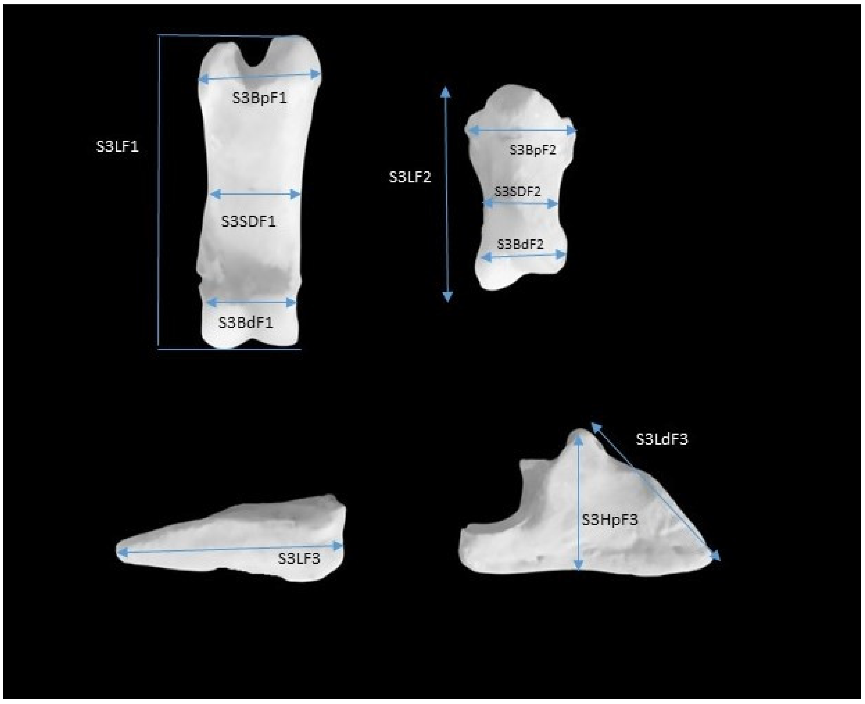
The first, second, and third phalanges were manually measured by the same person with the aid of a caliper, which had the capacity to measure up to 0.01 mm. The definition of linear measurements performed is depicted in Figure 1 and Table 2. The majority of measurements were taken according to the method previously described by von den Driesch [28], with an additional measurement in the third phalanx (height in the region of the extensor process). All linear measurements were in millimeters. In order to avoid errors during the measurements, the identical anatomical reference points, including the fovea articularis, the articular surface, the process, the protuberance, and the borders of the bones, were also considered. All parameters were measured three times, and the mean values were recorded. A series of indexes was calculated as the ratio of the length to the smaller width for the first and second phalanges and the ratio of the length to the height of the extensor process for the third phalanx (Table 3). The total length of the third and fourth digits was calculated by summing the lengths for the first and second phalanges and the length of the dorsal surface of the third phalanx of the relevant digits.
The data were analyzed using the statistical software package SPSS version 21. Paired-sample t-tests were run to compare the data obtained for each measurement between the fore and hind limbs and the third and fourth digit for each animal species. Pearson’s correlation coefficient was used to identify potential linear relationships among the parameters evaluated. Linear regression analysis was used to create prediction equations using variables with a Pearson correlation coefficient higher than 0.7. A significance level of p ≤ 0.05 was used in all comparisons.

3. Results

The results of all bone measurements obtained for sheep are presented in Table S1 in Supplementary Materials, while those obtained for goats are presented in Table S2.
In sheep and goats, the length of the second and third phalanges of the fore third digit were significantly higher than those of the fourth digit (p < 0.05; Table 4). No significant difference was detected on the length of the first phalanx between the third and the fourth digits. Furthermore, a significant decrease in the length of the first phalanx in the hind third digit compared to the fourth hind digit was observed (p < 0.05; Table 4) in both sheep and goats. Additionally, a similar trend was noted for the length of the third phalanx in goats (p < 0.05; Table 4). Conversely, in sheep, the length of the third phalanx was found to be significantly higher at the third digit compared to the fourth digit (p < 0.05; Table 4). In goats, the length of the second phalanx was significantly higher in the third than the fourth hind digit (p < 0.05; Table 4). However, no significant difference was detected in sheep (p>0.05; Table 4).
In sheep, the breadth of the proximal end of the first and second phalanges was significantly higher in the third than the fourth digit in fore and hind limbs (p < 0.05; Table 4). In goats, no such difference was observed (p > 0.05; Table 4), except for the first phalanx of the fore limbs (p < 0.05; Table 4). The breadth of the distal end of the phalanges was comparable between the third and fourth digits in both species in fore and hind limbs (p > 0.05; Table 4) with the exception of the first phalanx, where it was higher in the third than the fourth fore digit (p < 0.05; Table 4). Furthermore, the smallest breadth of the diaphysis of the first and second phalanx was significantly higher in the third than the fourth digit in fore and hind limbs in both species (p < 0.05; Table 4). However, this difference was not observed in the first digit of the hind limbs of goats (p > 0.05). In goats, the length of the dorsal surface of the third phalanx of the fore limbs was significantly higher in the third than the fourth digit (p < 0.05). In contrast, no significant differences were observed in sheep or hind limbs of goats (p > 0.05). However, in sheep and in the fore limb of goats, the height in the region of the extensor process of the third phalanx was significantly higher in the third than the fourth digit (p < 0.05). In the hind limb of goats, there was not such a difference (p > 0.05). In both species, the total length of the third digit was found to be significantly higher in the fore limb and significantly lower in the hind limb than the fourth digit.
The lengths of all phalanges of the third digit were found to be significantly higher in the fore compared to the hind limb in sheep and in goats (p < 0.05; Table 5). In the fourth digit, the lengths of the first and second phalanges were not significantly different between the fore and hind limb (p > 0.05; Table 5), whereas the length of the third phalanx was significantly higher in the fore than the hind limb (p < 0.05; Table 5). Furthermore, the breadth of the proximal and distal ends and the smallest breadth of the diaphysis of the first and second phalanges were significantly higher in the fore than the hind limb (p < 0.05; Table 5) in both species. Additionally, the length of the dorsal surface and the height in the region of the extensor process were also significantly higher in the fore compared to the hind limb (p < 0.05; Table 5). The total lengths of the third and fourth digits were significantly higher in the fore compared to the hind limb in both sheep and goats (p < 0.05; Table 5).
In sheep, the index in the fore first phalanx was significantly lower in the third digit compared to the fourth, but the opposite was observed for the second phalanx (p < 0.05; Table 6). In the hind limb, the indexes for the first and second phalanges were significantly lower in the third compared to the fourth digit. The index for the third phalanx was not significantly different between the third and the fourth digit in either the fore or the hind limb. All indexes of the phalanges were significantly different between the fore and the hind digits, and the indexes of the first phalanx of the third and the fourth digits and of the second phalanx of the fourth digit were significantly higher in the hind than the fore limbs (p < 0.05; Table 6). The indexes of the second phalanx of the third digit and of the third phalanx of the third and fourth digits exhibited significantly higher values in the fore compared to hind limb (p < 0.05; Table 6). In goats, the indexes of the first fore and of the first and second hind phalanges were significantly higher in the third than in the fourth digit (p < 0.05; Table 6). The other indexes were not significantly different between the third and the fourth digits (p > 0.05; Table 6). Furthermore, the indexes in the first and second phalanges of the third and fourth digits were significantly higher in the hind than the fore limb (p < 0.05; Table 6). Conversely, the indexes of the third phalanx were not significantly different between the fore and the hind limbs in either the third or fourth digit. Between species, all indexes calculated for the third phalanx, the index for the second phalanx of the third fore digit, and the index of the first phalanx of the third hind digit were significantly higher in goats than in sheep (p > 0.05; Table 6). However, no other significant difference was detected on the indexes between species (p > 0.05; Table 6).
The prediction equations between measurements with R > 0.7 are presented in Table 7. The length of the first phalanx of the fourth fore digit for sheep and the first phalanx of the fourth hind digit in goats were the most useful measurements for the prediction of the lengths of the other bones, with R2 values higher than 0.85. In sheep, the same measurement can be used for the prediction of the height in the region of the extensor process of the third phalanx, with R2 higher than 0.73.

4. Discussion

The objective of this study was to provide osteometric data on the phalanges of Karagouniko sheep and Hellenic goats. These breeds were selected due to their importance as indigenous local breeds of the Mediterranean basin, and their morphotypes are certainly close to those that can be found on ancient archaeological sites [29], despite their differences in size from modern breeds. All measurements were made manually with the aid of a caliper to determine the absolute bone dimensions. This method was selected over X-rays or digital images, because direct manual measurements have advantages in terms of reliable identification of the exact location of anatomical points, as well as direct visibility.
An interesting finding of this study is the difference between the lengths of the third and fourth digits, as reflected by the differences in phalanx lengths and their sums. In the fore limbs, the third digit is longer than the fourth digit and the opposite occurs in the hind limbs in both sheep and goats. To the best of our knowledge, such information is scarce in the extant literature on sheep and goats. However, similar results have been obtained in studies of cattle [1]. Muggli et al. [3] investigated the length asymmetry of the bovine digits of the fore and hind limbs, finding that the first and second phalanges of the fourth digit were significantly longer than their counterparts of the third digit, whereas the third phalanx of the third digit was longer than its lateral partner. Furthermore, Keller et al. [17] investigated the autopodia of four species of wild artiodactyls using X-rays, revealing that the paired digits differed in length, with the fourth digit being longer than the third. The authors proposed that a longer outer digit might be advantageous on soft ground to maximize the stability of the center of the body mass during walking and also at faster speeds [17]. Additionally, the latter authors hypothesized that this anatomical variation may confer a competitive edge in contexts such as intra-species combat or evasive maneuvers in predator–prey interactions, owing to an enhanced grip strength.
The differences in the length of digits or digital bones have been suggested as risk factors for locomotor disorders and lameness in cattle [1,3,30]. However, such a connection seems to occur, at least in sheep, for fore but not for hind limbs. In a previous study, it was observed that the majority of lesions that cause lameness occur in the inner claw in both fore and hind limbs [31]. Similar results were obtained regarding the prevalence of white line lesions in the fore limbs of sheep [32], where most of them were detected at the claw of the third digit. These observations suggest that bone length is likely to be only one of several contributing factors to lameness, with other anatomical structures such as joints, ligaments, and hooves also playing a role in locomotion. Furthermore, the presence of digital lesions and lameness is significantly influenced by infectious agents.
Another interesting finding was the observed difference in the length and the breadth of the proximal and distal ends and the smallest breadth of the diaphysis of the first and second phalanges and of the length of the digits between fore and hind limbs. The distinct functions of the fore limbs and hind limbs in cursorial quadrupeds suggest a potential correlation with the distribution of body weight across the limbs during locomotion. In sheep, for instance, approximately 30% of the body weight is allocated to each fore limb and 20% to each hind limb [33]. A similar phenomenon was observed in another study [34], where 31.34% of the body weight was found to be distributed between the forelimb and 18.79% between the hind limb in sheep. The direct relationship between these length differences and the prevalence of digital lesions and lameness remains uncertain and requires further investigation. Some studies have indicated a higher prevalence of lameness in the fore limb [35,36], while others have found it to be more prevalent in the hind limb [31,37,38].
The indexes have been demonstrated to be of significant value in facilitating comprehension of the morphology and functionality of the bones. The disparities observed between the fore and hind limbs in both species are indicative of the previously mentioned body weight distribution. Furthermore, from an archaeological perspective, the indexes are also useful for the differentiation of the bones between the different species. The metapodial index has been proposed as a means of differentiating between sheep and goats [39]. The results of the present study suggest that the index of the third phalanx can also be used for the differentiation between sheep and goats. Apart from the differentiation between species, the indexes seem to be useful for the differentiation between the fore and hind limb phalanges, but not for the third phalange of goats.
From an archaeological perspective, the prediction equations also proved to be of significant value. The results obtained from the analysis indicate that the most useful parameter for the prediction of the length of the first and second phalanges in sheep is the length of the first phalanx of the fourth fore digit. In goats, the equivalent parameter is the first phalanx of the fourth hind digit.
5. Conclusions

The findings of the present study suggest the presence of substantial disparities in the lengths and the breadths of the phalanges between the third and the fourth digits as well as between the fore and the hind limb. Furthermore, the indexes calculated were found to be effective in differentiating the third phalanx between sheep and goats as well as in distinguishing between the fore and hind digits. The length of the first phalanx of the fourth fore digit for sheep and the length of the first phalanx of the fourth hind digit for goats were identified as the most useful parameters for the prediction of the lengths of the first and second phalanges.

Supplementary Materials

The following supporting information can be downloaded at: https://www.mdpi.com/article/10.3390/ruminants5020013/s1. Table S1: Bone measurements obtained for sheep, Table S2: Bone measurements obtained for goat.

Author Contributions

Conceptualization, A.I.S., I.V. and A.P.; methodology, A.I.S., I.G. and A.P.; software, P.D.K.; investigation, T.C.; writing—original draft preparation, T.C., P.D.K.; writing—review and editing, A.P., I.G., A.I.S. and I.V.; visualization, supervision, A.P. All authors have read and agreed to the published version of the manuscript.

Funding

This research received no external funding.

Institutional Review Board Statement

This study did not require ethical approval, since the animal bones were collected from slaughterhouses.

Informed Consent Statement

Not applicable, since animals were used in this study and no humans were involved.

Data Availability Statement

The data used in the study will be made available to other researchers on request.

Conflicts of Interest

The authors declare no conflicts of interest.

References

  1. Ocal, M.K.; Sevil, F.; Parin, U. A quantitative study on the digital bones of cattle. Ann. Anat. 2004, 186, 165–168. [Google Scholar] [CrossRef]
  2. Maldre, L. Faunal remains from the settlement site of Pada. Est. J. Archaeol. 2007, 11, 59–79. [Google Scholar] [CrossRef]
  3. Muggli, E.; Sauter-Luis, C.; Braun, U.; Nuss, K. Length asymmetry of the bovine digits. Vet. J. 2011, 188, 295–300. [Google Scholar] [CrossRef]
  4. Muggli, E.; Weidmann, E.; Kircher, P.; Nuss, K. Radiographic Measurement of Hindlimb Digit Length in Standing Heifers. Anat. Histol. Embryol. 2016, 45, 463–468. [Google Scholar] [CrossRef] [PubMed]
  5. Gündemir, O.; Özkan, E.; Mutuş, R. Morphometric study on the digital bones in the domestic cattle. Kafkas Univ. Vet. Fak. Derg. 2020, 26, 75–82. [Google Scholar]
  6. Boessneck, J.; Müller, H.H.; Teichert, M. Osteologische unterscheidungsmerkmale zwischen Schaf (Ovis aries) und Ziege (Capra hircus). Kühn-Archiv 1964, 78, 1–129. [Google Scholar]
  7. Clutton-Brock, J.; Dennis-Bryan, K.; Armitage, P.L.; Jewell, P.A. Osteology of the Soay sheep. Bull. Br. Mus. (Nat. Hist.) Zool. Ser. 1990, 56, 1–56. [Google Scholar]
  8. Moran, N.C.; O’Connor, T.P. Age Attribution in Domestic Sheep by Skeletal and Dental Maturation: A Pilot Study of Available Sources. Int. J. Osteoarchaeol. 1994, 4, 267–285. [Google Scholar] [CrossRef]
  9. Zeder, M.A.; Lapham, H.A. Assessing the reliability of criteria used to identify postcranial bones in sheep, Ovis, and goats, Capra. J. Archaeol. Sci. 2010, 37, 2887–2905. [Google Scholar] [CrossRef]
  10. Blagojević, M.; Nikolić, Z.; Zorić, Z.; Ćupić Miladinović, D. Comparative Characteristics of metatarsals bones (ossa metatarsi) and finger articles (ossa digitorum pedis seu phalanges digitorum) of roe deer (Capreolus capreolus) and sheep (Ovis aries) in order to determine animal species. Vet. Glas. 2016, 70, 111–119. [Google Scholar] [CrossRef]
  11. Zeder, M.A. A Metrical Analysis of a Collection of Modern Goats (Capra hircus aegargus and C. h. hircus) from Iran and Iraq: Implications for the Study of Caprine Domestication. J. Archaeol. Sci. 2001, 28, 61–79. [Google Scholar] [CrossRef]
  12. Badenhorst, S.; Plug, I. The archaeozoology of goats, Capra hircus (Linnaeus, 1758): Their size variation during the last two millennia in southern Africa (Mammalia: Artiodactyla: Caprini). Ann. Transvaal Mus. 2003, 40, 91–121. [Google Scholar]
  13. Işbilir, F.; Güzel, B.C. Investigation of the metapodium and acropodium bones in Siirt-colored Mohair goat (Capra hircus) by 3D modelling. Harran Üniv. Vet. Fak. Derg. 2023, 12, 245–252. [Google Scholar]
  14. Demircioğlu, İ.; Koçyiğit, A.; Demiraslan, Y.; Yilmaz, B.; Gezer Ince, N.; Aydoğdu, S.; Orhun Dayan, M. Digit Bones (Acropodium) of Gazella (Gazella subgutturosa); Three-Dimensional Modelling and Morphometry. Pak. Vet. J. 2021, 41, 481–486. [Google Scholar] [CrossRef]
  15. Din, S. Digit and Sesamoid Bones of Adult Chinkara (Gazella bennettii) (Sykes, 1831) (Mammalia: Bovidae): Morphology and Osteometry. Pakistan J. Zool. 2023, 55, 1021–1029. [Google Scholar] [CrossRef]
  16. Keller, A.; Clauss, M.; Muggli, E.; Nuss, K. Even-toed but uneven in length: The digits of artiodactyla. Zoology 2009, 112, 270–278. [Google Scholar] [CrossRef]
  17. Trbojević Vukičević, T.; Alić, I.; Slavica, A.; Poletto, M.; Kužir, S. Preliminary osteometrical analysis of metapodium and acropodium bones of fallow deer (Dama dama L.) from the Brijuni Islands, Croatia. Vet. Arh. 2012, 82, 75–88. [Google Scholar]
  18. Bani Ismail, Z.; Alzghoul, M.B.; Daradka, M.; AL-Shiyab, A.H.; Tashman, O.G. Morphometric measurements of digital bones in juvenile male camels (Camelus dromedarius). J. Camel Pract. Res. 2008, 15, 117–120. [Google Scholar]
  19. Badawy, A. Computed tomographic anatomy of the fore foot in one-humped camel (Camelus dromedaries). Global Vet. 2011, 6, 417–423. [Google Scholar]
  20. Nourinezhad, J.; Mazaheri, Y.; Ahi, M.R. Metrical analysis of dromedary digital bones. Anat. Sci. Int. 2015, 90, 113–122. [Google Scholar] [CrossRef]
  21. Nourinezhad, J.; Mazaheri, Y.; Pourmahdi Borujeni, M.; Daneshi, M. Morphometric study on digital bones in Native Khuzestan Water Buffaloes (Bubalus bubalis). Bulg. J. Vet. Med. 2012, 15, 228–235. [Google Scholar]
  22. Galán López, A.B.; Costamagno, S.; Burke, A. Osteometric Study of Metapodial Bones and Phalanges as Indicators of the Behavioural Ecology of Modern Reindeer (Rangifer tarandus) and Implications for Reconstruction of Paleo Mobility. Open Quat. 2022, 8, 1–25. [Google Scholar]
  23. Choudhary, O.P.; Singh, I. Gross and Morphometrical Studies on Phalanges and Sesamoid Bones of Indian Blackbuck (Antilope cervicapra). J. Wildl. Res. 2016, 4, 1–5. [Google Scholar]
  24. Holder, N. Developmental constraints and the evolution of vertebrate digit patterns. J. Theor. Biol. 1983, 104, 451–471. [Google Scholar]
  25. Mastranestasis, I.; Ligda, C.; Theodorou, K.; Ekateriniadou, L.V. Genetic structure and diversity among three Greek sheep breeds using Random Amplified Polymorphic DNA-PCR. J. Hellenic Vet. Med. Soc. 2011, 62, 301–313. [Google Scholar]
  26. Anagnostopoulos, A.K.; Katsafadou, A.I.; Pierros, V.; Kontopidis, E.; Fthenakis, G.C.; Arsenos, G.; Karkabounas, S.C.; Tzora, A.; Skoufos, I.; Tsangaris, G.T. Milk of Greek sheep and goat breeds characterization by means of proteomics. J. Proteom. 2016, 147, 76–84. [Google Scholar]
  27. Wilson, R.T.; Durkin, J.W. Age at permanent incisor eruption in indigenous goats and sheep in semi-arid Africa. Livest. Prod. Sci. 1984, 11, 451–455. [Google Scholar]
  28. von den Driesch, A. A Guide to the Measurement of Animal Bones from Archaeological Sites; Peabody Museum Bulletin, Harvard University: Cambridge, MA, USA, 1976; pp. 97–101. [Google Scholar]
  29. Pourlis, A.; Katsoulos, P.; Chatzis, T. Metrical data of metapodial bones in female Karagouniko sheep and Hellenic goat. Revue Méd. Vét. 2017, 168, 10–12. [Google Scholar]
  30. Nuss, K.; Paulus, N. Measurements of claw dimensions in cows before and after functional trimming: A post mortem study. Vet. J. 2006, 172, 284–292. [Google Scholar] [PubMed]
  31. Gelasakis, A.I.; Oikonomou, G.; Bicallo, R.C.; Valergakis, G.E.; Fthenakis, G.C.; Arsenos, G. Clinical characteristics of lameness and potential risk factors in intensive and semi-intensive dairy sheep flocks in Greece. J. Hellenic Vet. Med. Soc. 2013, 64, 123–130. [Google Scholar]
  32. Winter, A.; Arsenos, G. White line lesions in sheep. Proc. Sheep Vet. Soc. 2006, 30, 141–142. [Google Scholar]
  33. Agostino, F.S.; Rahal, S.C.; Araujio, F.A.P.; Conceição, R.T.; Hussni, C.A.; El-Warrak, A.O.; Monteiro, F.O.B. Gait analysis in clinically healthy sheep from three different age groups using a pressure-sensitive walkway. BMC Vet. Res. 2012, 8, 87. [Google Scholar]
  34. Kim, J.; Breur, G.J. Temporospatial and kinetic characteristics of sheep walking on a pressure sensing walkway. Can. J. Vet. Res. 2008, 72, 50–55. [Google Scholar]
  35. Mgasa, M.N.; Arnbjerg, J. Occurrence of lameness and digital lesions in Tanzanian goats. Small Rum. Res. 1993, 10, 55–62. [Google Scholar]
  36. Bokko, B.P.; Adamu, S.S.; Mohammed, A. Limb conditions that predispose sheep to lameness in the arid zone of Nigeria. Small Rum. Res. 2003, 47, 165–169. [Google Scholar]
  37. Nonga, H.E.; Makungu, M.; Bittegeko, S.B.P.; Mpanduji, D.G. Occurrences and management of lameness in goats: A case study of Magadu farm, Morogoro, Tanzania. Small Rum. Res. 2009, 82, 149–151. [Google Scholar]
  38. Christodoulopoulos, G. Foot lameness in dairy goats. Res. Vet. Sci. 2009, 86, 281–284. [Google Scholar]
  39. Hillson, S. Mammal Bones and Teeth. An Introductory Guide to Methods of Identification; Henry Ling Ltd.: Dorchester, UK, 1999; 66p. [Google Scholar]
Figure 1. Measurements of the digital bones of the fore third digit.
Ruminants 05 00013 g001
Table 1. Research on the digital bones of artiodactyls.
AnimalSpeciesMethodology/Field of InvestigationReference
CattleBos taurusMeasurementsOcal et al., 2004 [1]
CattleBos taurusOsteoarcheologyMaldre 2007 [2]
CattleBos taurusRadiographyMuggli et al., 2011 [3]
CattleBos taurusRadiographyMuggli et al., 2016 [4]
CattleBos taurusMeasurementsGündemir et al., 2020 [5]
SheepOvis ariesMeasurementsBoessneck et al., 1964 [6]
SheepOvis ariesMeasurementsClutton-Brock et al., 1990 [7]
SheepOvis ariesOsteoarcheologyMoran and O’Connor 1994 [8]
SheepOvis ariesOsteoarcheologyMaldre 2007 [2]
SheepOvis ariesOsteoarcheologyZeder and Lapharm 2010 [9]
SheepOvis ariesComparative anatomyBlagojević et al., 2016 [10]
GoatCapra hircusMeasurementsBoessneck et al., 1964 [6]
GoatCapra hircusOsteoarcheologyZeder 2001 [11]
GoatCapra hircusOsteoarcheologyBadenhorst and Plug 2003 [12]
GoatCapra hircusOsteoarcheologyClutton-Brock et al., 1990 [7]
GoatCapra hircusOsteoarcheologyZeder and Lapharm 2010 [9]
GoatCapra hircusMeasurementsIşbilir and Güzel 2023 [13]
GazellaGazella subgutturosaMeasurementsDemircioğlu et al., 2021 [14]
ChinkaraCazella bennettiiMeasurementsDin 2023 [15]
Fallow deerDama damaRadiographyKeller et al., 2009 [16]
Fallow deerDama damaMeasurementsTrbojević Vukičević et al., 2012 [17]
Wild chamoisRupicarpa rupicarpaRadiographyKeller et al., 2009 [16]
BisonBison bisonRadiographyKeller et al., 2009 [16]
European mooseAlces alcesRadiographyKeller et al., 2009 [16]
Dromedary camelCamelus dromedariusMeasurementsBani Ismail et al., 2008 [18]
Dromedary camelCamelus dromedariusComputed tomographyBadawy 2011 [19]
Dromedary camelCamelus dromedariusMeasurementsNourinezhad et al., 2015 [20]
Roe deerCapreolus capreolusComparative anatomyBlagojević et al., 2016 [10]
Water buffaloBubalus bubalisMeasurementsNourinezhad et al., 2012 [21]
ReindeerRangifer tarandusMeasurementsGalán López et al., 2022 [22]
Indian BlakbuckAntilope cervicarpaMeasurementsChoudhary et al., 2016 [23]
Table 2. Definition of the measurements.
GoatSheepMeasurement
G3LF1S3LF1Length of the 1st phalanx of the fore 3rd digit
G4LF1S4LF1Length of the 1st phalanx of the fore 4th digit
G3LH1S3LH1Length of the 1st phalanx of the hind 3rd digit
G4LH1S4LH1Length of the 1st phalanx of the hind 4th digit
G3LF2S3LF2Length of the 2nd phalanx of the fore 3rd digit
G4LF2S4LF2Length of the 2nd phalanx of the fore 4th digit
G3LH2S3LH2Length of the 2nd phalanx of the hind 3rd digit
G4LH2S4LH2Length of the 2nd phalanx of the hind 4th digit
G3LF3S3LF3Length of the 3rd phalanx of the fore 3rd digit
G4LF3S4LF3Length of the 3rd phalanx of the fore 4th digit
G3LH3S3LH3Length of the 3rd phalanx of the hind 3rd digit
G4LH3S4LH3Length of the 3rd phalanx of the hind 4th digit
G3BpF1S3BpF1Breadth of the proximal end of the 1st phalanx of the fore 3rd digit
G4BpF1S4BpF1Breadth of the proximal end of the 1st phalanx of the fore 4th digit
G3BpH1S3BpH1Breadth of the proximal end of the 1st phalanx of the hind 3rd digit
G4BpH1S4BpH1Breadth of the proximal end of the 1st phalanx of the hind 4th digit
G3BdF1S3BdF1Breadth of the distal end of the 1st phalanx of the fore 3rd digit
G4BdF1S4BdF1Breadth of the distal end of the 1st phalanx of the fore 4th digit
G3BdH1S3BdH1Breadth of the distal end of the 1st phalanx of the hind 3rd digit
G4BdH1S4BdH1Breadth of the distal end of the 1st phalanx of the hind 4th digit
G3SDF1S3SDF1Smallest breadth of the diaphysis of the 1st phalanx of the fore 3rd digit
G4SDF1S4SDF1Smallest breadth of the diaphysis of the 1st phalanx of the fore 4th digit
G3SDH1S3SDH1Smallest breadth of the diaphysis of the 1st phalanx of the hind 3rd digit
G4SDH1S4SDH1Smallest breadth of the diaphysis of the 1st phalanx of the hind 4th digit
G3BpF2S3BpF2Breadth of the proximal end of the 2nd phalanx of the fore 3rd digit
G4BpF2S4BpF2Breadth of the proximal end of the 2nd phalanx of the fore 4th digit
G3BpH2S3BpH2Breadth of the proximal end of the 2nd phalanx of the hind 3rd digit
G4BpH2S4BpH2Breadth of the proximal end of the 2nd phalanx of the hind 4th digit
G3BdF2S3BdF2Breadth of the distal end of the 2nd phalanx of the fore 3rd digit
G4BdF2S4BdF2Breadth of the distal end of the 2nd phalanx of the fore 4th digit
G3BdH2S3BdH2Breadth of the distal end of the 2nd phalanx of the hind 3rd digit
G4BdH2S4BdH2Breadth of the distal end of the 2nd phalanx of the hind 4th digit
G3SDF2S3SDF2Smallest breadth of the diaphysis of the 2nd phalanx of the fore 3rd digit
G4SDF2S4SDF2Smallest breadth of the diaphysis of the 2nd phalanx of the fore 4th digit
G3SDH2S3SDH2Smallest breadth of the diaphysis of the 2nd phalanx of the hind 3rd digit
G4SDH2S4SDH2Smallest breadth of the diaphysis of the 2nd phalanx of the hind 4th digit
G3LdF3S3LdF3Length of the dorsal surface of the 3rd phalanx of the fore 3rd digit
G4LdF3S4LdF3Length of the dorsal surface of the 3rd phalanx of the fore 4th digit
G3LdH3S3LdH3Length of the dorsal surface of the 3rd phalanx of the hind 3rd digit
G4LdH3S4LdH3Length of the dorsal surface of the 3rd phalanx of the hind 4th digit
G3HpF3S3HpF3Height in the region of the extensor process of the 3rd phalanx of the fore 3rd digit
G4HpF3S4HpF3Height in the region of the extensor process of the 3rd phalanx of the fore 4th digit
G3HpH3S3HpH3Height in the region of the extensor process of the 3rd phalanx of the hind 3rd digit
G4HpH3S4HpH3Height in the region of the extensor process of the 3rd phalanx of the hind 4th digit
S3LFG3LFLength of the 1st phalanx + length of the 2nd phalanx + length of the dorsal surface of the 3rd phalanx
Table 3. Definition of the indexes.
SheepGoats
S3F1S3LF1/S3SDF1G3F1G3LF1/G3SDF1
S4F1S4LF1/S4SDF1G4F1G4LF1/G4SDF1
S3H1S3LH1/S3SDH1G3H1G3LH1/G3SDH1
S4H1S4LH1/S4SDH1G4H1G4LH1/G4SDH1
S3F2S3LF2/S3SDF2G3F2G3LF2/G3SDF2
S4F2S4LF2/S4SDF2G4F2G4LF2/G4SDF2
S3H2S3LH2/S3SDH2G3H2G3LH2/G3SDH2
S4H2S4LH2/S4SDH2G4H2G4LH2/G4SDH2
S3F3S3LF3/S3HpF3G3F3G3LF3/G3HpF3
S4F3S4LF3/S4HpF3G4F3G4LF3/G4HpF3
S3H3S3LH3/S3HpH3G3H3G3LH3/G3HpH3
S4H3S4LH3/S4HpH3G4H3G4LH3/G4HpH3
Table 4. Mean and standard error (mean± SE) of the dimensions measured in sheep and goat phalanges in the fore and hind limbs and comparisons between the third and fourth digits.
Mean ± SE Sig. Mean ± SE Sig.
S3LF138.95 ± 0.50NSG3LF139.521 ± 0.55NS
S4LF138.6 ± 0.58G4LF139.385 ± 0.54
S3LH137.7 ± 0.45***G3LH138.685 ± 0.64**
S4LH138.63 ± 0.57G4LH139.25 ± 0.62
S3LF224.46 ± 0.35***G3LF225.271 ± 0.37**
S4LF224.03 ± 0.38G4LF224.928 ± 0.38
S3LH224.17 ± 0.37NSG3LH224.735 ± 0.30*
S4LH224.06 ± 0.41G4LH224.985 ± 0.30
S3LF334.31 ± 0.43*G3LF340.91 ± 0.90**
S4LF333.91 ± 0.41G4LF339.6 ± 0.82
S3LH332.05 ± 0.43***G3LH336.63 ± 0.70NS
S4LH331.4 ± 0.45G4LH337.107 ± 0.59
S3BpF114.37 ± 0.19**G3BpF114.7 ± 015***
S4BpF113.98 ± 0.22G4BpF114.291 ± 0.19
S3BpH114.07 ± 0.18***G3BpH113.118 ± 0.20NS
S4BpH113.34 ± 0.19G4BpH113.1 ± 0.19
S3BdF113.92 ± 0.13*G3BdF114.525 ± 0.16NS
S4BdF113.67 ± 0.15G4BdF114.5 ± 0.20
S3BdH112.87 ± 0.09***G3BdH112.9 ± 0.18NS
S4BdH112.38 ± 0.12G4BdH112.72 ± 0.22
S3SDF112.3 ± 0.20***G3SDF112.508 ± 0.23*
S4SDF111.95 ± 0.23G4SDF112.358 ± 0.22
S3SDH111.46 ± 017***G3SDH111 ± 0.19NS
S4SDH110.91 ± 0.20G4SDH110.927 ± 0.21
S3BpF213.49 ± 0.14*G3BpF214 ± 0.22NS
S4BpF213.25 ± 0.18G4BpF213.966 ± 0.22
S3BpH212.32 ± 0.10*G3BpH212.145 ± 0.17NS
S4BpH212.05 ± 0.17G4BpH212.054 ± 0.18
S3BdF211.17 ± 0.18NSG3BdF211.891 ± 0.17NS
S4BdF211.1 ± 0.20G4BdF211.691 ± 0.21
S3BdH210.19 ± 0.1 ± 0.1NSG3BdH210.29 ± 0.12NS
S4BdH210.16 ± 0.16G4BdH210.154 ± 0.11
S3SDF210.33 ± 0.12*G3SDF210.533 ± 0.17***
S4SDF210.1 ± 0.16G4SDF210.341 ± 0.17
S3SDH29.2 ± 0.15***G3SDH29.118 ± 0.17*
S4SDH28.71 ± 0.17G4SDH28.954 ± 0.17
S3LdF323.59 ± 0.29NSG3LdF331.525 ± 0.75*
S4LdF323.63 ± 0.32G4LdF330.441 ± 0.63
S3LdH323.14 ± 0.37NSG3LdH328.127 ± 0.46NS
S4LdH322.92 ± 0.38G4LdH328.027 ± 0.45
S3HpF318.65 ± 0.21NSG3HpF318.858 ± 0.25*
S4HpF318.45 ± 0.26G4HpF318.591 ± 0.24
S3HpH318.08 ± 0.19***G3HpH317.063 ± 0.22NS
S4HpH317.53 ± 0.22G4HpH316.945 ± 0.21
S3LF87.00 ± 0.97**G3LF96.108 ± 0.90***
S4LF86.266 ± 1.07G4LF94.491 ± 0.89
S3LH85.025 ± 1.00*G3LH90.836 ± 0.85**
S4LH85.625 ± 1.11G4LH91.627 ± 0.80
NS: non-significant difference (p > 0.05); * significant difference at the level of p ≤ 0.05; ** significant difference at the level of p ≤ 0.01; *** significant difference at the level of p ≤ 0.001.
Table 5. Mean and standard error (mean ± SE) of the dimensions measured in sheep and goat phalanges in the third and fourth digits and comparisons between fore and hind limbs.
Mean ± SESig. Mean ± SESig.
S3LF138.95 ± 0.50***G3LF139.521 ± 0.55*
S3LH137.708 ± 0.45G3LH138.685 ± 0.63
S4LF138.6 ± 0.58NSG4LF139.385 ± 0.54NS
S4LH138.63 ± 0.57G4LH139.250 ± 0.61
S3LF224.467 ± 0.35**G3LF225.271 ± 0.37**
S3LH224.175 ± 0.37G3LH224.735 ± 0.30
S4LF224.033 ± 0.38NSG4LF224.928 ± 0.38NS
S4LH224.066 ± 0.41G4LH224.985 ± 0.30
S3LF334.316 ± 0.42***G3LF340.938 ± 0.97***
S3LH332.058 ± 0.43G3LH336.630 ± 0.71
S4LF333.916 ± 0.41***G4LF339.607 ± 0.82***
S4LH331.400 ± 0.45G4LH337.178 ± 0.55
S3BpF114.375 ± 0.19**G3BpF114.718 ± 0.16***
S3BpH114.075 ± 0.18G3BpH113118 ± 0.20
S4BpF113.983 ± 0.21***G4BpF114.272 ± 0.20***
S4BpH113.341 ± 0.19G4BpH113.100 ± 0.19
S3BdF113.925 ± 0.13***G3BdF114.490 ± 0.17***
S3BdH112.875 ± 0.1G3BdH112.900 ± 0.18
S4BdF113.675 ± 0.15***G4BdF114.472 ± 0.13***
S4BdH112.383 ± 0.2G4BdH112.727 ± 0.22
S3SDF112.300 ± 0.19***G3SDF112.300 ± 0.19***
S3SDH111.466 ± 0.16G3SDH111.466 ± 0.17
S4SDF111.950 ± 0.23***G4SDF112.300 ± 0.23***
S4SDH110.916 ± 0.20G4SDH110.927 ± 0.21
S3BpF213.491 ± 0.14***G3BpF213.954 ± 0.23***
S3BpH212.325 ± 0.1G3BpH212.145 ± 0.17
S4BpF213.250 ± 0.18***G4BpF213.890 ± 0.23***
S4BpH212.058 ± 0.17G4BpH212.054 ± 0.18
S3BdF211.175 ± 0.18***G3BdF211.872 ± 0.19***
S3BdH210.191 ± 0.10G3BdH210.290 ± 0.12
S4BdF211.108 ± 0.20***G4BdF211.709 ± 0.24***
S4BdH210.166 ± 0.15G4BdH210.154 ± 0.11
S3SDF210.333 ± 0,12***G3SDF210.472 ± 0.17***
S3SDH29.200 ± 0.15G3SDH29.118 ± 0.17
S4SDF210.108 ± 0.16***G4SDF210.281 ± 0.17***
S4SDH28.716 ± 0.17G4SDH28.954 ± 0.16
S3LdF323.591 ± 0.03*G3LdF331.518 ± 0.81***
S3LdH323.141 ± 0.29G3LdH328.127 ± 0.46
S4LdF323.633 ± 0.32***G4LdF330.509 ± 0.69***
S4LdH322.925 ± 0.38G4LdH328.027 ± 0.45
S3HpF318.658 ± 0.21***G3HpF318.763 ± 0.25***
S3HpH318.083 ± 0.20G3HpH317.063 ± 0.22
S4HpF318.458 ± 0.26***G4HpF318.518 ± 0.23***
S4HpH317.533 ± 0.22G4HpH316.945 ± 0.21
S3LF87.00 ± 0.97***G3LF95.663 ± 0.86***
S3LH85.025 ± 1.00G3LH90.836 ± 0.85
S4LF86.266 ± 1.07**G4LF94.054 ± 0.85**
S4LH85.625 ± 1.11G4LH91.627 ± 0.80
NS: non-significant difference (p > 0.05); * significant difference at the level of p ≤ 0.05; ** significant difference at the level of p ≤ 0.01; *** significant difference at the level of p ≤ 0.001.
Table 6. Mean and standard error (mean ± SE) of the dimensions measured in sheep and goat phalanges and comparisons between the third and fourth digits between the fore and hind limb between sheep and goats.
Third vs. Fourth DigitMean ± SE Sig.Fore vs. Hind Mean ± SE Sig.Sheep vs. GoatsMean ± SE Sig.
S3F13.171 ± 0.05*S3F13.171 ± 0.05***S3F1 3.171 ± 0.05NS
S4F13.236 ± 0.05S3H13.291 ± 0.04G3F13.157 ± 0.08
S3H13.291 ± 0.04***S4F13.236 ± 0.05***S4F13.236 ± 0.05NS
S4H13.543 ± 0.04S4H13.543 ± 0.04G4F13.176 ± 0.07
S3F22.661 ± 0.03***S3F22.661 ± 0.03*S3H13.291 ± 0.04*
S4F22.378 ± 0.02S3H22.629 ± 0.03G3H13.479 ± 0.1
S3H22.629 ± 0.03***S4F22.378 ± 0.02***S4H13.543 ± 0.04NS
S4H22.763 ± 0.02S4H22.763 ± 0.02G4H13.560 ± 0.1
S3F31.839 ± 0.02NSS3F31.839 ± 0.02***S3F22.661 ± 0.03***
S4F31.839 ± 0.03S3H31.773 ± 0.03G3F22.402 ± 0.06
S3H31.773 ± 0.03NSS4F31.839 ± 0.03*S4F22.378 ± 0.02NS
S4H31.792 ± 0.03S4H31.792 ± 0.03G4F22.414 ± 0.05
G3F13.157 ± 0.08NSG3F13.157 ± 0.08***S3H22.629 ± 0.03NS
G4F13.176 ± 0.07G3H13.479 ± 0.1G3H22.705 ± 0.07
G3H13.479 ± 0.1*G4F13.176 ± 0.07***S4H22.763 ± 0.02NS
G4H13.560 ± 0.1G4H13.560 ± 0.1G4H22.785 ± 0.07
G3F22.402 ± 0.06NSG3F22.402 ± 0.06***S3F31.839 ± 0.02***
G4F22.414 ± 0.05G3H22.705 ± 0.07G3F32.178 ± 0.06
G3H22.705 ± 0.07***G4F22.414 ± 0.05***S4F31.839 ± 0.03***
G4H22.785 ± 0.07G4H22.785 ± 0.07G4F32.141 ± 0.05
G3F32.178 ± 0.06NSG3F32.178 ± 0.06NSS3H31.773 ± 0.03***
G4F32.141 ± 0.05G3H32.134 ± 0.06G3H32.134 ± 0.06
G3H32.134 ± 0.06NSG4F32.141 ± 0.05NSS4H31.792 ± 0.03***
G4H32.178 ± 0.05G4H32.178 ± 0.05G4H32.178 ± 0.05
NS: non-significant difference (p > 0.05); * significant difference at the level of p ≤ 0.05; *** significant difference at the level of p ≤ 0.001.
Table 7. Pearson correlation coefficients (R) and regression equations between the linearly related measurements in sheep and goats.
SheepRR2Sig.GoatsRR2Sig.
S3LF1 = 8.049 + 0.801 × S4LF10.9260.858***G3LF1 = 6.598+0.839 × G4LH10.9350.875***
S3LH1 = 9.491 + 0.731 ×S4LF10.9390.881***G4LF1 = 6.591 + 0.836 × G4LH10.9610.924***
S4LH1 = 1.790 + 0.954 × S4LF10.9720.944***G3LH1 = 0.808 + 1.006 × G4LH10.9720.946***
S3LF2 = 2.386 + 0.572 × S4LF10.9450.894***G3LF2 = 3.355 + 0.558 × G4LH10.9250.856***
S4LF2 = 0.513 + 0.609 × S4LF10.9290.863***G4LF2 = 4.106 + 0.531 × G4LH10.8670.752***
S3LH2 = 0.770 + 0.606 × S4LF10.9400.883***G3LH2 = 6.864 + 0.455 × G4LH10.9430.889***
S4LH2 = 2.049 + 0.677 × S4LF10.9540.910***G4LH2 = 7.231 + 0.452 × G4LH10.9220.850***
S3HpF3 = 8.276 + 0.269 × S4LF10.7340.539**
S4HpF3 = 4.491 + 0.362 × S4LF10.8100.656**
S3HpH3 = 8.177 + 0.257 × S4LF10.7620.581**
S4HpH3 = 4.837 + 0.329 × S4LF10.8590.738***
** significant difference at the level of p ≤ 0.01; *** significant difference at the level of p ≤ 0.001.
Disclaimer/Publisher’s Note: The statements, opinions and data contained in all publications are solely those of the individual author(s) and contributor(s) and not of MDPI and/or the editor(s). MDPI and/or the editor(s) disclaim responsibility for any injury to people or property resulting from any ideas, methods, instructions or products referred to in the content.