1. Introduction
1.1. Background
In 1986, the first scientific study on cardioprotection [
1] was published. This study demonstrated the benefit of ischemic preconditioning in the reduction of myocardial infarction size.
Regarding ischaemic preconditioning, it is important to note that, during cardiac surgery, subsequent reperfusion causes myocardial damage in addition to damage induced by the ischaemia itself, such as arrhythmias, reversible contractile dysfunction, endothelial dysfunction, or irreversible damage due to the death of myocardial cells [
2].
Several studies have examined the pathogenesis of ischaemic myocardial injury and have concluded that it is a process involving depletion of high-energy phosphates and disruption of the regular homeostasis of intracellular calcium (Ca
2+) [
3]. Concerning reperfusion injury, it has been observed to depend on the extent of damage to the mitochondria of cardiomyocytes. More specifically, the magnitude of reperfusion injury is dependent on mitochondria, which is in turn proportional to the degree of opening of the mitochondrial permeability transition pore (MPTP), which remains completely closed during the ischaemic process. It has been observed, that the greater the degree of opening, the greater the extent of damage to the cardiomyocytes, which can result in cell death and lead to irreversible myocardial damage [
4].
The degree of opening of the mitochondrial permeability transition pore (MPTP) depends on the concentration of reactive oxygen species (ROS). The tissue re-oxygenation that occurs upon reperfusion triggers the release of ROS. The increase in ROS concentration disrupts electrochemical gradients over the mitochondrial membrane, triggering processes that result in complete disruption of mitochondrial function [
3,
4]. Therefore, depending on the extent of injury and the increase in MPTP opening, the possible outcomes of ischaemia are mitochondrial functional recovery or induction of apoptosis or cellular necrosis, leading to irreversible myocardial injury [
2].
To date, all studies that have analysed the mechanisms involved in cardioprotective processes have concluded that it is essential to support intracellular homeostasis through the preservation of mitochondria and their function [
3]. Consequently, to compensate for the damaging effects of myocardial ischaemia-reperfusion (IR) injury, a clinical strategy was proposed involving the application of brief cycles of ischaemia and reperfusion, known as ischaemic preconditioning and postconditioning [
5,
6,
7], which achieves an adaptive response that increases myocardial resistance to irreversible I/R injury.
One of the most relevant observations in this field was the identification of the cardioprotective effect of volatile halogenated agents (VHAs) in the 1980s [
8,
9,
10], similar to ischaemic preconditioning [
5], independent of its hypnotic properties [
3].
It has been shown that cardiomyocyte VHA exposure triggers several signalling pathways that promote the cell’s resistance to exposure to hypoxic stress, a process known as ‘anaesthetic conditioning’ [
6], prevents oxidative and nitrosative stress through the upregulation of antioxidant enzymes [
11], and induces lower postoperative levels of cardiac troponin (cTn) and a lower myocardial perfusion index [
12]. Furthermore, in clinical practice, VHAs have been observed to exert cardioprotective effects through anti-inflammatory, immunomodulatory, and antioxidant processes [
3].
The cardioprotective effect of VHAs has been widely studied by the scientific community. The research team responsible for this study protocol has conducted many studies regarding cardioprotection to show the molecular mechanisms involved in these cardioprotective processes [
13,
14,
15]. The results derived from our studies, and from other research groups, have shown different effects induced by VHA through cellular pathways, such as protein kinase C activation and the reduction of postoperative serum troponin I concentration [
16]. Moreover, the clinical benefit of the use of halogenated drugs in cardioprotection has been observed through the decrease in myocardial damage markers and the reduction of cardiac dysfunction in patients undergoing myocardial revascularisation surgery [
17]. Many other studies have been conducted at the gene expression level to investigate the initial mechanisms that trigger the cardioprotective effect induced by anaesthetic drugs and to identify biomarkers of cardioprotection, such as the microRNAs detected in the plasma of patients who have undergone surgery [
18,
19]. The results derived from these studies have shown key enzymes involved in the development of halogen-induced cardioprotection pathways such as the Akt enzyme (a type of serine-threonine kinase protein, also called protein kinase B2 (or AKT2), which is essential for cell survival, proliferation, and metabolism), the ERK 1⁄2 enzymes (protein kinases involved in the MAPK/ERK pathway and cell procedures such as survival, proliferation, mobility, and differentiation), and proteins of the STAT group (signal transducers and transcription activators, involved in cell apoptosis, proliferation, immunity, differentiation), all of which are related to the SAFE and RISK pathways [
20].
Regarding miRNAs at the clinical level, it has been observed that miRNAs can induce or inhibit the synthesis of enzymes related to myocardial conditioning, cardioprotection, and myocardial remodelling [
15,
18]. Many other studies have been developed to identify the effects of cardiovascular disease on several clinical aspects [
21,
22].
1.2. Justification
Despite all the current knowledge regarding the mechanisms involved in the induction of cardioprotective effects at the molecular level by VHAs, a standard protocol has not been established for the administration of volatile anaesthetics with cardioprotective properties. Therefore, it is necessary to contribute further scientific knowledge ‘from bench to bedside’.
Furthermore, different studies have reported conflicting results on the efficacy of various anaesthetic regimens on I/R injury, highlighting the difficulties in translating this cardioprotective strategy into clinical practice [
3], and, currently, there is no consensus within the scientific community about the clinically relevant cardioprotective effects of halogenated anaesthetics.
Moreover, it is important to note that there is considerable difficulty in obtaining samples from cardiac patients to increase knowledge. The availability and quantity of these samples for performing in vitro studies are limited, and therefore, the number of studies that can be performed with them is also limited. To increase the number of necessary in vitro studies, many more samples from patients would be necessary, and for this, the patient would be subjected repeatedly to invasive or surgical processes, in addition to those already needed for their own cardiac pathology, which could add risk to the integrity and survival of the patients.
An approach to overcome the barrier of the limitation of available patient samples was the in vitro study published by Carmona-Luque, MD et al. [
23]. This in vitro study was conducted using commercially available primary human cardiomyocytes. An in vitro anoxia/reperfusion process was developed to simulate the clinical ischemia/reperfusion process. In addition, the primary cardiomyocytes were exposed to different anaesthetic drugs during the anoxia/reperfusion process, and miRNA 197-3p was overexpressed via a transfection procedure. This miRNA was evaluated as an inducer of cardiac damage in ischemia/reperfusion processes, based on data from previously published studies [
18].
The results obtained in this study confirmed the cardioprotective effect induced by the overexpression of miARN197-3p, as had been observed in a previous clinical study.
From this experimental design, it was possible to make many observations of primary human cardiomyocytes in vitro, after they were subjected to the same conditions as the patient’s cardiomyocytes during the ischemia/reperfusion process, without subjecting the patient to additional risks.
To continue expanding the knowledge of the cardioprotective effect induced by halogenated anaesthetics in patients undergoing ischemia/reperfusion, this study protocol has been proposed. The main aim of this study protocol is to identify miRNAs in plasma samples with a cardioprotective function that are induced by exposure to halogenated anaesthetic drugs and analyse their cardioprotective effect when they are overexpressed in subjects that were previously subjected to the ischemia/reperfusion surgical process and exposed to halogenated drugs, to identify them as biomarkers of heart damage or heart protection.
Currently, several miRNAs have been identified as ‘cardioprotective reservoirs’ [
22], which are in extracellular vesicles (EVs) isolated from patients’ plasma. These miRNAs are being used as a therapeutic tool for the patients themselves and have not shown rejection-related complications or adverse reactions.
After conducting an exhaustive literature review, no published studies were found with a design like the one presented in this study protocol that would allow for the validation of an in vivo model for the experimental study of HAV-induced cardioprotection.
This validation will allow us to present the results obtained to the drug regulatory agency as a preclinical study to design and develop a phase I clinical study.
We consider that the publication of this protocol study can be fundamental for the transparency of current scientific research, contributing to knowledge development.
1.3. Proposed Scientific Rationale
The proposed scientific rationale is to design an in vivo experimental ischemia/reperfusion protocol involving exposure to halogenated drugs and the administration, as a cardioprotective treatment, of miRNAs with cardioprotective function induced by the halogenated drugs themselves, which will allow us to identify the main metabolic pathways and molecular targets through which halogenated drugs induce this cardioprotective effect.
In order to achieve this proposal, we have performed a detailed description of the experimental design, including the materials and methods required for its development, thereby ensuring its reproducibility.
In addition, a section on statistical analysis has been included, and a general description of its future implementation has been provided in the last
Section 3, entitled ‘Scientific impact and translational relevance of the application of the experimental design’.
2. Materials and Methods
2.1. Experimental Design
This study protocol has a translational aim: ‘from the bench to the bedside’. It has been designed to develop, at an experimental level, an in vivo model that will enable us to expand our knowledge at the molecular and genetic levels regarding the cardioprotective effect of halogenated agents when administered during the ischaemia-reperfusion procedure in cardiac patients who have undergone surgery, without the need to subject the patient to additional procedures.
This study protocol provides a detailed description of the proposed research plan, the experimental design, and the materials and methods required for its implementation, thereby ensuring its reproducibility. The ischemia/reperfusion murine model has been designed according to the literature [
24].
All the in vivo experiments will be performed on pathogen-free 8-week-old male and female isogenic Wistar rats. Animals will be housed in authorised facilities, in an air-conditioned room, with a light-dark cycle, a constant temperature of 20–22 °C, and ad libitum food and drink. The experimental protocols will begin after a 15-day adaptation period.
The in vivo model proposed has been described in
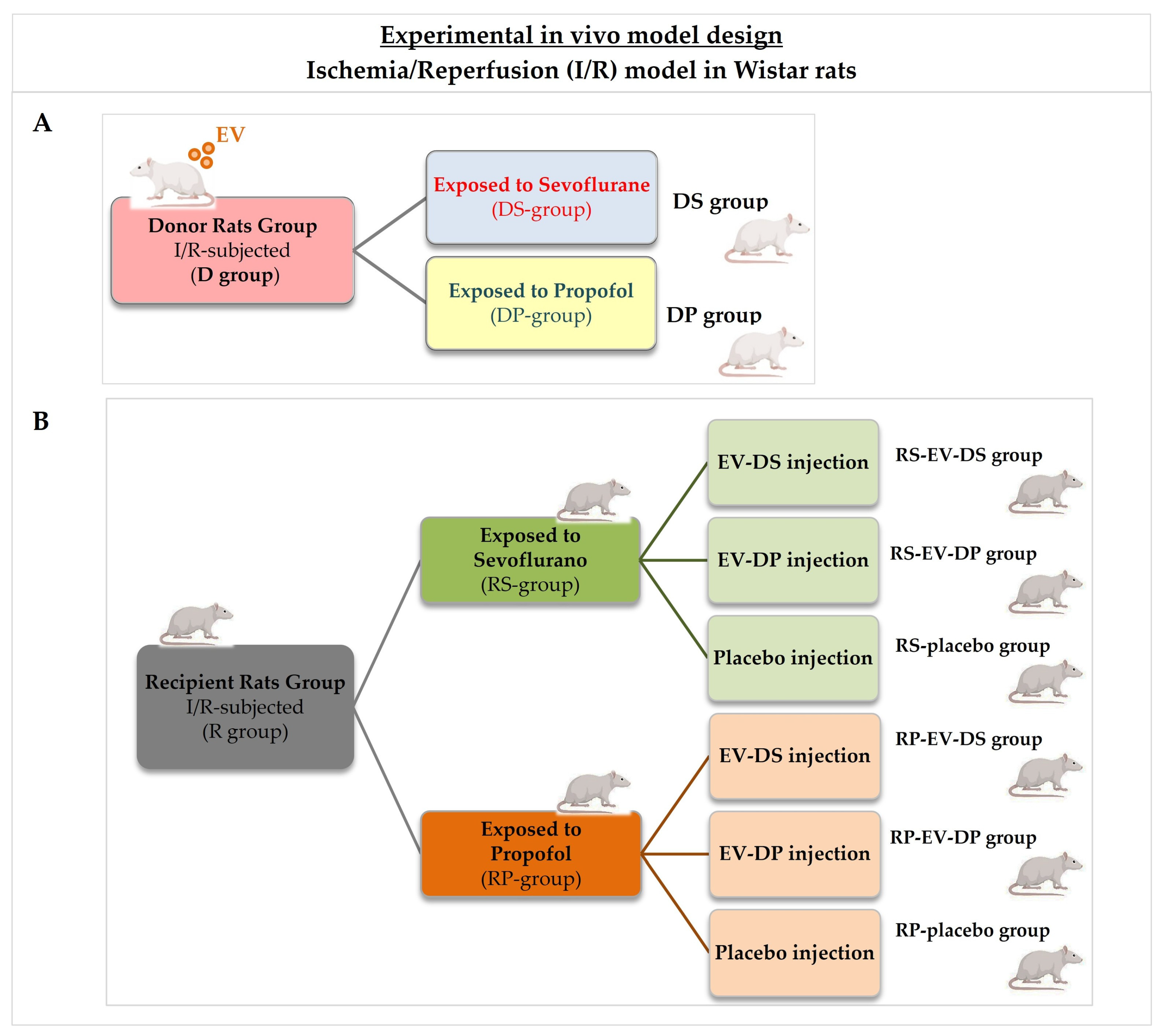
Figure 1. According to this design, the animals will be randomly divided into two experimental groups. One of them will be used as treatment donors, the Donor group (D-group), and the other group will serve as treatment recipients: the Recipient group (R-group).
All animals included in the Donor group will be subjected to an I/R procedure and randomly divided into two new groups according to the hypnotic drugs they will be exposed to: animals from the Donor group exposed to sevoflurane (DS-group), and animals from the Donor group exposed to propofol (DP-group).
Plasma samples will be extracted from each donor animal, and EVs will be isolated from the plasma samples to obtain a concentrated suspension of EVs. Plasma extraction and EV concentration procedures will be performed according to the methodology described in
Section 2.3. The suspension of concentrated EVs will be administered as treatment to the Recipient animal group. Depending on the Donor group, DS or DP group, EV treatments will be identified as EV-DS treatment and EV-DP treatment.
Regarding the Recipient group, animals will be randomly divided into six groups, as has been shown in
Figure 1. All of them will be subjected to an I/R procedure. Three of them will be exposed to sevoflurane (RS-groups), and the other three will be exposed to propofol (RP-groups).
The three Recipient groups exposed to sevoflurane (RS-groups) will be randomly divided into three new groups, according to the injected treatment: RS group injected with EVs derived from donor rats exposed to sevoflurane (RS-EV-DS group), RS group injected with EVs derived from donor rats exposed to propofol (RS-EV-DP group), and RS group injected with placebo (RS-placebo group).
The other three recipient groups exposed to propofol (RP-groups) will also be randomly divided into three groups according to the treatment they will receive: RP group injected with EVs derived from donor rats exposed to sevoflurane (RP-EV-DS group), RP group injected with EVs derived from donor rats exposed to propofol (RP-EV-DP group), and RP group injected with placebo (RP-placebo group).
All treatments, including placebo, will be administered by intravenous injection.
The cardioprotective effect generated by the injection of EVs isolated from animals exposed to sevoflurane into recipient animals will be analysed to identify the cardioprotective effect induced by halogenated drugs.
In this study, the control group will consist of animals subjected to ischaemia-reperfusion and injected with saline solution as a placebo treatment.
All protocols involving animals must be developed in accordance with the directives in force in each country. In Europe, this is currently Directive 2010/63/EU, whilst in Spain it is regulated by Royal Decree 53/2013 of 1 February, which covers the housing, handling, experimentation, and other scientific uses of animals, as well as staff training.
All animals will be treated humanely in accordance with Guidelines for the Care and Use of Laboratory Animals.
The Institutional Ethic Committee that approved the experimental protocol was the Research Ethics Committee of Málaga, Spain (CEI MALAGA NORTE), and the documentation can be consulted on the website of the Ethics Committee of Andalusia (Spain). The approval code is MIRNA-12/24, and the date of approval is 4 December 2024.
Based on the identified references of studies conducted with similar experimental models [
24], the sample size needed for the study, after performing a priori power analysis (α = 0.05, power = 0.8), for which the G*Power software, version 3.1.9.7. (free/open-source software, Heinrich Heine University in Düsseldorf, Düsseldorf, Germany) will be used, is estimated at a minimum of eight animals per group. This sample size detects a large effect size (Cohen’s d = 1.2) in outcome measures derived from ejection fraction measurements performed on animals after the I/R process and intravenous treatment administration. It is recommended to include ten rats per group to consider losses and ensure sufficient statistical power.
For unforeseeable reasons, certain animals included in the study may die and thus be excluded; therefore, a loss rate of up to 20% of the estimated minimum sample size has been considered. This loss rate would also consider cases where the animal’s state of health (i.e., situations where animals are in a critical condition or particularly ill) prevents them from undergoing any of the tests planned in the study.
2.2. In Vivo Model of Ischemia/Reperfusion in Wistar Rats
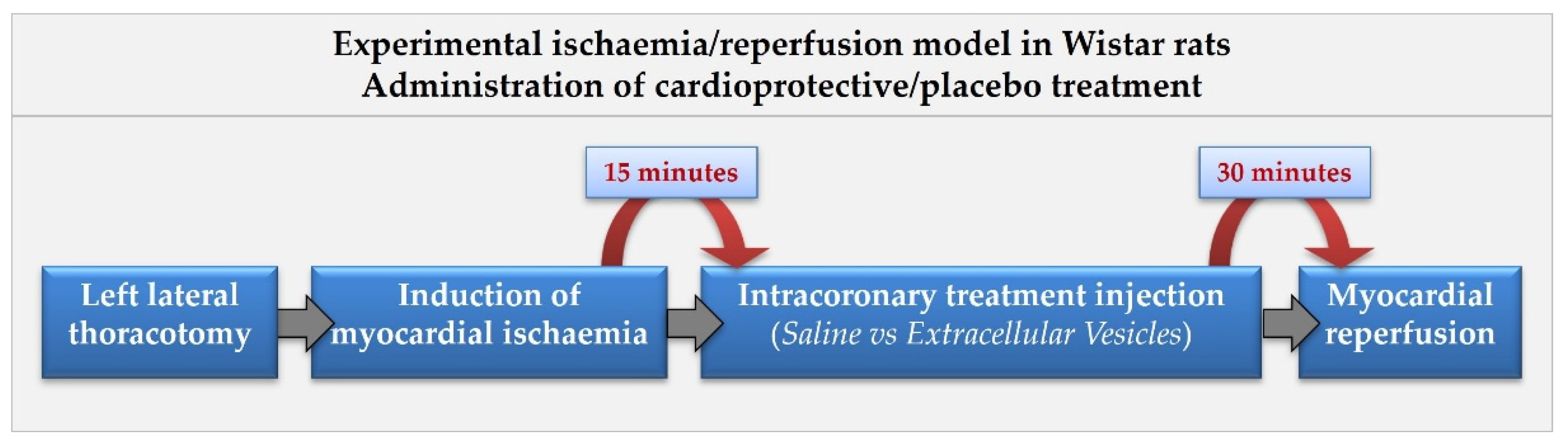
The validated I/R model proposed in this study protocol is performed according to the published literature [
24] and is shown in
Figure 2.
Both groups of rats included in the study, donors and recipients, will be subjected to the I/R procedure. The sevoflurane groups will be exposed to the sevoflurane anaesthetic as a hypnotic drug in the I/R process, and the propofol group will be injected with propofol anaesthetic as a hypnotic drug.
Sevoflurane will be administered by inhalation at a similar dose to that administered in humans, such as 1 CAM, mixed with an oxygen supplement. Propofol will be administered intravenously. The animals included in the propofol group will be intravenously injected with an equivalent dose to those administered in humans, such as ED 50.
After confirming the animals’ sedation, they will be intubated through the endotracheal route. The cardiorespiratory rate will be continuously monitored using a Harvard automatic small animal respirator (Harvard Apparatus, Holliston, MA, United States) or similar. The animal will be maintained at a respiratory ratio of 105–110 breaths/minute, and a volume of gas mixture, sevoflurane, and oxygen of 2.5 L/breath.
Fentanyl citrate analgesic will be administered intraperitoneally (IP) at a dose of 0.02 mg/kg of the animal’s weight.
The first incision will be performed in the anterior midline of the thorax, 2.5 cm in length. The second incision will be conducted in the midclavicular line of the intercostal space between the fourth and fifth ribs to penetrate the thoracic cavity. A Weitlaner retractor (FST, Heidelberg, Germany) will be used to separate the ribs, exposing the heart at the level of the left ventricle and apex. At this point, the left anterior descending coronary artery (LAD) will be located to perform an occlusion procedure, inducing transient ischemia. To this end, a 6.0-gauge suture thread will be used to create an occlusion suture approximately 2–3 mm from the apex of the left atrium; a double knot will be tied, and an occlusion tube and a myocardial protector will be placed.
After 15 min of ischemia, the EV/placebo treatment will be administered intravenously through tail vein injection.
After 30 min, reperfusion will be carried out by releasing the sliding knot made previously, removing the occlusion tube, and closing the chest cavity.
2.3. Collection of Blood Samples and Purification of the Plasma Extracellular Vesicle Suspension from Plasma
The rats in the Donor group (D-group) will serve as blood donors. Blood samples will be collected before and after the I/R procedure and exposure to hypnotic drugs, according to the experimental group (DS, sevoflurane; DP, propofol).
Blood samples will be extracted through the subclavian vein, according to a validated protocol [
25]. This sample will be collected in a blood collection tube coated with ethylenediaminetetraacetic acid (EDTA, DH Material Médico, Barcelona, Spain), an anticoagulant.
The plasma fraction will be isolated by performing centrifugation at 1500× g for 15 min. After centrifugation, the superior phase observed must be clear and yellowish and have a transparent appearance. This phase corresponds to blood plasma and contains the EVs, among other blood components.
To obtain a more concentrated EV suspension, we will perform ultracentrifugation (Ultracentrifuge Beckman Optima XPN100, Beckman Coulter, Barcelona, Spain) which will separate the blood components according to their size. The blood components larger than the EVs will appear in the supernatant, thereby concentrating the EVs in the pellet following ultracentrifugation.
The ultracentrifugation will be performed at 100,000× g for 6 h. The resulting pellet will be resuspended in saline solution and incubated overnight at 4 °C with gentle agitation to obtain a homogeneous EV suspension.
The EV suspensions must be concentrated before performing the characterisation protocols. For this purpose, the protocols applied are described below.
2.3.1. Size Exclusion Chromatography (SEC)
The SEC procedure will be performed using original® qEV/70 nm columns (Izon Science, Lyon, France), following the manufacturer’s instructions. Briefly, the Izon column (Izon Science, Lyon, France) will be placed at 4 °C, and the storage solution will be passed through it. The column will be equilibrated with 10 mL of phosphate-buffered saline (PBS, Cultek, Madrid, Spain) (pH 7.4) pre-filtered through 0.22 μm filters, and 0.5 mL of the EV samples will be added to the top. Once absorbed by the column, the FBS-buffered solution (BD Biosciences, Franklin Lakes, NJ, USA) is added again to prevent it from drying out, and the eluate is subsequently divided into 25 fractions of 0.5 mL each. Fractions 1 to 6 (3 mL) constitute the void volume, which is discarded. Fractions 7 to 10 (2 mL) contain the vesicular fraction obtained for further processing, with which we will continue processing, and fractions 11 to 25 (7.5 mL) contain the protein fraction, which will be discarded.
2.3.2. Ultrafiltration (UF)
The EV fractions collected by SEC will be enriched by UF using the Amicon® Ultra-4 100 KDa centrifugal filter (Merck Milli-pore, Darmstadt, Germany). For this purpose, the EV fraction will be resuspended in 70% ethanol with a 2:1 volume ratio and centrifuged at 4000× g for 10 min. The EVs will be blocked by the filter due to their size (≥100 KDa), and the rest of the cellular components (size < 100 KDa) will have passed through it. The filters must be washed to recover the retained EVs, and they will be resuspended at a final volume of 0.5 mL in FBS.
2.3.3. NanoDrop Spectrophotometry
To determine the purity of the enriched EV suspension, the concentration of protein will be quantified. This assay is a quality control to confirm the quality and efficiency of the enrichment procedure. This procedure will be conducted using a NanoDrop 2000C® spectrophotometer (Thermo Fisher Scientific, Waltham, MA, USA).
The final EV-enriched suspension will be divided into three aliquots, one of which will have low volume and the other two of which will have high volumes. All of them will be cryopreserved in a 10% dimethyl sulfoxide (DMSO, Sigma-aldrich MERK, Darmstadt, Germany) foetal bovine serum (FBS, BD Biosciences, Franklin Lakes, NJ, USA) solution, following the instructions included in the Mr. Frosty™ Freezing Container (Thermo Scientific™, Waltham, MA, USA) or similar.
EV-enriched aliquots will remain cryopreserved until their appropriate procedures are conducted. The cryopreserved low-volume EV-enriched aliquots will be used to perform the in vitro EV characterisation, and the high-volume ones will be administered as treatment to the rats included in the Recipient group (R-group), according to the protocol described below in
Section 2.6. These samples will be cryopreserved until their administration.
2.4. Thawing of Cryopreserved EV-Enriched Suspensions
Cryopreserved EV-enriched suspensions must be thawed before their use.
The thawing protocol will be the same for all cryopreserved samples. The cryopreserved samples will be thawed in a water bath at 37 °C for 2 min and centrifuged at 200× g for 5 min at 4 °C to remove the DMSO (Sigma-Aldrich MERK, Darmstadt, Germany), and the resulting pellet will be resuspended in 1 mL of saline solution.
2.5. Extracellular Vesicle Characterisation
The EV characterisation will be performed using the low-volume aliquots.
This characterisation will be performed according to the latest Minimal Information for Studies of Extracellular Vesicles (MISEV) guidelines issued by the International Society for Extracellular Vesicles (ISEV), such as size/concentration, morphology, and EV-specific protein markers [
26]. Moreover, to achieve the main aim of this study protocol design, differential miRNA expression analysis will be performed.
2.5.1. Quantification of the Concentration and Size of EVs
This quantification will be carried out by performing nanoparticle tracking analysis (NTAs) [
27], using a NanoSight NS300 (Malvern Panalytical, Great Malvern, Worcestershire, UK) equipment or similar.
In this analysis, particles in Brownian motion will be recorded in three videos for 1 min, using a magnification of 20×. The concentration and size of the nanoparticles will be calculated using NanoSight NTA software update v3.40 (Malvern Panalytical, Great Malvern, Worcestershire, UK) or similar, which allows visualisation of the nanoparticles in real time and calculates their concentration and size. To minimise data distortion based on large individual EVs, the ratio of total valid fingerprints to total complete footprints must always be ≤1:5. EVs larger than 10 nm to 2000 nm will be identified. Finally, the EV-enriched solution will contain 2 × 10
8 EVs/mL in FBS [
28].
2.5.2. Ultramorphology Analysis by Transmission Electron Microscopy (TEM)
To perform this assay, 25 μL of each sample will be placed on a 400-mesh holey film grid (Electron Microscopy Sciences, Morgantown, PA, USA). Samples will be stained with 2% uranyl acetate and incubated for 2 min. Once incubation is concluded, the samples can be observed using a JEOL JEM 1400 microscope, or similar, to analyse the ultrastructural morphology of the different samples. At least ten images for each sample will be acquired in .tiff mode, using microanalysis software such as Inca Energy 200 TEM (Oxford Instruments, Buckinghamshire, UK) and Aztec (Oxford Instruments, Buckinghamshire, UK) for analysis.
2.5.3. Identification of Specific Protein Markers for EV by Flow Cytometry [29]
The expression of specific protein markers such as CD9, CD63, and CD81 on EVs isolated from plasma samples (concentration 2 × 10⁸/mL) will be assessed to ensure that the isolated and injected particles are indeed EVs rather than cellular debris or protein aggregates.
This assay will be performed as follows. EVs will be resuspended in filtered PBS (Cultek, Madrid, Spain) containing 2% exosome-depleted FBS (BD Biosciences, Franklin Lakes, NJ, USA) and supplemented with protease and phosphatase inhibitors. EVs will be marked with the conjugated human antibodies: anti-CD9-FITC (Miltenyi Biotec, Bergisch Gladbach, Germany), human anti-CD63-PE (Miltenyi Biotec, Bergisch Gladbach, Germany), and human anti-CD81-APC (Miltenyi Biotec, Bergisch Gladbach, Germany). An isotype control, MOPC-21 (Miltenyi Biotec, Bergisch Gladbach, Germany), will be used. EVs and antibodies will be added in equivalent volumes, and staining will be allowed to proceed for 15 min at 4 °C in the dark. Once incubation is complete, EVs will be washed using a 300 kDa filter (Nanosep, ThermoFisher Scientific, Waltham, MA, USA) and resuspended in washing solution (2% exosome-depleted FBS in filtered PBS, BD Biosciences, Franklin Lakes, NJ, USA) for performing the flow cytometry analyses in a BD LSRFortessa SORP (Becton Dickinson, Madrid, Spain) apparatus, or similar. Data will be analysed using BD FlowJo v10 (Flow Cytometry Analysis Software, Beckman Coulter, CA, USA), flow cytometry analysis software, or similar.
2.5.4. Analysis of the Differential Expression of miRNAs
This analysis will be performed by ultrasequencing [
23], as follows.
miRNA extraction: The miRNA will be extracted with a specific kit (miRCURYTM RNA Isolation Kit-tissue and biofluids, Exiqon, Bio-Rad, CA, USA). One part will be used to perform sequencing of miRNA libraries to study the miRNA expression pattern (n = 4). Another aliquot will be used for confirmatory analyses of the results obtained from the sequencing by RT-PCR. The cDNA will be obtained using a Universal cDNA Synthesis Kit (Exiqon, Bio-Rad, CA, USA). Extraction quality and RT will be validated using a QC panel (miRCURY LNATM Universal RT miRNA PCR, 16 Ready-to-Use miRNA QC PCR Panels, Bio-Rad, CA, USA) on an Applied Biosystems Fast 7500 (ThermoFisher Scientif, Waltham, MA, USA).
Study of the quality of the RNA extracted for ultrasequencing: Once the different miRNA samples have been extracted, the RNA must meet minimum quantity and quality requirements to be used in the sequencing process. Quality will be analysed by on-chip electrophoresis using Bioanalyzer 2100 (Agilent Scientif Instrument, CA, USA) equipment. The samples will be quantified with fluorometric methods using a Qubit fluorometer (Life Technologies, Waltham, MA, USA) and stored in aliquots at −80 °C until processing. Once the miRNA libraries have been prepared, the sequencing will be performed with NextSeq 550 (Illumina, San Diego, CA, USA) instrument, or similar.
2.6. Administration of EV Treatment to Animals Subjected to I/R and Exposed to Anaesthetic Drugs
The high-volume cryopreserved EV-enriched aliquots will be thawed following the procedure previously described in
Section 2.4. These samples, isolated from donor rats, were cryopreserved to be used as treatment for the rats included in the Recipient group (R-group).
The EV-enriched samples will be thawed on the day of administration. Once the DMSO has been removed, the resulting pellet will be resuspended in 100 μL of saline solution, to a final concentration of 2 × 10
8 EV/mL [
28].
The EV suspension will be injected into the animals 15 min after ischemia induction, during the I/R procedure. The EV treatment, or saline as a placebo, will be injected through the central tail vein.
2.7. Functional Assessment by Echocardiography
To assess cardiac function, a transthoracic echocardiogram will be performed at three time points: before the induction of ischaemia/reperfusion, as a baseline control; before IV injection of the treatment; and 24 h after treatment. For this procedure, the animals will be anaesthetised with the hypnotic drug corresponding to the experimental group to which they have been assigned (sevoflurane or propofol) and placed in the lateral recumbent position. Echocardiography will be performed using an 8.9 MHz Acuson transducer ×300 ultrasound system, version 2.0, from Siemens Health Global. An M-mode trace of the left ventricle will be obtained by imagining the beating heart in two-dimensional mode. The left ventricular end-diastolic diameter (LVEDD), left ventricular end-systolic diameter (LVESD), and fractional shortening (FS) will be measured using at least three consecutive cardiac cycles. The ejection fraction (EF) will be calculated as follows: %EF = [(LVEDD
3 − LVESD
3)/LVEDD
3] × 100. The left ventricular end-diastolic volume (LVEDV) and the left ventricular end-systolic volume (LVESV) will also be measured [
30]. All echocardiographic studies will be performed by a single examiner who will be unaware of the treatments administered.
2.8. Animal Sacrifice and Heart Tissue Removal
Twenty-four hours after the beginning of the ischaemia-reperfusion process, all surviving rats will be euthanised in accordance with the strictest ethical and legal standards, as set out in the regional legislation.
Before euthanasia, a blood sample will be extracted from the subclavian, following the protocol described above in
Section 2.3, for quantification of biochemical parameters regarding cardiac function.
For euthanasia, the animals will be injected with a dose of sodium thiopental lower than the lethal dose 50 (L50 = 40 mg per kg of the animal’s weight) to preserve the heartbeat, and with a dose of 0.02 mg of fentanyl citrate per kg of the animal’s weight, as analgesia. Both drugs must be administered intraperitoneally. Once the animal loses reflexes in the footpads, its sedation state is confirmed, and the KCl solution can be perfused at a concentration of 60 mEq through an incision in the thoracic cavity. This perfusion must be made through two simultaneous routes, one of which is the apex of the heart, and the other of which is the inferior vena cava. Cardiac arrest is induced with this perfusion in the diastolic cardiac phase. A second infusion with saline solution will be performed through both routes, and a third perfusion with a 4% buffered paraformaldehyde solution to fix the heart tissue and block autolysis of tissue proteins will also be performed.
Finally, the heart will be excised and immersed in a 4% buffered paraformaldehyde solution to ensure that the fixative penetrates fully into the heart tissue. The cardiac tissue will be ready to perform histological (haematoxylin-eosin, Masson’s trichrome, etc.) and immunohistochemical procedures (α-actin, Ki 67, caspase 3, etc.).
2.9. Statistical Analysis
A descriptive analysis of the variables will be performed, calculating absolute and relative frequencies for the qualitative variables, as well as the arithmetic mean and standard deviation.
To compare means in the cross-sectional study (inter-group study), the appropriate parametric and non-parametric tests will be used (depending on whether the data for the different variables follow a normal or non-normal distribution, respectively, for which the Shapiro–Wilk test will be applied beforehand). Continuous quantitative variables will be compared using an ANOVA or Kruskal–Wallis test, as appropriate. The association between categorical variables will be analysed using a Chi-squared test. At the start of the baseline study, a comparison will be made between all groups, including the control group, using the statistical tests described above to observe differences between them.
In the longitudinal study (intra-group study), a comparison will be made between the different assessment time points in each group using repeated measures ANOVA or the Friedman test, depending on whether the data for the different variables follow a normal or non-normal distribution, respectively.
All comparisons will be two-sided, and a p-value < 0.05 will be considered statistically significant. All results will be expressed as the mean ± standard deviation (SD). If this is not the case, this must be stated. One of the software packages to perform all the statistical analyses required for this study is PASW Statistics 18 (IBM SPSS, Madrid, Spain).
3. Scientific Impact and Translational Relevance of the Application of the Experimental Design
This manuscript describes a future-direction protocol study and includes a new in vivo model of cardioprotection induced by halogenated hypnotic drugs to contribute to the design of future studies that will provide new insights into the cardioprotective effects of halogenated drugs. The expected development framework will allow identification of the main metabolic pathways and molecular targets through which halogenated drugs induce this cardioprotective effect. To this end, the miRNAs included in isolated EVs, with cardioprotective properties induced by exposure to halogenated agents, will be identified. These miRNAs could be considered as biomarkers of cardioprotection, and their administration could reduce cardiac damage in patients undergoing additional cardiac surgery.
Moreover, the data resulting from its application could be considered as preclinical study-derived results, and they might be submitted to the relevant regulatory authorities for approval to subsequently design a Phase I clinical trial, thereby achieving one of the study’s objectives: translation into clinical practice.
In this context, the identification of a cardioprotective biomarker would lead to the development of a universal ‘ready-to-use’ therapeutic product that could be administered to patients undergoing cardiac surgery or percutaneous coronary angioplasty, reducing the risk of morbidity and mortality for the patient and providing greater protection and improved cardiac remodelling.
In this scenario, we have designed this study protocol, which sets out the materials and methods required for its implementation, thereby ensuring its reproducibility.
The methodology described in this work has been included in various research projects to secure funding for its development. Furthermore, it is currently under review by the regional research ethics committee for approval.