Review

Peritoneal Metastasis as a Distinct Biological Entity: Mechanisms, Microenvironment, and Therapeutic Implications


Department of Surgical Oncology, Cukurova University, 01200 Adana, Turkey
*
Author to whom correspondence should be addressed.
Int. J. Transl. Med. 2026, 6(3), 27; https://doi.org/10.3390/ijtm6030027
Submission received: 10 May 2026 / Revised: 5 June 2026 / Accepted: 15 June 2026 / Published: 29 June 2026

Abstract

For decades, peritoneal metastases (PM) have been regarded as a terminal manifestation of advanced malignancies and managed primarily with palliative intent because of limited sensitivity to systemic therapies. Accumulating clinical, molecular, and immunological evidence now supports the view that PM is not merely an anatomic pattern of spread but a distinct metastatic niche with characteristic biological, microenvironmental, and therapeutic features. This review summarizes the major routes of PM development—transcoelomic, lymphatic, and hematologic dissemination—and emphasizes how these pathways converge through shared biological programs. Core mechanisms include epithelial–mesenchymal transition (EMT), adhesion signaling, extracellular matrix remodeling, and tumor–immune cell interactions. A central focus is the peritoneal tumor microenvironment: mesothelial-to-mesenchymal transition, cancer-associated fibroblast activity, adipocyte-derived metabolic support, macrophage polarization, and regulatory T-cell enrichment collectively shape an immunotolerant and treatment-resistant niche on the peritoneal surface. In addition, evidence from pre-metastatic niche biology suggests that primary tumor-derived exosomes and epitranscriptomic regulation can prime the peritoneal environment before overt implantation. These features provide a biological rationale for locoregional strategies such as cytoreductive surgery and hyperthermic intraperitoneal chemotherapy, as well as emerging intraperitoneal modalities and microenvironment-targeted approaches. Finally, organoid platforms, liquid biopsy-based minimal residual disease monitoring, and theranostic technologies may enable more personalized, biology-driven management of PM.

1. Introduction

Peritoneal metastases (PM) represent a dissemination phenotype with a distinct microenvironmental ecosystem, most commonly encountered in advanced gastrointestinal and gynecological malignancies. Historically, peritoneal involvement has been associated with relatively high therapeutic resistance compared with other metastatic sites and was often approached within a “terminal stage–palliative care” framework. Late diagnosis, extensive tumor burden, and limited response to systemic treatments are key contributors to this paradigm [1].
Clinically, peritoneal surface involvement may remain silent until disease burden increases; then, findings such as peritoneal nodules, omental caking, and malignant ascites become apparent. Malnutrition, bowel obstruction, and functional bowel disorders further reduce quality of life and adversely affect prognosis [2]. The frequent inadequacy of systemic chemotherapy in this setting has long restricted therapeutic options [2].
Over the last two decades, locoregional strategies have shifted the treatment landscape. Cytoreductive surgery (CRS) combined with hyperthermic intraperitoneal chemotherapy (HIPEC), systematized by Sugarbaker, can offer long-term survival and even curative potential in selected populations [3,4]. This approach is increasingly accepted as the standard of care for appendiceal pseudomyxoma peritonei and primary peritoneal mesothelioma [4,5].
Nevertheless, HIPEC benefit is not uniform across tumor types or patient subgroups. Pharmacokinetic constraints, heterogeneous drug penetration, and incomplete coverage of peritoneal surfaces are among the main limitations [6,7,8]. These clinical observations reinforce the need to conceptualize PM not simply as an anatomic spread pattern but as a biologically distinct metastatic niche with unique molecular and microenvironmental determinants [9,10]. In this context, the peritoneal tumor microenvironment and resident immune programs are particularly relevant for future targeted interventions [11,12,13].
This review aims to integrate the biological foundations of PM with their translational and clinical implications, focusing on dissemination mechanisms and microenvironmental ecology to inform biology-driven treatment strategies.
In parallel with these mechanistic advances, efforts have been made to translate biological understanding into clinically applicable frameworks. The Peritoneal Cancer Index (PCI)—integrating tumor size and anatomical distribution across 13 abdominal regions—remains the most widely used instrument linking disease burden to surgical resectability and serves as a central decision-making tool in CRS-HIPEC candidate selection [14]. A PCI threshold of approximately 15–20 is commonly applied in colorectal PM, though optimal cutoffs vary by tumor type and institutional experience [14,15,16]. Incorporating such burden-based stratification alongside emerging molecular markers may further refine patient selection beyond current anatomical criteria.

2. Peritoneal Surface Malignancies

Peritoneal surface malignancies (PSM) comprise a heterogeneous group including primary peritoneal tumors and secondary malignancies metastasizing to the peritoneum from intra-abdominal organs. A unifying model involves detachment of malignant cells from the primary tumor, attachment to the mesothelial surface, and proliferation into metastatic foci—conceptually aligned with the classic “seed and soil” hypothesis [17,18].
Early in implantation, malignant cells disrupt the mesothelial barrier and gain access to submesothelial structures, exposing the basement membrane and initiating a permissive stromal response. Angiogenesis is then triggered, supporting tumor growth [19,20,21]. This progression is not limited to proliferation and neovascularization; adaptive immune escape programs develop in parallel [22,23]. Thus, PM emerges from dynamic, bidirectional interactions between tumor cells and host microenvironmental components [10,11,12,13,22,23,24].
Clinically, the distinction between primary and secondary peritoneal malignancies carries meaningful prognostic and therapeutic weight. Appendiceal mucinous neoplasms and primary peritoneal mesothelioma tend to follow relatively predictable locoregional spread patterns and may achieve 5-year survival rates exceeding 50% after CRS-HIPEC in carefully selected patients [3,4]. Colorectal and gastric PM are generally associated with more heterogeneous biology and variable responses to locoregional therapy, underscoring the importance of integrating tumor origin, histological grade, and molecular features alongside PCI when defining treatment intent within multidisciplinary tumor boards [8,15].
Beyond their prognostic implications, primary and secondary peritoneal malignancies also differ in their biological origin and disease evolution. Primary peritoneal malignancies, including malignant peritoneal mesothelioma and primary peritoneal carcinoma, arise from the peritoneal lining itself and therefore develop within the peritoneal microenvironment from the earliest stages of disease [4,5]. In contrast, secondary peritoneal malignancies originate from primary tumors of intra-abdominal organs, most commonly colorectal, gastric, appendiceal, and ovarian cancers, and reach the peritoneal surface through metastatic dissemination [9,17,18].
These differences influence both biological behavior and therapeutic strategy. Primary peritoneal malignancies often remain confined to the peritoneal compartment for prolonged periods and may exhibit relatively predictable locoregional spread patterns, whereas secondary peritoneal disease reflects the metastatic capacity of the primary tumor and is shaped by mechanisms such as tumor cell detachment, dissemination, implantation, and microenvironmental adaptation. Consequently, primary malignancies such as malignant peritoneal mesothelioma and appendiceal pseudomyxoma peritonei are among the tumor types most likely to benefit from CRS-HIPEC, while secondary peritoneal metastases demonstrate greater biological heterogeneity and more variable responses to locoregional treatment [3,4,5,8,15].

3. Mechanisms and the Microenvironmental Ecosystem

3.1. Routes of Dissemination of Peritoneal Metastases

3.1.1. Transcoelomic Spread

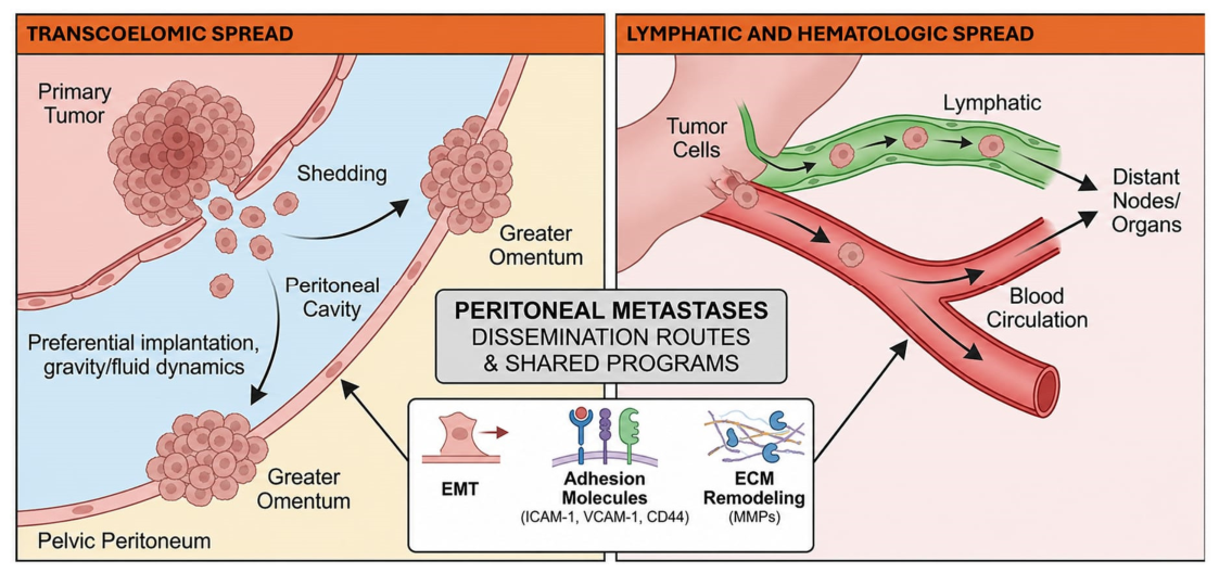
Transcoelomic dissemination is the most common and best-characterized route of peritoneal metastasis. Following serosal invasion, tumor cells detach from the primary lesion and are released into the peritoneal cavity [25,26]. These free-floating cells are subsequently distributed by peritoneal fluid dynamics and preferentially implant in specific anatomical regions rather than randomly. Metastatic burden frequently accumulates in the mesentery, greater omentum, paracolic gutters, and pelvic peritoneum, reflecting peritoneal fluid dynamics influenced by gravity, respiration, and intestinal peristalsis [25,26,27]. Pelvic fluid pooling, in particular, correlates well with observed clinical distribution patterns. Upon reaching the peritoneal surface, tumor cells encounter the mesothelium as an initial barrier, and successful implantation depends on tumor cell survival, adhesion, invasion capacity, and the permissiveness of the host microenvironment [10,11,12,13,19,23] (Table 1, Figure 1).
From a treatment perspective, these fluid-driven distribution patterns provide a biological rationale for the peritonectomy regions described by Sugarbaker, which systematically target the greater omentum, pelvic peritoneum, and paracolic gutters [3]. Building on the same principles, pressurized intraperitoneal aerosol chemotherapy (PIPAC) delivers aerosolized chemotherapy within a pressurized capnoperitoneum, potentially enabling more homogeneous intraperitoneal distribution compared with conventional liquid perfusion [28]. In addition, emerging clinical data suggest that repeated cycles may induce tumor regression that could allow conversion to resectability in selected patients [29].

3.1.2. Lymphatic and Hematologic Spread

In contrast to transcoelomic dissemination, lymphatic and hematologic spread contribute to more systemic distribution patterns. Tumor cells can enter lymphatic vessels via local invasion or reach blood circulation through intravasation [30,31].
Lymphatic stomata and submesothelial lymphatic regions may provide implantation opportunities, particularly for adenocarcinomas with serosal involvement, where local niche features can facilitate attachment and survival [18,21]. In hematogenous dissemination, circulating tumor cells tether to endothelium and extravasate through a multistep cascade regulated by adhesion molecules such as ICAM-1 and VCAM-1, which also activate intracellular signaling (e.g., PI3K/Akt, FAK) supporting survival in foreign microenvironments [32,33]. In selected settings, such processes may contribute to peritoneal seeding or combined spread patterns.
Importantly, lymphatic and hematologic routes are not mutually exclusive; transitions at lymphohematopoietic junctions can yield mixed dissemination patterns. Clinically, nodal metastasis is associated with worse outcomes and may reflect both regional and systemic spread risk [34,35] (Table 1, Figure 1).
In this context, the potential coexistence of lymphatic and hematogenous spread has direct bearing on staging and systemic treatment planning. Current ESMO guidelines recommend systemic chemotherapy as the primary approach for colorectal PM with concomitant distant metastases, generally reserving locoregional strategies for patients with isolated peritoneal disease [31]. Preoperative imaging—high-resolution CT, PET-CT, or diagnostic laparoscopy—to exclude occult extra-peritoneal involvement plays a key role before consideration of CRS-HIPEC, given that synchronous systemic metastasis has been associated with substantially reduced benefit from locoregional intervention [15].

3.1.3. Common Biological Programs: Points of Convergence

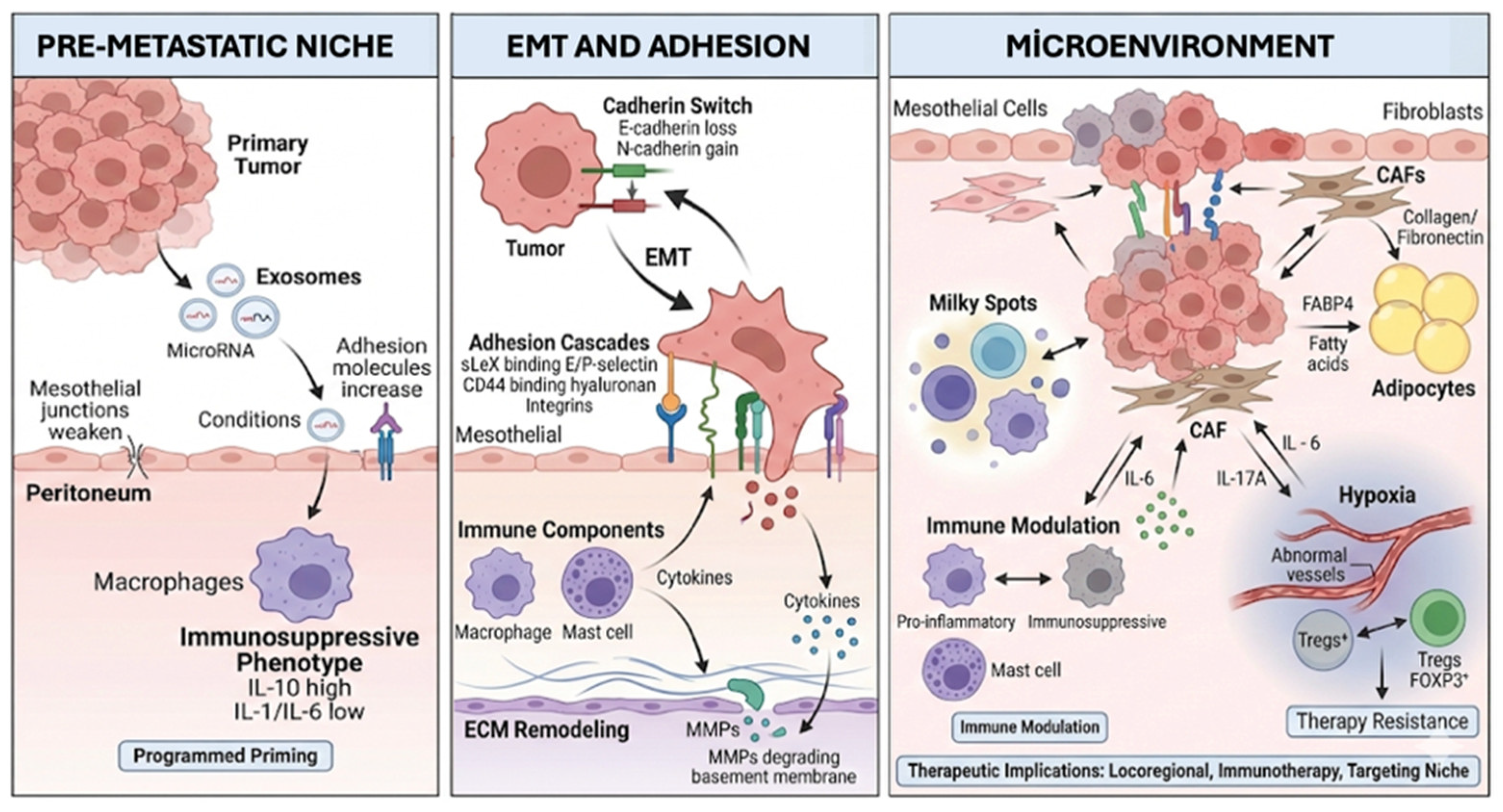
Although dissemination routes differ anatomically, PM development is orchestrated by shared biological programs that enable attachment, invasion, and immune evasion across compartments [10,11,21,22,23,24]. A central program is epithelial–mesenchymal transition (EMT), in which epithelial tumor cells lose polarity and junctional integrity while acquiring mesenchymal traits. Hallmarks include loss of E-cadherin and increased N-cadherin expression (“cadherin switch”). EMT enhances invasion, migration, and mechanical resistance under circulatory shear stress, thereby increasing dissemination competence [36] (Figure 2).
Early tumor–mesothelium interactions resemble inflammatory adhesion cascades. Sialyl Lewis X (sLeX) on tumor cells can bind E-selectin and P-selectin expressed by mesothelial and endothelial cells, facilitating tethering and rolling. CD44 on tumor cells interacts with the hyaluronan-rich mesothelial glycocalyx, while integrins reinforce adhesion and bidirectional signaling between tumor cells and the microenvironment [22,23,24,25,26,27,30,31,32,33,34,35,36,37,38].
Immune components are integral to this shared program: peritoneal macrophages and mast cells may exert context-dependent, bidirectional roles. Immunosuppressive macrophage polarization can promote tumor survival and proliferation, whereas mast cell-derived cytokines/chemokines can accelerate microenvironmental remodeling [22,23,24,25,26,27,30,31,32,33,34,35,36,37,38].
Extracellular matrix (ECM) remodeling further supports implantation. Matrix metalloproteinases, including MMP-3 and MMP-9, promote basement membrane degradation and ECM reorganization, facilitating invasion and stable metastatic outgrowth [39] (Table 2). These interactions—spanning EMT, selectin-mediated tethering, CD44-hyaluronan binding, and ECM remodeling—are summarized in Figure 2, which integrates the key molecular programs operating at the tumor–mesothelium interface within the peritoneal niche.
Beyond mechanistic interest, EMT-associated markers and CD44 overexpression have been linked to cancer stem cell phenotypes and chemoresistance in colorectal cancer [38,40], and circulating tumor cells with mesenchymal features are increasingly recognized as liquid biopsy candidates for early peritoneal recurrence detection [41,42].
While the molecular programs described above—EMT, adhesion signaling, ECM remodeling, and immune evasion—are not exclusive to peritoneal dissemination, their convergence within the peritoneal cavity creates a metastatic niche that differs in several important respects from other metastatic sites. Several characteristics are either unique to, or particularly prominent within, the peritoneal metastatic environment. First, transcoelomic spread—the detachment of viable tumor cells into a fluid-filled cavity and their redistribution by peritoneal fluid dynamics—has no direct anatomical equivalent in common parenchymal metastatic sites such as the liver or lung [25,26,27]. Second, mesothelial-to-mesenchymal transition (MMT) of resident peritoneal cells may generate an implantation-permissive surface that is specific to the serosal lining and contributes to metastatic colonization [19,43]. Third, omental milky spots represent specialized peritoneal immune microcompartments enriched in macrophages and lymphocytes and have been identified as preferential sites for metastatic implantation [44,45].
In addition, adipocyte-mediated metabolic support through FABP4-associated lipid transfer appears to play a particularly important role in omental metastasis, providing a favorable metabolic environment for tumor growth and adaptation [46]. Furthermore, emerging evidence suggests that primary tumor-derived exosomes may contribute to pre-metastatic niche formation through mechanisms involving METTL3-dependent epitranscriptomic regulation and immune conditioning of the peritoneal cavity before overt implantation occurs [47,48]. Collectively, these observations support the concept that PM represents a biologically distinct metastatic niche in which common metastatic programs are reshaped by the unique anatomical, immunological, and metabolic characteristics of the peritoneal cavity.

3.2. Pre-Metastatic Microenvironment and Exosomal Communication

The pre-metastatic niche concept implies that peritoneal implantation can be preceded by a biologically driven “priming” phase rather than arising solely from passive mechanical dispersion [47]. Primary tumor-derived exosomes may mediate remote conditioning of the peritoneal environment. Proposed mechanisms include microRNA cargo shaped by METTL3-dependent epitranscriptomic regulation, with downstream effects on stromal and immune populations—particularly peritoneal macrophages—skewing them toward immunosuppressive phenotypes. In this model, an IL-10–dominant cytokine milieu with relative suppression of IL-1 and IL-6 may develop, inhibiting anti-tumor T-cell proliferation and effector function; consequently, the peritoneal surface becomes more tolerogenic prior to overt implantation [47,48,49].
Beyond immune suppression, early mesothelial and ECM remodeling—such as weakening of cell–cell junctions and shifts in adhesion molecule expression—may further increase receptivity for subsequent tumor cell attachment [19]. Conceptually, this “programmed priming” should be distinguished from the established metastatic microenvironment.
These findings have important implications for prophylactic intraperitoneal intervention. The PROPHYLOCHIP-PRODIGE 15 trial evaluated second-look surgery with HIPEC in colorectal cancer patients at high clinical risk of peritoneal recurrence but did not demonstrate a survival advantage over surveillance [34]. This result may partly reflect the absence of molecular patient selection, although this hypothesis requires further validation. If only a subset of patients develop an activated pre-metastatic peritoneal niche, stratification based solely on clinical criteria may be insufficient to identify those most likely to benefit.
In this context, incorporating molecular indicators of niche priming—such as exosome cargo profiles or peritoneal washing-based liquid biopsy panels—into future trial designs remains an important direction [41,47,48].

3.3. Microenvironmental Effects in Established Metastasis

Once established, PM progression is shaped by complex interactions among tumor, stromal, and immune components. Cancer cell behavior is strongly influenced by bidirectional communication with the surrounding microenvironment, which can enhance invasion and support therapy resistance [10,11,12,13,22,23,26]. The peritoneal microenvironment contains mesothelial cells, fibroblasts, adipocytes, endothelial cells, macrophages, and lymphocyte subsets [19,21]. Single-cell analyses suggest that mesothelial cells comprise distinct functional subpopulations rather than a uniform barrier, potentially influencing metastatic behavior and treatment response [50].
Omental “milky spots,” enriched with macrophages and lymphocytes, can serve as immunological hubs and potential docking niches that support metastatic colonization [44,45]. In addition, adipocyte-derived lipid transfer—linked to factors such as FABP4—may provide metabolic support that favors tumor growth and adaptation in omental regions [46].

3.3.1. Mesothelial Transformation and Extracellular Matrix Remodeling

A subset of mesothelial cells can acquire mesenchymal features, lose barrier function, and convert into a phenotype that facilitates tumor adhesion. Another subset may adopt cancer-associated fibroblast-like behavior and increase ECM production. Collagen, fibronectin, and laminin-rich matrices provide mechanical scaffolding and function as reservoirs for growth factors and cytokines that intensify signaling within the niche [19,21]. Ongoing ECM remodeling through metalloproteinases further enhances invasion and migratory capacity [39,43].
In this context, mesothelial transformation and CAF activation represent potentially tractable intervention points, though direct clinical evidence in PM remains limited. IL-6/STAT3 signaling—a central pathway in CAF-mediated ECM remodeling and drug resistance—is under investigation in early-phase clinical settings combining IL-6 receptor blockade with systemic chemotherapy in gastric cancer with peritoneal involvement [51]. Agents that attenuate stromal collagen production have shown preclinical promise in improving drug penetration within fibrotic tumor microenvironments [19,43]; whether such approaches will translate meaningfully in PM will require prospective evaluation alongside companion biomarkers of stromal activation.

3.3.2. Immune Cells and Cytokine-Mediated Modulation

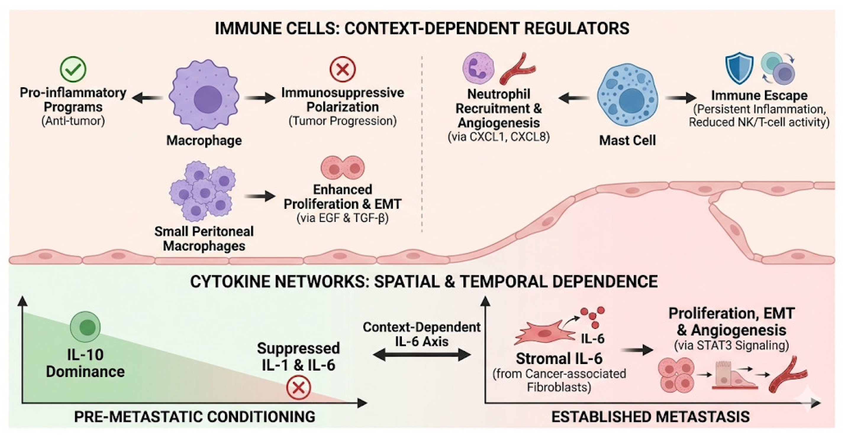
Macrophages are key regulators of immune directionality in the peritoneal niche. Pro-inflammatory programs can support anti-tumor responses, whereas immunosuppressive polarization promotes tumor progression [22,37,51]. Small peritoneal macrophage expansion has been linked to enhanced proliferation and EMT via EGF and TGF-β signaling in experimental settings [21,22,37,51].
Mast cells also display context-dependent effects. They may promote neutrophil recruitment and angiogenesis through chemokines such as CXCL1 and CXCL8, but persistent inflammation can contribute to immune escape programs, including reduced natural killer cell activity and T-cell exhaustion [11,12,13,33,50].
Cytokine networks operate in a spatially and temporally dependent manner. While pre-metastatic conditioning may favor IL-10 dominance with relative suppression of IL-1 and IL-6 [47,48,49], established metastasis often involves stromal IL-6—particularly from cancer-associated fibroblasts—driving proliferation, EMT, and angiogenesis through STAT3 signaling. Accordingly, the IL-6 axis should be interpreted as context-dependent, varying by cellular source, microcompartment, and disease stage rather than as a unidirectional effect [47,48,49] (Table 2).
Specifically, during pre-metastatic niche formation, tumor-derived exosomal signaling may contribute to an immunoregulatory milieu characterized by relative suppression of pro-inflammatory cytokines, facilitating immune tolerance before overt implantation [47,48]. In contrast, once metastatic deposits become established, IL-6 is predominantly produced by cancer-associated fibroblasts, macrophages, and tumor cells, where it promotes proliferation, epithelial–mesenchymal transition, angiogenesis, and therapy resistance through STAT3-dependent signaling pathways [52]. Thus, the impact of IL-6 is highly dependent on both disease stage and cellular source within the peritoneal microenvironment.
The interplay among macrophages, Tregs, cytokines, and tumor cells within the peritoneal niche is illustrated in Figure 3. The density and polarization state of tumor-associated macrophages and Tregs have been associated with treatment response in selected studies [50], highlighting their potential as predictive biomarkers. Checkpoint inhibitor combinations are being explored in this setting: the phase II DRAGON-09 trial evaluated intraperitoneal and intravenous paclitaxel plus S-1 combined with the anti-PD-1 antibody sintilimab as first-line treatment for gastric cancer with PM, reporting preliminary response rates that warrant further investigation [53]. These early data raise the possibility that modulating peritoneal immunosuppression alongside cytotoxic therapy may be important for more durable disease control, though larger randomized evidence is awaited.

3.3.3. Hypoxia, Immune Suppression, and Therapeutic Implications

Hypoxia is a hallmark of the peritoneal niche and can promote angiogenesis via HIF-1α activation and VEGF induction; however, the resulting abnormal vasculature may remain inefficient, limiting drug delivery and effective penetration of chemotherapeutic agents [20]. NF-κB-mediated inflammatory activation and IL-17A signaling have also been implicated in tumor-supportive inflammatory programs and proliferation [54,55,56]. Collectively, this stromal–immune architecture can reinforce treatment resistance in PM [54,55,56]. Regulatory T cells (Tregs) represent another relevant immunoregulatory population in PM [23]. FOXP3+ Tregs can suppress anti-tumor immunity, and high Treg infiltration is frequently associated with unfavorable outcomes. Therapeutic concepts include selectively targeting tumor-enriched effector Tregs (e.g., via CCR4 or CTLA-4–associated strategies) to enhance tumor immunity while limiting systemic autoimmunity risk [23]. These biological constraints help explain the limited efficacy of systemic treatments in PM and provide rationale for locoregional approaches [12,21,45,50,57]. Hyperthermia during HIPEC may promote immunogenic cell death programs and modulate the microenvironment, potentially increasing responsiveness to immunotherapy. Adjunct strategies that modulate mesothelial function, attenuate immunosuppressive immune circuits, or target ECM remodeling—potentially including nanoparticle-based carriers for controlled intraperitoneal drug release—may help overcome pharmacokinetic and niche barriers [19,58] (Table 2, Figure 3).
From a therapeutic standpoint, hyperthermia during HIPEC not only enhances drug cytotoxicity but may transiently improve microvascular perfusion within peritoneal nodules, potentially augmenting oxaliplatin and mitomycin-C penetration [6,7]. Anti-VEGF agents such as bevacizumab are incorporated into systemic regimens for colorectal PM, though their role in combination with HIPEC remains under investigation in the context of PM [31]. HIF-1α inhibition and vascular normalization strategies represent additional avenues that may render the hypoxic peritoneal niche more permissive to immune effector function and drug delivery; clinical validation in PM-specific trials is still needed.

4. Future Perspectives

The management of PM is shifting from symptom-focused palliation to biology-based, personalized strategies informed by the distinct metastatic niche concept. CRS plus HIPEC can provide long-term survival and potential cure in selected cohorts [3,4,54,59,60,61], but outcomes depend on patient selection, tumor biology, peritoneal tumor burden, and microenvironmental determinants [45,50,55]. The heterogeneity of clinical benefit supports the idea that PM comprises biologically distinct subgroups. Microenvironment-directed targeting is a major future direction. Mesothelial transformation, macrophage and mast cell immunoregulation, and ECM remodeling likely modulate intraperitoneal therapy efficacy. Reprogramming immunosuppression may also enhance immunotherapy responsiveness in PM [11,13,23,45,49,50,57].
Despite the growing adoption of CRS-HIPEC, important controversies remain regarding the relative contribution of hyperthermic intraperitoneal chemotherapy itself versus complete cytoreductive surgery. The PRODIGE 7 trial did not demonstrate an overall survival benefit with the addition of oxaliplatin-based HIPEC to complete cytoreduction in colorectal PM, despite prolonged survival observed in both treatment groups [8]. Similarly, prophylactic HIPEC strategies have yielded mixed results, as illustrated by the COLOPEC and PROPHYLOCHIP studies [34,59]. Current evidence suggests that the clinical benefit of HIPEC is likely to vary according to tumor biology and disease context rather than being uniform across all tumor types. Long-term outcomes appear most favorable in appendiceal neoplasms and peritoneal mesothelioma [4,5], whereas its role in colorectal and gastric PM remains an area of active investigation. Consequently, ongoing research increasingly focuses on identifying biological subgroups most likely to benefit from locoregional therapies and on clarifying the relative contributions of CRS and HIPEC to long-term oncological outcomes.
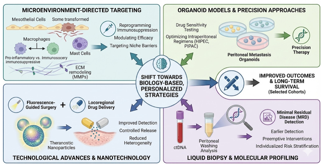
Beyond biological insights, technological advances are also reshaping the field. Organoid models derived from peritoneal metastases can help test drug sensitivity and optimize intraperitoneal regimens before HIPEC or pressurized intraperitoneal aerosol chemotherapy (PIPAC), supporting a transition from empirical to precision approaches [29,62,63,64].
In addition to organoid platforms, animal models remain important tools for translational PM research. Syngeneic, orthotopic, and patient-derived xenograft (PDX) models have been widely used to investigate tumor implantation, metastatic progression, and host–tumor interactions within the peritoneal cavity [65,66]. These models have also contributed substantially to the preclinical evaluation of locoregional therapies, including intraperitoneal chemotherapy, HIPEC, and PIPAC [66]. Although no single model fully reproduces the complexity of human PM, animal systems provide an essential bridge between mechanistic discoveries and clinical application, complementing patient-derived organoid platforms in the development of personalized therapeutic strategies [65,66].
These innovations further expand opportunities. Theranostic nanoparticles can combine fluorescence-guided surgery and locoregional drug delivery, potentially improving detection of small foci and enabling controlled release within subperitoneal niches [58]. Nanoparticle platforms and controlled release technologies may reduce heterogeneity in drug distribution and improve access to protected microcompartments [58,67,68,69]. Finally, liquid biopsy and molecular profiling may enable individualized risk stratification and monitoring. Minimal residual disease signals from peritoneal washings and circulating tumor DNA (ctDNA) may allow earlier detection of microscopic recurrence and facilitate preemptive interventions, including intraperitoneal strategies in selected cases [41,67,70] (Figure 4).
PIPAC offers an intraperitoneal alternative for patients ineligible for CRS-HIPEC due to high tumor burden or comorbidities [28]. By delivering aerosolized chemotherapy at physiological pressure within the capnoperitoneum, PIPAC achieves higher peritoneal tissue drug concentrations with a more favorable systemic toxicity profile. Emerging data suggest that sequential cycles may achieve tumor regression that could allow conversion to resectability in selected patients, positioning PIPAC as a potential bridging strategy [28,29]. Adaptive trial platforms incorporating biomarker stratification—including microsatellite instability status, HER2 amplification, and peritoneal immune signatures—are increasingly recognized as a priority for identifying subgroups that benefit most from immunotherapy–locoregional combinations. Establishing peritoneal-specific response criteria beyond conventional RECIST—which was not designed to capture diffuse, non-measurable peritoneal disease—remains an important unmet need that currently limits cross-trial comparability [15].
This limitation largely reflects the biological and radiological characteristics of PM. Peritoneal disease frequently presents as diffuse serosal thickening, omental caking, small nodules, or scattered implants that do not fulfill conventional RECIST definitions of measurable target lesions. Consequently, radiological response assessment remains challenging and may underestimate biologically meaningful treatment effects [15].

5. Conclusions

PM should be understood as a distinct metastatic niche shaped by specialized dissemination programs and a treatment-resistant microenvironment—not merely an anatomical pattern of spread. Integrating surgery, intraperitoneal therapies, and molecularly targeted approaches within multidisciplinary pathways offers the most coherent path toward improved outcomes. Organoid modeling, theranostics, and liquid biopsy collectively support a transition toward personalized, biology-driven management of peritoneal surface malignancies. The clinical impact of this paradigm shift is increasingly evident: biology-informed patient selection has contributed to improved long-term survival after CRS-HIPEC in selected tumor types [3,4,15], and the integration of intraperitoneal immunotherapy into prospective trials signals a move from empirical to precision-based locoregional oncology.

Author Contributions

All authors (S.G., U.T., I.C. and C.K.P.) contributed to the conception, drafting, critical revision, and final approval of the manuscript. All authors have read and agreed to the published version of the manuscript.

Funding

This research received no external funding.

Institutional Review Board Statement

Not applicable.

Informed Consent Statement

Not applicable.

Data Availability Statement

No new data were created or analyzed in this study. Data sharing is not applicable to this article.

Acknowledgments

During the preparation of this manuscript, the authors used OpenAI ChatGPT (GPT-5.5) and Google NotebookLM to assist with language refinement, text organization, and the conceptual planning of original figures. All generated content was critically reviewed, verified, and edited by the authors. The authors take full responsibility for the accuracy, interpretation, and content of this publication.

Conflicts of Interest

The authors declare no conflicts of interest.

References

  1. Franko, J.; Shi, Q.; Meyers, J.P.; Maughan, T.S.; Adams, R.A.; Seymour, M.T.; Saltz, L.; Punt, C.J.A.; Koopman, M.; Tournigand, C.; et al. Prognosis of patients with peritoneal metastatic colorectal cancer given systemic therapy: An analysis of individual patient data from prospective randomised trials from the Analysis and Research in Cancers of the Digestive System (ARCAD) database. Lancet Oncol. 2016, 17, 1709–1719. [Google Scholar] [CrossRef] [PubMed]
  2. Thomassen, I.; van Gestel, Y.R.; Lemmens, V.E.; de Hingh, I.H. Incidence, Prognosis, and Treatment Options for Patients with Synchronous Peritoneal Carcinomatosis and Liver Metastases from Colorectal Origin. Dis. Colon Rectum 2013, 56, 1373–1380. [Google Scholar] [CrossRef] [PubMed]
  3. Sugarbaker, P.H. Peritonectomy Procedures. Ann. Surg. 1995, 221, 29–42. [Google Scholar] [CrossRef] [PubMed]
  4. Sugarbaker, P.H. Review of Options to Traditional HIPEC for Prevention and Treatment of Peritoneal Metastases. J. Surg. Oncol. 2024, 130, 1163–1172. [Google Scholar] [CrossRef] [PubMed]
  5. Bhatt, A.; Goéré, D. Cytoreductive Surgery plus HIPEC for Peritoneal Metastases from Colorectal Cancer. Indian J. Surg. Oncol. 2016, 7, 177–187. [Google Scholar] [CrossRef] [PubMed][Green Version]
  6. Rietveld, P.C.S.; Guchelaar, N.A.D.; Sassen, S.D.T.; Koch, B.C.P.; Mathijssen, R.H.J.; Koolen, S.L.W. A Clinical Pharmacological Perspective on Intraperitoneal Chemotherapy. Drugs 2025, 85, 931–943. [Google Scholar] [CrossRef] [PubMed]
  7. Ceelen, W.P.; Flessner, M.F. Intraperitoneal therapy for peritoneal tumors: Biophysics and clinical evidence. Nat. Rev. Clin. Oncol. 2010, 7, 108–115. [Google Scholar] [PubMed]
  8. Quénet, F.; Elias, D.; Roca, L.; Goéré, D.; Ghouti, L.; Pocard, M.; Facy, O.; Arvieux, C.; Lorimier, G.; Pezet, D.; et al. Cytoreductive surgery plus hyperthermic intraperitoneal chemotherapy versus cytoreductive surgery alone for colorectal peritoneal metastases (PRODIGE 7): A multicentre, randomised, open-label, phase 3 trial. Lancet Oncol. 2021, 22, 256–266. [Google Scholar] [CrossRef] [PubMed]
  9. Roth, L.; Russo, L.; Ulugoel, S.; Freire dos Santos, R.; Breuer, E.; Gupta, A.; Lehmann, K. Peritoneal Metastasis: Current Status and Treatment Options. Cancers 2021, 14, 60. [Google Scholar] [CrossRef] [PubMed]
  10. Nguyen, D.X.; Bos, P.D.; Massagué, J. Metastasis: From dissemination to organ-specific colonization. Nat. Rev. Cancer 2009, 9, 274–284. [Google Scholar] [CrossRef] [PubMed]
  11. Ornella, M.S.C.; Badrinath, N.; Kim, K.A.; Kim, J.H.; Cho, E.; Hwang, T.H.; Kim, J.J. Immunotherapy for Peritoneal Carcinomatosis: Challenges and Prospective Outcomes. Cancers 2023, 15, 2383. [Google Scholar] [CrossRef] [PubMed]
  12. Sheng, R.; Yin, Y.; Wang, X. Mesothelial and immune cells interplay in the tumor microenvironment. Trends Mol. Med. 2025, 31, 895–908. [Google Scholar] [CrossRef] [PubMed]
  13. Sundström, P.; Hogg, S.; Quiding Järbrink, M.; Bexe Lindskog, E. Immune cell infiltrates in peritoneal metastases from colorectal cancer. Front. Immunol. 2024, 15, 1347900. [Google Scholar] [CrossRef] [PubMed]
  14. Jacquet, P.; Sugarbaker, P.H. Clinical research methodologies in diagnosis and staging of patients with peritoneal carcinomatosis. Cancer Treat. Res. 1996, 82, 359–374. [Google Scholar] [CrossRef] [PubMed]
  15. Verwaal, V.J.; van Ruth, S.; de Bree, E.; van Sloothen, G.W.; van Tinteren, H.; Boot, H.; Zoetmulder, F.A. Randomized trial of cytoreduction and hyperthermic intraperitoneal chemotherapy versus systemic chemotherapy and palliative surgery in patients with peritoneal carcinomatosis of colorectal cancer. J. Clin. Oncol. 2003, 21, 3737–3743. [Google Scholar] [CrossRef] [PubMed]
  16. Elias, D.; Mariani, A.; Cloutier, A.S.; Blot, F.; Goéré, D.; Dumont, F.; Honoré, C.; Billard, V.; Dartigues, P.; Ducreux, M. Modified selection criteria for complete cytoreductive surgery plus HIPEC based on peritoneal cancer index and small bowel involvement for peritoneal carcinomatosis of colorectal origin. Eur. J. Surg. Oncol. 2014, 40, 1467–1473. [Google Scholar] [CrossRef] [PubMed]
  17. Paget, S. The distribution of secondary growths in cancer of the breast. Lancet 1889, 133, 571–573. [Google Scholar] [CrossRef]
  18. Fidler, I.J. The pathogenesis of cancer metastasis: The “seed and soil” hypothesis revisited. Nat. Rev. Cancer 2003, 3, 453–458. [Google Scholar] [CrossRef] [PubMed]
  19. Mutsaers, S.E. Mesothelial cells: Their structure, function, and role in serosal repair. Respirology 2002, 7, 171–191. [Google Scholar] [CrossRef] [PubMed]
  20. Semenza, G.L. Hypoxia-Inducible Factors in Physiology and Medicine. Cell 2012, 148, 399–408. [Google Scholar] [CrossRef] [PubMed]
  21. Xia, W.; Geng, Y.; Hu, W. Peritoneal Metastasis: A Dilemma and Challenge in the Treatment of Metastatic Colorectal Cancer. Cancers 2023, 15, 5641. [Google Scholar] [CrossRef] [PubMed]
  22. Mantovani, A.; Marchesi, F.; Malesci, A.; Laghi, L.; Allavena, P. Tumour-associated macrophages as treatment targets in oncology. Nat. Rev. Clin. Oncol. 2017, 14, 399–416. [Google Scholar] [CrossRef] [PubMed]
  23. Tanaka, A.; Sakaguchi, S. Regulatory T cells in cancer immunotherapy. Cell Res. 2017, 27, 109–118. [Google Scholar] [PubMed]
  24. Cook-Mills, J.M.; Marchese, M.E.; Abdala-Valencia, H. Vascular Cell Adhesion Molecule-1 Expression and Signaling During Disease: Regulation by Reactive Oxygen Species and Antioxidants. Antioxid. Redox Signal. 2011, 15, 1607–1638. [Google Scholar] [CrossRef] [PubMed]
  25. Sugarbaker, P.H. Observations concerning cancer spread within the peritoneal cavity and concepts supporting an ordered pathophysiology. In Peritoneal Carcinomatosis: Principles of Management; Springer: Berlin/Heidelberg, Germany, 1996; pp. 79–100. [Google Scholar]
  26. Yonemura, Y.; Endo, Y.; Yamaguchi, T.; Fujimura, T.; Obata, T.; Kawamura, T.; Nojima, N.; Miyazaki, I.; Sasaki, T. Mechanisms of the formation of the peritoneal dissemination in gastric cancer. Int. J. Oncol. 1996, 8, 795–802. [Google Scholar] [CrossRef] [PubMed]
  27. Carmignani, C.P.; Sugarbaker, T.A.; Bromley, C.M.; Sugarbaker, P.H. Intraperitoneal cancer dissemination: Mechanisms of the patterns of spread. Cancer Metastasis Rev. 2003, 22, 465–472. [Google Scholar] [CrossRef] [PubMed]
  28. Solass, W.; Kerb, R.; Mürdter, T.; Giger-Pabst, U.; Strumberg, D.; Tempfer, C.; Zieren, J.; Schwab, M.; Reymond, M.A. Intraperitoneal chemotherapy of peritoneal carcinomatosis using pressurized aerosol as an alternative to liquid solution: First evidence for efficacy. Ann. Surg. Oncol. 2014, 21, 553–559. [Google Scholar] [PubMed]
  29. Case, A.; Prosser, S.; Peters, C.J.; Adams, R.; Gwynne, S. Pressurised intraperitoneal aerosolised chemotherapy (PIPAC) for gastric cancer with peritoneal metastases: A systematic review by the PIPAC UK collaborative. Crit. Rev. Oncol. Hematol. 2022, 180, 103846. [Google Scholar] [CrossRef] [PubMed]
  30. Amin, M.B.; Greene, F.L.; Edge, S.B.; Compton, C.C.; Gershenwald, J.E.; Brookland, R.K.; Meyer, L.; Gress, D.M.; Byrd, D.R.; Winchester, D.P. The Eighth Edition AJCC Cancer Staging Manual: Continuing to build a bridge from a population-based to a more “personalized” approach to cancer staging. CA Cancer J. Clin. 2017, 67, 93–99. [Google Scholar] [PubMed]
  31. Cervantes, A.; Adam, R.; Roselló, S.; Arnold, D.; Normanno, N.; Taïeb, J.; Seligmann, J.; De Baere, T.; Osterlund, P.; Yoshino, T.; et al. Metastatic colorectal cancer: ESMO Clinical Practice Guideline for diagnosis, treatment and follow-up. Ann. Oncol. 2023, 34, 10–32. [Google Scholar] [CrossRef] [PubMed]
  32. VanHeyst, K.A.; Choi, S.H.; Kingsley, D.T.; Huang, A.Y. Ectopic Tumor VCAM-1 Expression in Cancer Metastasis and Therapy Resistance. Cells 2022, 11, 3922. [Google Scholar] [CrossRef] [PubMed]
  33. Dupas, A.; Goetz, J.G.; Osmani, N. Extravasation of immune and tumor cells from an endothelial perspective. J. Cell Sci. 2024, 137, 262066. [Google Scholar] [CrossRef]
  34. Goéré, D.; Glehen, O.; Quenet, F.; Guilloit, J.M.; Bereder, J.M.; Lorimier, G.; Thibaudeau, E.; Ghouti, L.; Pinto, A.; Tuech, J.-J.; et al. Second-look surgery plus hyperthermic intraperitoneal chemotherapy versus surveillance in patients at high risk of developing colorectal peritoneal metastases (PROPHYLOCHIP–PRODIGE 15): A randomised, phase 3 study. Lancet Oncol. 2020, 21, 1147–1154. [Google Scholar] [CrossRef] [PubMed]
  35. Karaman, S.; Detmar, M. Mechanisms of lymphatic metastasis. J. Clin. Investig. 2014, 124, 922–928. [Google Scholar] [CrossRef] [PubMed]
  36. Manfioletti, G.; Fedele, M. Epithelial–Mesenchymal Transition (EMT). Int. J. Mol. Sci. 2023, 24, 11386. [Google Scholar] [CrossRef] [PubMed]
  37. Qi, Y.; Sun, D.; Zhai, X.; Chen, F.; Niu, J.; Zhu, H. Macrophages in the premetastatic and metastatic niche: Key functions and therapeutic directions. J. Transl. Med. 2025, 23, 602. [Google Scholar] [CrossRef] [PubMed]
  38. Lessan, K.; Aguiar, D.J.; Oegema, T.; Siebenson, L.; Skubitz, A.P.N. CD44 and β1 Integrin Mediate Ovarian Carcinoma Cell Adhesion to Peritoneal Mesothelial Cells. Am. J. Pathol. 1999, 154, 1525–1537. [Google Scholar] [CrossRef] [PubMed]
  39. Egeblad, M.; Werb, Z. New functions for the matrix metalloproteinases in cancer progression. Nat. Rev. Cancer 2002, 2, 161–174. [Google Scholar] [CrossRef] [PubMed]
  40. Du, L.; Wang, H.; He, L.; Zhang, J.; Ni, B.; Wang, X.; Jin, H.; Cahuzac, N.; Mehrpour, M.; Lu, Y.; et al. CD44 is of functional importance for colorectal cancer stem cells. Clin. Cancer Res. 2008, 14, 6751–6760. [Google Scholar] [CrossRef] [PubMed]
  41. Martelli, V.; Vidal, J.; Salvans, S.; Fernández, C.; Badia-Ramentol, J.; Linares, J.; Jiménez, M.; Sibilio, A.; Gibert, J.; Pérez, M.; et al. Liquid Biopsy in Peritoneal Carcinomatosis from Colorectal Cancer: Current Evidence and Future Perspectives. Cancers 2025, 17, 1461. [Google Scholar] [CrossRef] [PubMed]
  42. Ding, P.; Wu, H.; Wu, J.; Li, T.; Gu, R.; Zhang, L.; Yang, P.; Guo, H.; Tian, Y.; He, J.; et al. Transcriptomics-based liquid biopsy panel for early non-invasive identification of peritoneal recurrence and micrometastasis in locally advanced gastric cancer. J. Exp. Clin. Cancer Res. 2024, 43, 181. [Google Scholar] [CrossRef] [PubMed]
  43. Sandoval, P.; Jiménez-Heffernan, J.A.; Guerra-Azcona, G.; Pérez-Lozano, M.L.; Rynne-Vidal, Á.; Albar-Vizcaíno, P.; Gil-Vera, F.; Martín, P.; Coronado, M.J.; Barcena, C.; et al. Mesothelial-to-mesenchymal transition in the pathogenesis of post-surgical peritoneal adhesions. J. Pathol. 2016, 239, 48–59. [Google Scholar] [CrossRef] [PubMed]
  44. Clark, R.; Krishnan, V.; Schoof, M.; Rodriguez, I.; Theriault, B.; Chekmareva, M.; Rinker-Schaeffer, C. Milky Spots Promote Ovarian Cancer Metastatic Colonization of Peritoneal Adipose in Experimental Models. Am. J. Pathol. 2013, 183, 576–591. [Google Scholar] [CrossRef] [PubMed]
  45. Gerber, S.A.; Rybalko, V.Y.; Bigelow, C.E.; Lugade, A.A.; Foster, T.H.; Frelinger, J.G.; Lord, E.M. Preferential Attachment of Peritoneal Tumor Metastases to Omental Immune Aggregates and Possible Role of a Unique Vascular Microenvironment in Metastatic Survival and Growth. Am. J. Pathol. 2006, 169, 1739–1752. [Google Scholar] [CrossRef] [PubMed]
  46. Nieman, K.M.; Kenny, H.A.; Penicka, C.V.; Ladanyi, A.; Buell-Gutbrod, R.; Zillhardt, M.R.; Romero, I.L.; Carey, M.S.; Mills, G.B.; Hotamisligil, G.S.; et al. Adipocytes promote ovarian cancer metastasis and provide energy for rapid tumor growth. Nat. Med. 2011, 17, 1498–1503. [Google Scholar] [CrossRef] [PubMed]
  47. Lan, T.H.; Li, W.; Wang, X.; Geng, Y.J.; Pang, B.; Kang, L.F.; Li, D.-Y.; Pang, L.-Y.; Zhang, X.-X.; Zhang, C.-Q.; et al. METTL3 promotes peritoneal metastasis of colorectal cancer through regulating m6A modification of NRXN3 mRNA. iScience 2025, 28, 113165. [Google Scholar] [CrossRef] [PubMed]
  48. Liu, J.; Gao, S.; Liu, X.; Dong, J.; Zhen, D.; Liu, T. Exosomes: Their role and therapeutic potential in overcoming drug resistance of gastrointestinal cancers. Front. Oncol. 2025, 15, 1540643. [Google Scholar] [CrossRef] [PubMed]
  49. Zou, W. Regulatory T cells, tumour immunity and immunotherapy. Nat. Rev. Immunol. 2006, 6, 295–307. [Google Scholar] [CrossRef] [PubMed]
  50. Nakanishi, T.; Imano, M.; Kohda, M.; Kato, H.; Kounami, N.; Yamada, A.; Terada, M.; Hiraki, Y.; Shiraishi, O.; Yasuda, A.; et al. Intraperitoneal immune microenvironment and efficacy of intraperitoneal chemotherapy in patients with gastric cancer and peritoneal metastasis. Sci. Rep. 2025, 15, 44193. [Google Scholar] [CrossRef] [PubMed]
  51. Trikha, M.; Corringham, R.; Klein, B.; Rossi, J.F. Targeted anti-interleukin-6 monoclonal antibody therapy for cancer: A review of the rationale and clinical evidence. Clin. Cancer Res. 2003, 9, 4653–4665. [Google Scholar] [PubMed]
  52. Yang, W.; Jiang, F.; Xu, L.; Pang, N.; Yang, C.; Yu, R.; Chen, H. Small peritoneal macrophages are accelerators of peritoneal metastasis of colorectal cancer. Transl. Cancer Res. 2025, 14, 1626–1637. [Google Scholar] [CrossRef] [PubMed]
  53. Yuan, H.; Lu, S.; Sun, D.B.; Yao, X.X.; Liu, W.T.; Zheng, Y.N.; Hua, Z.-C.; Ni, Z.-T.; He, C.-Y.; Wang, Z.-Q.; et al. Intraperitoneal and intravenous paclitaxel plus S-1 and sintilimab as first-line treatment for gastric cancer with peritoneal metastasis: A single-arm phase 2 trial (DRAGON-09). EClinicalMedicine 2026, 91, 103731. [Google Scholar] [CrossRef] [PubMed]
  54. Yun, H.; Dong, F.; Wei, X.; Yan, X.; Zhang, R.; Zhang, X.; Wang, Y. Role and value of the tumor microenvironment in the progression and treatment resistance of gastric cancer (Review). Oncol. Rep. 2024, 53, 14. [Google Scholar] [CrossRef] [PubMed]
  55. Kryczek, I.; Wei, S.; Szeliga, W.; Vatan, L.; Zou, W. Endogenous IL-17 contributes to reduced tumor growth and metastasis. Blood 2009, 114, 357–359. [Google Scholar] [CrossRef] [PubMed]
  56. Karin, M. NF-κB as a Critical Link Between Inflammation and Cancer. Cold Spring Harb. Perspect. Biol. 2009, 1, a000141. [Google Scholar] [CrossRef] [PubMed]
  57. Sun, W.; Jiang, Z.; Deng, Z. Comprehensive analysis of the tumor immune microenvironment in gastric cancer and peritoneal metastasis based on single-cell RNA sequencing analysis. Sci. Rep. 2025, 15, 32090. [Google Scholar] [CrossRef] [PubMed]
  58. Pan, S.; Yuan, H.; Zhai, Q.; Zhang, Y.; He, H.; Yin, T.; Tang, X.; Gou, J. The journey of nanoparticles in the abdominal cavity: Exploring their in vivo fate and impact factors. J. Control. Release 2024, 376, 266–285. [Google Scholar] [CrossRef] [PubMed]
  59. Zwanenburg, E.S.; El Klaver, C.; Wisselink, D.D.; Punt, C.J.A.; Snaebjornsson, P.; Crezee, J.; Aalbers, A.G.; Brandt-Kerkhof, A.R.; Bremers, A.J.; Burger, P.J.; et al. Adjuvant Hyperthermic Intraperitoneal Chemotherapy in Patients With Locally Advanced Colon Cancer (COLOPEC): 5-Year Results of a Randomized Multicenter Trial. J. Clin. Oncol. 2024, 42, 140–145, Erratum in J. Clin. Oncol. 2026, 44, 612. [Google Scholar] [CrossRef] [PubMed]
  60. Saito, S.; Yamaguchi, H.; Saito, A.; Kaneko, Y.; Ohzawa, H.; Yokota, S.; Kitayama, J. Advances in Intraperitoneal Chemotherapy for Gastric Cancer Patients with Peritoneal Metastases: Current Status of Treatment and Institutional Insights. J. Clin. Med. 2025, 14, 3521. [Google Scholar] [CrossRef] [PubMed]
  61. Ishigami, H.; Fujiwara, Y.; Fukushima, R.; Nashimoto, A.; Yabusaki, H.; Imano, M.; Imamoto, H.; Kodera, Y.; Uenosono, Y.; Amagai, K.; et al. Phase III Trial Comparing Intraperitoneal and Intravenous Paclitaxel Plus S-1 Versus Cisplatin Plus S-1 in Patients With Gastric Cancer With Peritoneal Metastasis: PHOENIX-GC Trial. J. Clin. Oncol. 2018, 36, 1922–1929. [Google Scholar] [CrossRef] [PubMed]
  62. Ubink, I.; Bolhaqueiro, A.C.F.; Elias, S.G.; Raats, D.A.E.; Constantinides, A.; Peters, N.A.; Wassenaar, E.C.; de Hingh, I.H.J.T.; Rovers, K.P.; van Grevenstein, W.M.U.; et al. Organoids from colorectal peritoneal metastases as a platform for improving hyperthermic intraperitoneal chemotherapy. Br. J. Surg. 2019, 106, 1404–1414. [Google Scholar] [CrossRef] [PubMed]
  63. Varinelli, L.; Battistessa, D.; Guaglio, M.; Zanutto, S.; Illescas, O.; Lorenc, E.J.; Pisati, F.; Kusamura, S.; Cattaneo, L.; Sabella, G.; et al. Colorectal carcinoma peritoneal metastases-derived organoids: Results and perspective of a model for tailoring hyperthermic intraperitoneal chemotherapy from bench-to-bedside. J. Exp. Clin. Cancer Res. 2024, 43, 132, Correction in J. Exp. Clin. Cancer Res. 2024, 43, 162. [Google Scholar] [CrossRef] [PubMed]
  64. Liu, D.; Chen, Z.; Deng, W.; Lan, J.; Zhu, Y.; Wang, H.; Xu, X.; Zhang, Y.; Wu, X.; Yang, K.; et al. An Organoid Model for the Therapeutic Effect of Hyperthermic Intraperitoneal Chemotherapy for Colorectal Cancer. Ann. Surg. Oncol. 2025, 32, 1925–1940. [Google Scholar] [CrossRef] [PubMed]
  65. Gremonprez, F.; Willaert, W.; Ceelen, W. Animal models of colorectal peritoneal metastasis. Pleura Peritoneum 2016, 1, 23–43. [Google Scholar] [CrossRef] [PubMed]
  66. Rovers, K.P.; Bakkers, C.; van Erning, F.N.; Nienhuijs, S.W.; Burger, J.W.A.; de Hingh, I. Current preclinical models to study peritoneal metastasis: Opportunities and limitations. Cancers 2021, 13, 1034. [Google Scholar]
  67. Nowacki, M.; Peterson, M.; Kloskowski, T.; McCabe, E.; Guiral, D.C.; Polom, K.; Pietkun, K.; Zegarska, B.; Pokrywczynska, M.; Drewa, T.; et al. Nanoparticle as a novel tool in hyperthermic intraperitoneal and pressurized intraperitoneal aerosol chemotherapy to treat patients with peritoneal carcinomatosis. Oncotarget 2017, 8, 78208–78224. [Google Scholar] [CrossRef] [PubMed][Green Version]
  68. Liang, S.; Xiao, L.; Chen, T.; Roa, P.; Cocco, E.; Peng, Z.; Yu, L.; Wu, M.; Liu, J.; Zhao, X.; et al. Injectable Nanocomposite Hydrogels Improve Intraperitoneal Co-delivery of Chemotherapeutics and Immune Checkpoint Inhibitors for Enhanced Peritoneal Metastasis Therapy. ACS Nano 2024, 18, 18963–18979. [Google Scholar] [CrossRef] [PubMed]
  69. Larsen, S.G.; Graf, W.; Mariathasan, A.B.; Sørensen, O.; Spasojevic, M.; Goscinski, M.A.; Selboe, S.; Lundstrøm, N.; Holtermann, A.; Revheim, M.E.; et al. First experience with 224Radium-labeled microparticles (Radspherin®) after CRS-HIPEC for peritoneal metastasis in colorectal cancer (a phase 1 study). Front. Med. 2023, 10, 1070362. [Google Scholar] [CrossRef]
  70. Palieri, R.; De Luca, M.; Balestra, F.; Panzetta, G.; Lotesoriere, C.; Rizzi, F.; Ricci, A.D.; Mastrogiacomo, R.; Curri, M.L.; Laghi, L.A.; et al. Liquid biopsy in gastrointestinal oncology: Clinical applications and translational integration of ctDNA, CTCs, and sEVs. Oncol. Rev. 2025, 19, 1702932. [Google Scholar] [CrossRef] [PubMed]
Figure 1. Routes of Dissemination of Peritoneal Metastases. Routes of dissemination of peritoneal metastases. The left panel (blue background) illustrates transcoelomic spread: tumor cells shed from the primary tumor into the peritoneal cavity, where peritoneal fluid dynamics and gravity direct preferential implantation to the greater omentum and pelvic peritoneum. The right panel (pink/red background) illustrates lymphatic and hematologic spread: tumor cells enter lymphatic vessels toward distant nodes or invade blood vessels to reach systemic circulation. The central box summarizes shared biological programs underlying all dissemination routes, including epithelial–mesenchymal transition (EMT), adhesion molecules (ICAM-1, VCAM-1, CD44), and extracellular matrix (ECM) remodeling via matrix metalloproteinases (MMPs). Black arrows indicate direction of tumor cell movement and dissemination. Original figure created by the authors.
Ijtm 06 00027 g001
Figure 2. Microenvironmental modulation. The figure illustrates key interactions mediating peritoneal implantation, including selectin-mediated tethering (E-selectin, P-selectin binding to sialyl Lewis X on tumor cells), CD44–hyaluronan interactions with the mesothelial glycocalyx, and integrin-mediated adhesion and bidirectional signaling. Epithelial–mesenchymal transition (EMT) is depicted by the cadherin switch (loss of E-cadherin, gain of N-cadherin). Extracellular matrix (ECM) remodeling by matrix metalloproteinases (MMP-3, MMP-9) facilitates basement membrane degradation and invasion. Colored arrows indicate activating (green) or inhibitory/transitional (red) molecular interactions. Original figure created by the authors.
Ijtm 06 00027 g002
Figure 3. Peritoneal niche immune regulation. The figure depicts the interplay among tumor-associated macrophages (TAMs), regulatory T cells (Tregs), mast cells, and tumor cells. Pro-inflammatory macrophage polarization supports anti-tumor immunity, whereas immunosuppressive polarization promotes tumor progression via EGF and TGF-β signaling. Tregs (FOXP3+) suppress effector T cell activity, contributing to immune evasion. Mast cells promote neutrophil recruitment and angiogenesis through CXCL1 and CXCL8. Cytokine networks including IL-6, IL-10, IL-1, and STAT3 signaling are shown as key regulatory axes. Colored arrows indicate direction and nature of cellular interactions and cytokine signaling. Colored cellular icons represent different immune cell populations, whereas colored circles, symbols, and dots denote cytokines, immune states, and signaling mediators within the peritoneal niche. Original figure created by the authors.
Ijtm 06 00027 g003
Figure 4. Emerging therapeutic and diagnostic strategies in peritoneal metastasis. The figure integrates four converging domains: (1) organoid and animal model platforms (syngeneic, orthotopic, PDX models) for preclinical drug testing and locoregional therapy optimization; (2) theranostic nanoparticles combining fluorescence-guided surgery and controlled intraperitoneal drug delivery; (3) liquid biopsy approaches including circulating tumor DNA (ctDNA) and peritoneal washing analysis for minimal residual disease detection and risk stratification; and (4) immunotherapy integration, including checkpoint inhibitor combinations and intraperitoneal strategies. Arrows and symbols indicate translational pathways from bench to clinical application. Colored panels represent distinct translational domains, whereas icons and symbols denote biological processes, diagnostic approaches, therapeutic interventions, and clinical applications. Original figure created by the authors.
Ijtm 06 00027 g004
Table 1. Routes of dissemination in peritoneal metastasis and associated biological programs.
RouteClinical PatternKey Biological MechanismsMicroenvironmental InteractionTherapeutic Implication
TranscoelomicOmentum, pelvis, paracolic guttersEMT, cell detachment, adhesion moleculesMesothelial interaction, milky spots, adipocytesStrong rationale for locoregional therapy
LymphaticRegional nodes, mixed patternsLymphatic invasion, ICAM/VCAM signalingSubmesothelial lymphatic nichesIndicator of systemic spread risk
HematogenousCombined systemic diseaseEndothelial adhesion, extravasationLimited peritoneal specificityExplains limited efficacy of systemic therapy
Abbreviations: EMT, epithelial–mesenchymal transition; ICAM, intercellular adhesion molecule; VCAM, vascular cell adhesion molecule.
Table 2. Components of the peritoneal microenvironment and therapeutic relevance.
ComponentRole in PMEffect on DiseasePotential Targeting Strategies
Mesothelial cellsAdhesion platformImplantation facilitationBarrier modulation
CAFsECM productionInvasion, resistanceIL-6/STAT3 inhibition
AdipocytesMetabolic supportRapid tumor growthMetabolic targeting
MacrophagesImmune directionalityTumor promotionReprogramming TAMs
TregsImmune suppressionPoor prognosisSelective depletion
ECMMechanical scaffoldDrug barrierECM remodeling
HypoxiaAngiogenesisChemoresistanceVascular normalization
Disclaimer/Publisher’s Note: The statements, opinions and data contained in all publications are solely those of the individual author(s) and contributor(s) and not of MDPI and/or the editor(s). MDPI and/or the editor(s) disclaim responsibility for any injury to people or property resulting from any ideas, methods, instructions or products referred to in the content.