Review

Hyperuricemia and Insulin Resistance: Interplay and Potential for Targeted Therapies

by
Opeyemi. O. Deji-Oloruntoba
1,*,
James Onoruoiza Balogun
2,
Taiwo. O. Elufioye
3 and
Simeon Okechukwu Ajakwe
4,*
1
Biohealth Convergence Unit, Food and Drug Biotechnology, Inje University, Gimhae-si 50834, Republic of Korea
2
Department of Pharmacy, Obafemi Awolowo University, Ile-Ife 220282, Nigeria
3
Department of Pharmacognosy, Faculty of Pharmacy, University of Ibadan, Ibadan 200005, Nigeria
4
ICT Convergence Research Centre, Kumoh National Institute of Technology, Gumi 39177, Republic of Korea
*
Authors to whom correspondence should be addressed.
Int. J. Transl. Med. 2025, 5(3), 30; https://doi.org/10.3390/ijtm5030030
Submission received: 20 May 2025 / Revised: 4 July 2025 / Accepted: 7 July 2025 / Published: 10 July 2025

Abstract

Hyperuricemia, defined as elevated serum uric acid (SUA) levels (>6.8 mg/dL), is traditionally linked to gout and nephrolithiasis but is increasingly implicated in insulin resistance (IR) and type 2 diabetes mellitus (T2DM). Epidemiological studies, such as NHANES, suggest hyperuricemia increases the risk of T2DM by 1.6 to 2.5 times. Mechanistically, uric acid promotes IR via oxidative stress, chronic inflammation, endothelial dysfunction, and adipocyte dysregulation. Despite growing evidence, significant gaps remain in understanding these pathways, with existing studies often limited by observational designs and short intervention durations. A bibliographic analysis of studies from 2004–2024 using Web of Science and VOSviewer highlights a growing focus on hyperuricemia’s interplay with inflammation, oxidative stress, and metabolic disorders. However, inconsistencies in therapeutic outcomes and limited exploration of causality underscore the need for further research. We also explored the importance of gender stratification and the limitations of the binary model for the relationship between hyperuricemia and insulin resistance. This review emphasizes the importance of addressing these gaps to optimize hyperuricemia management as a potential strategy for diabetes prevention and metabolic health improvement.

1. Introduction

Hyperuricemia, defined as a serum uric acid (SUA) concentration exceeding 7.0 mg/dL in men and 6.0 mg/dL in women, has garnered increasing attention as a potential contributor to metabolic dysfunction, particularly insulin resistance (IR) and type 2 diabetes mellitus (T2DM) [1]. Traditionally recognized for its role in gout and nephrolithiasis, hyperuricemia is now being explored for its broader metabolic implications, including its association with obesity, dyslipidemia, and hypertension, key components of metabolic syndrome [2]. However, the debate persists as to whether hyperuricemia serves as a causal driver of metabolic dysfunction or merely as an epiphenomenon reflecting underlying pathological processes such as renal impairment or systemic inflammation.
Several mechanisms have been proposed to explain the relationship between hyperuricemia and IR. For instance, elevated SUA levels have been shown to impair insulin signaling pathways by inhibiting IRS1 and Akt phosphorylation, thereby reducing glucose uptake in peripheral tissues [3]. Additionally, Du et al. [4] demonstrated that hyperuricemia promotes oxidative stress, endothelial dysfunction, and chronic low-grade inflammation, key contributors to IR. Despite these findings, critical knowledge gaps remain, such as Population-Specific Variability. Most mechanistic studies have been conducted in homogeneous cohorts, leaving uncertainty about whether hyperuricemia’s metabolic effects differ by sex, age, ethnicity, or comorbid conditions (e.g., chronic kidney disease or obesity). Moreover, it is unclear whether the duration or severity of hyperuricemia influences metabolic outcomes, as longitudinal studies with serial SUA measurements are limited. While animal models support a causal role for uric acid in metabolic dysfunction [5], human studies remain observational, and Mendelian randomization analyses have yielded conflicting results. Furthermore, whether urate-lowering therapies (e.g., allopurinol or febuxostat) improve insulin sensitivity or prevent T2DM remains controversial, with clinical trials reporting inconsistent outcomes, and the relative contributions of intracellular urate accumulation versus extracellular signaling (e.g., via uric acid transporters like URAT1 or GLUT9) in disrupting metabolic homeostasis are poorly understood.
Epidemiological studies provide further insights into this association. Chen et al. [6] reported a strong correlation between SUA levels and individual components of metabolic syndrome, such as obesity, hypertension, and dyslipidemia. Interestingly, it has been demonstrated that elevated SUA levels predict the onset of diabetes and prediabetes independent of confounding factors like body mass index (BMI) or age [7], they also identified similar predictive associations in larger cohorts spanning diverse ethnic populations. However, these studies vary in their conclusions regarding the strength and specificity of the association, suggesting that environmental or genetic factors might modulate the impact of SUA on metabolic health. On the other hand, intervention studies examining the effects of lowering SUA on metabolic outcomes have yielded conflicting results. Bose et al. [8] observed that while pharmacological interventions such as xanthine oxidase inhibitors (e.g., allopurinol) reduced SUA levels, they did not consistently result in significant improvements in insulin sensitivity or glycemic control. This inconsistency raises important questions about whether hyperuricemia is a direct causative factor in metabolic dysfunction or a secondary marker influenced by underlying pathologies such as obesity or inflammation. This observed variability in therapeutic outcomes might stem from differences in study design, patient characteristics, or treatment duration.
Moreover, while studies like Maloberti et al. [1] suggest that hyperuricemia actively participates in metabolic dysregulation, gaps remain in understanding its causal role. For instance, is hyperuricemia a precursor to metabolic dysfunction, or does it amplify existing metabolic disturbances? These questions underscore the need for further mechanistic studies and long-term trials to clarify the precise relationship between SUA and IR. Overall, while there is substantial evidence supporting a link between hyperuricemia and metabolic dysfunction, inconsistencies in findings, particularly regarding the causal role of SUA, highlight significant gaps in the literature. Addressing these gaps through more rigorous, longitudinal studies and targeted interventions could provide critical insights into whether hyperuricemia management should be integrated into broader strategies for preventing and treating IR and T2DM (Figure 1).
This study aims to investigate the evolving research trends in hyperuricemia and its link to insulin resistance and metabolic dysfunction by reviewing extant literature. Specifically, we seek to answer the following key research questions:
  • How has research on hyperuricemia and metabolic diseases evolved over the past two decades?
  • What are the most frequently studied themes and emerging trends in this field?
  • What are the gaps in current knowledge regarding the role of SUA-lowering interventions in improving metabolic outcomes?
To ensure a comprehensive analysis, a more holistic view of research trends on the subject is covered, thereby providing an unbiased empirical outcome to the key research questions based on data from multiple sources. Additionally, while SUA-lowering strategies are explored, this study does not assess their clinical efficacy directly, emphasizing the need for further interventional studies to establish causality. By addressing these gaps, this research will contribute to a more nuanced understanding of hyperuricemia’s role in metabolic health and its potential as a modifiable therapeutic target.

2. Evolution of Hyperuricemia, Insulin Resistance, and Metabolic Syndrome

To establish the direction of hyperuricemia research, it is essential to examine the key thematic areas and research interests over a certain period under consideration. To ensure a comprehensive analysis, we retrieved bibliographic data from the Web of Science (WoS) while acknowledging potential limitations, such as language restrictions (English-only publications) and database selection bias. The search was conducted using the following keywords: hyperuricemia, insulin resistance, cardiovascular diseases, and type 2 diabetes, and analyzed with VOSviewer (Version: 1.6.19, Universiteit Leiden). A total of 1572 papers were downloaded based on our search, 7479 keywords were identified, and 926 met the threshold, occurring at least 5 times. Figure 2 shows the keyword evolution related to hyperuricemia and its comorbidities. We identified major themes such as hyperuricemia, insulin resistance, cardiovascular diseases, and type 2 diabetes. Larger nodes and thicker lines more frequently occurred concerning these terms, with a stronger association. The findings from Figure 2 highlight the major research themes, influential studies, and emerging areas of interest within hyperuricemia-related metabolic dysfunction. The network visualization reveals key clusters of research, each representing an important aspect of hyperuricemia and its impact on metabolic health. One of the most prominent themes in recent research is the link between hyperuricemia and insulin resistance. The analysis shows a strong connection between uric acid levels, obesity, and glucose metabolism, suggesting that hyperuricemia contributes to metabolic disturbances that increase the risk of type 2 diabetes. Studies have shown that excess uric acid can impair insulin signaling, reduce glucose uptake by cells, and promote adipose tissue dysfunction [9]. Additionally, obesity and visceral fat accumulation further exacerbate these metabolic abnormalities, reinforcing the association between hyperuricemia and diabetes progression.
The major themes identified are hyperuricemia, insulin resistance, cardiovascular diseases, and type 2 diabetes. Larger nodes and thicker lines more frequently occurred concerning these terms, with a stronger association. According to overlay visualization, early studies on uric acid metabolism (Figure 2 highlight gout, chronic kidney disease, uric acid, dyslipidemia, and visceral fats. More recent associations include cardiovascular diseases, obesity, insulin resistance, oxidative stress, inflammation, cardiovascular risk factors, and endothelial functions. Emerging topics include genome-wide studies, xanthine oxidase inhibitors, high-dose allopurinol, machine learning predictions, gut microbiota, and antioxidant mechanisms.

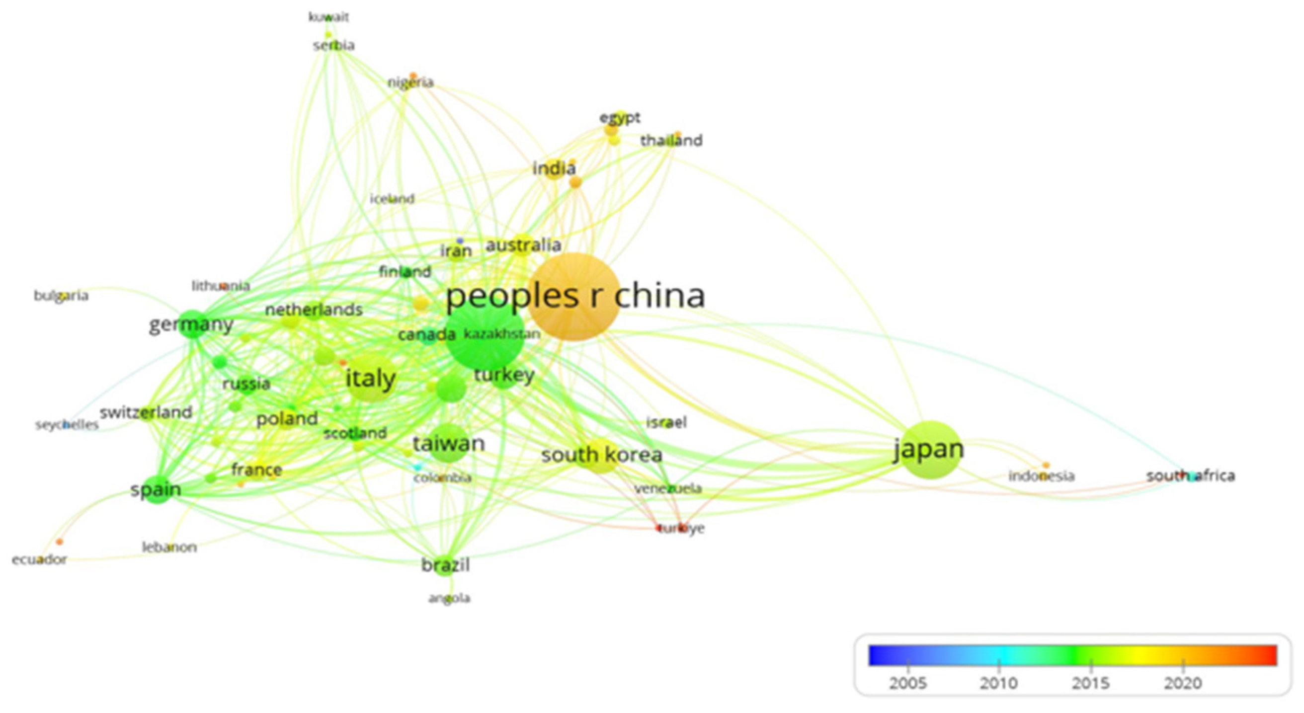
Research Trends over Time

Figure 2 provides insights into how research on hyperuricemia has evolved over the years (2005–2024). From 2012 to 2014, studies primarily focused on the cardiovascular effects of hyperuricemia, with particular attention to hypertension and blood pressure regulation. Between 2015 and 2020, the focus expanded to include insulin resistance, diabetes, obesity, mortality, and metabolic risk factors. More recent research, from 2020 onward, has explored the role of oxidative stress, inflammation, and fatty liver disease, indicating a shift toward understanding the broader systemic effects of hyperuricemia. Countries such as China, the USA, Italy, Japan, Germany, and South Korea have made leading contributions to hyperuricemia studies (Figure 3). These key players and trends in hyperuricemia research suggest limited research on the African population, as only Nigeria and South Africa are faintly represented on the map. Hence, collaboration with studies from such locations with key players may improve knowledge dissemination.

3. The Complex Interplay Between Hyperuricemia, Insulin Resistance, and Type 2 Diabetes

Recent research emphasizes the intricate relationship between hyperuricemia, insulin resistance (IR), and type 2 diabetes mellitus (T2DM), positioning hyperuricemia as a potential independent predictor of diabetes development [10]. This association appears bidirectional, as IR contributes to elevated uric acid levels, while hyperuricemia exacerbates IR through mechanisms such as chronic inflammation, oxidative stress, and endothelial dysfunction [11]. One proposed pathway suggests that hyperuricemia impairs insulin-dependent nitric oxide stimulation in endothelial cells, disrupting vascular function and insulin signaling. Intriguingly, hyperuricemia shares overlapping etiological and pathophysiological features with T2DM, prompting some researchers to suggest that it may not merely be a metabolic byproduct but an equivalent metabolic disorder in its own right [11]. These insights support the potential role of uric acid-lowering interventions in mitigating diabetes risk, though further studies are needed to establish causal relationships and define the extent of therapeutic benefits. Chronic low-grade inflammation and oxidative stress emerge as central mediators linking hyperuricemia to metabolic dysfunction. Elevated uric acid triggers the release of pro-inflammatory cytokines such as TNF-α and IL-6, perpetuating systemic inflammation that contributes to pancreatic β-cell dysfunction, IR, and disease progression [12] (Table 1). Concurrently, oxidative stress damages endothelial cells, increasing the risk of vascular complications in diabetic patients.
A key aspect of hyperuricemia research focuses on its role in cardiovascular health. The analysis reveals strong associations between hyperuricemia, hypertension, blood pressure regulation, and endothelial dysfunction, reinforcing its significance in both metabolic and cardiovascular pathology. These findings highlight the need for integrated metabolic and cardiovascular management strategies that address hyperuricemia as a potential risk factor.

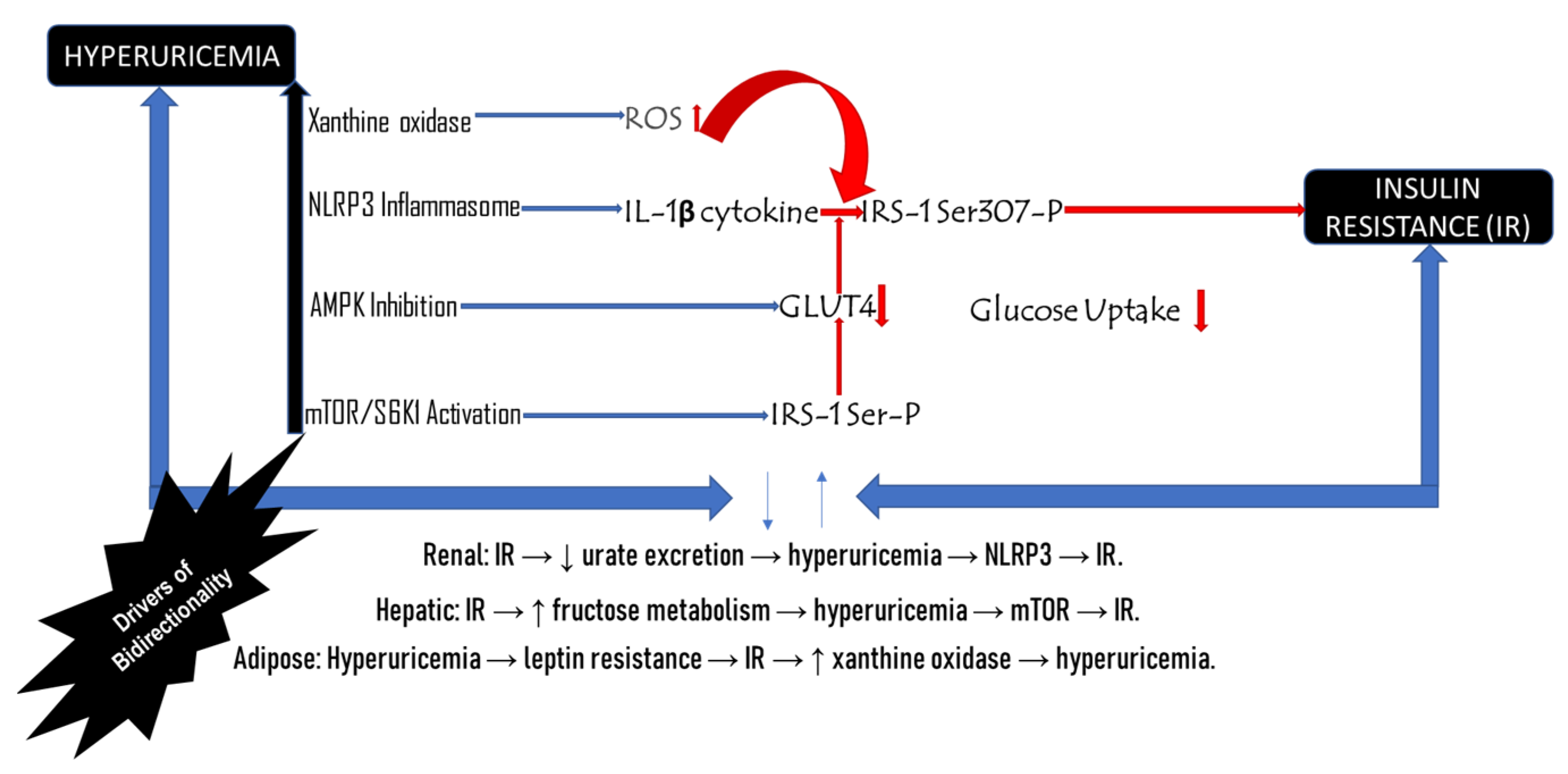
3.1. Bidirectional Relationship Between Hyperuricemia and Insulin Resistance

The scientific discourse on hyperuricemia has evolved over the past decade. Early research (2012–2016) primarily focused on its connection to oxidative stress, serum uric acid levels, and non-alcoholic fatty liver disease (NAFLD). In the past five years, hyperuricemia has been increasingly linked to T2DM, metabolic syndrome, and insulin resistance, cementing its place as a key marker of metabolic dysfunction. Compelling evidence suggests strong correlations between serum uric acid levels, IR, and metabolic syndrome components, particularly HDL cholesterol and triglycerides [12]. High uric acid levels are consistently observed in individuals with advanced metabolic syndrome, yet the precise cause-and-effect relationship remains a subject of debate [27].
To clarify this relationship, bidirectional Mendelian randomization studies have been employed [28]. However, some findings remain inconclusive, while some studies indicate that elevated uric acid does not directly induce IR; others suggest that higher fasting insulin levels may elevate serum uric acid levels, increasing the risk of gout [3,11]. Emerging research reveals that hyperuricemia and IR influence each other in a vicious cycle [22]. While high uric acid promotes IR via inflammation (NLRP3), oxidative stress, and impaired insulin signaling, IR also exacerbates hyperuricemia by reducing kidney urate excretion and increasing xanthine oxidase activity. For instance, insulin resistance upregulates renal URAT1 and GLUT9 transporters, trapping uric acid in the blood [29]. Conversely, hyperuricemia worsens IR by activating stress pathways like mTOR/S6K1. However, the relationship is not universal [30]. Some studies show no causal link in certain populations (e.g., East Asians with protective SLC2A9 gene variants), and paradoxically, uric acid can act as an antioxidant in early metabolic stages. Clinical trials reflect this complexity: While urate-lowering drugs (e.g., allopurinol) improve IR in prediabetes, they often fail in advanced diabetes, suggesting context-dependent effects. Sex differences present a higher level of complexity, with hyperuricemia being more strongly linked to IR in women [31]. Thus, the interplay is bidirectional, modifiable by genetics, metabolic health, and disease stage, demanding personalized treatment strategies.
Conversely, genetic predisposition to higher serum urate levels does not appear to contribute significantly to T2DM or glycemic traits [32]. This emphasizes the complex interplay between uric acid metabolism, IR, and metabolic disease progression. Furthermore, genome-wide association studies (GWAS) provide valuable insights into the hyperuricemia–IR connection. Research using electronic medical records and GWAS data suggests that high insulin levels drive hyperuricemia, but the reverse relationship is less evident [33]. These findings imply that reducing insulin resistance may naturally lower uric acid levels and potentially decrease gout risk. However, therapies aimed at lowering serum uric acid, such as allopurinol treatment, may have a limited impact on insulin resistance and its metabolic consequences.
Notably, emerging research presents a contrasting perspective. Low-dose allopurinol therapy has been linked to modest yet clinically meaningful improvements in glycemic control and variability in T2DM patients [34]. Additionally, longitudinal studies suggest that elevated uric acid may precede IR rather than result from it, particularly in individuals who later develop hypertension. Peripheral IR appears to play a more significant role than hepatic IR in the development of hypertension associated with hyperuricemia, inflammation, oxidative stress, and endothelial dysfunction [35]. These findings reinforce the need for further mechanistic studies to unravel the precise interactions between urate metabolism, insulin sensitivity, and metabolic health. Future clinical trials should evaluate the long-term efficacy of uric acid-lowering treatments in mitigating IR and related metabolic complications.

3.2. Gender Stratification in Hyperuricemia and Insulin Resistance

Hyperuricemia and insulin resistance exhibit notable gender-based differences in their prevalence, underlying mechanisms, and clinical consequences. These disparities are largely driven by hormonal, genetic, and lifestyle factors that influence disease expression and progression across the sexes (Table 2). Epidemiologically, men generally exhibit higher serum uric acid levels compared to premenopausal women, primarily due to the uricosuric effects of estrogen, which promotes uric acid excretion via the kidneys [36]. However, this gender gap diminishes after menopause, as estrogen levels fall and women’s uric acid levels rise. This shift also leads to an increased incidence of gout among postmenopausal women. Similarly, insulin resistance is less common in premenopausal women due to estrogen’s protective role in enhancing insulin sensitivity. After menopause, the risk of insulin resistance escalates in women, potentially exceeding that in men. Conditions such as polycystic ovary syndrome (PCOS) further exacerbate insulin resistance in younger women by altering hormonal balance and promoting visceral adiposity [37].
Biologically, sex hormones play a pivotal role. Estrogen not only lowers serum uric acid by enhancing renal clearance but also improves insulin sensitivity through its influence on adipokines such as adiponectin. In contrast, testosterone may contribute to insulin resistance in men by promoting the accumulation of visceral fat. Elevated androgen levels in women, as seen in PCOS, are associated with adverse metabolic profiles [38]. During the menopausal transition, declining estrogen levels lead to both increased uric acid retention and diminished insulin sensitivity, heightening cardiometabolic risk in aging women. Men are more prone to accumulating visceral fat, which is metabolically active and promotes both insulin resistance and hyperuricemia [31]. In contrast, premenopausal women typically have more subcutaneous fat, which is less harmful metabolically. Post menopause, however, women experience a redistribution of fat toward the visceral compartment, increasing their susceptibility to metabolic disorders.
These gender-based differences necessitate tailored approaches in diagnosis, screening, and treatment. Men should be evaluated earlier for hyperuricemia and gout due to their higher baseline risk. Postmenopausal women, on the other hand, require closer monitoring for insulin resistance and metabolic syndrome. Therapeutically, urate-lowering agents like allopurinol and febuxostat are more frequently indicated in men. For women, especially those with PCOS or postmenopausal metabolic dysfunction, insulin sensitizers such as metformin are particularly beneficial [37]. Lifestyle interventions also need to be gender-sensitive. In men, strategies should emphasize weight management, moderation of alcohol intake, and reduction in purine-rich foods. For women, especially those undergoing hormonal transitions, focus should be placed on preventing visceral fat accumulation and maintaining hormonal balance to reduce cardiometabolic risk.

4. Molecular Processes Linking Hyperuricemia and Insulin Resistance

Subclinical hyperuricemia has emerged as one of the many factors for impaired insulin sensitivity (IS) and an important component of the existing Abdominal obesity, metabolic syndrome, type II diabetes mellitus, and cardiovascular disease. The pathways between hyperuricemia and insulin resistance include oxidative stress and inflammation, vascular dysfunction, adipose dysfunction, and renin–angiotensin system (RAS) activation.

4.1. Oxidative Stress and Inflammation

Beyond the classical association of uric acid with crystal-induced arthropathy, elevated uric acid, both in soluble form and as monosodium urate (MSU) crystals, acts as a bioactive molecule that orchestrates a multifaceted assault on insulin signaling. Even in its soluble form, uric acid can stimulate pro-inflammatory pathways, potentially contributing to various chronic diseases beyond gout [39]. Central to this pathophysiology is the activation of the NLRP3 inflammasome, a molecular platform that senses metabolic danger and triggers downstream inflammatory cascades [40]. The innate immune sensor NLRP3 is activated in response to uric acid accumulation in immune cells such as macrophages and in metabolically active tissues, including adipocytes and hepatocytes [41]. Upon activation, NLRP3 promotes the cleavage of pro-caspase-1 into its active form, leading to the maturation and release of interleukin-1β (IL-1β) and interleukin-18. Among these, IL-1β exerts potent diabetogenic effects: it phosphorylates insulin receptor substrate-1 (IRS-1) on serine residues, preventing its interaction with the insulin receptor and inhibiting downstream Akt activation, which is essential for glucose uptake and glycogen synthesis [41]. Furthermore, chronic IL-1β exposure impairs glucose-stimulated insulin secretion (GSIS) in pancreatic β-cells, compounding systemic hyperglycemia. Moreover, solid epidemiologic evidence indicates that higher levels of uric acid stimulate the secretion of inflammatory cytokines such as IL-6 and TNF-α that sustain inflammation and influence insulin resistance adversely. These findings emphasize the pro-inflammatory underpinnings of uric acid-induced insulin resistance [42].
Pharmacologic inhibition of these pathways via IL-1 receptor antagonists such as anakinra, or urate-lowering agents like allopurinol and febuxostat has shown promise in attenuating inflammation-mediated insulin resistance. Furthermore, hyperuricemia is also related to the activation of inflammatory processes due to the nuclear factor kappa Beta (NF-κB) signaling pathway [10].
The AMPK serves as a metabolic master switch, regulating cellular energy homeostasis by promoting glucose uptake and fatty acid oxidation under low-energy conditions. However, hyperuricemia disrupts this regulatory axis through several converging mechanisms [43]. Uric acid-induced ROS primarily derived from xanthine oxidase activity, oxidatively inactivate AMPK. Additionally, the metabolism of fructose, which elevates intracellular uric acid levels, leads to ATP depletion and paradoxical AMPK suppression, further compromising cellular energy balance.
The downstream effects of AMPK inhibition are profound: reduced GLUT4 translocation in muscle and adipose tissue diminishes glucose uptake; activation of SREBP-1c promotes hepatic lipogenesis, contributing to non-alcoholic fatty liver disease (NAFLD); and suppression of PGC-1α, which downregulates mitochondrial biogenesis, and exacerbating mitochondrial dysfunction [44]. Metformin, a first-line therapy for type 2 diabetes, exerts its anti-hyperglycemic effects in part through AMPK activation, representing a therapeutic counterbalance to uric acid-mediated inhibition [45].
Efficient glucose uptake in insulin-sensitive tissues depends on the precise trafficking of GLUT4, the principal insulin-responsive glucose transporter. Hyperuricemia disrupts this process via oxidative and nutrient-sensing mechanisms. Uric acid-derived ROS oxidatively modifies TBC1D4, a key modulator of GLUT4 vesicle translocation, impairing its docking at the plasma membrane. Simultaneously, activation of the mTORC1/S6K1 pathway by uric acid leads to inhibitory phosphorylation of IRS-1 (e.g., Ser307), hindering the activation of Akt and subsequent GLUT4 mobilization [3].
These disruptions culminate in a paradoxical state where insulin is present, yet glucose cannot effectively enter cells, a hallmark feature of peripheral insulin resistance. Importantly, aerobic exercise can bypass this blockade by enhancing GLUT4 translocation via insulin-independent AMPK activation, while SGLT2 inhibitors (e.g., empagliflozin) facilitate glucose clearance independent of cellular uptake.
Furthermore, hyperuricemia is a potent instigator of oxidative stress, primarily through enhanced xanthine oxidase activity and stimulation of NADPH oxidase (NOX) enzymes in vascular and metabolic tissues. The resultant surge in ROS inflicts direct oxidative damage on core components of the insulin signaling cascade [46]. Proteins such as IRS-1 and Akt are oxidized, impairing their phosphorylation and function. In parallel, ROS activates cellular stress kinases, which further inhibit insulin signaling via serine phosphorylation of IRS-1.
This oxidative environment not only exacerbates insulin resistance but also promotes endothelial dysfunction and systemic inflammation. Therapeutic approaches involving antioxidants such as vitamin C, alpha-lipoic acid, and xanthine oxidase inhibitors offer mechanistic interventions to restore redox balance and preserve insulin sensitivity [44]. In the mTORC1/S6K1 axis, a nutrient-sensitive signaling hub becomes aberrantly activated in the setting of elevated uric acid. This overactivation phosphorylates IRS-1 at inhibitory serine sites, dampening insulin signal transduction. Additionally, hyperactive mTOR suppresses autophagy, leading to the accumulation of dysfunctional mitochondria and protein aggregates that further compromise cellular metabolism.
In the liver, uric acid-mediated mTOR activation enhances hepatic insulin resistance and augments gluconeogenesis, contributing to fasting hyperglycemia. While mTOR inhibitors like rapamycin offer metabolic benefits, their immunosuppressive and proliferative side effects limit long-term clinical utility. Dietary protein moderation and specific nutrient timing strategies may serve as adjunct approaches to indirectly temper mTOR overactivation [47].
These mechanistic threads; NLRP3 inflammasome activation, AMPK inhibition, GLUT4 dysregulation, oxidative stress, and mTOR overactivation do not operate in isolation (Figure 4). Rather, they form a self-reinforcing loop: uric acid-induced inflammation fuels ROS production, which in turn suppresses AMPK and activates mTORC1, further impairing GLUT4 trafficking and exacerbating insulin resistance. Compounding this, hyperinsulinemia, a consequence of insulin resistance reduces renal uric acid excretion, amplifying hyperuricemia in a deleterious feedback cycle [5].

4.2. Endothelial Dysfunction

Administering uric acid to persons with type 1 diabetes right away can help their endothelial health. The antioxidant capabilities of uric acid are responsible for this improvement [48]. However, a long-term rise in uric acid may hurt endothelial function in several ways, such as by increasing oxidative stress, inflammation, and arginase activity and lowering nitric oxide (NO) production and endothelial NO synthase phosphorylation [49]. Uric acid is linked to other heart disease risk factors and has two functions: an antioxidant and a pro-oxidant [49]. This makes the connection between uric acid and capillary function even more complicated. This reveals how uric acid and vascular function are complicatedly linked in many unhealthy situations.
Hyperuricemia decreases the availability of nitric gas, a critical molecule in the endothelium’s health and glucose uptake. Raised uric acid inhibits eNOS, reducing NO levels [50]. Percussive reduction in NO decreases the ability of vessels to expand, leading to endothelial dysfunction, which is closely correlated with insulin resistance. Hyperuricemia is associated with endothelial dysfunction, which has potentially dire consequences for glucose tolerance. Endothelial dysfunction is affected by decreased glucose uptake in peripheral tissues, which worsens insulin resistance [51]. However, the reduced ability of blood vessels to dilate may cause hypertension and enhance other metabolic decompensations. Thereby, establishing a circle of interdependence between hyperuricemia, endothelial dysfunction, and insulin resistance.

4.3. Adipocyte Dysfunction

Adipose tissue has a central role in the metabolic control of glucose homeostasis [52]. The current literature reveals that hyperuricemia compromises adipose tissue integrity and that adipokine release is affected. High uric acid levels, under perplexity, change the secretory capacity of adipose tissue by decreasing the production of adiponectin and increasing pro-inflammatory cytokines [53]. Adiponectin is involved in the improvement of insulin sensitivity; therefore, a decrease in its levels leads to insulin resistance. Hyperuricemia impairs adipokines and increases lipolysis with subsequent elevation of free fatty acid levels in the blood, which adds to hepatic and muscular insulin resistance [54]. This relationship also vindicates the significance of normal uric acid levels in adipose tissues and the general metabolic compartment.

4.4. Activation of the Renin–Angiotensin System (RAS)

Hyperuricemia also activates RAS, a blood pressure/metabolic regulation system actively involved in blood pressure regulation [55]. Uric acid amounts that are too high can make RAS work through pathways that depend on oxidative stress and inflammation [56]. RAS plays a role in increased pressure and compression resistance, which is recognized to be connected with metabolic syndrome and insulin resistance. Insulin resistance is most evident in cases of obesity or hypertension, and raised angiotensin II has been seen to interfere with cellular signaling pathways [57]. This impairment adds to the complication of the metabolic profile because it favors the creation of an environment associated with type 2 diabetes. Therefore, the control of hyperuricemia could be linked to effects on the activity of RAS and metabolic changes.
Research on uric acid’s role in activating the renin–angiotensin system (RAS) has yielded mixed results. Zhang et al. [58] found that uric acid upregulates RAS in adipose tissue through TLR2/4-mediated inflammation. Similarly, Yu et al. [59] demonstrated that uric acid induces oxidative stress and activates the local RAS in vascular endothelial cells, leading to endothelial dysfunction. However, McMullan et al. [60] conducted a randomized controlled trial contradicting these findings. They reported that lowering serum uric acid levels with allopurinol or probenecid had no significant effect on kidney-specific or systemic RAS activity or blood pressure. These conflicting results highlight the complexity of uric acid’s relationship with RAS activation and underscore the need for further research to clarify this association.

5. Clinical Evidence Supporting the Role of Hyperuricemia in Insulin Resistance

Recently, there has been growing interest in the association between hyperuricemia and metabolic disorders, particularly insulin resistance and type II diabetes mellitus. A review of clinical evidence, drawn from epidemiological studies, clinical trials, and observational data, sheds light on the relationship between hyperuricemia and these conditions. Numerous epidemiological studies have established a link between elevated uric acid levels in the blood and insulin resistance. For example, a case–control study involving 2530 participants found a significant difference in median plasma uric acid levels between individuals with and without insulin resistance (p < 0.001), with those having insulin resistance showing higher levels [61]. Additionally, a meta-analysis confirmed that elevated uric acid is an independent risk factor for developing insulin resistance [62]. A large-scale observational study of 15,403 participants demonstrated that high uric acid levels increased the risk of developing diabetes by 75% over five years [63]. These findings not only underscore the association between hyperuricemia and metabolic syndromes but also suggest that hyperuricemia could play a role in the development of insulin resistance.
Research further supporting the connection between hyperuricemia and type 2 diabetes is provided by clinical trials, which demonstrate that elevated uric acid levels increase the risk of developing diabetes. For example, hyperuricemia has been associated with a 1.63 odds ratio for metabolic syndrome. A study using data from 8669 participants in the Third National Health and Nutrition Examination Survey (NHANES-III) confirmed a link between hyperuricemia and type 2 diabetes [64]. Additionally, prospective studies have shown that variations in uric acid levels are linked to diabetes risk. For instance, individuals with persistent hyperuricemia are more likely to develop diabetes compared to those with normal uric acid levels [63]. This suggests that fluctuations in baseline uric acid concentrations may play a crucial role in assessing diabetes risk, rather than focusing solely on elevated uric acid levels.

Clinical Trials and Interventions

Evaluations of available anti-gout drugs have produced conflicting evidence regarding their effects on insulin sensitivity. In one study, the impact of allopurinol on fasting blood glucose (FBG) and glycated hemoglobin (HbA1c) was assessed. The results showed that pre-intervention FBG decreased in non-diabetic individuals but did not significantly alter HbA1c levels [65]. This suggests that uric acid-lowering treatments may improve glycemic control, though their effectiveness depends on baseline diabetic conditions. These variations can be attributed to methodological differences, sample characteristics, and the specific uric acid-lowering therapies used. For example, some studies have reported that allopurinol does not reduce FBG or HbA1c in diabetic patients [66]. This indicates that the glycemic benefits of uric acid-lowering therapy may be limited to individuals with prediabetes or without diabetes. Furthermore, Chen et al. [65] observed that patients receiving allopurinol at doses greater than 200 mg per day experienced a more significant reduction in FBG compared to those taking 200 mg or less. These inconsistencies highlight the need for further research to determine optimal therapeutic strategies for managing hyperuricemia in the context of insulin resistance.
Case reports have provided evidence supporting the proposed relationship between hyperuricemia and metabolic dysfunction. For example, patients undergoing gout treatment, a chronic hyperuricemia condition, have exhibited early signs of insulin resistance and an increased risk of type 2 diabetes [51]. These observations highlight the significance of addressing elevated uric acid levels and raise broader questions about the spectrum of metabolic disturbances. In individuals with metabolic syndrome, hyperuricemia is independently associated with insulin resistance [17]. Studies have shown that allopurinol, a uric acid-lowering agent, can improve insulin resistance, fasting glucose levels, and systemic inflammation, even in individuals without symptomatic hyperuricemia [67]. Moreover, elevated serum uric acid is considered a marker of insulin resistance syndrome, as higher levels are correlated with increased BMI, plasma triglycerides, and fasting insulin levels.
Data from specific population samples further support this relationship. Among patients with isolated hyperuricemia, particularly in ethnic groups with a high prevalence of gout, metabolic syndrome, obesity, or other risk factors, elevated uric acid levels and/or gout have been associated with an increased prevalence of insulin resistance and type 2 diabetes [68]. For instance, individuals diagnosed with metabolic syndrome tend to have higher serum uric acid concentrations compared to those without such a diagnosis [69]. Moreover, cross-sectional, prospective, and meta-analyses of epidemiological, interventional, and observational studies provide substantial evidence linking hyperuricemia directly to insulin resistance and type 2 diabetes. However, long-term studies are still needed to clarify causal mechanisms and establish treatment targets. Current evidence suggests that managing uric acid levels should be integrated into comprehensive metabolic disease management strategies.
The relationship between serum uric acid and insulin resistance has garnered increasing attention due to their shared involvement in metabolic dysfunction and cardiometabolic risk. Traditionally, epidemiological and clinical studies have employed binary classification models to investigate this association, often defining uric acid and insulin sensitivity based on fixed clinical thresholds [70]. It is an analytical framework that categorizes serum uric acid levels and insulin resistance status into dichotomous variables, typically as either elevated vs. normal or insulin resistant vs. insulin sensitive, based on predetermined thresholds. While such dichotomization offers analytical simplicity and clinical convenience, it may inadequately capture the complex, nonlinear, and context-dependent nature of the metabolic interactions involved [71]. However, several limitations of binary modeling approaches emphasize the need for continuous and dynamic analytical frameworks to enhance the precision of metabolic research and risk stratification (Table 3).

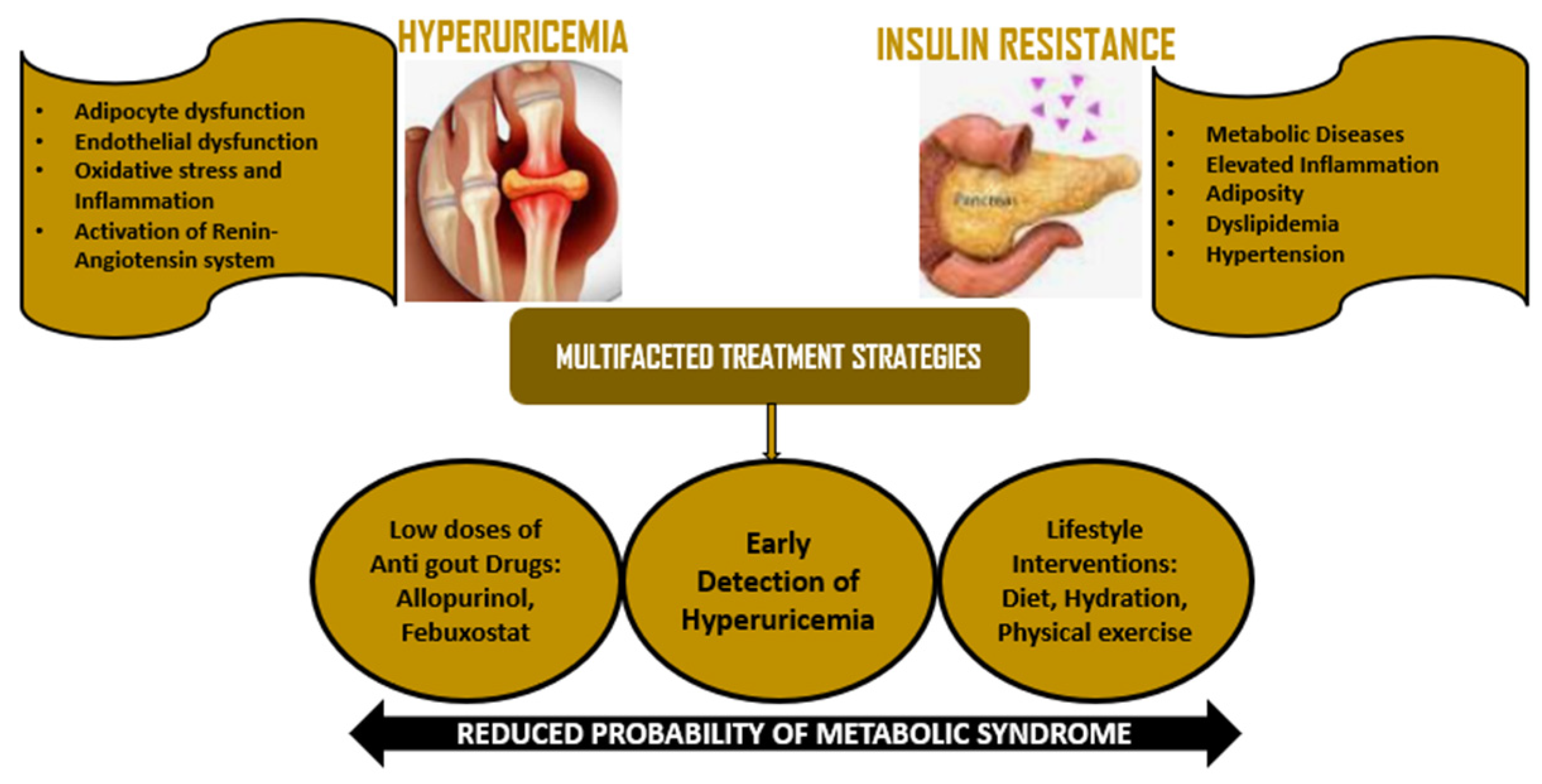
6. Implications for Management and Treatment

Hyperuricemia management, especially regarding the comorbidity with insulin resistance or type 2 diabetes, calls for an interdisciplinary model of management [77]. This section reviews existing pharmacological treatments, lifestyle modifications, and surveillance approaches that may be used to ameliorate the impact of hyperuricemia in the management of metabolic disorders (Table 1).

6.1. Drugs of Antigout and Antihyperuricemic Treatment

Treatment goals include managing acute attacks and lowering uric acid levels below 360 μmol/L (6 mg/dL) long-term [78]. First-line treatments include allopurinol and uricosuric agents, with febuxostat as an alternative when allopurinol is contraindicated. Acute gout attacks are typically managed with NSAIDs, colchicine, or glucocorticosteroids [79]. Recent developments in gout treatment include interleukin-1 inhibitors for refractory cases and patients with comorbidities [80]. Ongoing research focuses on new drugs targeting various mechanisms, including xanthine oxidase inhibitors, uricosurics, and dual inhibitors [81].
The main class of drugs used to prevent hyperuricemia is xanthine oxidase inhibitors, including allopurinol and febuxostat (Table 4). It has been the drug of choice in gout and hyperuricemia for many years by blocking the formation of uric acid. Other research conducted clinically has demonstrated that allopurinol has additional benefits, such as decreasing the concentration of serum uric acid while increasing insulin sensitivity in patients with hyperuricemia [82]. Based on a randomized control trial, allopurinol administration resulted in a large drop in HOMA-IR and fasting insulin, suggesting improvement in insulin resistance [83]. From clinical observation, febuxostat has been found to decrease uric acid levels effectively. The analysis of the effectiveness and safety of allopurinol vs. febuxostat revealed a higher efficacy of febuxostat in achieving the target serum uric acid level (<6 mg/dL) in both diabetic and non-diabetic [84]. Patients who do not achieve adequate control with allopurinol or have side effects with the drug may therefore prefer febuxostat.
The main value of medicines to lower uric acid levels is not only to treat gout but also potentially to stop and treat insulin resistance and diabetes mellitus. Observations provided by the extant literature support the notion that lower uric acid is associated with better metabolic profiles, such as lower fasting blood sugar and improved insulin sensitivity [85]. However, these therapies are not free from the hitch. Adverse effects obtained with both allopurinol and febuxostat include gastrointestinal disturbances and possible cardiovascular risks [86]. As such, the patients should be selected cautiously and supervised closely once these treatment modalities have been started.

6.2. Lifestyle Interventions

Lifestyle changes, specifically regarding diet, are central to the management of hyperuricemia and its metabolic complications. One of the most significant changes is the necessity to cut the quantities of fructose consumed, as it has been scientifically established that high fructose consumption results in excess production of uric acid [87]. Red meat, organ meat, and some fish and seafood are rich in purines, and should be avoided to some extent to avoid raising uric acid. They also show that food full of whole grains, fruits, veggies, and low-fat dairy is connected with low levels of uric acid and improved insulin sensitivity. Moreover, strategies such as hydration and a lower salt diet have also been shown to promote uric acid and glucose clearance in the kidneys. SGLT2 inhibitors, which increase renal glucose excretion, also lower uric acid levels, providing a dual benefit [88]. Thus, they promote enhanced renal function. Uric acid-lowering medications such as allopurinol and febuxostat may be combined with insulin sensitizers such as metformin or thiazolidinediones to improve insulin signaling [89]. These therapies produce a healthy metabolic profile, making them useful in a multifaceted therapy strategy.
The other factor that is also important while managing hyperuricemia and insulin resistance is weight control. Both of these diseases are strongly linked to obesity. So, achieving weight loss by reducing food intake and increasing patients’ physical activity would significantly improve their metabolic profile, according to the Cleveland Clinic [69]. Thus, one can claim that the mentioned approach to managing uric acid levels provides not only the direct therapeutic effect of weight loss and alterations in diet but also the positive effects of increased physical activity levels on insulin sensitivity [90]. Additionally, limiting purine and fructose intake and increasing physical activity naturally complement larger insulin resistance management methods.

6.3. Monitoring and Prevention Strategies

Monitoring of serum uric acid concentration is recommended in patients with a potential for insulin resistance and diabetes. Identifying hyperuricemia at an early stage will ensure that other interventions that might work to stop the worsening of different metabolic disorders are undertaken in good time. A study has shown the likelihood of developing gout in people with obesity, high blood pressure, or a history of diabetes. This suggests that more healthcare providers should order uric acid level tests regularly [91].
Preventive approaches should place more importance on life changes, with the use of drugs only when necessary. Some of these recommendations include: taking moderate purine and fructose diets, achieving normal body weight, engaging in routine exercises, and drinking adequate water [91]. Furthermore, informing the patient of the risk factors of untreated hyperuricemia, including gout flares or increased risk of diabetes, will change their lifestyle.
Table 4. Therapeutic Agents for Hyperuricemia and Insulin Resistance.
AgentMechanismEffect on IRKey EvidenceLimitationsReference
AllopurinolXanthine oxidase inhibitor lowers uric acidImproves HOMA-IR (~15%)RCT in metabolic syndromeWeak effect in advanced T2DMalorbeti et al. [1]
FebuxostatXanthine oxidase inhibitor (more potent)Improves hepatic IRReduced mTORC1 activation in NAFLDCardiovascular safety debatesYu et al. [58]
SGLT2 Inhibitors (e.g., Empagliflozin)Increases Urinary urate excretion and reduces inflammationImproves IR with reduced SUARCT in T2D (HOMA-IR reduced by 18%)Genital infections, volume depletionWang et al. [92]
MetforminAMPK activation leads to activation of GLUT4Indirectly counters uric acid’s AMPK blockadeReversed leptin resistance in adipocytesGI side effectsAgius et al. [93]
IL-1β Antagonists (e.g., Canakinumab)Blocks NLRP3 inflammasome, resulting in reduced IL-1βPreserves β-cell functionRestored GSIS in hyperuricemic miceHigh cost, infection riskMalorbeti et al. [1]
SGLT2i + AllopurinolDual urate-lowering + insulin-sensitizingSynergistic HOMA-IR reductionClinical trials are ongoing (e.g., NCT04881110), University of Campania, ItalyLimited long-term dataCaruso et al. [94]
Diet/LifestyleReduced fructose, an increase in fiber, and aerobic exerciseReduced SUA with increased AMPK/GLUT4Ketogenic diet improved IR despite increase in uric acidAdherence challengesYu et al. [58]

7. Current Research and Future Trends

As mentioned earlier, how high uric acid levels and insulin resistance are linked is an extensive topic that needs more comprehensive investigation. New gene data are being published, new therapeutic targets are being identified, and longitudinal work is necessary to increase understanding of these connections. This discussion elaborates on these aspects and highlights their significance for future research and clinical practice.

7.1. Insights from Large Cohort and Mendelian Randomization Studies

The relationship between serum uric acid (SUA) and insulin resistance (IR) has been extensively investigated through both large prospective cohort studies and Mendelian randomization (MR) analyses, yielding critical insights into the nature of their association. While observational data from major cohorts like the Atherosclerosis Risk in Communities (ARIC) study and the UK Biobank demonstrate consistent correlations between elevated SUA and incident type 2 diabetes (T2D) [95], these findings have been challenged by MR studies that interrogate causality using genetic instruments. Large cohort studies, including seminal work from ARIC, have shown hyperuricemia to be an independent predictor of T2D development, even after adjustment for traditional metabolic confounders. However, the attenuation of effect sizes following adjustment for obesity-related parameters suggests that residual confounding may partially explain these associations. More recent analyses of the UK Biobank cohort reveal complex temporal relationships, where the strength of the SUA-IR association varies substantially across subgroups, particularly by sex and baseline metabolic health [96]. MR studies have brought greater clarity to causal inference by leveraging genetic variants in urate transporters (SLC2A9, ABCG2, and URAT1) as proxies for lifelong SUA exposure. European-focused MR analyses have demonstrated modest but consistent causal effects of genetically predicted hyperuricemia on IR markers, supporting a pathophysiological role for SUA in metabolic dysfunction. However, these findings are not universal; East Asian MR studies using the same genetic instruments have failed to replicate this causal relationship, highlighting important ancestry-specific differences in urate metabolism and its metabolic consequences [97]. The most plausible explanation emerging from these data is that the SUA-IR relationship is bidirectional and context-dependent. While hyperuricemia may directly promote IR through inflammatory (NLRP3 inflammasome activation) and oxidative stress pathways, the metabolic milieu of IR simultaneously reduces renal urate excretion, creating a self-reinforcing cycle. This model accounts for why interventions like urate-lowering therapy show metabolic benefits primarily in early dysmetabolism, while having limited efficacy in established T2D where IR dominates the pathophysiology. These findings have important implications for both research and clinical practice. Future studies should prioritize: Ancestry-stratified analyses to clarify genetic modifiers of the SUA-IR relationship, mechanistic studies of tissue-specific urate effects (e.g., adipose vs. hepatic), and precision medicine trials that target hyperuricemia in metabolically defined subgroups. The convergence of evidence suggests that while hyperuricemia likely contributes to metabolic dysfunction in certain populations and disease stages, its clinical significance as a modifiable risk factor must be interpreted in the context of individual metabolic and genetic profiles. This nuanced understanding is essential for developing targeted prevention and treatment strategies.

7.2. Genetic Factors

New scientific studies have discovered some factors in genes that might be associated with hyperuricemia and insulin resistance. Several GWAS have been conducted to identify specific loci associated with serum uric acid levels, from which it can be inferred that hyperuricemia has a heritable background [97]. For example, polymorphisms associated with the solute carrier family SLC2A9 and SLC22A12 genes affect urate transport and can have implications for key metabolic pathways underlying insulin sensitivity [97]. A study by Vaskimo et al. [98] using Mendelian randomization showed that a higher genetically predetermined fasting insulin level is the prediction of increased serum uric acid concentration but not vice versa. Knowing these genetic connections would create opportunities and guidelines toward precision or precision medicine where prevention and treatments will depend on an individual’s genetic predisposition toward hyperuricemia and its metabolic consequences.
There is a great potential for customizing treatment. For those with hyperuricemia or insulin resistance, genetic testing will help the healthcare provider to make appropriate recommendations about lifestyle modifications or pharmacotherapy. For instance, the therapeutic target, certain genetic polymorphisms, may require stricter adherence to diets or prescription of uric acid-lowering medicines at an earlier stage [98]. This approach not only increases the chance of the desired result in treatment but also reduces the application of invasive procedures in low-risk individuals.

7.3. Novel Therapeutic Targets and Longitudinal Studies

The new evidence shows that there are new therapeutic points of contact associated with uric acid metabolism, the management of which can help to improve the therapy of hyperuricemia and related metabolic disorders. One of them comprises the investigation of compounds that selectively block urate transporters, including URAT1 and GLUT9, which are primary facilitative transporters implicated in the renal reabsorption of uric acid [97]. Therefore, targeting these transporters might promote uric acid urinary clearance and decrease serum concentrations more effectively than existing therapies. On the other hand, the new data indicates that more urate in the blood leads to oxidative stress and inflammation, which are two major factors in the formation of insulin resistance [98]. Consequently, antioxidant therapies or anti-inflammatory agents would appear to be new approaches to minimize the detrimental effects of hyperuricemia on metabolic health.
Identifying these new therapeutic targets defines new opportunities to improve the therapies. For instance, the novel dual-action drugs that not only calm the condition by reducing the level of uric acid but also have anti-inflammatory effects could solve both the hyperuricemia problem and the ensuing metabolic complications concomitantly. Moreover, there are other recent studies on gut microbiota contribution to uric acid metabolism, which may result in new probiotic or prebiotic interventions targeting the regulation of urate levels through diet [92].
To evaluate the causality of hyperuricemia and diabetes, a longitudinal study view has to be conducted. Some experiments carried out in the current age point to the fact that high rates of uric acid led to the growth of insulin resistance; however, this needs to be studied over time [92]. Further research using longitudinal designs may suggest whether managing uric acid levels reduces or alleviates a greater chance of developing insulin resistance and type 2 diabetes. Some of these issues should engage a diverse sample to identify specific genetic factors, local settings, and behaviors that may affect these linkages. This approach will help increase the likelihood of discovering the universality and population differences in metabolic health.
Different ethnic kinds of people should be covered in those interventions because different ethnic groups or other segments of the population could experience hyperuricemia or its treatment differently. For instance, specific genetic profiles would influence urate handling, or perhaps specific meals or food products would prompt abnormal serum uric acid levels [58]. By having a representation of all these cohorts in the research designs, the researchers can determine the individual risk factors leading to the problems and then provide appropriate solutions.

8. Harnessing Explainable AI for Early Prediction and Personalized Treatment of Hyperuricemia

Having established the relationship between hyperuricemia and its several comorbidities and metabolic diseases, such as insulin resistance, diabetes, hypertension, and obesity, Explainable Artificial Intelligence (XAI) offers promising avenues for enhancing early prediction and individualized treatment plans, thereby improving patient outcomes [4]. Traditional machine learning models, while effective in pattern recognition, often operate as “black boxes,” providing little insight into their decision-making processes [99]. XAI addresses this limitation by offering transparency and interpretability, crucial for clinical applications where understanding the rationale behind predictions is essential [100,101].
Recent studies have demonstrated the efficacy of XAI in predicting hyperuricemia. For instance, a model integrating Particle Swarm Optimization (PSO) with machine learning algorithms achieved an accuracy of 97.8% and sensitivity of 97.6% using routine blood test data. The incorporation of XAI techniques, such as SHAP (SHapley Additive exPlanations) and LIME (Local Interpretable Model-agnostic Explanations), provided insights into the model’s predictions, enabling healthcare professionals to understand and trust the outcomes [100]. Another study developed a stacking ensemble prediction model combining support vector machines, decision trees, and extreme gradient boosting. This model outperformed individual algorithms and utilized the iBreakDown technique to elucidate contributing risk factors, enhancing its clinical applicability [102].
XAI not only aids accurate prediction but also facilitates personalized health management. By interpreting individual risk factors, XAI enables tailored interventions, such as dietary modifications, lifestyle changes, and pharmacological treatments. For example, developing the Health Portrait Platform and integrating XAI-based risk prediction models allows for real-time online health risk assessments, empowering patients and clinicians to make informed decisions [103]. Moreover, integrating XAI with digital health data, including wearable biosensors and electronic health records, enhances the precision of personalized medicine [104]. This synergy enables continuous monitoring and dynamic adjustment of treatment plans based on real-time data, aligning with the principles of precision medicine [33].
Despite its potential, the implementation of XAI in clinical settings faces challenges. These include ensuring data privacy, addressing algorithmic biases, and integrating XAI tools into existing healthcare infrastructures. Additionally, the complexity of XAI models necessitates user-friendly interfaces to facilitate their adoption by healthcare professionals. With the incorporation of connected innovation intelligence into personalized health designs [105,106], the implementation challenges of XAI will be addressed. Future research should focus on developing standardized protocols for XAI integration, enhancing model interpretability, and conducting large-scale clinical trials to validate the efficacy of XAI-driven interventions. Collaborations between technologists, clinicians, and policymakers will be pivotal in overcoming these challenges and realizing the full potential of XAI in personalized health management. Undoubtedly, as research and technology evolve, the integration of XAI into healthcare systems will be instrumental in addressing the complexities of hyperuricemia and improving patient outcomes.

9. Study Limitations, Recommendations, and Future Outlook

9.1. Study Limitations

The preliminary aspect of the study was limited to literature indexed in the Web of Science database, potentially omitting relevant studies available in other repositories such as PubMed. Furthermore, restricting the analysis to English-language publications may have excluded valuable findings published in other languages. The review also covered a defined timeframe (2004–2024), which may not fully capture the most recent advancements in hyperuricemia research.

9.2. Recommendations

Given the growing recognition of hyperuricemia’s association with multiple comorbidities, including insulin resistance, diabetes, hypertension, and obesity, future healthcare strategies should explore the integration of personalized medicine for its management. Personalized approaches, tailored to individual metabolic profiles, genetic variability, and lifestyle factors, can optimize treatment outcomes. Early screening for hyperuricemia and its related disorders, especially insulin resistance, is strongly recommended to facilitate timely interventions. Additionally, the application of machine learning models trained on basic health checkup data holds promise for predicting hyperuricemia and other chronic conditions, enabling early detection and preventive healthcare.

9.3. Conclusion and Future Outlook

Hyperuricemia is increasingly recognized as a significant contributor to insulin resistance and type 2 diabetes mellitus (T2DM), largely through its links with oxidative stress, chronic inflammation, and metabolic syndrome. Its association with atherosclerotic diseases further highlights the need for its proactive management within broader metabolic health frameworks. Lifestyle modifications such as improved diet and increased physical activity combined with urate-lowering therapies offer practical and effective interventions. To integrate hyperuricemia management into diabetes prevention strategies, routine screening and early, targeted interventions should be prioritized. Future research should focus on uncovering the molecular mechanisms underlying hyperuricemia-related metabolic dysfunction, identifying new therapeutic targets, and investigating the role of gut microbiota. These insights will pave the way for more precise and effective strategies to mitigate insulin resistance and related disorders, ultimately enhancing global health outcomes.

Author Contributions

Conceptualization, O.O.D.-O., J.O.B., T.O.E. and S.O.A.; methodology, O.O.D.-O., J.O.B., T.O.E. and S.O.A., software, O.O.D.-O., J.O.B., T.O.E. and S.O.A.; validation, O.O.D.-O., J.O.B., T.O.E. and S.O.A. and investigation, O.O.D.-O. and S.O.A.; resources, T.O.E.; data curation, J.O.B. and O.O.D.-O.; writing—original draft preparation, O.O.D.-O., J.O.B., T.O.E. and S.O.A.; writing, review and editing, O.O.D.-O., J.O.B., T.O.E. and S.O.A.; visualization, O.O.D.-O., J.O.B., T.O.E. and S.O.A.; supervision, O.O.D.-O., J.O.B., T.O.E. and S.O.A.; project administration, O.O.D.-O., J.O.B., T.O.E. and S.O.A. All authors have read and agreed to the published version of the manuscript.

Funding

This research received no external funding.

Conflicts of Interest

The authors declare no conflicts of interest.

References

  1. Maloberti, A.; Vanoli, J.; Finotto, A.; Bombelli, M.; Facchetti, R.; Redon, P.; Mancia, G.; Grassi, G. Uric acid relationships with lipid profile and adiposity indices: Impact of different hyperuricemic thresholds. J. Clin. Hypertens. 2022, 25, 78–85. [Google Scholar] [CrossRef]
  2. Fritz, J.; Brozek, W.; Concin, H.; Nagel, G.; Kerschbaum, J.; Lhotta, K.; Ulmer, H.; Zitt, E. The Association of Excess Body Weight with Risk of ESKD Is Mediated Through Insulin Resistance, Hypertension, and Hyperuricemia. J. Am. Soc. Nephrol. 2022, 33, 1377–1389. [Google Scholar] [CrossRef] [PubMed]
  3. Nakamura, M.; Satoh, N.; Horita, S.; Nangaku, M. Insulin-induced mTOR signaling and gluconeogenesis in renal proximal tubules: A mini-review of current evidence and therapeutic potential. Front. Pharmacol. 2022, 13, 1015204. [Google Scholar] [CrossRef]
  4. Zhu, Y.; Hu, Y.; Huang, T.; Zhang, Y.; Li, Z.; Luo, C.; Luo, Y.; Yuan, H.; Hisatome, I.; Yamamoto, T.; et al. High uric acid directly inhibits insulin signalling and induces insulin resistance. Biochem. Biophys. Res. Commun. 2014, 447, 707–714. [Google Scholar] [CrossRef]
  5. Du, L.; Zong, Y.; Li, H.; Wang, Q.; Xie, L.; Yang, B.; Pang, Y.; Zhang, C.; Zhong, Z.; Gao, J. Hyperuricemia and its related diseases: Mechanisms and advances in therapy. Signal Transduct. Target. Ther. 2024, 9, 212. [Google Scholar] [CrossRef]
  6. Chen, J.; Ge, J.; Zha, M.; Miao, J.-J.; Sun, Z.-L.; Yu, J.-Y. Effects of Uric Acid-Lowering Treatment on Glycemia: A Systematic Review and Meta-Analysis. Front. Endocrinol. 2020, 11, 507731. [Google Scholar] [CrossRef]
  7. Sun, H.; Chang, X.; Bian, N.; An, Y.; Liu, J.; Leng, S.; Wang, G. Adipose Tissue Insulin Resistance Is Positively Associated with Serum Uric Acid Levels and Hyperuricemia in Northern Chinese Adults. Front. Endocrinol. 2022, 13, 835154. [Google Scholar] [CrossRef]
  8. Bose, B.; Badve, S.V.; Hiremath, S.S.; Boudville, N.; Brown, F.G.; Cass, A.; de Zoysa, J.R.; Fassett, R.G.; Faull, R.; Harris, D.C.; et al. Effects of uric acid-lowering therapy on renal outcomes: A systematic review and meta-analysis. Nephrol. Dial. Transplant. 2014, 29, 406–413. [Google Scholar] [CrossRef]
  9. Hu, X.; Rong, S.; Wang, Q.; Sun, T.; Bao, W.; Chen, L.; Liu, L. Association between plasma uric acid and insulin resistance in type 2 diabetes: A Mendelian randomization analysis. Diabetes Res. Clin. Pr. 2020, 171, 108542. [Google Scholar] [CrossRef]
  10. Zehra, F. Association of serum uric acid with diabetes mellitus type-2: A narrative review. Pak. J. Physiol. 2024, 20, 52–57. [Google Scholar]
  11. Shu, J.; Zhao, R.; Xu, H.; Liu, X.; Guo, H.; Lu, C. Hyperuricemia is associated with metabolic syndrome: A cross-sectional analysis of the National Health and Nutrition Examination Survey (NHANES). Prev. Med. Rep. 2023, 36, 102520. [Google Scholar] [CrossRef] [PubMed]
  12. Luis-Rodríguez, D.; Donate-Correa, J.; Martín-Núñez, E.; Ferri, C.; Tagua, V.G.; Castro, A.P.; Mora-Fernández, C.; Navarro-González, J.F. Serum urate is related to subclinical inflammation in asymptomatic hyperuricaemia. Rheumatology 2021, 60, 371–379. [Google Scholar] [CrossRef] [PubMed]
  13. Gao, Y.; Hua, R.; Hu, K.; Wang, Z. Carbohydrates deteriorate fatty liver by activating the inflammatory response. Nutr. Res. Rev. 2022, 35, 252–267. [Google Scholar] [CrossRef]
  14. Cuttone, A.; Cannavò, V.; Abdullah, R.M.S.; Fugazzotto, P.; Arena, G.; Brancati, S.; Muscarà, A.; Morace, C.; Quartarone, C.; Ruggeri, D.; et al. Expanding the Use of SGLT2 Inhibitors in T2D Patients Across Clinical Settings. Cells 2025, 14, 668. [Google Scholar] [CrossRef] [PubMed]
  15. Timsans, J.; Kauppi, J.; Rantalaiho, V.; Kerola, A.; Hakkarainen, K.; Lehto, T.; Kautiainen, H.; Kauppi, M. Serum Uric Acid Is Associated with Insulin Resistance in Non-Diabetic Subjects. J. Clin. Med. 2025, 14, 2621. [Google Scholar] [CrossRef]
  16. Zhang, J.; Sun, N.; Zhang, W.; Yue, W.; Qu, X.; Li, Z.; Xu, G. The impact of uric acid on musculoskeletal diseases: Clinical associations and underlying mechanisms. Front. Endocrinol. 2025, 16, 1515176. [Google Scholar] [CrossRef]
  17. Badii, M.; Klück, V.; Gaal, O.; Cabău, G.; Hotea, I.; Nica, V.; Mirea, A.M.; Bojan, A.; Zdrenghea, M.; Novakovic, B.; et al. Regulation of SOCS3-STAT3 in urate-induced cytokine production in human myeloid cells. Jt. Bone Spine 2024, 91, 105698. [Google Scholar] [CrossRef]
  18. Nasser, S.; Solé, T.; Vega, N.; Thomas, T.; Balcerczyk, A.; Strigini, M.; Pirola, L. Ketogenic diet administration to mice after a high-fat-diet regimen promotes weight loss, glycemic normalization and induces adaptations of ketogenic pathways in liver and kidney. Mol. Metab. 2022, 65, 101578. [Google Scholar] [CrossRef]
  19. Rodriguez-Iturbe, B.; Johnson, R.J.; Lanaspa, M.A.; Nakagawa, T.; Garcia-Arroyo, F.E.; Sánchez-Lozada, L.G. Sirtuin deficiency and the adverse effects of fructose and uric acid synthesis. Am. J. Physiol. Integr. Comp. Physiol. 2022, 322, R347–R359. [Google Scholar] [CrossRef]
  20. Jing, P.; Shi, M.; Ma, L.; Fu, P. Mechanistic insights of soluble uric acid-related kidney disease. Curr. Med. Chem. 2020, 27, 5056–5066. [Google Scholar]
  21. Sridharan, S.; Basu, A. Distinct Roles of mTOR Targets S6K1 and S6K2 in Breast Cancer. Int. J. Mol. Sci. 2020, 21, 1199. [Google Scholar] [CrossRef]
  22. Bahadoran, Z.; Mirmiran, P.; Kashfi, K.; Ghasemi, A. Hyperuricemia-induced endothelial insulin resistance: The nitric oxide connection. Pflüger Arch. Eur. J. Physiol. 2021, 474, 83–98. [Google Scholar] [CrossRef]
  23. Adnan, E.; Rahman, I.A.; Faridin, H.P. Relationship between insulin resistance, metabolic syndrome components and serum uric acid. Diabetes Metab. Syndr. Clin. Res. Rev. 2019, 13, 2158–2162. [Google Scholar]
  24. Wan, X.; Xu, C.; Lin, Y.; Lu, C.; Li, D.; Sang, J.; He, H.; Liu, X.; Li, Y.; Yu, C. Uric acid regulates hepatic steatosis and insulin resistance through the NLRP3 inflammasome-dependent mechanism. J. Hepatol. 2016, 64, 925–932. [Google Scholar] [CrossRef]
  25. Lanaspa, M.A.; Sanchez-Lozada, L.G.; Choi, Y.J.; Cicerchi, C.; Kanbay, M.; Roncal-Jimenez, C.A.; Johnson, R.J. Uric acid induces hepatic steatosis by generation of mitochondrial oxidative stress: Potential role in fructose-dependent and-independent fatty liver. J. Biol. Chem. 2012, 287, 40732–40744. [Google Scholar]
  26. Baharuddin, B. The impact of fructose consumption on human health: Effects on obesity, hyperglycemia, diabetes, uric acid, and oxidative stress with a focus on the liver. Cureus 2024, 16, e70095. [Google Scholar]
  27. Gong, M.; Wen, S.; Nguyen, T.; Wang, C.; Jin, J.; Zhou, L. Converging Relationships of Obesity and Hyperuricemia with Special Reference to Metabolic Disorders and Plausible Therapeutic Implications. Diabetes Metab. Syndr. Obesity Targets Ther. 2020, 13, 943–962. [Google Scholar] [CrossRef]
  28. McCormick, N.; O’cOnnor, M.J.; Yokose, C.; Merriman, T.R.; Mount, D.B.; Leong, A.; Choi, H.K. Assessing the Causal Relationships Between Insulin Resistance and Hyperuricemia and Gout Using Bidirectional Mendelian Randomization. Arthritis Rheumatol. 2021, 73, 2096–2104. [Google Scholar] [CrossRef]
  29. Dong, M.; Chen, H.; Wen, S.; Yuan, Y.; Yang, L.; Xu, D.; Zhou, L. The Mechanism of Sodium-Glucose Cotransporter-2 Inhibitors in Reducing Uric Acid in Type 2 Diabetes Mellitus. Diabetes Metab. Syndr. Obesity Targets Ther. 2023, 16, 437–445. [Google Scholar] [CrossRef]
  30. Yang, Y.; Wang, T.-T.; Xie, H.-A.; Hu, P.P.; Li, P. Experimental cell models of insulin resistance: Overview and appraisal. Front. Endocrinol. 2024, 15, 1469565. [Google Scholar] [CrossRef]
  31. Redon, P.; Maloberti, A.; Facchetti, R.; Redon, J.; Lurbe, E.; Bombelli, M.; Mancia, G.; Grassi, G. Gender-related differences in serum uric acid in treated hypertensive patients from central and east European countries: Findings from the Blood Pressure control rate and CArdiovascular Risk profilE study. J. Hypertens. 2019, 37, 380–388. [Google Scholar] [CrossRef] [PubMed]
  32. Yamase, Y.; Horibe, H.; Kato, K.; Oguri, M.; Fujimaki, T.; Hibino, T.; Kondo, T.; Sakuma, J.; Takeuchi, I.; Murohara, T.; et al. P4470Identification of four genes as novel susceptibility loci for early-onset type 2 diabetes mellitus, metabolic syndrome, or hyperuricemia in Japanese. Eur. Hear. J. 2019, 40, 867. [Google Scholar] [CrossRef]
  33. Alem, M.M. Effect of low dose allopurinol on glycemic control and glycemic variability in patients with type 2 diabetes mellitus: A cross-sectional study. Heliyon 2022, 8, e11549. [Google Scholar] [CrossRef] [PubMed]
  34. Han, T.; Lan, L.; Qu, R.; Xu, Q.; Jiang, R.; Na, L.; Sun, C. Temporal Relationship Between Hyperuricemia and Insulin Resistance and Its Impact on Future Risk of Hypertension. Hypertension 2017, 70, 703–711. [Google Scholar] [CrossRef]
  35. Sakr, H.F.; Sirasanagandla, S.R.; Das, S.; Bima, A.I.; Elsamanoudy, A.Z. Insulin Resistance and Hypertension: Mechanisms Involved and Modifying Factors for Effective Glucose Control. Biomedicines 2023, 11, 2271. [Google Scholar] [CrossRef]
  36. Li, L.; Zhang, Y.; Zeng, C. Update on the epidemiology, genetics, and therapeutic options of hyperuricemia. Am. J. Transl. Res. 2020, 12, 3167. [Google Scholar]
  37. Jung, J.H.; Song, G.G.; Lee, Y.H.; Kim, J.-H.; Hyun, M.H.; Choi, S.J. Serum uric acid levels and hormone therapy type: A retrospective cohort study of postmenopausal women. Menopause 2018, 25, 77–81. [Google Scholar] [CrossRef]
  38. Meloni, A.; Cadeddu, C.; Cugusi, L.; Donataccio, M.P.; Deidda, M.; Sciomer, S.; Gallina, S.; Vassalle, C.; Moscucci, F.; Mercuro, G.; et al. Gender Differences and Cardiometabolic Risk: The Importance of the Risk Factors. Int. J. Mol. Sci. 2023, 24, 1588. [Google Scholar] [CrossRef]
  39. Joosten, L.A.B.; Crişan, T.O.; Bjornstad, P.; Johnson, R.J. Asymptomatic hyperuricaemia: A silent activator of the innate immune system. Nat. Rev. Rheumatol. 2019, 16, 75–86. [Google Scholar] [CrossRef]
  40. Tan, H.; Zhang, S.; Liao, J.; Qiu, X.; Zhang, Z.; Wang, Z.; Geng, H.; Zhang, J.; Jia, E. Mechanism of macrophages in gout: Recent progress and perspective. Heliyon 2024, 10, e38288. [Google Scholar] [CrossRef]
  41. Kim, S.-K. The mechanism of the NLRP3 inflammasome activation and pathogenic implication in the pathogenesis of gout. J. Rheum. Dis. 2022, 29, 140–153. [Google Scholar] [CrossRef] [PubMed]
  42. Baratta, F.; Moscucci, F.; Ettorre, E.; Bocale, R.; Cicero, A.F.G.; Cirillo, P.; Fogacci, F.; Lospinuso, I.; Savoia, C.; Mengozzi, A.; et al. Influence of Uric Acid on Vascular and Cognitive Functions: Evidence for an Ambivalent Relationship. Metabolites 2024, 14, 642. [Google Scholar] [CrossRef] [PubMed]
  43. Guo, Q.; Jin, Y.; Chen, X.; Ye, X.; Shen, X.; Lin, M.; Zeng, C.; Zhou, T.; Zhang, J. NF-κB in biology and targeted therapy: New insights and translational implications. Signal Transduct. Target. Ther. 2024, 9, 1–37. [Google Scholar] [CrossRef]
  44. Liu, Y.; Wang, D.; Yang, L. Targeting AMPK Signaling in the Liver: Implications for Obesity and Type 2 Diabetes Mellitus. Curr. Drug Targets 2022, 23, 1057–1071. [Google Scholar] [CrossRef]
  45. Goel, S.; Singh, R.; Singh, V.; Singh, H.; Kumari, P.; Chopra, H.; Sharma, R.; Nepovimova, E.; Valis, M.; Kuca, K.; et al. Metformin: Activation of 5′ AMP-activated protein kinase and its emerging potential beyond anti-hyperglycemic action. Front. Genet. 2022, 13, 1022739. [Google Scholar] [CrossRef]
  46. Liu, N.; Xu, H.; Sun, Q.; Yu, X.; Chen, W.; Wei, H.; Jiang, J.; Xu, Y.; Lu, W.; Tocchetti, C.G. The Role of Oxidative Stress in Hyperuricemia and Xanthine Oxidoreductase (XOR) Inhibitors. Oxidative Med. Cell. Longev. 2021, 2021, 380. [Google Scholar] [CrossRef]
  47. Li, J.; Guo, C.; Yang, X.; Xie, W.; Mi, W.; Hua, C.; Tang, C.; Wang, H. Effects of natural products on macrophage immunometabolism: A new frontier in the treatment of metabolic diseases. Pharmacol. Res. 2025, 213, 107634. [Google Scholar] [CrossRef]
  48. Ndrepepa, G. Uric acid and cardiovascular disease. Clin. Chim. Acta 2018, 484, 150–163. [Google Scholar] [CrossRef]
  49. Kumar, A.N.; Aruna, P.; Naidu, J.N.; Kumar, R.; Srivastava, A.K. Review of concepts and controversies of uric acid as antioxidant and pro-oxidant. Arşiv Kaynak Tarama Derg. 2015, 24, 19–40. [Google Scholar] [CrossRef]
  50. Wei, X.; Zhang, M.; Huang, S.; Lan, X.; Zheng, J.; Luo, H.; He, Y.; Lei, W. Hyperuricemia: A key contributor to endothelial dysfunction in cardiovascular diseases. FASEB J. 2023, 37, e23012. [Google Scholar] [CrossRef]
  51. Rhea, E.M.; Banks, W.A.; Raber, J. Insulin Resistance in Peripheral Tissues and the Brain: A Tale of Two Sites. Biomedicines 2022, 10, 1582. [Google Scholar] [CrossRef] [PubMed]
  52. Dilworth, L.; Facey, A.; Omoruyi, F. Diabetes Mellitus and Its Metabolic Complications: The Role of Adipose Tissues. Int. J. Mol. Sci. 2020, 22, 7644. [Google Scholar] [CrossRef]
  53. Kosmas, C.E.; Bousvarou, M.D.; Kostara, C.E.; Papakonstantinou, E.J.; Salamou, E.; Guzman, E. Insulin resistance and cardiovascular disease. J. Int. Med Res. 2023, 51, 30006. [Google Scholar] [CrossRef]
  54. Manrique, C.; Lastra, G.; Sowers, J.R. New insights into insulin action and resistance in the vasculature. Ann. N. Y. Acad. Sci. 2014, 1311, 138–150. [Google Scholar] [CrossRef]
  55. La Russa, D.; Cerra, M.C.; Pellegrino, D.; Montesanto, A. Chronic kidney disease as an age-related disease: New study perspectives fron animal models to hospitalized patents. Dissertation 2019. [Google Scholar] [CrossRef]
  56. Russo, E.; Bertolotto, M.; Zanetti, V.; Picciotto, D.; Esposito, P.; Carbone, F.; Montecucco, F.; Pontremoli, R.; Garibotto, G.; Viazzi, F.; et al. Role of Uric Acid in Vascular Remodeling: Cytoskeleton Changes and Migration in VSMCs. Int. J. Mol. Sci. 2023, 24, 2960. [Google Scholar]
  57. Zhang, Z.; Jiang, S.-M.; Ma, Y.-P.; Dai, P.-L.; Wang, Y.-N.; Zou, G.-M.; Gao, H.-M.; Yang, Y.; Li, W.-G. Expression of the intrarenal angiotensin receptor and the role of renin-angiotensin system inhibitors in IgA nephropathy. Mol. Cell. Biochem. 2019, 453, 103–110. [Google Scholar] [CrossRef]
  58. Yu, W.; Xie, D.; Yamamoto, T.; Koyama, H.; Cheng, J. Mechanistic insights of soluble uric acid-induced insulin resistance: Insulin signaling and beyond. Rev. Endocr. Metab. Disord. 2023, 24, 327–343. [Google Scholar] [CrossRef]
  59. McMullen, M.K. Fructose increases uric acid contributing to metabolic syndrome-herbal, nutritional and dietary strategies to reduce uric acid. OBM Integr. Complement. Med. 2018, 3, 1–49. [Google Scholar]
  60. Rong, J.; Fang, C.; Chen, X.; Hong, C.; Huang, L. Association of serum uric acid with prognosis in patients with myocardial infarction: An update systematic review and meta-analysis. BMC Cardiovasc. Disord. 2023, 23, 512. [Google Scholar]
  61. Sakalli, A.S.A.; Küçükerdem, H.S.S.; Aygün, O.S. What is the relationship between serum uric acid level and insulin resistance? A case-control study. Medicine 2023, 102, e36732. [Google Scholar] [CrossRef]
  62. Liu, J.; Tao, L.; Zhao, Z.; Mu, Y.; Zou, D.; Zhang, J.; Guo, X. Two-Year Changes in Hyperuricemia and Risk of Diabetes: A Five-Year Prospective Cohort Study. J. Diabetes Res. 2018, 2018, 6905720. [Google Scholar] [CrossRef]
  63. Billing, A.M.; Kim, Y.C.; Gullaksen, S.; Schrage, B.; Raabe, J.; Hutzfeldt, A.; Demir, F.; Kovalenko, E.; Lassé, M.; Dugourd, A.; et al. Metabolic communication by SGLT2 inhibition. Circulation 2024, 149, 860–884. [Google Scholar] [CrossRef] [PubMed]
  64. Cheng, C.-Y.; Liu, J.-C.; Chen, H.-Y.; Chen, J.-J.; Hao, W.-R.; Cheng, T.-H. Hyperuricemia and epiretinal pathologies: A review of pathophysiological links and clinical implications. Explor. Med. 2024, 5, 732–749. [Google Scholar] [CrossRef]
  65. Dhungana, A.; Pandeya, A.; Pant, S.; Pokharel, B. Evaluation Of Serum Uric Acid, Glucose And Other Glycemic Parameter In Type II Diabetic Individuals. J. Chitwan Med. Coll. 2022, 12, 68–73. [Google Scholar]
  66. Krishnan, E.; Pandya, B.J.; Chung, L.; Hariri, A.; Dabbous, O. Hyperuricemia in Young Adults and Risk of Insulin Resistance, Prediabetes, and Diabetes: A 15-Year Follow-up Study. Am. J. Epidemiol. 2012, 176, 108–116. [Google Scholar] [CrossRef]
  67. Kanbay, M.; Xhaard, C.; Le Floch, E.; Dandine-Roulland, C.; Girerd, N.; Ferreira, J.P.; Boivin, J.-M.; Wagner, S.; Bacq-Daian, D.; Deleuze, J.-F.; et al. Weak association between genetic markers of hyperuricemia and cardiorenal outcomes: Insights from the STANISLAS study cohort with a 20-year follow-up. J. Am. Heart Assoc. 2022, 11, e023301. [Google Scholar] [CrossRef]
  68. Bashir, A.A.; Bathija, D.; Chandrakar, S. A Study Of Prevalence Of Metabolic Syndrome And Hyperuricemia In Type 2 Diabetes Mellitus. Int. J. Acad Med. Pharm. 2024, 6, 1153–1158. [Google Scholar] [CrossRef]
  69. Han, R.; Zhang, Y.; Jiang, X. Relationship Between Four Non-Insulin-Based Indexes of Insulin Resistance and Serum Uric Acid in Patients with Type 2 Diabetes: A Cross-Sectional Study. Diabetes Metab. Syndr. Obes. Targets Ther. 2022, 15, 1461–1471. [Google Scholar] [CrossRef]
  70. Maloberti, A.; Giannattasio, C.; Bombelli, M.; Desideri, G.; Cicero, A.F.G.; Muiesan, M.L.; Rosei, E.A.; Salvetti, M.; Ungar, A.; Rivasi, G.; et al. Working Group on Uric Acid and Cardiovascular Risk of the Italian Society of Hypertension (SIIA). Hyperuricemia and Risk of Cardiovascular Outcomes: The Experience of the URRAH (Uric Acid Right for Heart Health) Project. High Blood Press. Cardiovasc. Prev. 2020, 27, 121–128. [Google Scholar] [CrossRef]
  71. Fu, Y.; Yu, Y.; Wang, Y.; Sun, Y.; Zhang, K.; Xu, F.; Wang, N.; Wang, B.; Lu, Y. Nonlinear relationship between body mass index and serum uric acid: An observational and Mendelian randomization study among Chinese adults. Nutr. Metab. Cardiovasc. Dis. 2024, 34, 998–1007. [Google Scholar] [CrossRef] [PubMed]
  72. Pinz, M.P.; Medeiros, I.; Carvalho, L.A.d.C.; Meotti, F.C. Is uric acid a true antioxidant? Identification of uric acid oxidation products and their biological effects. Redox Rep. 2025, 30, 2498105. [Google Scholar] [CrossRef] [PubMed]
  73. Xiao, Y.; Wang, H.; Han, L.; Lyu, G.; Li, S. Effect of uric acid on lipid metabolism assessed via restricted cubic splines: A new insight. Heliyon 2024, 10, e37408. [Google Scholar] [CrossRef] [PubMed]
  74. Chien, H.-T.; Lin, Y.-W.; Shen, L.-J.; Hsieh, S.-C.; Lin, L.-Y.; Chen, Y.-A.; Lin, F.-J. Sex-specific associations between prolonged serum uric acid levels and risk of major adverse cardiovascular events. Int. J. Cardiol. Cardiovasc. Risk Prev. 2024, 22, 200302. [Google Scholar] [CrossRef]
  75. Gonzalez-Martin, G.; Cano, J.; Carriazo, S.; Kanbay, M.; Perez-Gomez, M.V.; Fernandez-Prado, R.; Ortiz, A. The dirty little secret of urate-lowering therapy: Useless to stop chronic kidney disease progression and may increase mortality. Clin. Kidney J. 2020, 13, 936–947. [Google Scholar] [CrossRef]
  76. Zhang, W. Hyperuricemia: Current State and Prospects. Explor. Res. Hypothesis Med. 2025, 10, 49–55. [Google Scholar]
  77. Kumaresan, S.; Palanisamy, S. Serum uric acid levels in type 2 diabetes patients: Exploring the association with cardiovascular disease-a systematic. Int. J. Acad. Med. Pharm. 2024, 6, 415–419. [Google Scholar] [CrossRef]
  78. Ruoff, G.; Edwards, N.L. Overview of Serum Uric Acid Treatment Targets in Gout: Why Less than 6 mg/dL? Postgrad. Med. 2016, 128, 706–715. [Google Scholar] [CrossRef]
  79. Latourte, A.; Pascart, T.; Flipo, R.-M.; Chalès, G.; Coblentz-Baumann, L.; Cohen-Solal, A.; Ea, H.-K.; Grichy, J.; Letavernier, E.; Lioté, F.; et al. 2020 Recommendations from the French Society of Rheumatology for the management of gout: Management of acute flares. Jt. Bone Spine 2020, 87, 387–393. [Google Scholar] [CrossRef]
  80. Yadav, S.; Khandelwal, N.; Nath, S.K.; Rai, S. A Hospital-Based Cross-Sectional Study of Patients with Plantar Fasciitis: Is Hyperuricemia Screening Needed? Cureus 2023, 15, e37088. [Google Scholar] [CrossRef]
  81. Richette, P.; Doherty, M.; Pascual, E.; Bardin, T. SUA levels should not be maintained <3 mg/dL for several years. Response to ‘EULAR gout treatment guidelines by Richette et al: Uric acid and neurocognition by Singh et al’. Ann. Rheum. Dis. 2018, 77, e21. [Google Scholar] [CrossRef] [PubMed]
  82. Tsunoda, S.; Kamide, K.; Minami, J.; Kawano, Y. Decreases in serum uric acid by amelioration of insulin resistance in overweight hypertensive patients: Effect of a low-energy diet and an insulin-sensitizing agent. Am. J. Hypertens. 2002, 15, 697–701. [Google Scholar] [CrossRef] [PubMed]
  83. Takir, M.; Kostek, O.; Ozkok, A.; Elcioglu, O.C.; Bakan, A.; Erek, A.; Mutlu, H.H.; Telci, O.; Semerci, A.; Odabas, A.R.; et al. Lowering Uric Acid with Allopurinol Improves Insulin Resistance and Systemic Inflammation in Asymptomatic Hyperuricemia. J. Investig. Med. 2015, 63, 924–929. [Google Scholar] [CrossRef] [PubMed]
  84. Becker, M.A.; MacDonald, P.A.; Hunt, B.J.; Jackson, R.L. Diabetes and gout: Efficacy and safety of febuxostat and allopurinol. Diabetes Obes. Metab. 2013, 15, 1049–1055. [Google Scholar] [CrossRef]
  85. Spatola, L.; Ferraro, P.M.; Gambaro, G.; Badalamenti, S.; Dauriz, M. Metabolic syndrome and uric acid nephrolithiasis: Insulin resistance in focus. Metabolism 2018, 83, 225–233. [Google Scholar] [CrossRef]
  86. Bai, Y.; Wu, B.; Gou, L.; Fang, Z.; Xu, T.; Zhang, T.; Li, Y. Cardiovascular Safety Evaluation of Febuxostat and Allopurinol: Findings from the FDA Adverse Event Reporting System. J. Clin. Med. 2022, 12, 6089. [Google Scholar] [CrossRef]
  87. Caliceti, C.; Calabria, D.; Roda, A.; Cicero, A.F.G. Fructose Intake, Serum Uric Acid, and Cardiometabolic Disorders: A Critical Review. Nutrients 2017, 9, 395. [Google Scholar] [CrossRef]
  88. Suijk, D.L.; van Baar, M.J.; van Bommel, E.J.; Iqbal, Z.; Krebber, M.M.; Vallon, V.; Touw, D.; Hoorn, E.J.; Nieuwdorp, M.; Kramer, M.M.; et al. SGLT2 Inhibition and Uric Acid Excretion in Patients with Type 2 Diabetes and Normal Kidney Function. Clin. J. Am. Soc. Nephrol. 2022, 17, 663–671. [Google Scholar] [CrossRef]
  89. Hegazi, O.; Alalalmeh, S.; Shahwan, M.; Jairoun, A.A.; Alourfi, M.M.; Bokhari, G.A.; Alkhattabi, A.; Alsharif, S.; Aljehani, M.A.; Alsabban, A.M.; et al. Exploring Promising Therapies for Non-Alcoholic Fatty Liver Disease: A ClinicalTrials.gov Analysis. Diabetes Metab. Syndr. Obesity Targets Ther. 2024, 17, 545–561. [Google Scholar] [CrossRef]
  90. Kakutani-Hatayama, M.; Kadoya, M.; Okazaki, H.; Kurajoh, M.; Shoji, T.; Koyama, H.; Tsutsumi, Z.; Moriwaki, Y.; Namba, M.; Yamamoto, T. Nonpharmacological Management of Gout and Hyperuricemia: Hints for Better Lifestyle. Am. J. Lifestyle Med. 2015, 11, 321–329. [Google Scholar] [CrossRef]
  91. Cleveland, C. Hyperuricemia (High Uric Acid Level): Symptoms, Causes & Treatment. Available online: https://my.clevelandclinic.org/health/diseases/17808-hyperuricemia-high-uric-acid-level (accessed on 19 December 2024).
  92. Wang, Z.; Li, Y.; Liao, W.; Huang, J.; Liu, Y.; Li, Z.; Tang, J. Gut microbiota remodeling: A promising therapeutic strategy to confront hyperuricemia and gout. Front. Cell Infect Microbiol. 2022, 12, 935723. [Google Scholar] [CrossRef]
  93. Agius, L.; Ford, B.E.; Chachra, S.S. The Metformin Mechanism on Gluconeogenesis and AMPK Activation: The Metabolite Perspective. Int. J. Mol. Sci. 2020, 21, 3240. [Google Scholar] [CrossRef]
  94. Caruso, P.; Maiorino, M.I.; Longo, M.; Porcellini, C.; Matrone, R.; Selvaggio, L.D.; Gicchino, M.; Carbone, C.; Scappaticcio, L.; Bellastella, G.; et al. Liraglutide for lower limb perfusion in people with type 2 diabetes and peripheral artery disease: The STARDUST randomized clinical trial. JAMA Netw. Open 2024, 7, e241545. [Google Scholar]
  95. Sullivan, V.K.; Kim, H.; Caulfield, L.E.; Steffen, L.M.; Selvin, E.; Rebholz, C.M. Plant-Based Dietary Patterns and Incident Diabetes in the Atherosclerosis Risk in Communities (ARIC) Study. Diabetes Care 2024, 47, 803–809. [Google Scholar] [CrossRef]
  96. Mulugeta, A.; Hyppönen, E.; Ala-Korpela, M.; Mäkinen, V.-P. Cross-sectional metabolic subgroups and 10-year follow-up of cardiometabolic multimorbidity in the UK Biobank. Sci. Rep. 2022, 12, 8905. [Google Scholar] [CrossRef]
  97. Cho, C.; Kim, B.; Kim, D.S.; Hwang, M.Y.; Shim, I.; Song, M.; Lee, Y.C.; Jung, S.-H.; Cho, S.K.; Park, W.-Y.; et al. Large-scale cross-ancestry genome-wide meta-analysis of serum urate. Nat. Commun. 2024, 15, 3411. [Google Scholar] [CrossRef]
  98. Vaskimo, L.M.; Gomon, G.; Naamane, N.; Cordell, H.J.; Pratt, A.; Knevel, R. The Application of Genetic Risk Scores in Rheumatic Diseases: A Perspective. Genes 2023, 14, 2167. [Google Scholar] [CrossRef]
  99. Yang, B.; Xin, M.; Liang, S.; Xu, X.; Cai, T.; Dong, L.; Wang, C.; Wang, M.; Cui, Y.; Song, X.; et al. New insight into the management of renal excretion and hyperuricemia: Potential therapeutic strategies with natural bioactive compounds. Front. Pharmacol. 2022, 13, 1026246. [Google Scholar] [CrossRef]
  100. Armand, T.P.T.; Mozumder, A.I.; Carole, K.S.; Deji-Oloruntoba, O.; Kim, H.-C.; Ajakwe, S.O. ELIPF: Explicit Learning Framework for Pre-Emptive Forecasting, Early Detection and Curtailment of Idiopathic Pulmonary Fibrosis Disease. BioMedInformatics 2024, 4, 1807–1821. [Google Scholar] [CrossRef]
  101. Metta, C.; Beretta, A.; Pellungrini, R.; Rinzivillo, S.; Giannotti, F. Towards Transparent Healthcare: Advancing Local Explanation Methods in Explainable Artificial Intelligence. Bioengineering 2024, 11, 369. [Google Scholar] [CrossRef]
  102. Ajakwe, S.O.; Okoloegbo, C.A.; Lee, J.M.; Kim, D.S. Machine Learning Models for Drone Security: Cognitive Versus Cyber Intelligence for Safety Operations. In Machine Learning for Drone-Enabled IoT Networks; Springer: Cham, Switzerland, 2025; pp. 121–139. [Google Scholar]
  103. Alkhanbouli, R.; Almadhaani, H.M.A.; Alhosani, F.; Simsekler, M.C.E. The role of explainable artificial intelligence in disease prediction: A systematic literature review and future research directions. BMC Med. Inform. Decis. Mak. 2025, 25, 110. [Google Scholar] [CrossRef]
  104. Zhang, Y.; Zhang, L.; Lv, H.; Zhang, G. Ensemble machine learning prediction of hyperuricemia based on a prospective health checkup population. Front. Physiol. 2024, 15, 1357404. [Google Scholar] [CrossRef]
  105. Ajakwe, S.O.; Saviour, I.I.; Ihekoronye, V.U.; Nwankwo, O.U.; Dini, M.A.; Uchechi, I.U.; Kim, D.-S.; Lee, J.M. Medical IoT Record Security and Blockchain: Systematic Review of Milieu, Milestones, and Momentum. Big Data Cogn. Comput. 2024, 8, 121. [Google Scholar] [CrossRef]
  106. Ajakwe, S.O.; Ihekoronye, V.U.; Ajakwe, I.U.; Jun, T.; Kim, D.S.; Lee, J.M. Connected Intelligence for Smart Water Quality Monitoring System in IIoT. In Proceedings of the 2022 13th International Conference on Information and Communication Technology Convergence (ICTC), Jeju, Republic of Korea, 9–21 October 2022; pp. 2386–2391. [Google Scholar]
Figure 1. Overview of Hyperuricemia and Insulin Resistance.
Ijtm 05 00030 g001
Figure 2. Keyword evolution of hyperuricemia, insulin resistance, and other comorbidities (2004–2024).
Ijtm 05 00030 g002
Figure 3. Contributions of C countries to Hyperuricemia Studies.
Ijtm 05 00030 g003
Figure 4. Mechanism by which Hyperuricemia affects insulin resistance.
Ijtm 05 00030 g004
Table 1. Studies on Hyperuricemia, Insulin Resistance, and Potential Therapies.
StudyStudy Type Key FindingsMechanistic InsightsTherapeutic Implications
Gao et al. [13]Human studies Uric acid activates mTORC1-S6K1 in hepatocytes, worsening hepatic insulin resistance.Uric acid disrupts IRS-1/Akt signaling via oxidative stress.Febuxostat improves insulin sensitivity in NAFLD patients.
Cuttone et al. [14]Human studies on SGLT2 inhibitors SGLT2 inhibitors (empagliflozin) lower uric acid and improve insulin sensitivity in T2D.Reduced renal urate reabsorption (URAT1 inhibition) with anti-inflammatory effects. Other insights are increased urinary UA excretion and anti-inflammatory and AMPK activation effects.SGLT2 may be dual-purpose for hyperuricemia and diabetes.
Hu et al. [15] Human studies Mendelian randomization confirms a causal link between uric acid and insulin resistance, and Variants in SLC2A9 (urate transporter) are linked to higher T2D risk.Genetic variants in SLC2A9/ABCG2 affect both urate and glucose metabolism.Supports early urate-lowering therapy (ULT) in prediabetes.
Zhang et al. [16]Human studies on xanthine oxidase inhibitors Allopurinol reduces fasting insulin in hyperuricemic patients with metabolic syndrome. Additionally, Alopurinol with metformin reduces HOMA-IR more than alone.Xanthine oxidase inhibition lowers TNF-α and IL-6, improving insulin signaling. Also, Xanthine oxidase inhibition reduces oxidative stress but may not fully reverse IR pathways.Lowers HOMA-IR by ~15% in 6 months.
Zhang et al. [16]Human Studies on Lifestyle InterventionLow-purine diet and exercise reduce SUA and improve IR; also, Mediterranean diet lowers UA and IR.Reduced fructose intake decreases UA synthesis, and exercise enhances insulin sensitivity.First-line strategy for HU and metabolic syndrome.
Badii et al. [17]Human studies Leptin resistance mediates hyperuricemia-induced insulin resistance in adipose tissue.Uric acid upregulates SOCS3, blocking leptin/insulin receptor crosstalk.Potential for leptin sensitizers (e.g., metformin adjunct).
Nasser et al. [18]Animal studiesKetogenic diets raise uric acid but may paradoxically improve insulin sensitivity via β-hydroxybutyrate. This suggests a complex context-dependent effect. Confirms context-dependent effects of uric acid (antioxidant vs. pro-oxidant).Cautions against high-purine diets in susceptible individuals.
Rodriguez-Iturbe et al. [19]Human studiesNLRP3 inflammasome activation by uric acid crystals drives pancreatic β-cell dysfunction.Uric acid reduces GSIS (glucose-stimulated insulin secretion).Anakinra (IL-1 antagonist) trials show promise in T2D.
Yu et al. [20]Human studiesUric acid-induced inflammasome activation promotes hepatic insulin resistance.NLRP3 inflammasome drives hepatic inflammation.Anti-inflammatory agents (e.g., IL-1β antagonists) may improve hepatic insulin sensitivity.
Sridharan, S. and Basu, A. [21]Human studies Uric acid activates mTOR/S6K1 pathway, inducing insulin receptor substrate-1 (IRS-1) serine phosphorylation.mTOR/S6K1 pathway mediates insulin resistance.mTOR inhibitors (e.g., rapamycin) may have adjunct benefits.
Bahadoran et al. [22]Human Studies Uric acid impairs insulin signaling in endothelial cells.ROS generation via NADPH oxidase activation.Antioxidant therapies (e.g., vitamin C, allopurinol) may restore insulin sensitivity.
Adnan et al. [23]Human studies Elevated serum uric acid (SUA) is associated with a higher incidence of metabolic syndrome and insulin resistance.Uric acid impairs endothelial function and reduces nitric oxide (NO) bioavailability.Xanthine oxidase inhibitors (e.g., allopurinol) may improve insulin sensitivity.
Wan et al. [24]Animal studies Hyperuricemia independently predicts insulin resistance and type 2 diabetes (T2D) development.Uric acid activates inflammatory pathways (e.g., NLRP3 inflammasome).Urate-lowering therapy (ULT) may delay T2D onset.
Lanaspa et al. [25], Baharudin [26]Animal studies (uric acid-induced IR models)Fructose metabolism increases uric acid, leading to mitochondrial oxidative stress and insulin resistance.Fructose-induced uric acid production inhibits AMPK.Reducing fructose intake or blocking uric acid synthesis may improve metabolic health.
Gong et al. [27]Review Hyperuricemia induces adipocyte dysfunction and systemic inflammation.Uric acid stimulates leptin resistance and adipokine dysregulation.Targeting adipocyte-uric acid interaction may mitigate insulin resistance.
Gong et al. [27]Animal studies on uricosurics Probenecid improves IR in obese mice.Enhances UA excretion, and reduces renal lipotoxicity.URAT1 inhibitors may be promising.
Yu et al. [10]Animal studies (anti-inflammatory approach)IL-1β knockout mice resist fructose-induced IR UA triggers NLRP3 inflammasome leading to IL-1β and then IR.IL-1 blocker may help gout and metabolic syndrome.
Zhang et al. [16]Animal studies on xanthine oxidase inhibitorsAllopurinol/febuxostat reverse IR in fructose-fed rats.Reduces oxidative stress and improves endothelial function.Stronger IR benefits in animals than humans.
Gao et al. [13]Animal study on gene therapy (uricase)PEGylated uricase reverses IR in KO mice Degrades UA reduces oxidative stress and inflammation.Potential for severe HU, but human trials are needed.
Table 2. Gender-specific differences in mechanisms, risks, and potential interventions.
AspectMenWomen (Premenopausal)Women (Postmenopausal)Ref.
Uric Acid LevelsHigher (androgen-driven reabsorption)Lower (estrogen promotes excretion)Rises (loss of estrogen protection)Li et al. [36]
Insulin Resistance (IR) RiskEarlier onset (visceral fat dominance)Lower risk (estrogen protective)Sharply increases (visceral fat shift)Redon et al. [31]
Hyperuricemia–IR LinkStronger association (oxidative stress, endothelial dysfunction)Weaker (estrogen-mediated protection)Strengthens (resembles male pattern)Meloni et al. [38]
Key InfluencesTestosterone, muscle mass, and dietEstrogen, subcutaneous fatDeclining estrogen, rising androgensMeloni et al. [38]
Clinical ImplicationsEarly urate-lowering therapy may benefit metabolic healthMonitor postmenopausal transitionHormone replacement therapy may modulate risk and screen for metabolic syndrome.Jung et al. [37]
Table 3. Limitations of Binary Models of Uric Acid and Insulin Resistance.
Issue/DimensionTraditional Binary ApproachLimitations of the Binary ApproachProposed Continuous/Stratified ApproachReferences
Classification of SUA and IRHyperuricemia vs. Normouricemia; Insulin Resistant vs. Insulin SensitiveOversimplifies dynamic biological relationships; ignores gradient riskModel SUA and IR as continuous variables to capture the full physiological range and subtle trends.Han et al. [69]
Threshold EffectsFixed cutoffs (e.g., SUA > 6.8 mg/dL)Misses early metabolic risks; may delay interventionUse data-driven, sex-specific thresholds (e.g., 5.5 mg/dL in men, 4.6 mg/dL in women). Malorbeti et al. [1]
Nonlinear AssociationsAssumes linear or stepwise riskIgnores U- or J-shaped patterns; overlooks potential harm at low SUA levelsEmploy nonlinear modeling (e.g., splines) to detect risk inflection points across the SUA spectrum.Fu et al. [71] Pinz et al. [72]
Gender DifferencesUniform cutoffs across sexesFails to account for hormonal and physiological variability; menopause alters SUA-IR linkageConduct sex-stratified analyses; adjust for menopausal status. -
Biological InterpretationUric acid as an isolated metabolic markerMisrepresents uric acid’s dual role as an antioxidant and pro-oxidant based on context and concentrationView SUA as a context-sensitive biomarker requiring nuanced interpretation.Pinz et al. [72]
Statistical ModelingLogistic regression or categorical analysisReduces statistical power and granularityApply flexible modeling techniques: restricted cubic splines and quantile regression.Xiao et al. [73]
Clinical ImplicationsOne-size-fits-all diagnostic and therapeutic thresholdsPoor risk stratification may overlook at-risk patients with “normal” SUAEnable early detection, individualized risk scoring, and targeted interventionsHu et al. [9]
Research DesignCross-sectional studies with single-time-point measurementsCannot capture temporal dynamics or causalityPromote longitudinal studies with repeated SUA/IR assessments; use Mendelian randomization.Chien et al. [74]
Therapeutic TargetingUniversal urate-lowering approachRisk of overcorrection in low-SUA individuals; unintended oxidative stressTest SUA modulation across stratified levels to identify safe and effective intervention windows.Gonzalez-Martin et al. [75]
Guideline DevelopmentStatic cutoffs dominate clinical protocolsLimit precision medicine applicationsAdvocate for dynamic sex- and age-sensitive clinical guidelines.Zhang, [76]
Disclaimer/Publisher’s Note: The statements, opinions and data contained in all publications are solely those of the individual author(s) and contributor(s) and not of MDPI and/or the editor(s). MDPI and/or the editor(s) disclaim responsibility for any injury to people or property resulting from any ideas, methods, instructions or products referred to in the content.

