
Int. J. Transl. Med., Volume 5, Issue 2 (June 2025) – 12 articles
Cover Story (view full-size image):
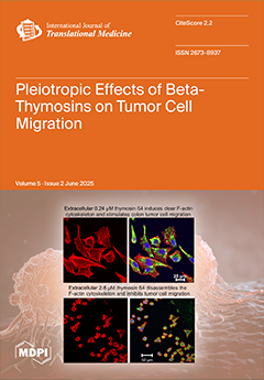
Tumor cell migration depends on the dynamic actin cytoskeleton, regulated by actin-binding proteins such as cofilin and thymosin beta 4 (Tß4). Existing reports have revealed diverging effects of the peptide Tß4 (5 kDa) on tumor cell migration. Therefore, we investigated the effects of varying concentrations of Tß4, either intra- or extracellularly, on the migration of human colon and mammary carcinoma cells. Both the mammary MDA-MB-231 and the colon 3LNLN cells exhibited a biphasic migratory response to increasing Tß4, with activity being highest at 0.24 µM but decreasing at 2.4 µM Tß4. Cells with high migratory activity showed a well-developed intracellular actin filament system, increased phosphorylation of AKT1 and AKT2, and secretion of matrix metalloproteases. The arrested cells at 2.4 µM Tß4 were rounded and lacked clear actin filaments and AKT1 and AKT2 phosphorylation. View this paper
- Issues are regarded as officially published after their release is announced to the table of contents alert mailing list.
- You may sign up for e-mail alerts to receive table of contents of newly released issues.
- PDF is the official format for papers published in both, html and pdf forms. To view the papers in pdf format, click on the "PDF Full-text" link, and use the free Adobe Reader to open them.
Previous Issue
Next Issue