Review

The Natural History of SARS-CoV-2-Incurred Disease: From Infection to Long COVID

1
Department of Medical Research, Taipei Veterans General Hospital, Taipei 112, Taiwan
2
Biosafety Level 3 Laboratory, Taipei Veterans General Hospital, Taipei 112, Taiwan
3
Institute of Biomedical Informatics, National Yang Ming Chiao Tung University, Taipei 112, Taiwan
4
Institute of Food Safety and Health Risk Assessment, National Yang Ming Chiao Tung University, Taipei 112, Taiwan
5
Institute of Biophotonics, National Yang-Ming Chiao Tung University, Taipei 112, Taiwan
*
Authors to whom correspondence should be addressed.
Int. J. Transl. Med. 2024, 4(1), 72-86; https://doi.org/10.3390/ijtm4010004
Submission received: 23 October 2023 / Revised: 28 December 2023 / Accepted: 31 December 2023 / Published: 8 January 2024
(This article belongs to the Special Issue Translational Medicine Approach against the COVID-19 Pandemic 2.0)

Abstract

:
The coronavirus SARS-CoV-2 is the causative pathogen of the COVID-19 pandemic that has been causing global upheaval since 2019. The widespread administration of vaccines has partially deterred the spread of SARS-CoV-2, yet the virus is mutating its genome to reduce its antigenicity and evade the human herd immunity. It seems that SARS-CoV-2 will co-exist with the human population for many decades to come. While most infected individuals only experience mild to moderate symptoms, some develop severe pulmonary and systemic disease that can result in hospitalization or even death. The natural history model of SARS-CoV-2 infection has been proposed which includes three sequential stages: the early infection stage, pulmonary stage, and hyper-inflammatory stage. Recently, it has been observed that many people who recovered from an acute infection still experience persistent symptoms for weeks or months, a condition known as long COVID. Furthermore, some COVID-19 patients display escalated rates of both macro- and micro-thrombosis due to endotheliopathy. Hence, we added the thrombosis and convalescent stages to the natural history model, encompassing the entire period from early infection to long COVID. The early infection stage is characterized by symptomatic or asymptomatic elevation of viral titers. Some patients progress to the pulmonary stage characterized by opacities in chest X-rays and computed tomography. The thrombosis stage is characterized by heightened rates of pulmonary thrombosis and consistently elevated D-dimer levels. The hyper-inflammatory stage is characterized by storms of cytokines, such as IL-6, IL-17, and interferons, which is a systemic effect. In the convalescent stage, some people recover completely, while others suffer from long COVID with persistent symptoms such as fatigue, shortness of breath, or brain fog. The natural history model of SARS-CoV-2 infection can be used to elucidate treatment and care.

1. Background

The worldwide pandemic of an infectious respiratory disease that emerged in late 2019 and termed by the World Health Organization as coronavirus disease 2019 (COVID-19) has been causing global upheaval until the present moment [1]. The causative pathogen, severe acute respiratory syndrome coronavirus 2 (SARS-CoV-2), is characterized by high transmission rates, which allowed its quick spread to all human-populated continents [1,2,3,4,5] and disturbed local medical, economic, social, and educational systems [6,7]. The airborne transmission of SARS-CoV-2, either from symptomatic or asymptomatic carriers, frequently occurs in indoor environments [8]. The lack of air change in indoor environments, directional airflow/air circulation systems, and activities such as talking and singing can increase infection rates via aerosol transmission [8]. Therefore, the scientific experiments of this airborne virus need to be performed in biological safety level 3 laboratories for the proper containment of the virus.
The relatives of SARS-CoV-2, SARS-CoV-1 and MERS-CoV, have previously caused human-to-human infections [9,10] but they only lasted for a short period. In contrast, SARS-CoV-2 has persisted in human populations to date. To cope with the pandemic, preventive measures such as school closures, lockdowns, travel restrictions, and personal behavioral changes were exercised in 2020–2022. These measures not only reduced the spread of SARS-CoV-2, but also reduced seasonal infections, including bronchitis, flu, croup, upper respiratory tract infection, otitis media, and tonsillitis [11]. In the meantime, mass vaccination against COVID-19 has been effective in adults [12,13], children, and adolescents [14]. Despite the herd immunity in humans, the virus has not been fully contained [15]. Rather, it has been mutating its genome to repeatedly generate breakthrough infections [16]. Variants of concerns such as alpha, delta, omicron BA4/5, BQ1/1.1, and XBB emerged one after another to prevail in the human population [17]. Although the vaccine cannot fully contain the spread of the virus, it can reduce the rate of infection, as well as the rate of disease progression to severe disease.
While most infected individuals only experience mild to moderate symptoms and recover within a few weeks, some develop severe pulmonary and systemic disease that can result in hospitalization or even death. The natural history stages reflect the impact of the infection on the individual, with each stage representing different physiological and clinical manifestations as the infection progresses and the immune response evolves.
A natural history model has been conceptualized to delineate COVID-19, which is comprised of three sequential stages: the early infection stage, pulmonary stage, and hyper-inflammatory stage [18]. Additionally, a model with three stages, the viral proliferation stage, the cytokine injury stage, and the thrombosis stages, was proposed [19]. These models have shed light on the clinical course of the infectious disease, as well as providing a rationale for treatments. Recently, it was observed that many people who recover from an acute infection still experience persistent symptoms such as fatigue, chest pain, palpitations, cough, dyspnea, abdominal pain, nausea, sleep problem, memory loss, tinnitus, and cognitive impairment [20]. Such a combination of symptoms is referred to as long COVID, which is defined as a condition in patients with confirmed SARS-CoV-2 infection approximately 3 months after the initial onset, and with these symptoms lasting at least 2 months, which cannot be attributed to any other diagnosis [21]. Therefore, we incorporated all these aspects and proposed a comprehensive natural history model, supported by this narrative review of the literature, and has five stages: the early infection stage, pulmonary stage, thrombosis stage, hyper-inflammatory stage, and convalescent stage. Not all the stages will manifest in a patient, and the symptoms and severity of a SARS-CoV-2 infection varies among individuals.

2. The Natural History of SARS-CoV-2 and Human Interactions

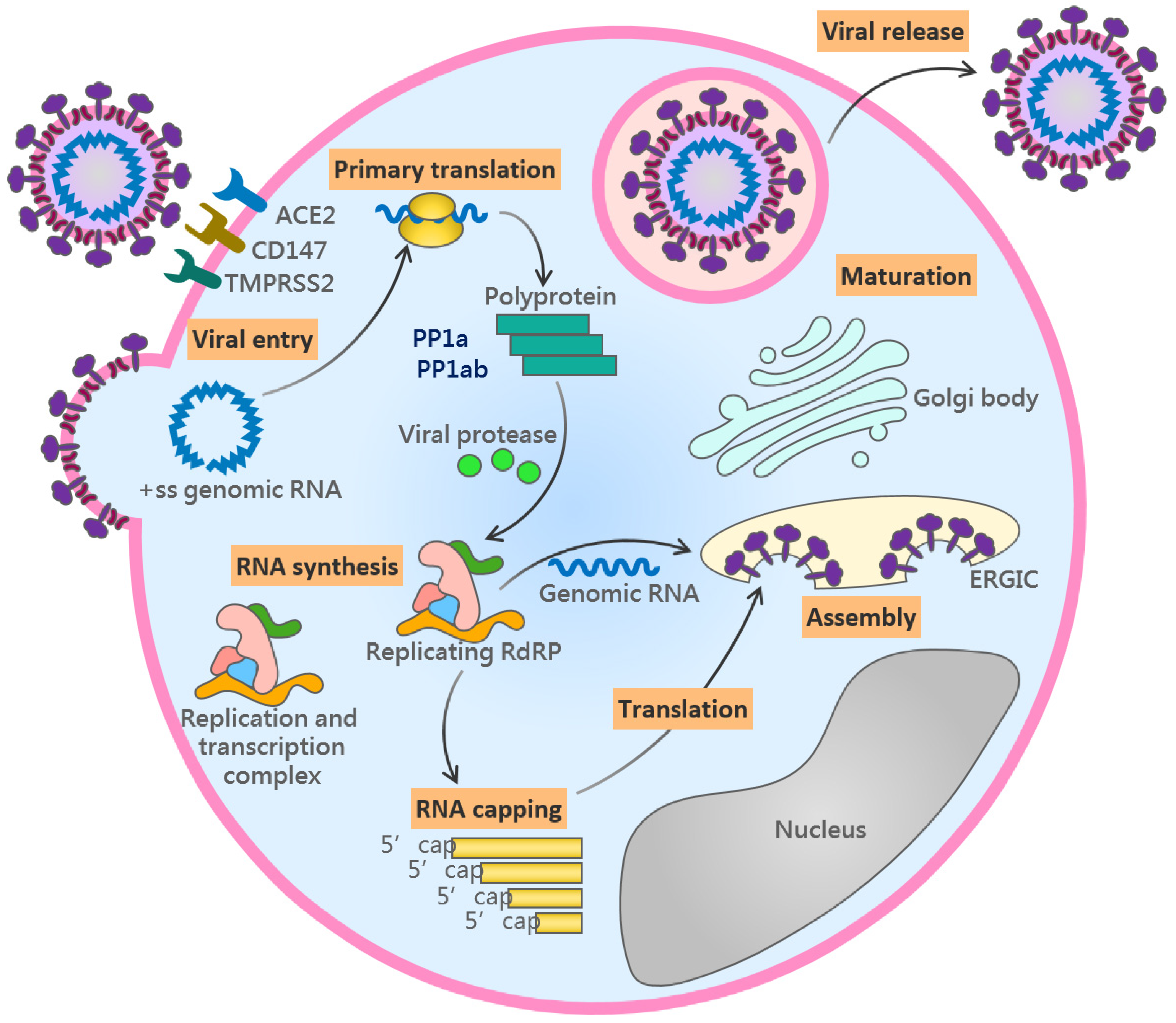
2.1. The Viral Life Cycle

We will start by reviewing the life cycle of SARS-CoV-2 which underlies the natural history of SARS-CoV-2 infection. SARS-CoV-2 is an enveloped, positive-strand RNA virus, with a genome of ~30,000 nucleotide bases encoding a total of 29 proteins, which are either structural or non-structural [18]. The structural proteins include spike (S), membrane (M), nucleocapsid (N), and envelope (E) proteins [22]. The genomic RNA forms a complex with the nucleocapsid protein to create a helical capsid, which is encapsulated in a thick membrane decorated with other structural proteins [22]. The spike protein contains a receptor-binding domain which extrudes outside the virion for binding with host cell membrane proteins [18]. The virion with the spike protein forms a crown-like and solar corona-like structure, hence the virus is named coronavirus. The entry of SARS-CoV-2 virus into the host cells is a major process in establishing an infection within the host. During this process, the spike proteins of the virions interact with angiotensin-converting enzyme 2 (ACE2) receptors on human cells in the respiratory tract. This interaction mediates the fusion of the virion to the human cell membranes which then triggers a sequence of molecular events constituting the life cycle of SARS-CoV-2. Apart from the ACE2 receptors on the surface of these cells, the human protease TMPRSS2 and possibly CD147 also play roles in the cleavage of spike proteins which facilitates viral entry into human cells [18,23] (Figure 1).
Once the virus enters the human cells, it hijacks the host’s molecular machinery to sustain the viral life cycle and to replicate itself, thereby generating multiple virus particles which can invade surrounding host cells (Figure 1). The structural and non-structural proteins, including the components of the replicase–transcriptase complex and proteases, are synthesized according to the subgenomic viral RNA templates, which are produced by the transcription of the viral genomic RNA (Figure 1) [18]. Initially, the open reading frames 1a and 1b (ORF1a and ORF1b) of the viral RNA are translated to produce polyproteins PP1a and PP1ab, the latter being a product of a programmed -1 ribosomal frameshift to avoid the stop codon in ORF1a [24].
The viral proteases, chymotrypsin-like protease (3CLpro or Mpro) and papain-like protease (PLpro), cut two polyproteins into a total of 16 non-structural proteins (nsp1-nsp16) [24,25], providing the components of the SARS-CoV-2 replicase–transcriptase complex. PLpro and Mpro are incorporated within the polyproteins nsp3 and nsp5, respectively [24,25]. The replicase–transcriptase complex is responsible for the next stages of the viral life cycle by producing subgenomic RNAs encoding structural proteins as well as replicating full viral genomes for subsequent packaging into virions (Figure 1). The major enzymatic component of this complex is RNA-dependent RNA polymerase (RdRp, nsp12), which is conserved among multiple coronaviruses and serves as an important target for antiviral drugs [5,26]. During genome replication, a negative-strand template is synthesized, which serves as the template for synthesizing new positive-strand viral genomes [27].
The structural proteins are translated by the host ribosomes and further localized to specialized endoplasmic reticulum–Golgi intermediate compartment (ERGIC) vesicles. The assembly stage occurring on the ERGIC includes the binding of the positive-strand viral genomic RNA with the N protein into a string-like viral ribonucleoprotein (vRNP) structure [28]. The M and E proteins are important for the final viral assembly as well as for viral release [29]. The E protein is responsible for the viral budding from the ERGIC [2]. These particles are then released from the infected host cells; however, the exact mechanism of this exit remains unclear, but it can occur either through the secretory, exosomal, or lysosomal pathways [30].
The viral life cycle in the human body triggers the virus–host interactions which underlie the natural history of SARS-CoV-2 infections, comprising five sequential stages: the early infection stage, pulmonary stage, thrombosis stage, hyper-inflammatory stage, and convalescent stage (Figure 2). Each stage is characterized by distinct clinical features, biomarkers and virus–host interactions. Different medical interventions are required accordingly (Table 1).

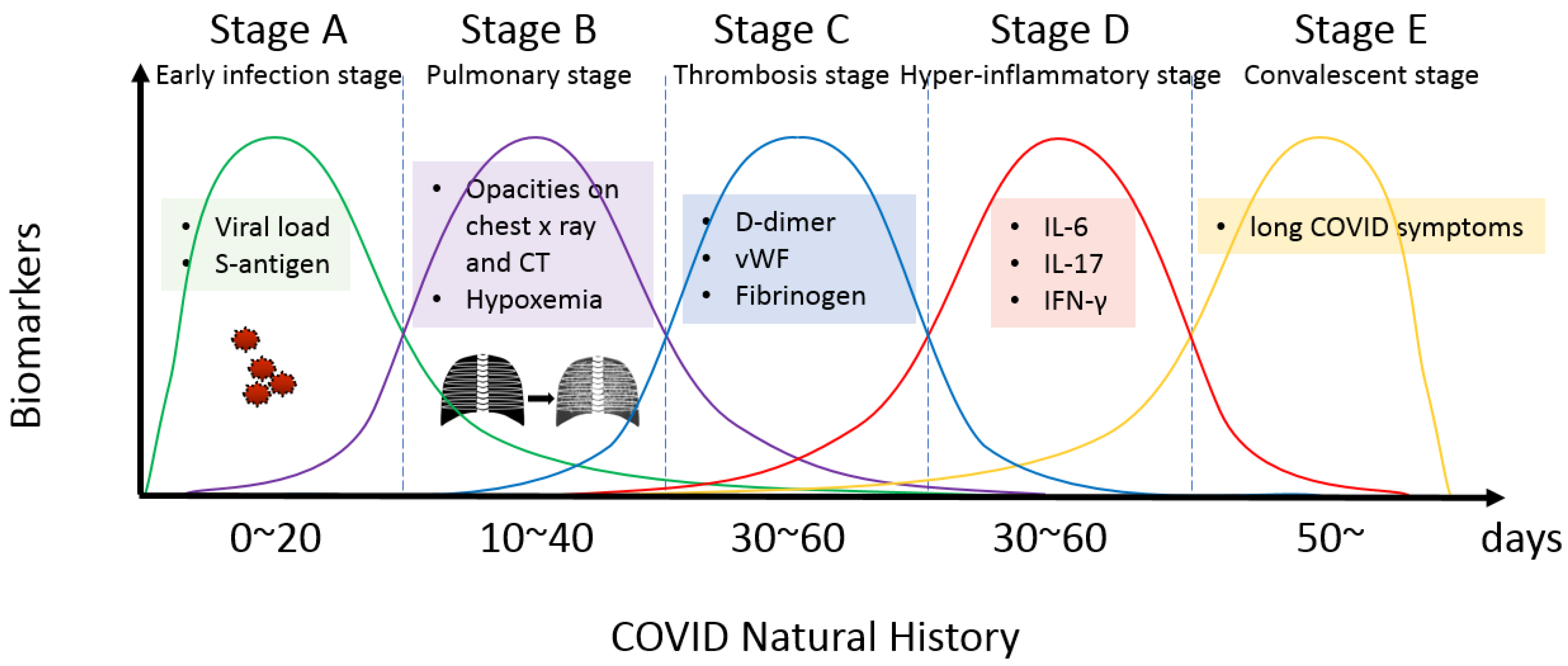
2.2. The Early Infection Stage

This stage starts when SARS-CoV-2 enters the human body through the respiratory tract. After exposure to the virus, people usually feel symptom-free during the incubation period which can last for a few days (with a median of 5.1 days) [31]. Afterwards, the infected people, with or without prior vaccinations, either remain asymptomatic or exhibit mild flu-like symptoms which will subside within 1~2 weeks. The immune system responds to the infection by producing an inflammatory response, leading to the characteristic symptoms of COVID-19, such as fever, cough, and shortness of breath. Some people may also experience muscle pain, fatigue, diarrhea, and loss of taste and/or smell [32].
In this early stage, real-time polymerase chain reaction (RT-PCR) or viral antigen tests are generally needed for the diagnosis of SARS-CoV-2 and the quantitation of viral titers in the human body. The viral titer can vary widely, ranging from a low titer of 103 to a high titer of 104–108 RNA copies/mL in persons with viral shedding capability [33]. The viral titer can change over the course of the infection.
However, the disease can progress into pneumonia, especially in some high-risk populations such as elderly people or patients with underlying type 2 diabetes [34]. To prevent these unfavorable conditions, antiviral drugs and neutralizing antibodies are given to reduce the risk of disease progression [35]. Patients at this stage can also be treated by the plasma from donors who have already fully recovered from the infection, also known as the COVID-19 convalescent plasma (CCP) [36].
A meta-analysis of studies conducted in India among healthcare workers, examining the prophylactic use of hydroxychloroquine either alone or with zinc, revealed a 75% efficacy rate and indicated a dose–response relationship. Specifically, among a subset of the 5 studies where healthcare workers received a minimum of six doses of weekly hydroxychloroquine prophylaxis, the efficacy was notably higher (75%) compared to the collective efficacy of all 11 studies (44%) [37]. Moreover, a recent retrospective cohort study involving 30,423 patients demonstrated substantial efficacy associated with the combined protocol of hydroxychloroquine and azithromycin, especially when administered at the earliest stage of the illness [38].

2.3. The Pulmonary Stage

In the pulmonary stage, the infection primarily affects the lungs, leading to the development of pneumonia symptoms. Breathing difficulties, shortness of breath, and lung inflammation are common during this stage. As the virus replicates in many host cells and causes damage, the immune system responds to the SARS-CoV-2 infection by activating immune cells, such as T cells/natural killer cells, and producing antibodies to target and destroy the virus. On the one hand, this response can help to clear the virus from the body and prevent the infection from spreading. On the other hand, the immune response can also cause inflammation and damage to healthy tissue, leading to pneumonia symptoms. This damage can make it difficult for the body to perform the exchange of oxygen and carbon dioxide, thus causing problems with metabolism. In some cases, the infection can progress even further to the hyper-inflammatory stage characterized by acute respiratory distress syndrome (ARDS), and multi-organ failure [39]. Chest X-rays or computed tomography (CT) may show evidence of inflammation in the lungs. This can manifest as whitened patches of increased density or as areas of ground-glass opacity on the X-ray or CT images [33]. These findings may be accompanied by other manifestations such as pleural effusion or lymphadenopathy.
As SARS-CoV-2 interacts with the host cells at the pulmonary stage, targeting the molecular machinery of the viral life cycle may prevent further disease progression to the hyper-inflammatory stage. Oral antiviral drugs are designed to antagonize the life cycle of SARS-CoV-2 including viral entry into human cells, viral replication, viral assembly, and release [2]. One strategy of effective antiviral treatments is to target the replicase–transcriptase complex. During virus replication, the polymerases form a particular 3D structure that specifically binds natural host nucleotides and incorporates them into a nascent chain of viral RNA. The antiviral nucleoside/nucleotide analogues are designed artificial molecules which can fit into the enzymatic pocket of the viral polymerases. This results either in the termination of the polymerization reaction in the case of analogues with a blocked 3′-hydroxyl group or the introduction of multiple mutations into the viral genome due to incorporation of unnatural nucleotides. At the beginning of the pandemic, an adenosine analogue remdesivir, originally developed by Gilead Sciences for treating Ebola virus, was quickly identified as one potential candidate for treating COVID-19 [29,40,41,42,43]. More potent antiviral nucleoside analogs were subsequently developed [43].
Apart from nucleoside analogs that can impair viral genome polymerization, the inhibition of the proteases is another strategy for viral life cycle intervention [29]. Protease inhibitors are drugs that target the viral protease enzyme, the machinery involved in the cleavage of viral proteins into smaller peptides, which are then assembled into new viral particles. By inhibiting this enzyme, protease inhibitors can prevent the virus from replicating and spreading, thereby reducing the severity of the infection. Such inhibitors usually mimic protease substrates and their binding to the active site prevents the protease from cleaving viral polypeptides. This binding can either block the enzyme’s activity entirely or reduce its activity to a level that is no longer sufficient for viral replication.
Paxlovid is an approved combination of a protease inhibitor and polymerase inhibitor that was authorized by the US Federal Drug Administration (FDA) for emergency use in SARS-CoV-2-infected patients at elevated risks of progression in late 2021. It is a combination of two inhibitors: an Mpro inhibitor (PF-07321332/nirmatrelvir) and a polymerase inhibitor (ritonavir). Nirmatrelvir is a modified form of the Mpro inhibitor originally developed to antagonize the SARS-CoV-1 virus, which was responsible for the pandemic in 2003 [40]. In a phase 2–3 randomized controlled clinical trial of unvaccinated and previously uninfected people during the delta variant period (July 2021–December 2021), Paxlovid treatment within 3 days and 5 days of the onset of symptoms could effectively reduce hospitalization and death from all causes at day 28 by 88.9% [44]. The clinical guidelines indicate that Paxlovid should be considered for groups at high risk for severe COVID-19, where age is the primary risk factor [45]. In real-world data from a total of 4737 people treated with Paxlovid in Israel between January 2022 and February 2022, the omicron variant-dominated period, the hospitalization and death rate reduction by Paxlovid was 46% at day 28. The protective effect of Paxlovid is independent of vaccination, which can result in an 80% reduction in hospitalization and death rates compared with individuals of with similar ages and co-mobility factors but who were not treated [45]. Recent data in the US showed that Paxlovid treatment was associated with a decreased hospitalization rate among adults with COVID-19. The latest real-world data from the US, spanning 1 April to 31 August 2022, included 198,927 adult patients (≥18 years old), including those with prior infections or vaccinations, who took Paxlovid within 5 days of diagnosis, as well as 500,921 patients who did not take Paxlovid. The data revealed that patients receiving Paxlovid were 51% less likely to be hospitalized within 30 days of diagnosis compared to those who did not take Paxlovid [46].
Molnupiravir is a nucleoside analog prodrug that targets the SARS-CoV-2 polymerase. The emergency use authorization for molnupiravir was issued by the FDA for the treatment of mild-to-moderate COVID-19. Molnupiravir is a polymerase inhibitor that was previously developed to antagonize Venezuelan equine encephalitis virus (VEEV), a mosquito-borne pathogen [40]. It showed a broad-spectrum action on RdRp of various viruses including SARS-CoV-1 and SARS-CoV-2 in preclinical models [40]. The incorporation of molnupiravir by the polymerase does not stop viral RNA elongation, rather, the molnupiravir-containing RNA template strands guide the wrong bases into the new viral RNA [40]. In a phase 3 randomized controlled trial of non-hospitalized patients enrolled during the delta variant period (May 2021–October 2021), the use of molnupiravir within 5 days of the onset of symptoms could effectively reduce hospitalization or death by 30% at day 29 [47]. The single-center study conducted by the Yale-New Haven Transplant Center during an omicron variant-dominated period (January 2022–February 2022) analyzed patients with Paxlovid contraindication due to organ transplantation. The use of molnupiravir in such patients could reduce the rate of hospitalization and death (from 19/48 to 9/49) within 30 days, and no deaths were observed in the molnupiravir-treated group [48].
The nasopharyngeal swab, often used to diagnose SARS-CoV-2, does not detect viral infection in other parts of the body. In a case study, negative tests from swabs led to the stopping of antiviral treatments. However, a severe viral reactivation followed, and the virus was found from bronchoalveolar lavage samples [49]. This emphasizes the need for caution when relying solely on swab results, as they might not signify a full recovery. Relying solely on nasopharyngeal swabs at this stage is insufficient, potentially leading to the misinterpretation of recovery and the inadvertent discontinuation of treatments, considering the virus can localize in lungs and other organs, emphasizing the need for more comprehensive diagnostic approaches.

2.4. Thrombosis Stage

Thrombosis linked to endotheliopathy in COVID-19 remains a critical concern. Many patients exhibit elevated pulmonary thrombosis [50] and consistently elevated D-dimer levels [51]. Viral infection of the endothelial cells often leads to a cascade of events, triggering inflammation and thrombotic complications, including microvascular thrombosis, venous thromboembolism, acute limb ischemia, cardiovascular events, cerebrovascular events, and neurologic invasion [52]. Endothelial dysfunction together with the compromised innate immunity facilitate viral-induced acute endothelialitis in the pulmonary microcirculation, leading to vasoconstrictive anomalies, luminal blockage by inflammatory cells, and intravascular thrombosis [53]. Autopsies revealed pulmonary micro-thrombosis and embolism, emphasizing endothelial injuries in oxygen desaturation [54].
Several biomarkers closely associated with endotheliopathy in COVID-19 have emerged as crucial indicators in understanding and managing the disease. Acutely ill COVID-19 patients often exhibit elevated levels of D-dimer, a marker consistently associated with an increased risk of deep venous thrombosis and pulmonary embolism [51]. Elevated levels of von Willebrand factor (vWF; a glycoprotein crucial for blood clotting) [52,55], fibrinogen [55], soluble P-selectin [56], E-selectin, and angiopoietin-2 [57] reflect endotheliopathy. Additionally, decreased levels of thrombomodulin and endothelial protein C receptor (EPCR) shed light on anticoagulant dysfunction [58]. Furthermore, measuring endothelial-derived microparticles [59] and circulating endothelial cells [60] provides insights into the severity of endothelial injury and the potential for thrombotic events.
The dysfunction of endothelial cells not only underlies the severity of COVID-19 but also influences therapeutic approaches, affecting patient care and outcomes. The early administration of aspirin (325 mg/day) is advised for its antiplatelet and anti-inflammatory effects [61]. Anticoagulant therapy, particularly low-molecular-weight heparin or novel oral anticoagulants, reduces mortality in COVID-19 patients, especially in severe cases with thrombotic complications or elevated D-dimer levels [62]. The consideration of systemic anticoagulation and aspirin is recommended for high-risk patients with a history of heart, lung, kidney, or malignant disease [63].

2.5. The Hyper-Inflammatory Stage

Some individuals progress all the way to this advanced stage, characterized by an excessive immune response known as a cytokine storm. In such a case, the immune system overreacts, causing widespread inflammation throughout the body, which can lead to organ damage and severe complications. At this stage, the disease can progress to severe complications including ARDS, sepsis, and organ failure. The virus can cause fluid to build up in the lungs, particularly within the bronchioles, and make it hard to breathe. This happens because the virus damages the protective coating of the lungs and causes inflammation [64]. The immune system becomes hyper-activated and causes more damage, leading to the accumulation of reactive oxygen species, cell debris, and proteases [64].
The patients at this stage often require intensive care. Septic shock and multi-organ failure are the major causes of death [65]. The risk of death is generally higher in elderly people, people with underlying medical conditions (heart disease, diabetes, or lung disease), and people with compromised immune systems. Additionally, some patients progress to a cytokine storm which is characterized by an overproduction of pro-inflammatory cytokines and other immune mediators [66]. This can lead to a range of clinical manifestations, including fever, inflammation, and organ dysfunction. During the hyper-inflammatory stage, cytokines such as IL-6, IL-8, IL-17, tumor necrosis factor-α (TNF-α), and interferons (e.g., IFN-γ) are elevated [56,67], which is not only an indicator of this stage, but can also lead to the development and progression of the long COVID condition in the next stage (Figure 2) [18,22,66]. These cytokines are the reaction to the SARS-CoV-2 infection and are involved in the activation and recruitment of immune cells, such as T cells and natural killer cells, to the site of infection. The hyper-inflammation causes organ damage and multisystem inflammatory syndrome in children (MIS-C), which is potentially life-threatening. A recent study analyzed the virus-reactive T cells, serological responses to viral proteins, and plasma and peripheral blood mononuclear cells in children with acute MIS-C, in children recovering from COVID-19, and in healthy controls. The children with MIS-C exhibited significantly lower virus-specific CD4+ and CD8+ T-cell responses to major SARS-CoV-2 antigens compared with children who had recovered from a SARS-CoV-2 infection. The titers in MIS-C patients were similar or lower than those in healthy controls [68].
In patients with severe COVID-19, the development of virus-specific circulating T follicular helper cells (cTfh) is delayed compared to patients with mild disease. This in turn leads to a delay in the production of high-quality neutralizing antibodies, which may contribute to the progression to severe disease [69].
Corticosteroids, such as prednisone or dexamethasone, may be used to reduce inflammation and improve oxygenation in patients with severe COVID-19. Dexamethasone, a corticosteroid commonly used for conditions such as inflammation and allergic reactions, has shown effectiveness in reducing inflammation and improving outcomes in patients with severe or critical COVID-19. The RECOVERY trial conducted in the United Kingdom showed that dexamethasone reduced the risk of 28-day mortality in patients hospitalized with COVID-19 (age-adjusted rate ratio: 0.83) [70]. It was also found that dexamethasone improved survival in critically ill COVID-19 patients [70]. Dexamethasone is typically used in conjunction with other medications and supportive care measures, such as oxygen therapy, to manage the symptoms and complications of severe COVID-19. It is important to note that dexamethasone may be harmful to patients with mild or moderate COVID-19 [71].
Tocilizumab is a medication used to treat conditions like rheumatoid arthritis and certain types of cytokine release syndrome (including COVID) by blocking the interleukin-6 receptor. One phase 3 clinical trial assessed tocilizumab alongside standard care in hospitalized COVID-19 patients with hypoxia and systemic inflammation [72]. Among the 4116 patients, those receiving tocilizumab had lower 28-day mortality rates (31% vs. 35%) and were more likely to be discharged within 28 days compared to those receiving standard care alone [72]. Tocilizumab also reduced the risk of reaching the composite endpoint of invasive mechanical ventilation or death among patients not initially on invasive ventilation, demonstrating its efficacy in improving survival and clinical outcomes irrespective of respiratory support, in conjunction with systemic corticosteroids [72]. In another phase 3 trial, among the hospitalized patients who received either tocilizumab or placebo, the primary outcome, measured on a scale from discharge readiness to death at day 28, showed no significant difference between the two groups [73]. Both groups had similar rates of serious adverse events and mortality at 28 days, indicating that tocilizumab did not notably improve the clinical status or reduce mortality compared to the placebo in this trial [73].
Additionally, patients can also receive supportive care to help their bodies recover from the severe inflammation and organ damage at this stage. This may include oxygen therapy to aid breathing, medications to reduce fever and pain, and electrolyte replacement to correct fluid/electrolyte imbalances. In severe cases, patients may also require mechanical ventilation to help them breathe or extracorporeal membrane oxygenation (ECMO) to support their heart and lungs.

2.6. The Convalescent Stage

In the convalescent stage, the acute symptoms subside. The virus may be gradually cleared by the immune system. However, a substantial proportion of people transition into the long COVID condition, defined by WHO as persistent symptoms lasting at least 2 months after the initial onset of a confirmed SARS-CoV-2 infection, occurring approximately 3 months post-infection, and cannot be explained by other diagnoses [21]. Long COVID can affect people of all ages, including those who had mild or asymptomatic acute infections [14,74]. The Long COVID in Scotland Study (Long-CISS) tracked adults with confirmed SARS-CoV-2 infections over 18 months and discovered that certain symptoms like altered taste and smell improved over time, while issues like cough and hearing problems were more common after SARS-CoV-2 infection compared to those who had never been infected [75]. After the acute infection, SARS-CoV-2 may persist at low titers in the body, causing ongoing inflammation and damage. Also, the immune system may remain activated after the acute phases, leading to a prolonged inflammatory response. Long COVID may be caused by ongoing inflammation and damage to various organs, including the lungs, heart, brain, and immune system. The dysregulated production of cytokines leads to chronic inflammation. SARS-CoV-2 breaching the blood–brain barrier and infecting neuronal cells might contribute to neurological symptoms like headaches, brain fog, and memory issues. Pre-existing conditions such as diabetes [34,76] and hypertension [77] are associated with an increased risk of severe acute infection and subsequent long-term symptoms. Risk factors associated with the occurrence of long COVID have been identified [78,79], including female sex, elderly age, socioeconomic deprivation, smoking, obesity, and a wide range of comorbidities [80].
In a study of 312 cancer patients with a median age of 57 years, 60% experienced prolonged COVID-19 symptoms for up to 14 months after diagnosis, mainly consisting of fatigue (82%), sleep issues (78%), myalgias (67%), and gastrointestinal symptoms (61%). More females (63%) than males (37%) reported persistent symptoms, and only 8.5% of patients with prolonged symptoms were re-admitted for COVID-19-related reasons [81]. In a study of 5133 patients (mean age 61 years, 62.9% male), pre-existing cardiovascular disease (CVD) was linked to a 1.15 higher odds of death after adjusting for various factors, while no independent association with cardiovascular events was found. Myocardial injury upon ICU admission was independently associated with increased odds of death (1.93) and cardiovascular events (1.82), irrespective of pre-existing cardiovascular disease [82]. Among the 1682 eligible patients (median age 59, predominantly African American (34.4%) and male (54.5%)), 12% experienced mortality within 60 days. The 60–74 (adjusted OR 3.30) and 75–100 (adjusted OR 4.52) age groups, along with medical histories of atrial fibrillation (adjusted OR 2.47) and venous thromboembolism (aOR 2.00) were significant predictors of a higher risk for 60-day mortality compared to the 19–39 age group [83]. Headache is a common long COVID symptom that can persist after COVID-19, with limited but growing evidence suggesting features such as a migrainous or tension-type-like phenotype, often presents with other long COVID symptoms such as hyposmia, and treatment recommendations are currently based on the existing guidelines for primary headaches with similar phenotypes [84]. The SARS-CoV-2 virus may use the ACE2 receptor to breach the blood–brain barrier and has been shown to infect and replicate in neuronal cells in cultures and postmortem examinations have detected virus RNA in the brain in about 30–40% of cases [85]. This symptom of COVID-19 is believed to be mediated by innate immune responses and cytokine-mediated pathogenic processes, including the activation of afferent neurons through pattern recognition receptors [86].
Vaccination might help alleviate the overall impact of long COVID. After COVID-19 vaccination, the frequency of enduring long COVID symptoms is lowered [87]. There is currently no specific treatment for long COVID, but supportive care can help alleviate symptoms. This may include pulmonary rehabilitation for respiratory symptoms, cognitive-behavioral therapy for mental health symptoms, and medication for specific symptoms such as pain and fatigue. Researchers are also investigating potential therapies, such as antiviral drugs and immunomodulatory agents, that may target the underlying mechanisms of long COVID. Seroconversion is observed in some but not all patients [20].
A newly suggested treatment approach has emerged for long COVID, centered on the hypothesis that the sustained alleviation of symptoms necessitates equipping the body with enzymes capable of eliminating residual viral spike proteins present in the bloodstream [88,89].
At the convalescent stage, T cell responses have been noticed [90]. The common symptoms of sequelae include anosmia, dysgeusia, fatigue, post-exertional malaise, respiratory problems such as cough, shortness of breath, and tightness of chest, neurological problems such as headache, brain fog, and memory issues, as well as gastrointestinal problems such as diarrhea and nausea [74]. Statistics in the US showed that one in five of previously infected people developed long COVID [91]. Recently, a comprehensive multi-omic study of post-acute sequelae of COVID-19 was reported [34]. This study reported proteomic, metabolomic, and single-cell measurements of a cohort of patients at the diagnosis stage of a SARS-CoV-2 infection, acute phase, and convalescence stage. It was revealed that diabetes, SARS-CoV-2 RNA viremia, Epstein–Barr virus viremia, and specific auto-antibodies are major risk factors for post-acute COVID-19 sequelae [34]. Interestingly, other metabolic diseases commonly occurring in adults, such as hypertension, coronary artery disease, and liver fibrosis, did not appear to significantly elevate the risk of sequelae [34]. The molecular characteristics at the acute stage are correlated with long COVID [92]. In long COVID, the production of cytokines can become dysregulated, leading to chronic inflammation and the persistence of symptoms such as fatigue, muscle aches, and difficulty breathing.

3. Discussion

We added the convalescent stage to the natural history model of SARS-CoV-2 infection to incorporate the complex and multi-faceted long COVID condition, which we believe is an important part of the disease which should not be overlooked. Long COVID has the following characteristics and impact on the patients. First, long COVID has a diverse range of respiratory, neurological, cardiovascular, and gastrointestinal symptoms that persist beyond the acute phase of the illness. These diverse symptoms and their occurrence months after the initial infection make them difficult to identify precisely. Nevertheless, the prolonged physical and mental discomfort associated with long COVID can significantly impact the quality of life of the affected individuals. The symptoms, such as fatigue, cognitive difficulties/brain fog, and persistent pain, can hinder daily activities and reduce overall well-being. Yet, widespread vaccination could reduce the rate of infection, thereby mitigate the overall impact of long COVID on a population level.
During the early infection stage of COVID-19, oral antiviral treatments such as Paxlovid and molnupiravir are recommended for high-risk populations, including elderly individuals and patients with metabolic syndrome, to effectively manage the disease. However, this population is often afflicted by age-related chronic illnesses and may be taking medications such as blood thinners, cholesterol-lowering drugs, and medications for urinary incontinence. Given that these individuals are on multiple medications simultaneously, it is crucial to evaluate potential interactions with the anti-SARS-CoV-2 drugs. This evaluation falls under the realm of pharmacogenomics, which addresses variations in drug responses and potential drug–drug interactions based on an individual’s genetic makeup. For instance, one of the key components of Paxlovid, ritonavir, is known to inhibit major cytochrome P450 isoforms 3A4 and 2D6, thus affecting liver metabolic activity [93].
Solid organ transplant patients, who often receive immunosuppressants to prevent organ rejection, face an increased risk of severe COVID-19 and mortality due to their compromised immune response to infections and vaccines. A recent study demonstrated a potential solution by harvesting autologous T cells from these patients and stimulating them with a SARS-CoV-2 antigen outside the body. These T cells can be genetically engineered to develop resistance to the commonly used immunosuppressant tacrolimus. This approach aims to provide protection against COVID-19 for immunosuppressed patients, ensuring their safety and well-being [94].

4. Conclusions

Due to the evolving nature of the SARS-CoV-2 virus, its continued coexistence with the human population for an extended period is expected. We illustrated the multi-stage natural history of COVID-19, encompassing early infection, pulmonary involvement, thrombotic complications, hyper-inflammatory responses, and a convalescent phase which includes long COVID. This comprehensive model aids in delineating treatment strategies and care protocols to address the varying clinical manifestations and prolonged effects of the disease. We reviewed the disease in the light of the viral life cycle of SARS-CoV-2. By focusing on these crucial disease characteristics and treatment options, healthcare providers can better address the various risks associated with COVID-19, ultimately improving patient outcomes, and reducing morbidity and mortality rates. Paxlovid and molnupiravir are suitable for managing high-risk patients during the acute infection and pulmonary stages, whereas dexamethasone is an option for hospitalized patients experiencing the hyper-inflammatory stage.

Author Contributions

Conceptualization, K.-H.L. and M.-L.W.; figure generation, K.-H.L., Y.-C.T. and Y.-T.L.; literature analysis, K.-H.L., S.-H.C., W.-C.H., C.-Y.H., E.-T.T., T.-J.C. and D.-M.Y.; writing—original draft preparation, K.-H.L.; writing—review and editing, K.-H.L., A.A.Y. and M.-L.W. All authors have read and agreed to the published version of the manuscript.

Funding

This research was funded by Taipei Veterans General Hospital (Grant number: V113E-002-4) which is gratefully appreciated.

Institutional Review Board Statement

The study did not require ethical approval.

Informed Consent Statement

The study did not require informed consent.

Data Availability Statement

We did not have any data apart from the literature which has already ben cited in the manuscript.

Conflicts of Interest

The authors declare no conflict of interest.

References

  1. Sohrabi, C.; Alsafi, Z.; O’Neill, N.; Khan, M.; Kerwan, A.; Al-Jabir, A.; Iosifidis, C.; Agha, R. World health organization declares global emergency: A review of the 2019 novel coronavirus (COVID-19). Int. J. Surg. 2020, 76, 71–76. [Google Scholar] [CrossRef] [PubMed]
  2. Yang, H.; Rao, Z. Structural biology of SARS-CoV-2 and implications for therapeutic development. Nat. Rev. Microbiol. 2021, 19, 685–700. [Google Scholar] [CrossRef] [PubMed]
  3. de Wit, E.; van Doremalen, N.; Falzarano, D.; Munster, V.J. Sars and mers: Recent insights into emerging coronaviruses. Nat. Rev. Microbiol. 2016, 14, 523–534. [Google Scholar] [CrossRef] [PubMed]
  4. Zhang, R.; Li, Y.; Zhang Annie, L.; Wang, Y.; Molina Mario, J. Identifying airborne transmission as the dominant route for the spread of COVID-19. Proc. Natl. Acad. Sci. USA 2020, 117, 14857–14863. [Google Scholar] [CrossRef] [PubMed]
  5. Malone, B.; Urakova, N.; Snijder, E.J.; Campbell, E.A. Structures and functions of coronavirus replication-transcription complexes and their relevance for SARS-CoV-2 drug design. Nat. Rev. Mol. Cell. Biol. 2022, 23, 21–39. [Google Scholar] [CrossRef] [PubMed]
  6. Giuntella, O.; Hyde, K.; Saccardo, S.; Sadoff, S. Lifestyle and mental health disruptions during COVID-19. Proc. Natl. Acad. Sci. USA 2021, 118, e2016632118. [Google Scholar] [CrossRef] [PubMed]
  7. Shang, Y.; Li, H.; Zhang, R. Effects of pandemic outbreak on economies: Evidence from business history context. Front. Public Health 2021, 9, 632043. [Google Scholar] [CrossRef]
  8. Duval, D.; Palmer, J.C.; Tudge, I.; Pearce-Smith, N.; O’Connell, E.; Bennett, A.; Clark, R. Long distance airborne transmission of SARS-CoV-2: Rapid systematic review. BMJ 2022, 377, e068743. [Google Scholar] [CrossRef]
  9. Cui, J.; Li, F.; Shi, Z.-L. Origin and evolution of pathogenic coronaviruses. Nat. Rev. Microbiol. 2018, 17, 181–192. [Google Scholar] [CrossRef]
  10. Dhama, K.; Patel, S.K.; Sharun, K.; Pathak, M.; Tiwari, R.; Yatoo, M.I.; Malik, Y.S.; Sah, R.; Rabaan, A.A.; Panwar, P.K.; et al. SARS-CoV-2 jumping the species barrier: Zoonotic lessons from sars, mers and recent advances to combat this pandemic virus. Travel Med. Infect. Dis. 2020, 37, 101830. [Google Scholar] [CrossRef]
  11. Kadambari, S.; Goldacre, R.; Morris, E.; Goldacre, M.J.; Pollard, A.J. Indirect effects of the COVID-19 pandemic on childhood infection in England: Population based observational study. BMJ 2022, 376, e067519. [Google Scholar] [CrossRef] [PubMed]
  12. El Sahly, H.M.; Baden, L.R.; Essink, B.; Doblecki-Lewis, S.; Martin, J.M.; Anderson, E.J.; Campbell, T.B.; Clark, J.; Jackson, L.A.; Fichtenbaum, C.J.; et al. Efficacy of the mrna-1273 SARS-CoV-2 vaccine at completion of blinded phase. N. Engl. J. Med. 2021, 385, 1774–1785. [Google Scholar] [CrossRef] [PubMed]
  13. Polack, F.P.; Thomas, S.J.; Kitchin, N.; Absalon, J.; Gurtman, A.; Lockhart, S.; Perez, J.L.; Pérez Marc, G.; Moreira, E.D.; Zerbini, C.; et al. Safety and efficacy of the bnt162b2 mrna COVID-19 vaccine. N. Engl. J. Med. 2020, 383, 2603–2615. [Google Scholar] [CrossRef] [PubMed]
  14. Liang, K.-H.; Hung, K.-F.; Wang, M.-L.; Chang, T.-J.; Cheng, Y.-F.; Chiang, S.-H.; Chen, M.-F.; Liao, Y.-T.; Chiou, S.-H.; Yang, D.-M. SARS-CoV-2 vaccines in children and adolescents: Can immunization prevent hospitalization? J. Chin. Med. Assoc. 2022, 85, 891–895. [Google Scholar] [CrossRef] [PubMed]
  15. Zheng, C.; Shao, W.; Chen, X.; Zhang, B.; Wang, G.; Zhang, W. Real-world effectiveness of COVID-19 vaccines: A literature review and meta-analysis. Int. J. Infect. Dis. 2022, 114, 252–260. [Google Scholar] [CrossRef] [PubMed]
  16. Mallapaty, S. Where did omicron come from? Three key theories. Nature 2022, 602, 26–28. [Google Scholar] [CrossRef] [PubMed]
  17. Kurhade, C.; Zou, J.; Xia, H.; Liu, M.; Chang, H.C.; Ren, P.; Xie, X.; Shi, P.Y. Low neutralization of SARS-CoV-2 omicron ba.2.75.2, bq.1.1, and xbb.1 by parental mrna vaccine or a ba.5-bivalent booster. Nat. Med. 2022, 29, 344–347. [Google Scholar] [CrossRef] [PubMed]
  18. dos Santos, W.G. Natural history of COVID-19 and current knowledge on treatment therapeutic options. Biomed. Pharmacother. 2020, 129, 110493. [Google Scholar] [CrossRef]
  19. McCullough, P.A.; Alexander, P.E.; Armstrong, R.; Arvinte, C.; Bain, A.F.; Bartlett, R.P.; Berkowitz, R.L.; Berry, A.C.; Borody, T.J.; Brewer, J.H.; et al. Multifaceted highly targeted sequential multidrug treatment of early ambulatory high-risk SARS-CoV-2 infection (COVID-19). Rev. Cardiovasc. Med. 2020, 21, 517–530. [Google Scholar]
  20. Davis, H.E.; McCorkell, L.; Vogel, J.M.; Topol, E.J. Long COVID: Major findings, mechanisms and recommendations. Nat. Rev. Microbiol. 2023, 21, 133–146. [Google Scholar] [CrossRef]
  21. Soriano, J.B.; Murthy, S.; Marshall, J.C.; Relan, P.; Diaz, J.V. A clinical case definition of post-COVID-19 condition by a delphi consensus. Lancet Infect. Dis. 2022, 22, e102–e107. [Google Scholar] [CrossRef] [PubMed]
  22. Diamond, M.S.; Kanneganti, T.D. Innate immunity: The first line of defense against SARS-CoV-2. Nat. Immunol. 2022, 23, 165–176. [Google Scholar] [CrossRef] [PubMed]
  23. Seth, S.; Batra, J.; Srinivasan, S. COVID-19: Targeting proteases in viral invasion and host immune response. Front. Mol. Biosci. 2020, 7, 215. [Google Scholar] [CrossRef] [PubMed]
  24. Lv, Z.; Cano, K.E.; Jia, L.; Drag, M.; Huang, T.T.; Olsen, S.K. Targeting SARS-CoV-2 proteases for COVID-19 antiviral development. Front. Chem. 2022, 9, 1221. [Google Scholar] [CrossRef] [PubMed]
  25. Chen, Y.; Liu, Q.; Guo, D. Emerging coronaviruses: Genome structure, replication, and pathogenesis. J. Med. Virol. 2020, 92, 418–423. [Google Scholar] [CrossRef] [PubMed]
  26. Hardenbrook, N.J.; Zhang, P. A structural view of the SARS-CoV-2 virus and its assembly. Curr. Opin. Virol. 2022, 52, 123–134. [Google Scholar] [CrossRef] [PubMed]
  27. Sawicki, S.G.; Sawicki, D.L.; Younker, D.; Meyer, Y.; Thiel, V.; Stokes, H.; Siddell, S.G. Functional and genetic analysis of coronavirus replicase-transcriptase proteins. PLoS Pathog. 2005, 1, e39. [Google Scholar] [CrossRef]
  28. Cubuk, J.; Alston, J.J.; Incicco, J.J.; Singh, S.; Stuchell-Brereton, M.D.; Ward, M.D.; Zimmerman, M.I.; Vithani, N.; Griffith, D.; Wagoner, J.A.; et al. The SARS-CoV-2 nucleocapsid protein is dynamic, disordered, and phase separates with rna. Nat. Commun. 2021, 12, 1936. [Google Scholar] [CrossRef]
  29. Huang, J.; Song, W.; Huang, H.; Sun, Q. Pharmacological therapeutics targeting rna-dependent rna polymerase, proteinase and spike protein: From mechanistic studies to clinical trials for COVID-19. J. Clin. Med. 2020, 9, 1131. [Google Scholar] [CrossRef]
  30. Bracquemond, D.; Muriaux, D. Betacoronavirus assembly: Clues and perspectives for elucidating SARS-CoV-2 particle formation and egress. mBio 2021, 12, e0237121. [Google Scholar] [CrossRef]
  31. Lauer, S.A.; Grantz, K.H.; Bi, Q.; Jones, F.K.; Zheng, Q.; Meredith, H.R.; Azman, A.S.; Reich, N.G.; Lessler, J. The incubation period of coronavirus disease 2019 (COVID-19) from publicly reported confirmed cases: Estimation and application. Ann. Intern. Med. 2020, 172, 577–582. [Google Scholar] [CrossRef] [PubMed]
  32. Looi, M.K. How are COVID-19 symptoms changing? BMJ 2023, 380, 3. [Google Scholar] [CrossRef] [PubMed]
  33. Kevadiya, B.D.; Machhi, J.; Herskovitz, J.; Oleynikov, M.D.; Blomberg, W.R.; Bajwa, N.; Soni, D.; Das, S.; Hasan, M.; Patel, M.; et al. Diagnostics for SARS-CoV-2 infections. Nat. Mater. 2021, 20, 593–605. [Google Scholar] [CrossRef] [PubMed]
  34. Su, Y.; Yuan, D.; Chen, D.G.; Ng, R.H.; Wang, K.; Choi, J.; Li, S.; Hong, S.; Zhang, R.; Xie, J.; et al. Multiple early factors anticipate post-acute COVID-19 sequelae. Cell 2022, 185, 881–895.e820. [Google Scholar] [CrossRef] [PubMed]
  35. Ceriello, A.; Prattichizzo, F. Pharmacological management of COVID-19 in type 2 diabetes. J. Diabetes Complicat. 2021, 35, 107927. [Google Scholar] [CrossRef] [PubMed]
  36. Marconato, M.; Abela, I.A.; Hauser, A.; Schwarzmuller, M.; Katzensteiner, R.; Braun, D.L.; Epp, S.; Audige, A.; Weber, J.; Rusert, P.; et al. Antibodies from convalescent plasma promote SARS-CoV-2 clearance in individuals with and without endogenous antibody response. J. Clin. Investig. 2022, 132, e158190. [Google Scholar] [CrossRef] [PubMed]
  37. Stricker, R.B.; Fesler, M.C. Hydroxychloroquine pre-exposure prophylaxis for COVID-19 in healthcare workers from india: A meta-analysis. J. Infect. Public Health 2021, 14, 1161–1163. [Google Scholar] [CrossRef] [PubMed]
  38. Brouqui, P.; Million, M.; Parola, P.; McCullough, P.A.; Raoult, D. Outcomes after early treatment with hydroxychloroquine and azithromycin: An analysis of a database of 30,423 COVID-19 patients. New Microbes New Infect. 2023, 55, 101188. [Google Scholar] [CrossRef]
  39. Jolly, M.K.; Ward, C.; Eapen, M.S.; Myers, S.; Hallgren, O.; Levine, H.; Sohal, S.S. Epithelial-mesenchymal transition, a spectrum of states: Role in lung development, homeostasis, and disease. Dev. Dyn. 2018, 247, 346–358. [Google Scholar] [CrossRef]
  40. Cully, M. A tale of two antiviral targets—And the COVID-19 drugs that bind them. Nat. Rev. Drug Discov. 2021, 21, 3–5. [Google Scholar] [CrossRef]
  41. Sanders, J.M.; Monogue, M.L.; Jodlowski, T.Z.; Cutrell, J.B. Pharmacologic treatments for coronavirus disease 2019 (COVID-19). JAMA 2020, 323, 1824–1836. [Google Scholar] [CrossRef] [PubMed]
  42. Wang, M.; Cao, R.; Zhang, L.; Yang, X.; Liu, J.; Xu, M.; Shi, Z.; Hu, Z.; Zhong, W.; Xiao, G. Remdesivir and chloroquine effectively inhibit the recently emerged novel coronavirus (2019-ncov) in vitro. Cell Res. 2020, 30, 269–271. [Google Scholar] [CrossRef] [PubMed]
  43. Samaddar, A.; Grover, M.; Nag, V.L. Pathophysiology and potential therapeutic candidates for COVID-19: A poorly understood arena. Front. Pharmacol. 2020, 11, 585888. [Google Scholar] [CrossRef] [PubMed]
  44. Hammond, J.; Leister-Tebbe, H.; Gardner, A.; Abreu, P.; Bao, W.; Wisemandle, W.; Baniecki, M.; Hendrick, V.M.; Damle, B.; Simón-Campos, A.; et al. Oral nirmatrelvir for high-risk, nonhospitalized adults with COVID-19. N. Engl. J. Med. 2022, 386, 1397–1408. [Google Scholar] [CrossRef] [PubMed]
  45. Najjar-Debbiny, R.; Gronich, N.; Weber, G.; Khoury, J.; Amar, M.; Stein, N.; Goldstein, L.H.; Saliba, W. Effectiveness of paxlovid in reducing severe COVID-19 and mortality in high risk patients. Clin. Infect. Dis. 2022, 76, e342–e349. [Google Scholar] [CrossRef] [PubMed]
  46. Shah, M.M.; Joyce, B.; Plumb, I.D.; Sahakian, S.; Feldstein, L.R.; Barkley, E.; Paccione, M.; Deckert, J.; Sandmann, D.; Gerhart, J.L.; et al. Paxlovid associated with decreased hospitalization rate among adults with COVID-19—United States, April–September 2022. MMWR Morb. Mortal. Wkly. Rep. 2022, 71, 1531–1537. [Google Scholar] [CrossRef] [PubMed]
  47. Jayk Bernal, A.; Gomes da Silva, M.M.; Musungaie, D.B.; Kovalchuk, E.; Gonzalez, A.; Delos Reyes, V.; Martin-Quiros, A.; Caraco, Y.; Williams-Diaz, A.; Brown, M.L.; et al. Molnupiravir for oral treatment of COVID-19 in nonhospitalized patients. N. Engl. J. Med. 2022, 386, 509–520. [Google Scholar] [CrossRef] [PubMed]
  48. Radcliffe, C.; Palacios, C.F.; Azar, M.M.; Cohen, E.; Malinis, M. Real-world experience with available, outpatient COVID-19 therapies in solid organ transplant recipients during the omicron surge. Am. J. Transplant. 2022, 22, 2458–2463. [Google Scholar] [CrossRef]
  49. Luciani, F.; Cione, E.; Caroleo, M.C.; Colosimo, M.; Zanolini, A.; Barca, A.; Cosimo, S.; Pasqua, P.; Gallelli, L. SARS-CoV-2 translocate from nasopharyngeal to bronchoalveolar site: A case presentation. Reports 2020, 3, 23. [Google Scholar] [CrossRef]
  50. McGonagle, D.; Bridgewood, C.; Meaney, J.F.M. A tricompartmental model of lung oxygenation disruption to explain pulmonary and systemic pathology in severe COVID-19. Lancet Respir. Med. 2021, 9, 665–672. [Google Scholar] [CrossRef]
  51. Artifoni, M.; Danic, G.; Gautier, G.; Gicquel, P.; Boutoille, D.; Raffi, F.; Neel, A.; Lecomte, R. Systematic assessment of venous thromboembolism in COVID-19 patients receiving thromboprophylaxis: Incidence and role of d-dimer as predictive factors. J. Thromb. Thrombolysis 2020, 50, 211–216. [Google Scholar] [CrossRef] [PubMed]
  52. Gavriilaki, E.; Anyfanti, P.; Gavriilaki, M.; Lazaridis, A.; Douma, S.; Gkaliagkousi, E. Endothelial dysfunction in COVID-19: Lessons learned from coronaviruses. Curr. Hypertens. Rep. 2020, 22, 63. [Google Scholar] [CrossRef]
  53. Vrints, C.J.M.; Krychtiuk, K.A.; Van Craenenbroeck, E.M.; Segers, V.F.; Price, S.; Heidbuchel, H. Endothelialitis plays a central role in the pathophysiology of severe COVID-19 and its cardiovascular complications. Acta Cardiol. 2021, 76, 109–124. [Google Scholar] [CrossRef] [PubMed]
  54. Ackermann, M.; Verleden, S.E.; Kuehnel, M.; Haverich, A.; Welte, T.; Laenger, F.; Vanstapel, A.; Werlein, C.; Stark, H.; Tzankov, A.; et al. Pulmonary vascular endothelialitis, thrombosis, and angiogenesis in COVID-19. N. Engl. J. Med. 2020, 383, 120–128. [Google Scholar] [CrossRef] [PubMed]
  55. Siddiqi, H.K.; Libby, P.; Ridker, P.M. COVID-19—A vascular disease. Trends Cardiovasc. Med. 2021, 31, 1–5. [Google Scholar] [CrossRef]
  56. Gando, S.; Wada, T. Thromboplasminflammation in COVID-19 coagulopathy: Three viewpoints for diagnostic and therapeutic strategies. Front. Immunol. 2021, 12, 649122. [Google Scholar] [CrossRef]
  57. Smadja, D.M.; Guerin, C.L.; Chocron, R.; Yatim, N.; Boussier, J.; Gendron, N.; Khider, L.; Hadjadj, J.; Goudot, G.; Debuc, B.; et al. Angiopoietin-2 as a marker of endothelial activation is a good predictor factor for intensive care unit admission of COVID-19 patients. Angiogenesis 2020, 23, 611–620. [Google Scholar] [CrossRef]
  58. Won, T.; Wood, M.K.; Hughes, D.M.; Talor, M.V.; Ma, Z.; Schneider, J.; Skinner, J.T.; Asady, B.; Goerlich, E.; Halushka, M.K.; et al. Endothelial thrombomodulin downregulation caused by hypoxia contributes to severe infiltration and coagulopathy in COVID-19 patient lungs. EBioMedicine 2022, 75, 103812. [Google Scholar] [CrossRef]
  59. Garnier, Y.; Claude, L.; Hermand, P.; Sachou, E.; Claes, A.; Desplan, K.; Chahim, B.; Roger, P.M.; Martino, F.; Colin, Y.; et al. Plasma microparticles of intubated COVID-19 patients cause endothelial cell death, neutrophil adhesion and netosis, in a phosphatidylserine-dependent manner. Br. J. Haematol. 2022, 196, 1159–1169. [Google Scholar] [CrossRef]
  60. Giordo, R.; Paliogiannis, P.; Mangoni, A.A.; Pintus, G. SARS-CoV-2 and endothelial cell interaction in COVID-19: Molecular perspectives. Vasc. Biol. 2021, 3, R15–R23. [Google Scholar] [CrossRef]
  61. Chow, J.H.; Khanna, A.K.; Kethireddy, S.; Yamane, D.; Levine, A.; Jackson, A.M.; McCurdy, M.T.; Tabatabai, A.; Kumar, G.; Park, P.; et al. Aspirin use is associated with decreased mechanical ventilation, intensive care unit admission, and in-hospital mortality in hospitalized patients with coronavirus disease 2019. Anesth. Analg. 2021, 132, 930–941. [Google Scholar] [CrossRef] [PubMed]
  62. Tang, N.; Bai, H.; Chen, X.; Gong, J.; Li, D.; Sun, Z. Anticoagulant treatment is associated with decreased mortality in severe coronavirus disease 2019 patients with coagulopathy. J. Thromb. Haemost. 2020, 18, 1094–1099. [Google Scholar] [CrossRef] [PubMed]
  63. Yamakawa, M.; Kuno, T.; Mikami, T.; Takagi, H.; Gronseth, G. Clinical characteristics of stroke with COVID-19: A systematic review and meta-analysis. J. Stroke Cerebrovasc. Dis. 2020, 29, 105288. [Google Scholar] [CrossRef] [PubMed]
  64. Machhi, J.; Herskovitz, J.; Senan, A.M.; Dutta, D.; Nath, B.; Oleynikov, M.D.; Blomberg, W.R.; Meigs, D.D.; Hasan, M.; Patel, M.; et al. The natural history, pathobiology, and clinical manifestations of SARS-CoV-2 infections. J. Neuroimmune Pharmacol. 2020, 15, 359–386. [Google Scholar] [CrossRef] [PubMed]
  65. Elezkurtaj, S.; Greuel, S.; Ihlow, J.; Michaelis, E.G.; Bischoff, P.; Kunze, C.A.; Sinn, B.V.; Gerhold, M.; Hauptmann, K.; Ingold-Heppner, B.; et al. Causes of death and comorbidities in hospitalized patients with COVID-19. Sci. Rep. 2021, 11, 4263. [Google Scholar] [CrossRef] [PubMed]
  66. Rabaan, A.A.; Al-Ahmed, S.H.; Muhammad, J.; Khan, A.; Sule, A.A.; Tirupathi, R.; Mutair, A.A.; Alhumaid, S.; Al-Omari, A.; Dhawan, M.; et al. Role of inflammatory cytokines in COVID-19 patients: A review on molecular mechanisms, immune functions, immunopathology and immunomodulatory drugs to counter cytokine storm. Vaccines 2021, 9, 436. [Google Scholar] [CrossRef] [PubMed]
  67. Ambrosino, P.; Calcaterra, I.L.; Mosella, M.; Formisano, R.; D’Anna, S.E.; Bachetti, T.; Marcuccio, G.; Galloway, B.; Mancini, F.P.; Papa, A.; et al. Endothelial dysfunction in COVID-19: A unifying mechanism and a potential therapeutic target. Biomedicines 2022, 10, 812. [Google Scholar] [CrossRef]
  68. Singh, V.; Obregon-Perko, V.; Lapp, S.A.; Horner, A.M.; Brooks, A.; Macoy, L.; Hussaini, L.; Lu, A.; Gibson, T.; Silvestri, G.; et al. Limited induction of SARS-CoV-2-specific t cell responses in children with multisystem inflammatory syndrome compared with COVID-19. JCI Insight 2022, 7, e155145. [Google Scholar] [CrossRef]
  69. Yu, M.; Charles, A.; Cagigi, A.; Christ, W.; Osterberg, B.; Falck-Jones, S.; Azizmohammadi, L.; Ahlberg, E.; Falck-Jones, R.; Svensson, J.; et al. Delayed generation of functional virus-specific circulating t follicular helper cells correlates with severe COVID-19. Nat. Commun. 2023, 14, 2164. [Google Scholar] [CrossRef]
  70. Group, R.C.; Horby, P.; Lim, W.S.; Emberson, J.R.; Mafham, M.; Bell, J.L.; Linsell, L.; Staplin, N.; Brightling, C.; Ustianowski, A.; et al. Dexamethasone in hospitalized patients with COVID-19. N. Engl. J. Med. 2021, 384, 693–704. [Google Scholar]
  71. Kocks, J.; Kerkhof, M.; Scherpenisse, J.; van de Maat, A.; van Geer-Postmus, I.; le Rütte, T.; Schaart, J.; Gans, R.O.B.; Kerstjens, H.A.M. A potential harmful effect of dexamethasone in non-severe COVID-19: Results from the copper-pilot study. ERJ Open Res. 2022, 8, 00129–02022. [Google Scholar] [CrossRef] [PubMed]
  72. Group, R.C. Tocilizumab in patients admitted to hospital with COVID-19 (recovery): A randomised, controlled, open-label, platform trial. Lancet 2021, 397, 1637–1645. [Google Scholar]
  73. Rosas, I.O.; Brau, N.; Waters, M.; Go, R.C.; Hunter, B.D.; Bhagani, S.; Skiest, D.; Aziz, M.S.; Cooper, N.; Douglas, I.S.; et al. Tocilizumab in hospitalized patients with severe COVID-19 pneumonia. N. Engl. J. Med. 2021, 384, 1503–1516. [Google Scholar] [CrossRef] [PubMed]
  74. Davis, H.E.; Assaf, G.S.; McCorkell, L.; Wei, H.; Low, R.J.; Re’em, Y.; Redfield, S.; Austin, J.P.; Akrami, A. Characterizing long COVID in an international cohort: 7 months of symptoms and their impact. EClinicalMedicine 2021, 38, 101019. [Google Scholar] [CrossRef]
  75. Hastie, C.E.; Lowe, D.J.; McAuley, A.; Mills, N.L.; Winter, A.J.; Black, C.; Scott, J.T.; O’Donnell, C.A.; Blane, D.N.; Browne, S.; et al. Natural history of long-COVID in a nationwide, population cohort study. Nat. Commun. 2023, 14, 3504. [Google Scholar] [CrossRef]
  76. Ebinger, J.E.; Achamallah, N.; Ji, H.; Claggett, B.L.; Sun, N.; Botting, P.; Nguyen, T.T.; Luong, E.; Kim, E.H.; Park, E.; et al. Pre-existing traits associated with COVID-19 illness severity. PLoS ONE 2020, 15, e0236240. [Google Scholar] [CrossRef]
  77. Fernandez-de-Las-Penas, C.; Torres-Macho, J.; Velasco-Arribas, M.; Plaza-Canteli, S.; Arias-Navalon, J.A.; Hernandez-Barrera, V.; Guijarro, C. Preexisting hypertension is associated with a greater number of long-term post-COVID symptoms and poor sleep quality: A case-control study. J. Hum. Hypertens. 2022, 36, 582–584. [Google Scholar] [CrossRef]
  78. Yang, D.M.; Chang, T.J.; Hung, K.F.; Wang, M.L.; Cheng, Y.F.; Chiang, S.H.; Chen, M.F.; Liao, Y.T.; Lai, W.Q.; Liang, K.H. Smart healthcare: A prospective future medical approach for COVID-19. J. Chin. Med. Assoc JCMA 2023, 86, 138–146. [Google Scholar] [CrossRef]
  79. Caze, A.B.; Cerqueira-Silva, T.; Bomfim, A.P.; de Souza, G.L.; Azevedo, A.C.; Brasil, M.Q.; Santos, N.R.; Khouri, R.; Dan, J.; Bandeira, A.C.; et al. Prevalence and risk factors for long COVID after mild disease: A cohort study with a symptomatic control group. J. Glob. Health 2023, 13, 06015. [Google Scholar] [CrossRef]
  80. Subramanian, A.; Nirantharakumar, K.; Hughes, S.; Myles, P.; Williams, T.; Gokhale, K.M.; Taverner, T.; Chandan, J.S.; Brown, K.; Simms-Williams, N.; et al. Symptoms and risk factors for long COVID in non-hospitalized adults. Nat. Med. 2022, 28, 1706–1714. [Google Scholar] [CrossRef]
  81. Dagher, H.; Chaftari, A.-M.; Subbiah, I.M.; Malek, A.E.; Jiang, Y.; Lamie, P.; Granwehr, B.; John, T.; Yepez, E.; Borjan, J.; et al. Long COVID in cancer patients: Preponderance of symptoms in majority of patients over long time period. eLife 2023, 12, e81182. [Google Scholar] [CrossRef]
  82. Vasbinder, A.; Meloche, C.; Azam, T.U.; Anderson, E.; Catalan, T.; Shadid, H.; Berlin, H.; Pan, M.; O’Hayer, P.; Padalia, K.; et al. Relationship between preexisting cardiovascular disease and death and cardiovascular outcomes in critically ill patients with COVID-19. Circ. Cardiovasc. Qual. Outcomes 2022, 15, e008942. [Google Scholar] [CrossRef] [PubMed]
  83. Kerolos, M.M.; Ruge, M.; Gill, A.; Planek, M.I.; Volgman, A.S.; Du-Fay-De-Lavallaz, J.M.; Gomez, J.M.D.; Suboc, T.M.; Williams, K.A.; Abusin, S. Clinical outcomes of COVID-19 infection in patients with pre-existing cardiovascular disease. Am. Heart J. Plus 2022, 20, 100189. [Google Scholar] [CrossRef] [PubMed]
  84. Tana, C.; Bentivegna, E.; Cho, S.J.; Harriott, A.M.; Garcia-Azorin, D.; Labastida-Ramirez, A.; Ornello, R.; Raffaelli, B.; Beltran, E.R.; Ruscheweyh, R.; et al. Long COVID headache. J. Headache Pain 2022, 23, 93. [Google Scholar] [CrossRef] [PubMed]
  85. Waliszewska-Prosol, M.; Budrewicz, S. The unusual course of a migraine attack during COVID-19 infection—Case studies of three patients. J. Infect. Public Health 2021, 14, 903–905. [Google Scholar] [CrossRef] [PubMed]
  86. Straburzynski, M.; Kuca-Warnawin, E.; Waliszewska-Prosol, M. COVID-19-related headache and innate immune response—A narrative review. Neurol. Neurochir. Pol. 2023, 57, 43–52. [Google Scholar] [CrossRef] [PubMed]
  87. Ayoubkhani, D.; Bermingham, C.; Pouwels, K.B.; Glickman, M.; Nafilyan, V.; Zaccardi, F.; Khunti, K.; Alwan, N.A.; Walker, A.S. Trajectory of long COVID symptoms after COVID-19 vaccination: Community based cohort study. BMJ 2022, 377, e069676. [Google Scholar] [CrossRef]
  88. Hulscher, N.; Procter, B.C.; Wynn, C.; McCullough, P.A. Clinical approach to post-acute sequelae after COVID-19 infection and vaccination. Cureus 2023, 15, e49204. [Google Scholar] [CrossRef]
  89. McCullough, P.A.; Procter, B.C.; Wynn, C. Clinical rationale for SARS-CoV-2 base spike protein detoxification in post COVID-19 and vaccine injury syndromes. J. Am. Physicians Surg. 2023, 28, 90–93. [Google Scholar]
  90. Kared, H.; Redd, A.D.; Bloch, E.M.; Bonny, T.S.; Sumatoh, H.; Kairi, F.; Carbajo, D.; Abel, B.; Newell, E.W.; Bettinotti, M.P.; et al. SARS-CoV-2-specific cd8+ t cell responses in convalescent COVID-19 individuals. J. Clin. Investig. 2021, 131, e145476. [Google Scholar] [CrossRef]
  91. Bull-Otterson, L.; Baca, S.; Saydah, S.; Boehmer, T.K.; Adjei, S.; Gray, S.; Harris, A.M. Post–COVID conditions among adult COVID-19 survivors aged 18–64 and ≥65 years—United States, March 2020–November 2021. MMWR Morb. Mortal. Wkly. Rep. 2022, 71, 713–717. [Google Scholar] [CrossRef]
  92. Thompson, R.C.; Simons, N.W.; Wilkins, L.; Cheng, E.; Del Valle, D.M.; Hoffman, G.E.; Cervia, C.; Fennessy, B.; Mouskas, K.; Francoeur, N.J.; et al. Molecular states during acute COVID-19 reveal distinct etiologies of long-term sequelae. Nat. Med. 2022, 29, 236–246. [Google Scholar] [CrossRef] [PubMed]
  93. Loos, N.H.C.; Beijnen, J.H.; Schinkel, A.H. The mechanism-based inactivation of cyp3a4 by ritonavir: What mechanism? Int. J. Mol. Sci. 2022, 23, 9866. [Google Scholar] [CrossRef] [PubMed]
  94. Peter, L.; Wendering, D.J.; Schlickeiser, S.; Hoffmann, H.; Noster, R.; Wagner, D.L.; Zarrinrad, G.; Munch, S.; Picht, S.; Schulenberg, S.; et al. Tacrolimus-resistant SARS-CoV-2-specific t cell products to prevent and treat severe COVID-19 in immunosuppressed patients. Mol. Ther. Methods Clin. Dev. 2022, 25, 52–73. [Google Scholar] [CrossRef]
Figure 1. The life cycle of SARS-CoV-2 from viral entry and genome replication/transcription to assembly/release.
Ijtm 04 00004 g001
Figure 2. The natural history of COVID-19 includes five stages. The first stage is the early infection stage, which is characterized by the elevation of the viral load and S-antigen levels in the serum. The second stage is the pulmonary stage, which is characterized by opacities on chest X-rays, glass opacities on computed tomography (CT), and hypoxemia. The third stage is the thrombosis stage which is characterized by elevated D-dimer, vWF, and fibrinogen levels. The fourth stage is the hyper-inflammatory stage which is characterized by the elevation of cytokines such as IL-6, IL-17, and interferons. The fifth stage is the convalescent stage where long COVID symptoms may be observed.
Ijtm 04 00004 g002
Table 1. The clinical features, biomarkers, virus–host interactions, and treatments for the stages of COVID-19.
StageClinical FeaturesBiomarkersVirus–Host Interactions/ImmunologyTreatments
Early Infection StageFever, cough, shortness of breath, muscle pain, fatigue, diarrhea, loss of taste or smellViral load by RT-PCR or viral antigens by antigenic assays from nasal swab or sputum samplesViral entry through ACE2 receptors on host cells leading to replication and symptom developmentAntiviral drugs and neutralizing antibodies to reduce disease progression
Pulmonary StageBreathing difficulties, lung inflammationChest X-ray or computed tomography showing lung inflammationImmune response produces antibodies and activates immune cells (T cells, NK cells) to target and destroy the virusAntiviral drugs targeting viral replication and assembly/release
Thrombosis StageShortness of breath, chest pain, respiratory distress, oxygen desaturationElevated D-dimer and von Willebrand factor (vWF) levelsSARS-CoV-2 infection triggering endothelial injury and inflammation, leading to a prothrombotic stateAspirin, low-molecular-weight heparin, novel oral anticoagulants
Hyper-Inflammatory StageCytokine storm, widespread inflammation, severe illness, ARDS, sepsis, organ failureElevated levels of proinflammatory cytokines (e.g., IL-6, IL-17, IFN-γ)Overproduction of proinflammatory cytokines and immune mediators causing organ damage and complicationsCorticosteroids (e.g., dexamethasone) to reduce inflammation and improve oxygenation
Supportive care (oxygen therapy, mechanical ventilation, ECMO)
Convalescent StageGradual recovery after acute infection subsides
Long COVID conditions with persisting symptoms
Supportive care to alleviate symptoms (pulmonary rehabilitation, cognitive-behavioral therapy)
ARDS: acute respiratory distress syndrome; ECMO: extracorporeal membrane oxygenation.
Disclaimer/Publisher’s Note: The statements, opinions and data contained in all publications are solely those of the individual author(s) and contributor(s) and not of MDPI and/or the editor(s). MDPI and/or the editor(s) disclaim responsibility for any injury to people or property resulting from any ideas, methods, instructions or products referred to in the content.

