Review

Early Neural Changes as Underlying Pathophysiological Mechanism in Diabetic Retinopathy

by
Antolín Cantó
1,
Javier Martínez
1,
Giuliana Perini-Villanueva
2,
María Miranda
1,* and
Eloy Bejarano
1,*
1
Department of Biomedical Sciences, School of Health Sciences, Universidad Cardenal Herrera-CEU, CEU Universities, Alfara del Patriarca, 46115 Valencia, Spain
2
Laboratory for Nutrition and Vision Research, USDA Human Nutrition Research Center on Aging, Tufts University, Boston, MA 02111, USA
*
Authors to whom correspondence should be addressed.
Int. J. Transl. Med. 2022, 2(1), 1-16; https://doi.org/10.3390/ijtm2010001
Submission received: 12 November 2021 / Revised: 21 December 2021 / Accepted: 23 December 2021 / Published: 30 December 2021
(This article belongs to the Special Issue Diabetic Retinopathy)

Abstract

:
Diabetes mellitus is a chronic disease often accompanied by diabetic retinopathy (DR), one of the most common diabetic complications. DR is an eye condition that causes vision deficiency and often leads to blindness. DR develops when blood vessels damage the retina, the light-sensitive tissue at the back of the eye. Before changes in retinal blood vessel permeability, different molecular and anatomical modifications take place in the retina, including early neural changes. This review will summarize the current status of knowledge regarding pathophysiological mechanisms underlying DR, with a special focus on early neural modifications associated with DR. We describe hyperglycemia-associated molecular and cellular alterations linked to the initiation and progression of DR. We also discuss retinal neurodegeneration as a shared feature in different in vitro and in vivo models of DR. Given how ubiquitous diabetes is and how severe the effects of DR are, we also examine the current pharmacological and genetic approaches for combatting this disease.

1. Diabetic Retinopathy as a World-Wide Burden on Health Systems

Diabetes Mellitus is a disease that affects over 240 million people around the world, and cases are expected to increase to 300 million people by 2025 [1]. Of the standard side effects of diabetes, hyperglycemia is the most common, and the extreme glycemic levels associated with hyperglycemia have their own dangerous complications. This can include complications such as diabetic neuropathy and diabetic retinopathy [2]. Diabetic retinopathy (DR) is the main cause of visual impairment in Europe [3]. Around 93 million people are affected by DR, and 28 million by vision-threatening diabetic retinopathy (VTDR), a more severe form of DR. Despite the rising cases of DR, it represents the most common cause of preventable blindness in work-aged adults in developed countries [4]. This makes DR a growing global health problem with significant economic consequences. Around 40% of people with type 2 diabetes and 86% of people with type 1 diabetes suffer from DR in the United States [5]. It is estimated that the number of people affected by diabetic eye disease in Europe will increase to 8.6 million in 2050 [6]. The incidence of DR is lower in developing countries such as India [7,8], but this could change due to rising diabetes cases worldwide and because people affected by diabetes are living longer than before.
The duration of diabetes appears to be a very important risk factor for the development of DR. In diabetic adults, DR is extremely rare in the first 5 years. However, this risk increases from 25% to 50% between 10 and 15 years, 75% to 90% after 15 years, and 95% after 30 years [9]. Other well-known risk factors for DR in type 1 diabetic people are pregnancy and puberty [10]. Several studies investigating the prevalence of DR remark the importance of ethnic origin differences. People of African American, south Asian or Hispanic American descent appear to be more likely to have this condition than white Europeans [11]. Nevertheless, it is unknown whether this condition is related to medical care or to variability in the genetic predisposition for microvascular damage [11]. According to recent studies, gender is another risk factor and also plays a role in the propensity for developing DR. Diabetic retinopathy patients are more likely to be elderly, male, hypertensive, and to have high body mass index. Furthermore, the presence of diabetic neuropathy, diabetic nephropathy, diabetic foot ulcers, and high fasting blood glucose are associated with DR [12].
A systematic review revealed that the annual incidence of DR ranged from 2.2% to 12.7% since 2000 [13]. Successful screening for DR, adopted by several countries in the last 20 years, has led to an important improvement in prognosis. Systematic screening performed on patients at risk of developing DR allows for earlier detection and better control of the disease. In this respect, a country’s level of economic development is paramount. For example, China is currently the largest developing country [14] and their awareness, treatment, and control of diabetes among rural populations is lower compared to their urban population.
The global increase of diabetic patients predicts that diabetes will cause a tremendous economic burden on the health system, as well as increase costs associated with DR and other common diabetic complications [4]. Available strategies such as long-term management of glucose and blood pressure or intraocular clinical treatments such as laser photocoagulation have concerning limitations. Therefore, in order to prevent visual loss in diabetic patients, new therapeutic targets and drug development are required. For this reason, it is imperative to gain a better understanding of the pathophysiological mechanism of DR to avoid structural and functional damage to the retina in this growing diabetic population.

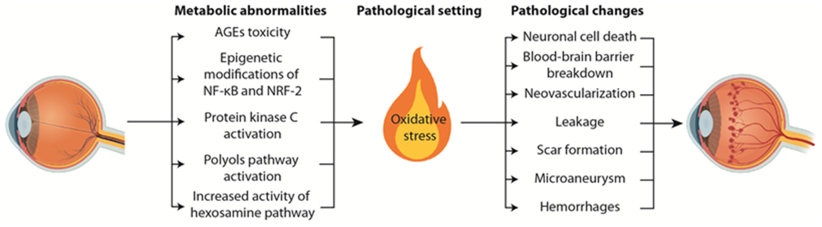
2. Pathophysiological Mechanisms Underlying Diabetic Retinopathy

It has long been believed that the main structural and functional changes in the retina generated by DR are related to the modification of blood vessel structure. Retinal blood supply is divided into two plexuses: the choroid (which supplies blood to the outer part of the retina) and the intraretinal vasculature (which supplies blood to the inner part of the retina). Each plexus changes as the stages of DR progress. These changes include pericyte loss, thickening of the basement membrane, neovascularization, breakdown of the blood retinal barrier (BRB), leakage, hemorrhaging, and microaneurysms [10,15].
DR has been divided into two stages that are based on microvascular proliferation: the first one is a non-proliferative phase (NPDP) composed of three levels (mild, moderate, and severe), followed by a proliferative phase (PDP). The main feature of the first stage is the degeneration and pericyte depletion of capillary blood vessels. Pericyte death is followed by endothelial cell death, both of which occur via apoptosis [16]. This severe form of cell death leads to the formation of acellular capillaries and the loss of BRB functionality [15,16]. BRB breakdown induces the formation of microaneurysms, retinal edema, and hemorrhages [15,16,17]. BRB breakdown is one of the main early pathological changes that takes place in DR. This is a multifactorial process that involves hypoxia, oxidative stress, and inflammation [18]. There are multiple molecular pathways involved in the process. These include hypoxia-inducible factors-1 and -2 (HIF-1 and -2), placental growth factor (PlGF), TNFα, IL-1β, platelet-activating factor (PAF), adenosine, histamine, prostaglandins (PGE1, PGE2, and PGF2α), platelet-derived growth factors A and B (PDGF-A and -B), insulin-like growth factor-1 (IGF-1), ICAM-1, VCAM-1, P-selectin, E-selectin, and VEGF [19]. Several reports point out the importance of VEGF in BRB breakdown, and all of them are indicative of the capability of decreasing the number of tight junctions between the cells. In turn, this leads to a decrease in the strength of the force that maintains cells side by side and an increase in permeability of the blood vessels. All these factors contribute to the appearance of microaneurysms, although the pathogenesis is multifactorial, and other factors such as pericyte loss, abnormalities of the adjacent retina, increased intraluminal pressure, among others, may also be of importance [20,21]. Lipoprotein precipitation results from fluid absorption in areas of inflammation. Exudates are distinct yellow-white retinal deposits made up of extracellular lipids, and located generally within the retina, although some may be on the retinal surface. Exudates can be identified by ophthalmoscopy as a sign of DR progression [22].
All these vascular changes occur as a result of ischemia and non-perfusion of the retinal tissue. In turn, there is upregulation of angiogenic cytokines, causing intra-retinal and intra-vitreous neovascularization [22]. The new vessels which grow on the retinal surface are usually fenestrated, leaky and brittle, and can result in vitreous hemorrhages. Vitreous hemorrhages are related to gliosis and scar formation, and the contraction of this tissue is associated with retinal detachment and blindness [22]. At this point DR is classified as PDP [16,22,23] (Figure 1).
Different sight-threatening factors have been identified as potential triggers of DR. For example, genetic data has identified polymorphisms in different genes as potential risk factors for the susceptibility to DR [24,25,26,27]. In addition, several types of experimental data indicate that oxidative stress, the excessive accumulation of reactive oxygen species (ROS), contribute to the onset and development of DR. The retina has a tremendous oxygen demand with even higher oxygen consumption than the cerebral cortex or muscle, making the retina highly susceptible to the accelerated accumulation of ROS [28]. This implies that significant damage in the retina can be attributed to oxidative stress and the accumulation of ROS. Oxidative stress is thought to induce lipid peroxidation, mitochondrial dysfunction, inflammation, and apoptosis leading to functional and structural damage in the retina.
An important factor of DR is the inflammation process, and it has been proven that inflammation plays an essential role in DR and can also be a promising therapeutic target [29]. Inflammation is a non-specific response to an injury, a pathological process, or stress, which utilizes a tremendous variety of molecules. Inflammation in DR can be characterized by an increase in gliosis, in other words the increased function of Muller glial cells, a source of proinflammatory modulators. This hypothesis is supported by the higher levels of adhesion molecules, ICAM-1, neutrophil chemotactic MCP-2, cytokines (IL-1β, IL-6, IL-8, IL10, TNF-α, IFN-γ, and MCP-1), and vitreous levels of neurotrophins (NTs) [30,31,32,33,34]. Increased expression of these molecules occurs at the same time as the rise of other growth factors such as VEGF, insulin-like growth factor (IGF), platelet-derived growth factor (PDGF), basic fibroblast growth factor (bFGF), and hepatocyte growth factor (HGF) – factors which are involved in the blood vessel progression associated with DR [30,31,32,33,34].
A plethora of hyperglycemia-associated molecular and cellular alterations are linked to the initiation and progression of DR and to high levels of oxidative stress (Figure 1). These hyperglycemia-associated pathways are categorized by 4 metabolic abnormalities: accumulation of toxic advanced glycation end products (AGEs), increased polyol pathway flux, upregulation of the hexosamine pathway, and activation of the protein kinase C pathway [15,35,36]. These metabolic routes are not independent, instead they are interacting with each other (reviewed in [37]). At a cellular level, dysfunction of these routes leads to oxidative stress, inflammation, and vascular abnormalities. Malfunction in signaling and transcriptional regulation triggers the upregulation of factors associated with the pathogenesis of DR including vascular endothelial growth factor (VEGF), receptor for advanced glycation end products (RAGE), tumor necrosis factor alpha (TNF-alpha), and more.
Firstly, chronic exposure of the retina to high blood glucose levels gives rise to the accelerated formation of AGEs, including carboxyethyl lysine, carboxymethyl lysine, and pentosidine [38,39]. Those harmful compounds are the result of a non-enzymatic process called glycation by which reactive sugars or metabolites derived from glycolysis are covalently bound to different macromolecules, inactivating them [40]. During hyperglycemia, AGEs accumulate in the retina, colocalize with RAGE in the retinal vasculature of diabetic individuals, and correlate to the severity of retinopathy [41,42,43]. The pathological role of AGEs in DR is complex, impacting the retinal function at different levels. AGEs form cross-linking between biomolecules of the extracellular matrix and the cytosol, influencing cellular metabolism. For example, sugar-derived damage can inactivate proteolytic pathways required for maintaining retinal cellular homeostasis [44,45,46]. The most understood AGEs-induced impairment takes place through the receptor for advanced glycation end products (RAGE). AGEs bind RAGE on the cell surface, triggering aberrant signaling cascade function. Some signaling factors affected by glycative stress are non-receptor tyrosine kinases (Janus kinases, JAK), intracellular transcription factors (signal transducer and activator of transcription, STAT), nuclear factor kappa B (NFkB), and recruitment of phosphatidylinositol 3′ kinase to Ras (reviewed in [47]). AGEs binding to RAGE in the vascular endothelium initiate changes that raise the expression of adhesion molecules and increase the secretion of cytokines TNF-alpha and VEGF. Also, high levels of RAGE expression are found in retinal ganglion cells and glial cells [48]. In addition, AGEs disrupt integrin signaling by cross-linking basement membrane proteins and stimulating plasminogen activator inhibitor-1 (PAI-1) through an interaction with RAGE. Several studies also indicate that the elevation of VEGF is dependent on AGE-RAGE interaction and integrin-linked kinases. All these forms of experimental data illustrate that AGEs are a pathological factor of DR contributing to vascular injury by increasing retinal vascular endothelial permeability and perycite apoptosis [49]. Lowering AGEs levels has been proposed as a therapeutic strategy to prevent the onset of this sight-threatening diabetic complication [45], however there is no effective clinical treatment currently available.
Secondly, hyperglycemia leads to upregulation of the polyol pathway. The consequences of this metabolic abnormality are (1) osmotic damage derived from sorbitol production, (2) accelerated formation of AGEs through the production and metabolism of generated fructose and, finally, (3) unbalanced redox status by limiting the synthesis of glutathione due to the overutilization of NADPH. All these hyperglycemia-induced changes might contribute to retinal capillary cell death. However, therapeutic strategies based on the dysregulation of the polyol pathway failed in clinical trials and did not prevent the progression of DR [50].
Thirdly, increased activity of the hexosamine pathway is found in the retina of diabetic patients. This pathway is a branch of glycolysis responsible for the production of uracil-N-acetylglucosamine, which is involved in glycosylation. Thus, in a diabetic context, hyperglycemia increases glucose flux by the hexosamine pathway and stimulates the production of high levels of this glucosamine. The increased glucosamine leads to overproduction of ROS that has a significant impact on redox status [51]. In addition, excessive production of uracil-N-acetylglucosamine alters glycosylation levels of proteins and lipids, impacting the neuroprotective effect of insulin and leading to retinal neurodegeneration [52]. Although there is compelling evidence pointing to the pathological role of increased glucose flux by the hexosamine pathway in diabetes, the information in relation to retinal tissues is limited.
Fourthly, hyperglycemia-induced activation of the protein kinase C (PKC) pathway is thought to aggravate the pathological imbalance in retinal cells and contribute to the pathogenesis of DR. Different isoforms of this serine/threonine kinase family (α, -β, -δ, and -ε) are reported to be upregulated in DR [53]. The activity of these isoforms is enhanced by diacylglycerol, whose de novo synthesis is accelerated because hyperglycemia elevates glucose flux through the glycolysis pathway. PKC isoforms are relevant signaling transducers that impact the activity of different transcription factors involved in vital physiological processes in retinal cells. PKC-β regulates the expression of VEGF and microvascular abnormalities [54]. In addition, PKC activation augments the activity of NADPH oxidase and nuclear factor-κB (NFkB), stimulating the production of ROS and inflammation [55]. Interestingly, ruboxistaurin, an orally administrated PKCβ inhibitor, has demonstrated benefits during clinical trials [56]. All this data suggests that PKC activation could be behind some of the pathological abnormalities present in DR.
Furthermore, emerging data indicates that hyperglycemia-induced epigenetic modifications, along with malfunction of nuclear factors such as NF-κB or nuclear factor erythroid 2 related factor 2 (NRF2), could contribute to DR through aberrant transcriptional modulation of genes coding for anti-inflammatory and antioxidant proteins [57,58,59,60].
Despite mounting evidence from experimental diabetic models supporting the role of oxidative stress in neuroretinal damage, antioxidants in clinical trials were not shown to be sufficient for combatting DR [61,62]. Thus, there is a need to uncover other pathological mechanisms that contribute to DR and design pharmacological approaches to fight this sight-threatening disease.

3. Early Neural Alterations Are Associated to DR Pathological Mechanisms

DR has been characterized as a vascular pathology, with pericyte loss being considered the earliest sign of DR [63]. However, recent data suggests that neurodegeneration appears in the diabetic retina earlier than any other microvasculopathic manifestation.
The induction of neuronal retinal degeneration during diabetes has long been recognized. In 1998 Barber et al. demonstrated that neuronal apoptosis occurs in diabetic rats and in humans prior to the appearance of microvascular lesions [64]. This evidence has been confirmed in recent years due to advancements in retinal imaging technology and the generalized use of optical coherence tomography (OCT). A clear example is the study of Sohn et al. using diabetic mice [65]. In this study the researchers used OCT to confirm that there were no differences in the density of retinal pericytes and the number of acellular capillaries in mice with diabetic retinas, but neural changes were detected [65].
These diabetes-induced neural changes have been observed in relation to the “neurovascular unit.” This unit refers to the coupling of neurons, glia, and vasculature in the central nervous system. In the retina, the neurovascular unit maintains the integrity of the BRB [66]. The known retinal alterations during DR are: (1) neural apoptosis of ganglion, amacrine, and Müller cells, as well as photoreceptors, (2) increased expression of glial fibrillary acidic protein (GFAP) in Müller cells, (3) microglial and glial activation, (4) glutamate metabolism alterations, (5) increased expression of neurotrophic factors, and (6) reduction of optic nerve axons [67].
As previously mentioned, the key pathways involved in the pathogenesis of DR include accumulation of AGEs, increased polyol pathway flux, upregulation of the hexosamine pathway, and activation of protein kinase C. It is important to note that the primary retinal neuronal diabetic change theory is compatible with the importance of these pathways in DR pathogenesis. Therefore, we can assume that no single pathway abnormality is responsible for DR development, instead it is a combination of several abnormalities and their interactions with each other. Of note, some of these pathways have been directly related to neuronal alterations in diabetes. For example, the accumulation of AGEs and the upregulation of RAGEs has been directly linked to the activation of Müller cells and the upregulation of GFAP, both of which contribute to DR [68]. Regarding PKC alterations, it has been demonstrated that PKC inhibition ameliorates neural dysfunction in diabetic rats [69]. PKC activation also induces overexpression of NADPH oxidase and NFκB, while increasing oxidative stress [70]. It has been hypothesized that damage created by oxidative stress is common across the avenues that induce DR. The evidence presented here shows that retinal neurons and photoreceptors are also significantly exposed to ROS and therefore, have a high propensity for oxidative stress that increases the severity and propensity for DR.
These retinal neuronal abnormalities lead to functional changes that sometimes occur before diabetes diagnosis and include deficits in pattern electroretinograms (ERG), increased implicit times in multifocal ERGs, changes in oscillatory potentials, and altered microperimetric, and perimetric psychophysical testing [67]. These functional alterations have clinical implications that include abnormal dark adaptation, decreased contrast sensitivity, abnormal color vision, and abnormal visual fields [71].

4. In Vivo and In Vitro Models for the Study of DR

Although a lot of important information can be obtained from human studies of DR, the exact pathological mechanism for DR remains unknown. Therefore, in vivo and in vitro models are needed to better understand the molecular and cellular changes that occur in the retina at the beginning of DR development. In addition, these models may provide us with new tools to find novel therapeutic agents for this disease.
Rabbits, cats, dogs, and monkeys have been used to study DR. However, mice and rats have been the most frequently used animal models. Three types of mice and rat models have been used over the years: (1) models with pharmacologically induced hyperglycemia, (2) spontaneous diabetic animals, and (3) models of angiogenesis without diabetes [72].
Regarding pharmacological diabetes induction, type 1 diabetes can be induced in rats or mice by injection of chemicals such as streptozotocin (STZ) and alloxan (both of which are toxic to pancreatic β-cells), or by feeding them galactose [72]. STZ diabetes-induced mice show vascular alterations in the retina as early as 8 days after hyperglycemia induction and rats show alterations 2 weeks after hyperglycemia induction [73]. Alloxan induction of diabetes is less commonly used in mice and rats, but alloxan-induced rat retinal neovascularization has been observed 2 months after induction [74]. Neuronal cell death is observed in both models. Galactose feeding-induced hyperglycemia showed that mice and rats develop vascular alterations 6 months after hyperglycemia [75].
Spontaneous diabetic mice models (including Ins2Akita, non-obese diabetic (NOD), db/db, and KKAy mice) have developed vascular lesions between 8 and 18 weeks [76,77,78]. Several rats with spontaneous onset of diabetes have been identified and include biobreeding (BB) rats, Wistar Bonn/Kobori (WBN/Kob) rats, Zucker diabetic fatty (ZDF) rats, Otsuka Long-Evans Tokushima fatty (OLETF) rats, non-obese GotoKakizaki (GK) rats, and non-obese spontaneously diabetic Torii (SDT) rats. Vasculature lesions have been observed after 1, 2, or 5 months of hyperglycemia [79,80,81,82]. Neuronal degeneration in all these animal models has been traditionally studied with electrophysiological methods.
Models that specifically target retinal neovascularization without diabetes are oxygen-induced retinopathy (OIR), retinal occlusion and injection, or genetic induction of VEGF [83]. OIR is an animal model for premature retinopathy that is characterized by the presence of neovascularization and consists of exposing newborn mice to high oxygen concentration for several days before returning them to standard room air. Examples of occlusion models used to study DR include ligation of pterygopalatine artery and external carotid artery, retinal vein occlusion, and increased intraocular pressure (IOP). TrVEGF029 is a model of genetic induction of neovascularization due to its overexpression of VEGF16 [72]. The time points associated with changes in the vasculature of diabetic retina vary significantly based on the alteration evaluated, the method used to detect the alteration, or even the laboratory that performed the analysis.
Neuronal degeneration in these animal models has generally been studied with electrophysiological methods and via detection of apoptosis in neural cells or degree of thinning in different retinal layers. This parameter has been determined by immunohistochemical techniques or by OCT. Our group has reported a decrease in the ERG amplitude of diabetic mice induced by alloxan only one week after hyperglycemic induction [84]. In the STZ model, a decrease in the b-wave amplitude of the ERG and an increase in the percentage of apoptotic cells in retinal layers has been seen 14 days after the induction of diabetes [85]. A thinning in the retina has been observed in STZ rats after 12 weeks of diabetes [86].
An increase in apoptosis has been found in Ins2Akita mice after 4 weeks of hyperglycemia [76] and OCT revealed a progressive thinning of the retina from 3 months onwards [87]. Retinal thinning has been observed at 3 months of diabetes in db/db mice as well [88]. In this animal model, alterations in the ERG have been observed after 12 weeks and a significant increase in TUNEL-positive cells was documented 20, 24, and 28 weeks after hyperglycemia induction [89]. Other studies have reported a loss of ganglion cells and a reduction of neuroretinal thickness at week 8 [90].
Irrespective of the existence of a remarkable number of animal models, all fail to show the complete set of human clinical aspects of DR. Some animal models only show early DR features and others only show the late proliferative and angiogenesis hallmarks [72]. A proper selection of the model used to study DR is therefore of great importance for improving DR research.
Other models have also been used and are currently being developed to study DR. For example, in vitro cell culture using isolated endothelial cells or pericytes to mimic the human angiogenic process have been used [91]. Valdés et al. recently demonstrated the utility of retinal explants as a novel tool to study the neural mechanisms of DR [92]. In vitro studies using retinal explants may help researchers uncover the cellular response triggered by cytotoxic effects that are directly caused by high glucose. Moreover, retinal explants undergo rapid vasoregression and loss of microglial cells and may thus be of utility to study hyperglycemia effects, independently of vascular- or immune-related effects. This may allow for the establishment and characterization of the initial neural mechanisms of DR. One of the advantages of retinal explant cultures is that cells are kept within their normal environment and preserve cell-to-cell interactions. At the same time, the defined chemical culture conditions allow precise and reproducible experimental environments. This expands the potential to study new pharmaceutical treatments, thus retinal explants are ideal for drug screening purposes [92]. The use of retinal explants presents other advantages such as helping to reduce the number of experimental models. Retinal explant cultures with retinal pigment epithelium can also be maintained in vitro for several weeks, and drug treatment screening is simplified due to the lack of body fluids or organ excretion that can dilute or degrade the drug treatments [93].
Regarding the previously mentioned metabolic pathways linked to DR, we can confirm that they are all important pathological mechanisms in the STZ rat model. An accumulation of AGEs, as well as an increase in diacylglycerol (DAG) levels and PKC activity have been found in the retina of STZ-induced diabetic rats [94,95,96,97]. This diabetes animal model is one of the most studied, however, there is much less information available about hyperglycemia-induced metabolic changes in the STZ model, in comparison to other models. In alloxan-induced diabetic rats and galactose-fed rats it has been demonstrated that there is some increase in AGEs in tissues such as the kidney, but not in the retina [75]. Mice with short-term STZ-induced diabetes also lack some of these biochemical changes (i.e. accumulation of retinal sorbitol and fructose) that are clearly present in the retina of STZ-diabetic rats [98]. Furthermore, increases in PKC activity have been observed in experimental galactosemia [99], in genetically determined diabetic BB rats [97] and in Zucker Diabetic Fatty rats [100]. Alterations in the hexosamine pathway of the Zucker Diabetic Fatty rats have also been observed [100].
With respect to the in vitro models, AGEs levels increase with glucose concentration and time of exposure in retinal explants [101]. However, there is no information regarding possible alterations in the polyol or hexosamine pathways or about modifications in PKC activity. The addition of glucose or AGEs to the culture medium of retinal endothelial cells is another well-established, in-vitro model that has previously demonstrated that the addition of these supplementations increases sorbitol and PKC activity [102].
All the animal models described in this review have limitations. For example, there is not an agreement about the doses of alloxan or STZ that should be used to induce diabetes, or about the possible treatment of these animals with insulin in order to increase their survival. Moreover, the use of these drugs induces type 1 diabetes animal models, but the prevalence of type 2 diabetes is greater and there are far fewer type 2 diabetes animal models. The use of retinal explants requires technical resources and does not allow for the study of vascular or microglial alterations. Finally, as previously stated, some diabetes models are related to all the pathological and metabolic alterations and other models only show a relationship between some of these alterations. All these factors should be considered when selecting a model to study DR and test new effective therapies.

5. Novel Therapeutic Targets in DR

The treatments for DR have improved dramatically in recent years and include proper metabolic control of patients, laser treatments, vitrectomy surgery, and the administration of drugs such as steroids and anti-vascular endothelial growth factor (VEGF) treatments [103]. However, metabolic control is difficult to achieve for most diabetic patients and repeated anti-VEGF injections have been shown to reduce retinopathy severity in less than one-third of eyes treated over the course of 2 years [104].
The main barrier for preventing and treating retinal failure in a diabetic context is the incomplete understanding of the retinal pathophysiology of diabetes. The early neuronal alterations in retinal diabetes described in this review are clinically asymptomatic but represent important opportunities to find earlier treatments that may prevent vision loss in diabetic patients [105].
In this regard, apoptosis of neurons is an essential pharmacological target [106]. The main advantage of these upstream treatments is that they would be preventive, and they could be administered before symptoms appear. Inhibiting apoptosis could be a potential treatment for preventing DR-associated neurodegeneration. Latanoprost and calpain inhibitors have demonstrated the ability to decrease this apoptosis [107]. Blockade of glutamate receptors, through the administration of drugs such as memantin, may also help to maintain the integrity of the neuroretina during diabetes [107]. Glucagon-like peptide 1 (GLP-1) is an endogenous substance that has neuroprotective qualities that has also been related to the inhibition of vascular leakage. Pigment epithelium-derived factor (PEDF) and brain derived neurotrophic factor (BDNF) have been demonstrated to rescue dying neurons in animal models of diabetes. Somatostatin, another potential neurotrophic agent, is being evaluated in a clinical trial and its administration may be topical, which is advantageous when compared to intravitreal injections or systemic administration [107].
As outlined before, oxidative stress may be considered a unifying mechanism among all the pathways with established relationships to DR. However, there is conflicting evidence regarding the effects of antioxidants on DR. The discrepancies in the results observed from the use of antioxidants may be due to the type of antioxidant, the method of administration, or the dosage.
Enzymatic antioxidants, non-enzymatic antioxidants (for example, vitamin A, C, or E), and other oral supplements such as alpha lipoic acid, omega 3 poly-unsaturated fatty acids, calcium dobesilate, and Gingkgo biloba have been used in human studies [108,109,110,111,112]. The results of these studies have shown little effect on vascular changes in the retina, but have shown a beneficial impact on retinal function, suggesting that they can be adequate adjuvant therapies in the early phases of DR [113]. Flavonoids and carotenoids are antioxidants that have been shown to inhibit DR neuronal damage [114]. An antioxidant that has demonstrated positive effects on retinal neurodegeneration in animal models of diabetes is sulforaphane. This antioxidant is found in cruciferous plants and has demonstrated protective effects on the diabetic retina because of the delay in photoreceptor degeneration associated to diabetes and its potential for activating the AMPK (AMP-activated protein kinase) pathway [115].
A new approach for DR treatments is to identify novel epigenetic changes associated with DR and target those specific microRNAs (miRNAs). miRNA alterations have been observed in many disorders including DR. Most miRNA alterations in diabetes have been related to vascular changes, but some of them have also been related to inflammation [116]. A direct relationship between miRNAs and diabetic neural retinopathy has been established: miR29b and its potential target PKR associated protein X (RAX) are localized in retinal ganglion cells and cells of the inner nuclear layer and are upregulated in diabetes and, therefore, may be considered a therapeutic target [117].
Finally, genetic therapies can be applied to DR treatment because they are already being applied to other retinal degenerative diseases. For example, Leber’s congenital amaurosis, a rare type of inherited eye disorder that causes severe vision loss at birth, is currently being combated with Luxturna, a gene therapy product used for the treatment of patients with retinal disease due to mutations in both copies of the RPE65 gene. Gene therapy could be used to impact two targets: neovascularization and leakage treatment, or blood vessel and neuronal damage protection [118,119].
Genetic treatments focused specifically on solving blood vessel damage are demonstrating the potential of gene therapy for DR. Inoculation with adeno-associated virus (AAV) that increases the expression of the protein Flt23k, an anti-VEGF intraceptor, showed a decrease in retinal neovascularization [118,120,121,122]. Injection of a plasmid targeting VEGF by expressing siRNA inhibited neovascularization in animal models. AAV injection in the retina that delivered PEDF demonstrated the capability to decrease VEGF levels. In addition, angiostatin and endostatin encoded in a lentivirus proved to be potential retinal degeneration treatments through the inhibition of endothelial proliferation [119,123]. AAV encoding inhibitors such as metalloproteinase-3 and CAD confirmed the capability to avoid neovascularization in a DR mice model. HGFK1 carried in AAV can save the ocular architecture and reduce the amount of VEGF. Also, retinal transduction of AAV, which encodes for proteins sFlt-1 and vasoinhibin, reduced the BRB breakdown and decreased the number of microvascular abnormalities [118,119,124,125].
Another possible use of genetic therapy is for neuronal and vascular protection. It has been shown that an AAV vector encoding small hairpin RNAs against EGR1 could reduce apoptosis in the retinal inner nuclear layer (INL) and outer nuclear layer (ONL) [118,119,126]. Injection of AAV containing anti microRNA-204 enhanced the expression of the autophagosomal component LC3B-II—increasing autophagy and decreasing the evolution of DR [127]. AAV carrying CD59 was proven to reduce vascular leakage [70]. AAV that encodes brain derived neurotropic factor (BDNF) increased the survival of retinal ganglion cells and improved retinal function [128]. Subretinal AAV2-mediated overexpression of erythropoietin (EPO) prevented BRB breakdown and reduced neuronal apoptosis. AAV encoding manganese superoxide dismutase (MnSOD) was proven to reduce oxidative stress in the retina, leading to a decrease in DR progression and preventing the onset of the metabolic memory phenomena, whose main feature is that the glycemic control in diabetes signs and symptoms are “remembered” [119]. AAV carrying ACE2 and Ang-(1-7) led to a reduction of acellular capillaries, inflammation, oxidative stress, and vasculature leakage [129]. Furthermore, because of the novel CRISPR-Cas Gene editing system, different gene therapies are being developed. For example, AGN-151587 (EDIT-101) might remove a point mutation in the CEP290 gene which causes type 10 Leber congenital amaurosis, the most common inherited retinal degenerative disease that induces childhood blindness [130]. In addition, disruption of VEGFR2 and prevention of retinal angiogenesis can be achieved with the CRISPR-SpCas9 system [118,131].
Furthermore, another new and innovative DR treatment is stem cells. Mesenchymal stromal cells (MSCs), specifically groups such as adipose stem cells (ASC) and bone marrow-derived mesenchymal stem cells (BM-MSC), have shown promising results by producing paracrine factors which preserve the ganglion cell layer and assist in the regeneration of optic nerve cells. DR treatment with ASC has been proven to protect and accelerate recovery of the vasculature by releasing vasoprotective cytokines and differentiating them into vascular cells such as pericytes [132,133,134]. Furthermore, ASC treatment has been shown to decrease vascular leakage and cell apoptosis, while improving electroretinogram activity in rat models [133]. Regarding BM-MSC, CD14+ cells have demonstrated the capability to restore vasculature, making them a powerful, potential form of DR therapy [135]. Another possible BM-MSC treatment is the use of CD34+ cells, which can differentiate into endothelial cells whose main function is to repair the vasculature through paracrine mechanisms. Despite being a promising treatment, due to the pro-inflammatory environment of diabetic patients, most CD34+ cells are trapped in the bone marrow, making them very difficult to acquire and use [135]. Finally, it has been corroborated that neural stem cells from umbilical cord-derived mesenchymal stem cells (UC-MSC) can restore retinal morphology and retinal vision function [136]. In summation, MSC, especially ASC, are interesting forms of DR treatment because of their paracrine abilities. These kinds of cells are able to produce growth factors, chemokines, and cytokines which can counter the high immune response in the body of someone experiencing DR and act as immunosuppressor agents [137]. Currently, there are several DR stem cell treatments in development. Some examples are NCT01736059, NCT03403283, and NCT03403699.

6. Concluding Remarks

DR represents the most common cause of preventable blindness of work-aged adults in developed countries. Taking into account the increasing number of people who suffer from diabetes in developed countries, it could be predicted that the future incidence of DR will increase. Furthermore, it is estimated that persons with diabetic eye disease in Europe will increase to 8.6 million in 2050. This indicates that DR may cause a substantial economic burden for our society over the next few decades. For these reasons, it is important to understand the pathophysiological mechanisms involved in DR in order to develop new and functional therapies.
Despite the previous belief that the main structural and functional changes in a diabetic retina are related to the modification of blood vessel structure, neurodegeneration is a novel discovery that is also caused by diabetes and appears earlier than any other microvascupolathy manifestation. Given this new information and evolving technology, DR could be considered a neurological disorder. As a neurological disorder new, neuroprotective therapeutic strategies could become a different and effective way of preventing or slowing loss of vision incurred by diabetes.

Author Contributions

A.C., J.M., M.M. and E.B. contributed to the writing of the original draft. A.C., J.M., M.M., G.P.-V. and E.B. contributed to the review and editing of the manuscript. M.M. and E.B. designed the structure of the review and supervised the writing. All authors have read and agreed to the published version of the manuscript.

Funding

This manuscript has been supported by grants RYC2018-024434-I, MINECO PID2020-119466RB-I00, FUSP-PPC-19-B53C4C64, ACIF 199/2019, FPU20/06277.

Institutional Review Board Statement

Not applicable.

Informed Consent Statement

Not applicable.

Data Availability Statement

Not applicable.

Acknowledgments

We thank Elizabeth A. Whitcomb for reviewing and editing the manuscript.

Conflicts of Interest

The authors declare no conflict of interest.

References

  1. Romero-Aroca, P.; Baget-Bernaldiz, M.; Pareja-Rios, A.; Lopez-Galvez, M.; Navarro-Gil, R.; Verges, R. Diabetic Macular Edema Pathophysiology: Vasogenic versus Inflammatory. J. Diabetes Res. 2016, 2016, 2156273. [Google Scholar] [CrossRef] [PubMed] [Green Version]
  2. Le Floch, J.P.; Doucet, J.; Bauduceau, B.; Verny, C. Retinopathy, nephropathy, peripheral neuropathy and geriatric scale scores in elderly people with Type 2 diabetes. Diabet. Med. A J. Br. Diabet. Assoc. 2014, 31, 107–111. [Google Scholar] [CrossRef]
  3. Bourne, R.R.; Stevens, G.A.; White, R.A.; Smith, J.L.; Flaxman, S.R.; Price, H.; Jonas, J.B.; Keeffe, J.; Leasher, J.; Naidoo, K.; et al. Causes of vision loss worldwide, 1990-2010: A systematic analysis. Lancet Glob. Health 2013, 1, e339–e349. [Google Scholar] [CrossRef] [Green Version]
  4. Yau, J.W.; Rogers, S.L.; Kawasaki, R.; Lamoureux, E.L.; Kowalski, J.W.; Bek, T.; Chen, S.J.; Dekker, J.M.; Fletcher, A.; Grauslund, J.; et al. Global prevalence and major risk factors of diabetic retinopathy. Diabetes Care 2012, 35, 556–564. [Google Scholar] [CrossRef] [Green Version]
  5. Kempen, J.H.; O’Colmain, B.J.; Leske, M.C.; Haffner, S.M.; Klein, R.; Moss, S.E.; Taylor, H.R.; Hamman, R.F. The prevalence of diabetic retinopathy among adults in the United States. Arch. Ophthalmol. 2004, 122, 552–563. [Google Scholar] [CrossRef] [Green Version]
  6. Li, J.Q.; Welchowski, T.; Schmid, M.; Letow, J.; Wolpers, C.; Pascual-Camps, I.; Holz, F.G.; Finger, R.P. Prevalence, incidence and future projection of diabetic eye disease in Europe: A systematic review and meta-analysis. Eur. J. Epidemiol. 2020, 35, 11–23. [Google Scholar] [CrossRef] [PubMed]
  7. Rema, M.; Premkumar, S.; Anitha, B.; Deepa, R.; Pradeepa, R.; Mohan, V. Prevalence of diabetic retinopathy in urban India: The Chennai Urban Rural Epidemiology Study (CURES) eye study, I. Investig. Ophthalmol. Vis. Sci. 2005, 46, 2328–2333. [Google Scholar] [CrossRef] [Green Version]
  8. Raman, R.; Rani, P.K.; Reddi Rachepalle, S.; Gnanamoorthy, P.; Uthra, S.; Kumaramanickavel, G.; Sharma, T. Prevalence of diabetic retinopathy in India: Sankara Nethralaya Diabetic Retinopathy Epidemiology and Molecular Genetics Study report 2. Ophthalmology 2009, 116, 311–318. [Google Scholar] [CrossRef] [PubMed]
  9. Klein, R.; Klein, B.E.; Moss, S.E.; Davis, M.D.; DeMets, D.L. The Wisconsin epidemiologic study of diabetic retinopathy. II. Prevalence and risk of diabetic retinopathy when age at diagnosis is less than 30 years. Arch. Ophthalmol. 1984, 102, 520–526. [Google Scholar] [CrossRef]
  10. Cheung, N.; Mitchell, P.; Wong, T.Y. Diabetic retinopathy. Lancet 2010, 376, 124–136. [Google Scholar] [CrossRef]
  11. Raymond, N.T.; Varadhan, L.; Reynold, D.R.; Bush, K.; Sankaranarayanan, S.; Bellary, S.; Barnett, A.H.; Kumar, S.; O’Hare, J.P. Higher prevalence of retinopathy in diabetic patients of South Asian ethnicity compared with white Europeans in the community: A cross-sectional study. Diabetes Care 2009, 32, 410–415. [Google Scholar] [CrossRef] [Green Version]
  12. Yin, L.; Zhang, D.; Ren, Q.; Su, X.; Sun, Z. Prevalence and risk factors of diabetic retinopathy in diabetic patients: A community based cross-sectional study. Medicine 2020, 99, e19236. [Google Scholar] [CrossRef]
  13. Knowler, W.C.; Barrett-Connor, E.; Fowler, S.E.; Hamman, R.F.; Lachin, J.M.; Walker, E.A.; Nathan, D.M. Reduction in the incidence of type 2 diabetes with lifestyle intervention or metformin. N. Engl. J. Med. 2002, 346, 393–403. [Google Scholar] [CrossRef]
  14. Song, P.; Yu, J.; Chan, K.Y.; Theodoratou, E.; Rudan, I. Prevalence, risk factors and burden of diabetic retinopathy in China: A systematic review and meta-analysis. J. Glob. Health 2018, 8, 010803. [Google Scholar] [CrossRef]
  15. Hammes, H.P. Diabetic retinopathy: Hyperglycaemia, oxidative stress and beyond. Diabetologia 2018, 61, 29–38. [Google Scholar] [CrossRef] [Green Version]
  16. Spencer, B.G.; Estevez, J.J.; Liu, E.; Craig, J.E.; Finnie, J.W. Pericytes, inflammation, and diabetic retinopathy. Inflammopharmacology 2020, 28, 697–709. [Google Scholar] [CrossRef]
  17. Eshaq, R.S.; Aldalati, A.M.Z.; Alexander, J.S.; Harris, N.R. Diabetic retinopathy: Breaking the barrier. Pathophysiol. Off. J. Int. Soc. Pathophysiol. 2017, 24, 229–241. [Google Scholar] [CrossRef]
  18. Yang, X.; Yu, X.W.; Zhang, D.D.; Fan, Z.G. Blood-retinal barrier as a converging pivot in understanding the initiation and development of retinal diseases. Chin. Med. J. 2020, 133, 2586–2594. [Google Scholar] [CrossRef]
  19. Vinores, S.A. Breakdown of the Blood–Retinal Barrier. Encycl. Eye 2010, 216–222. [Google Scholar] [CrossRef]
  20. Omri, S.; Behar-Cohen, F.; Rothschild, P.-R.; Gélizé, E.; Jonet, L.; Jeanny, J.C.; Omri, B.; Crisanti, P. PKCζ mediates breakdown of outer blood-retinal barriers in diabetic retinopathy. PLoS ONE 2013, 8, e81600. [Google Scholar] [CrossRef] [Green Version]
  21. Huang, H.; Gandhi, J.K.; Zhong, X.; Wei, Y.; Gong, J.; Duh, E.J.; Vinores, S.A. TNFalpha is required for late BRB breakdown in diabetic retinopathy, and its inhibition prevents leukostasis and protects vessels and neurons from apoptosis. Investig. Ophthalmol. Vis. Sci. 2011, 52, 1336–1344. [Google Scholar] [CrossRef] [Green Version]
  22. Lechner, J.; O’Leary, O.E.; Stitt, A.W. The pathology associated with diabetic retinopathy. Vis. Res. 2017, 139, 7–14. [Google Scholar] [CrossRef] [PubMed]
  23. Wilkinson, C.P.; Ferris, F.L., 3rd; Klein, R.E.; Lee, P.P.; Agardh, C.D.; Davis, M.; Dills, D.; Kampik, A.; Pararajasegaram, R.; Verdaguer, J.T. Proposed international clinical diabetic retinopathy and diabetic macular edema disease severity scales. Ophthalmology 2003, 110, 1677–1682. [Google Scholar] [CrossRef]
  24. Cilenšek, I.; Mankoč, S.; Globočnik Petrovič, M.; Petrovič, D. The 4a/4a genotype of the VNTR polymorphism for endothelial nitric oxide synthase (eNOS) gene predicts risk for proliferative diabetic retinopathy in Slovenian patients (Caucasians) with type 2 diabetes mellitus. Mol. Biol. Rep. 2012, 39, 7061–7067. [Google Scholar] [CrossRef]
  25. Jafarzadeh, F.; Javanbakht, A.; Bakhtar, N.; Dalvand, A.; Shabani, M.; Mehrabinejad, M.M. Association between diabetic retinopathy and polymorphisms of cytokine genes: A systematic review and meta-analysis. Int. Ophthalmol. 2021. [Google Scholar] [CrossRef] [PubMed]
  26. Balasubbu, S.; Sundaresan, P.; Rajendran, A.; Ramasamy, K.; Govindarajan, G.; Perumalsamy, N.; Hejtmancik, J.F. Association analysis of nine candidate gene polymorphisms in Indian patients with type 2 diabetic retinopathy. BMC Med. Genet. 2010, 11, 158. [Google Scholar] [CrossRef] [PubMed] [Green Version]
  27. Hampton, B.M.; Schwartz, S.G.; Brantley, M.A., Jr.; Flynn, H.W., Jr. Update on genetics and diabetic retinopathy. Clin. Ophthalmol. 2015, 9, 2175–2193. [Google Scholar] [CrossRef] [PubMed] [Green Version]
  28. Wilkinson-Berka, J.L.; Rana, I.; Armani, R.; Agrotis, A. Reactive oxygen species, Nox and angiotensin II in angiogenesis: Implications for retinopathy. Clin. Sci. 2013, 124, 597–615. [Google Scholar] [CrossRef] [PubMed] [Green Version]
  29. Forrester, J.V.; Kuffova, L.; Delibegovic, M. The Role of Inflammation in Diabetic Retinopathy. Front. Immunol. 2020, 11, 583687. [Google Scholar] [CrossRef]
  30. Rübsam, A.; Parikh, S.; Fort, P.E. Role of Inflammation in Diabetic Retinopathy. Int. J. Mol. Sci. 2018, 19, 942. [Google Scholar] [CrossRef] [Green Version]
  31. Ucgun, N.I.; Zeki-Fikret, C.; Yildirim, Z. Inflammation and diabetic retinopathy. Mol. Vis. 2020, 26, 718–721. [Google Scholar] [PubMed]
  32. Hong, F.; Yang, D.Y.; Li, L.; Zheng, Y.F.; Wang, X.J.; Guo, S.R.N.; Jiang, S.; Zhu, D.; Tao, Y. Relationship Between Aqueous Humor Levels of Cytokines and Axial Length in Patients With Diabetic Retinopathy. Asia-Pac. J. Ophthalmol. 2020, 9, 149–155. [Google Scholar] [CrossRef] [PubMed]
  33. Zeng, Y.; Cao, D.; Yu, H.; Hu, Y.; He, M.; Yang, D.; Zhuang, X.; Zhang, L. Comprehensive analysis of vitreous humor chemokines in type 2 diabetic patients with and without diabetic retinopathy. Acta Diabetol. 2019, 56, 797–805. [Google Scholar] [CrossRef] [PubMed]
  34. Zhou, J.; Wang, S.; Xia, X. Role of intravitreal inflammatory cytokines and angiogenic factors in proliferative diabetic retinopathy. Curr. Eye Res. 2012, 37, 416–420. [Google Scholar] [CrossRef] [PubMed]
  35. Brownlee, M. Biochemistry and molecular cell biology of diabetic complications. Nature 2001, 414, 813–820. [Google Scholar] [CrossRef] [PubMed]
  36. Dagher, Z.; Park, Y.S.; Asnaghi, V.; Hoehn, T.; Gerhardinger, C.; Lorenzi, M. Studies of rat and human retinas predict a role for the polyol pathway in human diabetic retinopathy. Diabetes 2004, 53, 2404–2411. [Google Scholar] [CrossRef] [PubMed] [Green Version]
  37. Kang, Q.; Yang, C. Oxidative stress and diabetic retinopathy: Molecular mechanisms, pathogenetic role and therapeutic implications. Redox Biol. 2020, 37, 101799. [Google Scholar] [CrossRef]
  38. Uchiki, T.; Weikel, K.A.; Jiao, W.; Shang, F.; Caceres, A.; Pawlak, D.; Handa, J.T.; Brownlee, M.; Nagaraj, R.; Taylor, A. Glycation-altered proteolysis as a pathobiologic mechanism that links dietary glycemic index, aging, and age-related disease (in nondiabetics). Aging Cell 2012, 11, 1–13. [Google Scholar] [CrossRef] [PubMed] [Green Version]
  39. Rowan, S.; Jiang, S.; Korem, T.; Szymanski, J.; Chang, M.L.; Szelog, J.; Cassalman, C.; Dasuri, K.; McGuire, C.; Nagai, R.; et al. Involvement of a gut-retina axis in protection against dietary glycemia-induced age-related macular degeneration. Proc. Natl. Acad. Sci. USA 2017, 114, E4472–E4481. [Google Scholar] [CrossRef] [Green Version]
  40. Rowan, S.; Bejarano, E.; Taylor, A. Mechanistic targeting of advanced glycation end-products in age-related diseases. Biochim. Biophys. Acta. Mol. Basis Dis. 2018, 1864, 3631–3643. [Google Scholar] [CrossRef] [PubMed]
  41. Stitt, A.W. Advanced glycation: An important pathological event in diabetic and age related ocular disease. Br. J. Ophthalmol. 2001, 85, 746–753. [Google Scholar] [CrossRef]
  42. Stitt, A.W.; Li, Y.M.; Gardiner, T.A.; Bucala, R.; Archer, D.B.; Vlassara, H. Advanced glycation end products (AGEs) co-localize with AGE receptors in the retinal vasculature of diabetic and of AGE-infused rats. Am. J. Pathol. 1997, 150, 523–531. [Google Scholar]
  43. Chen, M.; Curtis, T.M.; Stitt, A.W. Advanced glycation end products and diabetic retinopathy. Curr. Med. Chem. 2013, 20, 3234–3240. [Google Scholar] [CrossRef] [PubMed]
  44. Aragonès, G.; Dasuri, K.; Olukorede, O.; Francisco, S.G.; Renneburg, C.; Kumsta, C.; Hansen, M.; Kageyama, S.; Komatsu, M.; Rowan, S.; et al. Autophagic receptor p62 protects against glycation-derived toxicity and enhances viability. Aging Cell 2020, 19, e13257. [Google Scholar] [CrossRef] [PubMed]
  45. Aragonès, G.; Rowan, S.; Francisco, S.G.; Yang, W.; Weinberg, J.; Taylor, A.; Bejarano, E. Glyoxalase System as a Therapeutic Target against Diabetic Retinopathy. Antioxidants 2020, 9, 1062. [Google Scholar] [CrossRef] [PubMed]
  46. Queisser, M.A.; Yao, D.; Geisler, S.; Hammes, H.P.; Lochnit, G.; Schleicher, E.D.; Brownlee, M.; Preissner, K.T. Hyperglycemia impairs proteasome function by methylglyoxal. Diabetes 2010, 59, 670–678. [Google Scholar] [CrossRef] [PubMed] [Green Version]
  47. Safi, S.Z.; Qvist, R.; Kumar, S.; Batumalaie, K.; Ismail, I.S. Molecular mechanisms of diabetic retinopathy, general preventive strategies, and novel therapeutic targets. BioMed Res. Int. 2014, 2014, 801269. [Google Scholar] [CrossRef] [Green Version]
  48. Tezel, G.; Luo, C.; Yang, X. Accelerated aging in glaucoma: Immunohistochemical assessment of advanced glycation end products in the human retina and optic nerve head. Investig. Ophthalmol. Vis. Sci. 2007, 48, 1201–1211. [Google Scholar] [CrossRef] [PubMed]
  49. Yamagishi, S.; Nakamura, K.; Matsui, T.; Inagaki, Y.; Takenaka, K.; Jinnouchi, Y.; Yoshida, Y.; Matsuura, T.; Narama, I.; Motomiya, Y.; et al. Pigment epithelium-derived factor inhibits advanced glycation end product-induced retinal vascular hyperpermeability by blocking reactive oxygen species-mediated vascular endothelial growth factor expression. J. Biol. Chem. 2006, 281, 20213–20220. [Google Scholar] [CrossRef] [Green Version]
  50. Sorbinil Retinopathy Triail Research Group. A Randomized Trial of Sorbinil, an Aldose Reductase Inhibitor, in Diabetic Retinopathy. Arch. Ophthalmol. 1990, 108, 1234–1244. [Google Scholar] [CrossRef] [PubMed]
  51. Du, X.; Matsumura, T.; Edelstein, D.; Rossetti, L.; Zsengellér, Z.; Szabó, C.; Brownlee, M. Inhibition of GAPDH activity by poly(ADP-ribose) polymerase activates three major pathways of hyperglycemic damage in endothelial cells. J. Clin. Investig. 2003, 112, 1049–1057. [Google Scholar] [CrossRef] [PubMed] [Green Version]
  52. Nakamura, M.; Barber, A.J.; Antonetti, D.A.; LaNoue, K.F.; Robinson, K.A.; Buse, M.G.; Gardner, T.W. Excessive hexosamines block the neuroprotective effect of insulin and induce apoptosis in retinal neurons. J. Biol. Chem. 2001, 276, 43748–43755. [Google Scholar] [CrossRef] [PubMed] [Green Version]
  53. Idris, I.; Gray, S.; Donnelly, R. Protein kinase C activation: Isozyme-specific effects on metabolism and cardiovascular complications in diabetes. Diabetologia 2001, 44, 659–673. [Google Scholar] [CrossRef] [PubMed] [Green Version]
  54. Aiello, L.P.; Clermont, A.; Arora, V.; Davis, M.D.; Sheetz, M.J.; Bursell, S.E. Inhibition of PKC beta by oral administration of ruboxistaurin is well tolerated and ameliorates diabetes-induced retinal hemodynamic abnormalities in patients. Investig. Ophthalmol. Vis. Sci. 2006, 47, 86–92. [Google Scholar] [CrossRef] [Green Version]
  55. Xia, P.; Inoguchi, T.; Kern, T.S.; Engerman, R.L.; Oates, P.J.; King, G.L. Characterization of the mechanism for the chronic activation of diacylglycerol-protein kinase C pathway in diabetes and hypergalactosemia. Diabetes 1994, 43, 1122–1129. [Google Scholar] [CrossRef] [PubMed]
  56. Aiello, L.P.; Vignati, L.; Sheetz, M.J.; Zhi, X.; Girach, A.; Davis, M.D.; Wolka, A.M.; Shahri, N.; Milton, R.C. Oral protein kinase c β inhibition using ruboxistaurin: Efficacy, safety, and causes of vision loss among 813 patients (1,392 eyes) with diabetic retinopathy in the Protein Kinase C β Inhibitor-Diabetic Retinopathy Study and the Protein Kinase C β Inhibitor-Diabetic Retinopathy Study 2. Retina 2011, 31, 2084–2094. [Google Scholar] [CrossRef]
  57. Kowluru, R.A. Retinopathy in a Diet-Induced Type 2 Diabetic Rat Model and Role of Epigenetic Modifications. Diabetes 2020, 69, 689–698. [Google Scholar] [CrossRef] [PubMed]
  58. Kumari, N.; Karmakar, A.; Ganesan, S.K. Targeting epigenetic modifications as a potential therapeutic option for diabetic retinopathy. J. Cell. Physiol. 2020, 235, 1933–1947. [Google Scholar] [CrossRef] [PubMed]
  59. Sui, A.; Chen, X.; Demetriades, A.M.; Shen, J.; Cai, Y.; Yao, Y.; Yao, Y.; Zhu, Y.; Shen, X.; Xie, B. Inhibiting NF-κB Signaling Activation Reduces Retinal Neovascularization by Promoting a Polarization Shift in Macrophages. Investig. Ophthalmol. Vis. Sci. 2020, 61, 4. [Google Scholar] [CrossRef]
  60. Miller, W.P.; Sunilkumar, S.; Giordano, J.F.; Toro, A.L.; Barber, A.J.; Dennis, M.D. The stress response protein REDD1 promotes diabetes-induced oxidative stress in the retina by Keap1-independent Nrf2 degradation. J. Biol. Chem. 2020, 295, 7350–7361. [Google Scholar] [CrossRef] [Green Version]
  61. Williams, M.; Hogg, R.E.; Chakravarthy, U. Antioxidants and diabetic retinopathy. Curr. Diabetes Rep. 2013, 13, 481–487. [Google Scholar] [CrossRef] [PubMed]
  62. Mann, J.F.; Lonn, E.M.; Yi, Q.; Gerstein, H.C.; Hoogwerf, B.J.; Pogue, J.; Bosch, J.; Dagenais, G.R.; Yusuf, S. Effects of vitamin E on cardiovascular outcomes in people with mild-to-moderate renal insufficiency: Results of the HOPE study. Kidney Int. 2004, 65, 1375–1380. [Google Scholar] [CrossRef] [Green Version]
  63. Hammes, H.P.; Lin, J.; Renner, O.; Shani, M.; Lundqvist, A.; Betsholtz, C.; Brownlee, M.; Deutsch, U. Pericytes and the pathogenesis of diabetic retinopathy. Diabetes 2002, 51, 3107–3112. [Google Scholar] [CrossRef] [Green Version]
  64. Barber, A.J.; Lieth, E.; Khin, S.A.; Antonetti, D.A.; Buchanan, A.G.; Gardner, T.W. Neural apoptosis in the retina during experimental and human diabetes. Early onset and effect of insulin. J. Clin. Investig. 1998, 102, 783–791. [Google Scholar] [CrossRef] [PubMed] [Green Version]
  65. Sohn, E.H.; van Dijk, H.W.; Jiao, C.; Kok, P.H.; Jeong, W.; Demirkaya, N.; Garmager, A.; Wit, F.; Kucukevcilioglu, M.; van Velthoven, M.E.; et al. Retinal neurodegeneration may precede microvascular changes characteristic of diabetic retinopathy in diabetes mellitus. Proc. Natl. Acad. Sci. USA 2016, 113, E2655–E2664. [Google Scholar] [CrossRef] [PubMed] [Green Version]
  66. Simó, R.; Stitt, A.W.; Gardner, T.W. Neurodegeneration in diabetic retinopathy: Does it really matter? Diabetologia 2018, 61, 1902–1912. [Google Scholar] [CrossRef] [Green Version]
  67. Lynch, S.K.; Abràmoff, M.D. Diabetic retinopathy is a neurodegenerative disorder. Vis. Res. 2017, 139, 101–107. [Google Scholar] [CrossRef] [PubMed]
  68. Stitt, A.W.; Curtis, T.M.; Chen, M.; Medina, R.J.; McKay, G.J.; Jenkins, A.; Gardiner, T.A.; Lyons, T.J.; Hammes, H.P.; Simó, R.; et al. The progress in understanding and treatment of diabetic retinopathy. Prog. Retin. Eye Res. 2016, 51, 156–186. [Google Scholar] [CrossRef] [PubMed]
  69. Nakamura, J.; Kato, K.; Hamada, Y.; Nakayama, M.; Chaya, S.; Nakashima, E.; Naruse, K.; Kasuya, Y.; Mizubayashi, R.; Miwa, K.; et al. A protein kinase C-beta-selective inhibitor ameliorates neural dysfunction in streptozotocin-induced diabetic rats. Diabetes 1999, 48, 2090–2095. [Google Scholar] [CrossRef] [PubMed]
  70. Whitehead, M.; Wickremasinghe, S.; Osborne, A.; Van Wijngaarden, P.; Martin, K.R. Diabetic retinopathy: A complex pathophysiology requiring novel therapeutic strategies. Expert Opin. Biol. Ther. 2018, 18, 1257–1270. [Google Scholar] [CrossRef]
  71. Trento, M.; Durando, O.; Lavecchia, S.; Charrier, L.; Cavallo, F.; Costa, M.A.; Hernández, C.; Simó, R.; Porta, M. Vision related quality of life in patients with type 2 diabetes in the EUROCONDOR trial. Endocrine 2017, 57, 83–88. [Google Scholar] [CrossRef] [PubMed]
  72. Lai, A.K.; Lo, A.C. Animal models of diabetic retinopathy: Summary and comparison. J. Diabetes Res. 2013, 2013, 106594. [Google Scholar] [CrossRef]
  73. Kim, J.H.; Kim, J.H.; Yu, Y.S.; Cho, C.S.; Kim, K.W. Blockade of angiotensin II attenuates VEGF-mediated blood-retinal barrier breakdown in diabetic retinopathy. J. Cereb. Blood Flow Metab. Off. J. Int. Soc. Cereb. Blood Flow Metab. 2009, 29, 621–628. [Google Scholar] [CrossRef] [PubMed]
  74. Schröder, S.; Palinski, W.; Schmid-Schönbein, G.W. Activated monocytes and granulocytes, capillary nonperfusion, and neovascularization in diabetic retinopathy. Am. J. Pathol. 1991, 139, 81–100. [Google Scholar]
  75. Kern, T.S.; Tang, J.; Mizutani, M.; Kowluru, R.A.; Nagaraj, R.H.; Romeo, G.; Podesta, F.; Lorenzi, M. Response of capillary cell death to aminoguanidine predicts the development of retinopathy: Comparison of diabetes and galactosemia. Investig. Ophthalmol. Vis. Sci. 2000, 41, 3972–3978. [Google Scholar]
  76. Barber, A.J.; Antonetti, D.A.; Kern, T.S.; Reiter, C.E.; Soans, R.S.; Krady, J.K.; Levison, S.W.; Gardner, T.W.; Bronson, S.K. The Ins2Akita mouse as a model of early retinal complications in diabetes. Investig. Ophthalmol. Vis. Sci. 2005, 46, 2210–2218. [Google Scholar] [CrossRef] [PubMed] [Green Version]
  77. Shaw, S.G.; Boden, J.P.; Biecker, E.; Reichen, J.; Rothen, B. Endothelin antagonism prevents diabetic retinopathy in NOD mice: A potential role of the angiogenic factor adrenomedullin. Exp. Biol. Med. 2006, 231, 1101–1105. [Google Scholar]
  78. Midena, E.; Segato, T.; Radin, S.; di Giorgio, G.; Meneghini, F.; Piermarocchi, S.; Belloni, A.S. Studies on the retina of the diabetic db/db mouse. I. Endothelial cell-pericyte ratio. Ophthalmic Res. 1989, 21, 106–111. [Google Scholar] [CrossRef]
  79. Sima, A.A.; Chakrabarti, S.; Garcia-Salinas, R.; Basu, P.K. The BB-rat--an authentic model of human diabetic retinopathy. Curr. Eye Res. 1985, 4, 1087–1092. [Google Scholar] [CrossRef] [PubMed]
  80. Bhutto, I.A.; Miyamura, N.; Amemiya, T. Vascular architecture of degenerated retina in WBN/Kob rats: Corrosion cast and electron microscopic study. Ophthalmic Res. 1999, 31, 367–377. [Google Scholar] [CrossRef]
  81. Yang, Y.S.; Danis, R.P.; Peterson, R.G.; Dolan, P.L.; Wu, Y.Q. Acarbose partially inhibits microvascular retinopathy in the Zucker Diabetic Fatty rat (ZDF/Gmi-fa). J. Ocul. Pharmacol. Ther. Off. J. Assoc. Ocul. Pharmacol. Ther. 2000, 16, 471–479. [Google Scholar] [CrossRef]
  82. Miyamoto, K.; Ogura, Y.; Nishiwaki, H.; Matsuda, N.; Honda, Y.; Kato, S.; Ishida, H.; Seino, Y. Evaluation of retinal microcirculatory alterations in the Goto-Kakizaki rat. A spontaneous model of non-insulin-dependent diabetes. Investig. Ophthalmol. Vis. Sci. 1996, 37, 898–905. [Google Scholar]
  83. Grossniklaus, H.E.; Kang, S.J.; Berglin, L. Animal models of choroidal and retinal neovascularization. Prog. Retin. Eye Res. 2010, 29, 500–519. [Google Scholar] [CrossRef] [Green Version]
  84. Miranda, M.; Muriach, M.; Johnsen, S.; Bosch-Morell, F.; Araiz, J.; Romá, J.; Romero, F.J. [Oxidative stress in a model for experimental diabetic retinopathy: Treatment with antioxidants]. Arch. Soc. Esp. Oftalmol. 2004, 79, 289–294. [Google Scholar] [CrossRef] [PubMed]
  85. Hernández, C.; García-Ramírez, M.; Corraliza, L.; Fernández-Carneado, J.; Farrera-Sinfreu, J.; Ponsati, B.; González-Rodríguez, A.; Valverde, A.M.; Simó, R. Topical administration of somatostatin prevents retinal neurodegeneration in experimental diabetes. Diabetes 2013, 62, 2569–2578. [Google Scholar] [CrossRef] [PubMed] [Green Version]
  86. Arnal, E.; Miranda, M.; Johnsen-Soriano, S.; Alvarez-Nölting, R.; Díaz-Llopis, M.; Araiz, J.; Cervera, E.; Bosch-Morell, F.; Romero, F.J. Beneficial effect of docosahexanoic acid and lutein on retinal structural, metabolic, and functional abnormalities in diabetic rats. Curr. Eye Res. 2009, 34, 928–938. [Google Scholar] [CrossRef] [PubMed]
  87. Hombrebueno, J.R.; Chen, M.; Penalva, R.G.; Xu, H. Loss of synaptic connectivity, particularly in second order neurons is a key feature of diabetic retinal neuropathy in the Ins2Akita mouse. PLoS ONE 2014, 9, e97970. [Google Scholar] [CrossRef]
  88. Sheskey, S.R.; Antonetti, D.A.; Rentería, R.C.; Lin, C.M. Correlation of Retinal Structure and Visual Function Assessments in Mouse Diabetes Models. Investig. Ophthalmol. Vis. Sci. 2021, 62, 20. [Google Scholar] [CrossRef]
  89. Yang, Q.; Xu, Y.; Xie, P.; Cheng, H.; Song, Q.; Su, T.; Yuan, S.; Liu, Q. Retinal Neurodegeneration in db/db Mice at the Early Period of Diabetes. J. Ophthalmol. 2015, 2015, 757412. [Google Scholar] [CrossRef] [PubMed]
  90. Bogdanov, P.; Corraliza, L.; Villena, J.A.; Carvalho, A.R.; Garcia-Arumí, J.; Ramos, D.; Ruberte, J.; Simó, R.; Hernández, C. The db/db mouse: A useful model for the study of diabetic retinal neurodegeneration. PLoS ONE 2014, 9, e97302. [Google Scholar] [CrossRef] [Green Version]
  91. Mi, X.S.; Yuan, T.F.; Ding, Y.; Zhong, J.X.; So, K.F. Choosing preclinical study models of diabetic retinopathy: Key problems for consideration. Drug Des. Dev. Ther. 2014, 8, 2311–2319. [Google Scholar] [CrossRef] [Green Version]
  92. Valdés, J.; Trachsel-Moncho, L.; Sahaboglu, A.; Trifunović, D.; Miranda, M.; Ueffing, M.; Paquet-Durand, F.; Schmachtenberg, O. Organotypic retinal explant cultures as in vitro alternative for diabetic retinopathy studies. Altex 2016, 33, 459–464. [Google Scholar] [CrossRef] [PubMed] [Green Version]
  93. Schnichels, S.; Paquet-Durand, F.; Löscher, M.; Tsai, T.; Hurst, J.; Joachim, S.C.; Klettner, A. Retina in a dish: Cell cultures, retinal explants and animal models for common diseases of the retina. Prog. Retin. Eye Res. 2021, 81, 100880. [Google Scholar] [CrossRef]
  94. Gardiner, T.A.; Anderson, H.R.; Stitt, A.W. Inhibition of advanced glycation end-products protects against retinal capillary base-ment membrane expansion during long-term diabetes. J. Pathol. 2003, 201, 328–333. [Google Scholar] [CrossRef]
  95. Schlotterer, A.; Kolibabka, M.; Lin, J.; Acunman, K.; Dietrich, N.; Sticht, C.; Fleming, T.; Nawroth, P.; Hammes, H.P. Methylglyoxal induces retinopathy-type lesions in the absence of hyperglycemia: Studies in a rat model. FASEB J. 2019, 33, 4141–4153. [Google Scholar] [CrossRef] [Green Version]
  96. Karachalias, N.; Babaei-Jadidi, R.; Ahmed, N.; Thornalley, P.J. Accumulation of fructosyl-lysine and advanced glycation end products in the kidney, retina and peripheral nerve of streptozotocin-induced diabetic rats. Biochem. Soc. Trans. 2003, 31, 1423–1425. [Google Scholar] [CrossRef] [PubMed] [Green Version]
  97. Shiba, T.; Inoguchi, T.; Sportsman, J.R.; Heath, W.F.; Bursell, S.; King, G.L. Correlation of diacylglycerol level and protein kinase C activity in rat retina to retinal circulation. Am. J. Physiol. 1993, 265, E783–E793. [Google Scholar] [CrossRef]
  98. Saxena, R.; Singh, D.; Saklani, R.; Gupta, S.K. Clinical biomarkers and molecular basis for optimized treatment of diabetic retinopathy: Current status and future prospects. Eye Brain 2016, 8, 1–13. [Google Scholar] [CrossRef] [Green Version]
  99. Kowluru, R.A.; Engerman, R.L.; Kern, T.S. Abnormalities of retinal metabolism in diabetes or experimental galactosemia VIII. Prevention by aminoguanidine. Curr. Eye Res. 2000, 21, 814–819. [Google Scholar] [CrossRef]
  100. Bosch, R.R.; Janssen, S.W.; Span, P.N.; Olthaar, A.; van Emst-de Vries, S.E.; Willems, P.H.; Martens, J.M.G.; Hermus, A.R.; Sweep, C.C. Exploring levels of hexosamine biosynthesis pathway intermediates and protein kinase C isoforms in muscle and fat tissue of Zucker Diabetic Fatty rats. Endocrine 2003, 20, 247–252. [Google Scholar] [CrossRef]
  101. Villa, M.; Parravano, M.; Micheli, A.; Gaddini, L.; Matteucci, A.; Mallozzi, C.; Facchiano, F.; Malchiodi-Albedi, F.; Pricci, F. A quick, simple method for detecting circulating fluorescent advanced glycation end-products: Correlation with in vitro and in vivo non-enzymatic glycation. Metab. Clin. Exp. 2017, 71, 64–69. [Google Scholar] [CrossRef] [PubMed]
  102. Lee, T.S.; Saltsman, K.A.; Ohashi, H.; King, G.L. Activation of protein kinase C by elevation of glucose concentration: Proposal for a mechanism in the development of diabetic vascular complications. Proc. Natl. Acad. Sci. USA 1989, 86, 5141–5145. [Google Scholar] [CrossRef] [PubMed] [Green Version]
  103. Gardner, T.W.; Chew, E.Y. Future opportunities in diabetic retinopathy research. Curr. Opin. Endocrinol. Diabetes Obes. 2016, 23, 91–96. [Google Scholar] [CrossRef] [PubMed] [Green Version]
  104. Bressler, N.M.; Varma, R.; Suñer, I.J.; Dolan, C.M.; Ward, J.; Ehrlich, J.S.; Colman, S.; Turpcu, A. Vision-related function after ranibizumab treatment for diabetic macular edema: Results from RIDE and RISE. Ophthalmology 2014, 121, 2461–2472. [Google Scholar] [CrossRef] [PubMed]
  105. Abramoff, M.D.; Fort, P.E.; Han, I.C.; Jayasundera, K.T.; Sohn, E.H.; Gardner, T.W. Approach for a Clinically Useful Comprehensive Classification of Vascular and Neural Aspects of Diabetic Retinal Disease. Investig. Ophthalmol. Vis. Sci. 2018, 59, 519–527. [Google Scholar] [CrossRef] [PubMed]
  106. Barber, A.J. A new view of diabetic retinopathy: A neurodegenerative disease of the eye. Prog. Neuro-Psychopharmacol. Biol. Psychiatry 2003, 27, 283–290. [Google Scholar] [CrossRef]
  107. Stem, M.S.; Gardner, T.W. Neurodegeneration in the pathogenesis of diabetic retinopathy: Molecular mechanisms and therapeutic implications. Curr. Med. Chem. 2013, 20, 3241–3250. [Google Scholar] [CrossRef] [Green Version]
  108. Age-Related Eye Disease Study Research Group. A randomized, placebo-controlled, clinical trial of high-dose supplementation with vitamins C and E and beta carotene for age-related cataract and vision loss: AREDS report no. 9. Arch. Ophthalmol. 2001, 119, 1439–1452. [Google Scholar] [CrossRef]
  109. Sala-Vila, A.; Díaz-López, A.; Valls-Pedret, C.; Cofán, M.; García-Layana, A.; Lamuela-Raventós, R.M.; Castañer, O.; Zanon-Moreno, V.; Martinez-Gonzalez, M.A.; Toledo, E.; et al. Dietary Marine ω-3 Fatty Acids and Incident Sight-Threatening Retinopathy in Middle-Aged and Older Individuals With Type 2 Diabetes: Prospective Investigation From the PREDIMED Trial. JAMA Ophthalmol. 2016, 134, 1142–1149. [Google Scholar] [CrossRef]
  110. Ribeiro, M.L.; Seres, A.I.; Carneiro, A.M.; Stur, M.; Zourdani, A.; Caillon, P.; Cunha-Vaz, J.G. Effect of calcium dobesilate on progression of early diabetic retinopathy: A randomised double-blind study. Graefe’s Arch. Clin. Exp. Ophthalmol. 2006, 244, 1591–1600. [Google Scholar] [CrossRef]
  111. Lanthony, P.; Cosson, J.P. [The course of color vision in early diabetic retinopathy treated with Ginkgo biloba extract. A preliminary double-blind versus placebo study]. J. Fr. D’ophtalmologie 1988, 11, 671–674. [Google Scholar]
  112. Huang, S.Y.; Jeng, C.; Kao, S.C.; Yu, J.J.; Liu, D.Z. Improved haemorrheological properties by Ginkgo biloba extract (Egb 761) in type 2 diabetes mellitus complicated with retinopathy. Clin. Nutr. 2004, 23, 615–621. [Google Scholar] [CrossRef]
  113. Alfonso-Muñoz, E.A.; Burggraaf-Sánchez de Las Matas, R.; Mataix Boronat, J.; Molina Martín, J.C.; Desco, C. Role of Oral Antioxidant Supplementation in the Current Management of Diabetic Retinopathy. Int. J. Mol. Sci. 2021, 22, 4020. [Google Scholar] [CrossRef]
  114. Eggers, E.D.; Carreon, T.A. The effects of early diabetes on inner retinal neurons. Vis. Neurosci. 2020, 37, E006. [Google Scholar] [CrossRef]
  115. Lv, J.; Bao, S.; Liu, T.; Wei, L.; Wang, D.; Ye, W.; Wang, N.; Song, S.; Li, J.; Chudhary, M.; et al. Sulforaphane delays diabetes-induced retinal photoreceptor cell degeneration. Cell Tissue Res. 2020, 382, 477–486. [Google Scholar] [CrossRef]
  116. Smit-McBride, Z.; Morse, L.S. MicroRNA and diabetic retinopathy-biomarkers and novel therapeutics. Ann. Transl. Med. 2021, 9, 1280. [Google Scholar] [CrossRef]
  117. Silva, V.A.; Polesskaya, A.; Sousa, T.A.; Corrêa, V.M.; André, N.D.; Reis, R.I.; Kettelhut, I.C.; Harel-Bellan, A.; De Lucca, F.L. Expression and cellular localization of microRNA-29b and RAX, an activator of the RNA-dependent protein kinase (PKR), in the retina of streptozotocin-induced diabetic rats. Mol. Vis. 2011, 17, 2228–2240. [Google Scholar]
  118. Lin, F.L.; Wang, P.Y.; Chuang, Y.F.; Wang, J.H.; Wong, V.H.Y.; Bui, B.V.; Liu, G.S. Gene Therapy Intervention in Neovascular Eye Disease: A Recent Update. Mol. Ther. J. Am. Soc. Gene Ther. 2020, 28, 2120–2138. [Google Scholar] [CrossRef]
  119. Wang, J.H.; Roberts, G.E.; Liu, G.S. Updates on Gene Therapy for Diabetic Retinopathy. Curr. Diabetes Rep. 2020, 20, 22. [Google Scholar] [CrossRef]
  120. Prea, S.M.; Chan, E.C.; Dusting, G.J.; Vingrys, A.J.; Bui, B.V.; Liu, G.S. Gene Therapy with Endogenous Inhibitors of Angiogenesis for Neovascular Age-Related Macular Degeneration: Beyond Anti-VEGF Therapy. J. Ophthalmol. 2015, 2015, 201726. [Google Scholar] [CrossRef] [PubMed]
  121. Zhang, X.; Das, S.K.; Passi, S.F.; Uehara, H.; Bohner, A.; Chen, M.; Tiem, M.; Archer, B.; Ambati, B.K. AAV2 delivery of Flt23k intraceptors inhibits murine choroidal neovascularization. Mol. Ther. J. Am. Soc. Gene Ther. 2015, 23, 226–234. [Google Scholar] [CrossRef] [PubMed] [Green Version]
  122. Ludwig, P.E.; Freeman, S.C.; Janot, A.C. Novel stem cell and gene therapy in diabetic retinopathy, age related macular degeneration, and retinitis pigmentosa. Int. J. Retin. Vitr. 2019, 5, 7. [Google Scholar] [CrossRef]
  123. Igarashi, T.; Miyake, K.; Kato, K.; Watanabe, A.; Ishizaki, M.; Ohara, K.; Shimada, T. Lentivirus-mediated expression of angiostatin efficiently inhibits neovascularization in a murine proliferative retinopathy model. Gene Ther. 2003, 10, 219–226. [Google Scholar] [CrossRef] [PubMed]
  124. Bainbridge, J.W.; Mistry, A.; De Alwis, M.; Paleolog, E.; Baker, A.; Thrasher, A.J.; Ali, R.R. Inhibition of retinal neovascularisation by gene transfer of soluble VEGF receptor sFlt-1. Gene Ther. 2002, 9, 320–326. [Google Scholar] [CrossRef] [Green Version]
  125. Lai, Y.K.; Shen, W.Y.; Brankov, M.; Lai, C.M.; Constable, I.J.; Rakoczy, P.E. Potential long-term inhibition of ocular neovascularisation by recombinant adeno-associated virus-mediated secretion gene therapy. Gene Ther. 2002, 9, 804–813. [Google Scholar] [CrossRef] [Green Version]
  126. Agarwal, A.; Ingham, S.A.; Harkins, K.A.; Do, D.V.; Nguyen, Q.D. The role of pharmacogenetics and advances in gene therapy in the treatment of diabetic retinopathy. Pharmacogenomics 2016, 17, 309–320. [Google Scholar] [CrossRef]
  127. Mao, X.B.; Cheng, Y.H.; Xu, Y.Y. miR-204-5p promotes diabetic retinopathy development via downregulation of microtubule-associated protein 1 light chain 3. Exp. Ther. Med. 2019, 17, 2945–2952. [Google Scholar] [CrossRef] [Green Version]
  128. Gong, Y.; Chang, Z.P.; Ren, R.T.; Wei, S.H.; Zhou, H.F.; Chen, X.F.; Hou, B.K.; Jin, X.; Zhang, M.N. Protective Effects of Adeno-associated Virus Mediated Brain-derived Neurotrophic Factor Expression on Retinal Ganglion Cells in Diabetic Rats. Cell. Mol. Neurobiol. 2012, 32, 467–475. [Google Scholar] [CrossRef]
  129. Verma, A.; Shan, Z.; Lei, B.; Yuan, L.; Liu, X.; Nakagawa, T.; Grant, M.B.; Lewin, A.S.; Hauswirth, W.W.; Raizada, M.K.; et al. ACE2 and Ang-(1-7) confer protection against development of diabetic retinopathy. Mol. Ther. J. Am. Soc. Gene Ther. 2012, 20, 28–36. [Google Scholar] [CrossRef] [PubMed] [Green Version]
  130. First CRISPR therapy dosed. Nat. Biotechnol. 2020, 38, 382. [CrossRef] [Green Version]
  131. Ruan, G.X.; Barry, E.; Yu, D.; Lukason, M.; Cheng, S.H.; Scaria, A. CRISPR/Cas9-Mediated Genome Editing as a Therapeutic Approach for Leber Congenital Amaurosis 10. Mol. Ther. J. Am. Soc. Gene Ther. 2017, 25, 331–341. [Google Scholar] [CrossRef] [PubMed] [Green Version]
  132. Cronk, S.M.; Kelly-Goss, M.R.; Ray, H.C.; Mendel, T.A.; Hoehn, K.L.; Bruce, A.C.; Dey, B.K.; Guendel, A.M.; Tavakol, D.N.; Herman, I.M.; et al. Adipose-derived stem cells from diabetic mice show impaired vascular stabilization in a murine model of diabetic retinopathy. Stem Cells Transl. Med. 2015, 4, 459–467. [Google Scholar] [CrossRef] [PubMed] [Green Version]
  133. Rajashekhar, G.; Ramadan, A.; Abburi, C.; Callaghan, B.; Traktuev, D.O.; Evans-Molina, C.; Maturi, R.; Harris, A.; Kern, T.S.; March, K.L. Regenerative therapeutic potential of adipose stromal cells in early stage diabetic retinopathy. PLoS ONE 2014, 9, e84671. [Google Scholar] [CrossRef]
  134. Ezquer, M.; Urzua, C.A.; Montecino, S.; Leal, K.; Conget, P.; Ezquer, F. Intravitreal administration of multipotent mesenchymal stromal cells triggers a cytoprotective microenvironment in the retina of diabetic mice. Stem Cell Res. Ther. 2016, 7, 42. [Google Scholar] [CrossRef] [Green Version]
  135. Caballero, S.; Hazra, S.; Bhatwadekar, A.; Li Calzi, S.; Paradiso, L.J.; Miller, L.P.; Chang, L.J.; Kern, T.S.; Grant, M.B. Circulating mononuclear progenitor cells: Differential roles for subpopulations in repair of retinal vascular injury. Investig. Ophthalmol. Vis. Sci. 2013, 54, 3000–3009. [Google Scholar] [CrossRef] [Green Version]
  136. Zhang, W.; Wang, Y.; Kong, J.; Dong, M.; Duan, H.; Chen, S. Therapeutic efficacy of neural stem cells originating from umbilical cord-derived mesenchymal stem cells in diabetic retinopathy. Sci. Rep. 2017, 7, 408. [Google Scholar] [CrossRef]
  137. Yu, S.; Cheng, Y.; Zhang, L.; Yin, Y.; Xue, J.; Li, B.; Gong, Z.; Gao, J.; Mu, Y. Treatment with adipose tissue-derived mesenchymal stem cells exerts anti-diabetic effects, improves long-term complications, and attenuates inflammation in type 2 diabetic rats. Stem Cell Res. Ther. 2019, 10, 333. [Google Scholar] [CrossRef]
Figure 1. Hypothetical model of metabolic abnormalities associated to DR. Hyperglycemia disturbs essential molecular pathways involved in retinal homeostasis. These metabolic abnormalities lead to pathological changes in retinal tissues that ultimately cause DR. This figure does not describe the chronological order of the metabolic abnormalities or the pathological changes, instead it demonstrates that different metabolic abnormalities could happen at the same time.
Ijtm 02 00001 g001
Publisher’s Note: MDPI stays neutral with regard to jurisdictional claims in published maps and institutional affiliations.

