
Microbial Degradation of Plastics in Freshwater Environments

Abstract
1. Introduction
1.1. Microbial Communities in Rivers, Ponds, and Creeks
1.2. Research Questions
- Which microbial communities are associated with LDPE, PET, PLA, and PHA plastics in freshwater environments?
- How do degradation patterns differ between non-biodegradable (LDPE and PET) and biodegradable (PLA and PHA) plastics?
- Do degradation rates differ among freshwater environments, including pond, river, and creek systems?
2. Materials and Methods
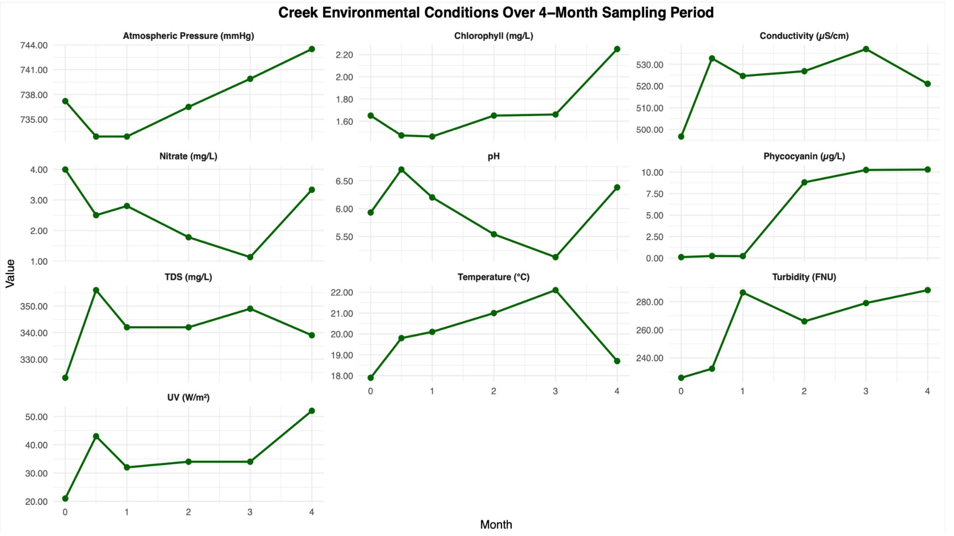
2.1. Environmental Measurements
2.1.1. Water Quality Parameters
2.1.2. Water Sampling and Filtration
2.2. Plastic Enrichment Trap Installation
2.2.1. Location of Film Enrichment
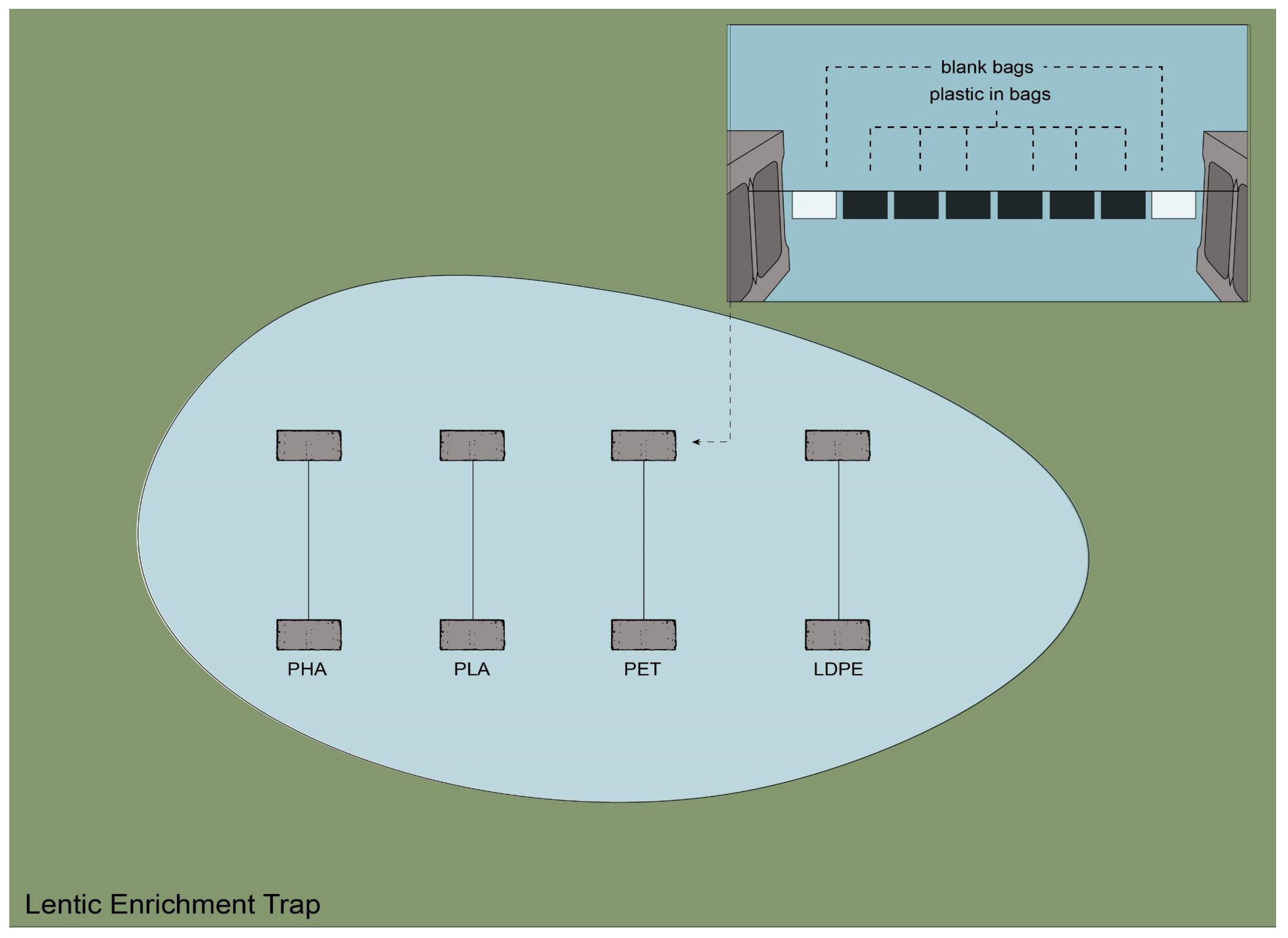
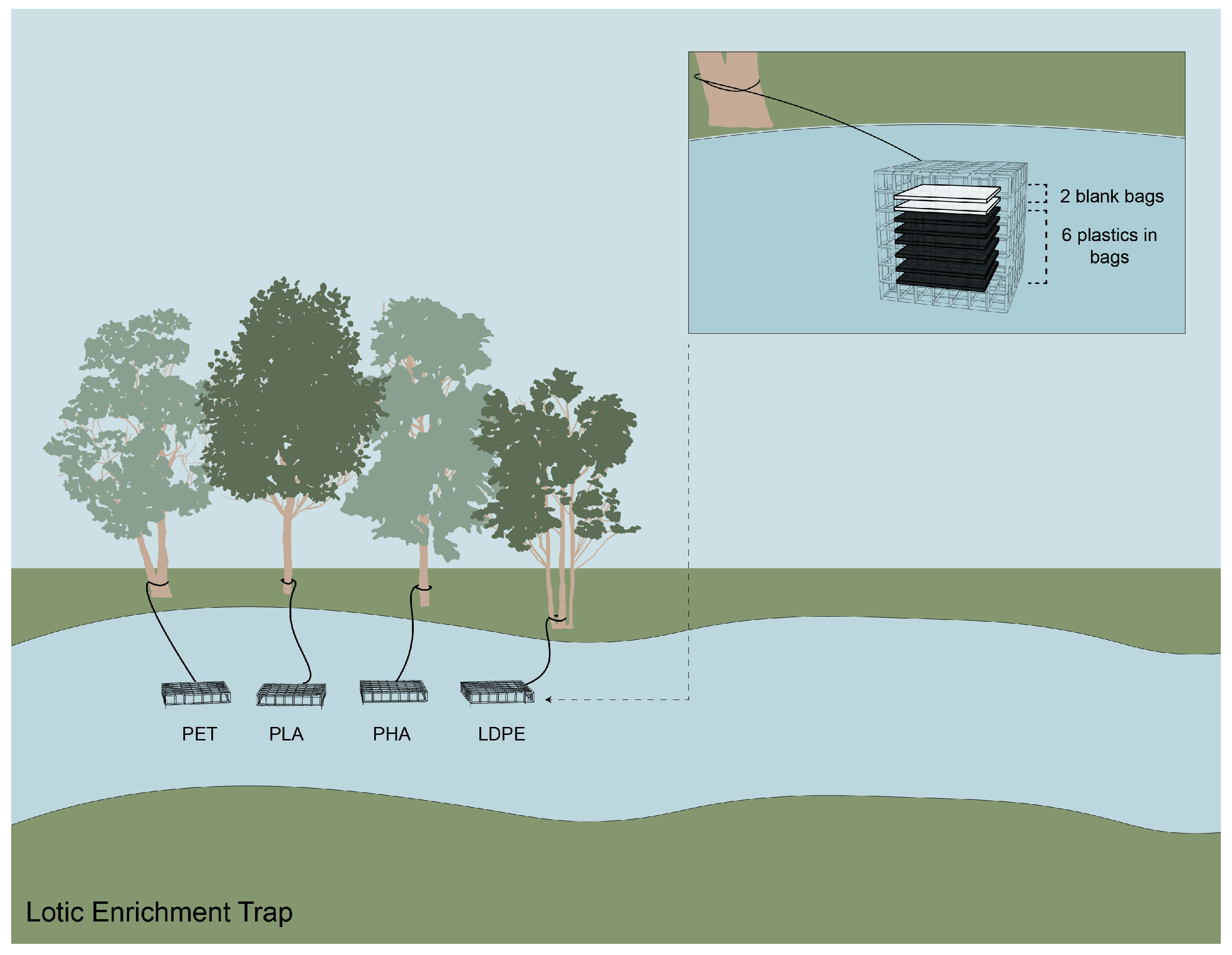
2.2.2. Plastic Film Enrichment Trap
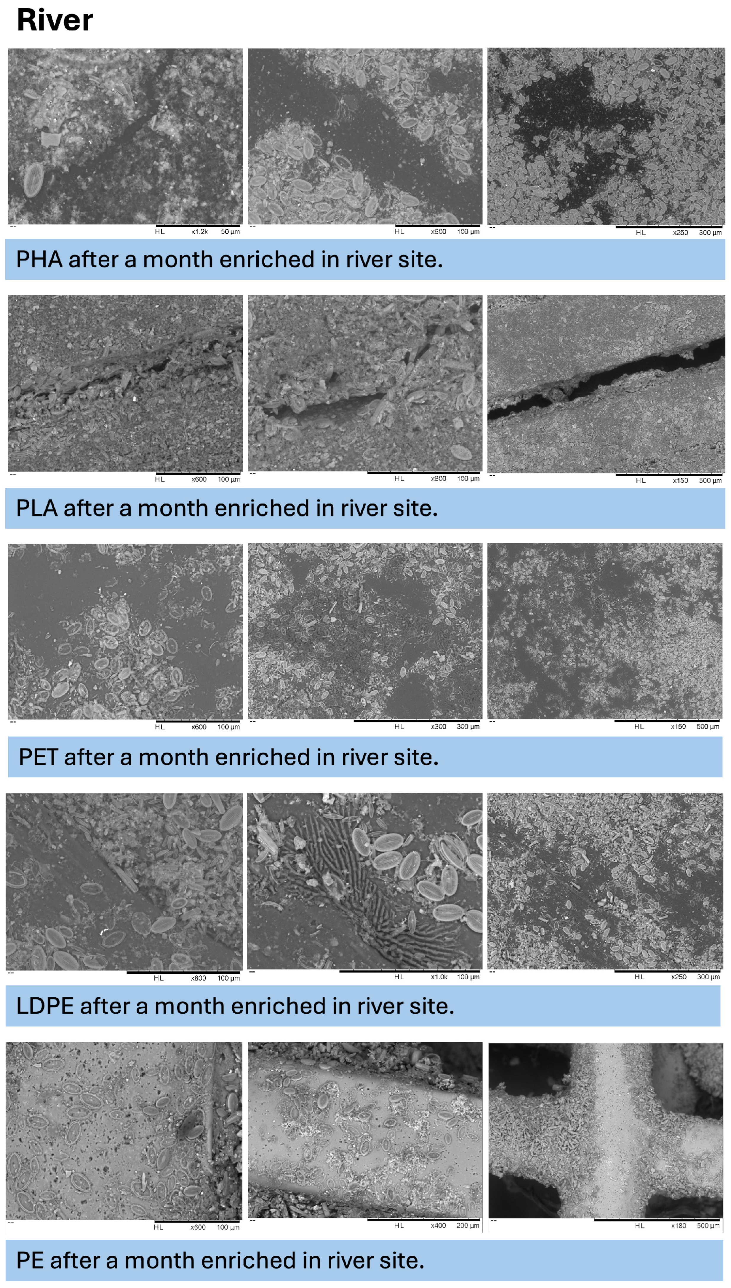
2.3. Scanning Electron Microscopy
2.4. Liquid Degradation Assay
2.5. Microbial Community
2.5.1. DNA Extraction
2.5.2. 16S rRNA Gene Amplification and Library Preparation
2.5.3. Instrument Loading
2.6. Statistical Analyses
2.6.1. Environmental Conditions
2.6.2. Plastic Weight Loss
2.6.3. Microbial Community Composition
3. Results
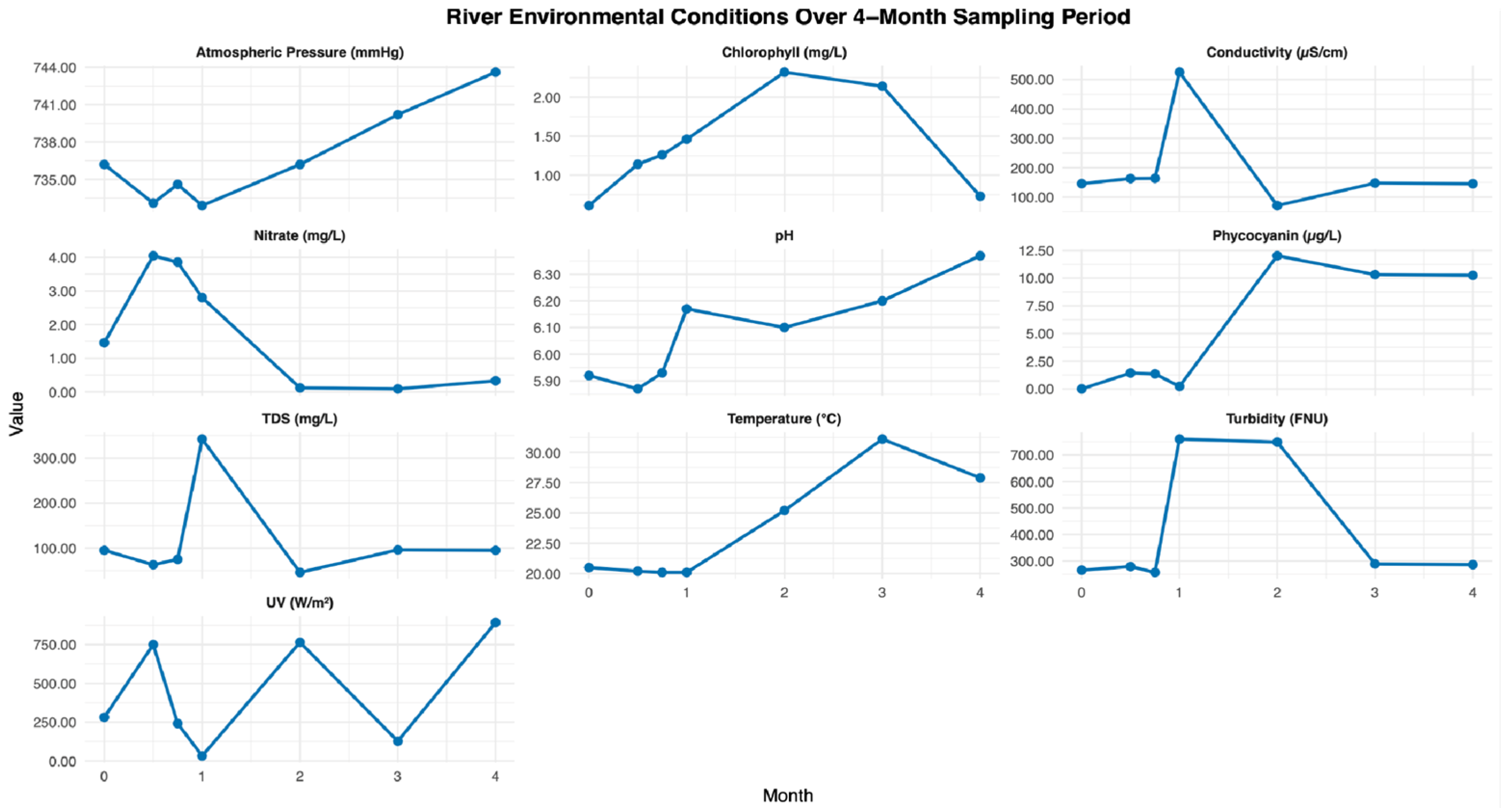
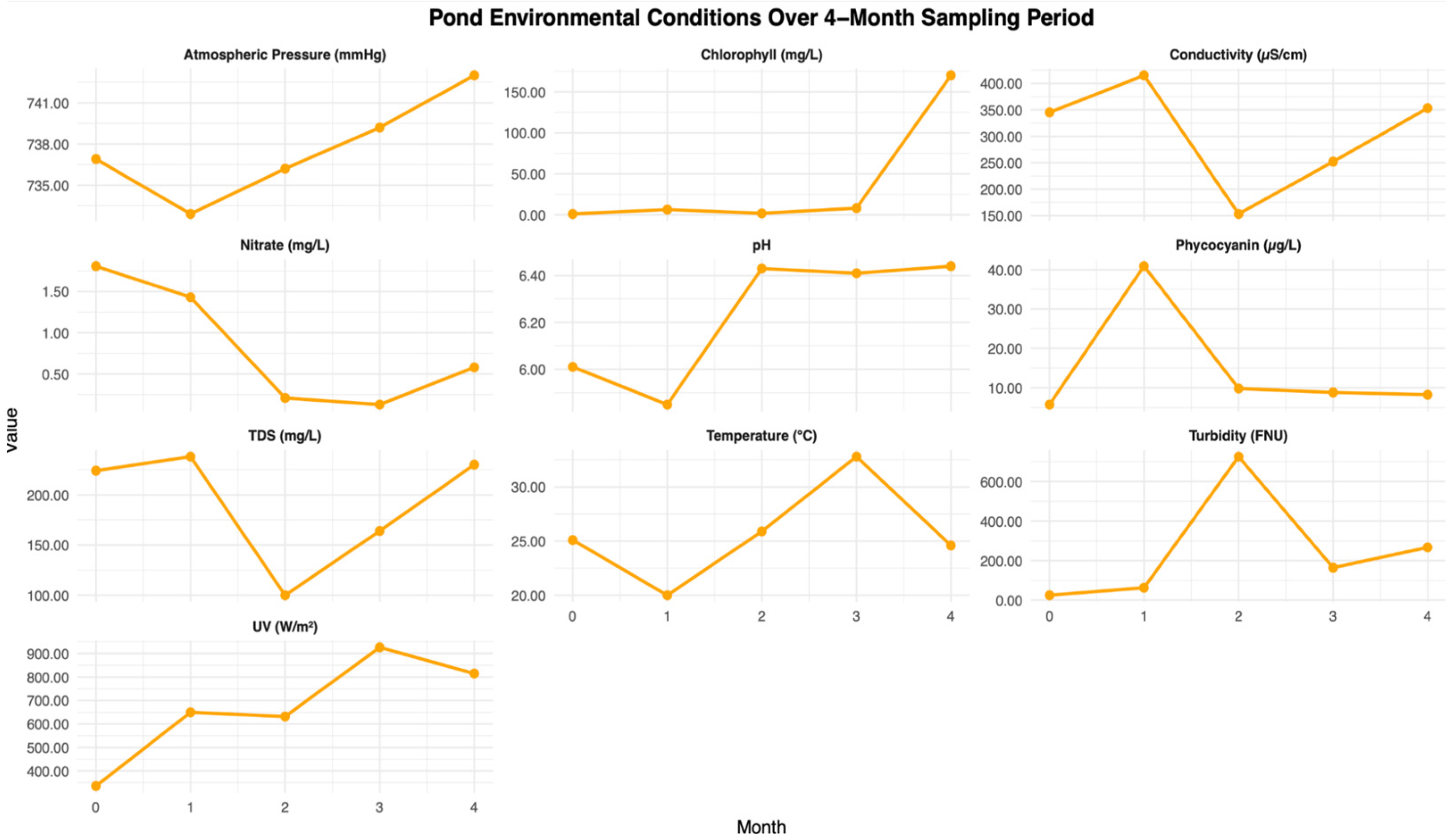
3.1. Environmental Test Analysis
3.2. Plastic Weight Loss Analyses
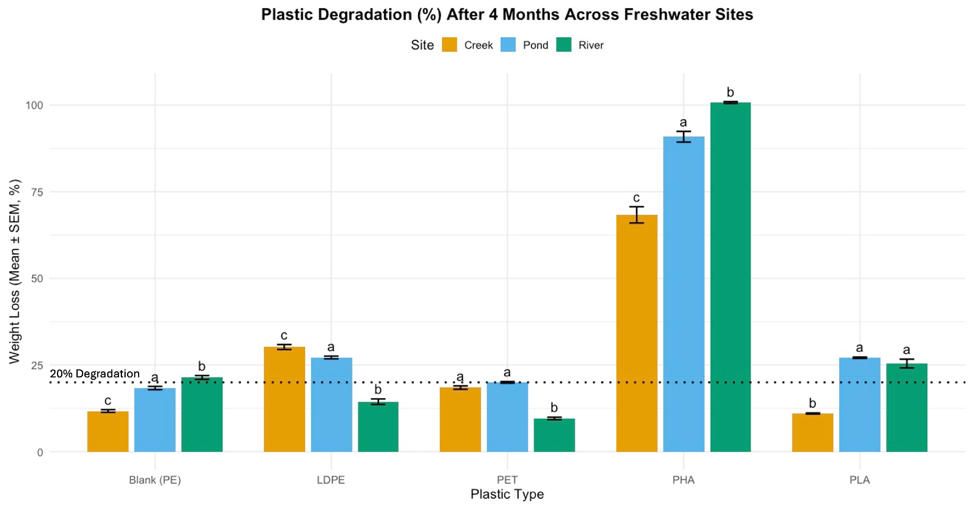
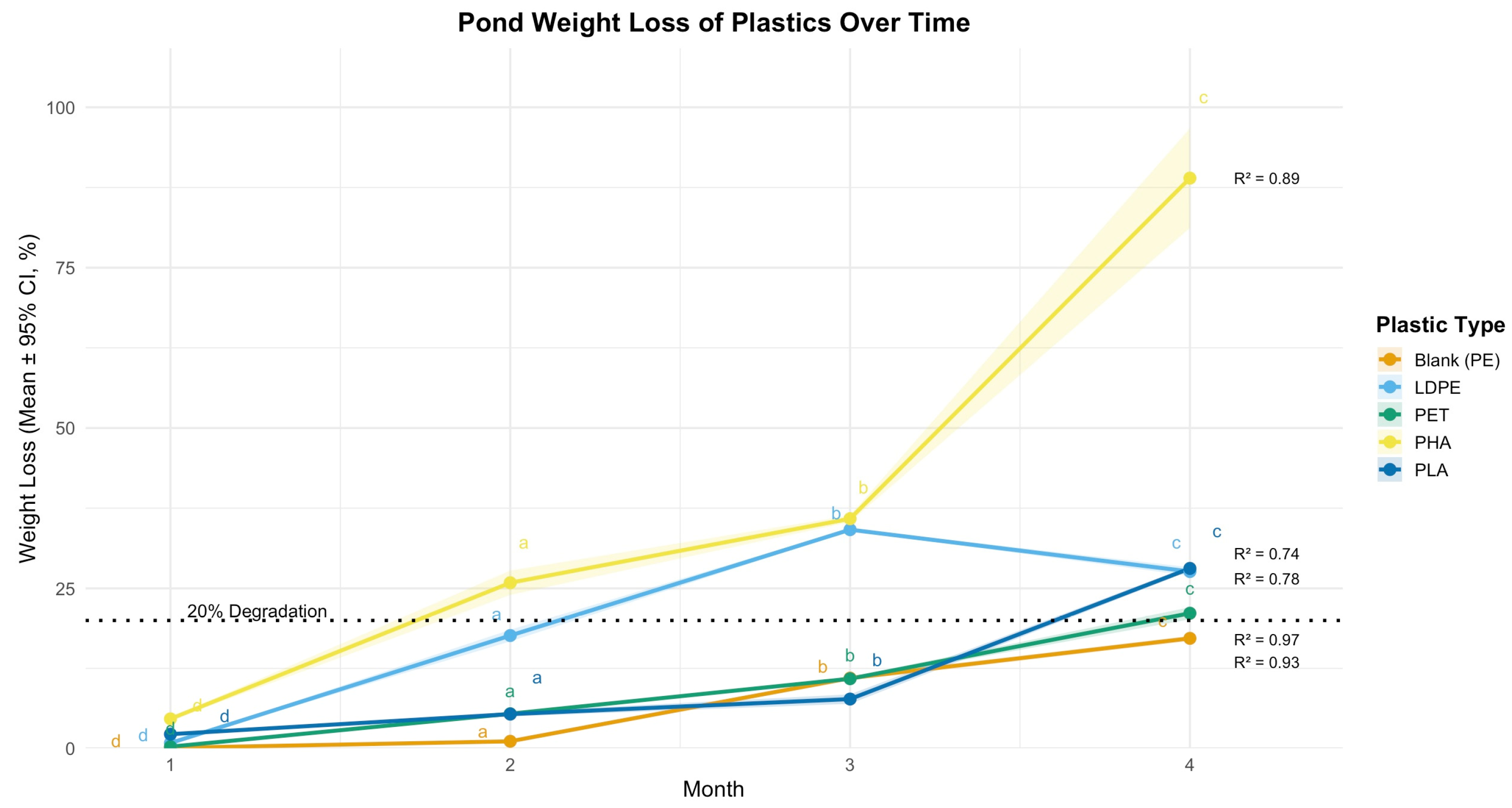
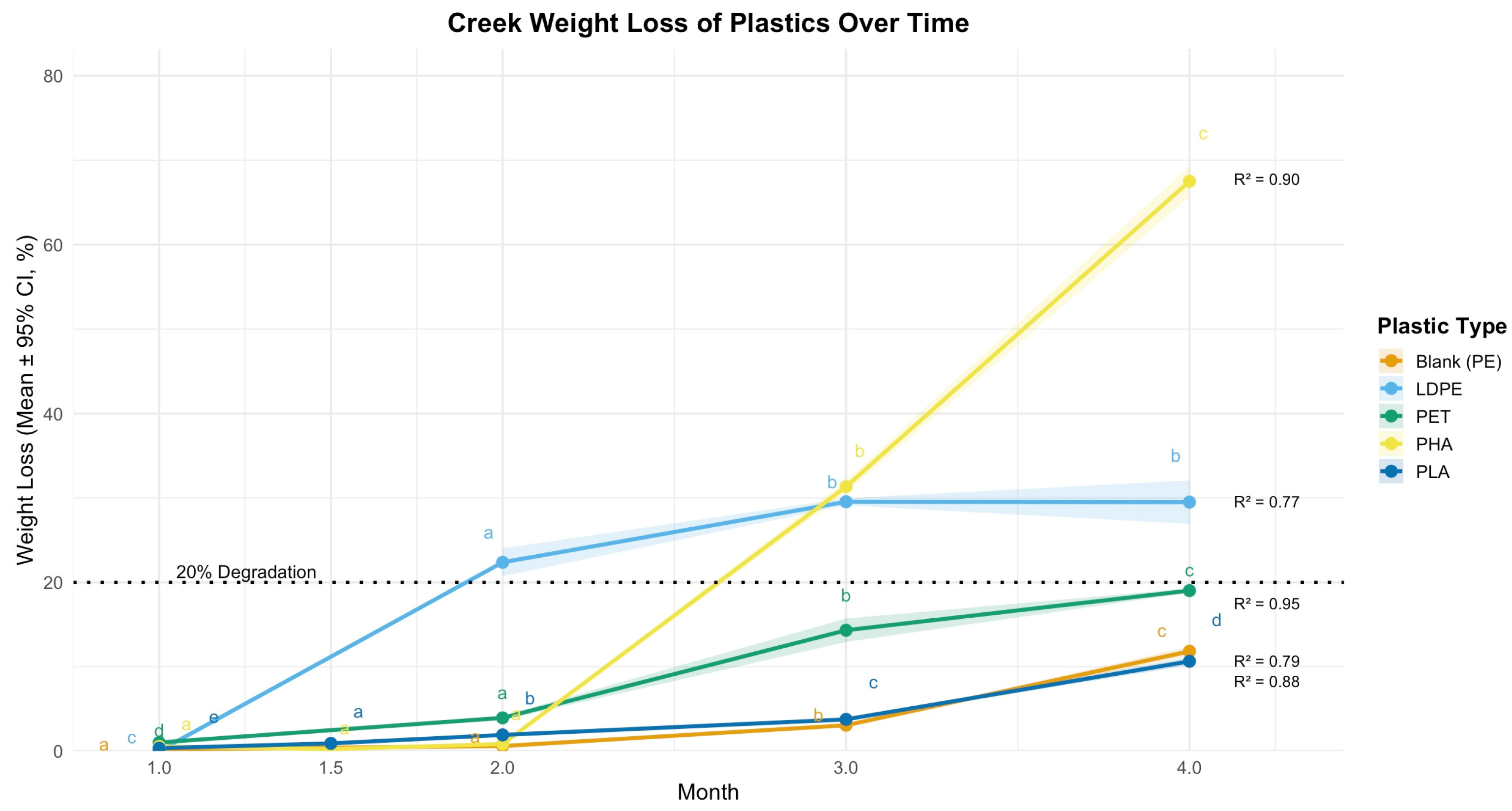
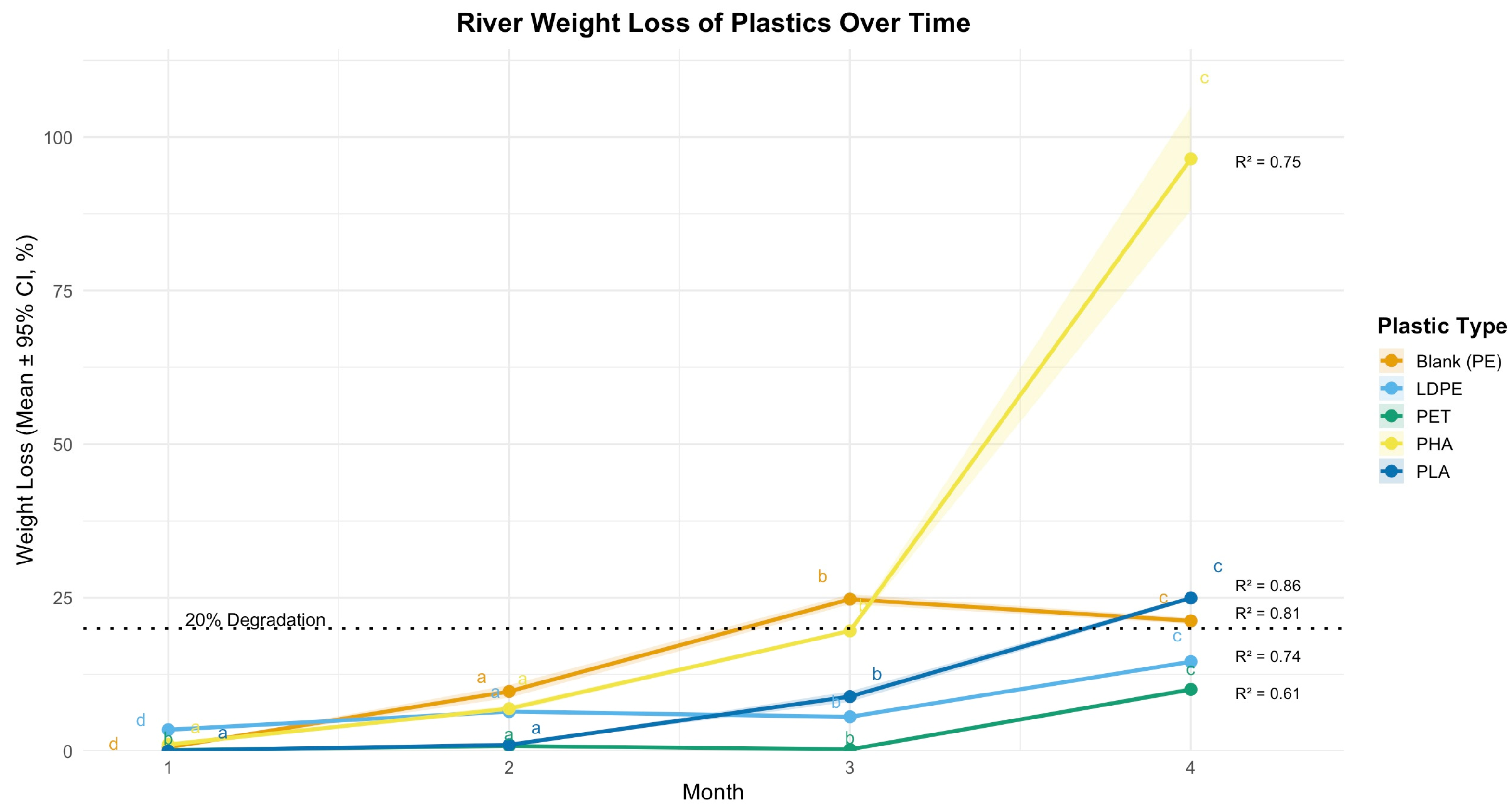
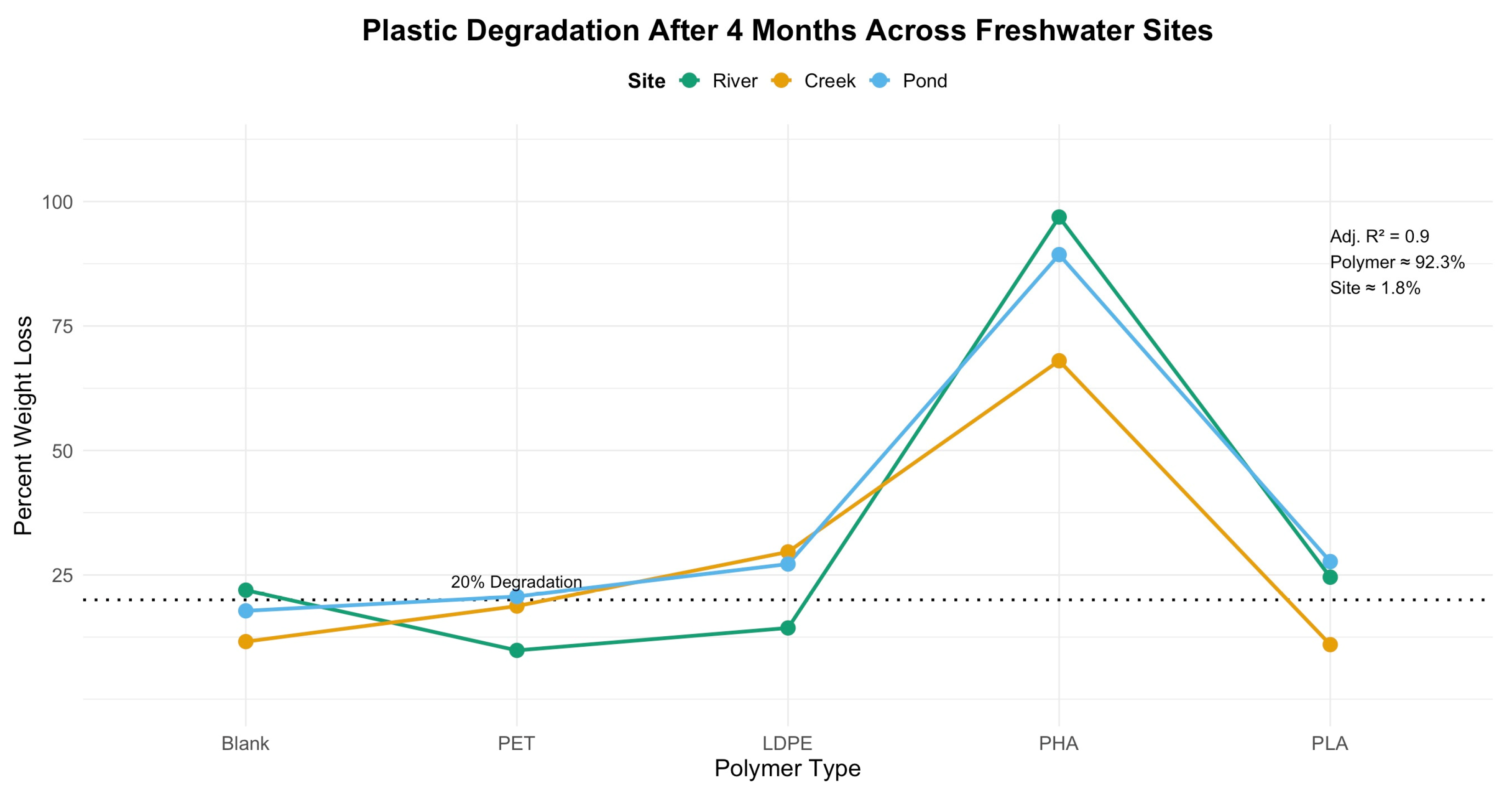
3.2.1. Measured Plastic Weight Loss
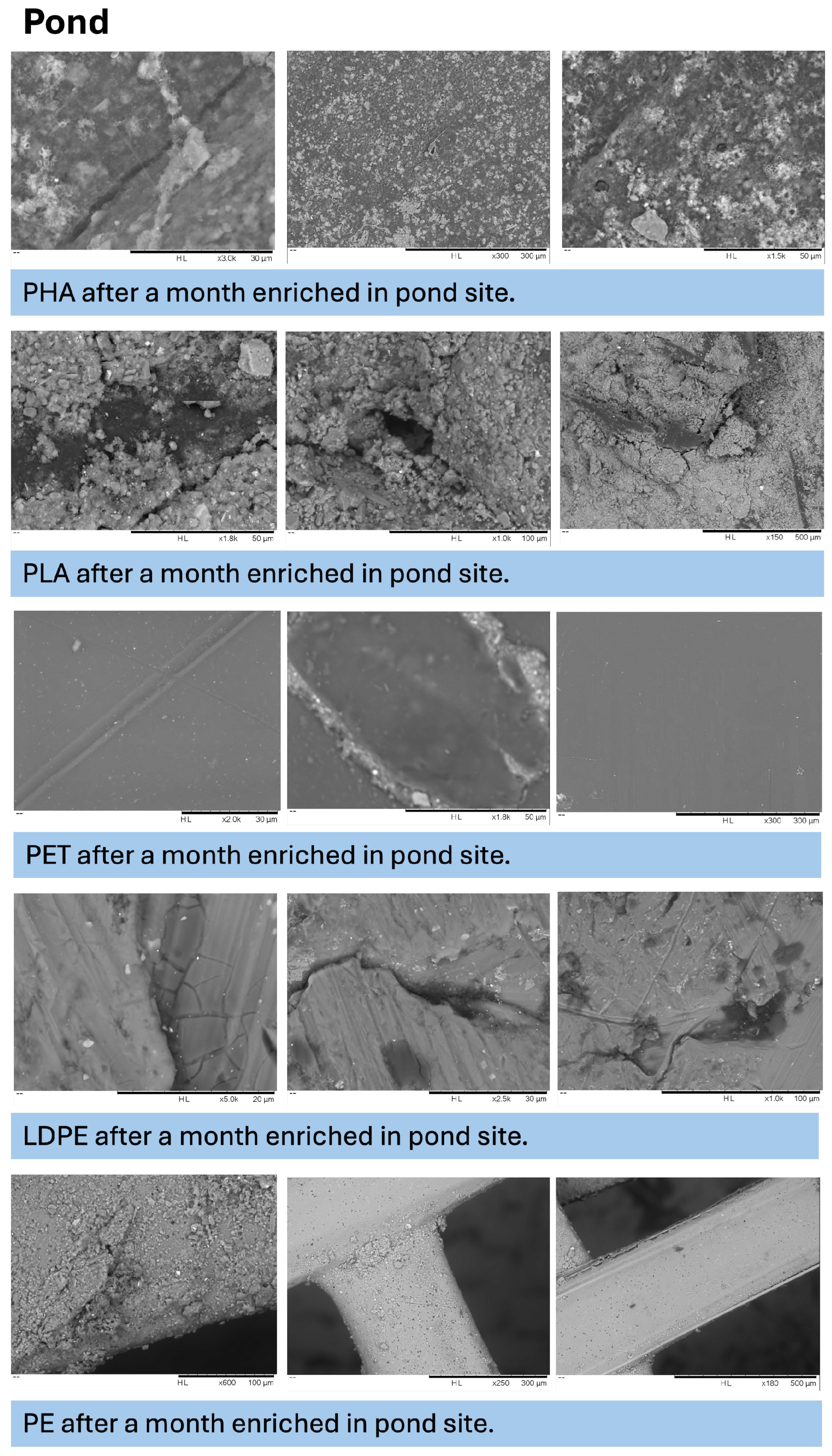
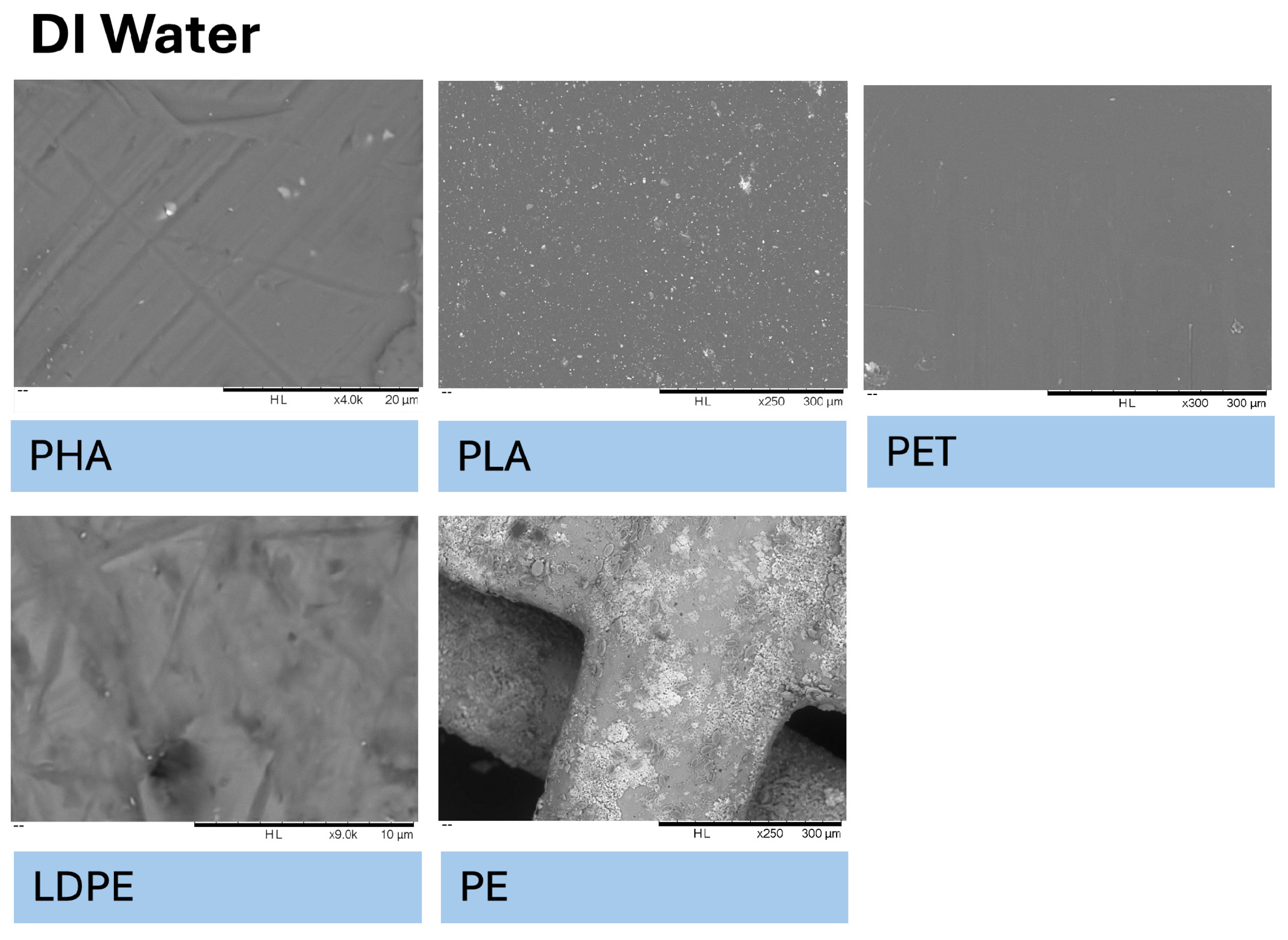
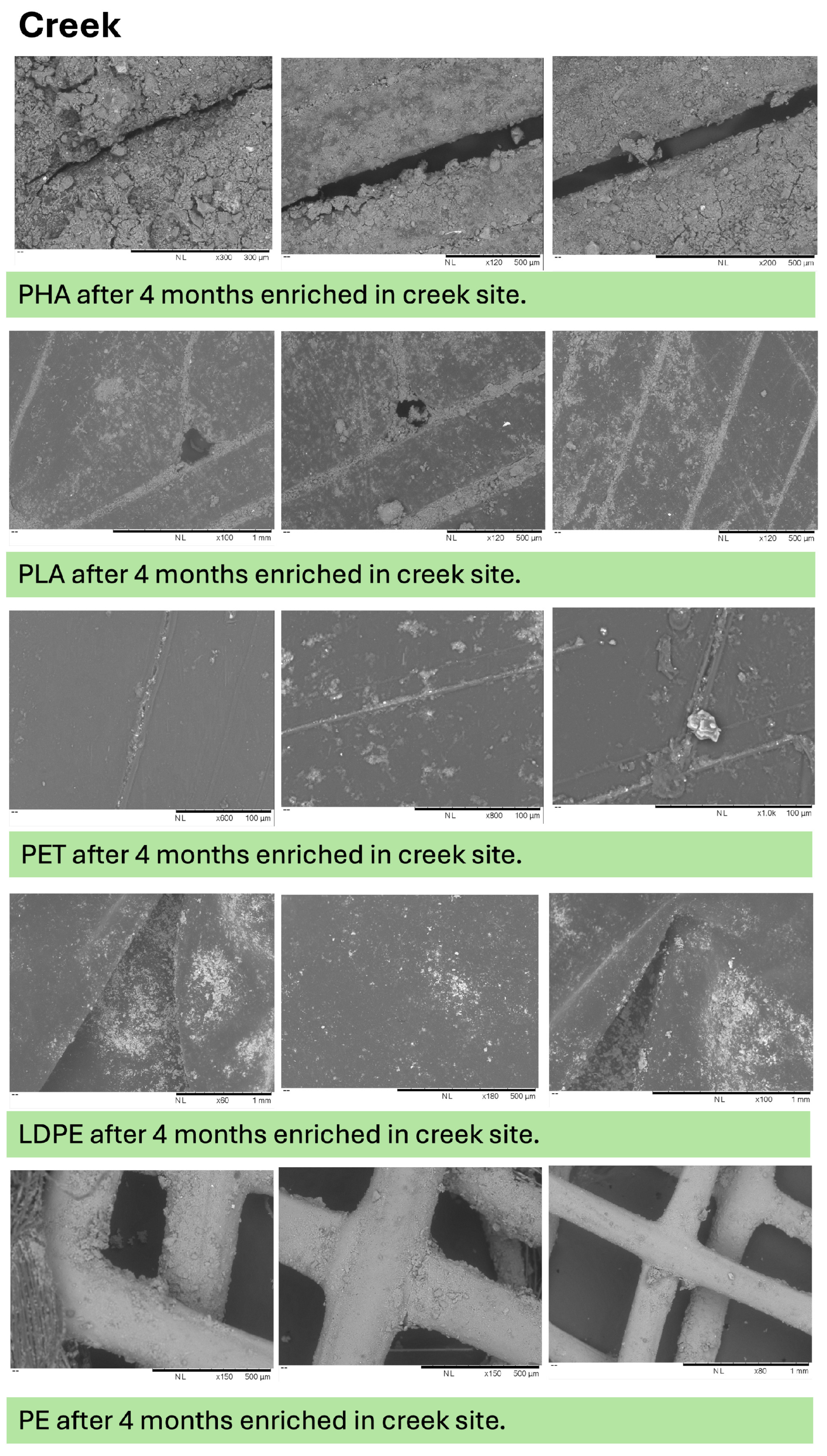
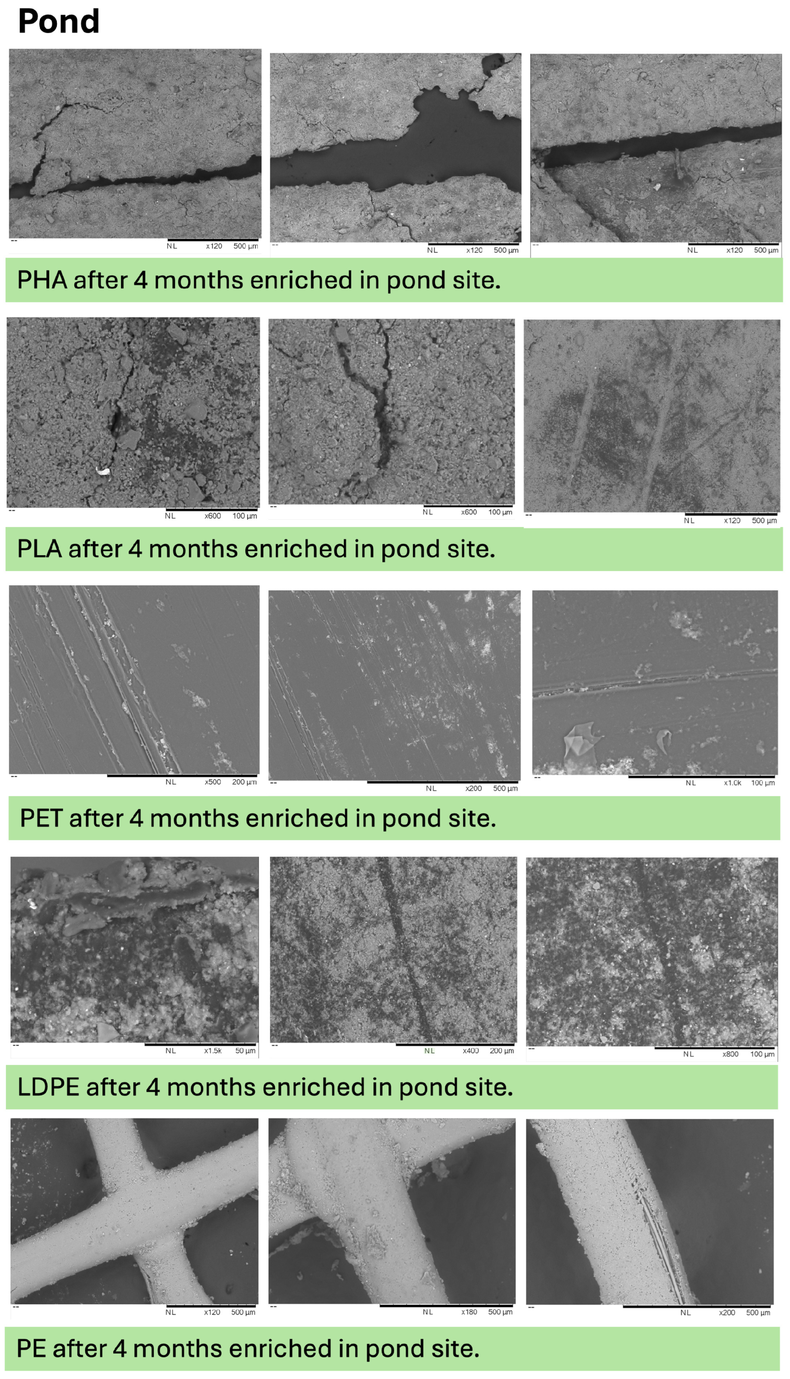
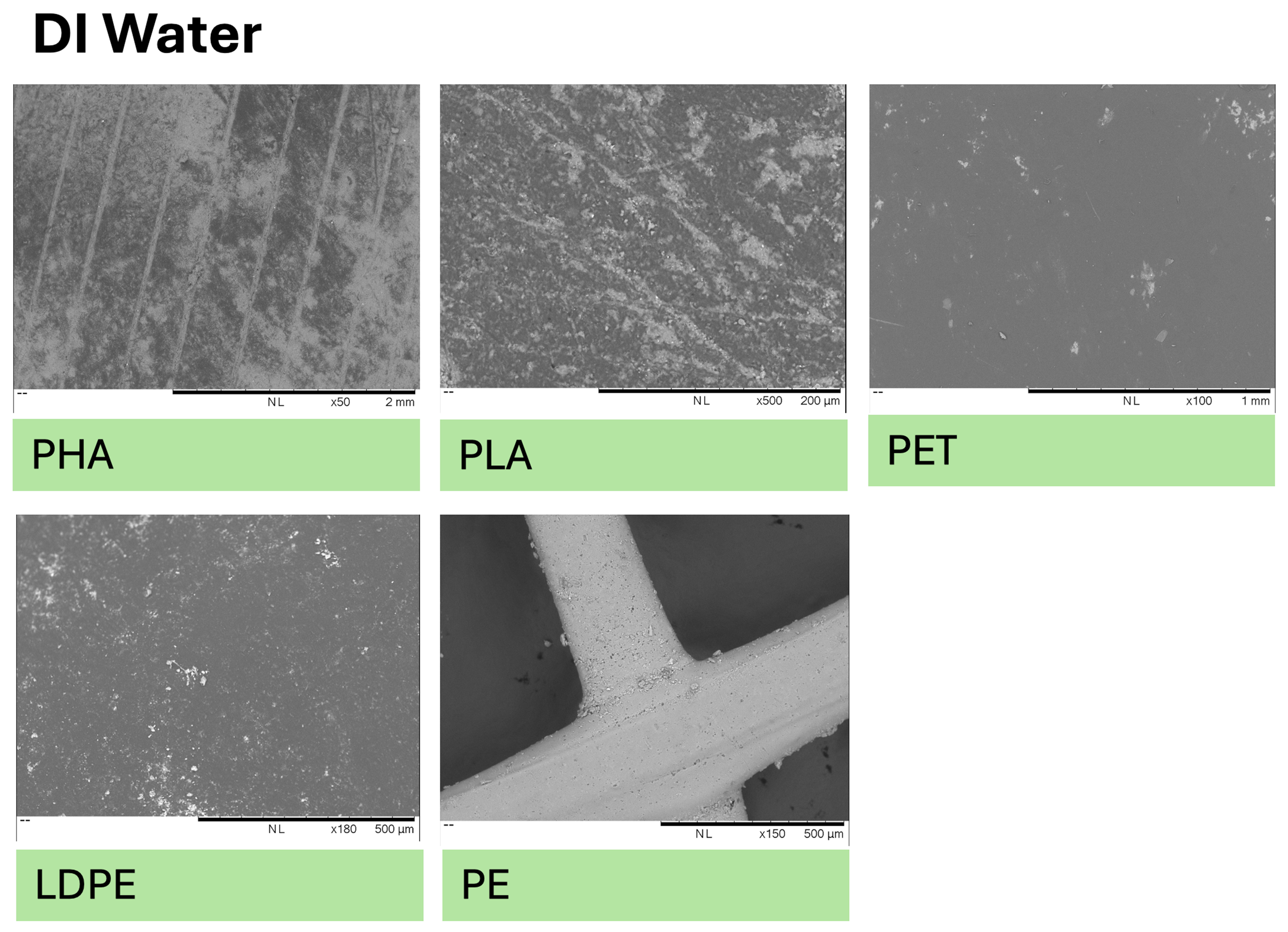
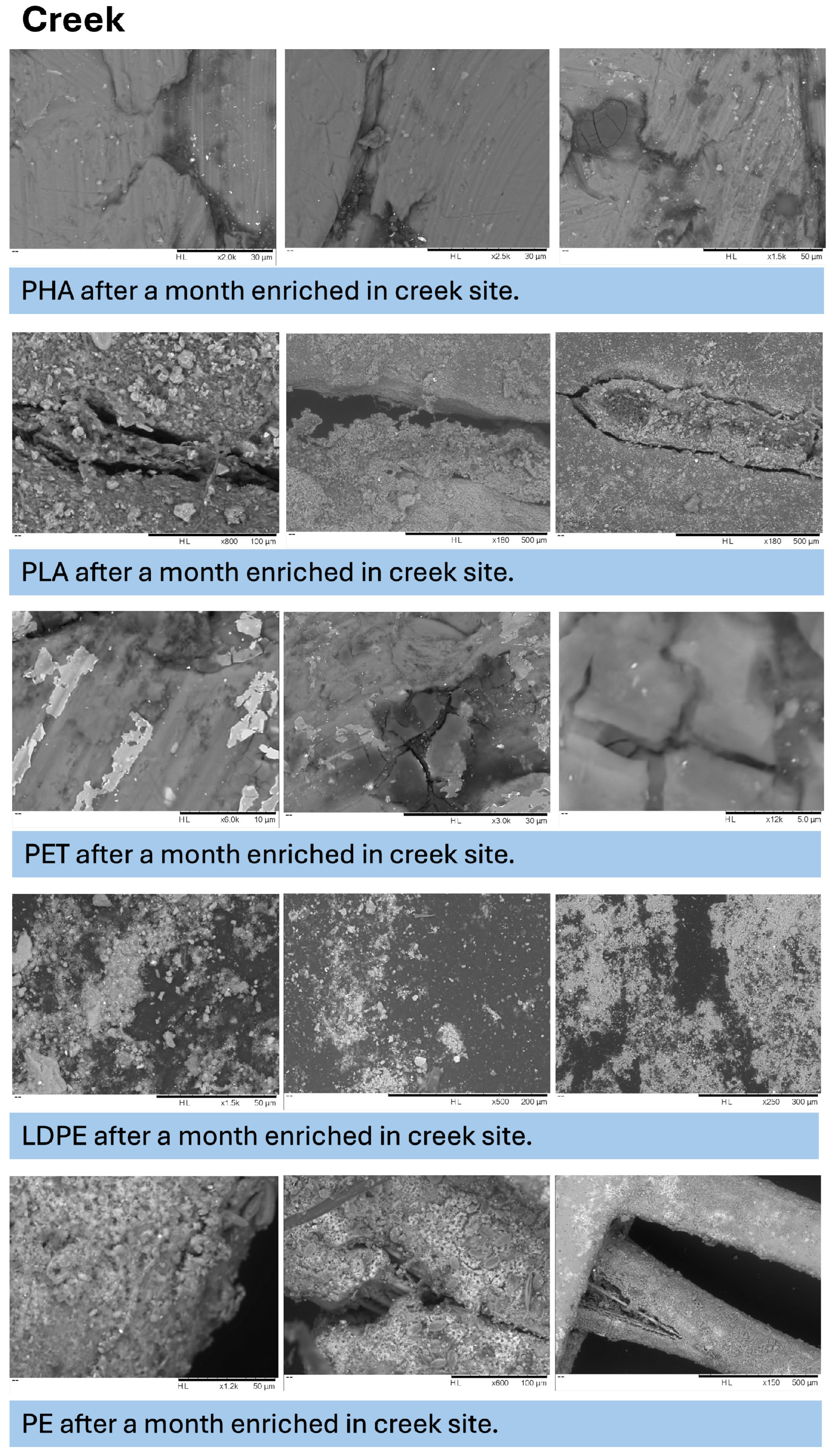
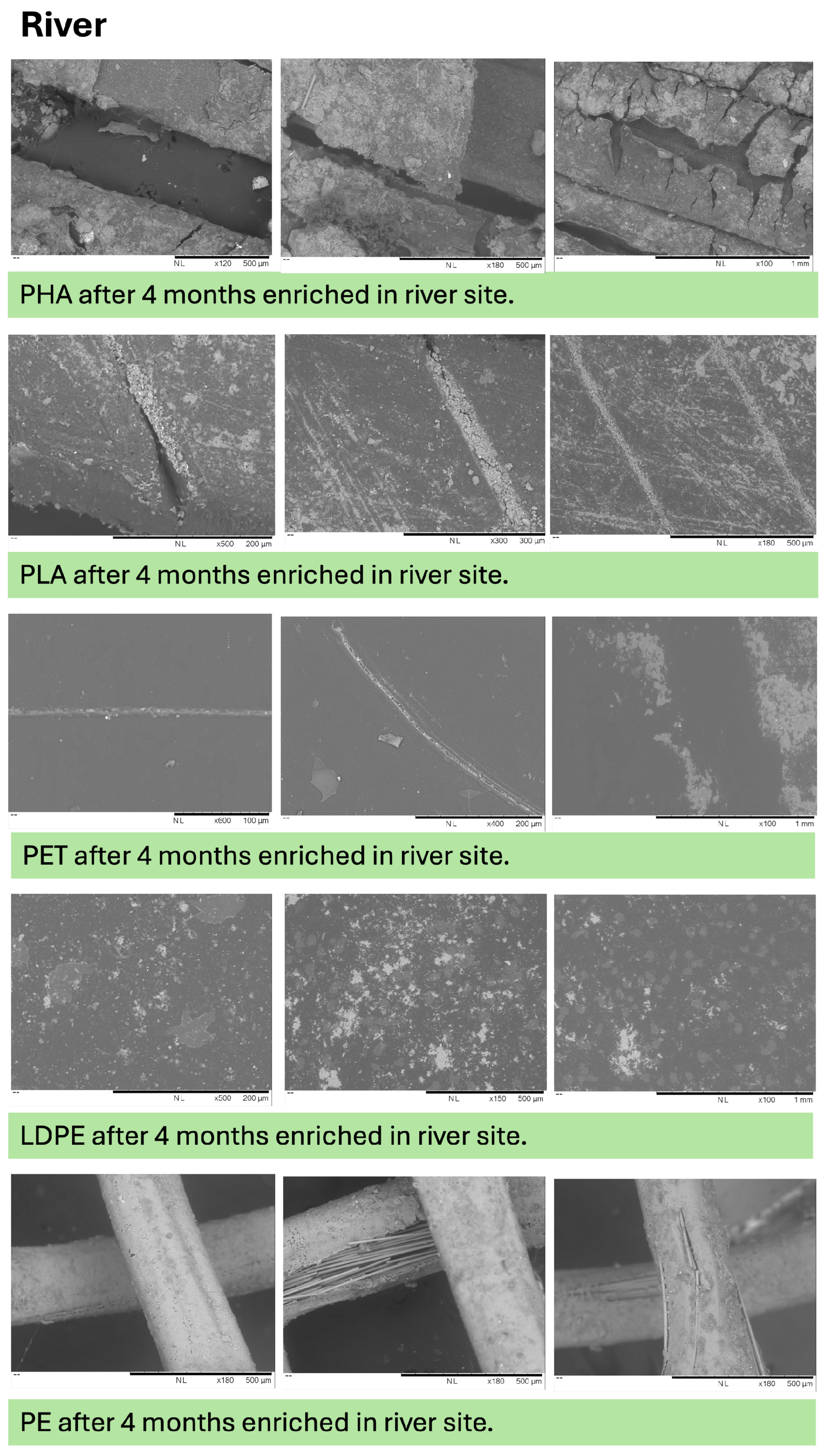
3.2.2. Visually Observed Plastic Changes with Time
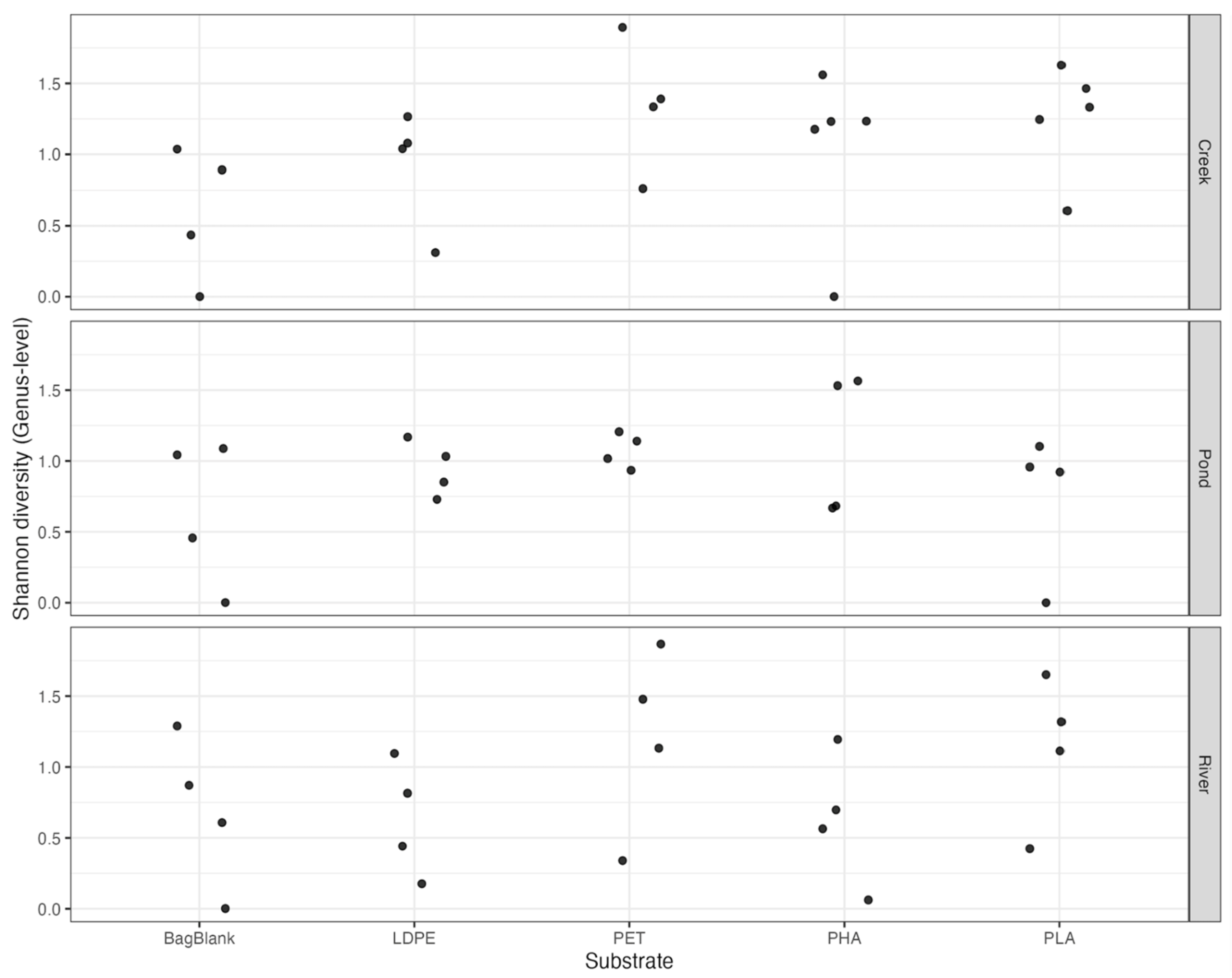
3.3. Alpha Diversity of Plastics Associated with Microbial Communities
3.4. Beta Diversity and Community Composition
3.4.1. Water Versus Enriched Plastic Communities
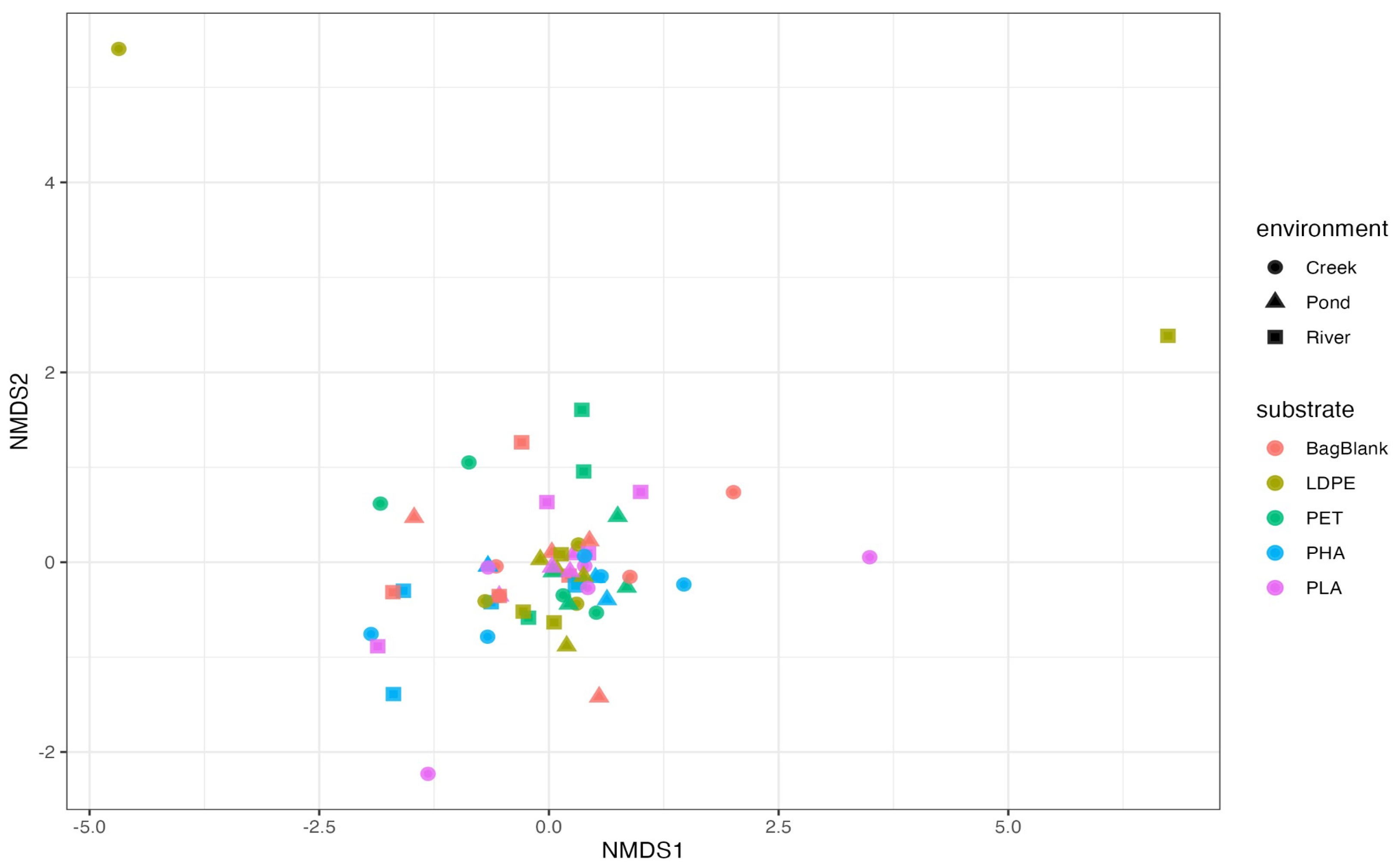
3.4.2. Incubated Plastics: Environment, Substrate, and Time Effects
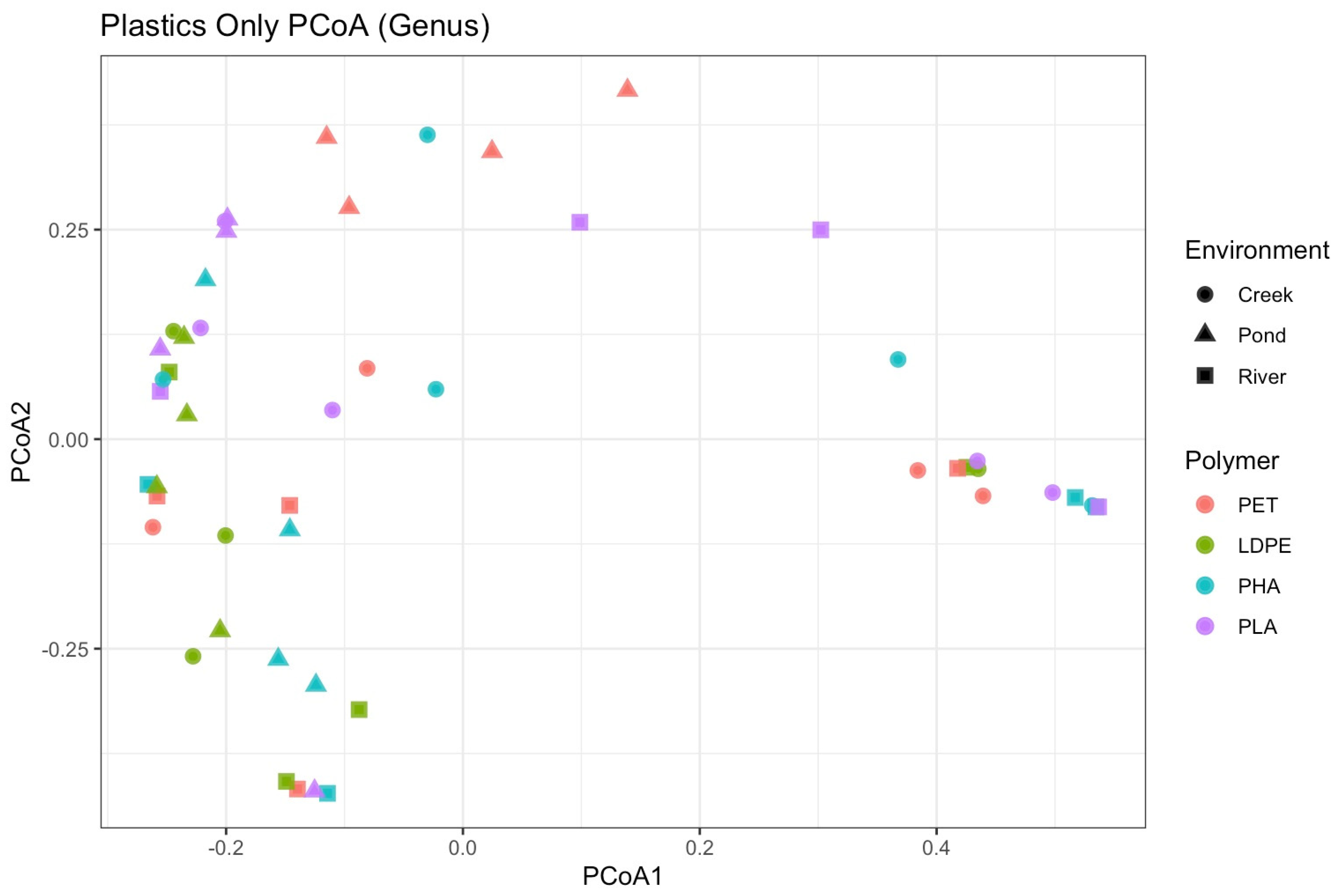
3.4.3. Plastic Community Patterns
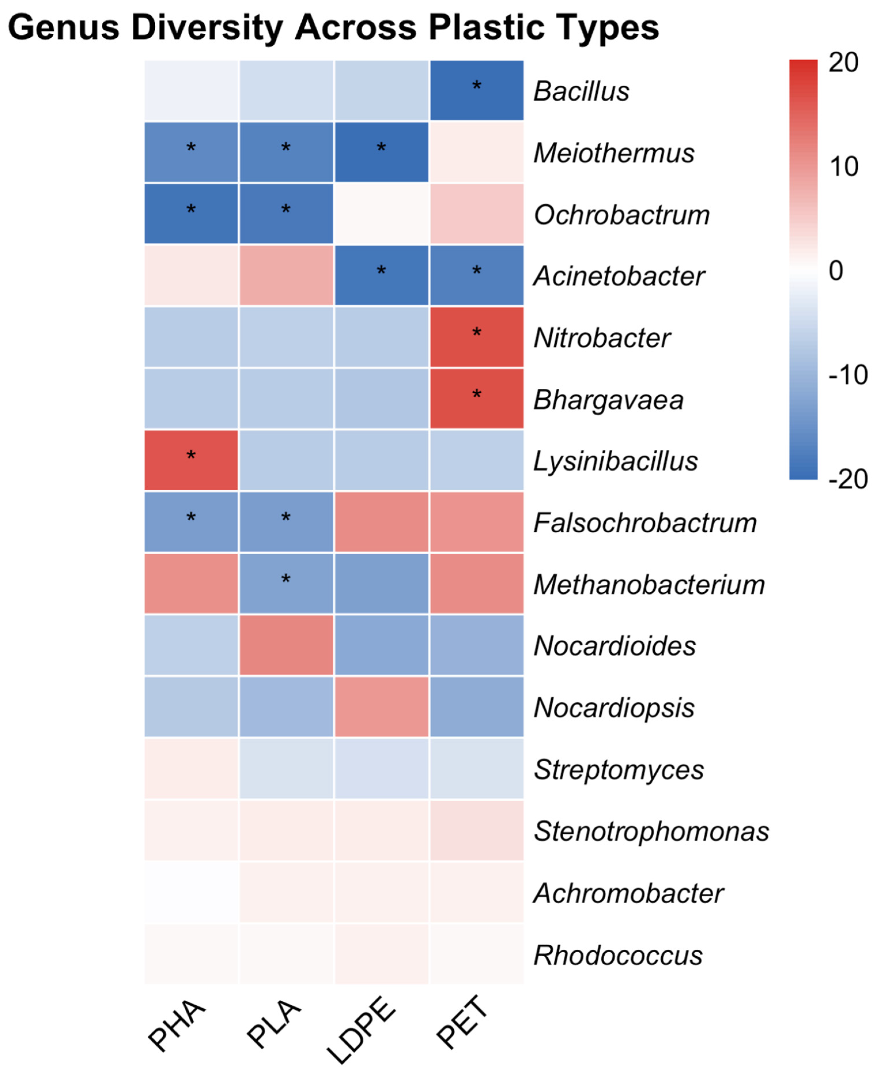
3.5. Differential Abundance Analyses
3.5.1. Differential Abundance of Plastisphere Communities
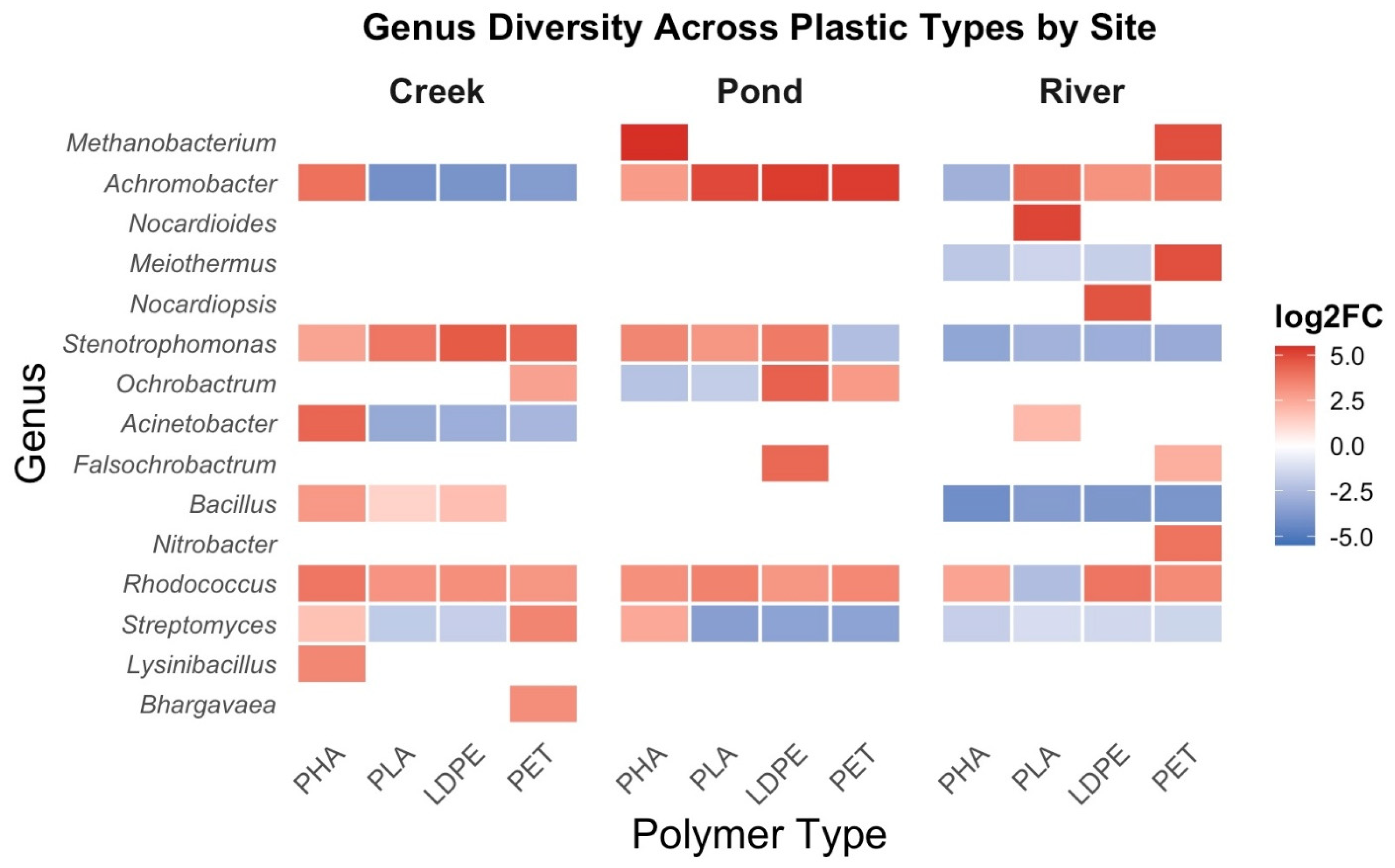
3.5.2. Polymer Specific Comparisons to Bag Blanks
3.5.3. Environmental Comparisons
3.6. Relationship Between Microbial Communities and Plastic Mass Loss
4. Discussion
4.1. Environmental Influences
4.2. Plastic Degradation Behavior
4.3. Biofilm Formation Versus Mass Loss
4.4. Plastisphere Community Structure and Selectivity
4.5. Relationship Between Microbial Communities and Degradation
4.5.1. Environmental Microbial Communities
4.5.2. Plastisphere Microbial Communities
5. Conclusions
5.1. Limitations
5.2. Future Directions
Author Contributions
Funding
Institutional Review Board Statement
Informed Consent Statement
Data Availability Statement
Acknowledgments
Conflicts of Interest
Abbreviations
| ANOVA | Attenuated total reflectance Fourier-transform infrared spectroscopy |
| ATR-FTIR | Analysis of variance |
| BCL | Binary Base Call |
| DESeq2 | Differential expression analysis for sequence count data 2 |
| DI | Deionized |
| DOC | Dissolved organic carbon |
| DOM | Dissolved organic matter |
| FNU | Formazin Nephelometric Unit |
| GPC | Gel permeation chromatography |
| HDPE | High-density polyethylene |
| HPSC | High Performance and Scientific Computing |
| ISAAC-NG | Infrastructure for Scientific Applications and Advanced Computing-Next Generation |
| LDPE | Low-density polyethylene |
| NMDS | Non-metric multidimensional scaling |
| NOB | Nitrite-oxidizing bacteria |
| OD600 | Optical density at 600 nm |
| PBAT | Polybutylene adipate terephthalate |
| PC | Phycocyanin |
| PCoA | Principal coordinates analysis |
| PE | Polyethylene |
| PERMANOVA | Permutational multivariate analysis of variance |
| PET | Polyethylene terephthalate |
| PHA | Polyhydroxyalkanoate |
| PHBV | Poly(3-hydroxybutyrate-co-3-hydroxyvalerate) |
| PLA | Polylactic acid/Polylactide |
| PP | Polypropylene |
| PS | Polystyrene |
| PVC | Polyvinyl chloride |
| SEM | Scanning electron microscopy |
| TDS | Total dissolved solids |
| TSS | Total suspended solids |
| UV | Ultraviolet |
Appendix A







| Site | Sample Month | Plastic Type | Dilution | OD600 |
|---|---|---|---|---|
| Third Creek | 1 | PET | ×10 | 0.005 |
| Third Creek | 1 | LDPE | ×10 | 0.103 |
| Third Creek | 1 | PHA | ×10 | 0.017 |
| Third Creek | 1 | PLA | ×10 | 0.121 |
| Third Creek | 1 | Blank PE | ×10 | 0.409 |
| Third Creek | 1.5 | PHA | ×10 | 0.142 |
| Third Creek | 1.5 | PLA | ×10 | 0.233 |
| Third Creek | 2 | PET | ×10 | 0.276 |
| Third Creek | 2 | LDPE | ×100 | 0.254 |
| Third Creek | 2 | PHA | ×10 | 0.372 |
| Third Creek | 2 | PLA | ×10 | 0.255 |
| Third Creek | 2 | Blank PE | ×100 | 0.250 |
| Third Creek | 3 | PET | ×100 | 0.343 |
| Third Creek | 3 | LDPE | ×100 | 0.438 |
| Third Creek | 3 | PHA | ×10 | 0.121 |
| Third Creek | 3 | PLA | ×10 | 0.470 |
| Third Creek | 3 | Blank PE | ×10 | 0.228 |
| Third Creek | 4 | PET | ×100 | 0.386 |
| Third Creek | 4 | LDPE | ×100 | 0.479 |
| Third Creek | 4 | PHA | ×100 | 0.456 |
| Third Creek | 4 | PLA | ×10 | 0.223 |
| Third Creek | 4 | Blank PE | ×100 | 0.386 |
| Seven Islands | 1 | PET | ×10 | 0.303 |
| Seven Islands | 1 | LDPE | ×10 | 0.133 |
| Seven Islands | 1 | PHA | ×10 | 0.113 |
| Seven Islands | 1 | PLA | ×10 | 0.130 |
| Seven Islands | 1 | Blank PE | ×10 | 0.089 |
| Seven Islands | 2 | PET | ×10 | 0.120 |
| Seven Islands | 2 | LDPE | ×10 | 0.060 |
| Seven Islands | 2 | PHA | ×100 | 0.091 |
| Seven Islands | 2 | PLA | ×10 | 0.091 |
| Seven Islands | 2 | Blank PE | ×100 | 0.104 |
| Seven Islands | 3 | PET | ×100 | 0.457 |
| Seven Islands | 3 | LDPE | ×10 | 0.403 |
| Seven Islands | 3 | PHA | ×10 | 0.315 |
| Seven Islands | 3 | PLA | ×10 | 0.061 |
| Seven Islands | 3 | Blank PE | ×10 | 0.340 |
| Seven Islands | 4 | PET | ×10 | 0.251 |
| Seven Islands | 4 | LDPE | ×10 | 0.109 |
| Seven Islands | 4 | PHA | ×10 | 0.288 |
| Seven Islands | 4 | PLA | ×100 | 0.425 |
| Seven Islands | 4 | Blank PE | ×10 | 0.365 |
| Compost | 1 | PET | ×10 | 0.005 |
| Compost | 1 | LDPE | ×10 | 0.432 |
| Compost | 1 | PHA | ×100 | 0.230 |
| Compost | 1 | PLA | ×10 | 0.057 |
| Compost | 1 | Blank PE | ×10 | 0.244 |
| Compost | 2 | PET | ×10 | 0.351 |
| Compost | 2 | LDPE | ×10 | 0.233 |
| Compost | 2 | PHA | ×10 | 0.013 |
| Compost | 2 | PLA | ×10 | 0.141 |
| Compost | 2 | Blank PE | ×10 | 0.167 |
| Compost | 3 | PET | ×10 | 0.402 |
| Compost | 3 | LDPE | ×10 | 0.500 |
| Compost | 3 | PHA | ×10 | 0.379 |
| Compost | 3 | PLA | ×100 | 0.412 |
| Compost | 3 | Blank PE | ×100 | 0.370 |
| Compost | 4 | PET | ×100 | 0.288 |
| Compost | 4 | LDPE | ×10 | 0.465 |
| Compost | 4 | PHA | ×100 | 0.133 |
| Compost | 4 | PLA | ×100 | 0.405 |
| Compost | 4 | Blank PE | ×100 | 0.317 |
| Genus | Base Mean | Log2 Fold Change | SE | Wald Statistic | p-Value | Adjusted p-Value (padj) |
|---|---|---|---|---|---|---|
| Rhodococcus | 34.614 | 7.778 | 1.019 | 7.634 | 2.28 × 10⁻¹⁴ | 1.12 × 10⁻¹² |
| Meiothermus | 3.077 | −14.372 | 2.102 | −6.836 | 8.12 × 10⁻¹² | 1.99 × 10⁻¹⁰ |
| Magnetospirillum | 0.185 | −21.439 | 3.915 | −5.476 | 4.35 × 10⁻⁸ | 7.10 × 10⁻⁷ |
| Candidatus omnitrophus | 0.146 | −20.928 | 3.915 | −5.346 | 9.00 × 10⁻⁸ | 1.10 × 10⁻⁶ |
| Limnohabitans | 11.318 | −6.863 | 1.531 | −4.482 | 7.40 × 10⁻⁶ | 7.25 × 10⁻⁵ |
| Achromobacter | 9.968 | 5.463 | 1.280 | 4.269 | 1.97 × 10⁻⁵ | 1.61 × 10⁻⁴ |
| Stenotrophomonas | 4.462 | 3.479 | 1.550 | 2.244 | 0.0248 | 0.174 |
| Paramesorhizobium | 1.263 | 3.308 | 1.665 | 1.987 | 0.0469 | 0.287 |
| Methanosaeta | 1.224 | −4.684 | 2.853 | −1.642 | 0.101 | 0.494 |
| Candidatus lainarchaeum | 1.086 | −4.382 | 2.671 | −1.641 | 0.101 | 0.494 |
| Brucella | 1.588 | 3.103 | 1.996 | 1.555 | 0.120 | 0.534 |
| Methanoregula | 0.759 | −4.195 | 2.999 | −1.399 | 0.162 | 0.661 |
| Streptomyces | 1.567 | 2.763 | 2.428 | 1.138 | 0.255 | 0.872 |
| Bacillus | 1.887 | 2.428 | 2.645 | 0.918 | 0.359 | 0.872 |
| Truepera | 0.356 | 1.001 | 3.915 | 0.256 | 0.798 | 0.872 |
| Candidatus nitrosotalea | 0.177 | −1.814 | 3.904 | −0.465 | 0.642 | 0.872 |
| Nitrosarchaeum | 0.319 | −2.440 | 3.896 | −0.626 | 0.531 | 0.872 |
| Nocardioides | 0.247 | 0.968 | 3.916 | 0.247 | 0.805 | 0.872 |
| Aquabacterium | 0.094 | −1.246 | 3.909 | −0.319 | 0.750 | 0.872 |
| Candidatus methanoperedens | 0.515 | −3.325 | 3.445 | −0.965 | 0.335 | 0.872 |
| AR20 | 0.286 | −2.206 | 3.899 | −0.566 | 0.572 | 0.872 |
| Acinetobacter | 0.714 | 1.198 | 3.913 | 0.306 | 0.760 | 0.872 |
| Methanosarcina | 0.199 | −2.105 | 3.898 | −0.540 | 0.589 | 0.872 |
| CL500-29 marine group | 0.818 | −3.143 | 3.022 | −1.040 | 0.298 | 0.872 |
| Pseudorhabdium | 0.411 | 0.871 | 3.913 | 0.223 | 0.824 | 0.872 |
| Aquamicrobium | 0.201 | 0.982 | 3.916 | 0.251 | 0.802 | 0.872 |
| Paenibacillus | 0.460 | 1.249 | 3.914 | 0.319 | 0.750 | 0.872 |
| Methanosphaera | 0.108 | −1.351 | 3.908 | −0.346 | 0.730 | 0.872 |
| hgcl_clade | 0.587 | −3.343 | 2.893 | −1.155 | 0.248 | 0.872 |
| Cyanobium PCC-6307 | 0.744 | −3.537 | 3.819 | −0.926 | 0.354 | 0.872 |
| Ochrobactrum | 0.378 | 1.346 | 3.917 | 0.344 | 0.731 | 0.872 |
| Candidatus Planktophila | 0.237 | −2.234 | 3.898 | −0.573 | 0.567 | 0.872 |
| Methanospirillum | 0.173 | −1.582 | 3.907 | −0.405 | 0.685 | 0.872 |
| Hyphomicrobium | 0.317 | 0.728 | 3.913 | 0.186 | 0.852 | 0.872 |
| Pseudarcicella | 0.669 | −2.771 | 2.646 | −1.047 | 0.295 | 0.872 |
| Glutamicibacter | 0.098 | 0.781 | 3.917 | 0.199 | 0.842 | 0.872 |
| SN8 | 0.720 | 1.727 | 2.930 | 0.589 | 0.556 | 0.872 |
| Pseudomonas | 0.496 | 1.097 | 3.068 | 0.358 | 0.721 | 0.872 |
| Polynucleobacter | 0.355 | −1.915 | 3.902 | −0.491 | 0.623 | 0.872 |
| Bhargavaea | 0.102 | 0.740 | 3.917 | 0.189 | 0.850 | 0.872 |
| Orrella | 0.325 | 1.146 | 3.914 | 0.293 | 0.770 | 0.872 |
| Lysinibacillus | 0.234 | 0.710 | 3.915 | 0.181 | 0.856 | 0.872 |
| Leucobacter | 0.252 | 1.051 | 3.915 | 0.268 | 0.788 | 0.872 |
| Candidatus aquiluna | 0.199 | −1.244 | 3.909 | −0.318 | 0.750 | 0.872 |
| Dinghuibacter | 0.207 | −0.734 | 3.913 | −0.188 | 0.851 | 0.872 |
| Flavobacterium | 0.220 | −0.655 | 3.914 | −0.167 | 0.867 | 0.872 |
| Methanobacterium | 1.632 | −3.329 | 2.499 | −1.332 | NA | NA |
| Nocardiopsis | 0.224 | 0.952 | 3.916 | 0.243 | NA | NA |
| Nitrobacter | 0.532 | 0.958 | 3.914 | 0.245 | NA | NA |
| Falsochrobactrum | 0.238 | 1.166 | 3.917 | 0.298 | NA | NA |
| Genus | Base Mean | Log2 Fold Change | SE | Wald Statistic | p-Value | Adjusted p-Value (padj) |
|---|---|---|---|---|---|---|
| Rhodococcus | 47.15 | 0.97 | 1.13 | 0.86 | 0.389 | 1.000 |
| Achromobacter | 12.97 | 0.91 | 1.50 | 0.60 | 0.546 | 1.000 |
| Meiothermus | 0.39 | −1.26 | 4.70 | −0.27 | 0.788 | 1.000 |
| Methanobacterium | 0.70 | 0.00 | 4.71 | 0.00 | 1.000 | 1.000 |
| Streptomyces | 1.93 | −2.97 | 2.63 | −1.13 | 0.258 | 1.000 |
| Nocardiopsis | 0.24 | 1.60 | 4.70 | 0.34 | 0.733 | 1.000 |
| Bacillus | 2.15 | −2.33 | 2.77 | −0.84 | 0.400 | 1.000 |
| Stenotrophomonas | 5.79 | 0.87 | 1.85 | 0.47 | 0.640 | 1.000 |
| Brucella | 2.15 | 1.69 | 2.22 | 0.76 | 0.447 | 1.000 |
| Truepera | 0.15 | 0.25 | 4.70 | 0.05 | 0.958 | 1.000 |
| Nocardioides | 0.30 | 0.00 | 4.71 | 0.00 | 1.000 | 1.000 |
| Acinetobacter | 0.81 | −1.67 | 4.70 | −0.36 | 0.722 | 1.000 |
| Pseudochrobactrum | 0.51 | −0.58 | 4.70 | −0.12 | 0.902 | 1.000 |
| Paramesorhizobium | 1.61 | 1.33 | 1.88 | 0.71 | 0.480 | 1.000 |
| Aquamicrobium | 0.25 | −1.57 | 4.69 | −0.34 | 0.737 | 1.000 |
| Paenibacillus | 0.59 | −1.77 | 4.69 | −0.38 | 0.706 | 1.000 |
| Falsochrobactrum | 0.34 | 2.26 | 4.69 | 0.48 | 0.630 | 1.000 |
| Anaerococcus | 0.14 | 0.00 | 4.70 | 0.00 | 1.000 | 1.000 |
| Hyphomicrobium | 0.34 | 0.00 | 4.70 | 0.00 | 1.000 | 1.000 |
| Glutamicibacter | 0.11 | −1.17 | 4.70 | −0.25 | 0.804 | 1.000 |
| SN8 | 0.95 | −1.94 | 3.26 | −0.60 | 0.552 | 1.000 |
| Pseudomonas | 0.66 | −0.20 | 3.42 | −0.06 | 0.957 | 1.000 |
| Lysinibacillus | 0.34 | 0.00 | 4.70 | 0.00 | 1.000 | 1.000 |
| Pseudomonas | 0.66 | −0.20 | 3.42 | −0.06 | 0.957 | 1.000 |
| Lysinibacillus | 0.34 | 0.00 | 4.70 | 0.00 | 1.000 | 1.000 |
| Leucobacter | 0.30 | −0.25 | 4.70 | −0.05 | 0.957 | 1.000 |
| Mycobacterium | 0.21 | −1.66 | 4.70 | −0.35 | 0.724 | 1.000 |
| Genus | Base Mean | Log2 Fold Change | SE | Wald Statistic | p-Value | Adjusted p-Value |
|---|---|---|---|---|---|---|
| Rhodococcus | 47.15 | 0.44 | 1.11 | 0.39 | 0.693 | 1.000 |
| Achromobacter | 12.97 | 0.26 | 1.48 | 0.18 | 0.859 | 1.000 |
| Meiothermus | 0.39 | −1.07 | 4.62 | −0.23 | 0.817 | 1.000 |
| Methanobacterium | 0.70 | 9.39 | 4.62 | 2.03 | 0.042 | 1.000 |
| Streptomyces | 1.93 | 0.98 | 2.54 | 0.39 | 0.699 | 1.000 |
| Nocardiopsis | 0.24 | 0.00 | 4.62 | 0.00 | 1.000 | 1.000 |
| Bacillus | 2.15 | −1.10 | 2.71 | −0.41 | 0.684 | 1.000 |
| Stenotrophomonas | 5.79 | 2.43 | 1.81 | 1.34 | 0.179 | 1.000 |
| Brucella | 2.15 | 0.96 | 2.19 | 0.44 | 0.662 | 1.000 |
| Truepera | 0.15 | 0.00 | 4.61 | 0.00 | 1.000 | 1.000 |
| Nocardioides | 0.30 | 0.00 | 4.62 | 0.00 | 1.000 | 1.000 |
| Nitrobacter | 0.57 | 0.00 | 4.62 | 0.00 | 1.000 | 1.000 |
| Acinetobacter | 0.81 | 1.30 | 4.59 | 0.28 | 0.777 | 1.000 |
| CL500-29_marine_group | 0.07 | 0.00 | 4.62 | 0.00 | 1.000 | 1.000 |
| Pseudochrobactrum | 0.51 | −0.35 | 4.61 | −0.07 | 0.940 | 1.000 |
| Aquamicrobium | 0.25 | −2.13 | 4.61 | −0.46 | 0.644 | 1.000 |
| Paenibacillus | 0.59 | −1.68 | 4.61 | −0.36 | 0.716 | 1.000 |
| Ochrobactrum | 0.47 | −1.14 | 4.61 | −0.25 | 0.805 | 1.000 |
| Falsochrobactrum | 0.34 | 0.00 | 4.62 | 0.00 | 1.000 | 1.000 |
| Anaerococcus | 0.14 | 0.00 | 4.62 | 0.00 | 1.000 | 1.000 |
| Hyphomicrobium | 0.34 | 0.00 | 4.62 | 0.00 | 1.000 | 1.000 |
| Glutamicibacter | 0.11 | −1.62 | 4.61 | −0.35 | 0.726 | 1.000 |
| SN8 | 0.95 | 1.77 | 3.17 | 0.56 | 0.576 | 1.000 |
| Pseudomonas | 0.66 | −0.36 | 3.36 | −0.11 | 0.914 | 1.000 |
| Bhargavaea | 0.09 | 0.00 | 4.62 | 0.00 | 1.000 | 1.000 |
| Orrella | 0.41 | 0.79 | 4.61 | 0.17 | 0.864 | 1.000 |
| Lysinibacillus | 0.34 | 1.98 | 4.60 | 0.43 | 0.667 | 1.000 |
| Leucobacter | 0.30 | 1.53 | 4.61 | 0.33 | 0.740 | 1.000 |
| Mycobacterium | 0.21 | −1.37 | 4.61 | −0.30 | 0.765 | 1.000 |
| Genus | Base Mean | Log2 Fold Change | SE | Wald Statistic | p-Value | Adjusted p-Value |
|---|---|---|---|---|---|---|
| Rhodococcus | 47.15 | −0.24 | 1.11 | −0.22 | 0.830 | 1.000 |
| Achromobacter | 12.97 | 1.13 | 1.47 | 0.77 | 0.443 | 1.000 |
| Meiothermus | 0.39 | −0.92 | 4.62 | −0.20 | 0.842 | 1.000 |
| Methanobacterium | 0.70 | 0.00 | 4.62 | 0.00 | 1.000 | 1.000 |
| Streptomyces | 1.93 | −1.63 | 2.57 | −0.63 | 0.526 | 1.000 |
| Nocardiopsis | 0.24 | 0.00 | 4.62 | 0.00 | 1.000 | 1.000 |
| Bacillus | 2.15 | −2.21 | 2.72 | −0.81 | 0.416 | 1.000 |
| Stenotrophomonas | 5.79 | 3.15 | 1.81 | 1.75 | 0.080 | 1.000 |
| Brucella | 2.15 | −1.28 | 2.22 | −0.58 | 0.563 | 1.000 |
| Truepera | 0.15 | 0.00 | 4.62 | 0.00 | 1.000 | 1.000 |
| Nitrobacter | 0.57 | 0.00 | 4.62 | 0.00 | 1.000 | 1.000 |
| Acinetobacter | 0.81 | 0.23 | 4.60 | 0.05 | 0.960 | 1.000 |
| CL500-29_marine_group | 0.07 | 0.00 | 4.62 | 0.00 | 1.000 | 1.000 |
| Pseudochrobactrum | 0.51 | −1.04 | 4.61 | −0.23 | 0.821 | 1.000 |
| Paramesorhizobium | 1.61 | 0.29 | 1.87 | 0.15 | 0.878 | 1.000 |
| Aquamicrobium | 0.25 | −2.24 | 4.61 | −0.49 | 0.627 | 1.000 |
| Paenibacillus | 0.59 | −0.39 | 4.60 | −0.09 | 0.932 | 1.000 |
| Ochrobactrum | 0.47 | −1.28 | 4.61 | −0.28 | 0.781 | 1.000 |
| Falsochrobactrum | 0.34 | 0.00 | 4.62 | 0.00 | 1.000 | 1.000 |
| Anaerococcus | 0.14 | 0.00 | 4.62 | 0.00 | 1.000 | 1.000 |
| Hyphomicrobium | 0.34 | 0.00 | 4.62 | 0.00 | 1.000 | 1.000 |
| SN8 | 0.95 | −1.88 | 3.20 | −0.59 | 0.557 | 1.000 |
| Pseudomonas | 0.66 | 0.26 | 3.35 | 0.08 | 0.938 | 1.000 |
| Bhargavaea | 0.09 | 0.00 | 4.62 | 0.00 | 1.000 | 1.000 |
| Orrella | 0.41 | 0.84 | 4.61 | 0.18 | 0.855 | 1.000 |
| Lysinibacillus | 0.34 | 0.00 | 4.62 | 0.00 | 1.000 | 1.000 |
| Leucobacter | 0.30 | 0.42 | 4.61 | 0.09 | 0.928 | 1.000 |
| Mycobacterium | 0.21 | −1.46 | 4.61 | −0.32 | 0.751 | 1.000 |
| Genus | Base Mean | Log2 Fold Change | SE | Wald Statistic | p-Value | Adjusted p-Value |
|---|---|---|---|---|---|---|
| Nitrobacter | 0.57 | 18.36 | 4.71 | 3.90 | 9.67 × 10⁻⁵ | 0.003 |
| Methanobacterium | 0.70 | 12.59 | 4.70 | 2.68 | 0.007 | 0.111 |
| Rhodococcus | 47.15 | 0.21 | 1.13 | 0.19 | 0.850 | 1.000 |
| Achromobacter | 12.97 | 0.97 | 1.50 | 0.65 | 0.516 | 1.000 |
| Meiothermus | 0.39 | 1.09 | 4.69 | 0.23 | 0.816 | 1.000 |
| Streptomyces | 1.93 | −1.39 | 2.62 | −0.53 | 0.597 | 1.000 |
| Nocardiopsis | 0.24 | 0.00 | 4.71 | 0.00 | 1.000 | 1.000 |
| Bacillus | 2.15 | −3.32 | 2.78 | −1.20 | 0.232 | 1.000 |
| Stenotrophomonas | 5.79 | 2.54 | 1.84 | 1.38 | 0.167 | 1.000 |
| Brucella | 2.15 | 1.00 | 2.23 | 0.45 | 0.653 | 1.000 |
| Truepera | 0.15 | 0.37 | 4.70 | 0.08 | 0.937 | 1.000 |
| Nocardioides | 0.30 | 0.00 | 4.71 | 0.00 | 1.000 | 1.000 |
| Acinetobacter | 0.81 | −1.34 | 4.70 | −0.29 | 0.775 | 1.000 |
| CL500-2_marine_group | 0.07 | 0.86 | 4.70 | 0.18 | 0.854 | 1.000 |
| Pseudochrobactrum | 0.51 | 1.33 | 4.69 | 0.28 | 0.777 | 1.000 |
| Paramesorhizobium | 1.61 | 0.66 | 1.90 | 0.35 | 0.728 | 1.000 |
| Aquamicrobium | 0.25 | −2.25 | 4.70 | −0.48 | 0.631 | 1.000 |
| Paenibacillus | 0.59 | −0.71 | 4.69 | −0.15 | 0.879 | 1.000 |
| Ochrobactrum | 0.47 | 1.19 | 4.69 | 0.25 | 0.800 | 1.000 |
| Falsochrobactrum | 0.34 | 0.62 | 4.71 | 0.13 | 0.895 | 1.000 |
| Anaerococcus | 0.14 | 1.00 | 4.70 | 0.21 | 0.832 | 1.000 |
| Hyphomicrobium | 0.34 | 2.00 | 4.69 | 0.43 | 0.669 | 1.000 |
| Glutamicibacter | 0.11 | −1.58 | 4.70 | −0.34 | 0.736 | 1.000 |
| SN8 | 0.95 | −1.01 | 3.26 | −0.31 | 0.757 | 1.000 |
| Pseudomonas | 0.66 | 0.31 | 3.42 | 0.09 | 0.927 | 1.000 |
| Bhargavaea | 0.09 | 0.92 | 4.70 | 0.19 | 0.846 | 1.000 |
| Orrella | 0.41 | −0.11 | 4.70 | −0.02 | 0.981 | 1.000 |
| Lysinibacillus | 0.34 | 0.00 | 4.71 | 0.00 | 1.000 | 1.000 |
| Leucobacter | 0.30 | 0.00 | 4.71 | 0.00 | 1.000 | 1.000 |
| Mycobacterium | 0.21 | 0.19 | 4.69 | 0.04 | 0.968 | 1.000 |
| Genus | Base Mean | Log2 Fold Change | SE | Wald Statistic | p-Value | Adjusted p-Value |
|---|---|---|---|---|---|---|
| Methanobacterium | 0.70 | −10.17 | 3.57 | −2.85 | 0.004 | 0.131 |
| Rhodococcus | 47.15 | −0.83 | 0.86 | −0.97 | 0.331 | 1.000 |
| Achromobacter | 12.97 | −1.40 | 1.13 | −1.24 | 0.215 | 1.000 |
| Meiothermus | 0.39 | 0.00 | 3.57 | 0.00 | 1.000 | 1.000 |
| Streptomyces | 1.93 | 1.69 | 1.98 | 0.86 | 0.391 | 1.000 |
| Nocardiopsis | 0.24 | 0.00 | 3.57 | 0.00 | 1.000 | 1.000 |
| Bacillus | 2.15 | 2.07 | 2.12 | 0.98 | 0.329 | 1.000 |
| Stenotrophomonas | 5.79 | −0.02 | 1.38 | −0.02 | 0.987 | 1.000 |
| Brucella | 2.15 | 0.19 | 1.68 | 0.11 | 0.912 | 1.000 |
| Truepera | 0.15 | 1.41 | 3.56 | 0.39 | 0.693 | 1.000 |
| Nocardioides | 0.30 | 0.00 | 3.57 | 0.00 | 1.000 | 1.000 |
| Nitrobacter | 0.57 | 0.00 | 3.57 | 0.00 | 1.000 | 1.000 |
| Acinetobacter | 0.81 | 2.25 | 3.55 | 0.63 | 0.527 | 1.000 |
| CL500-29_marine_group | 0.07 | 0.00 | 3.57 | 0.00 | 1.000 | 1.000 |
| Pseudochrobactrum | 0.51 | 1.80 | 3.56 | 0.51 | 0.612 | 1.000 |
| Paramesorhizobium | 1.61 | −0.66 | 1.41 | −0.46 | 0.642 | 1.000 |
| Aquamicrobium | 0.25 | −0.51 | 3.56 | −0.14 | 0.886 | 1.000 |
| Paenibacillus | 0.59 | 0.00 | 3.57 | 0.00 | 1.000 | 1.000 |
| Ochrobactrum | 0.47 | −1.03 | 3.56 | −0.29 | 0.773 | 1.000 |
| Anaerococcus | 0.14 | 0.98 | 3.57 | 0.27 | 0.784 | 1.000 |
| Hyphomicrobium | 0.34 | 1.05 | 3.56 | 0.29 | 0.768 | 1.000 |
| Glutamicibacter | 0.11 | 0.77 | 3.57 | 0.22 | 0.829 | 1.000 |
| SN8 | 0.95 | 0.62 | 2.46 | 0.25 | 0.801 | 1.000 |
| Pseudomonas | 0.66 | −0.02 | 2.59 | −0.01 | 0.994 | 1.000 |
| Bhargavaea | 0.09 | 0.87 | 3.57 | 0.24 | 0.808 | 1.000 |
| Orrella | 0.41 | −0.65 | 3.56 | −0.18 | 0.856 | 1.000 |
| Lysinibacillus | 0.34 | 0.89 | 3.56 | 0.25 | 0.803 | 1.000 |
| Leucobacter | 0.30 | 0.62 | 3.56 | 0.17 | 0.861 | 1.000 |
| Mycobacterium | 0.21 | 1.07 | 3.56 | 0.30 | 0.764 | 1.000 |
| Genus | Base Mean | Log2 Fold Change | SE | Wald Statistic | p-Value | Adjusted p-Value |
|---|---|---|---|---|---|---|
| Nitrobacter | 0.57 | −19.94 | 3.57 | −5.59 | 2.33 × 10⁻⁸ | 6.99 × 10⁻⁷ |
| Methanobacterium | 0.70 | −12.92 | 3.57 | −3.62 | 0.0003 | 0.004 |
| Rhodococcus | 47.15 | −0.93 | 0.86 | −1.09 | 0.275 | 0.975 |
| Achromobacter | 12.97 | 0.62 | 1.14 | 0.55 | 0.584 | 0.975 |
| Meiothermus | 0.39 | −1.70 | 3.56 | −0.48 | 0.634 | 0.975 |
| Streptomyces | 1.93 | 2.57 | 1.98 | 1.29 | 0.196 | 0.975 |
| Nocardiopsis | 0.24 | −1.31 | 3.56 | −0.37 | 0.713 | 0.975 |
| Bacillus | 2.15 | −1.09 | 2.09 | −0.52 | 0.601 | 0.975 |
| Stenotrophomonas | 5.79 | 0.57 | 1.39 | 0.41 | 0.680 | 0.975 |
| Brucella | 2.15 | 0.59 | 1.69 | 0.35 | 0.729 | 0.975 |
| Truepera | 0.15 | 1.17 | 3.56 | 0.33 | 0.742 | 0.975 |
| Nocardioides | 0.30 | −1.43 | 3.56 | −0.40 | 0.689 | 0.975 |
| Acinetobacter | 0.81 | 1.86 | 3.56 | 0.52 | 0.601 | 0.975 |
| CL500-29_marine_group | 0.07 | −0.81 | 3.56 | −0.23 | 0.821 | 0.975 |
| Pseudochrobactrum | 0.51 | 1.85 | 3.56 | 0.52 | 0.603 | 0.975 |
| Paramesorhizobium | 1.61 | 0.18 | 1.43 | 0.13 | 0.899 | 0.975 |
| Aquamicrobium | 0.25 | 0.53 | 3.56 | 0.15 | 0.881 | 0.975 |
| Paenibacillus | 0.59 | −2.39 | 3.56 | −0.67 | 0.501 | 0.975 |
| Falsochrobactrum | 0.34 | −0.91 | 3.56 | −0.26 | 0.797 | 0.975 |
| Anaerococcus | 0.14 | 0.73 | 3.56 | 0.21 | 0.837 | 0.975 |
| Hyphomicrobium | 0.34 | 0.26 | 3.56 | 0.07 | 0.942 | 0.975 |
| Glutamicibacter | 0.11 | 0.21 | 3.56 | 0.06 | 0.953 | 0.975 |
| SN8 | 0.95 | 1.51 | 2.47 | 0.61 | 0.541 | 0.975 |
| Pseudomonas | 0.66 | −0.08 | 2.59 | −0.03 | 0.975 | 0.975 |
| Bhargavaea | 0.09 | 0.62 | 3.56 | 0.17 | 0.862 | 0.975 |
| Orrella | 0.41 | 0.75 | 3.56 | 0.21 | 0.834 | 0.975 |
| Lysinibacillus | 0.34 | 1.04 | 3.56 | 0.29 | 0.770 | 0.975 |
| Leucobacter | 0.30 | 1.17 | 3.56 | 0.33 | 0.742 | 0.975 |
| Mycobacterium | 0.21 | 0.49 | 3.56 | 0.14 | 0.890 | 0.975 |
| Genus | Base Mean | Log2 Fold Change | SE | Wald Statistic | p-Value | Adjusted p-Value |
|---|---|---|---|---|---|---|
| Nitrobacter | 0.57 | 24.37 | 3.65 | 6.68 | 2.37 × 10⁻¹¹ | 7.12 × 10⁻¹⁰ |
| Rhodococcus | 47.15 | 0.10 | 0.87 | 0.12 | 0.907 | 1.000 |
| Achromobacter | 12.97 | −2.02 | 1.16 | −1.75 | 0.080 | 1.000 |
| Meiothermus | 0.39 | 1.89 | 3.64 | 0.52 | 0.603 | 1.000 |
| Methanobacterium | 0.70 | 2.75 | 3.64 | 0.76 | 0.450 | 1.000 |
| Streptomyces | 1.93 | −0.87 | 2.04 | −0.43 | 0.669 | 1.000 |
| Nocardiopsis | 0.24 | 1.52 | 3.64 | 0.42 | 0.677 | 1.000 |
| Bacillus | 2.15 | 3.16 | 2.15 | 1.47 | 0.142 | 1.000 |
| Stenotrophomonas | 5.79 | −0.59 | 1.42 | −0.42 | 0.675 | 1.000 |
| Brucella | 2.15 | −0.40 | 1.73 | −0.23 | 0.816 | 1.000 |
| Truepera | 0.15 | 0.00 | 3.65 | 0.00 | 1.000 | 1.000 |
| Nocardioides | 0.30 | 1.35 | 3.64 | 0.37 | 0.709 | 1.000 |
| Acinetobacter | 0.81 | 0.39 | 3.64 | 0.11 | 0.915 | 1.000 |
| CL500-29_marine_group | 0.07 | 0.91 | 3.64 | 0.25 | 0.802 | 1.000 |
| Pseudochrobactrum | 0.51 | −0.05 | 3.64 | −0.01 | 0.989 | 1.000 |
| Paramesorhizobium | 1.61 | −0.84 | 1.46 | −0.58 | 0.564 | 1.000 |
| Aquamicrobium | 0.25 | −1.04 | 3.64 | −0.29 | 0.775 | 1.000 |
| Paenibacillus | 0.59 | 2.42 | 3.63 | 0.67 | 0.505 | 1.000 |
| Ochrobactrum | 0.47 | −1.66 | 3.64 | −0.46 | 0.647 | 1.000 |
| Anaerococcus | 0.14 | 0.00 | 3.65 | 0.00 | 1.000 | 1.000 |
| Hyphomicrobium | 0.34 | 0.79 | 3.64 | 0.22 | 0.823 | 1.000 |
| Glutamicibacter | 0.11 | 0.56 | 3.64 | 0.15 | 0.878 | 1.000 |
| SN8 | 0.95 | −0.89 | 2.53 | −0.35 | 0.725 | 1.000 |
| Pseudomonas | 0.66 | 0.06 | 2.64 | 0.02 | 0.981 | 1.000 |
| Bhargavaea | 0.09 | 0.00 | 3.64 | 0.00 | 1.000 | 1.000 |
| Orrella | 0.41 | −1.39 | 3.64 | −0.38 | 0.702 | 1.000 |
| Lysinibacillus | 0.34 | 0.00 | 3.64 | 0.00 | 1.000 | 1.000 |
| Leucobacter | 0.30 | −0.55 | 3.64 | −0.15 | 0.880 | 1.000 |
| Mycobacterium | 0.21 | 0.58 | 3.64 | 0.16 | 0.874 | 1.000 |
| Genus (This Study) | LDPE Degradation | Evidence | Interpretation | Source |
|---|---|---|---|---|
| Rhodococcus | Yes | LDPE film weight loss; surface morphological changes; reduced hydrophobicity and tensile strength; formation of oxygen-containing groups; genomic/transcriptomic enzyme evidence | Degrades LDPE via enzymatic oxidation and depolymerization pathways, with GPx and other enzymes implicated | [87] |
| Achromobacter | Yes | 6.5% weight loss of LDPE; FTIR showed oxidation products; SEM showed surface alterations; enhanced degradation after thermo-oxidative pretreatment | Contributes to LDPE breakdown via oxidative modification of polymer chains, demonstrated by chemical and physical changes | [88] |
| Meiothermus | No | No published experimental evidence of LDPE polymer degradation | Associated with polymer degrading communities, but not evidence of LDPE biodegradation | [89] |
| Methanobacterium | No | No published experimental evidence of LDPE polymer degradation | Contributes to downstream anaerobic metabolism (methanogenesis) within plastic-associated microbial consortia, but no direct LDPE degradation evidence | [90] |
| Streptomyces | Yes | Weight loss; CO2 evolution in Sturm test; surface morphological changes; chemical group changes detected by FTIR/GC-MS | Can biodegrade LDPE, likely via enzymatic breakdown | [91] |
| Nocardiopsis | Yes | Weight loss; SEM shows surface deterioration; FTIR shows new functional groups | Shows degradation potential by polymer weight loss and chemical/physical changes | [92] |
| Bacillus | Yes | Weight loss; SEM surface erosion and cracks; ATR-FTIR shows increased carbonyl/double bond indices; reduced crystallinity | Degradation potential that could cause physical and chemical changes consistent with biodegradation | [93] |
| Stenotrophomonas | Yes | Weight loss; SEM surface cracks and erosion; FTIR detected functional group changes; biofilm formation | Degradation potential that could cause measurable physical and chemical alterations consistent with microbial biodegradation | [94] |
| Brucella | Yes | Weight loss (~8.1% and ~18.85%); decreased media pH; FTIR and GC–MS detected chemical changes | Isolates actively degrade LDPE under lab conditions, shown by mass loss and chemical breakdown consistent with biodegradation | [95] |
| Truepera | No | No published experimental evidence of LDPE polymer degradation | Known to colonize plastic debris, but it is not shown to degrade LDPE | [96] |
| Nocardioides | No | No published experimental evidence of LDPE polymer degradation | Has metabolic capacity for degrading complex compounds and is associated with plastic polluted environments supporting its potential role in LDPE biodegradation ecosystems | [97] |
| Nitrobacter | No | Weight loss and abundance correlated with zones of high methane oxidation and plastic degradation | Contributes indirectly within landfill microbial consortia by supporting nitrification, which enhances biodegradation | [98] |
| Acinetobacter | Yes | Weight loss (~15 ± 0.85%); SEM/AFM observed pits and surface damage; FTIR/XPS showed carbonyl formation; hydrophilicity increase | Degrades under lab conditions causing physical and chemical changes consistent with microbial biodegradation | [99] |
| CL500-29 marine group | No | No published experimental evidence of LDPE polymer degradation | Supported as a common/abundant biofilm member on plastic surfaces not a confirmed degrader | [100] |
| Pseudochrobactrum | No | No published experimental evidence of LDPE polymer degradation | Measurable biodegradation of polyethylene microplastics suggesting it can contribute to LDPE degradation under lab conditions | [101] |
| Paramesorhizobium | No | No published experimental evidence of LDPE polymer degradation | Aerobic soil bacterium with no reported extracellular LDPE depolymerase activity or direct evidence of external LDPE polymer degradation | [1] |
| Aquamicrobium | No | No published experimental evidence of LDPE polymer degradation | Shown to increase during LDPE enrichment, suggesting ecological association with plastic biodegradation processes, but direct biodegradation is not confirmed | [102] |
| Paenibacillus | Yes | Weight loss; FTIR shows formation of carboxylic/ester/ether groups; SEM shows surface cracks/pits | Demonstrates direct LDPE biodegradation under laboratory conditions, with physical and chemical evidence of polymer breakdown | [103] |
| Ochrobactrum | Yes | Weight loss (~3.46 ± 0.37%) after 30 d; AFM/roughness increase; decreased tensile strength | Demonstrated direct LDPE biodegradation under lab conditions, causing measurable polymer weight loss and physical surface alterations consistent with microbial degradation | [102] |
| Falsochrobactrum | No | No published experimental evidence of LDPE polymer degradation | Aerobic, Gram-negative environmental bacterium described from animal-associated sources, with no reported extracellular LDPE depolymerase activity or evidence of degradation of external LDPE polymers | [104] |
| Anaerococcus | No | No published experimental evidence of LDPE polymer degradation | Anaerobic, host-associated genus described based on taxonomic and physiological traits, with no reported extracellular LDPE depolymerase activity or evidence of degradation of external LDPE polymers | [105] |
| Hyphomicrobium | No | No published experimental evidence of LDPE polymer degradation | Associated with plastics, but there is no evidence showing it directly degrades LDPE | [106] |
| Glutamicibacter | No | No published experimental evidence of LDPE polymer degradation | Aerobic, Gram-positive soil bacterium described based on taxonomic and physiological traits, with no reported extracellular LDPE depolymerase activity or evidence of degradation of external LDPE polymers | [107] |
| Unclassified (strain SN8) | No | Strain designation without taxonomic resolution | Strain SN8 was reported without assignment to a recognized genus, so genus level inference cannot be made | [108] |
| Bhargavaea | No | No published experimental evidence of LDPE polymer degradation | Aerobic, Gram-positive genus described based on taxonomic and physiological traits, with no reported extracellular LDPE depolymerase activity or evidence of degradation of external LDPE polymers | [109] |
| Orrella | No | No published experimental evidence of LDPE polymer degradation | Plant associated genus described from leaf tissues that does not report extracellular LDPE depolymerase activity or degradation of external LDPE polymers | [110] |
| Genus (This Study) | PHA Degradation | Evidence | Interpretation | Source |
|---|---|---|---|---|
| Bacillus | Yes | PHB film mass loss, molecular weight decrease (GPC), SEM surface erosion, detection of PHB depolymerase genes (phaZ, bdhA) | Rapid and near-complete PHB degradation (~98% within 5 days), indicating strong enzymatic depolymerization capacity | [111] |
| Pseudomonas | Yes | Growth on PHB as sole carbon source; extracellular PHB depolymerase isolation; enzymatic assays | Demonstrated enzymatic PHB degradation and utilization without enrichment | [112] |
| Paenibacillus | Yes | Isolation from soil PLA/PHB degradation assays | Recovered from actively degrading environments, indicating likely contribution to PHA biodegradation | [113] |
| Lysinibacillus | Yes | Isolation via PHB enrichment; PHB utilization assays | Demonstrated ability to utilize PHB as carbon source | [114] |
| Streptomyces | Yes | Isolation from soil PLA/PHB degradation assays | Associated with active PLA/PHB degradation in soil communities | [113] |
| Rhodococcus | Yes | Isolation from soil PLA/PHB degradation assays | Part of microbial communities capable of degrading PHB-containing bioplastics | [113] |
| Stenotrophomonas | No | PHA biosynthesis (PHB-co-3HV production) | Demonstrates PHA production but not extracellular degradation | [115] |
| Methanobacterium | No | No published degradation assays | Lacks metabolic pathways associated with extracellular polymer degradation | [116] |
| Achromobacter | No | PHA accumulation during hydrocarbon degradation | Produces PHBV but no evidence of external polymer degradation | [117] |
| Brucella | No | No published degradation assays | Host-associated lifecycle; no extracellular polymer degradation evidence | [118] |
| Truepera | No | No published degradation assays | Known for radiation/thermal resistance; no PHA depolymerase activity reported | [119] |
| Nocardiopsis | Yes | Agar clearing zones; growth on PHB; SEM erosion; extracellular depolymerase activity | Produced extracellular PHB depolymerase and degraded PHB/PHBV | [120] |
| Nocardioides | Yes | PHB-degrading strain isolated; substrate utilization; biofilm formation | Demonstrated functional PHB biodegradation ability | [121] |
| Nitrobacter | No | Co-culture degradation only | No independent PHB degradation demonstrated | [122] |
| Acinetobacter | No | PHA accumulation during hydrocarbon degradation | Demonstrated internal PHA synthesis, not external degradation | [117] |
| Pseudorhodobacter | No | No published degradation assays | No reported extracellular PHA depolymerase activity | [123] |
| Paramesorhizobium | No | No published degradation assays | No evidence of external PHA degradation | [1] |
| Aquamicrobium | No | No published degradation assays | No reported extracellular depolymerase activity | [124] |
| Ochrobactrum | Yes | PHB degradation plate assays; association with degrading communities | Likely involved in PHA polymer breakdown | [125] |
| Falsochrobactrum | No | No published degradation assays | No evidence of extracellular PHA degradation | [104] |
| Anaerococcus | No | No published degradation assays | No reported depolymerase activity | [105] |
| Hyphomicrobium | No | PHB production from methane; no degradation assays | Demonstrates synthesis, not external degradation | [126] |
| Glutamicibacter | No | No published degradation assays | No evidence of external PHA polymer breakdown | [107] |
| Unclassified (SN8) | No | Strain without taxonomic resolution | Genus-level degradation inference cannot be made | [108] |
| Bhargavaea | No | No published degradation assays | No reported extracellular PHA depolymerase activity | [110] |
| Orrella | No | No published degradation assays | No reported external PHA degradation | [110] |
| Leucobacter | No | No published degradation assays | No evidence of extracellular PHA degradation | [127] |
| Mycobacterium | Yes | Isolates from tropical soil PHA biodegradation tests | Part of active PHA-degrading soil communities | [86] |
| CL500-29 marine group | No | 16S rRNA community profiling only | No cultured isolate or degradation evidence | [128] |
| Genus (This Study) | PLA Degradation | Evidence | Interpretation | Source |
|---|---|---|---|---|
| Rhodococcus | No | No published experimental evidence of PLA degradation | Direct PLA depolymerization has not been experimentally confirmed | [129] |
| Achromobacter | No | No published experimental evidence of PLA degradation | No demonstrated role in PLA depolymerization | [129] |
| Meiothermus | No | No published experimental evidence of PLA degradation | Detected in polymer-associated communities but not shown to degrade PLA | [129] |
| Methanobacterium | No | No published experimental evidence of PLA degradation | Obligate methanogen with no demonstrated PLA depolymerization activity | [129] |
| Streptomyces | Yes | Extracellular depolymerase activity; enzymatic ester bond hydrolysis | Capable of PLA chain scission under laboratory conditions | [130] |
| Nocardiopsis | Yes | Agar clearing zones; PLA film surface degradation (≤37 °C) | Demonstrated enzymatic PLA depolymerization independent of abiotic hydrolysis | [131] |
| Bacillus | Yes | Depolymerization of high-molecular-weight PLA; lactic acid release; SEM surface changes | Demonstrated enzymatic hydrolysis of ester bonds leading to film degradation | [132] |
| Stenotrophomonas | No | No direct PLA degradation assays | May contribute in pre-treated systems, but no confirmed depolymerization | [133] |
| Brucella | No | No published experimental evidence of PLA degradation | No specific evidence supporting PLA depolymerization | [95] |
| Truepera | No | No published experimental evidence of PLA degradation | No reported extracellular PLA depolymerase activity | [119] |
| Nocardioides | Yes | PLA film weight loss; molecular weight reduction; enzymatic activity in consortium | Participates in PLA degradation within cooperative communities | [68] |
| Nitrobacter | No | No direct degradation assays | May contribute indirectly within microbial consortia but not shown to depolymerize PLA | [98] |
| Acinetobacter | No | No experimental evidence of PLA degradation | No demonstrated PLA depolymerization activity | [134] |
| CL500-29 marine group | No | 16S community profiling only | No cultured isolates or functional evidence for PLA degradation | [128] |
| Pseudochrobactrum | No | No published experimental evidence of PLA degradation | No evidence of PLA depolymerization | [135] |
| Paramesorhizobium | No | No published experimental evidence of PLA degradation | No reported extracellular PLA depolymerase activity | [1] |
| Aquamicrobium | No | No published experimental evidence of PLA degradation | No evidence of external PLA depolymerization | [124] |
| Paenibacillus | Yes | Culture-based polymer degradation assays (PLA/PHB blends) | Demonstrated biodegradation of polyester blends under soil conditions | [113] |
| Ochrobactrum | Yes | Clearing zones on PLA plates; molecular weight reduction; extracellular depolymerase activity | Demonstrated enzymatic PLA depolymerization | [129] |
| Falsochrobactrum | No | No published experimental evidence of PLA degradation | No reported extracellular PLA depolymerase activity | [104] |
| Anaerococcus | No | No published experimental evidence of PLA degradation | No reported depolymerase activity | [105] |
| Hyphomicrobium | No | No published experimental evidence of PLA degradation | No evidence of PLA depolymerization | [136] |
| Glutamicibacter | No | No published experimental evidence of PLA degradation | No reported extracellular PLA depolymerase activity | [107] |
| Unclassified (SN8) | No | Strain without taxonomic resolution | Genus-level inference cannot be made | [108] |
| Pseudomonas | Yes | Extracellular esterase/depolymerase activity; molecular weight reduction | Demonstrated enzymatic PLA depolymerization via ester bond hydrolysis | [130] |
| Bhargavaea | No | No published experimental evidence of PLA degradation | No reported extracellular PLA depolymerase activity | [110] |
| Orrella | No | No published experimental evidence of PLA degradation | No evidence of external PLA depolymerization | [110] |
| Lysinibacillus | No | No published experimental evidence of PLA degradation | No demonstrated PLA depolymerization activity | [114] |
| Leucobacter | No | No published experimental evidence of PLA degradation | No reported extracellular PLA depolymerase activity | [127] |
| Mycobacterium | No | No published experimental evidence of PLA degradation | No demonstrated PLA depolymerization activity | [86] |
| Genus (This Study) | PET Degradation | Evidence | Interpretation | Source |
|---|---|---|---|---|
| Nitrobacter | No | No published experimental evidence of PET degradation | May contribute indirectly within landfill consortia via nitrification, but no direct depolymerization | [98] |
| Methanobacterium | No | No published experimental evidence of PET degradation | Contributes to methanogenesis in consortia; no direct PET degradation evidence | [90] |
| Rhodococcus | Yes | Growth on PET film; biofilm formation; PET depolymerization | Demonstrated PET esterase activity and enzymatic depolymerization | [137] |
| Achromobacter | No | No direct PET degradation assays | PET degradation reported only with pretreatment; no confirmed depolymerization alone | [138] |
| Meiothermus | No | No published experimental evidence of PET degradation | Associated with polymer communities but no PET biodegradation evidence | [89] |
| Streptomyces | Yes | PET surface modification; LipA PET-hydrolyzing esterase activity | Encodes PET esterase capable of PET depolymerization | [139] |
| Nocardiopsis | No | No direct PET degradation assays | Demonstrates degradation of other plastics, not PET | [92] |
| Bacillus | No | Growth in PET-degrading consortium; hydrolysis product metabolism | Contributed within community but no independent PET depolymerization shown | [140] |
| Stenotrophomonas | Yes | SEM/FTIR surface changes; TPA, MHET, BHET production; cutinase-like enzyme | Produces enzyme capable of PET depolymerization at mesophilic temperature | [141] |
| Brucella | Yes | PET sheet weight loss (~26%); SEM erosion; FTIR changes; GC-MS monomers; lipase/esterase activity | Demonstrated measurable PET depolymerization and monomer release | [142] |
| Truepera | No | No published experimental evidence of PET degradation | Colonizes plastics but no PET degradation evidence | [96] |
| Nocardioides | Yes | Weight loss (~1.8%); SEM cracks; biofilm formation; UPLC-MS hydrolysis products | Utilizes PET as sole carbon source via enzymatic hydrolysis | [143] |
| Acinetobacter | No | No direct PET degradation assays | Demonstrates degradation of other plastics, not PET | [99] |
| CL500-29 marine group | No | 16S community profiling only | No cultured isolate or functional evidence of PET degradation | [128] |
| Pseudochrobactrum | No | No published PET degradation assays | Demonstrated polyethylene degradation; no PET depolymerization evidence | [101] |
| Paramesorhizobium | No | No published experimental evidence of PET degradation | No reported extracellular PET depolymerase activity | [1] |
| Aquamicrobium | No | Enrichment during PET incubation | Ecological association only; no confirmed depolymerization | [102] |
| Paenibacillus | No | No direct PET degradation assays | Produces ester-hydrolyzing enzymes but not shown to degrade PET | [144] |
| Ochrobactrum | No | No published experimental evidence of PET degradation | No confirmed PET depolymerase activity | [145] |
| Falsochrobactrum | No | No published experimental evidence of PET degradation | No extracellular PET depolymerase evidence | [104] |
| Anaerococcus | No | No published experimental evidence of PET degradation | No reported PET depolymerase activity | [105] |
| Hyphomicrobium | No | No published experimental evidence of PET degradation | Associated with plastics but not shown to degrade PET | [106] |
| Glutamicibacter | No | No published experimental evidence of PET degradation | No reported extracellular PET depolymerase activity | [107] |
| Unclassified (SN8) | No | Strain without taxonomic resolution | Genus-level degradation inference cannot be made | [108] |
| Pseudomonas | No | Conversion of PET hydrolysis products (BHET → TPA + EG) | Contributed in consortium but no independent PET depolymerization demonstrated | [140] |
| Bhargavaea | No | No published experimental evidence of PET degradation | No reported extracellular PET depolymerase activity | [109] |
| Orrella | No | No published experimental evidence of PET degradation | No evidence of PET depolymerization | [110] |
| Lysinibacillus | No | No published PET degradation assays | Demonstrated LDPE degradation, not PET | [146] |
| Leucobacter | No | No published experimental evidence of PET degradation | No reported extracellular PET depolymerase activity | [127] |
| Mycobacterium | No | No published experimental evidence of PET degradation | May contribute to community processes, but no direct PET depolymerization shown | [102] |
References
- Li, C.; Busquets, R.; Campos, L.C. Assessment of microplastics in freshwater systems: A review. Sci. Total Environ. 2020, 707, 135578. [Google Scholar] [CrossRef] [Scilit] [PubMed]
- Rangel-Buitrago, N.; Neal, W.; Williams, A. The Plasticene: Time and rocks. Mar. Pollut. Bull. 2022, 185, 114358. [Google Scholar] [CrossRef] [Scilit] [PubMed]
- Eerkes-Medrano, D.; Thompson, R.C.; Aldridge, D.C. Microplastics in freshwater systems: A review of the emerging threats, identification of knowledge gaps and prioritisation of research needs. Water Res. 2015, 75, 63–82. [Google Scholar] [CrossRef] [Scilit]
- Jambeck, J.R.; Geyer, R.; Wilcox, C.; Siegler, T.R.; Perryman, M.; Andrady, A.; Narayan, R.; Law, K.L. Plastic waste inputs from land into the ocean. Science 2015, 347, 768–771. [Google Scholar] [CrossRef] [Scilit]
- Yang, J.; Monnot, M.; Sun, Y.; Asia, L.; Wong-Wah-Chung, P.; Doumenq, P.; Moulin, P. Microplastics in different water samples (seawater, freshwater, and wastewater): Removal efficiency of membrane treatment processes. Water Res. 2023, 232, 119673. [Google Scholar] [CrossRef] [Scilit]
- Azoulay, D.; Villa, P.; Arellano, Y.; Gordon, M.F.; Moon, D.; Miller, K.A.; Thompson, K. Plastic & Health: The Hidden Costs of a Plastic Planet; Center for International Environmental Law: Washington, DC, USA, 2019. [Google Scholar]
- Bridson, J.H.; Gaugler, E.C.; Smith, D.A.; Northcott, G.L.; Gaw, S. Leaching and extraction of additives from plastic pollution to inform environmental risk: A multidisciplinary review of analytical approaches. J. Hazard. Mater. 2021, 414, 125571. [Google Scholar] [CrossRef] [Scilit]
- Bhagat, J.; Zang, L.; Nishimura, N.; Shimada, Y. Zebrafish: An emerging model to study microplastic and nanoplastic toxicity. Sci. Total Environ. 2020, 728, 138707. [Google Scholar] [CrossRef] [Scilit]
- Omura, T.; Isobe, N.; Miura, T.; Ishii, S.; Mori, M.; Ishitani, Y.; Kimura, S.; Hidaka, K.; Komiyama, K.; Suzuki, M.; et al. Microbial decomposition of biodegradable plastics on the deep-sea floor. Nat. Commun. 2024, 15, 568. [Google Scholar] [CrossRef] [Scilit] [PubMed]
- Demirel, B.; Daver, F. The effects on the properties of PET bottles of changes to bottle-base geometry. J. Appl. Polym. Sci. 2009, 114, 3811–3818. [Google Scholar] [CrossRef] [Scilit]
- Perz, V.; Zumstein, M.T.; Sander, M.; Zitzenbacher, S.; Ribitsch, D.; Guebitz, G.M. Biomimetic approach to enhance enzymatic hydrolysis of the synthetic polyester poly(1,4-butylene adipate): Fusing binding modules to esterases. Biomacromolecules 2015, 16, 3889–3896. [Google Scholar] [CrossRef] [Scilit]
- Taniguchi, I.; Yoshida, S.; Hiraga, K.; Miyamoto, K.; Kimura, Y.; Oda, K. Biodegradation of PET: Current status and application aspects. ACS Catal. 2019, 9, 4089–4105. [Google Scholar] [CrossRef] [Scilit]
- Debroas, D.; Domaizon, I.; Humbert, J.-F.; Jardillier, L.; Lepère, C.; Oudart, A.; Taïb, N. Overview of freshwater microbial eukaryotes diversity: A first analysis of publicly available metabarcoding data. FEMS Microbiol. Ecol. 2017, 93, fix023. [Google Scholar] [CrossRef] [Scilit]
- Newton, R.J.; Jones, S.E.; Eiler, A.; McMahon, K.D.; Bertilsson, S. A guide to the natural history of freshwater lake bacteria. Microbiol. Mol. Biol. Rev. 2011, 75, 14–49. [Google Scholar] [CrossRef] [Scilit] [PubMed]
- Chen, M.; Hur, J.; Gu, J.-D.; Yamashita, Y. Microbial degradation of various types of dissolved organic matter in aquatic ecosystems and its influencing factors. Sci. China Earth Sci. 2023, 66, 169–189. [Google Scholar] [CrossRef] [Scilit]
- Thorp, J.H.; Rogers, D.C. Thorp and Covich’s Freshwater Invertebrates: Ecology and General Biology, 4th ed.; Elsevier Science & Technology: Amsterdam, The Netherlands, 2014. [Google Scholar]
- Pérez-García, P.; Sass, K.; Wongwattanarat, S.; Amann, J.; Feuerriegel, G.; Neumann, T.; Bäse, N.; Schmitz, L.S.; Dierkes, R.F.; Gurschke, M.F.; et al. Microbial plastic degradation: Enzymes, pathways, challenges, and perspectives. Microbiol. Mol. Biol. Rev. 2025, 89, e0008724. [Google Scholar] [CrossRef] [Scilit]
- Cai, Z.; Li, M.; Zhu, Z.; Wang, X.; Huang, Y.; Li, T.; Gong, H.; Yan, M. Biological degradation of plastics and microplastics: A recent perspective on associated mechanisms and influencing factors. Microorganisms 2023, 11, 1661. [Google Scholar] [CrossRef] [Scilit]
- Zettler, E.R.; Mincer, T.J.; Amaral-Zettler, L.A. Life in the “Plastisphere”: Microbial communities on plastic marine debris. Environ. Sci. Technol. 2013, 47, 7137–7146. [Google Scholar] [CrossRef] [Scilit] [PubMed]
- Zhai, X.; Zhang, X.-H.; Yu, M. Microbial colonization and degradation of marine microplastics in the plastisphere: A review. Front. Microbiol. 2023, 14, 1127308. [Google Scholar] [CrossRef] [Scilit]
- Blettler, M.C.M.; Abrial, E.; Khan, F.R.; Sivri, N.; Espinola, L.A. Freshwater plastic pollution: Recognizing research biases and identifying knowledge gaps. Water Res. 2018, 143, 416–424. [Google Scholar] [CrossRef] [Scilit]
- Galloway, T.S.; Cole, M.; Lewis, C. Interactions of microplastic debris throughout the marine ecosystem. Nat. Ecol. Evol. 2017, 1, 0116. [Google Scholar] [CrossRef] [Scilit]
- Amaral-Zettler, L.A.; Zettler, E.R.; Mincer, T.J. Ecology of the plastisphere. Nat. Rev. Microbiol. 2020, 18, 139–151. [Google Scholar] [CrossRef] [Scilit] [PubMed]
- Rendueles, O.; Beloin, C.; Latour-Lambert, P.; Ghigo, J.-M. A new biofilm-associated colicin with increased efficiency against biofilm bacteria. ISME J. 2014, 8, 1275–1288. [Google Scholar] [CrossRef] [Scilit] [PubMed]
- Bocci, V.; Galafassi, S.; Levantesi, C.; Crognale, S.; Amalfitano, S.; Congestri, R.; Matturro, B.; Rossetti, S.; Di Pippo, F. Freshwater plastisphere: A review on biodiversity, risks, and biodegradation potential with implications for the aquatic ecosystem health. Front. Microbiol. 2024, 15, 1395401. [Google Scholar] [CrossRef] [Scilit]
- Emmerik, T.; Schwarz, A. Plastic debris in rivers. Wiley Interdiscip. Rev. Water 2020, 7, e1398. [Google Scholar] [CrossRef] [Scilit]
- Oberbeckmann, S.; Osborn, A.M.; Duhaime, M.B. Microbes on a bottle: Substrate, season and geography influence community composition of microbes colonizing marine plastic debris. PLoS ONE 2016, 11, e0159289. [Google Scholar] [CrossRef] [Scilit]
- Di Pippo, F.; Bocci, V.; Amalfitano, S.; Crognale, S.; Levantesi, C.; Pietrelli, L.; Di Lisio, V.; Martinelli, A.; Rossetti, S. Microbial colonization patterns and biodegradation of petrochemical and biodegradable plastics in lake waters: Insights from a field experiment. Front. Microbiol. 2023, 14, 1290441. [Google Scholar] [CrossRef] [Scilit]
- Choi, J.; Kim, H.; Ahn, Y.-R.; Kim, M.; Yu, S.; Kim, N.; Lim, S.Y.; Park, J.-A.; Ha, S.-J.; Lim, K.S.; et al. Recent advances in microbial and enzymatic engineering for the biodegradation of micro- and nanoplastics. RSC Adv. 2024, 14, 9943–9966. [Google Scholar] [CrossRef] [Scilit]
- Boström, K.H.; Simu, K.; Hagström, Å.; Riemann, L. Optimization of DNA extraction for quantitative marine bacterioplankton community analysis. Limnol. Oceanogr. Methods 2004, 2, 365–373. [Google Scholar] [CrossRef] [Scilit]
- Somerville, C.C.; Knight, I.T.; Straube, W.L.; Colwell, R.R. Simple, rapid method for direct isolation of nucleic acids from aquatic environments. Appl. Environ. Microbiol. 1989, 55, 548–554. [Google Scholar] [CrossRef] [Scilit]
- Zhou, J.; Bruns, M.A.; Tiedje, J.M. DNA recovery from soils of diverse composition. Appl. Environ. Microbiol. 1996, 62, 316–322. [Google Scholar] [CrossRef] [Scilit] [PubMed]
- Tennessee Department of Environment and Conservation, Division of Water Resources. Posted Streams, Rivers, and Reservoirs in Tennessee. Available online: https://www.tn.gov/content/dam/tn/health/documents/wr_wq_fish-advisories.pdf (accessed on 5 May 2026).
- Choe, S.; Kim, Y.; Park, G.; Lee, D.H.; Park, J.; Mossisa, A.T.; Lee, S.; Myung, J. Biodegradation of 3D-printed biodegradable/non-biodegradable plastic blends. ACS Appl. Polym. Mater. 2022, 4, 5077–5090. [Google Scholar] [CrossRef] [Scilit]
- McDonnell, G.; Russell, A.D. Antiseptics and disinfectants: Activity, action, and resistance. Clin. Microbiol. Rev. 1999, 12, 147–179. [Google Scholar] [CrossRef] [Scilit] [PubMed]
- Chen, C.-C.; Dai, L.; Ma, L.; Guo, R.-T. Enzymatic degradation of plant biomass and synthetic polymers. Nat. Rev. Chem. 2020, 4, 114–126. [Google Scholar] [CrossRef] [Scilit] [PubMed]
- Lear, G.; Maday, S.D.M.; Gambarini, V.; Northcott, G.; Abbel, R.; Kingsbury, J.M.; Weaver, L.; Wallbank, J.A.; Pantos, O. Microbial abilities to degrade global environmental plastic polymer waste are overstated. Environ. Res. Lett. 2022, 17, 043002. [Google Scholar] [CrossRef] [Scilit]
- Pooja, N.; Chakraborty, I.; Rahman, M.H.; Mazumder, N. An insight on sources and biodegradation of bioplastics: A review. 3 Biotech 2023, 13, 220. [Google Scholar] [CrossRef] [Scilit]
- Alam, O.; Yang, L.; Yanchun, X. Determination of the selected heavy metal and metalloid contents in various types of plastic bags. J. Environ. Health Sci. Eng. 2019, 17, 161–170. [Google Scholar] [CrossRef] [Scilit]
- Gilewski, A.L.; Shrestha, S.; Kahara, S.N.; Stasulli, N.M. Microbial composition on microplastics mediated by stream impairment. Environ. Microbiome 2025, 20, 32. [Google Scholar] [CrossRef] [Scilit] [PubMed]
- Febria, F.A.; Syafrita, A.; Putra, A.; Hidayat, H.; Febrion, C. Degradation of low-density polyethylene by a novel strain of bacteria isolated from the plastisphere of marine ecosystems. Glob. J. Environ. Sci. Manag. 2024, 10, 805–820. [Google Scholar] [CrossRef] [Scilit]
- Relucenti, M.; Familiari, G.; Donfrancesco, O.; Taurino, M.; Li, X.; Chen, R.; Artini, M.; Papa, R.; Selan, L. Microscopy methods for biofilm imaging: Focus on SEM and VP-SEM pros and cons. Biology 2021, 10, 51. [Google Scholar] [CrossRef] [Scilit]
- Chao, Y.; Zhang, T. Optimization of fixation methods for observation of bacterial cell morphology and surface ultrastructures by atomic force microscopy. Appl. Microbiol. Biotechnol. 2011, 92, 381–392. [Google Scholar] [CrossRef] [Scilit]
- Dassanayake, R.P.; Falkenberg, S.M.; Stasko, J.A.; Shircliff, A.L.; Lippolis, J.D.; Briggs, R.E. Identification of a reliable fixative solution to preserve the complex architecture of bacterial biofilms for scanning electron microscopy evaluation. PLoS ONE 2020, 15, e0233973. [Google Scholar] [CrossRef] [Scilit]
- Talbot, M.J.; White, R.G. Methanol fixation of plant tissue for scanning electron microscopy improves preservation of tissue morphology and dimensions. Plant Methods 2013, 9, 36. [Google Scholar] [CrossRef] [Scilit] [PubMed]
- Fischer, E.R.; Hansen, B.T.; Nair, V.; Hoyt, F.H.; Schwartz, C.L.; Dorward, D.W. Scanning electron microscopy. Curr. Protoc. 2024, 4, e1034. [Google Scholar] [CrossRef] [Scilit] [PubMed]
- Alhede, M.; Qvortrup, K.; Liebrechts, R.; Høiby, N.; Givskov, M.; Bjarnsholt, T. Combination of microscopic techniques reveals a comprehensive visual impression of biofilm structure and composition. FEMS Immunol. Med. Microbiol. 2012, 65, 335–342. [Google Scholar] [CrossRef] [Scilit] [PubMed]
- Ali, R.; El-Boubbou, K.; Boudjelal, M. An easy, fast and inexpensive method of preparing a biological specimen for scanning electron microscopy (SEM). MethodsX 2021, 8, 101521. [Google Scholar] [CrossRef] [Scilit]
- Ru, J.; Huo, Y.; Yang, Y. Microbial degradation and valorization of plastic wastes. Front. Microbiol. 2020, 11, 442. [Google Scholar] [CrossRef] [Scilit]
- Stevenson, E.M.; Buckling, A.; Cole, M.; Murray, A. Culturing the plastisphere: Comparing methods to isolate culturable bacteria colonising microplastics. Front. Microbiol. 2023, 14, 1259287. [Google Scholar] [CrossRef] [Scilit] [PubMed]
- Lathrop, S.K.; Cooper, K.G.; Binder, K.A.; Starr, T.; Mampilli, V.; Detweiler, C.S.; Steele-Mortimer, O. Salmonella Typhimurium infection of human monocyte-derived macrophages. Curr. Protoc. Microbiol. 2018, 50, e56. [Google Scholar] [CrossRef] [Scilit]
- Beal, J.; Farny, N.G.; Haddock-Angelli, T.; Selvarajah, V.; Baldwin, G.S.; Buckley-Taylor, R.; Gershater, M.; Kiga, D.; Marken, J.; Sanchania, V.; et al. Robust estimation of bacterial cell count from optical density. Commun. Biol. 2020, 3, 512. [Google Scholar] [CrossRef] [Scilit]
- Tsuchiya, K.; Cao, Y.-Y.; Kurokawa, M.; Ashino, K.; Yomo, T.; Ying, B.-W. A decay effect of the growth rate associated with genome reduction in Escherichia coli. BMC Microbiol. 2018, 18, 101. [Google Scholar] [CrossRef] [Scilit]
- Jarerat, A.; Tokiwa, Y.; Tanaka, H. Production of poly(l-lactide)-degrading enzyme by Amycolatopsis orientalis for biological recycling of poly(l-lactide). Appl. Microbiol. Biotechnol. 2006, 72, 726–731. [Google Scholar] [CrossRef] [Scilit]
- Elbing, K.L.; Brent, R. Recipes and tools for culture of Escherichia coli. Curr. Protoc. Mol. Biol. 2019, 125, e83. [Google Scholar] [CrossRef] [Scilit]
- Yoshida, S.; Hiraga, K.; Takehana, T.; Taniguchi, I.; Yamaji, H.; Maeda, Y.; Toyohara, K.; Miyamoto, K.; Kimura, Y.; Oda, K. Response to comment on “A bacterium that degrades and assimilates poly(ethylene terephthalate)”. Science 2016, 353, 759. [Google Scholar] [CrossRef] [Scilit] [PubMed]
- QIAGEN. QIAamp PowerFecal Pro DNA Kit: Quick-Start Protocol (HB-2559-002); QIAGEN: Hilden, Germany, 2019. [Google Scholar]
- Illumina, Inc. 16S Metagenomic Sequencing Library Preparation: Preparing 16S Ribosomal RNA Gene Amplicons for the Illumina MiSeq System, Part #15044223 Rev. B; Illumina, Inc.: San Diego, CA, USA, 2019. [Google Scholar]
- Reis, S.N.; Aldridge, D.C. Biofilm development on macroplastics along a freshwater-estuarine gradient with attention to diatom assemblages. ACS ES&T Water 2024, 4, 5522–5530. [Google Scholar] [CrossRef] [Scilit]
- Jiang, C.; Almuhtaram, H.; McKie, M.J.; Andrews, R.C. Assessment of biofilm growth on microplastics in freshwaters using a passive flow-through system. Toxics 2023, 11, 987. [Google Scholar] [CrossRef] [Scilit] [PubMed]
- Restrepo-Flórez, J.-M.; Bassi, A.; Thompson, M.R. Microbial degradation and deterioration of polyethylene—A review. Int. Biodeterior. Biodegrad. 2014, 88, 83–90. [Google Scholar] [CrossRef] [Scilit]
- Raddadi, N.; Fava, F. Biodegradation of oil-based plastics in the environment: Existing knowledge and needs of research and innovation. Sci. Total Environ. 2019, 679, 148–158. [Google Scholar] [CrossRef] [Scilit]
- Shah, A.A.; Hasan, F.; Hameed, A.; Ahmed, S. Biological degradation of plastics: A comprehensive review. Biotechnol. Adv. 2008, 26, 246–265. [Google Scholar] [CrossRef] [Scilit]
- Lyshtva, P.; Voronova, V.; Barbir, J.; Leal Filho, W.; Kröger, S.D.; Witt, G.; Miksch, L.; Saborowski, R.; Gutow, L.; Frank, C.; et al. Degradation of a poly(3-hydroxybutyrate-co-3-hydroxyvalerate) (PHBV) compound in different environments. Heliyon 2024, 10, e24770. [Google Scholar] [CrossRef] [Scilit] [PubMed]
- Bocci, V.; De Vivo, M.; Alfano, S.; Rossetti, S.; Di Pippo, F.; Pietrelli, L.; Martinelli, A. Biodegradation in freshwater: Comparison between compostable plastics and their biopolymer matrices. Polymers 2025, 17, 2236. [Google Scholar] [CrossRef] [Scilit] [PubMed]
- Mori-Bazzano, L.; Nguyen, N.H.A.; Sevcu, A.; Riha, J.; Fu, T.; Slaveykova, V.I.; Ibelings, B.W. Plastic degradation in Lake Geneva: Influence of depth, seasonal shifts, and bacterial community dynamics. Sci. Total Environ. 2025, 983, 179615. [Google Scholar] [CrossRef] [Scilit] [PubMed]
- Gong, M.; Yang, G.; Zhuang, L.; Zeng, E.Y. Microbial biofilm formation and community structure on low-density polyethylene microparticles in lake water microcosms. Environ. Pollut. 2019, 252, 94–102. [Google Scholar] [CrossRef] [Scilit]
- Mistry, A.N.; Kachenchart, B.; Wongthanaroj, A.; Somwangthanaroj, A.; Luepromchai, E. Rapid biodegradation of high-molecular-weight semi-crystalline polylactic acid at ambient temperature via enzymatic and alkaline hydrolysis by a defined bacterial consortium. Polym. Degrad. Stab. 2022, 202, 110051. [Google Scholar] [CrossRef] [Scilit]
- Yoshida, S.; Hiraga, K.; Takehana, T.; Taniguchi, I.; Yamaji, H.; Maeda, Y.; Toyohara, K.; Miyamoto, K.; Kimura, Y.; Oda, K. A bacterium that degrades and assimilates poly(ethylene terephthalate). Science 2016, 351, 1196–1199. [Google Scholar] [CrossRef] [Scilit]
- Gewert, B.; Plassmann, M.M.; MacLeod, M. Pathways for degradation of plastic polymers floating in the marine environment. Environ. Sci. Process. Impacts 2015, 17, 1513–1521. [Google Scholar] [CrossRef] [Scilit]
- Hossain, M.R.; Jiang, M.; Wei, Q.; Leff, L.G. Microplastic surface properties affect bacterial colonization in freshwater. J. Basic Microbiol. 2019, 59, 54–61. [Google Scholar] [CrossRef] [Scilit]
- Thauer, R.K.; Kaster, A.-K.; Seedorf, H.; Buckel, W.; Hedderich, R. Methanogenic archaea: Ecologically relevant differences in energy conservation. Nat. Rev. Microbiol. 2008, 6, 579–591. [Google Scholar] [CrossRef] [Scilit]
- Carvalho, C.C.C.R.; Costa, S.S.; Fernandes, P.; Couto, I.; Viveiros, M. Membrane transport systems and the biodegradation potential and pathogenicity of genus Rhodococcus. Front. Physiol. 2014, 5, 133. [Google Scholar] [CrossRef] [Scilit] [PubMed]
- Alam, K.; Mazumder, A.; Sikdar, S.; Zhao, Y.-M.; Hao, J.; Song, C.; Wang, Y.; Sarkar, R.; Islam, S.; Zhang, Y.; et al. Streptomyces: The biofactory of secondary metabolites. Front. Microbiol. 2022, 13, 968053. [Google Scholar] [CrossRef] [Scilit] [PubMed]
- Danso, D.; Chow, J.; Streit, W.R. Plastics: Environmental and biotechnological perspectives on microbial degradation. Appl. Environ. Microbiol. 2019, 85, e01095-19. [Google Scholar] [CrossRef] [Scilit]
- Ghatge, S.; Yang, Y.; Ahn, J.-H.; Hur, H.-G. Biodegradation of polyethylene: A brief review. Appl. Biol. Chem. 2020, 63, 27. [Google Scholar] [CrossRef] [Scilit]
- Crump, B.C.; Adams, H.E.; Hobbie, J.E.; Kling, G.W. Biogeography of bacterioplankton in lakes and streams of an Arctic tundra catchment. Ecology 2007, 88, 1365–1378. [Google Scholar] [CrossRef] [Scilit]
- Liu, M.; Han, X.; Tong, J.; Zhu, H.; Bai, X. Mutual environmental drivers of the community composition, functional attributes and co-occurrence patterns of bacterioplankton in the composite aquatic ecosystem of Taihu watershed in China. FEMS Microbiol. Ecol. 2020, 96, fiaa137. [Google Scholar] [CrossRef] [Scilit]
- Niño-García, J.P.; Ruiz-González, C.; del Giorgio, P.A. Interactions between hydrology and water chemistry shape bacterioplankton biogeography across boreal freshwater networks. ISME J. 2016, 10, 1755–1766. [Google Scholar] [CrossRef] [Scilit]
- Stadler, M.; del Giorgio, P.A. Terrestrial connectivity, upstream aquatic history and seasonality shape bacterial community assembly within a large boreal aquatic network. ISME J. 2022, 16, 937–947. [Google Scholar] [CrossRef] [Scilit]
- Bai, C.; Cai, J.; Zhou, L.; Jiang, X.; Hu, Y.; Dai, J.; Shao, K.; Tang, X.; Yang, X.; Gao, G. Geographic patterns of bacterioplankton among lakes of the middle and lower reaches of the Yangtze River Basin, China. Appl. Environ. Microbiol. 2020, 86, e02423-19. [Google Scholar] [CrossRef] [Scilit]
- Savio, D.; Sinclair, L.; Ijaz, U.Z.; Parajka, J.; Reischer, G.H.; Stadler, P.; Blaschke, A.P.; Blöschl, G.; Mach, R.L.; Kirschner, A.K.T.; et al. Bacterial diversity along a 2600 km river continuum. Environ. Microbiol. 2015, 17, 4994–5007. [Google Scholar] [CrossRef] [Scilit] [PubMed]
- Wurtsbaugh, W.A.; Paerl, H.W.; Dodds, W.K. Nutrients, eutrophication and harmful algal blooms along the freshwater to marine continuum. Wiley Interdiscip. Rev. Water 2019, 6, e1373. [Google Scholar] [CrossRef] [Scilit]
- Jones, I.D.; Smol, J.P. Wetzel’s Limnology: Lake and River Ecosystems, 4th ed.; Elsevier Academic Press: Amsterdam, The Netherlands, 2024. [Google Scholar]
- Read, D.S.; Gweon, H.S.; Bowes, M.J.; Newbold, L.K.; Field, D.; Bailey, M.J.; Griffiths, R.I. Catchment-scale biogeography of riverine bacterioplankton. ISME J. 2015, 9, 516–526. [Google Scholar] [CrossRef] [Scilit] [PubMed]
- Boyandin, A.N.; Prudnikova, S.V.; Karpov, V.A.; Ivonin, V.N.; Đỗ, N.L.; Nguyễn, T.H.; Lê, T.M.H.; Filichev, N.L.; Levin, A.L.; Filipenko, M.L.; et al. Microbial degradation of polyhydroxyalkanoates in tropical soils. Int. Biodeterior. Biodegrad. 2013, 83, 77–84. [Google Scholar] [CrossRef] [Scilit]
- Rong, Z.; Ding, Z.-H.; Wu, Y.-H.; Xu, X.-W. Degradation of low-density polyethylene by the bacterium Rhodococcus sp. C-2 isolated from seawater. Sci. Total Environ. 2024, 907, 167993. [Google Scholar] [CrossRef] [Scilit]
- Maleki Rad, M.; Moghimi, H.; Azin, E. Biodegradation of thermo-oxidative pretreated low-density polyethylene (LDPE) and polyvinyl chloride (PVC) microplastics by Achromobacter denitrificans Ebl13. Mar. Pollut. Bull. 2022, 181, 113830. [Google Scholar] [CrossRef] [Scilit] [PubMed]
- Atanasova, N.; Paunova-Krasteva, T.; Stoitsova, S.; Radchenkova, N.; Boyadzhieva, I.; Petrov, K.; Kambourova, M. Degradation of poly(ε-caprolactone) by a thermophilic community and Brevibacillus thermoruber strain 7 isolated from a Bulgarian hot spring. Biomolecules 2021, 11, 1488. [Google Scholar] [CrossRef] [Scilit]
- Mohan, M.; Abedien, Z.U.; Kaparaju, P. Thermophilic anaerobic digestion of polylactic acid, polyethylene and polypropylene microplastics: Effect of inoculum–substrate ratio and microbiome. Biodegradation 2025, 36, 95. [Google Scholar] [CrossRef] [Scilit]
- Abraham, J.; Ghosh, E.; Mukherjee, P.; Gajendiran, A. Microbial degradation of low-density polyethylene. Environ. Prog. Sustain. Energy 2017, 36, 147–154. [Google Scholar] [CrossRef] [Scilit]
- Oliveira, J.; Almeida, P.L.; Sobral, R.G.; Lourenço, N.D.; Gaudêncio, S.P. Marine-derived actinomycetes: Biodegradation of plastics and formation of PHA bioplastics—A circular bioeconomy approach. Mar. Drugs 2022, 20, 760. [Google Scholar] [CrossRef] [Scilit] [PubMed]
- Jebashalomi, V.; Emmanuel Charles, P.; Rajaram, R. Microbial degradation of low-density polyethylene (LDPE) and polystyrene using Bacillus cereus (OR268710) isolated from a plastic-polluted tropical coastal environment. Sci. Total Environ. 2024, 924, 171580. [Google Scholar] [CrossRef] [Scilit]
- Mazaheri, H.; Nazeri, S. Biodegradation and detoxification of low-density polyethylene (LDPE) by Stenotrophomonas sp. and an Alcaligenaceae bacterium. Bull. Environ. Contam. Toxicol. 2024, 112, 19. [Google Scholar] [CrossRef] [Scilit]
- Alamer, N.J.; Aldayel, M.F.; Khalifa, A. Isolation and characterization of Brucella spp., low-density polyethylene (LDPE) plastic-degrading bacteria in the Al-Ahsa region, Saudi Arabia. Appl. Sci. 2023, 13, 4629. [Google Scholar] [CrossRef] [Scilit]
- Singleton, S.L.; Davis, E.W.; Arnold, H.K.; Daniels, A.M.Y.; Brander, S.M.; Parsons, R.J.; Sharpton, T.J.; Giovannoni, S.J. Identification of rare microbial colonizers of plastic materials incubated in a coral reef environment. Front. Microbiol. 2023, 14, 1259014. [Google Scholar] [CrossRef] [Scilit]
- Ma, Y.; Wang, J.; Liu, Y.; Wang, X.; Zhang, B.; Zhang, W.; Chen, T.; Liu, G.; Xue, L.; Cui, X. Nocardioides: “Specialists” for hard-to-degrade pollutants in the environment. Molecules 2023, 28, 7433. [Google Scholar] [CrossRef] [Scilit]
- Muenmee, S.; Chiemchaisri, W.; Chiemchaisri, C. Enhancement of biodegradation of plastic wastes via methane oxidation in semi-aerobic landfill. Int. Biodeterior. Biodegrad. 2016, 113, 244–255. [Google Scholar] [CrossRef] [Scilit]
- Zhang, H.; Lu, Y.; Wu, H.; Liu, Q.; Sun, W. Effect of an Acinetobacter pittobacter on low-density polyethylene. Environ. Sci. Pollut. Res. Int. 2023, 30, 10495–10504. [Google Scholar] [CrossRef] [Scilit]
- Szabó, I.; Al-Omari, J.; Dąbrowska, A.; Szerdahelyi, S.G.; Sebők, R.; Szabó, G.; Micsinai, A.; Kaszab, E.; Szoboszlay, S.; Kriszt, B. Long-term study of the bacterial colonization of polypropylene microplastics in a freshwater lake by optical and molecular methods. J. Mol. Liq. 2024, 393, 123637. [Google Scholar] [CrossRef] [Scilit]
- Zhong, R.; Li, M.; Zhu, Z.; Fang, X.; Wang, X.; Gong, H.; Yan, M. Bacterial degradation of polyethylene and polypropylene microplastics in a mangrove ecosystem. Chemosphere 2024, 369, 143908. [Google Scholar] [CrossRef] [Scilit] [PubMed]
- Muangchinda, C.; Pinyakong, O. Enrichment of LDPE-degrading bacterial consortia: Community succession and enhanced degradation efficiency through various pretreatment methods. Sci. Rep. 2024, 14, 28795. [Google Scholar] [CrossRef] [Scilit] [PubMed]
- Bardají, D.K.R.; Furlan, J.P.R.; Stehling, E.G. Isolation of a polyethylene-degrading Paenibacillus sp. from a landfill in Brazil. Arch. Microbiol. 2019, 201, 699–704. [Google Scholar] [CrossRef] [Scilit]
- Kämpfer, P.; Glaeser, S.P.; Busse, H.-J.; Eisenberg, T.; Scholz, H.C. Falsochrobactrum ovis gen. nov., sp. nov., isolated from a sheep. Int. J. Syst. Evol. Microbiol. 2013, 63, 3841–3847. [Google Scholar] [CrossRef] [Scilit]
- Ezaki, T.; Kawamura, Y.; Li, N.; Li, Z.-Y.; Zhao, L.; Shu, S. Proposal of the genera Anaerococcus gen. nov., Peptoniphilus gen. nov. and Gallicola gen. nov. for members of the genus Peptostreptococcus. Int. J. Syst. Evol. Microbiol. 2001, 51, 1521–1528. [Google Scholar] [CrossRef] [Scilit]
- Park, W.J.; Hwangbo, M.; Chu, K.-H. Plastisphere and microorganisms involved in polyurethane biodegradation. Sci. Total Environ. 2023, 886, 163932. [Google Scholar] [CrossRef] [Scilit]
- Busse, H.-J. Review of the taxonomy of the genus Arthrobacter, emendation of the genus Arthrobacter sensu lato, proposal to reclassify selected species of the genus Arthrobacter in the novel genera Glutamicibacter gen. nov., Paeniglutamicibacter gen. nov., Pseudoglutamicibacter gen. nov., Paenarthrobacter gen. nov. and Pseudarthrobacter gen. nov., and emended description of Arthrobacter roseus. Int. J. Syst. Evol. Microbiol. 2016, 66, 9–37. [Google Scholar] [CrossRef] [Scilit]
- Stackebrandt, E.; Goebel, B.M. Taxonomic note: A place for DNA–DNA reassociation and 16S rRNA sequence analysis in the present species definition in bacteriology. Int. J. Syst. Bacteriol. 1994, 44, 846–849. [Google Scholar] [CrossRef] [Scilit]
- Greene, A.C.; Euzéby, J.P.; Tindall, B.J.; Patel, B.K.C. Proposal of Frondihabitans gen. nov. to replace the illegitimate genus name Frondicola Zhang et al. 2007. Int. J. Syst. Evol. Microbiol. 2009, 59, 447–448. [Google Scholar] [CrossRef] [Scilit] [PubMed]
- Carlier, A.; Cnockaert, M.; Fehr, L.; Vandamme, P.; Eberl, L. Draft genome and description of Orrella dioscoreae gen. nov., sp. nov., a new species of Alcaligenaceae isolated from leaf acumens of Dioscorea sansibarensis. Syst. Appl. Microbiol. 2017, 40, 11–21. [Google Scholar] [CrossRef] [Scilit] [PubMed]
- Jeon, Y.; Jin, H.; Kong, Y.; Cha, H.-G.; Lee, B.W.; Yu, K.; Yi, B.; Kim, H.T.; Joo, J.C.; Yang, Y.-H.; et al. Poly(3-hydroxybutyrate) degradation by Bacillus infantis sp. isolated from soil and identification of phaZ- and bdhA-expressing PHB depolymerase. J. Microbiol. Biotechnol. 2023, 33, 1076–1083. [Google Scholar] [CrossRef] [Scilit] [PubMed]
- Di, Y.; Xia, H.; Jiao, Y.; Zhang, X.; Fang, Q.; Li, F.; Chen, S. Biodegradation of polyhydroxybutyrate by Pseudomonas sp. DSDY0501 and purification and characterization of polyhydroxybutyrate depolymerase. 3 Biotech 2019, 9, 359. [Google Scholar] [CrossRef] [Scilit]
- Jeszeová, L.; Puškárová, A.; Bučková, M.; Kraková, L.; Grivalský, T.; Danko, M.; Mosnáčková, K.; Chmela, Š.; Pangallo, D. Microbial communities responsible for the degradation of poly(lactic acid)/poly(3-hydroxybutyrate) blend mulches in soil burial respirometric tests. World J. Microbiol. Biotechnol. 2018, 34, 101. [Google Scholar] [CrossRef] [Scilit]
- Raghuwanshi, S.; Agarwal, T.; Yadav, A.; Zaidi, M.; Shouche, Y.; Goel, R. Selection of poly(R)-3-hydroxybutyric acid-utilising bacteria by enrichment, optimisation and compatibility testing for consortia development. Chem. Ecol. 2016, 32, 583–597. [Google Scholar] [CrossRef] [Scilit]
- Karimnezhad, H.; Rahimpour, F.; Samadi, S. Bioconversion of food waste to poly(3-hydroxybutyrate-co-3-hydroxyvalerate) biopolymer using Stenotrophomonas geniculata: Study of operating parameters and process optimization. Biodegradation 2025, 36, 57. [Google Scholar] [CrossRef] [Scilit]
- Jiménez, N.; Richnow, H.H.; Vogt, C.; Treude, T.; Krüger, M. Methanogenic hydrocarbon degradation: Evidence from field and laboratory studies. J. Mol. Microbiol. Biotechnol. 2016, 26, 227–242. [Google Scholar] [CrossRef] [Scilit]
- Patel, P.D.; Patel, R.B.; Munshi, N.S. Exploring the poly(3-hydroxybutyrate-co-3-hydroxyvalerate) production potential of total petroleum hydrocarbon-degrading bacteria using oily sludge waste as feedstock. ACS EST Eng. 2024, 4, 1622–1634. [Google Scholar] [CrossRef] [Scilit]
- Celli, J. The intracellular life cycle of Brucella spp. Microbiol. Spectr. 2019, 7, BAI-0006-2019. [Google Scholar] [CrossRef] [Scilit]
- Albuquerque, L.; Simões, C.; Nobre, M.F.; Pino, N.M.; Battista, J.R.; Silva, M.T.; Rainey, F.A.; da Costa, M.S. Truepera radiovictrix gen. nov., sp. nov., a new radiation-resistant species and the proposal of Trueperaceae fam. nov. FEMS Microbiol. Lett. 2005, 247, 161–169. [Google Scholar] [CrossRef] [Scilit]
- Mabrouk, M.E.S.; El-Badan, D.E.S.; Ghanem, N.B.; Sabry, S.A. Degradation of polyesters by a novel marine Nocardiopsis aegyptia sp. nov.: Application of Plackett–Burman experimental design for the improvement of PHB depolymerase activity. J. Gen. Appl. Microbiol. 2005, 51, 151–158. [Google Scholar] [CrossRef] [Scilit]
- Suzuki, M.; Tachibana, Y.; Takizawa, R.; Morikawa, T.; Takeno, H.; Kasuya, K. A novel poly(3-hydroxybutyrate)-degrading actinobacterium isolated from the plastisphere formed on marine plastic debris. Polym. Degrad. Stab. 2021, 183, 109461. [Google Scholar] [CrossRef] [Scilit]
- Al-Khattaf, F.S.; Al-Ansari, M.M.; Maruthamuthu, M.K.; Dyona, L.; Agastian, P. Polyhydroxybutyrate degradation by biocatalyst of municipal sludge water and degradation efficacy in a sequencing batch biofilm reactor. Environ. Res. 2022, 204, 112336. [Google Scholar] [CrossRef] [Scilit] [PubMed]
- Yokota, A.; Hamada, T.; Uchino, Y. Proposal of Pseudorhodobacter ferrugineus gen. nov., comb. nov., for a non-photosynthetic marine bacterium, Agrobacterium ferrugineum, related to the genus Rhodobacter. J. Gen. Appl. Microbiol. 2002, 48, 309–319. [Google Scholar] [CrossRef] [Scilit] [PubMed]
- Kämpfer, P.; Martin, E.; Lodders, N.; Jäckel, U. Transfer of Defluviibacter lusatiensis to the genus Aquamicrobium as Aquamicrobium lusatiense comb. nov. and description of Aquamicrobium aerolatum sp. nov. Int. J. Syst. Evol. Microbiol. 2009, 59, 2468–2470. [Google Scholar] [CrossRef] [Scilit]
- Vigneswari, S.; Vijaya, S.; Majid, M.I.A.; Sudesh, K.; Sipaut, C.S.; Azizan, M.N.M.; Amirul, A.A. Enhanced production of poly(3-hydroxybutyrate-co-4-hydroxybutyrate) copolymer with manipulated variables and its properties. J. Ind. Microbiol. Biotechnol. 2009, 36, 547–556. [Google Scholar] [CrossRef] [Scilit]
- Cattaneo, C.R.; Rodríguez, Y.; Rene, E.R.; García-Depraect, O.; Muñoz, R. Biogas bioconversion into poly(3-hydroxybutyrate) by a mixed microbial culture in a novel Taylor flow bioreactor. Waste Manag. 2022, 150, 364–372. [Google Scholar] [CrossRef] [Scilit]
- Takeuchi, M.; Weiss, N.; Schumann, P.; Yokota, A. Leucobacter komagatae gen. nov., sp. nov., a new aerobic, Gram-positive, nonsporulating rod with 2,4-diaminobutyric acid in the cell wall. Int. J. Syst. Bacteriol. 1996, 46, 967–971. [Google Scholar] [CrossRef] [Scilit]
- Rodríguez, J.; Andersson, A.; Björn, E.; Timonen, S.; Brugel, S.; Skrobonja, A.; Rowe, O. Inputs of terrestrial dissolved organic matter enhance bacterial production and methylmercury formation in oxic coastal water. Front. Microbiol. 2022, 13, 809166. [Google Scholar] [CrossRef] [Scilit]
- Jarerat, A.; Pranamuda, H.; Tokiwa, Y. Poly(L-lactide)-degrading activity in various actinomycetes. Macromol. Biosci. 2002, 2, 420–428. [Google Scholar] [CrossRef] [Scilit]
- Tokiwa, Y.; Calabia, B.P. Biodegradability and biodegradation of poly(lactide). Appl. Microbiol. Biotechnol. 2006, 72, 244–251. [Google Scholar] [CrossRef] [Scilit]
- Kawashima, N.; Tokuda, J.; Yagi, T.; Takahashi, K. Isolation of a Nocardiopsis chromatogenes strain that degrades PLA (polylactic acid) in pig waste-based compost. Arch. Microbiol. 2022, 204, 599. [Google Scholar] [CrossRef] [Scilit] [PubMed]
- Bonifer, K.S.; Wen, X.; Hasim, S.; Phillips, E.K.; Dunlap, R.N.; Gann, E.R.; DeBruyn, J.M.; Reynolds, T.B. Bacillus pumilus B12 degrades polylactic acid and degradation is affected by changing nutrient conditions. Front. Microbiol. 2019, 10, 2548. [Google Scholar] [CrossRef] [Scilit] [PubMed]
- Jeon, H.J.; Kim, M.N. Biodegradation of poly(L-lactide) (PLA) exposed to UV irradiation by a mesophilic bacterium. Int. Biodeterior. Biodegrad. 2013, 85, 289–293. [Google Scholar] [CrossRef] [Scilit]
- Pourhajibagher, M.; Pourakbari, B.; Bahador, A. Contribution of antimicrobial photo-sonodynamic therapy in wound healing: An in vivo effect of curcumin–nisin-based poly(L-lactic acid) nanoparticles on Acinetobacter baumannii biofilms. BMC Microbiol. 2022, 22, 28. [Google Scholar] [CrossRef] [Scilit]
- Mao, Z.; Yu, C.; Xin, L. Enhancement of phenol biodegradation by Pseudochrobactrum sp. through ultraviolet-induced mutation. Int. J. Mol. Sci. 2015, 16, 7320–7333. [Google Scholar] [CrossRef] [Scilit]
- Zhang, M.; Li, Q.; Nie, R.; Bai, X.; Hu, G.; Pan, K.; Liu, H.; Zhu, Q.; Yan, X.; Hong, Q. Unveiling the highly efficient degradation mechanism of strobilurin fungicides by strain Hyphomicrobium sp. DY-1. Int. Biodeterior. Biodegrad. 2024, 187, 105722. [Google Scholar] [CrossRef] [Scilit]
- Guo, W.; Duan, J.; Shi, Z.; Yu, X.; Shao, Z. Biodegradation of PET by the membrane-anchored PET esterase from the marine bacterium Rhodococcus pyridinivorans P23. Commun. Biol. 2023, 6, 1090. [Google Scholar] [CrossRef] [Scilit] [PubMed]
- Krainara, S.; Mistry, A.N.; Malee, C.; Chavananikul, C.; Pinyakong, O.; Assavalapsakul, W.; Jitpraphai, S.M.; Kachenchart, B.; Luepromchai, E. Development of a plastic-waste treatment process by combining deep eutectic solvent pretreatment and bioaugmentation with a plastic-degrading bacterial consortium. J. Hazard. Mater. 2023, 460, 132507. [Google Scholar] [CrossRef] [Scilit]
- Verschoor, J.-A.; Croese, M.R.J.; Lakemeier, S.E.; Mugge, A.; Burgers, C.M.C.; Innocenti, P.; Willemse, J.; Crooijmans, M.E.; van Wezel, G.P.; Ram, A.F.J.; et al. Polyester degradation by soil bacteria: Identification of conserved BHETase enzymes in Streptomyces. Commun. Biol. 2024, 7, 725. [Google Scholar] [CrossRef] [Scilit]
- Roberts, C.; Edwards, S.; Vague, M.; León-Zayas, R.; Scheffer, H.; Chan, G.; Swartz, N.A.; Mellies, J.L. Environmental consortium containing Pseudomonas and Bacillus species synergistically degrades polyethylene terephthalate plastic. mSphere 2020, 5, e01151-20. [Google Scholar] [CrossRef] [Scilit]
- Din, S.U.; Satti, S.M.K.; Uddin, S.; Mankar, S.V.; Ceylan, E.; Hasan, F.; Khan, S.; Badshah, M.; Beldüz, A.O.; Canakci, S.; et al. Purification and characterization of a cutinase-like enzyme with activity on polyethylene terephthalate (PET) from a newly isolated bacterium, Stenotrophomonas maltophilia PRS8, at a mesophilic temperature. Appl. Sci. 2023, 13, 3686. [Google Scholar] [CrossRef] [Scilit]
- Singh, S.; Thakur, R.S.; Manickam, N. Insights into the molecular mechanism of plasticizer biodegradation in Dietzia kunjamensis IITR165 and Brucella intermedia IITR166 isolated from a solid-waste dumpsite. J. Appl. Microbiol. 2023, 134, lxad231. [Google Scholar] [CrossRef] [Scilit]
- Zhao, S.; Liu, R.; Wang, J.; Lv, S.; Zhang, B.; Dong, C.; Shao, Z. Biodegradation of polyethylene terephthalate (PET) by diverse marine bacteria in deep-sea sediments. Environ. Microbiol. 2023, 25, 2719–2731. [Google Scholar] [CrossRef] [Scilit]
- Carr, C.M.; Clarke, D.J.; Dobson, A.D.W. Microbial polyethylene terephthalate hydrolases: Current and future perspectives. Front. Microbiol. 2020, 11, 571265. [Google Scholar] [CrossRef] [Scilit] [PubMed]
- Ramasamy, S.; Mathiyalagan, P.; Chandran, P. Characterization and optimization of EPS-producing and diesel oil-degrading Ochrobactrum anthropi MP3 isolated from refinery wastewater. Pet. Sci. 2014, 11, 439–445. [Google Scholar] [CrossRef] [Scilit]
- Esmaeili, A.; Pourbabaee, A.A.; Alikhani, H.A.; Shabani, F.; Esmaeili, E. Biodegradation of low-density polyethylene (LDPE) by mixed culture of Lysinibacillus xylanilyticus and Aspergillus niger in soil. PLoS ONE 2013, 8, e71720. [Google Scholar] [CrossRef] [Scilit]
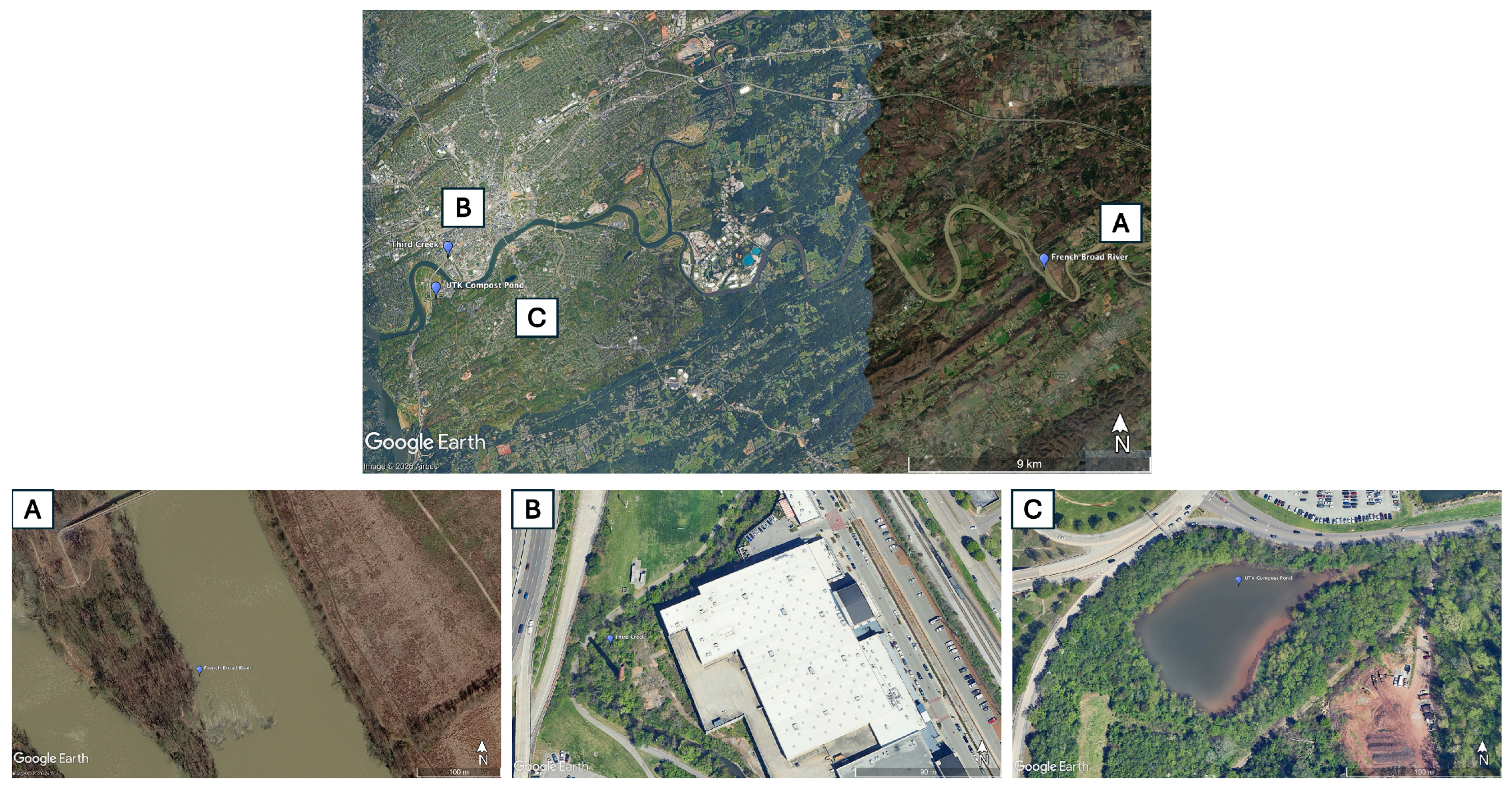
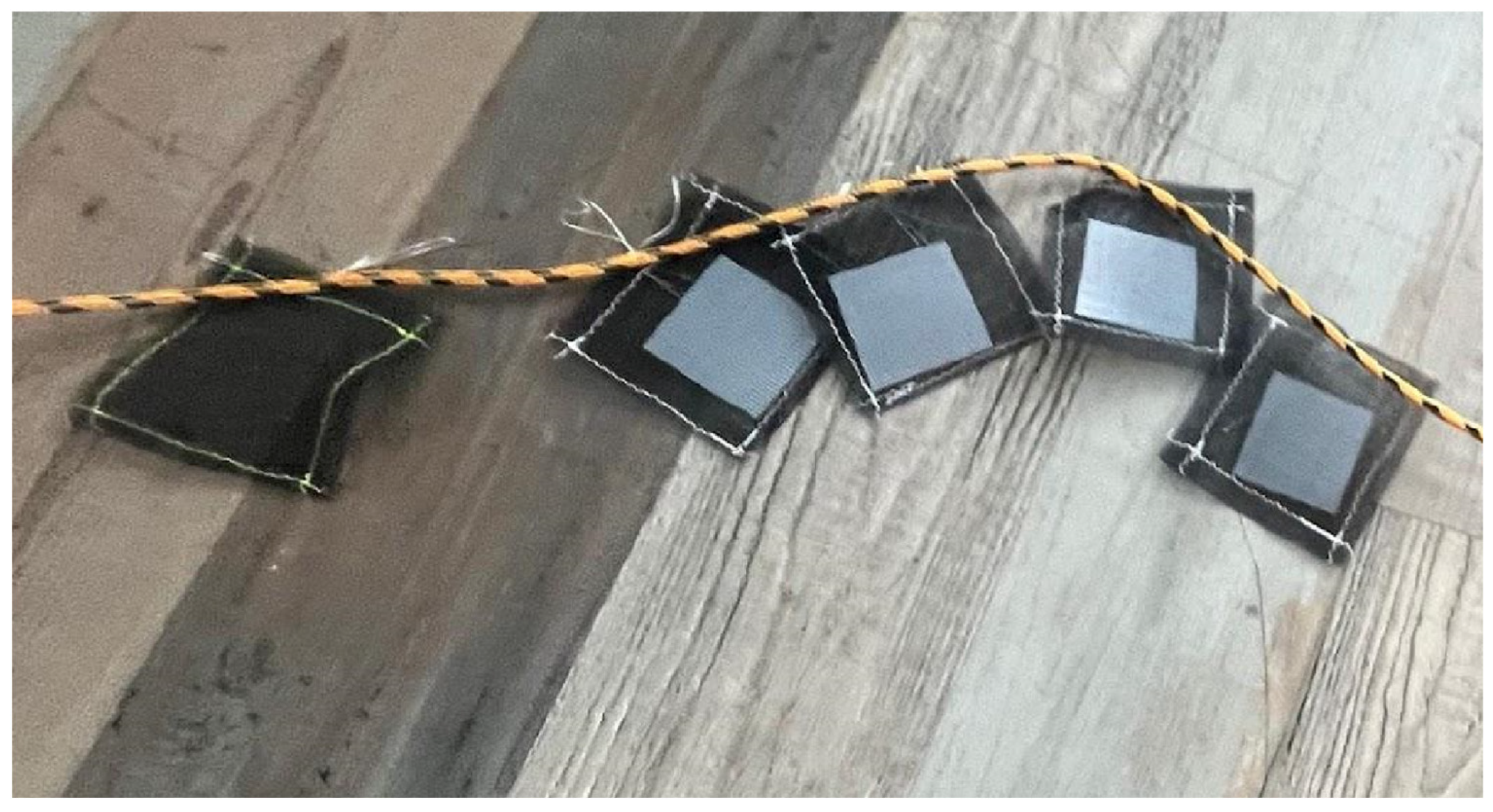


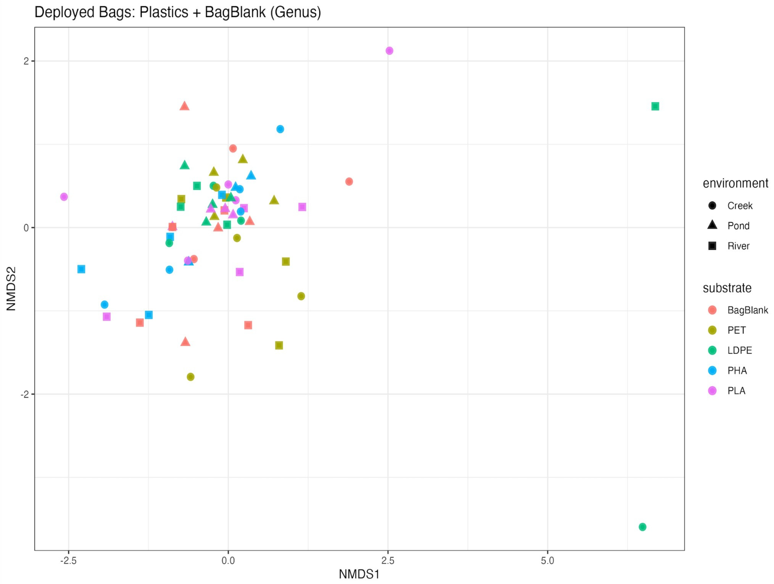
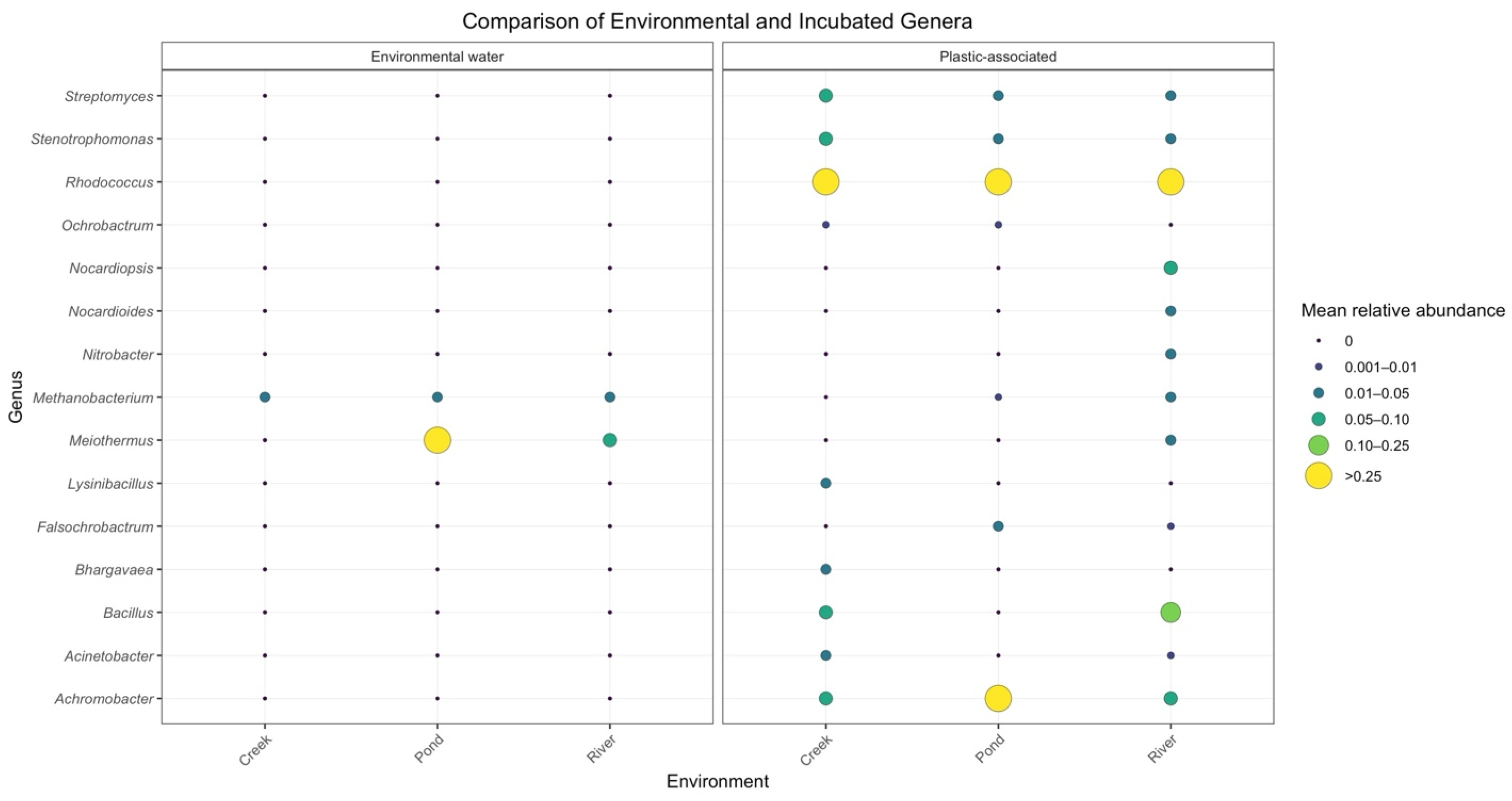


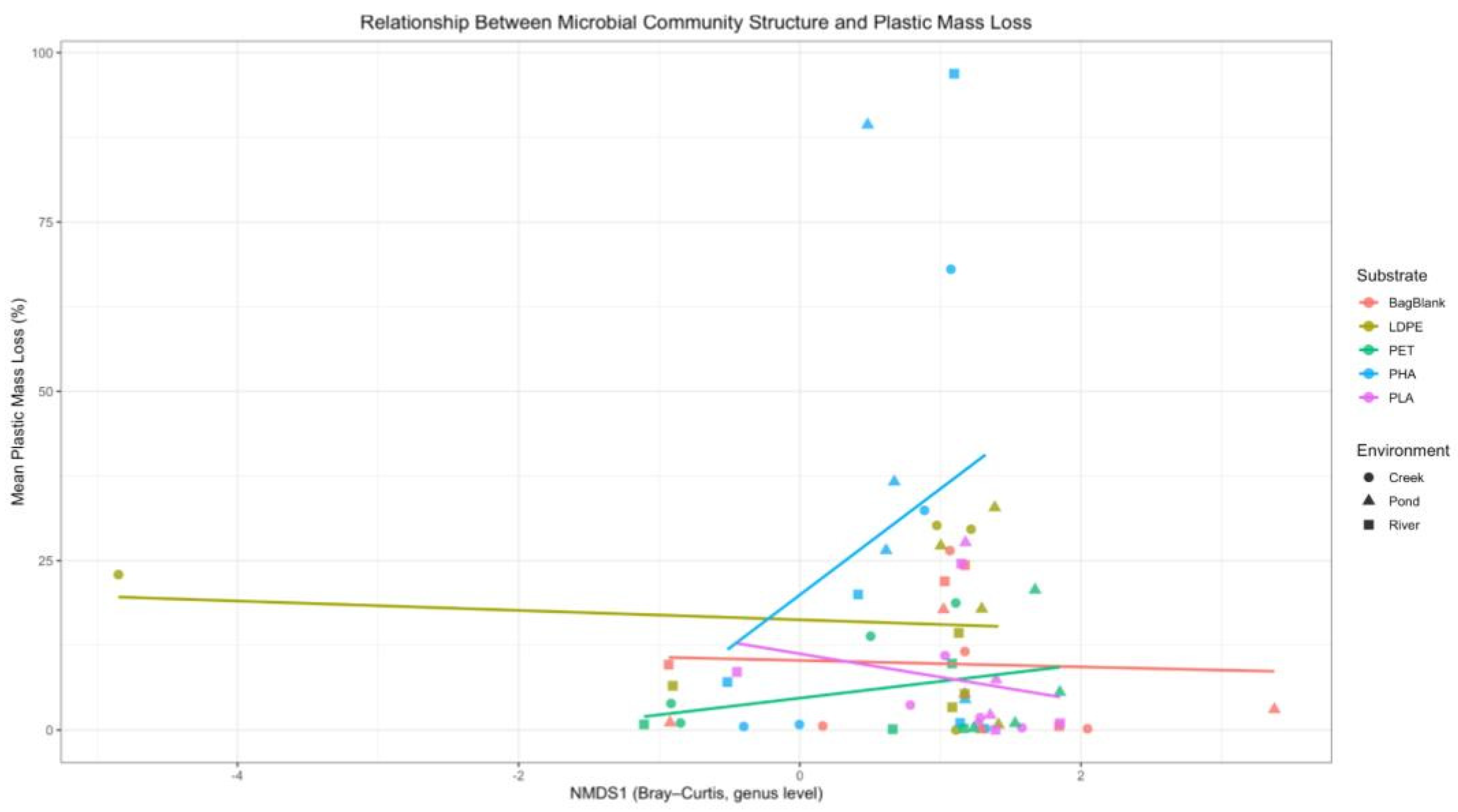
| Compound | Amount (g) |
|---|---|
| NaCl | 2.5 |
| (NH4)2SO4 | 5 |
| KH2PO4 | 15 |
| Na2HPO4 | 32.5 |
| Site | pH | UV (W m−2) | Temperature (°C) | Nitrate (mg L−1) | Turbidity (FNU) | Conductivity (µS cm−1) | TDS (mg L−1) | Chl (mg L−1) | PC (µg L−1) |
|---|---|---|---|---|---|---|---|---|---|
| River | 6.08 ± 0.17 | 441.14 ± 324.11 | 23.59 ± 4.19 | 1.82 ± 1.62 | 412.08 ± 216.69 | 194.73 ± 137.79 | 116.00 ± 93.92 | 1.38 ± 0.60 | 5.09 ± 5.06 |
| Creek | 5.98 ± 0.52 | 36.00 ± 9.61 | 19.93 ± 1.39 | 2.59 ± 0.94 | 262.99 ± 25.07 | 523.13 ± 12.93 | 341.83 ± 10.12 | 1.69 ± 0.26 | 4.99 ± 4.82 |
| Pond | 6.23 ± 0.25 | 671.80 ± 199.97 | 25.68 ± 4.11 | 0.83 ± 0.67 | 248.91 ± 253.00 | 303.80 ± 91.63 | 191.20 ± 52.59 | 37.52 ± 66.36 | 14.71 ± 13.17 |
| Source | Df | Sum of Squares | R2 | F | Pr (>F) |
|---|---|---|---|---|---|
| Model | 10 | 10.7286 | 0.3608 | 3.8380 | 0.001 |
| Residual | 68 | 19.0086 | 0.6392 | — | — |
| Total | 78 | 29.7373 | 1.0000 | — | — |
| Analysis | Factors Tested | Df | F Statistic | R2 | p-Value |
|---|---|---|---|---|---|
| Water vs. Deployed Bags | Sample type (water vs. bag-associated) | 10 | 3.84 | 0.361 | 0.001 |
| Environment × Substrate × Month | Environment, substrate, month | 18 | 1.49 | 0.384 | 0.001 |
| Plastics Only | Polymer type, environment, month | 15 | 1.56 | 0.408 | 0.003 |
| Source | Df | Sum of Squares | R2 | F | Pr (>F) |
|---|---|---|---|---|---|
| Model | 15 | 5.6115 | 0.284 | 1.22 | 0.069 |
| Residual | 46 | 14.1465 | 0.716 | — | — |
| Total | 61 | 19.7580 | 1.0000 | — | — |
| Source | Df | Sum of Squares | R2 | F | Pr (>F) |
|---|---|---|---|---|---|
| Model | 12 | 5.6471 | 0.263 | 1.10 | 0.047 |
| Residual | 37 | 15.8602 | 0.737 | — | — |
| Total | 49 | 21.5073 | 1.0000 | — | — |
| Study | Polymer(s) | Setting | Duration | Methods Used | Key Findings | What This Supports in Our Study |
|---|---|---|---|---|---|---|
| [28] | LDPE, PET, PLA, Mater-Bi | Natural lake (field) | Up to 77 days | SEM, ATR-FTIR, contact angle, sequencing | All plastics rapidly colonized by biofilms; limited chemical evidence of polymer degradation over study period | Supports our finding that biofilm formation occurs quickly (≤30 days) but does not necessarily indicate polymer degradation, especially for PET and LDPE |
| [66] | PET, LDPE | Lake Geneva (field; depth gradient) | 45 weeks | Spectroscopy, microscopy, contact angle, community analysis | PET showed no detectable surface degradation after 45 weeks; LDPE showed depth-dependent oxidation | Supports our observation of minimal PET degradation despite prolonged exposure and surface colonization |
| [65] | PLA, PBAT blends, PHBV, PP | Lentic fresh- water (field) | 120 days | Morphology, chemical characterization, thermal/ mechanical analyses | Limited degradation of compostable plastics in natural freshwater; lab tests overestimate degradability | Supports our finding that PLA and conventional plastics exhibit limited degradation over ~4 months in freshwater |
| [59] | PP, PET, PS | River contin- uum (field) | 21 days | Biofilm development and community composition | Biofilm structure driven by environment and exposure time more than polymer type | Supports our site dependent SEM patterns across river, creek, and pond environments |
| [60] | PVC, PP, PET, HDPE, LDPE (weathered & virgin) | Fresh- water intake sites (in situ) | 21 weeks | Biofilm growth, community composition | Water quality and prior weathering strongly influence colonization | Supports our interpretation that environmental context shapes plastisphere structure more than polymer identity |
| [67] | LDPE microplastics | Lab incubation in natural lake water | 30 days | Microscopy, community analysis | Rapid colonization and structured biofilms in freshwater | Supports our 30-day SEM evidence of early biofilm establishment |
| [64] | PHBV compound | River (Elbe) and controlled aquatic systems | Method based not duration | Mass loss, spectroscopy, morphology, mechanical properties | Biodegradable polyesters show measurable degradation under freshwater conditions | Supports our high PHA mass loss and increasing SEM surface disruption |
| [68] | PLA | Lab aqueous biodegradation | 35 days | Molecular weight (GPC), FTIR, SEM, mass loss | PLA degradation evidenced by molecular weight reduction and FTIR bond changes | Provides contrast showing that true PLA degradation requires conditions beyond ambient freshwater exposure |
| [25] | PE, LDPE, HDPE, PP, PET, PS, PVC, PLA, PHA/PHBV, Mater-Bi | Freshwater systems | Hours to >1 year | SEM, spectroscopy, omics synthesis | Plastisphere colonization does not equate to mineralization | Supports our decoupling of biofilm formation and mass loss |
Disclaimer/Publisher’s Note: The statements, opinions and data contained in all publications are solely those of the individual author(s) and contributor(s) and not of MDPI and/or the editor(s). MDPI and/or the editor(s) disclaim responsibility for any injury to people or property resulting from any ideas, methods, instructions or products referred to in the content. |
© 2026 by the authors. Licensee MDPI, Basel, Switzerland. This article is an open access article distributed under the terms and conditions of the Creative Commons Attribution (CC BY) license.
Share and Cite
Verble, J.A.; McKinney, M.L. Microbial Degradation of Plastics in Freshwater Environments. Microplastics 2026, 5, 119. https://doi.org/10.3390/microplastics5020119
Verble JA, McKinney ML. Microbial Degradation of Plastics in Freshwater Environments. Microplastics. 2026; 5(2):119. https://doi.org/10.3390/microplastics5020119
Chicago/Turabian StyleVerble, Jillian A., and Michael L. McKinney. 2026. "Microbial Degradation of Plastics in Freshwater Environments" Microplastics 5, no. 2: 119. https://doi.org/10.3390/microplastics5020119
APA StyleVerble, J. A., & McKinney, M. L. (2026). Microbial Degradation of Plastics in Freshwater Environments. Microplastics, 5(2), 119. https://doi.org/10.3390/microplastics5020119