Article

Bioluminescent ATP-Metry in Assessing the Impact of Various Microplastic Particles on Fungal, Bacterial, and Microalgal Cells

Faculty of Chemistry, Lomonosov Moscow State University, Lenin Hills 1/3, Moscow 119991, Russia
*
Author to whom correspondence should be addressed.
Microplastics 2025, 4(4), 72; https://doi.org/10.3390/microplastics4040072
Submission received: 5 August 2025 / Revised: 9 September 2025 / Accepted: 1 October 2025 / Published: 3 October 2025

Abstract

The concentration of intracellular adenosine triphosphate (ATP) is one of the most important characteristics of the metabolic state of the cells of microorganisms and their viability. This indicator, monitored by bioluminescent ATP-metry, and accumulation of the suspension biomass in the medium were used to assess the effect of particles of different synthetic microplastics (MPs) (non-biodegradable and biodegradable) on the cells of yeast, filamentous fungi, bacteria and phototrophic microorganisms (microalgae and cyanobacteria) co-exposed with polymer samples in different environments and concentrations. It was found that the effect of MPs on microorganisms depends on the concentration of MPs (1–5 g/L), as well as on the initial concentration of cells (104 or 107 cells/mL) in the exposure medium with polymers. It was shown that the lack of a sufficient number of nutrition sources in the medium with MPs is not fatal for the cells. The study of the effect of MPs on the photobacteria Photobacterium phosphoreum, widely used as a bioindicator for assessing the ecotoxicity of various environments, demonstrated a correlation between the residual bioluminescence of these cells and the level of their intracellular ATP in media with biodegradable polycaprolactone and polylactide, which had an inhibitory effect on these cells. Marine representatives of phototrophic microorganisms showed the greatest sensitivity to the presence of MPs, which was confirmed by both a decrease in the level of intracellular ATP and the concentration of their biomass. Among the eight microorganisms studied, bacteria of the genus Pseudomonas turned out to be not only the most tolerant to the presence of the seven MP samples used in the work, but also actively growing in their presence.

1. Introduction

Microorganisms, being an integral part of ecobiosystems, participate in soil formation and the purification of water systems and also serve as biocatalysts for many ecological and eco-adaptive processes. Microalgae and cyanobacteria occupy a key place in ecosystems due to their phototrophic type of nutrition, which makes them the initial link in trophic chains. As a result, their reactions to the presence of various pollutants in environmental conditions are extremely important [1,2]. Mycelial fungi, yeasts, and bacteria are consumers of organic substances produced by phototrophs and, at the same time, reducers that play an important role in food chains by completing them. In this regard, they are most often the biocatalysts of various processes of decomposition and transformation of xenobiotics [3,4].
Given the interest in research into the impact of microplastics (MPs) on various ecosystems [5,6,7], as the most discussed and widespread contaminants in the modern world, it is especially relevant to study the impact of MPs on microorganisms, which appears to be quite diverse. It has been established that microorganisms are participants in the processes of destruction of synthetic polymers in the environment and producers of enzymes capable of carrying out this biocatalytic destruction [8]. Traditionally, synthetic polymers are also matrices for cell sorption and the formation of structures, such as biofilms, that are resistant to the presence and influence of MPs [9].
Of exceptional interest are studies devoted to the influence of MPs on photobacteria, which are generally recognized bioindicators used to assess the toxicity of various environments [10]. It has already been noted that the effect of MPs on the bioluminescence and cell viability of photobacteria varies [11]; however, the key factors influencing this variation in the recorded cell reactions (type or concentration of MP, the environment in which the cells are located, their concentration, etc.) have not been determined.
Quite a few studies are devoted to the influence of MPs on phototrophic microorganisms. Moreover, it has been established that this influence is versatile. On the one hand, particles of synthetic polymers predominantly negatively affect the growth and photosynthesis occurring in cells [12]. Growth inhibition and changes in chlorophyll functioning in green microalgae cells in response to the presence of MP particles were established [13]. On the other hand, MPs can be a material for the adhesion of the cells of microorganisms, associated with the synthesis of exopolysaccharides (EPSs) by phototrophs and bacteria [9,14]. The accumulation of EPS leads to cell flocculation and increased sorption of MP nanoparticles on the cells aggregated in this way.
Today, to determine the presence of toxicants and assess the general condition of phototrophic cells in the presence of MPs, growth and physiological parameters such as cell density, chlorophyll a, and the maximum quantum yield Fv/Fm are mainly detected [15]. However, these parameters may not always give a true result, indicating the state of the cells or the state of the system as a whole [16].
The concentration of intracellular adenosine triphosphate (ATP) in the cells of different microorganisms is one of the common and reliable criteria for assessing their metabolic activity and viability. Luminescent ATP-metry allows for rapid, highly specific, and sensitive determination of the level of this indicator in different cells [17].
The aim of this work was to study, using bioluminescent ATP-metry, the influence of different MPs on the concentration of intracellular ATP in the cells of individual microorganisms (filamentous fungi and yeast, bacteria, and phototrophs), which are participants in a variety of natural ecosystems existing in different conditions.
As noted above, there are now quite a few studies devoted to the influence of microplastics on various groups of microorganisms, especially phototrophic ones, in which the influence of the chemical nature of the polymer, as well as the concentration and size of microplastic particles on microbial cells, is studied [18,19,20,21,22,23,24,25]. But until now, changes in the concentration of ATP in the presence of microplastics have been carried out only in the cells of the cephalopod Sepia esculenta [26]. In this paper, we decided to approach the study of such phenomena using bioluminescent ATP-metry as one of the main methods for studying the reaction of microorganisms to the presence of various microplastics.
Synthetic polymers were selected based on their prevalence in various environmental objects. The impact of both conditionally non-biodegradable [27,28] and biodegradable polymer analogs was assessed [29,30], since it has already been proven that the rate of formation of microplastics of biodegradable polymers is significantly higher than that of non-biodegradable ones, and they are currently no less relevant xenobiotics in environmental objects [27,29].
The effect of MP particles on microorganisms must certainly be considered depending on the chemical nature of the polymers and their concentration in the media with cells, but it is no less important in such studies to take into account the state of the cells themselves. It is known that cells of different microorganisms in a state of highly concentrated populations (~107 cells/mL), that is, the so-called quorum sensing (QS) state, exhibit increased resistance to negative environmental factors, compared to cell populations in a lower concentration and in the logarithmic growth phase (~104 cells/mL). This is due to the fact that, being in the QS state, they have a different proteomic composition as a result of the realized genetic potential of protein synthesis encoded by the so-called “silent genes” [31]. It is known that the presence of nutrients in the media promotes active cell growth and/or biosynthesis of essential metabolites that help overcome negative effects on microorganisms [17].
In this regard, the effect of MPs on microorganisms was assessed under conditions of different environments and cell concentrations. When interpreting data on changes in the concentration of intracellular ATP in the presence of MP particles, not only was information on the initial specific concentration of ATP in cells (at the beginning of contact with MP) (mol/g DCW) taken into account, but also information on the growth phase (logarithmic or stationary) of microorganisms in which they are located. The QS state for some cells is characterized by a sharp increase in the concentration of intracellular ATP [32]. This fact was also taken into account in the work.

2. Materials and Methods

2.1. Chemicals

Microplastic particles were obtained by a milling process from the following: cling film—low-density polyethylene (LDPE); bottle cap—high-density polyethylene (HDPE); bottle from drinking water—polyethylene terephthalate (PET); and food box—polypropylene (PP). All used containers were clean. After grinding, the particles were sifted through a sieve with a mesh size of 5 mm (Scharlab, Barcelona, Spain). The biodegradable polymer particles were commercial: polyvinyl alcohol (PVA; Sinopec Corp., Beijing, China), polycaprolactone (PCL; Polymorphous, Moscow, Russia), and polylactic acid (PLA; Ahlstrom-Munksjö, Helsinki, Finland). MP samples were obtained by grinding the starting materials in a GM 300 knife mill (Retsch, Haan, Germany), after which the resulting particles were sifted through a sieve with a mesh size of 5 mm. Microparticles were washed with deionized water, dried, and exposed under UV light for 12 h with an intensity of 0.63 W/m2 at 254 nm (TUV 15W Philips).
The components of nutrient media (with ≥98% purity) for biomass accumulation of microorganisms were purchased from Chimmed (Moscow, Russia).

2.2. Microorganisms

The phototrophic cells of Nannochloropsis sp. rsemsu-N-1 and Arthrospira (Spirulina) platensis (Nordst.) Geitl. rsemsu 1/02 are in the collection of the staff of the laboratory of ecobiocatalysis of the Faculty of Chemistry of the Lomonosov Moscow State University [21]. Chlorella vulgaris C-2 was obtained from the IPPAS collection of microalgae (Moscow, Russia) (http://cellreg.org/catalog/ accessed on 2 July 2025). Tetraselmis suecica was purchased as a commercial preparation “Starter culture Tetraselmis suecica” (Aqa-shop, Moscow, Russia). The yeast cells of Saccharomyces cerevisiae Y-185 and Candida maltose Y-2256, the fungal cells of Aspergillus niger F-1057 and Rhizopus oryzae F-814, and the bacterial strains Bacillus subtilis B-5522, Pseudomonas putida B-6741, and Photobacterium phosphoreum B-12307 were purchased from the All-Russian Collection of Industrial Microorganisms (Moscow, Russia) (https://vkpm.genetika.ru/ accessed on 2 July 2025) [33,34].
Chlorella vulgaris C-2 strain cells were maintained on a Tamiya nutrient medium (g/L): KNO3—5; KH2PO4—1.25; MgSO4·7H2O—2.5; FeSO4·7H2O—0.009; EDTA—0.037; H3BO3—2.86 × 10⁻³; MnCI2·4H2O—1.81 × 10⁻³; ZnSO4·7H2O—0.22 × 10⁻³; MnO3—0.018 × 10⁻³; NH4VO3—0.023 × 10⁻³.
Nannochloropsis sp. rsemsu-N-1 cells were maintained on a BG-11 medium (g/L): NaNO3—1.5, K2HPO4·3H2O—0.04, MgSO4·7H2O—0.075, CaCl2·2H2O—0.04, Na2CO3—0.02, citric acid—0.006, and Trilon B—0.001; microelement solution A—1 mL; and microelement solution B—1 mL. Microelement solution A contains the following (g/L): H3BO3—2.86; MnCl2 × 4H2O—1.81; ZnSO4 × 7H2O—0.22; CuSO4 × 5H2O—0.08; and MoO3—0.015. Microelement solution B contains the following (g/L): K2Cr2(SO4)4 × 24H2O—0.096; NH4VO3—0.023; NiSO4 × 7H2O—0.048; Na2WO4 × 2H2O—0.018; Ti2(SO4)3—0.04; and Co(NO3)2 × 6H2O—0.044.
Arthrospira/Spirulina platensis strain 1/02-P cells were maintained on Zarrouk’s medium (g/L): NaHCO3—16.8, NaNO3—2.5, K2HPO4·3H2O—1, K2SO4—1, MgSO4·7H2O—0.2, NaCl—1.0, CaCl2—0.04, FeSO4·7H2O—0.025, and Trilon B—0.03; microelement solution A—1 mL; and microelement solution B—1 mL.
Tetraselmis suecica strain cells were maintained on Goldberg’s medium (per L of sterilized sea water): 2 mL of solution #1 (KNO3—101 g/L); 0.5 mL of solution #2 (Na2HPO4—14.21 g/L); 1 mL of solution #3 (MnCl2 × 4H2O—0.2 g/L and CoCl2 × 6H2O—0.24 g/L); and 1 mL of solution #4 (FeCl3 × 6H2O—0.27 g/L).
The above-mentioned nutrient media were used for experiments with phototrophic microorganisms. Suspensions of phototrophic microorganism cells were diluted to the required concentration using calibration curves established between the optical density of cell suspensions and biomass concentration at a wavelength of 540 nm measured using an Agilent UV-8453 spectroscopy system (Agilent Technology, Waldbronn, Germany). Microalgae and cyanobacteria were cultivated in 50%-filled glass flasks at 24 ± 2 °C under lighting (regime 16/8) with Osram Fluora 77 luminescent lamps (30 W, Munich, Germany) for 96 h and manually shaken once a day to prevent the sedimentation of phototrophic microorganisms. A 2% NaCl solution was used as a medium to assess the effect of MP particles on P. phosphoreum photobacteria cells. The analysis was carried out for 24 h.
Bacterial cells of B. subtilis and P. putida were cultured and maintained on a Luria–Bertani nutrient medium (g/L): NaCl—10; tryptone—10; and yeast extract—5. Parameters of pH 7 at 28  ±  2 °C, 150 rpm, were used (thermostatically controlled Adolf Kuhner AG shaker, Basel, Switzerland).
Yeast biomass was accumulated as a result of culturing cells on a medium of the following composition (g/L): glucose—20; yeast extract and peptone—5; K2HPO4—3; KH2PO4—2; citric acid—1; ascorbic acid—1; and MgSO4—0.5. Parameters such as a pH of 5.6 at 26 ± 2 °C, as well as 150 rpm for 20 h, were used. The composition of the liquid medium for fungi was as follows (g/L): glucose—100; (NH4)2SO4—2.36; MgSO4 × 7H2O—0.2; ZnSO4 × 7H2O—0.07; and K2HPO4 × 3 H2O—1. Parameters of pH 6.8 at 28 ± 2 °C, 180 rpm for 36 h, were used.
The accumulated biomass of microorganisms was separated in each case from the culture broth by centrifugation at 10,000× g for 12 min (Avanti J25, Beckman, Coulter, Germany) and transferred to the medium with the MP sample at the required concentration under sterile conditions. For experiments with fungi and bacteria, glass vials filled to 50% were used, and cultivation was carried out under aerobic conditions at 24 °C and 180 rpm using a thermostatically controlled Adolf Kuhner AG shaker (Basel, Switzerland). To assess the effect of MP particles, cultivation was carried out for 24 h.

2.3. Analytical Methods

Microbial growth was controlled by cell counting using an improved Neubauer hemocytometer (Rohem Instruments, Nashik, Maharashtra, India) through an optical microscope (Biomed, Russia) with a Biomed Lum 206070112209 nozzle and a Myscope 500 M digital camera for the microscope. Concurrently, the OD540 of the cell suspensions was controlled with an Agilent UV-8453 spectroscopy system (Agilent Technology, Waldbronn, Germany) to investigate the kinetic curve of microbial growth. The calibration graphs reflected a correlation between the OD540 and dry cell weight (DCW) of each culture of the studied microorganisms.
The DCW of biomass was determined by a standard gravimetrical method. Briefly, the weighed biomass samples were dried to a constant weight at 105 °C, and their mass loss was calculated.
For the determination of the ATP concentration in microbial cells, the known procedures were used [17]. For ATP analysis, each sample was centrifuged at 10,000× g for 12 min. The mass of the sediment, which was a mixture of biomass and polymer, was determined. Next, the sediment was mixed with dimethyl sulfoxide (DMSO, ≥99.9%, Sigma Aldrich, Darmstadt, Germany) in a ratio of 1:9 for 3 min by Vortex (Biosan, Riga, Lithuania) and then maintained at room temperature for 2 h for complete extraction of ATP from the cells. To determine the concentration of intracellular ATP of cells adsorbed on the polymer, MP particles were extracted from the culture medium by filtration through a sieve with a cell size of 300 μm (Sigma Aldrich, Darmstadt, Germany), washed with saline, weighed, and filled with DMSO in a ratio of 1:9. Then, the procedure for ATP extraction was similar to that described above. Next, a 50 μL aliquot of the aqueous phase was analyzed for ATP using a 3560 Microluminometer (New Horizons Diagnostics Co., Columbia, MD, USA). To prepare ATP standards, the commercial sodium adenosine triphosphate (OOO Ellara, Moscow, Russia) was used. For bioluminescent analysis of intracellular ATP concentration, the luciferin–luciferase reagent (>98%, Lumtek, Moscow, Russia) was used [35].
The pH value was determined using a Corning Pinnacle 530 pH meter (Corning Incorporated, Corning, NY, USA).

2.4. Data Analysis

Data on changes in the concentration of ATP and cell biomass as a result of their cultivation in media in the presence of different MP particles were processed, are presented in the figures in the article as a ratio to the control, and were calculated as the ratio of the parameter value for a sample in the presence of particles of a specific MP to the value of the same parameter in the control (in the absence of MP).
The data are shown as means of at least three independent experiments ± standard deviations (±SD). The statistical analysis was realized using SigmaPlot 12.5 (ver. 12.5, Systat Software Inc., San Jose, CA, USA). The significant (p ≤ 0.05) differences between the obtained results were estimated by a one-way analysis of variance (ANOVA) (unless otherwise noted, the significance level was set at p < 0.05). Data were expressed as average ± standard deviation in the study.

3. Results and Discussion

3.1. Changes in Intracellular ATP Concentration in Yeast and Filamentous Fungi in the Presence of MP Particles

Mycelial fungi have several natural and unique abilities: to produce hydrophobins for attaching hyphae to hydrophobic substrates and to ensure penetration of their enzymes into substrates. This allows them to be effective destructors, including MPs. Studies on MP biodegradation have shown that fungi are able to use these materials as sources of carbon and energy, as well as matrices for the formation of stable cellular formations [36]. Yeasts of the genera Saccharomyces and Candida (Figure 1) and filamentous fungi Rhizopus oryzae (a representative of the zygomycetes) and Aspergillus niger (a representative of the ascomycetes) (Figure 2) are widespread in various terrestrial ecosystems and therefore became one of the main objects of study in this work. To study the effect on them, as well as on all other microorganisms, particles of seven different MPs were used in this study: LDPE, HDPE, PET, PP, PVA, PCL, and PLA (Figure S1). When selecting the working concentrations of microplastics for this work, information was taken into account that in coastal zones, pollution by them can range from 1–5 g/L to 100 g/L [37]. In addition, according to existing estimates, one of the main objects of accumulation of microplastics is soil, in which the concentration of microplastics reaches 5 g/kg [38]. In this study, it was decided to use 5 g/L for all microplastics as the base concentration for comparison of all cells except phototrophic cells, for which a concentration of 1 g/L was used.
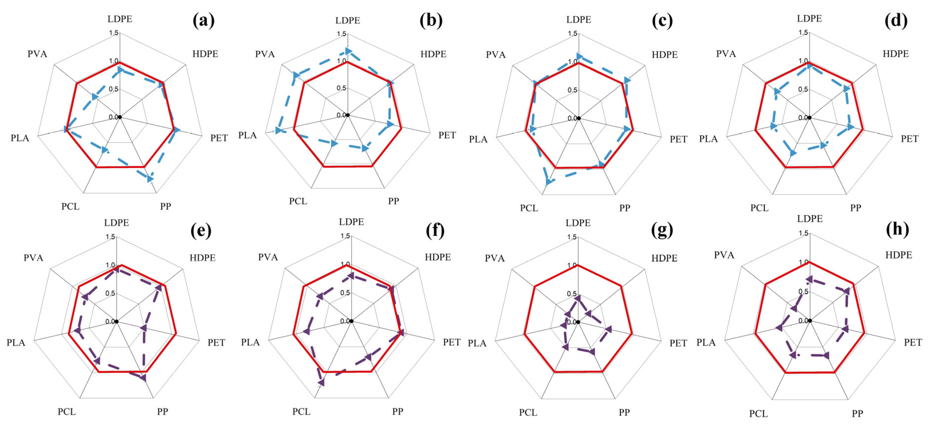
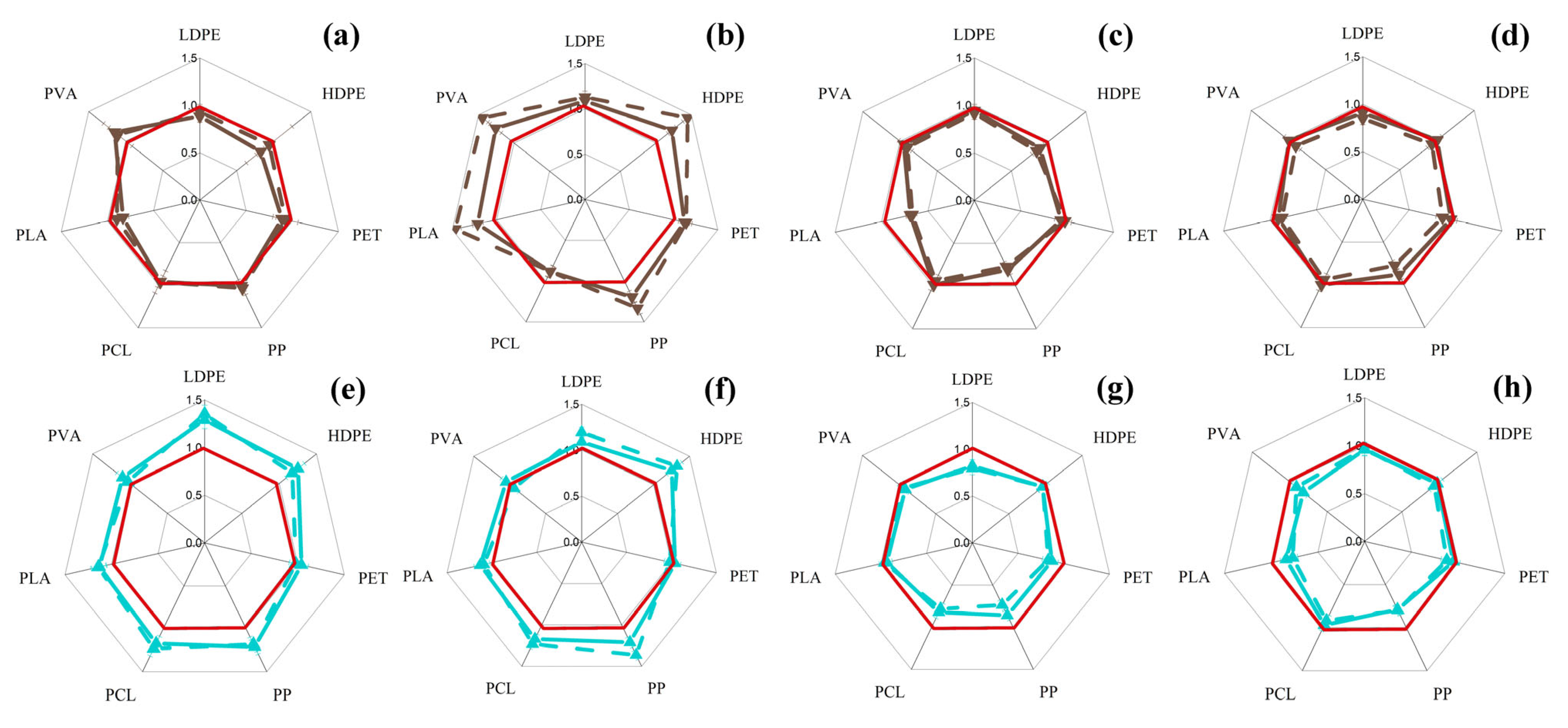
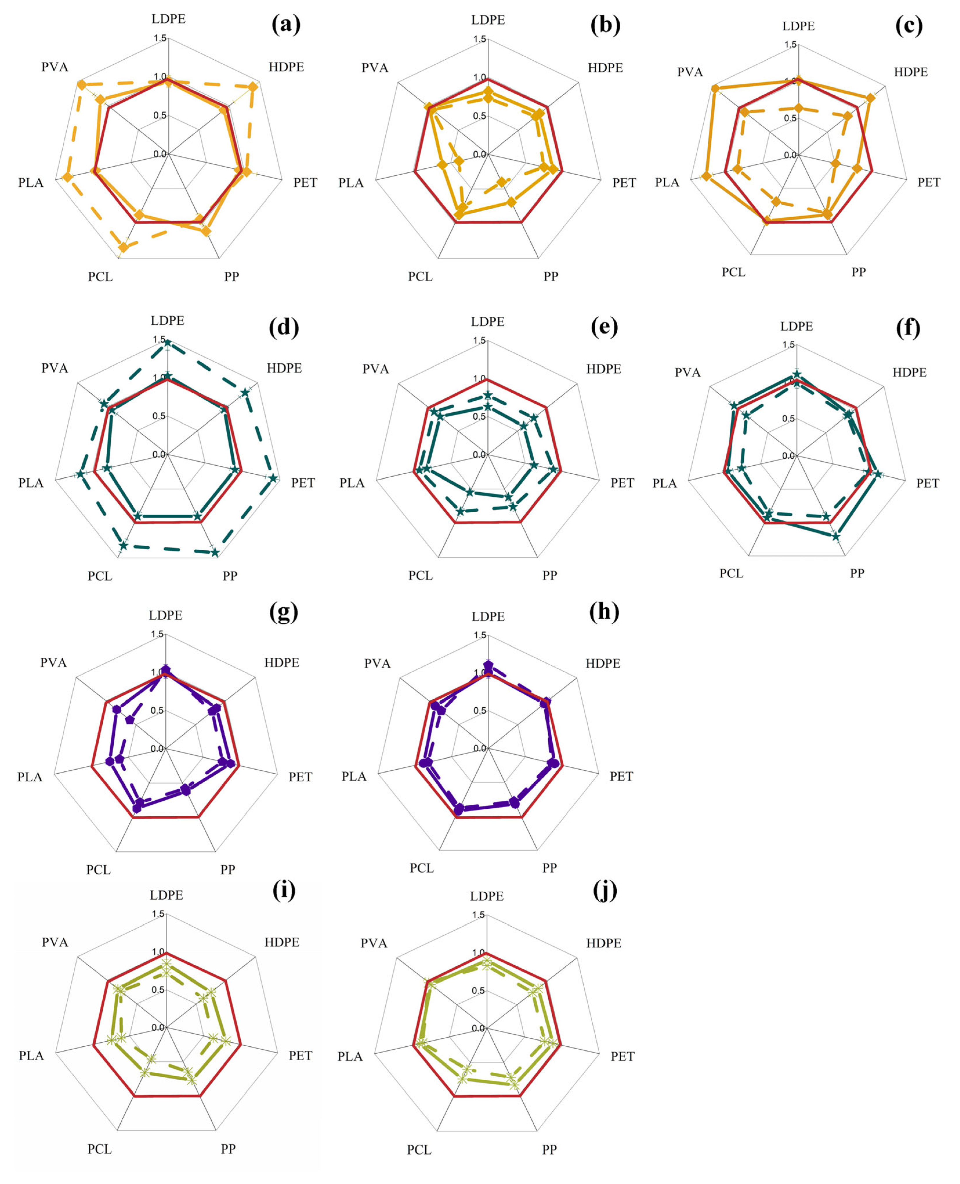
It was found that the presence/absence of nutrients in the cell exposure medium and variation in cell concentrations in media with the same concentration of MP particles led to a change in the ratio of these concentrations of fungal cells and MP particles, and also had a significant effect on the concentration of intracellular ATP (Figure 1 and Figure 2). When analyzing the data obtained for yeast cells, it was noted that the high initial concentration of cells (characteristic of the QS state) allows the yeast S. cerevisiae and C. maltose cells to tolerate the presence of MPs with minimal response (without significant changes in biomass accumulation and intracellular ATP levels) relative to the control (in a medium without MPs). An exception to this trend was only in the case of PET, where the stimulation of cells was pronounced (19–25%) and did not indicate the presence of cell inhibition by MP (Figure 1b,f). The presence of nutrients in the medium with cells with a lower initial concentration (104 cells/mL) led to the fact that in yeast of the genus Saccharomyces, in the presence of MP particles, the level of intracellular ATP significantly increased compared to the control in the presence of almost all the polymers studied except PVA (Figure 1a). In this case, in yeasts of the genus Saccharomyces and Candida, under similar conditions, an increase in the biomass concentration was also detected (Figure 1a,e) compared to the control in media with different polymers, also with the exception of the medium with PVA.
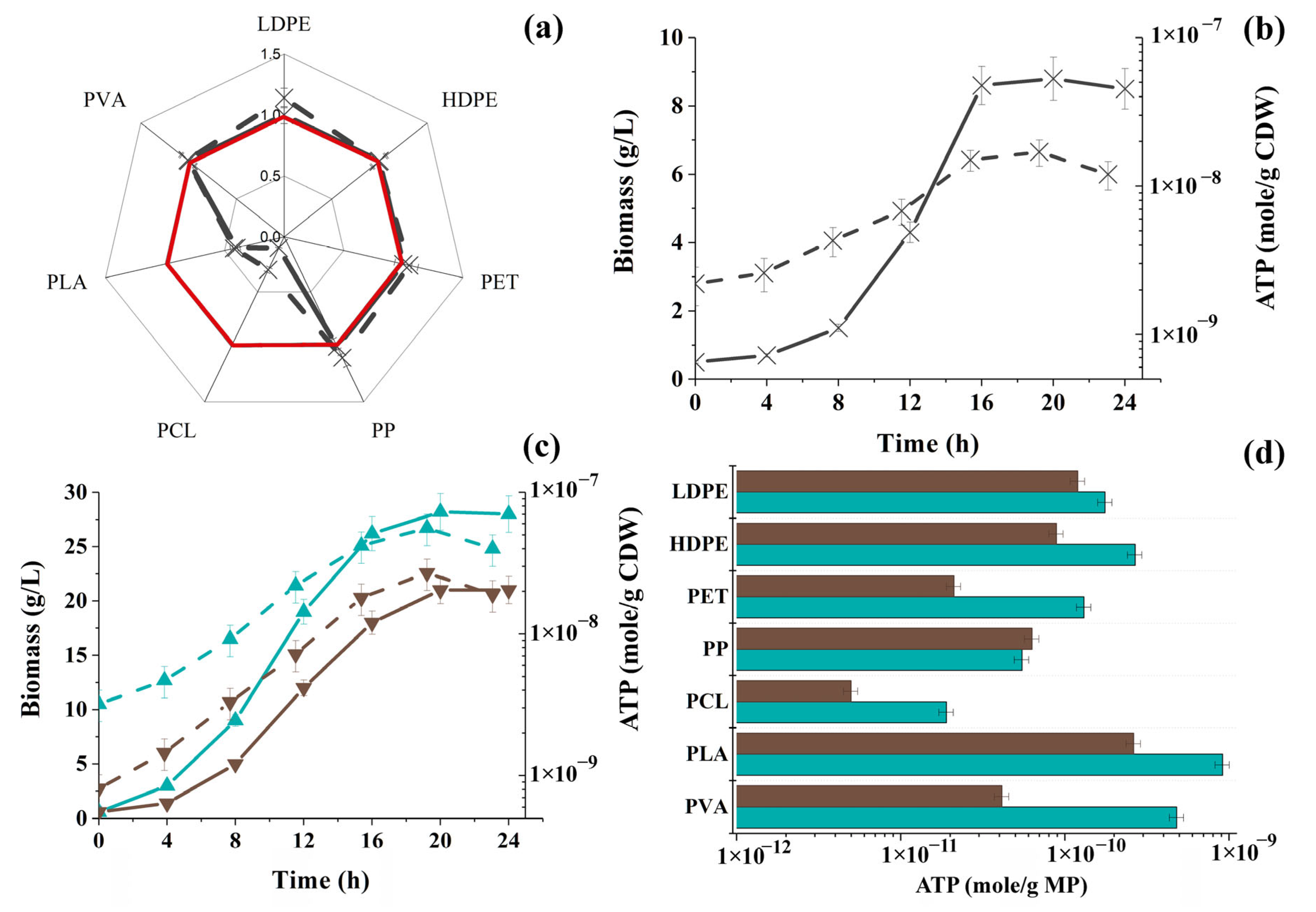
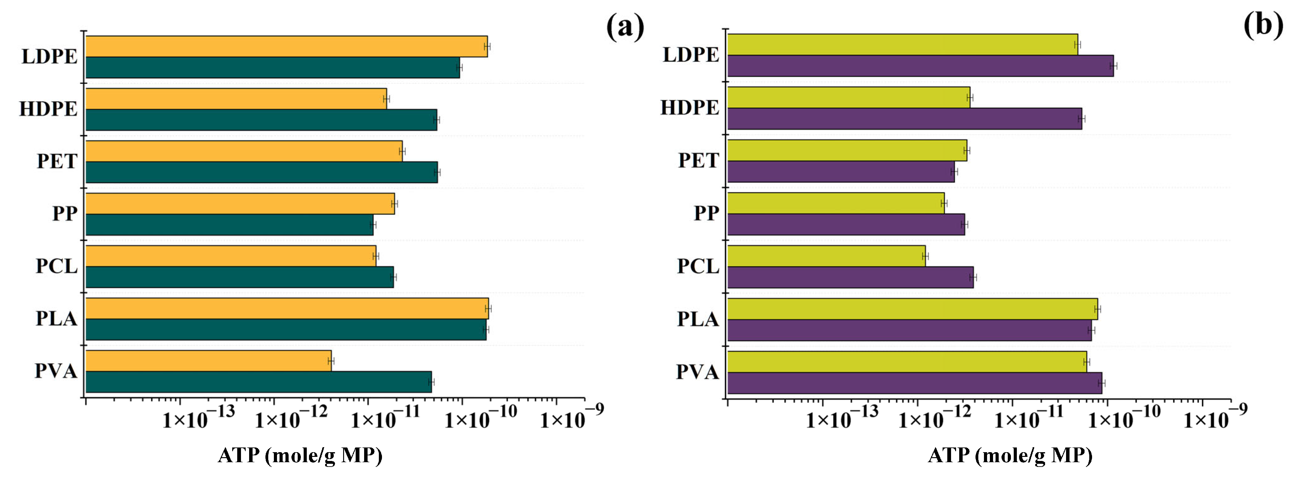
This fact indicates a clear growth of yeast cells in the presence of MPs, and the growth was not only in the medium, as in the absence of MP particles, but also in MP particles, since the presence of ATP in samples of polymer particles due to adhered cells was shown (Figure 3).
It is interesting to note that for C. maltose cells, special preferences in adhesion to MP particles were revealed in the case of PLA, whereas PVA clearly “repelled” the same yeast cells. It is possible that such a positive predisposition of Candida cells to PLA is due to the fact that these cells have esterase (lipase) activity [39] and are capable of participating in the hydrolysis of ester bonds in the PLA structure. Some inhibitory effect of PCL (up to 30%) on yeast biomass of the genus Candida (Figure 1e,f,g) is probably due to the fact that in an aqueous environment PCL undergoes combined hydrolysis under the influence of abiotic and biotic factors with the formation of caproic acid, which in turn is an inhibitor of growth and metabolism for these cells [40].
In the absence of nutrients, the inhibitory effect of MP particles on C. maltose yeast cells, manifested in a decrease in the concentration of intracellular ATP, was more pronounced. This may be due to the fact that ATP in these cases is spent on internal cell reorganization, as well as the synthesis of the necessary enzymes and adhesins to prepare for the use of MPs as probable substrates. A decrease in the ATP level in C. maltose yeast cells was also observed in the presence of HDPE, PCL, PLA, and PVA at their low initial concentration in the nutrient medium in the presence of MPs. However, in this case, ATP was obviously spent not only on the synthesis of metabolites necessary for cell adhesion to MP particles (Figure 3c), but also on the growth of the cells themselves. This was noted for five of seven MP samples studied (the exceptions were PCL and PVA). These data positively correlate with the results published earlier for Candida cells, when it was found that C. parapsilosis yeast is capable of adhesion and subsequent biodegradation of particles of synthetic polymers such as polystyrene (PS) and polyvinyl chloride (PVC). Favorable conditions for MP degradation were provided by a high cell density (>107 cells/mL) and a slightly acidic pH of the medium (pH 4.9–5.6) [18].
S. cerevisiae yeast cells were less sensitive to the presence of MPs compared to Candida yeast cells (Figure 1). It is interesting to note that the inhibition of S. cerevisiae yeast by particles of MPs such as polyethylene (PE), PS, PP, PVC, and PET at a concentration of 1 g/L in the medium with cells was previously demonstrated [19], whereas in our study, such serious inhibitory effects, leading to a change in the concentration of ATP by more than an order of magnitude, were not detected either in the nutrient medium or in the physiological solution, although the MP concentration was increased and was 5 g/L. It should be noted that all changes in the concentration of intracellular ATP within the same order, according to well-known studies [17], indicate the stability of the metabolic state of cells and the absence of its significant restructuring. The differences in the achieved effects were probably due to the fact that in our work, the exposure of yeast cells was carried out under aerobic conditions, similar to how they exist under such environmental conditions in ecobiosystems, whereas in the work with which the comparison is being made, the authors used anaerobic conditions and the process of sucrose fermentation by yeast. Thus, the conditions for oxidation–reduction reactions were different, and this means that the negative effect of the MPs in the absence or presence of an electron acceptor (in our case, O2) may or may not be there.
Analysis of the effect of MPs on mycelial fungi, represented in the study by asco- and zygomycetes, showed that zygomycetes (R. oryzae) are generally more tolerant to the presence of MPs than ascomycetes (A. niger) (Figure 2). Cells of the mycelial fungus A. niger predominantly reacted negatively to the presence of MP particles. It is important to note here that it was the mycelium of the fungi that was studied, which was preliminarily accumulated in a nutrient medium and transferred to a medium at the desired concentration with different MP particles. At the same time, some of the MP samples (Figure S1) were located on the interface between the liquid and gas phases, facilitating the access of oxygen to the cells, which mycelial fungi usually need.
When comparing the data on the change in ATP concentration and sorption of fungi on MP particles for the mycelium of A. niger, it was noted that the sorption of A. niger cells was worse than this indicator for R. oryzae (in particular, on PCL and PP particles) (Figure 3). Probably, due to the better sorption, the intracellular ATP concentration of A. niger cells was even higher than in the control in the presence of PP (Figure 2e) and PCL (Figure 2f) in the nutrition medium. This can probably be explained by the partial difference in the composition of the cell wall of asco- and zygomycetes, namely, the presence of β-glucan in ascomycetes along with chitin, which, as has already been established, better sorbs MP particles [41]. In this regard, it is the mycelium of A. niger that can be considered as a means for the sorption and collection of MP particles in environmental objects [42]. In general, it should be noted that the fungal mycelium actively colonized the MP particles, penetrating the structure of the polymers or enclosing them inside the structures formed by the mycelium.
A decrease in the concentration of intracellular ATP in R. oryzae cells in the presence of all studied MPs (Figure 2) was observed only in the case of a high biomass concentration in the medium in the absence of nutrients, which was obviously due to the expenditure of ATP on the active reorganization of the enzymatic systems of the cells in order to use MP particles as a source of nutrition. This is generally consistent with the literature data, according to which mycelial fungi of the genus Rhizopus or their enzymes are known destructors of various synthetic polymers [43,44].

3.2. Changes in the Concentration of Intracellular ATP in Bacterial Cells in the Presence of MP Particles

Bacterial cells are the most active participants in trophic chains in various ecosystems, which are characterized by high reproduction rates and a wide variety of biochemical pathways that they can use to develop new substrates or overcome the negative effects of various substances. In this regard, they are important test objects in identifying the possible impact of MPs on ecosystem participants. This is why they are most often used in such studies [44,45]. Since Gram-positive and Gram-negative bacteria have different cell wall structures, which predetermine the presence of peptidoglycan or outer membranes on the cell surface, respectively, they can respond differently to the presence of MP particles in their microenvironment. In this regard, in this work, both Gram-negative bacteria of the genus Pseudomonas and Gram-positive bacteria of the genus Bacillus were used as study objects (Figure 4). In addition, to study the effect of particles of different MPs on bacteria, this work used Gram-negative cells of the photobacteria Photobacteriam phosphoreum (Figure 5a,b), which are widely used in international practice to assess the overall ecotoxicity of various environments [10].
Their use as a bioindicator for determining the ecotoxicity of environments is based on their ability to luminesce and their loss in the presence of toxins. At the same time, immobilized preparations of these cells are actively used for these purposes, including those obtained by incorporating them into matrices of synthetic polymers [10]. At the same time, the study undertaken had an obvious novelty, since it was not only about assessing the state of photobacteria cells in the presence of MP particles by their residual bioluminescence, but also by the intracellular concentration of ATP. And if the absence of a significant effect of gel polymer matrices on ATP in photobacteria cells was previously confirmed [10], then the absence of the effect of MP particles on the same parameter was initially not obvious. In addition, a similar study of the effect of MP particles themselves on photobacteria cells seemed important, since these particles can sorb various toxins (pesticides, pharmaceuticals, etc.) in environmental conditions and then act as carriers for them, participating in their distribution in ecobiosystems [9]. An assessment of the potential toxicity of MP particles themselves in relation to photobacteria for subsequent differentiation of this effect from the toxic effect of various toxins sorbed on MP on the same cells seemed to be practically significant. Analysis of the data obtained for bacteria showed (Figure 5) that Gram-positive cells exhibited lower sorption on almost all the studied MP particles compared to Gram-negative P. putida cells.
It is known that in the presence of nutrients, bacterial cells use MP particles as a carrier matrix, which allows them to more actively accumulate suspension biomass, especially at a high initial cell density in the inoculum [46,47]. In this study, the concentration of accumulated biomass and intracellular ATP of Gram-positive B. subtilis cells in the nutrition medium with high cell population density was higher than for Gram-negative bacteria P. putida (Figure 4b,f). In the presence of nutrients and a low initial cell concentration, the differences between the response of B. subtilis and P. putida bacteria to the presence of MP in the medium were explained by different cell growth rates and a corresponding change in the specific concentration of intracellular ATP in the cells (Figure 5c).
According to the literature, for bacteria of the genus Bacillus, an ambiguous effect of MPs on their cells was previously revealed. Thus, growth inhibition by 21–27% of B. amyloliquefaciens cells was established in the presence of 100 mg/L PLA particles, primarily due to oxidative damage to cell membranes [20]. The introduction of 160 mg/L PS particles also had a negative effect on the growth of B. cereus cells, and intracellular concentrations of reactive oxygen species in these cells increased by 1.5 times as the concentration of PS particles increased to 320 mg/L [21]. In our study, bacteria of the genus Bacillus were cultured on a rich nutrient medium, and MP particles clearly acted as a carrier for the cells (Figure 5d).
A maximal negative effect (~30%) on B. subtilis bacteria among all MPs was revealed for PLA particles that were present in saline at a concentration of 5 g/L, and the concentration of the cells themselves in the exposure medium was not high (104 cells/mL) (Figure 4c). Since it is known that Bacillus cells are capable of participating in the degradation of various MPs by forming primary biofilms on the surfaces of synthetic polymers [22], it was interesting to pay attention to the results of colonization of different MP samples by these cells for 24 h in our study (Figure 5d). It turned out that Bacillus cells are noticeably inferior in MP colonization to Gram-negative bacteria such as P. putida, with the exception of PP particles (Figure 5d). It is interesting to note that earlier (Figure 3d) filamentous fungi also demonstrated an analogous sorption preference with respect to the same PP particles.
It was previously established that the negative effect of MP particles on the growth of P. putida bacteria may depend on the type, shape, size, and concentration of the polymer. Thus, when studying the effect of PE, PP, PS, PVC, and PET particles (size 200–600 μm and concentration 50–1000 mg/L), an increase in cell inhibition was noted with an increase in the concentration of MPs. The size of PS and PP particles did not show a pronounced effect on cell growth. In other cases, inhibition increased with a decrease in particle size [19].
Under the conditions of our experiment, such pronounced negative effects on P. putida cells were not observed, which indirectly confirms the fact that the effect of MP on cells strongly depends on the state of the cells and the density of their populations (Figure 4e,f). A synchronous decrease in the ATP concentration and the intensity of residual luminescence in Photobacterium phosphoreum cells was noted depending on the presence of MPs such as PLA and PCL in the medium (Figure 5a). Such an effect may be due to the fact that in an aqueous medium, these MPs can undergo weak hydrolysis, and the resulting PLA and PCL monomers (lactic and caproic acids, respectively), like other fatty acids, can affect the function of cell membranes, in particular, their permeability, which is determined by the composition and characteristics of the lipid layer [11]. Since such a negative effect of PLA and PCL was detected on P. phosphoreum cells for the first time, it can be taken into account for potential practical use in assessing the toxicity of media with these MPs.

3.3. Changes in the Concentration of Intracellular ATP in the Cells of Phototrophic Microorganisms in the Presence of MP Particles

Phototrophic microorganisms are the initial link for food chains in different ecosystems, as they live in soil, freshwater, and saltwater bodies. To date, adhesion of synthetic polymer particles has been demonstrated on green microalgae cells of Chlorella vulgaris and Chlamydomonas reinhardtii and cyanobacteria Limnospira maxima and Anabaena variabilis [13,14]. Phototrophic microorganisms thus promote the deposition of MP particles by changing the buoyancy of polymer particles. It has been found that cyanobacteria form large and dense cellular aggregates that include MP particles in the accumulation of their biomass, causing them to sediment and making them part of the bottom sediments that affect the functioning of sludge ecosystems [14].
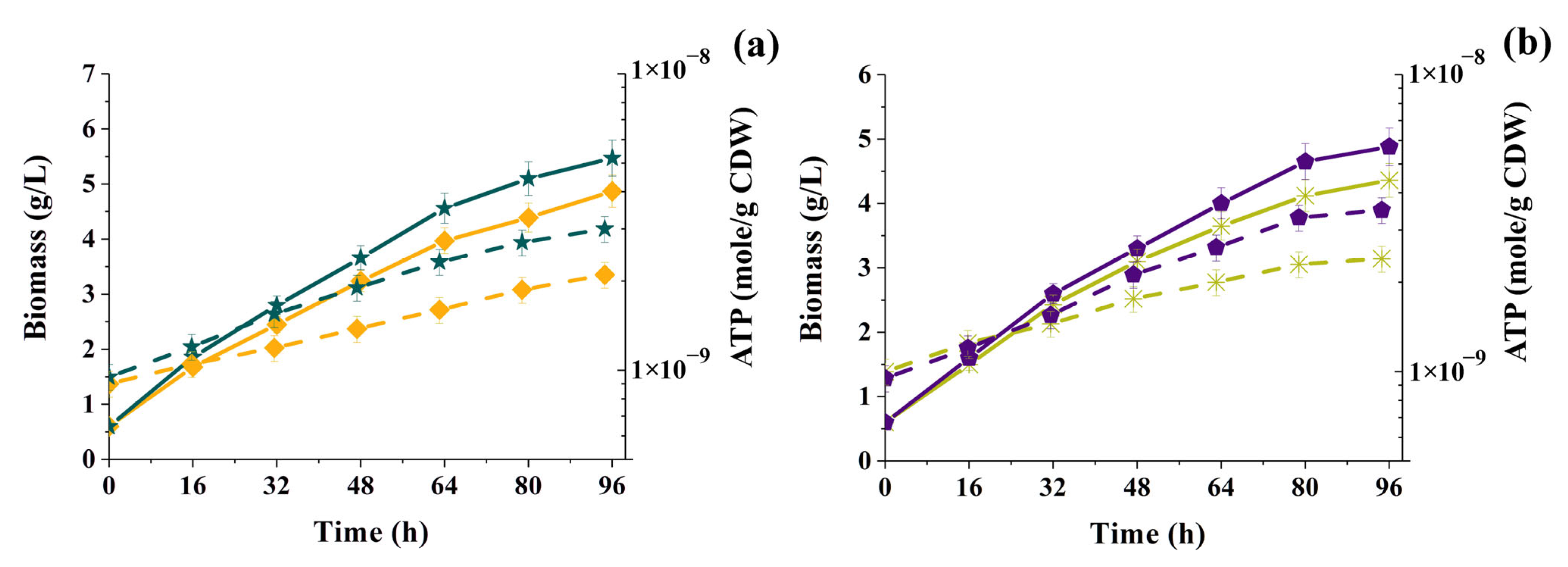
In this work, we used freshwater green microalgae Chlorella vulgaris, cyanobacteria Arthrospira platensis, and marine green microalgae Nannochloropsis sp. and Tetraselmis suecica to assess the effect of the presence of MP particles in their microenvironment on their intracellular ATP levels (Figure 6).
Microalgae Chlorella vulgaris are one of the most studied cultures of phototrophic microorganisms. They are widely distributed in ecosystems [32]. They are actively used in biotechnological processes [23] and aquaculture [48].
Cyanobacteria Arthrospira platensis are widely used for wastewater treatment and destruction of xenobiotics [49,50], and are also of interest as a natural, renewable raw material for the production of food additives rich in vitamins and antioxidants [51].
Nannochloropsis sp. and Tetraselmis suecica are marine microalgae of importance in aquaculture for fish and shellfish production [52,53].
The established pattern of dependence of the change in the concentration of intracellular ATP and biomass of both green microalgae Chlorella vulgaris and cyanobacteria Arthrosporic platensis on the concentration of the initial inoculum and the concentration of MPs has a similar trend. This was most clearly seen from the results obtained during the exposure of high cell concentrations (107 cells/mL) in the presence of 5 g/L MP. In this case, inhibition of cell growth and a decrease in the ATP concentration were observed (Figure 7).
The growth rates of phototrophic microorganisms under autotrophic conditions are lower than those of bacteria and fungi (Figure 3, Figure 5 and Figure 6); therefore, the change in the concentration of ATP in phototrophic cells and their growth in the presence of different MP samples were analyzed not for 24 h, but over a longer period of time (96 h) (Figure 7).
At an initially low concentration of phototrophic cells and a high concentration of polymers (5 g/L), the concentration of accumulating cell biomass remained virtually unchanged compared to the control or changed very slightly, but the ATP concentration increased in the presence of most MP samples (Figure 7b,e), indicating high metabolic activity of cells. At the same time, both C. vulgaris and A. platensis cells responded highly positively in an equal manner to the presence of HDPE, PCL, and PLA. One of the reasons for such a metabolic response of phototrophic cells to these MPs was the ability of these particles to be retained in the surface layer of liquid media with cells and to facilitate the retention of phototrophic cells in the same layer. This contributed to the improvement of conditions for photosynthesis, which, apparently, was reflected in the controlled level of intracellular ATP in C. vulgaris and A. platensis.
Initially high cell concentrations in media with MPs in the case of A. platensis and the presence of synthetic polymers at a concentration of 1 g/L (Figure 7d) led to significantly less pronounced effects of MP exposure (5–10%) on cells. The detected decrease in the concentration of intracellular ATP in A. platensis cells in the presence of different MPs was not significant. In the case of C. vulgaris microalgae cells, which were exposed to high concentrations in media with 1 g/L of different MPs (Figure 7a), a clear increase in cells in the case of PLA, PVA, and HDPE particles was established, which was accompanied by noticeable sorption on MP particles (Figure 8a). The concentration of intracellular ATP in C. vulgaris cells slightly decreased, which, in comparison with the accumulation of biomass, indicated that the cells spent ATP on reproduction, rebuilding their metabolism with an orientation to new conditions of existence and adaptation to them.
In general, the tendency to stimulate the growth of phototrophic microorganisms by low concentrations of MPs has, on the one hand, a negative connotation, since in objects contaminated with MPs, cultures responsible for harmful blooming can develop more intensively. On the other hand, such a phenomenon can be used to stimulate the growth of phototrophs in biotechnological processes, for example, for the subsequent processing of accumulated biomass into biofuels or polymers [23].
A similar effect has been previously noted for cells of microalgae of the genus Chlorella [24], but also for other phototrophs. In particular, it was shown that the growth of cells of cyanobacteria Anabaena variabilis accelerated or slowed down depending on the concentration of HDPE microparticles present in the medium [14].
It has been previously shown for Arthrospira platensis cyanobacteria cells that LDPE microparticles at concentrations of up to 100 mg/L can have an inhibitory effect on growth and provoke oxidative stress in these cells [25]. Moreover, the authors noted that MPs lead to the rupture of Arthrospira platensis trichomes. The same effect was noted by other researchers in combination with increased secretion of polysaccharides and cell aggregation [54]. In our study, such an effect was not recorded (Figures S2–S5), probably due to the different duration of the experiments, and, possibly, due to the different concentrations of cells that were used in the mentioned studies. In the cited works, these concentrations are not indicated at all, and in our study, we showed for the first time that the role of the current concentration of cells, in which they find themselves next to different MPs, is of significant importance for the phototrophs to manifest their properties and metabolic preferences.
For marine microalgae Nannochloropsis sp. and Tetraselmis suecica, inhibition of growth and metabolism of both studied cultures was detected (Figure 7g–j), which increased with an increase in the MP concentration from 1 to 5 g/L in the cell exposure media. An exception was observed only in the case of the simultaneous presence of Nannochloropsis sp. cells and LDPE particles in the medium. This was probably due to the fact that LDPE particles rose to the gas–liquid interface along with the sorbed microalgae cells, which facilitated gas exchange for the cells (Figures S2–S5). For microalgae of the genus Nannochloropsis, increased cell aggregation was previously noted in the presence of polystyrene [55]. In this study, a similar effect was noted for these cells only in the presence of MPs such as PCL and HDPE in the medium.
According to the literature, a decrease in the concentration of Tetraselmis suecica cell biomass in the presence of MPs may be due to a decrease in cell size rather than their number [56], as well as increased cell aggregation around MP particles [57]. However, in our study, we did not observe a similar effect. In the experiment, which was carried out for 96 h (Figure 8), the cell size remained unchanged (Figures S2–S5), but the population size decreased (Figure 7i,j). Moreover, this picture was observed in the media with all MP samples, regardless of the initial cell concentration. This trend was most obvious when the cell concentration in the medium with MP was increased, which was associated with cell sorption on polymer particles (Figure 8b), and obviously, the subsequent death of these phototrophs. Only in the medium with PVA particles was the level of intracellular ATP the same for T. suecica cells as in the control (without MP particles). This was only observed for cells at a concentration of 104 cells/mL exposed to MPs (Figure 7i).

3.4. General Analysis of Obtained Results

Analyzing the obtained results and generalizing the identified trends in the change in the concentration of intracellular ATP in the cells of different microorganisms in the presence of particles of different MPs (Table 1), it can be noted that fungi (especially yeast) and bacteria showed greater tolerance to the presence of most samples of synthetic polymers than phototrophic microorganisms.
The decrease in intracellular ATP levels was observed in most cases, predominantly in the absence of nutrients in the cell exposure media. High cell density also improved their resistance to the presence of MPs.
Changes in ATP levels in cells under such conditions, including the time interval used, were most likely due to the adaptation of the microorganisms’ biochemical systems to the degradation of synthetic polymers and their consumption or their degradation products as food sources. The increase in ATP concentration in many cases coincides with an increase in cell biomass, so the assessment of cell biomass concentration helps explain the reasons for the increase in ATP and the correlation between these cell characteristics (Table 1; Figure 1, Figure 2, Figure 4 and Figure 7). However, according to the data in Table 1, for a number of cells in the presence of microplastics, cases of an increase in ATP levels in cells were observed that were not associated with an increase in biomass concentration. This applies to Candida maltose, Chlorella vulgaris, and Arthrospira platensis cells in the presence of mainly two types of microplastics: HDPE and PCL.
With regard to PCL, which is widely used in medicine and packaging materials, it is known from the literature that it is in the presence of PCL that membrane-tropic effects on cells are established, not from the polymers themselves, but from the products of their degradation [58]. It should be noted that PCLs are biodegradable polymers, and the appearance of their degradation products in the presence of different microorganism cells is quite probable. In our experiments, it turned out that it was the two indicated microalgae variants that showed an increase in ATP concentration against the background of a decrease in their numbers (Table 1), which may be associated with the membrane-tropic effect of the indicated microplastic and damage to the photosynthetic system of these cells, which has a membrane localization.
With regard to HDPE, it has been established that yeasts of the genus Candida have increased adhesion to this polymer [59,60]. A similar trend has been established for the microalgae Chlorella vulgaris; moreover, it has been shown that HDPE inhibits the growth of these cells and changes their biochemical composition [61]. The presence of HDPE in the environment leads to damage to the surface of Arthrospira platensis cells and significant changes in the functional groups of proteins that make up the biomass [62].
The results of the study show that MPs that enter ecobiosystems can quickly colonize various microorganisms and serve as an effective substrate for adhesion and reproduction. A clearly expressed negative effect was most often noted for synthetic biodegradable MPs (PLA and PCL), the monomers of which have membrane-tropic properties [58,63]. The most sensitive microorganism to the presence of MPs was the mycelial fungus Aspergillus niger.
Analysis of the data presented in Table 1 indicates that the cells of filamentous fungi are predominantly in the stage of reorganization of biochemical pathways for MP consumption. Yeast S. cerevisiae and bacteria (both P. putida and B. subtillus, especially at concentrations typical for QS) are obviously the most successful in maintaining not only the levels of metabolism and growth that are typical for similar cells in the absence of MPs, but are also characterized by an excess of the control level of the analyzed parameters (intracellular ATP and cell number). This clearly indicates that the development of populations of these microorganisms will be stimulated by the presence of MP particles in ecosystems. Thus, the number and metabolic activity of consumers and reducers will be stimulated by the presence of most of the studied synthetic polymers.
In experiments with phototrophs, the influence of MPs was different. Thus, for freshwater phototrophs, at low cell concentrations, practically neutral reactions to the presence of MPs were possible, which changed to negative with an increase in the density of cell populations. For marine variants of microalgae, the effect of MPs was predominantly pronounced negative. The toxic effect of microplastics on various marine phototrophic microorganisms has been previously noted in various publications [12,64]. The authors note the aggregation of these phototrophic cells as the main reason for the toxic effect of microplastics on marine microalgae [64,65]. Such aggregation of microalgae leads to a decrease in the functions of their photosynthetic apparatus, localized in the cell membrane, which leads to sedimentation of the coarsening particles and their removal from the light source. At the same time, the researchers emphasize the selective toxicity to different species of microalgae [65]. Similar information was not found among the known publications in relation to Nanochloropsis cells, and therefore, the results obtained in this work are new; in relation to cells of the genus Tetraselmis, it was shown that the decrease in growth rate can reach ~20% in the presence of 1.47 mg/L PE in the medium with these marine microalgae [66].
For photobacteria with their own luminescence, a multidirectional effect of MPs on cells was revealed. It was shown that they can be used to determine the presence of biodegradable polymers such as PLA and PCL, which had a clearly negative effect on photobacteria used as an indicator of general ecotoxicity. It should be noted that the negative effect of these polymers on photobacteria was revealed in this work for the first time.
The aim of this work was to study the effect of a number of microplastics on the cells of various microorganisms. However, it is known that MPs are found not only in the environment, but also in the human body, including blood [67,68]. Twenty-four types of polymers were identified, with concentrations reaching 4.6 μg/mL. Among the discovered MPs, polyethylene, polystyrene, and PP were the most common. It has also been shown that MPs cause aggregation of erythrocytes and platelets, as well as enhance adhesion to endothelial cells, which can lead to thrombosis and cardiovascular diseases [69]. In our study, it was shown that cells of microorganisms with different cell wall structures (yeast, mycelial fungi, microalgae, and Gram-positive and Gram-negative bacteria) could be adsorbed on MPs. The greatest adsorption for all the studied cells was observed in the case of polyethylene (HDPE and LDPE), PLA, and PVA (Figure 3, Figure 5 and Figure 8). In this regard, the possible adsorption of blood cells on the surface of the same polymer microparticles, followed by an increase in the size of these formations (aggregates), is of particular concern. Due to our work and other investigations’ results, it can also be expected in the case of polyethylene, but the situation with PLA and PVA should be studied specially. That is because PLA is used in the production of various medical tablets, and PVA, considered GRAS, is widely used in foods.
When discussing possible candidates among microorganisms that could attract the attention of researchers based on the data obtained in this study, the following should be noted. According to literature data, yeasts are capable of carrying out PET degradation using their enzyme systems, the action of which leads to the formation of polymer degradation products, the consumption of which by yeast cells is possible as substrates [70]. Mono(2-hydroxyethyl) terephthalate is most effectively utilized [71]. Simultaneously with the utilization of the products of enzymatic destruction of PET, a change in the characteristics of this polymer surface is observed [72]. Researchers have found that in the presence of surfactants, in particular, Tween 20, the change in the plastic surface under the influence of yeast accelerated. Since yeasts themselves are capable of synthesizing biologically active substances [73], it can be assumed that all these properties of yeast cells taken together can contribute to the result that we observed in the experiments in the form of a fairly rapid response of yeast to the presence of PET in the medium, especially in the absence of other nutrition sources (Figure 1a,b,e,f).
Comparison of the results obtained in this work with the results of works on destructors of synthetic polymers [74] can also allow us to identify promising destructors of microplastics and note that the synthesis of exohydrolases is characteristic of mycelial fungi capable of transforming synthetic polymers [8]. These microorganisms are of interest primarily as producers of enzymes that catalyze the destruction of MPs.

4. Conclusions

The presence of nutrients in media with any initial concentration of the cells of microorganisms (bacteria and fungi) mainly contributed to the fact that MP particles either did not have a significant effect on the metabolic activity of microorganisms, or in some cases stimulated their growth and viability. In the absence of readily available nutrients, MPs inhibited cell development, but this inhibition was not fatal and was probably associated with the restructuring of metabolic pathways in the cells of microorganisms. In general, the following general trends were observed for the interaction between phototrophic microorganisms and MP particles: (i) MP particles at a concentration of 5 g/L inhibited the growth of freshwater and marine cultures at high population densities, and in most cases, at low population densities of cells of marine microalgae. Since this fact did not depend on the chemical structure of the polymers, it can be assumed that this is due to changes in the physical properties of the system. (ii) Polymer concentrations of 1 g/L inhibited the growth and metabolism of the studied microalgae and cyanobacteria in most cases. (iii) At an MP concentration of 5 g/L and low density of freshwater phototrophic cultures, a different nature of the reaction was observed: the A. platensis culture began to restructure the enzymatic systems for the consumption of new substrates; C. vulgaris microalgae in this case were completely inhibited by LDPE and tolerant to the presence of PVA, and in the cases of HDPE, PCL, and PLA, a restructuring of the enzymatic system of cells was observed.

Supplementary Materials

The following supporting information can be downloaded at https://www.mdpi.com/article/10.3390/microplastics4040072/s1: Figure S1: Samples of various microplastic particles used in the work. PVA—polyvinyl alcohol; PLA—polylactic acid; PCL—polycaprolactone; PP—polypropylene; PET—polyethylene terephthalate; HDPE—high-density polyethylene; LDPE—low-density polyethylene. Figure S2: Microphotographs of Chlorella vulgaris cells after cultivation for 96 h with microplastic particles (5 g/L). C—control (without microplastics). Other abbreviations are disclosed in the caption of Figure S1. Figure S3: Microphotographs of Arthrospira platensis after cultivation for 96 h with microplastic particles (5 g/L). All abbreviations are disclosed in the caption of Figure S1. Figure S4: Microphotographs of Nannochloropsis sp. after cultivation for 96 h with microplastic particles (5 g/L). All abbreviations are disclosed in the caption of Figure S1. Figure S5: Microphotographs of Tetraselmis suecica after cultivation for 96 h with microplastic particles (5 g/L). All abbreviations are disclosed in the caption of Figure S1.

Author Contributions

Conceptualization, E.E.; investigation, O.S., N.S., A.A., and E.E.; data curation, O.S. and E.E.; writing—original draft preparation, O.S., N.S., and E.E.; writing—review and editing, O.S. and E.E.; supervision, E.E. All authors have read and agreed to the published version of the manuscript.

Funding

This research was financially supported by the Russian Science Foundation (25-44-01003).

Institutional Review Board Statement

Not applicable.

Informed Consent Statement

Not applicable.

Data Availability Statement

The original contributions presented in this study are included in the article. Further inquiries can be directed to the corresponding author.

Conflicts of Interest

The authors declare no conflicts of interest.

Abbreviations

The following abbreviations are used in this manuscript:
ATPIntracellular adenosine triphosphate
MPsMicroplastics
EPSExopolysaccharides
QSQuorum Sensing
DCWDry cell weight
LDPELow-density polyethylene
HDPEHigh-density polyethylene
PETPolyethylene terephthalate
PPPolypropylene
PVAPolyvinyl alcohol
PCLPolycaprolactone
PLAPolylactic acid
DMSODimethyl sulfoxide
PSPolystyrene
PVCPolyvinyl chloride
PEPolyethylene

References

  1. Martínez-Burgos, W.J.; Pozzan, R.; de Carvalho, J.C.; Cavali, M.; Mariano, A.B.; Vargas, J.V.; Ordonez, J.; Severo, I.A.; Soccol, C.R. The role of microalgae as bioindicators of aquatic contaminations. In Algae as a Natural Solution for Challenges in Water-Food-Energy Nexus; Kurniawan, T.A., Anouzla, A., Eds.; Springer Nature: Singapore, 2024; pp. 323–347. [Google Scholar] [CrossRef]
  2. Yadav, S.; Singh, A.K.; Verma, E. Cyanobacteria as bioindicator of water pollution. In Cyanobacterial Biotechnology in the 21st Century; Neilan, B., Passarini, M.R.Z., Singh, P.K., Kumar, A., Eds.; Springer Nature: Singapore, 2023; pp. 149–179. [Google Scholar] [CrossRef]
  3. Miglani, R.; Parveen, N.; Kumar, A.; Ansari, M.A.; Khanna, S.; Rawat, G.; Panda, A.K.; Bisht, S.S.; Upadhyay, J.; Ansari, M.N. Degradation of xenobiotic pollutants: An environmentally sustainable approach. Metabolites 2022, 12, 818. [Google Scholar] [CrossRef]
  4. Nicula, N.-O.; Lungulescu, E.-M.; Rîmbu, G.A.; Marinescu, V.; Corbu, V.M.; Csutak, O. Bioremediation of wastewater using yeast strains: An assessment of contaminant removal efficiency. Int. J. Environ. Res. Public Health 2023, 20, 4795. [Google Scholar] [CrossRef] [PubMed]
  5. Slaveykova, V.I.; Marelja, M. Progress in research on the bioavailability and toxicity of nanoplastics to freshwater plankton. Microplastics 2023, 2, 389–410. [Google Scholar] [CrossRef]
  6. Santonicola, S.; Volgare, M.; Cocca, M.; Colavita, G. Abundance and characteristics of fibrous microplastics and microfibers isolated in Mullus barbatus from the Adriatic Sea—Preliminary investigation. Microplastics 2023, 2, 411–421. [Google Scholar] [CrossRef]
  7. Hrovat, B.; Uurasjärvi, E.; Koistinen, A. Analysis of microplastics in industrial processes—Systematic analysis of digestion efficiency of samples from forestry, wastewater treatment plants and biogas industries. Microplastics 2024, 3, 634–652. [Google Scholar] [CrossRef]
  8. Efremenko, E.N.; Lyagin, I.V.; Maslova, O.V.; Senko, O.V.; Stepanov, N.A.; Aslanli, A.G. Catalytic degradation of microplastics. Russ. Chem. Rev. 2023, 92, 5069. [Google Scholar] [CrossRef]
  9. Ayush, P.T.; Ko, J.-H.; Oh, H.-S. Characteristics of initial attachment and biofilm formation of Pseudomonas aeruginosa on microplastic surfaces. Appl. Sci. 2022, 12, 5245. [Google Scholar] [CrossRef]
  10. Efremenko, E.; Lyagin, I.; Stepanov, N.; Senko, O.; Maslova, O.; Aslanli, A.; Ugarova, N. Luminescent bacteria as bioindicators in screening and selection of enzymes detoxifying various mycotoxins. Sensors 2024, 24, 763. [Google Scholar] [CrossRef]
  11. De Jesus, R.; Iqbal, S.; Mundra, S.; AlKendi, R. Heterogenous bioluminescence patterns, cell viability, and biofilm formation of Photobacterium leiognathi strains exposed to ground microplastics. Front. Toxicol. 2024, 6, 1479549. [Google Scholar] [CrossRef]
  12. Zhang, C.; Chen, X.; Wang, J.; Tan, L. Toxic effects of microplastic on marine microalgae Skeletonema costatum: Interactions between microplastic and algae. Environ. Pollut. 2017, 220, 1282–1288. [Google Scholar] [CrossRef]
  13. Pencik, O.; Molnarova, K.; Durdakova, M.; Kolackova, M.; Klofac, D.; Kucsera, A.; Capal, P.; Svec, C.; Bytesnikova, Z.; Richtera, L.; et al. Not so dangerous? PET microplastics toxicity on freshwater microalgae and cyanobacteria. Environ. Pollut. 2023, 329, 121628. [Google Scholar] [CrossRef]
  14. Gopalakrishnan, K.K.; Kashian, D.R. Complex interactions among temperature, microplastics and cyanobacteria may facilitate cyanobacteria proliferation and microplastic deposition. Ecotoxicol. Environ. Saf. 2023, 263, 115259. [Google Scholar] [CrossRef] [PubMed]
  15. Das, S.; Lizon, F.; Gevaert, F.; Bialais, C.; Duong, G.; Ouddane, B.; Souissi, S. Assessing indicators of arsenic toxicity using variable fluorescence in a commercially valuable microalgae: Physiological and toxicological aspects. J. Hazard. Mater. 2023, 452, 131215. [Google Scholar] [CrossRef] [PubMed]
  16. Gu, M.; Yin, G.; Gan, T.; Zhao, N.; Qi, P.; Chen, M.; Ding, Z.; Jia, R.; Liu, J.; Ma, M.; et al. Construction of characterization parameters of algal photosynthetic inhibition method for detection of comprehensive toxicity in water. Ecol. Indic. 2022, 136, 108651. [Google Scholar] [CrossRef]
  17. Efremenko, E.; Senko, O.; Stepanov, N.; Maslova, O.; Lomakina, G.Y.; Ugarova, N. Luminescent Analysis of ATP: Modern Objects and Processes for Sensing. Chemosensors 2022, 10, 493. [Google Scholar] [CrossRef]
  18. Bule Možar, K.; Miloloža, M.; Martinjak, V.; Cvetnić, M.; Ocelić Bulatović, V.; Mandić, V.; Bafti, A.; Ukić, Š.; Kučić Grgić, D.; Bolanča, T. Bacteria and yeasts isolated from the environment in biodegradation of PS and PVC microplastics: Screening and treatment optimization. Environments 2023, 10, 207. [Google Scholar] [CrossRef]
  19. Miloloža, M.; Bule, K.; Prevarić, V.; Cvetnić, M.; Ukić, Š.; Bolanča, T.; Kučić Grgić, D. Assessment of the influence of size and concentration on the ecotoxicity of microplastics to microalgae Scenedesmus sp., bacterium Pseudomonas putida and yeast Saccharomyces cerevisiae. Polymers 2022, 14, 1246. [Google Scholar] [CrossRef]
  20. Li, R.; Tao, J.; Huang, D.; Zhou, W.; Gao, L.; Wang, X.; Chen, H.; Huang, H. Investigating the effects of biodegradable microplastics and copper ions on probiotic (Bacillus amyloliquefaciens): Toxicity and application. J. Hazard. Mater. 2023, 443, 130081. [Google Scholar] [CrossRef]
  21. Yi, X.; Li, W.; Liu, Y.; Yang, K.; Wu, M.; Zhou, H. Effect of polystyrene microplastics of different sizes to Escherichia coli and Bacillus cereus. Bull. Environ. Contam. Toxicol. 2021, 107, 626–632. [Google Scholar] [CrossRef]
  22. Hossain, S.; Shukri, Z.N.A.; Waiho, K.; Ibrahim, Y.S.; Kamaruzzan, A.S.; Rahim, A.I.A.; Draham, A.S.; Wahab, W.; Khatoon, H.; Kasan, N.A. Biodegradation of polyethylene (PE), polypropylene (PP), and polystyrene (PS) microplastics by floc-forming bacteria, Bacillus cereus strain SHBF2, isolated from a commercial aquafarm. Environ. Sci. Pollut. Res. 2024, 31, 32225–32245. [Google Scholar] [CrossRef]
  23. Senko, O.; Stepanov, N.; Maslova, O.; Efremenko, E. Transformation of enzymatic hydrolysates of chlorella–fungus mixed biomass into poly(hydroxyalkanoates). Catalysts 2023, 13, 118. [Google Scholar] [CrossRef]
  24. Abomohra, A.; Hanelt, D. Recent advances in micro-/nanoplastic (MNPs) removal by microalgae and possible integrated routes of energy recovery. Microorganisms 2022, 10, 2400. [Google Scholar] [CrossRef]
  25. Abbasi, S.; Amiranipour, S.; Karimi, J.; Mohsenzadeh, S.; Turner, A. Impacts of polyethylene microplastics on the microalga, Spirulina (Arthrospira platensis). Environ. Pollut. 2023, 327, 121611. [Google Scholar] [CrossRef] [PubMed]
  26. Liu, X.; Yang, J.; Wang, W.; Bao, X.; Chang, D.; Hao, Z.; Qiu, M.; Liu, Y.; Teng, X.; Li, Z. Mechanisms of nanoplastic-induced energy metabolism reprogramming in juvenile Sepia esculenta: mRNA profile, miRNA/mRNA network, and ceRNA network. J. Environ. Chem. Ecotoxicol. 2025, 7, 1836–1849. [Google Scholar] [CrossRef]
  27. Simon-Sánchez, L.; Grelaud, M.; Lorenz, C.; Garcia-Orellana, J.; Vianello, A.; Liu, F.; Vollertsen, J.; Ziveri, P. Can a sediment core reveal the plastic age? Microplastic preservation in a coastal sedimentary record. Environ. Sci. Technol. 2022, 56, 16780–16788. [Google Scholar] [CrossRef] [PubMed]
  28. Singh, S.; Chakma, S.; Alawa, B.; Kalyanasundaram, M.; Diwan, V. Microplastic pollution in terrestrial environment: Identification, characterization, and risk assessment in Indore, Central India. Soil Use Manag. 2024, 40, e13053. [Google Scholar] [CrossRef]
  29. Feng, S.; Wang, H.; Wang, Y.; Cheng, Q. A review of the occurrence and degradation of biodegradable microplastics in soil environments. Sci. Total Environ. 2023, 904, 166855. [Google Scholar] [CrossRef]
  30. Nigro, L.; Magni, S.; Ortenzi, M.A.; Gazzotti, S.; Della Torre, C.; Binelli, A. Are “liquid plastics” a new environmental threat? The case of polyvinyl alcohol. Aquat. Toxicol. 2022, 248, 106200. [Google Scholar] [CrossRef]
  31. Abbamondi, G.R.; Tommonaro, G. Research progress and hopeful strategies of application of quorum sensing in food, agriculture and nanomedicine. Microorganisms 2022, 10, 1192. [Google Scholar] [CrossRef]
  32. Senko, O.; Maslova, O.; Aslanli, A.; Efremenko, E. Impact of perfluorocarbons with gas transport function on growth of phototrophic microorganisms in a free and immobilized state and in consortia with bacteria. Appl. Sci. 2023, 13, 1868. [Google Scholar] [CrossRef]
  33. Efremenko, E.N.; Nikolskaya, A.B.; Lyagin, I.V.; Senko, O.V.; Makhlis, T.A.; Stepanov, N.A.; Maslova, O.V.; Mamedova, F.; Varfolomeev, S.D. Production of biofuels from pretreated microalgae biomass by anaerobic fermentation with immobilized Clostridium acetobutylicum cells. Bioresour. Technol. 2012, 114, 342–348. [Google Scholar] [CrossRef] [PubMed]
  34. Algae. Consolidated Catalogue of Microbial Cultures Held in Russian Non-Medical Collections. Available online: https://vkm.ru/eCatalogue.htm (accessed on 4 July 2025).
  35. Ugarova, N.; Koksharov, M.; Lomakina, G. Reagent for Adenosine-5-Triphosphate Determination. Patent RU 2420594, 20 May 2009. [Google Scholar]
  36. Sánchez, C. Fungal potential for the degradation of petroleum-based polymers: An overview of macro-and microplastics biodegradation. Biotechnol. Adv. 2020, 40, 107501. [Google Scholar] [CrossRef] [PubMed]
  37. Baztan, J.; Carrasco, A.; Chouinard, O.; Cleaud, M.; Gabaldon, J.E.; Huck, T.; Jaffres, L.; Jorgensen, B.; Miquelez, A.; Paillard, C.; et al. Protected areas in the Atlantic facing the hazards of micro-plastic pollution: First diagnosis of three islands in the Canary current. Mar. Pollut. Bull. 2014, 80, 302–311. [Google Scholar] [CrossRef] [PubMed]
  38. Meizoso-Regueira, T.; Fuentes, J.; Cusworth, S.J.; Rillig, M.C. Prediction of future microplastic accumulation in agricultural soils. Environ. Pollut. 2024, 359, 124587. [Google Scholar] [CrossRef]
  39. Mueller, R.-J. Biological degradation of synthetic polyesters—Enzymes as potential catalysts for polyester recycling. Process Biochem. 2006, 41, 2124–2128. [Google Scholar] [CrossRef]
  40. Hunková, Z.; Fencl, A. Toxic effects of fatty acids on yeast cells: Possible mechanisms of action. Biotechnol. Bioeng. 1978, 20, 1235–1247. [Google Scholar] [CrossRef]
  41. Fu, X.; Zhang, S.; Zhang, X.; Zhang, Y.; Li, B.; Jin, K.; Feng, X.; Hong, J.; Huang, X.; Cao, H.; et al. Sustainable microplastic remediation with record capacity unleashed via surface engineering of natural fungal mycelium framework. Adv. Funct. Mater. 2023, 33, 2212570. [Google Scholar] [CrossRef]
  42. Hadiyanto, H.; Joelyna, F.A.; Khoironi, A.; Sudarno, S.; Safaat, J.A.; Pratama, W.D.; Nur, M.M.A. Harnessing Chlorella vulgaris-Aspergilus niger Interactions for effective microplastic removal in aquatic ecosystems. Waste Biomass Valor. 2025, 1–17. [Google Scholar] [CrossRef]
  43. Krumov, N.; Atanasova, N.; Boyadzhieva, I.; Petrov, K.; Petrova, P. Biodegradation of Poly (ε-caprolactone): Microorganisms, Enzymes, and Mechanisms. Int. J. Mol. Sci. 2025, 26, 5826. [Google Scholar] [CrossRef]
  44. Báez-Flores, M.E.; Tiznado-Hernández, M.E.; Gracia-Valenzuela, M.H.; Troncoso-Rojas, R. Biosphere plastic contamination and microbial alternatives for a sustainable degradation of plastic waste. Microorganisms 2025, 13, 1246. [Google Scholar] [CrossRef]
  45. Fang, X.; Cai, Z.; Wang, X.; Liu, Z.; Lin, Y.; Li, M.; Gong, H.; Yan, M. Isolation and identification of four strains of bacteria with potential to biodegrade polyethylene and polypropylene from Mangrove. Microorganisms 2024, 12, 2005. [Google Scholar] [CrossRef]
  46. Boyandin, A.N.; Prudnikova, S.V.; Karpov, V.A.; Ivonin, V.N.; Dỗ, N.L.; Nguyễn, T.H.; Lê, T.M.H.; Filichev, N.L.; Levin, A.L.; Filipenko, M.L. Microbial degradation of polyhydroxyalkanoates in tropical soils. Int. Biodeterior. Biodegrad. 2013, 83, 77–84. [Google Scholar] [CrossRef]
  47. Cholewińska, P.; Moniuszko, H.; Wojnarowski, K.; Pokorny, P.; Szeligowska, N.; Dobicki, W.; Polechoński, R.; Górniak, W. The occurrence of microplastics and the formation of biofilms by pathogenic and opportunistic bacteria as threats in aquaculture. Int. J. Environ. Res. Public Health 2022, 19, 8137. [Google Scholar] [CrossRef]
  48. Aly, S.M.; ElBanna, N.I.; Fathi, M. Chlorella in aquaculture: Challenges, opportunities, and disease prevention for sustainable development. Aquac. Int. 2024, 32, 1559–1586. [Google Scholar] [CrossRef]
  49. Araujo, G.S.; Santiago, C.S.; Moreira, R.T.; Neto, M.P.D.; Fernandes, F.A. Nutrient removal by Arthrospira platensis cyanobacteria in cassava processing wastewater. J. Water Process Eng. 2021, 40, 101826. [Google Scholar] [CrossRef]
  50. Tabagari, I.; Kurashvili, M.; Varazi, T.; Adamia, G.; Gigolashvili, G.; Pruidze, M.; Chokheli, L.; Khatisashvili, G.; von Fragstein und Niemsdorff, P. Application of Arthrospira (Spirulina) platensis against chemical pollution of water. Water 2019, 11, 1759. [Google Scholar] [CrossRef]
  51. Gentscheva, G.; Nikolova, K.; Panayotova, V.; Peycheva, K.; Makedonski, L.; Slavov, P.; Radusheva, P.; Yotkovska, I. Application of Arthrospira platensis for medicinal purposes and the food industry: A review of the literature. Life 2023, 13, 845. [Google Scholar] [CrossRef] [PubMed]
  52. Park, B.S.; Choi, W.J.; Guo, R.; Kim, H.; Ki, J.S. Changes in free-living and particle-associated bacterial communities depending on the growth phases of marine green algae Tetraselmis suecica. J. Mar. Sci. Eng. 2021, 9, 171. [Google Scholar] [CrossRef]
  53. Marchi, A.; Benini, E.; Dondi, F.; Ferrari, M.G.; Scicchitano, D.; Palladino, G.; Candela, M.; Cerri, R.; Di Biase, F.; Vizcaino, A.J.; et al. The use of fishery and aquaculture by-products with Nannochloropsis sp. allows total dietary replacement of wild-caught fishmeal, fish oil and soy protein in European sea bass juveniles. Aquaculture 2024, 590, 741015. [Google Scholar] [CrossRef]
  54. Yue, Z.; Qian, J.; Li, W.; Liu, X.; Dai, H.; Liu, X.; Pi, F.; Wang, J. Spotlight on the long-term effects of micro/nanoplastics exposure on Spirulina platensis: Algal cells, extracellular polymeric substances, and phycocyanin. Food Chem. 2025, 472, 142940. [Google Scholar] [CrossRef]
  55. Xu, H.; Li, L.A.; Wang, Y.; Qiu, K.; Chen, S.; Zeng, J.; Liu, R.; Yang, Q.; Huang, W. Differential physiological response of marine and freshwater microalgae to polystyrene microplastics. J. Hazard. Mater. 2023, 448, 130814. [Google Scholar] [CrossRef] [PubMed]
  56. Mendonça, I.; Cunha, C.; Kaufmann, M.; Faria, M.; Cordeiro, N. Microplastics reduce microalgal biomass by decreasing single-cell weight: The barrier towards implementation at scale. Sci. Total Environ. 2023, 877, 162950. [Google Scholar] [CrossRef] [PubMed]
  57. Cunha, C.; Faria, M.; Nogueira, N.; Ferreira, A.; Cordeiro, N. Marine vs freshwater microalgae exopolymers as biosolutions to microplastics pollution. Environ. Pollut. 2019, 249, 372–380. [Google Scholar] [CrossRef] [PubMed]
  58. Li, Z.; Qiu, S.; Xu, J.; Ge, S. Effect of undissociated n-caproic acid on methanogen activity and subsequent recovery: Methane anabolism and community structure. ACS ES&T Eng. 2024, 4, 706–716. [Google Scholar] [CrossRef]
  59. Oliveira, M.M.; Proenca, A.M.; Moreira-Silva, E.; Dos Santos, F.M.; Marconatto, L.; de Castro, A.M.; Medina-Silva, R. Biochemical features and early adhesion of marine Candida parapsilosis strains on high-density polyethylene. J. Appl. Microbiol. 2022, 132, 1954–1966. [Google Scholar] [CrossRef]
  60. Akinbobola, A.; Kean, R.; Quilliam, R.S. Plastic pollution as a novel reservoir for the environmental survival of the drug resistant fungal pathogen Candida auris. Mar. Pollut. Bull. 2024, 198, 115841. [Google Scholar] [CrossRef]
  61. Sponza, D.T.; Öztekin, R. Removals of some high-and low-density polyethylene (HDPE and LDPE), polypropylene (PP) and polyvinyl chloride (PVC) microplastics using some microalgae types, energy production and energy recovery. Mol. Sci. Appl. 2023, 3, 66–68. [Google Scholar] [CrossRef]
  62. Hadiyanto, H.; Khoironi, A.; Dianratri, I.; Suherman, S.; Muhammad, F.; Vaidyanathan, S. Interactions between polyethylene and polypropylene microplastics and Spirulina sp. microalgae in aquatic systems. Heliyon 2021, 7, e07676. [Google Scholar] [CrossRef]
  63. Alakomi, H.L.; Skytta, E.; Saarela, M.; Mattila-Sandholm, T.; Latva-Kala, K.; Helander, I.M. Lactic acid permeabilizes gram-negative bacteria by disrupting the outer membrane. Appl. Environ. Microbiol. 2000, 66, 2001–2005. [Google Scholar] [CrossRef]
  64. Su, Y.; Gao, L.; Xu, E.G.; Peng, L.; Diao, X.; Zhang, Y.; Bao, R. When microplastics meet microalgae: Unveiling the dynamic formation of aggregates and their impact on toxicity and environmental health. Water Res. 2025, 273, 123008. [Google Scholar] [CrossRef]
  65. Wang, M.; Jiang, S.; Tan, H.; Wang, H.; Wang, J.; Song, K.; Xu, D.; Zhang, B.; Liu, Z.; Liu, X.; et al. Responses of different species of marine microalgae and their community to gear-derived microplastics. Water Res. 2025, 281, 123528. [Google Scholar] [CrossRef] [PubMed]
  66. Davarpanah, E.; Guilhermino, L. Single and combined effects of microplastics and copper on the population growth of the marine microalgae Tetraselmis chuii. Estuar. Coast. Shelf Sci. 2015, 167, 269–275. [Google Scholar] [CrossRef]
  67. Leonard, S.V.; Liddle, C.R.; Atherall, C.A.; Chapman, E.; Watkins, M.; Calaminus, S.D.; Rotchell, J.M. Microplastics in human blood: Polymer types, concentrations and characterization using mFTIR. Environ. Int. 2024, 188, 108751. [Google Scholar] [CrossRef] [PubMed]
  68. Lee, D.W.; Jung, J.; Park, S.; Lee, Y.; Kim, J.; Han, C.; Kim, H.C.; Lee, J.H.; Hong, Y.C. Microplastic particles in human blood and their association with coagulation markers. Sci. Rep. 2024, 14, 30419. [Google Scholar] [CrossRef]
  69. Rajendran, D.; Chandrasekaran, N. Journey of micronanoplastics with blood components. RSC Adv. 2023, 13, 31435–31459. [Google Scholar] [CrossRef]
  70. da Costa, A.M.; de Oliveira Lopes, V.R.; Vidal, L.; Nicaud, J.M.; de Castro, A.M.; Coelho, M.A.Z. Poly (ethylene terephthalate)(PET) degradation by Yarrowia lipolytica: Investigations on cell growth, enzyme production and monomers consumption. Process Biochem. 2020, 95, 81–90. [Google Scholar] [CrossRef]
  71. Loll-Krippleber, R.; Sajtovich, V.A.; Ferguson, M.W.; Ho, B.; Burns, A.R.; Payliss, B.J.; Bellisssimo, J.; Peters, S.; Roy, P.G.; Wyatt, H.D.M.; et al. Development of a yeast whole-cell biocatalyst for MHET conversion into terephthalic acid and ethylene glycol. Microb. Cell Fact. 2022, 21, 280. [Google Scholar] [CrossRef]
  72. Giyahchi, M.; Moghimi, H. Acceleration a yeast-based biodegradation process of polyethylene terephthalate microplastics by Tween 20: Efficiency, by-product analysis, and metabolic pathway Prediction. Environ. Pollut. 2024, 351, 124106. [Google Scholar] [CrossRef]
  73. Araujo, J.; Monteiro, J.; Silva, D.; Alencar, A.; Silva, K.; Coelho, L.; Pacheco, W.; Silva, D.; Silva, M.; Silva, L.; et al. Surface-Active Compounds Produced by Microorganisms: Promising Molecules for the Development of Antimicrobial, Anti-Inflammatory, and Healing Agents. Antibiotics 2022, 11, 1106. [Google Scholar] [CrossRef]
  74. Senko, O.; Maslova, O.; Stepanov, N.; Aslanli, A.; Lyagin, I.; Efremenko, E. Role of Humic Substances in the (Bio)Degradation of Synthetic Polymers under Environmental Conditions. Microorganisms 2024, 12, 2024. [Google Scholar] [CrossRef]
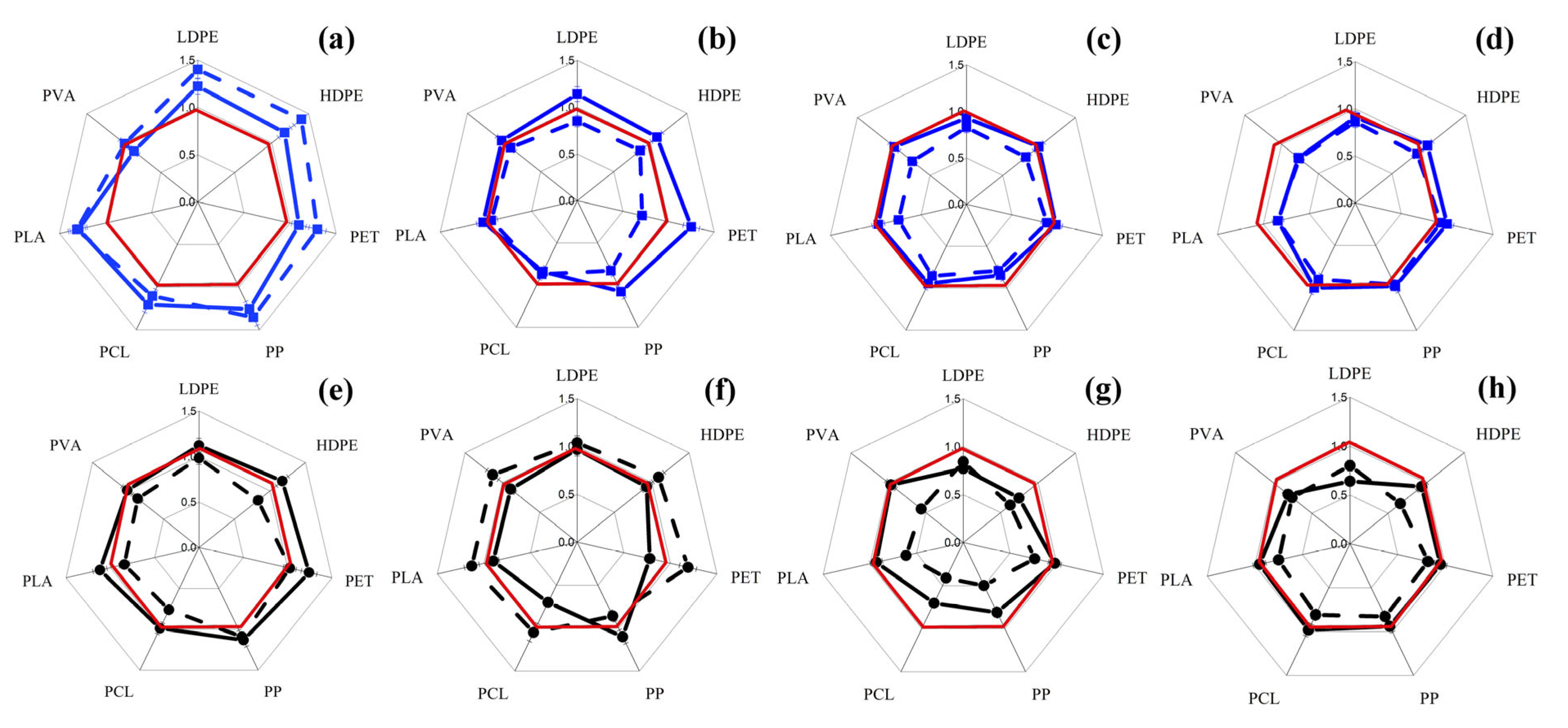
Figure 1. Changes in total intracellular ATP concentration (dashed lines) in yeast cells S. cerevisiae (ad) and ●—C. maltose (eh), and their biomass concentrations (solid lines) in a medium with MP particles (5 g/L) as a result of their exposure for 24 h in a nutrient medium with an initial inoculum concentration of 104 cells/mL (a,e) and 107 cells/mL (b,f) and in a saline solution with an inoculum concentration of 104 cells/mL (c,g) and 107 cells/mL (d,h), respectively. The axes corresponding to each type of MP particle used in the work show the number of times the change (increase or decrease) in the studied parameters occurs in relation to the control (cells without MP under the same conditions) (red lines).
Microplastics 04 00072 g001
Figure 2. Changes in total intracellular ATP concentration (dashed lines) in filamentous fungal cells R. oryzae (ad) and A. niger (eh) when exposed to MP particles (5 g/L) for 24 h under different conditions: a nutrient medium with 7.5 g dry bm/L (a,e) and 45 g dry bm/L and (b) a saline solution with 7.5 g dry bm/L (c,g) and 45 g dry bm/L (d,h), respectively. The axes corresponding to each type of MP particles used in the work show the number of times the change (increase or decrease) in the studied parameters occurs in relation to the control (cells without MP under the same conditions) (red lines).
Microplastics 04 00072 g002
Figure 3. Biomass accumulation (solid lines) and changes in specific intracellular concentration of ATP (dashed lines) during cultivation of yeast cells S. cerevisiae () and C. maltose (●) (a) and filamentous fungi Rhizopus oryzae () and Aspergillus niger () in the absence of MP (b) in a nutrient medium. The concentration of intracellular ATP detected on MP particles (5 g/L) after culturing of yeast S. cerevisiae () and C. maltose (■) (c) and filamentous fungi R. oryzae () and A. niger () (d), respectively, in a nutrient medium with them for 24 h. The initial cell concentration was 107 cells/mL.
Microplastics 04 00072 g003
Figure 4. Changes in total intracellular ATP concentration (dashed lines) in bacterial cells Bacillus subtills (ad) and Pseudomonas putida (eh), as well as the concentration of their biomass (solid lines) in a medium with MP particles (5 g/L) as a result of their exposure for 24 h in a nutrient medium with an initial inoculum concentration of 104 cells/mL (a,e) and 107 cells/mL (b,f) and in a saline solution with an inoculum concentration of 104 cells/mL (c,g) and 107 cells/mL (d,h), respectively. The axes corresponding to each type of MP particle used in the work show the number of times the change (increase or decrease) in the studied parameters occurs in relation to the control (cells without MP under the same conditions) (red lines).
Microplastics 04 00072 g004
Figure 5. (a) Relative residual luminescence (solid line) and change in intracellular ATP concentration (dashed line) in ✕—Photobacterium phosphoreum cells when exposed to particles of different MPs (5 g/L) in a 2% NaCl solution. The axes corresponding to each type of MP particles used in the work show the number of times the change (increase or decrease) in the studied parameters occurs in relation to the control (cells without MP under the same conditions) (red lines). (b,c) Biomass accumulation (solid lines) and changes in intracellular ATP concentration (dashed lines) during cultivation of various bacteria are assessed for (b) P. phosphoreum (), (c) B. subtilis (), and P. putida (). (d) Intracellular ATP concentration detected on MP particles (5 g/L) after exposure of B. subtilis () and P. putida () cells to the medium containing them within 24 h at an initial cell concentration of 107 cells/mL.
Microplastics 04 00072 g005
Figure 6. Biomass accumulation (solid lines) and changes in intracellular ATP concentration (dashed lines) during cultivation of phototrophic microorganisms: Arthrospira platensis and Chlorella vulgaris (a); Nanochloropsis sp. and Tetraselmis suecica (b).
Microplastics 04 00072 g006
Figure 7. Biomass accumulation (solid lines) and changes in intracellular ATP concentration (dashed lines) during exposition for 96 h of green microalgae Chlorella vulgaris (ac) and cyanobacteria Arthrospira platensis (df) and marine microalgae Nannochloropsis sp. (g,h) and Tetraselmis suecica (i,j) with 1 g/L MP and initial cell concentration of 107 cells/mL (c,f), with 5 g/L MP and 104 cells/mL (a,d,g,i) or 107 cells/mL (b,e,h,j). The axes corresponding to each type of MP particles used in the work show the number of times the change (increase or decrease) in the studied parameters occurs in relation to the control (cells without MP under the same conditions) (red lines).
Microplastics 04 00072 g007
Figure 8. Intracellular ATP concentration detected on MP particles (5 g/L) after cultivation of cells of C. vulgaris () and A. platensis () (a) and Nannochloropsis sp. () and Tetraselmis suecica () (b), respectively, in a nutrient medium with them for 96 h at an initial cell concentration of 107 cells/mL.
Microplastics 04 00072 g008
Table 1. Trends in changes in intracellular ATP concentration and biomass (left and right halves of rectangles, respectively) revealed in different microorganisms in the presence of MP particles.
MPScCmRoAnBsPpPhpCvApNsTs
5 g/L MP, Nutrient Medium, 104 Cells/mL2% NaCl5 g/L MP, MNM *, 104 Cells/mL
LDPEABABABABABABABABABABAB
HDPEABABABABABABABABABABAB
PETABABABABABABABABABABAB
PPABABABABABABABABABABAB
PCLABABABABABABABABABABAB
PLAABABABABABABABABABABAB
PVAABABABABABABABABABABAB
5 g/L MP, nutrient medium, 107 cells/mL5 g/L MP, MNM, 107 cells/mL
LDPEABABABABABAB ABABABAB
HDPEABABABABABAB ABABABAB
PETABABABABABAB ABABABAB
PPABABABABABAB ABABABAB
PCLABABABABABAB ABABABAB
PLAABABABABABAB ABABABAB
PVAABABABABABAB ABABABAB
5 g/L MP, saline, 104 cells/mL1 g/LMP, MNM, 107 cells/mL
LDPEABABABABABAB ABAB
HDPEABABABABABAB ABAB
PETABABABABABAB ABAB
PPABABABABABAB ABAB
PCLABABABABABAB ABAB
PLAABABABABABAB ABAB
PVAABABABABABAB ABAB
5 g/L MP, saline, 107 cells/mLATP (A) and biomass (B) decreases in the parameters are highlighted in red, increases in green, and constant levels in yellow. Yeasts: Sc—S. cerevisiae and Cm—C. maltose; bacteria: Bs—B. subtillus, Pp—P. putida, and Php—P. phosphoreum; mycelial fungi: Ro—R. oryzae and An—A. niger; microalgae: Cv -C. vulgaris, Ns—Nannochloropsis sp., and Ts—T. suecica; cyanobacteria: Ap—A. platensis in the presence of MP under various experimental conditions.
LDPEABABABABABAB
HDPEABABABABABAB
PETABABABABABAB
PPABABABABABAB
PCLABABABABABAB
PLAABABABABABAB
PVAABABABABABAB
* MNM—minimum nutrient medium. Autotrophic cultivation conditions: Microplastics 04 00072 i001—stimulation of cell growth and metabolism; Microplastics 04 00072 i002, Microplastics 04 00072 i003—survival of cells due to the death of some of them, which means there is toxicity or lack of available nutrition; Microplastics 04 00072 i004—cells are tolerant to the presence of MPs, behaving as in the control in the absence of MPs; Microplastics 04 00072 i005—the culture cells are in the growth stage; Microplastics 04 00072 i006—maintenance of metabolism occurs at the expense of the death of some cells, which indicates the presence of toxicity or lack of available nutrition; Microplastics 04 00072 i007—inhibition of cell growth and metabolism; Microplastics 04 00072 i008—the culture is in the process of restructuring biochemical pathways to consume MPs; Microplastics 04 00072 i009—the culture is in the stage of division and restructuring of enzymatic systems for MP consumption.
Disclaimer/Publisher’s Note: The statements, opinions and data contained in all publications are solely those of the individual author(s) and contributor(s) and not of MDPI and/or the editor(s). MDPI and/or the editor(s) disclaim responsibility for any injury to people or property resulting from any ideas, methods, instructions or products referred to in the content.