Review

Dermatogenomic Insights into Systemic Diseases: Implications for Primary and Preventive Medicine


1
Faculty of Medicine, University of British Columbia, Vancouver, BC V6T 1Z3, Canada
2
Department of Dermatology and Skin Science, University of British Columbia, Vancouver, BC V5Z 4E8, Canada
*
Author to whom correspondence should be addressed.
Submission received: 23 July 2025 / Revised: 2 October 2025 / Accepted: 11 November 2025 / Published: 6 January 2026

Abstract

The emerging field of dermatogenomics, which examines visible dermatologic phenotypes alongside their polygenic factors, offers insights for early disease recognition and initiation of preventative measures. This review explores key dermatologic manifestations serving as clinical markers of systemic diseases, emphasizing cardiovascular, autoimmune, neuropsychiatric, metabolic/endocrine, and cancer-related conditions. Importantly, the pathogenesis of certain skin conditions including psoriasis, atopic dermatitis, vitiligo, and hidradenitis suppurativa is linked to systemic disease through shared genetic and epigenetic mechanisms. The diagnostic markers for these integumentary diseases are discussed alongside their shared mechanisms to systemic diseases, highlighting the clinical manifestation typically seen in primary care settings. This narrative review integrates dermatology with genomics, primary care, preventative care, public health, and internal medicine perspectives, underscoring the importance of an interdisciplinary and collaborative approach to patient care. Lastly, this review advocates for standardized dermatogenomic screening thresholds, inclusivity and expansion of genomic datasets, and the leverage of artificial intelligence and multi-omic technologies in preventative healthcare.

1. Introduction

Dermatogenomics, the integration of dermatological conditions with genomic data, enables clinicians to interpret visible skin findings as indicators of underlying genetic susceptibilities to diseases. The skin, the body’s largest and most visible organ, provides a unique clinical vantage point for detecting genetic and epigenetic markers predictive of systemic conditions [1]. As this narrative review will address, skin disease may precede or follow systemic disease or multi-organ malfunction due to the complex interplay between multiple gene variants, providing a window of time for preventative care and early intervention. Despite advancements in genomics, significant limitations remain, particularly the inadequate representation of diverse populations in genome studies, limiting the global applicability of genetic risk assessments [2,3,4]. Dermatogenomics can address these limitations by combining phenotypic observations from individual examinations with genomic analysis, offering precision medicine applications in specialties such as dermatology, internal medicine, and primary care. Existing research has identified dermatogenomics as a useful tool for assessing familial melanoma risk [5]. Moreover, genome-wide association studies (GWAS) have provided useful insights in understanding dermatology case studies [6]. More recently, with the development of high-throughput technologies such as next generation sequencing and the discovery of new genetic sites for targeted therapies, dermatogenomics have been rising in popularity amongst academics and clinicians alike [7]. This narrative review aims to consolidate dermatogenomic correlates of systemic diseases to facilitate predictive screening and prevention strategies for health practitioners. We further aim to identify current limitations in the application of dermatogenomics and future directions for research.

2. Dermatological Manifestations as Windows to Systemic Diseases

2.1. Psoriasis and Cardiovascular Risks

Psoriasis is an autoimmune skin condition that causes chronic inflammation, leading to scaly eruption throughout the body [8]. Despite its classification as a skin disease, recent studies have shown a strong link between psoriasis and cardiovascular diseases [9]. The mechanisms behind inflammatory processes in psoriasis involves mediators such as tumor necrosis factor-alpha (TNF-α), IL-17, IL-23 and IL-6, which are all linked to vascular inflammation and atherosclerosis development [10]. Furthermore, transcriptomics has been able to elucidate several key ferroptosis-related genes and necroptosis-related genes that showed differential enrichment during psoriatic inflammation and atherosclerotic events; more in vivo studies exploring these genetic markers need to be replicated to validate the connection [11]. The same upstream hub genes have been implicated to be involved in the pathology of both diseases, raising interest and insights in the study of these common pathogenic mechanisms [12]. Given these shared mechanisms, individuals suffering from psoriasis are up to 50% more likely to have cardiovascular diseases, with the likelihood increasing depending on the severity of psoriasis, accenting the importance of appropriate primary and preventative care before the development of cardiovascular symptoms [10]. Common treatments for psoriasis are TNF-α and IL-17 inhibitors. Due to the role that these cytokines play in both autoimmune responses and atherosclerosis, these inhibitors have demonstrated significant efficacy in diminishing non-calcified plaque burden and thereby improving cardiovascular disease outcomes [13]. Similarly, first-line medications for atherosclerosis such as oral statins have shown positive effects in severe psoriasis, particularly due to their anti-inflammatory effects [14]. Given that patients with psoriasis present with a significantly greater risk for cardiovascular diseases, further research should use high-end genomic technologies to explore the markers involved in patients with isolated psoriasis versus those with concurrent cardiovascular disease. The interplay between specific genetic markers correlated with cardiovascular disease in patients with psoriasis could be elucidated and key targets for therapy development could be identified.

2.2. Atopic Dermatitis and Atopy

Atopic dermatitis or eczema is a prevalent chronic skin disease, characterized by dryness, pruritus, and chronic inflammation of the skin [15]. Although the exact molecular mechanism of atopic dermatitis has not been fully elucidated, implicated genes have been identified and well-studied [15]. Intensive research has linked the filaggrin (FLG) gene as the strongest genetic risk factor for atopic dermatitis, with 20–30% of people with atopic dermatitis having a FLG gene mutation [16,17,18]. This gene codes for the filaggrin protein, which plays a critical role in the structural integrity of the epidermis [16,19]. In the absence of mutations, previous studies have demonstrated that IL-4 and IL-13, two interleukins implicated in the allergic airway response pathway, suppress FLG expression [19,20]. Recent epigenetic studies have found a shared link in deoxyribonucleic acid (DNA) methylation changes in FLG that are shared between patients with atopic dermatitis, asthma, and even food allergies [21]. The epigenetic modifications of genes common between skin conditions and other inflammatory diseases represent new predictors of disease risk with applications in both primary care and preventative medicine. This is an evolving area of research that draws powerful conclusions based on family history and environmental factors and allows for tailored screening recommendations across the wide spectrum of available atopic dermatitis findings.

2.3. Vitiligo and Autoimmunity

Vitiligo is a multifactorial autoimmune skin condition that causes depigmentation, with the affected area typically increasing in size over time [22]. As such, active research is done on the interplay between genetic and environmental factors in understanding its pathogenesis. Two such genes include protein tyrosine phosphatase non-receptor type 22 (PTPN22) and tyrosinase (TYR), linked with many other autoimmune diseases [23,24,25]. In addition to PTPN22 mutations being strongly associated with generalized vitiligo, it has been shown comorbid with expanded autoimmunity phenotypes [23]. In the same regard, TYR has also been shown to play a mutually exclusive relationship between the susceptibility to vitiligo and melanoma, highlighting the value of proper analyses of genetic markers and the myriad of diseases they may be affected in [24]. Recent GWAS have continued to identify new key loci implicated in vitiligo development, many of which interact in complex ways with the environment and other genes, leading to other autoimmune conditions such as Addison’s disease, multiple sclerosis, type 1 diabetes and many others [25]. Even though two patients may both present with vitiligo, depending on the genetic marker that is affected, different autoimmune conditions can arise, each requiring appropriate methods for screening and specific treatments. Although the exact mechanism by which vitiligo presents is not entirely known, healthcare providers can identify risk factors for these autoimmune conditions, suggest treatments for symptom relief, and improve patients’ quality of life through understanding the likelihood of comorbidities using dermatogenomic cues. As more genetic markers are identified through GWAS, screening will become more thorough and the correlation between specific mutations in genetic markers and a patient’s risk for subsequent pathological states will be more certain. When dealing with skin diseases, where the presentation and comorbidities can vary greatly across different ethnicities, genetic testing to determine susceptibility for systemic diseases represents the future of preventive healthcare.

3. Connections Between the Skin and the Brain

3.1. Neurocutaneous Syndromes

Both arising from the ectoderm, the skin often displays changes alongside neurologic and psychiatric disorders. Neurocutaneous syndromes or phakomatoses are disorders that involve the nervous system and the integumentary system simultaneously [26]. Neurofibromatosis (NF) and tuberous sclerosis complex (TSC), examples of phakomatoses, both arise from genetic mutations and are associated with unique dermatologic presentations.
NF consists of tumors on nerve sheaths all across the body and skin manifestations such as café au lait spots (flat brown spots), freckling (armpit and groin areas), and neurofibromas (benign lesions over the skin) [27]. NF can result from mutations in the NF1 or NF2 genes. A mutation in the NF1 gene (chromosome region 17q11.2) results in decreased neurofibromin production and, thus, disinhibition of Ras [26]. Ras is an oncogene that, when mutated, can contribute to uncontrolled cell proliferation and the development of tumors. Specifically, the NF1 mutation results in constitutively active or GTP-bound Ras proteins. This in turn results in the overactivation of MAPK/ERK pathway and PI3K/AKT/mTOR pathways [28]. The overactivation of these two pathways can result in excessive proliferation of Schwann cells and increased melanin synthesis, clinically presenting as neurofibromas and café au lait spots, respectively, both characteristic signs of neurofibromatosis [28]. Similarly, a mutation in the NF2 gene (chromosome region 22q1.11) results in decreased production of merlin, another tumor suppressor protein [26].
TSC is a rare phakomatose. TSC results in non-cancerous tumor growth in various organs and include dermatologic manifestations such as hypomelanotic macules (often leaf-shaped), angiofibromas (facial papules in a butterfly distribution), ungual fibromas (benign growth in the nail folds), and shagreen patches (“orange-peel” texture lesions) [29]. TSC results from mutations in TSC1 (chromosome region 9q34.13) or TSC2 (16p13.3) genes, encoding for tumor suppressors hamartin and tuberin, respectively [29].

3.2. Cutaneous Manifestations of Common Psychiatric Conditions

Aside from neurocutaneous syndromes, skin manifestations are also shown to be connected to psychiatric conditions [30]. Research has shown significant correlations of stress, depression, and anxiety among skin patients [31]. A systematic review of 2904 studies reported that stress is highly prevalent in patients with acne (75.7%) [31]. A retrospective case–control study including 7061 children and adolescent patients with depression showed significant association with atopic dermatitis, nail disorders, and hair loss [32]. Furthermore, skin potentials, the differences in electrical signals between two locations on the skin due to sweat gland activity, have been shown to be a biological marker of affective disorders. Abnormal skin potentials were found in patients with bipolar depression and major depressive disorder, suggesting that skin characteristics can be a useful marker for affective disorder [33]. The overlapping pathogenesis of skin conditions and affective disorders highlight the link between the skin and other organ systems. For example, psoriasis and depression have common inflammatory and immune mechanisms resulting from the dysregulation of the gut–brain–skin axis, in relation to the hypothalamic–pituitary–adrenal (HPA) axis and gut microbiome [34]. Genes shown to be implicated in both conditions include CTLA4, LCK, ITK, IL7R, CD3D, SOCS1, IL4R, PRKCQ, SOCS3, IL23A, PDGFB, PAG1, TGFA, FGFR1, RELN, ITGB5, and TNXB [35]. Many of these genes are involved in adaptive immunity, with their dysfunction underlying the presentation of both psoriasis and depression [35]. While further research needs to be done to identify the specific genetic component(s) underlying the connection between the conditions, these associations highlight the importance of assessing mental health disorders amongst patients evaluated for dermatologic conditions. Moreover, common genetic sites between skin conditions and comorbid neurologic or psychiatric conditions can be targets for novel therapies.

4. Cutaneous Presentation of Metabolic and Endocrine Disorders

4.1. Acanthosis Nigricans and Insulin Resistance

Acanthosis nigricans (AN) is a skin condition that causes velvet-like darkening of the skin, typically in intertriginous areas [36]. AN has been primarily linked to increased growth factor pathway signaling and metabolic or hormonal conditions such as type 2 diabetes or prediabetes [36,37]. A study involving overweight adolescents identified AN as a clinical marker of insulin resistance, though its association with disease progression and ethnicity remains to be studied [38]. Despite the pathogenesis of AN being more complex, a mutation in fibroblast growth factor receptor 3 (FGFR3) gene has been implicated in promoting AN and potentially leading to early-onset diabetes [39,40].
The Center for Disease Control and Prevention reports that approximately 1 in 5 people with diabetes are not aware they have the condition, with this ratio potentially being higher amongst children [41]. For this reason, visible skin markers such as AN have a powerful role in early disease identification for metabolic and endocrine concerns, as most people who present with AN due to insulin resistance later develop diabetes [38]. Thus, in addition to existing measures of insulin resistance like Homeostatic Model Assessment of Insulin Resistance (HOMA-IR) or hemoglobin A1C (HbA1C) levels, populations with this skin presentation should also be screened for metabolic disorders [42].

4.2. Lipid Disorders and Xanthomas

Studies have also found skin markers that strongly indicate underlying genetic lipid disorders. One such example is familial hypercholesterolemia which is a genetic disorder that results in high levels of low-density lipoprotein cholesterol. This genetic predisposition puts individuals at a higher risk for cardiovascular disease or myocardial infarction from a younger age [43]. Familial hypercholesterolemia is an autosomal dominant disorder and, due to the elevated blood cholesterol levels, it can lead to cholesterol deposits under the skin known as xanthomas [44,45]. Despite xanthomas being present in most cases of familial hypercholesterolemia, they are often misdiagnosed or mistreated with research suggesting an urgent need for education and appropriate testing of this condition amongst healthcare professionals [44,46]. Genetically, over 99% of all familial hypercholesterolemia cases occur from mutations in the low-density lipoprotein receptor (LDLR) gene (86–88%) or the apolipoprotein B (APOB) gene (12%, highlighting the need for genetic testing following identification of xanthomas [47].
Another example of lipid disease that exhibits powerful skin markers is that of sitosterolemia. This is a rare inherited condition in which fatty substances, typically plant sterols, accumulate in the blood and tissues to levels 30–100× greater than normal [48]. Patients often develop tendon xanthomas, something typically also found in those diagnosed with familial hypercholesterolemia [48]. However, sitosterolemia is caused by mutations in the ATP-binding cassette subfamily G member 5 (ABCG5) or ABCG8 genes and follows an autosomal recessive inheritance type (9), allowing both lipid diseases presenting with xanthomas to be clearly distinguished through genetic testing [49].

4.3. Hidradenitis Suppurativa and Metabolic Diseases

Hidradenitis suppurativa (HS) is an autoinflammatory disease in which the body attacks its own hair follicles, causing painful lumps to form under the skin [50]. Although the exact pathogenesis is unknown, studies have shown that dysregulated innate immunity tends to drive HS, as seen through autoinflammatory events or keratinization [50]. Although this aberrant keratinization and autoinflammation may subsequently put individuals at a higher risk for related diseases, a unique link was found between HS and metabolic diseases. For example, HS is found to be associated with metabolic syndrome in young and older patient populations, suggesting the need for metabolic syndrome screening in vulnerable populations with HS [51]. In addition, a strong correlation is found between HS and irritable bowel syndrome, possibly due to their shared mechanistic target of rapamycin (mTOR) and Notch signaling pathways [52]. These studies demonstrate the importance of genetic markers in not only understanding skin disease, but as to how they often predate serious systemic disease. With developments in transcriptomics and epigenomics studies, the application of dermatogenomics can aid in providing effective preventative care and early identification of disease for patients. This is especially important for determining the shared pathogenesis between the two clinical entities that arise from specific combinations of genes being involved and not others, leading to the concurrent disease states.

5. Dermatogenomics in Preventative Medicine and Primary Care

5.1. Skin Lesions in Inherited Cancer Syndromes

Skin lesions offer opportunities for early identification and preventative care for inherited cancer syndromes. Two well-established examples are BAP1-inactivated nevi (BIN) and sebaceous neoplasms, which may appear benign but function as dermatologic indicators of underlying cancer risk [53,54].
BAP1 tumor predisposition syndrome (BAP1-TPDS) is an autosomal dominant condition caused by pathogenic variants in BRCA1-associated protein 1 gene (BAP1), conferring increased risk for uveal melanoma, cutaneous melanoma, mesothelioma, and renal cell carcinoma [53]. BIN, the most common dermatologic manifestation, are benign melanocytic lesions that typically present as pink, dome-shaped papules about 5 mm in size [53,55]. In a study with 22 families with BAP1-TPDS, 36% of probands were referred for genetic testing after BIN diagnosis by a dermatologist [56]. As early skin manifestations of BAP1-TPDS, BIN are critical for identifying at-risk individuals, especially given the aggressive nature of uveal melanoma where early detection and treatment significantly improves the survival duration by approximately three times [57]. Once a pathogenic BAP1 variant is identified, first-degree relatives have a 50% chance of carrying the mutation and may benefit from targeted surveillance and avoidance of environmental triggers like ultraviolet (UV) exposure, asbestos, and arc welding [55]. At-risk individuals should be regularly surveyed for BIN by their primary care physicians to allow for earlier detection, improving prognosis and survival outcomes.
Similarly, sebaceous neoplasms, including adenomas and carcinomas can indicate Muir-Torre syndrome (MTS), a subtype of Lynch syndrome associated with mutations in the mismatch repair genes MutS Homolog 2 (MSH2) and MutL Homolog 1 (MLH1) [54]. Sebaceous neoplasms range in appearance, from small papules and ulcers to protruding nodules [58]. Though clinically subtle, these lesions may signal increased risk for colorectal, endometrial, and other Lynch-associated cancers [58]. Up to 30% of individuals with sebaceous neoplasms harbor germline mutations, particularly if lesions are multiple or occur at a young age [59]. Immunohistochemistry (IHC) testing for mismatch repair (MMR) protein loss and clinical scoring systems such as the Mayo MTS score can help identify patients for germline testing [60].
In both cases, educating primary care physicians and encouraging surveillance of these cutaneous findings enables earlier diagnosis, improving the overall prognosis and survival outcomes for patients.

5.2. Skin Lesions in Germline Syndromes

Several germline syndromes also present with broader skin findings that inform oncologic risk. Cyclin-dependent kinase inhibitor 2A (CDKN2A) locus mutations including those of p16INK4a and p14ARF tumor suppressor genes, the most common genetic causes of familial atypical multiple mole melanoma (FAMMM) syndrome, predispose carriers to multiple atypical nevi, early-onset melanoma, and pancreatic cancer [61]. Lifetime melanoma risk among carriers ranges from 30–70% (B11) and pancreatic cancer risk is elevated 13- to 37-fold compared to the general population [62,63]. Geographic variation influences both penetrance and mutation frequency, with increased melanoma risk given higher UV exposure and regional differences across Europe (57%), North America (45%), and Australia (20%) (B13). Although the lack of p16INK4A is the primary contributor to melanoma susceptibility, emerging data suggest that mutations affecting p14ARF may also confer broader oncologic risk, including predisposition to sarcomas and neural tumors [61]. These pleiotropic risks denote the importance of genetic counseling to guide screening strategies, reinforce sun protection, and encourage lifestyle modifications such as smoking cessation.
Another example is Gorlin syndrome, as known as basal cell nevus syndrome, a heritable cancer syndrome most commonly caused by germline mutations in the Patched Homolog 1 gene (PTCH1) and less commonly Suppressor of Fused Homolog gene (SUFU), both central to the hedgehog signaling pathway [64]. Early-onset basal cell carcinomas (BCC) are the hallmark dermatologic manifestation, often presenting before age 20. Other clinical features include jaw keratocysts, palmar or plantar pits, and skeletal anomalies. SUFU mutation carriers have a reported 20- to 33-fold increased risk of developing medulloblastoma compared to PTCH1 carriers, necessitating genotype-specific surveillance approaches [65]. Cutaneous signs of Gorlin syndrome can prompt early genetic testing and cascade screening, allowing for genotype-specific cancer surveillance and preventive care [65]. For PTCH1 carriers, annual skin exams should begin by age 10, with dentistry and jaw imaging starting around age 8 to monitor odontogenic keratocysts [65]. For SUFU mutation carriers, who face a significantly higher childhood medulloblastoma risk, brain MRIs every 3–4 months until age 3, then every 6 months until age 5 are strongly recommended [65]. Both genotypes should also undergo brain MRI every 3–5 years from age ~30 for meningioma surveillance [65]. Avoidance of ionizing radiation is essential, given the enhanced risk of radiation-induced BCC.

5.3. Preventative and Primary Care

Primary care providers, particularly family physicians, possess detailed family histories, awareness of visible skin clues, and familiarity with referral pathways, making them well-positioned to integrate dermatogenomic applications into routine practice. With the growing availability of direct-to-consumer and panel-based testing such as 23andMe, AncestryDNA, and Invitae’s genetic health screening, clinicians must help patients interpret results in a medical context, confirm findings with validated testing, and coordinate downstream care [66,67,68]. These tools present opportunities for preventive lifestyle changes, including UV protection, smoking cessation, and radiation avoidance, in addition to the initiation of regular screening of at-risk cancers for early detection.

6. Skin Pigmentation and Existing Health Disparities

Skin pigmentation arises from variation in melanin content and distribution which are primarily regulated by genes encoding for the sodium/potassium/calcium exchanger 5 (SLC24A5), membrane-associated transporter protein (SLC45A2), melanocortin 1 receptor (MC1R), and P protein (OCA2) [69,70]. These genetic differences influence skin sensitivity to UV damage and the associated risk of skin cancers, including basal cell carcinoma, squamous cell carcinoma and melanoma [71]. While melanin-rich skin offers greater protection against UV-induced DNA damage, individuals with skin of color face disparities in diagnostic accuracy as a result of Eurocentric genomic datasets and inadequate representation in the dermatologic field and medical education [72].
A major driver of diagnostic disparity in dermatology is the persistent underrepresentation of non-European populations in genomic research. Over 80% of participants in GWAS are of European descent, despite comprising less than 20% of the global population [73]. This imbalance is reflected in the poor characterization of single-nucleotide polymorphism (SNP) associated with pigmentation, UV response, and DNA repair in African, South Asian, and Indigenous populations. As a result, polygenic risk scores, a metric that informs cancer screening, are less predictive and sometimes inaccurate for individuals of non-European descent [70]. In the context of melanoma, patients with UV radiation-independent subtypes such as acral lentiginous melanoma, a subtype more prevalent in pigmented populations, may be overlooked by current genetic screening models based on eurocentric data [74]. These gaps are compounded by the underrepresentation of skin disease presentations in colored skin within medical training materials [75,76]. Consequently, less-experienced clinicians may miss or misidentify conditions in richly pigmented skin, perpetuating inequities in health outcomes [77]. Collaboration between clinicians and underrepresented populations may be further hindered by mistrust stemming from historical inequities and differential treatment. Therefore, it is tantamount that physicians and researchers advocate for the rights of diverse populations to build strong trust and patient rapport. Only then can multicentric cohort studies and other community engagement practices yield accurate and inclusive data for appropriate diagnosis and treatment.

7. Artificial Intelligence in Dermatological Settings

Artificial intelligence (AI) is increasingly being applied in dermatology and genomics to improve diagnostic accuracy, streamline research, and personalize care. Paired with large genomic databases, AI offers the potential to identify complex patterns across ancestries and reduce inequities in disease prediction and detection [78,79].
Recent applications include deep learning models like ResNet-50 and EfficientNet-B4, which were shown to perform up to 40% worse on images of darkly pigmented skin when trained on conventional datasets [80]. Retraining these models using the Diverse Dermatology Images (DDI) dataset significantly improved accuracy across skin tones, illustrating the importance of representative training data. To address these limitations, researchers are increasingly turning to generative AI as a strategy to diversify dermatologic datasets [80]. Models like Dermatology Diffusion Transformer (DermDiT) use vision-language diffusion to synthetically generate high-resolution images of skin conditions across a range of pigmentation, offering a scalable method to supplement underrepresented skin tones in AI training [81].
In genomics, AI models are being developed to improve the accuracy of polygenic risk scores (PRS), particularly in underrepresented populations [82,83]. Deep learning frameworks like DeepNull capture nonlinear genetic effects to enhance phenotype prediction, while transfer learning and ancestry-disentangled representation learning have improved PRS performance across diverse ancestries [82,84,85]. Tools such as PhyloFrame further reduce ancestral bias by integrating population-specific variation into disease risk models [86]. Applying these approaches to dermatology, especially for UV radiation-independent melanoma subtypes, could bridge gaps in risk predictions for skin-of-color populations.
Ancestry-informed preventive screening marks a step towards equity in dermatologic care. Due to disparities in diagnosis, the five-year survival rate for melanoma is approximately 66% in non-Hispanic Black individuals, compared to roughly 90% in non-Hispanic white individuals [87]. In non-Hispanic Black men specifically, the mortality is 26% higher than their White counterparts, despite lower overall incidence [88].
These inequities are rooted in risk tools and screening guidelines based on predominantly European data, which overlook pigmentation and ancestry factors that shift disease risk and presentation [80,89]. While AI models like DermDiT and DermDiff can fill phenotypic data gaps by generating synthetic images of diverse skin tones, efforts must begin with diversifying genomic databases and validating PRS across ancestries [83,90].
For equitable implementation, these tools should be integrated into primary care and community clinics, paired with training for non-specialists, and stratified with culturally sensitive care education, especially in areas where access to dermatology specialists is limited [91]. For example, high-sensitivity digital AI tools that assess the risk of melanoma from digital skin images can be integrated into clinics without dermatology specialists or even through patients’ mobile devices. These clinics can then refer patients for a specialist assessment if the model deems the skin lesions to be suspicious. This would overall reduce patient wait time, anxiety, and healthcare costs. Such systems can also work cohesively with telemedicine to flag any unknown phenotypes, such as those of rare conditions or those with limited training data, for further assessment by a specialist.
Community stakeholders should also be consulted and engaged in the design of screening protocols, facilitating trust and clinical relevance to local communities. Together, these strategies can enable earlier detection and improve skin cancer outcomes for historically underserved populations.

8. Strengths, Limitations, and Future Directions

8.1. Strengths of Genomic Tools

Clinicians now have access to a wide array of omics resources ranging from genomics and transcriptomics to proteomics and metabolomics via databases such as Online Mendelian Inheritance in Man (OMIM), Rare Disease and Orphan Drug Database (Orphanet), Genotype-Tissue Expression Project (GTEx), and the GWAS catalog [92]. Among the most transformative innovations for dermatology are single-cell RNA sequencing and spatial transcriptomics, which offer unprecedented resolution of the skin’s diverse cellular landscape. These technologies have revealed previously unrecognizable immune subsets, fibroblast states, and keratinocyte signatures that contribute to the pathogenesis of psoriasis, atopic dermatitis, and cutaneous T-cell lymphoma; the data provided by these technologies guide new diagnostic and therapeutic strategies [93]. Spatial transcriptomics extends these findings by mapping gene expression to intact tissue architecture, enabling the correlation of molecular phenotypes with histopathological features.
Complementing these transcriptomic tools, proteomic profiling identifies skin-specific cytokine and chemokine signatures, helping differentiate conditions with overlapping presentations (e.g., Th2-driven eczema vs. Th17-driven psoriasis), monitor therapeutic response, and discover early disease biomarkers [94]. However, these approaches face important barriers: high cost, limited scalability for routine clinical use, and the need for specialized infrastructure and data interpretation. Additionally, findings from studies using omics techniques may not generalize across diverse populations unless ancestral and phenotypic representation is prioritized during sample collection.

8.2. Limitations of Genomic Tools

While PRS have emerged as a promising avenue for personalized skin cancer screening, their predictive power remains constrained in non-European populations due to biased SNP reference datasets [90].
Genomic testing in dermatology can also uncover incidental findings, pathogenic variants unrelated to the primary indication, which raise complex ethical questions. For instance, whole-exome or genome sequencing for a rare genodermatosis might uncover a BRCA mutation, posing implications for cancer risk in family members [95]. While guidelines exist for returning medically actionable findings, the patient’s right not to know may conflict with clinical obligations, especially if the information could prevent future harm.
For informed consent to occur, patients must be made aware of the potential for incidental findings, the implications of long-term data storage, and the possibility of future reinterpretation as genomic knowledge evolves. This is particularly challenging in dermatology when tests are used in pediatric populations or in direct-to-consumer (DTC) context that often lack professional guidance.
Access to pediatric, elective, or non-medically essential genomic testing is often restricted by clinical guidelines, insurance payers, and national regulations. In many healthcare systems, only tests with clear medical indications, such as symptomatic disease or high-risk family history, qualify for approval, while elective or exploratory testing is typically not reimbursed [96]. Jurisdictions such as Germany have implemented strict legislation (Gendiagnostikgesetz, the German Genetic Diagnostics Act) requiring physician involvement and prohibiting predictive genetic testing in minors for adult-onset conditions [97]. Meanwhile, DTC companies frequently bypass traditional clinical channels and offer testing without medical oversight, resulting in variable quality and unclear obligations for follow-up care [96].
The proliferation of DTC platforms (e.g., 23andMe, AncestryDNA) has made genetic testing more accessible but often at the expense of clinical guidance. While these platforms report PRS or single-gene variants linked to dermatologic traits like pigmentation or skin cancer predisposition, lay interpretations can be misleading or inaccurate, and few frameworks exist to ensure proper confirmatory testing or clinical follow-up [96]. Additionally, this creates inequitable access: individuals who can self-fund, often via DTC platforms, gain potentially life altering insights, while others are excluded.
Early detection of genetic risks through genomic testing not only improves clinical outcomes by enabling timely intervention but also has important financial implications. For example, insurance policies increasingly include “early discovery benefits” which provide partial lump-sum payouts if a covered condition is diagnosed at an early stage [98,99]. These benefits can help patients manage the high costs and loss in income associated with treatment and recovery. However, genetic information revealing elevated risk could also influence life insurance negatively as genetic risk profiles may be assessed by insurers to determine eligibility or premiums. The potential for genetic discrimination creates a significant ethical and practical concern for genetic testing as individuals may hesitate to pursue beneficial testing due to fears about insurance repercussions. Therefore, clear policies and legal safeguards are essential to ensure that the advantages of early detection through genomic tools are not undermined by socioeconomic barriers.

8.3. Future Directions

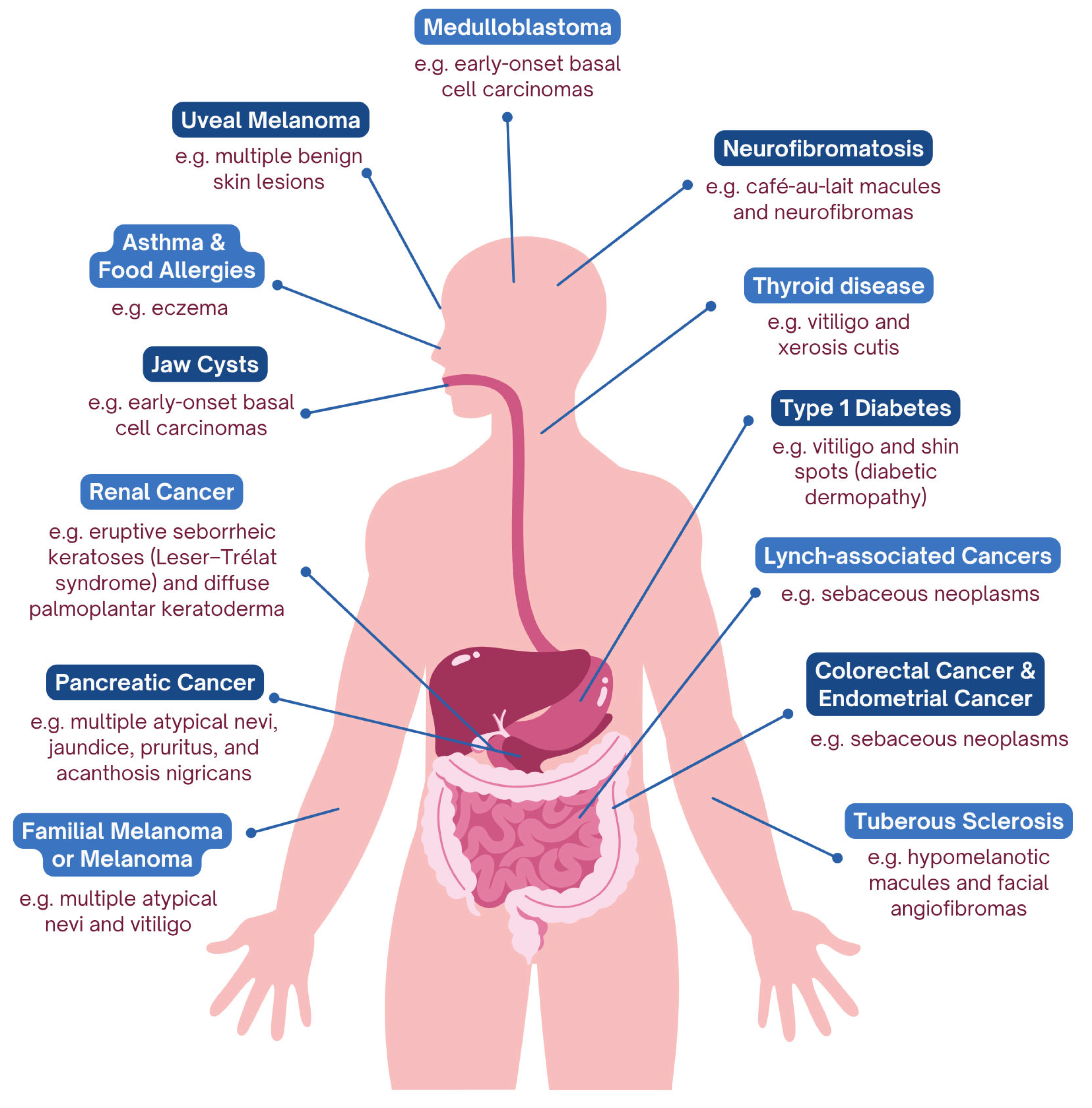
While ethical concerns around dermatologic genomics often focus on consent, access, and regulation, there remains an equally important need to translate visible skin findings into genomic risk assessment in primary care, where such clues are often first encountered. Dermatologic signs could be integrated into routine assessments to guide downstream testing or referrals. For example, a child presenting with café-au-lait macules may warrant evaluation for NF type 1 while a young adult with numerous atypical nevi may prompt consideration of familial melanoma syndromes [100,101]. In these cases, early recognition and triage can lead to meaningful interventions well before systemic disease manifests [100,101]. To support this approach, we encourage a framework in which unique skin presentations are mapped to their potential systemic risks and corresponding primary care or preventative actions (Table 1). Results from a review of published literature on skin manifestations of various systemic diseases is summarized in a schematic figure (Figure 1).

9. Conclusions

The field of dermatogenomics offers powerful insights for systemic disease prediction and prevention, through personalized care and targeted therapies. It is plausible that, as the genetic links and pathogenetic mechanisms behind integumentary diseases are better understood, the genetic components of many systemic diseases are also revealed. Interdisciplinary collaborations between dermatologists, internists, primary care providers, and genomic specialists are vital to standardizing dermatogenomic screening, achieving inclusivity in research, and harnessing AI-based tools. Diverse genomic datasets representative of different skin pigmentations and rare conditions are critical for the development of equitable AI tools. With such tools, dermatogenomics can offer deeply personalized care and suggest effective preventative treatment early in the progression of skin diseases, decreasing the comorbidity and severity of systemic diseases in patients.

Author Contributions

Conceptualization, Y.X.J.; methodology, Y.X.J., D.A.A. and M.Y.Z.; software, not applicable; validation, Y.X.J., D.A.A., M.Y.Z. and A.P.; formal analysis, Y.X.J., D.A.A. and M.Y.Z.; investigation, Y.X.J., D.A.A. and M.Y.Z.; resources, not applicable; data curation, Y.X.J., D.A.A. and M.Y.Z.; writing—original draft preparation, Y.X.J., D.A.A. and M.Y.Z.; writing—review and editing, Y.X.J., D.A.A., M.Y.Z., A.P. and C.L.; visualization, not applicable; supervision, Y.X.J., A.P. and C.L.; project administration, Y.X.J.; funding acquisition, not applicable. All authors have read and agreed to the published version of the manuscript.

Funding

This research received no external funding.

Institutional Review Board Statement

Not applicable.

Informed Consent Statement

Not applicable.

Data Availability Statement

No new data was created in the production of the work.

Conflicts of Interest

Liu is a consultant for Sanofi, Arcutis, L’Oréal, Neutrogena, Sun Pharma, and a speaker for Sanofi, Arcutis, Sun Pharma, Celltrion, and Pfizer. All authors declare no conflicts of interest.

Abbreviations

The following abbreviations are used in this manuscript:
GWASGenome-Wide Association Studies
TNF-αTumor Necrosis Factor Alpha
ILInterleukin
FLGFilaggrin
DNADeoxyribonucleic Acid
CDKN2ACyclin-Dependent Kinase Inhibitor 2A
BAP1BRCA1-Associated Protein 1
MSH2MutS Homolog 2
MLH1MutL Homolog 1
MTSMuir-Torre Syndrome
PTCH1Patched Homolog 1
SUFUSuppressor of Fused Homolog
PTPN22Protein Tyrosine Phosphatase Non-Receptor Type 22
TYRTyrosinase
NFNeurofibromin
TSCTuberous Sclerosis
RasRat Sarcoma Virus
T1DType 1 Diabetes
RARheumatoid Arthritis
CNSCentral Nervous System
IHCImmunohistochemistry
MMRMismatch Repair
BPBlood Pressure
HPAHypothalamic–Pituitary–Adrenal
ANAcanthosis Nigricans
FGFR3Fibroblast Growth Factor Receptor 3
HOMA-IRHomeostatic Model Assessment of Insulin Resistance
HbA1CHemoglobin A1C
LDLRLow-Density Lipoprotein Receptor
APOBApolipoprotein B
ABCDGATP-Binding Cassette Subfamily G
HSHidradenitis Suppurativa
mTORMechanistic Target of Rapamycin
BINBAP1-Inactivated Nevi
BAP1-TPDSBAP1 Tumor Predisposition Syndrome
FAMMMFamilial Atypical Multiple Mole Melanoma Syndrome
p16INK4ACyclin-Dependent Kinase Inhibitor 2A Protein Isoform (p16)
p14ARFAlternate Reading Frame Protein of CDKN2A
BCCBasal Cell Carcinoma
SLC24A5Solute Carrier Family 24 Member 5
SCL45A2Solute Carrier Family 45 Member 2
MC1RMelanocortin 1 Receptor
OCA2Oculocutaneous Albinism II
UVUltraviolet
SLC24A5Sodium/Potassium/Calcium Exchange 5 Gene
SLC45A2Membrane-Associated Transporter Gene
MC1RMelanocortin 1 Receptor Gene
OCA2P Protein Gene
AIArtificial Intelligence
DDIDiverse Dermatology Images
PRSPolygenic Risk Scores
OMIMOnline Mendelian Inheritance in Man
OrphanetRare Disease and Orphan Drug Database
GTExGenotype-Tissue Expression Project
SNPSingle Nucleotide Polymorphism
BRCABreast Cancer Gene
DTCDirect-to-Consumer

References

  1. Tenchov, R.; Sasso, J.M.; Wang, X.; Zhou, Q.A. Aging Hallmarks and Progression and Age-Related Diseases: A Landscape View of Research Advancement. ACS Chem. Neurosci. 2023, 15, 1–30. [Google Scholar] [CrossRef] [PubMed]
  2. Tam, V.; Patel, N.; Turcotte, M.; Bossé, Y.; Paré, G.; Meyre, D. Benefits and Limitations of Genome-Wide Association Studies. Nat. Rev. Genet. 2019, 20, 467–484. [Google Scholar] [CrossRef] [PubMed]
  3. Altshuler, D.; Daly, M.J.; Lander, E.S. Genetic Mapping in Human Disease. Science 2008, 322, 881–888. [Google Scholar] [CrossRef] [PubMed]
  4. Rosenberg, N.A.; Huang, L.; Jewett, E.M.; Szpiech, Z.A.; Jankovic, I.; Boehnke, M. Genome-Wide Association Studies in Diverse Populations. Nat. Rev. Genet. 2010, 11, 356–366. [Google Scholar] [CrossRef]
  5. Primiero, C.A.; Baker, A.M.; Wallingford, C.K.; Maas, E.J.; Yanes, T.; Fowles, L.; Janda, M.; Young, M.-A.; Nisselle, A.; Terrill, B.; et al. Attitudes of Australian Dermatologists on the Use of Genetic Testing: A Cross-Sectional Survey with a Focus on Melanoma. Front. Genet. 2022, 13, 919134. [Google Scholar] [CrossRef]
  6. Shen, S.; Sobczyk, M.K.; Paternoster, L.; Brown, S.J. From GWASs toward Mechanistic Understanding with Case Studies in Dermatogenetics. J. Investig. Dermatol. 2024, 144, 1189–1199.e8. [Google Scholar] [CrossRef]
  7. Betz, R.C.; Hüffmeier, U. Dermatological Diseases from a Genetic Perspective. Med. Genet. 2023, 35, 1–2. [Google Scholar] [CrossRef]
  8. Raharja, A.; Mahil, S.K.; Barker, J.N. Psoriasis: A Brief Overview. Clin. Med. 2021, 21, 170–173. [Google Scholar] [CrossRef]
  9. Piaserico, S.; Orlando, G.; Messina, F. Psoriasis and Cardiometabolic Diseases: Shared Genetic and Molecular Pathways. Int. J. Mol. Sci. 2022, 23, 9063. [Google Scholar] [CrossRef]
  10. Garshick, M.S.; Ward, N.L.; Krueger, J.G.; Berger, J.S. Cardiovascular Risk in Patients with Psoriasis. J. Am. Coll. Cardiol. 2021, 77, 1670–1680. [Google Scholar] [CrossRef]
  11. Fan, J.; Zhu, T.; Tian, X.; Liu, S.; Zhang, S.-L. Exploration of Ferroptosis and Necroptosis-Related Genes and Potential Molecular Mechanisms in Psoriasis and Atherosclerosis. Front. Immunol. 2024, 15, 1372303. [Google Scholar] [CrossRef] [PubMed]
  12. Su, W.; Zhao, Y.; Wei, Y.; Zhang, X.; Ji, J.; Yang, S. Exploring the Pathogenesis of Psoriasis Complicated with Atherosclerosis via Microarray Data Analysis. Front. Immunol. 2021, 12, 667690. [Google Scholar] [CrossRef] [PubMed]
  13. Ji, L.; Ravi, S.; Wright, L.; Nguyen, V.; Wiley, J.; Vukelic, M.; Kim, S. Psoriasis Treatments in the Stabilization of Atherosclerosis: A Systematic Review. Arch. Dermatol. Res. 2025, 317, 159. [Google Scholar] [CrossRef] [PubMed]
  14. Socha, M.; Pietrzak, A.; Grywalska, E.; Pietrzak, D.; Matosiuk, D.; Kiciński, P.; Rolinski, J. The Effect of Statins on Psoriasis Severity: A Meta-Analysis of Randomized Clinical Trials. Arch. Med. Sci. 2019, 16, 1–7. [Google Scholar] [CrossRef]
  15. Lugović-Mihić, L.; Meštrović-Štefekov, J.; Potočnjak, I.; Cindrić, T.; Ilić, I.; Lovrić, I.; Skalicki, L.; Bešlić, I.; Pondeljak, N. Atopic Dermatitis: Disease Features, Therapeutic Options, and a Multidisciplinary Approach. Life 2023, 13, 1419. [Google Scholar] [CrossRef]
  16. David Boothe, W.; Tarbox, J.A.; Tarbox, M.B. Atopic Dermatitis: Pathophysiology. In Management of Atopic Dermatitis: Methods and Challenges; Fortson, E.A., Feldman, S.R., Strowd, L.C., Eds.; Springer International Publishing: Cham, Switzerland, 2017; pp. 21–37. ISBN 978-3-319-64804-0. [Google Scholar]
  17. Moosbrugger-Martinz, V.; Leprince, C.; Méchin, M.-C.; Simon, M.; Blunder, S.; Gruber, R.; Dubrac, S. Revisiting the Roles of Filaggrin in Atopic Dermatitis. Int. J. Mol. Sci. 2022, 23, 5318. [Google Scholar] [CrossRef]
  18. FLG Gene: MedlinePlus Genetics. Available online: https://medlineplus.gov/genetics/gene/flg/ (accessed on 17 September 2025).
  19. Gupta, J.; Margolis, D.J. Filaggrin Gene Mutations with Special Reference to Atopic Dermatitis. Curr. Treat. Options Allergy 2020, 7, 403–413. [Google Scholar] [CrossRef]
  20. Gour, N.; Wills-Karp, M. IL-4 and IL-13 Signaling in Allergic Airway Disease. Cytokine 2015, 75, 68–78. [Google Scholar] [CrossRef]
  21. Baloh, C.H.; Mathias, R.A. Recent Progress in the Genetic and Epigenetic Underpinnings of Atopy. J. Allergy Clin. Immunol. 2023, 151, 60–69. [Google Scholar] [CrossRef]
  22. Joge, R.R.; Kathane, P.U.; Joshi, S.H.; Joge, R.R.; Kathane, P.U.; Joshi, S.H. Vitiligo: A Narrative Review. Cureus 2022, 14, e29307. [Google Scholar] [CrossRef]
  23. LaBerge, G.S.; Bennett, D.C.; Fain, P.R.; Spritz, R.A. PTPN22 Is Genetically Associated with Risk of Generalized Vitiligo, but CTLA4 Is Not. J. Investig. Dermatol. 2008, 128, 1757–1762. [Google Scholar] [CrossRef]
  24. Jin, Y.; Birlea, S.A.; Fain, P.R.; Gowan, K.; Riccardi, S.L.; Holland, P.J.; Mailloux, C.M.; Sufit, A.J.D.; Hutton, S.M.; Amadi-Myers, A.; et al. Variant of TYR and Autoimmunity Susceptibility Loci in Generalized Vitiligo. N. Engl. J. Med. 2010, 362, 1686–1697. [Google Scholar] [CrossRef]
  25. Shen, C.; Gao, J.; Sheng, Y.; Dou, J.; Zhou, F.; Zheng, X.; Ko, R.; Tang, X.; Zhu, C.; Yin, X.; et al. Genetic Susceptibility to Vitiligo: GWAS Approaches for Identifying Vitiligo Susceptibility Genes and Loci. Front. Genet. 2016, 7, 3. [Google Scholar] [CrossRef] [PubMed]
  26. Purkait, R.; Samanta, T.; Thakur, S.; Dhar, S. Neurocutaneous Syndrome: A Prospective Study. Indian J. Dermatol. 2011, 56, 375. [Google Scholar] [CrossRef] [PubMed]
  27. Choi, J.; An, S.; Lim, S.Y. Current Concepts of Neurofibromatosis Type 1: Pathophysiology and Treatment. Arch. Craniofac Surg. 2022, 23, 6–16. [Google Scholar] [CrossRef] [PubMed]
  28. Ozarslan, B.; Russo, T.; Argenziano, G.; Santoro, C.; Piccolo, V. Cutaneous Findings in Neurofibromatosis Type 1. Cancers 2021, 13, 463. [Google Scholar] [CrossRef]
  29. UYSAL, S.; ŞAHİN, M. Tuberous Sclerosis: A Review of the Past, Present, and Future. Turk. J. Med. Sci. 2020, 50, 1665–1676. [Google Scholar] [CrossRef]
  30. Osman, O.T.; Mufaddel, A.; Almugaddam, F.; Augusterfer, E.F. The Psychiatric Aspects of Skin Disorders. Expert. Rev. Dermatol. 2011, 6, 195–209. [Google Scholar] [CrossRef]
  31. Salari, N.; Heidarian, P.; Hosseinian-Far, A.; Babajani, F.; Mohammadi, M. Global Prevalence of Anxiety, Depression, and Stress Among Patients with Skin Diseases: A Systematic Review and Meta-Analysis. J. Prev. 2024, 45, 611–649. [Google Scholar] [CrossRef]
  32. Teichgräber, F.; Jacob, L.; Koyanagi, A.; Shin, J.I.; Seiringer, P.; Kostev, K. Association between Skin Disorders and Depression in Children and Adolescents: A Retrospective Case-Control Study. J. Affect. Disord. 2021, 282, 939–944. [Google Scholar] [CrossRef]
  33. Lyu, H.; Huang, H.; He, J.; Zhu, S.; Hong, W.; Lai, J.; Gao, T.; Shao, J.; Zhu, J.; Li, Y.; et al. Task-State Skin Potential Abnormalities Can Distinguish Major Depressive Disorder and Bipolar Depression from Healthy Controls. Transl. Psychiatry 2024, 14, 110. [Google Scholar] [CrossRef]
  34. Wang, X.; Li, Y.; Wu, L.; Xiao, S.; Ji, Y.; Tan, Y.; Jiang, C.; Zhang, G. Dysregulation of the Gut-Brain-Skin Axis and Key Overlapping Inflammatory and Immune Mechanisms of Psoriasis and Depression. Biomed. Pharmacother. 2021, 137, 111065. [Google Scholar] [CrossRef] [PubMed]
  35. Hołdrowicz, A.; Żebrowska, A. Molecular Link Between Psoriasis and Depression—Update on Pathophysiology. Int. J. Mol. Sci. 2025, 26, 2467. [Google Scholar] [CrossRef] [PubMed]
  36. Kutlubay, Z.; Engin, B.; Bairamov, O.; Tüzün, Y. Acanthosis Nigricans: A Fold (Intertriginous) Dermatosis. Clin. Dermatol. 2015, 33, 466–470. [Google Scholar] [CrossRef] [PubMed]
  37. Hughes, E.K.; Brady, M.F.; Rawla, P. Acanthosis Nigricans. In StatPearls; StatPearls Publishing: Treasure Island, FL, USA, 2025. [Google Scholar]
  38. Videira-Silva, A.; Albuquerque, C.; Fonseca, H. Acanthosis Nigricans as a Clinical Marker of Insulin Resistance among Overweight Adolescents. Ann. Pediatr. Endocrinol. Metab. 2019, 24, 99–103. [Google Scholar] [CrossRef]
  39. Mustafa, M.; Moghrabi, N.; Bin-Abbas, B. Hypochondroplasia, Acanthosis Nigricans, and Insulin Resistance in a Child with FGFR3 Mutation: Is It Just an Association? Case Rep. Endocrinol. 2014, 2014, 840492. [Google Scholar] [CrossRef]
  40. Das, A.; Datta, D.; Kassir, M.; Wollina, U.; Galadari, H.; Lotti, T.; Jafferany, M.; Grabbe, S.; Goldust, M. Acanthosis Nigricans: A Review. J. Cosmet. Dermatol. 2020, 19, 1857–1865. [Google Scholar] [CrossRef]
  41. CDC National Diabetes Statistics Report. Available online: https://www.cdc.gov/diabetes/php/data-research/index.html (accessed on 18 July 2025).
  42. Önal, Z.E.; Atasayan, V.; Gürbüz, T.; Hepkaya, E.; Nuhoğlu, Ç. Association of Glycosylated Hemoglobin (HbA1c) Levels with Iinsulin Resistance in Obese Children. Afr. Health Sci. 2014, 14, 533–538. [Google Scholar] [CrossRef]
  43. Dai, X.; Wiernek, S.; Evans, J.P.; Runge, M.S. Genetics of Coronary Artery Disease and Myocardial Infarction. World J. Cardiol. 2016, 8, 1–23. [Google Scholar] [CrossRef]
  44. Bell, A.; Shreenath, A.P. Xanthoma. In StatPearls; StatPearls Publishing: Treasure Island, FL, USA, 2025. [Google Scholar]
  45. Pejic, R.N. Familial Hypercholesterolemia. Ochsner J. 2014, 14, 669–672. [Google Scholar]
  46. Alnouri, F.; Al-Allaf, F.A.; Athar, M.; Abduljaleel, Z.; Alabdullah, M.; Alammari, D.; Alanazi, M.; Alkaf, F.; Allehyani, A.; Alotaiby, M.A.; et al. Xanthomas Can Be Misdiagnosed and Mistreated in Homozygous Familial Hypercholesterolemia Patients: A Call for Increased Awareness Among Dermatologists and Health Care Practitioners. Glob. Heart 2020, 15, 19. [Google Scholar] [CrossRef] [PubMed]
  47. Lv, X.; Wang, C.; Liu, L.; Yin, G.; Zhang, W.; Abdu, F.A.; Shi, T.; Zhang, Q.; Che, W. Screening and Verifying the Mutations in the LDLR and APOB Genes in a Chinese Family with Familial Hypercholesterolemia. Lipids Health Dis. 2023, 22, 175. [Google Scholar] [CrossRef] [PubMed]
  48. Williams, R.M.; Primig, M.; Washburn, B.K.; Winzeler, E.A.; Bellis, M.; Sarrauste de Menthière, C.; Davis, R.W.; Esposito, R.E. The Ume6 Regulon Coordinates Metabolic and Meiotic Gene Expression in Yeast. Proc. Natl. Acad. Sci. USA 2002, 99, 13431–13436. [Google Scholar] [CrossRef] [PubMed]
  49. Graf, G.A.; Cohen, J.C.; Hobbs, H.H. Missense Mutations in ABCG5 and ABCG8 Disrupt Heterodimerization and Trafficking*. J. Biol. Chem. 2004, 279, 24881–24888. [Google Scholar] [CrossRef] [PubMed]
  50. Seyed Jafari, S.M.; Hunger, R.E.; Schlapbach, C. Hidradenitis Suppurativa: Current Understanding of Pathogenic Mechanisms and Suggestion for Treatment Algorithm. Front. Med. 2020, 7, 68. [Google Scholar] [CrossRef] [PubMed]
  51. Nomura, T. Hidradenitis Suppurativa as a Potential Subtype of Autoinflammatory Keratinization Disease. Front. Immunol. 2020, 11, 847. [Google Scholar] [CrossRef]
  52. Demïrbaş, A.; Elmas, Ö.F.; Eker, H.; Demïrbaş, G.U.; Dursun, R.; Atasoy, M.; Türsen, Ü.; Lottï, T. The Relationship between Hidradenitis Suppurativa and Irritable Bowel Syndrome: A Cross-Sectional Study. Turk. J. Med. Sci. 2022, 52, 97–104. [Google Scholar] [CrossRef]
  53. Repo, P.; Staskiewicz, A.; Sutinen, E.; Rönty, M.; Kivelä, T.T.; Myllärniemi, M.; Turunen, J.A. BAP1 Germline Variants in Finnish Patients with Malignant Mesothelioma. Lung Cancer 2022, 165, 102–107. [Google Scholar] [CrossRef]
  54. Fujii, C.; Mochizuki, A.; Dreike, S.; Jeter, J.M. Genetic Drivers in Sebaceous Neoplasms: A Review of Germline and Somatic Mutations and Their Role in Treatment and Management Strategies. Cancers 2025, 17, 659. [Google Scholar] [CrossRef]
  55. Pilarski, R.; Byrne, L.; Carlo, M.I.; Hanson, H.; Cebulla, C.; Abdel-Rahman, M. BAP1 Tumor Predisposition Syndrome. In GeneReviews®; Adam, M.P., Feldman, J., Mirzaa, G.M., Pagon, R.A., Wallace, S.E., Amemiya, A., Eds.; University of Washington, Seattle: Seattle, WA, USA, 1993. [Google Scholar]
  56. Chau, C.; van Doorn, R.; van Poppelen, N.M.; van der Stoep, N.; Mensenkamp, A.R.; Sijmons, R.H.; van Paassen, B.W.; van den Ouweland, A.M.W.; Naus, N.C.; van der Hout, A.H.; et al. Families with BAP1-Tumor Predisposition Syndrome in The Netherlands: Path to Identification and a Proposal for Genetic Screening Guidelines. Cancers 2019, 11, 1114. [Google Scholar] [CrossRef]
  57. Saldanha, E.F.; Ribeiro, M.F.; Hirsch, I.; Spreafico, A.; Saibil, S.D.; Butler, M.O. How We Treat Patients with Metastatic Uveal Melanoma. ESMO Open 2025, 10, 104496. [Google Scholar] [CrossRef] [PubMed]
  58. Papadimitriou, I.; Vakirlis, E.; Sotiriou, E.; Bakirtzi, K.; Lallas, A.; Ioannides, D. Sebaceous Neoplasms. Diagnostics 2023, 13, 1676. [Google Scholar] [CrossRef] [PubMed]
  59. Everett, J.N.; Raymond, V.M.; Dandapani, M.; Marvin, M.; Kohlmann, W.; Chittenden, A.; Koeppe, E.; Gustafson, S.L.; Else, T.; Fullen, D.R.; et al. Screening for Germline Mismatch Repair Mutations Following Diagnosis of Sebaceous Neoplasm. JAMA Dermatol. 2014, 150, 1315–1321. [Google Scholar] [CrossRef] [PubMed]
  60. Roberts, M.E.; Riegert-Johnson, D.L.; Thomas, B.C.; Rumilla, K.M.; Thomas, C.S.; Heckman, M.G.; Purcell, J.U.; Hanson, N.B.; Leppig, K.A.; Lim, J.; et al. A Clinical Scoring System to Identify Patients with Sebaceous Neoplasms at Risk for the Muir-Torre Variant of Lynch Syndrome. Genet. Med. 2014, 16, 711–716. [Google Scholar] [CrossRef]
  61. Zheng, C.; Sarin, K.Y. Unveiling the Genetic Landscape of Hereditary Melanoma: From Susceptibility to Surveillance. Cancer Treat. Res. Commun. 2024, 40, 100837. [Google Scholar] [CrossRef]
  62. Cremin, C.; Howard, S.; Le, L.; Karsan, A.; Schaeffer, D.F.; Renouf, D.; Schrader, K.A. CDKN2A Founder Mutation in Pancreatic Ductal Adenocarcinoma Patients without Cutaneous Features of Familial Atypical Multiple Mole Melanoma (FAMMM) Syndrome. Hered. Cancer Clin. Pract. 2018, 16, 7. [Google Scholar] [CrossRef]
  63. Hruban, R.H.; Canto, M.; Goggins, M.; Schulick, R.; Klein, A.P. Update on Familial Pancreatic Cancer. Adv. Surg. 2010, 44, 293–311. [Google Scholar] [CrossRef]
  64. Bresler, S.C.; Padwa, B.L.; Granter, S.R. Nevoid Basal Cell Carcinoma Syndrome (Gorlin Syndrome). Head. Neck Pathol. 2016, 10, 119–124. [Google Scholar] [CrossRef]
  65. Guerrini-Rousseau, L.; Smith, M.J.; Kratz, C.P.; Doergeloh, B.; Hirsch, S.; Hopman, S.M.J.; Jorgensen, M.; Kuhlen, M.; Michaeli, O.; Milde, T.; et al. Current Recommendations for Cancer Surveillance in Gorlin Syndrome: A Report from the SIOPE Host Genome Working Group (SIOPE HGWG). Fam. Cancer 2021, 20, 317–325. [Google Scholar] [CrossRef]
  66. Gammal, R.S.; Mayes, J.; Caudle, K.E. Ready or Not, Here It Comes: Direct-to-Consumer Pharmacogenomic Testing and Its Implications for Community Pharmacists. J. Am. Pharm. Assoc. 2019, 59, 646–650. [Google Scholar] [CrossRef]
  67. Combs, L.D. “Turns Out, I’m 100% That B—”: A Scholarly Essay on DNA Ancestry Tests and Family Relationships. Genealogy 2025, 9, 73. [Google Scholar] [CrossRef]
  68. Kobayashi, Y.; Chen, E.; Facio, F.M.; Metz, H.; Poll, S.R.; Swartzlander, D.; Johnson, B.; Aradhya, S. Clinical Variant Reclassification in Hereditary Disease Genetic Testing. JAMA Netw. Open 2024, 7, e2444526. [Google Scholar] [CrossRef]
  69. Wakamatsu, K.; Kavanagh, R.; Kadekaro, A.L.; Terzieva, S.; Sturm, R.A.; Leachman, S.; Abdel-Malek, Z.; Ito, S. Diversity of Pigmentation in Cultured Human Melanocytes Is Due to Differences in the Type as Well as Quantity of Melanin. Pigment. Cell Res. 2006, 19, 154–162. [Google Scholar] [CrossRef] [PubMed]
  70. Hurbain, I.; Romao, M.; Sextius, P.; Bourreau, E.; Marchal, C.; Bernerd, F.; Duval, C.; Raposo, G. Melanosome Distribution in Keratinocytes in Different Skin Types: Melanosome Clusters Are Not Degradative Organelles. J. Investig. Dermatol. 2018, 138, 647–656. [Google Scholar] [CrossRef] [PubMed]
  71. Mosca, S.; Morrone, A. Human Skin Pigmentation: From a Biological Feature to a Social Determinant. Healthcare 2023, 11, 2091. [Google Scholar] [CrossRef] [PubMed]
  72. Swope, V.; Alexander, C.; Starner, R.; Schwemberger, S.; Babcock, G.; Abdel-Malek, Z.A. Significance of the Melanocortin 1 Receptor in the DNA Damage Response of Human Melanocytes to Ultraviolet Radiation. Pigment. Cell Melanoma Res. 2014, 27, 601–610. [Google Scholar] [CrossRef]
  73. Norton, H.L. The Color of Normal: How a Eurocentric Focus Erases Pigmentation Complexity. Am. J. Hum. Biol. 2021, 33, e23554. [Google Scholar] [CrossRef]
  74. Carvalho, L.A.D.; Aguiar, F.C.; Smalley, K.S.M.; Possik, P.A. Acral Melanoma: New Insights into the Immune and Genomic Landscape. Neoplasia 2023, 46, 100947. [Google Scholar] [CrossRef]
  75. Perlman, K.L.; Klein, E.J.; Park, J.H. Racial Disparities in Dermatology Training: The Impact on Black Patients. Cutis 2020, 106, 300–301. [Google Scholar] [CrossRef]
  76. Marco-Contelles, J.; Fall, Y. Editorial: Multitarget Molecules for Alzheimer’s Disease Therapy. Curr. Top. Med. Chem. 2017, 17, 3318. [Google Scholar] [CrossRef]
  77. Slaught, C.; Madu, P.; Chang, A.Y.; Williams, V.L.; Kebaetse, M.B.; Nkomazana, O.; Molefe-Baikai, O.J.; Bekele, N.A.; Omech, B.; Kellman, P.J.; et al. Novel Education Modules Addressing the Underrepresentation of Skin of Color in Dermatology Training. J. Cutan. Med. Surg. 2022, 26, 17–24. [Google Scholar] [CrossRef] [PubMed]
  78. Chiu, C.-H.; Chen, Y.-J.; Wu, Y.; Shi, Y.; Ho, T.-Y. Achieve Fairness without Demographics for Dermatological Disease Diagnosis. Med. Image Anal. 2024, 95, 103188. [Google Scholar] [CrossRef] [PubMed]
  79. Esteva, A.; Kuprel, B.; Novoa, R.A.; Ko, J.; Swetter, S.M.; Blau, H.M.; Thrun, S. Dermatologist-Level Classification of Skin Cancer with Deep Neural Networks. Nature 2017, 542, 115–118. [Google Scholar] [CrossRef] [PubMed]
  80. Daneshjou, R.; Vodrahalli, K.; Novoa, R.A.; Jenkins, M.; Liang, W.; Rotemberg, V.; Ko, J.; Swetter, S.M.; Bailey, E.E.; Gevaert, O.; et al. Disparities in Dermatology AI Performance on a Diverse, Curated Clinical Image Set. Sci. Adv. 2022, 8, eabq6147. [Google Scholar] [CrossRef]
  81. Munia, N.; Imran, A.-A.-Z. Prompting Medical Vision-Language Models to Mitigate Diagnosis Bias by Generating Realistic Dermoscopic Images. arXiv 2025, arXiv:2504.01838. [Google Scholar] [CrossRef]
  82. Gyawali, P.K.; Le Guen, Y.; Liu, X.; Belloy, M.E.; Tang, H.; Zou, J.; He, Z. Improving Genetic Risk Prediction across Diverse Population by Disentangling Ancestry Representations. Commun. Biol. 2023, 6, 964. [Google Scholar] [CrossRef]
  83. Martin, A.R.; Kanai, M.; Kamatani, Y.; Okada, Y.; Neale, B.M.; Daly, M.J. Current Clinical Use of Polygenic Scores Will Risk Exacerbating Health Disparities. Nat. Genet. 2019, 51, 584–591. [Google Scholar] [CrossRef]
  84. Kelemen, M.; Xu, Y.; Jiang, T.; Zhao, J.H.; Anderson, C.A.; Wallace, C.; Butterworth, A.; Inouye, M. Performance of Deep-Learning-Based Approaches to Improve Polygenic Scores. Nat. Commun. 2025, 16, 5122. [Google Scholar] [CrossRef]
  85. Gao, Y.; Cui, Y. Optimizing Clinico-Genomic Disease Prediction across Ancestries: A Machine Learning Strategy with Pareto Improvement. Genome Med. 2024, 16, 76. [Google Scholar] [CrossRef]
  86. Smith, L.A.; Cahill, J.A.; Lee, J.-H.; Graim, K. Equitable Machine Learning Counteracts Ancestral Bias in Precision Medicine. Nat. Commun. 2025, 16, 2144. [Google Scholar] [CrossRef]
  87. Culp, M.B. Melanoma Among Non-Hispanic Black Americans. Prev. Chronic Dis. 2019, 16, E79. [Google Scholar] [CrossRef] [PubMed]
  88. Fernandez, J.M.; Mata, E.M.; Bubic, B.N.; Kwan, K.R.; Whitley, M.J.; Wysong, A. Racial and Ethnic Differences in Males with Melanoma: A Retrospective Cohort Study of 205,125 Cases from the National Cancer Database. J. Am. Acad. Dermatol. 2023, 89, 828–832. [Google Scholar] [CrossRef] [PubMed]
  89. Chalitsios, C.V.; Tsilidis, K.K.; Tzoulaki, I. Psoriasis and COVID-19: A Bidirectional Mendelian Randomization Study. J. Am. Acad. Dermatol. 2023, 88, 893–895. [Google Scholar] [CrossRef] [PubMed]
  90. Munia, N.; Imran, A.-A.-Z. DermDiff: Generative Diffusion Model for Mitigating Racial Biases in Dermatology Diagnosis. arXiv 2025, arXiv:2503.17536. [Google Scholar] [CrossRef]
  91. Halachmi, S.; Marquart, L. Regulation of Medical Devices for Dermatology. Dermatol. Clin. 2022, 40, 297–305. [Google Scholar] [CrossRef]
  92. Doolan, B.J.; McGrath, J.A.; Onoufriadis, A. A Clinician’s Guide to Omics Resources in Dermatology. Clin. Exp. Dermatol. 2022, 47, 858–866. [Google Scholar] [CrossRef]
  93. Ascensión, A.M.; Araúzo-Bravo, M.J.; Izeta, A. Challenges and Opportunities for the Translation of Single-Cell RNA Sequencing Technologies to Dermatology. Life 2022, 12, 67. [Google Scholar] [CrossRef]
  94. Song, B.; Ning, X.; Guo, L.; Liu, W.; Jin, H. Comparative Proteomics Analysis Reveals Distinct Molecular Phenotype and Biomarkers in Patients with Erythrodermic Atopic Dermatitis and Erythrodermic Psoriasis. Inflammation 2025, 48, 331–345. [Google Scholar] [CrossRef]
  95. Ganatra, H.; Tan, J.K.; Simmons, A.; Bigogno, C.M.; Khurana, V.; Ghose, A.; Ghosh, A.; Mahajan, I.; Boussios, S.; Maniam, A.; et al. Applying Whole-Genome and Whole-Exome Sequencing in Breast Cancer: A Review of the Landscape. Breast Cancer 2024, 31, 999–1009. [Google Scholar] [CrossRef]
  96. Housholder, A.L. Genetic Medicine Arrives in the Outpatient Clinic: Ethical Concerns for Dermatologists. In Dermatoethics: Contemporary Ethics and Professionalism in Dermatology; Bercovitch, L., Perlis, C.S., Stoff, B.K., Grant-Kels, J.M., Eds.; Springer International Publishing: Cham, Switzerland, 2021; pp. 25–38. ISBN 978-3-030-56861-0. [Google Scholar]
  97. Schneider, S.A.; Schneider, U.H. Rights and Duties of Genetic Counsellors in Germany Related to Relatives at Risk: Comparative Thoughts on the German Genetic Diagnostics Act. J. Med. Ethics 2024, 50, 324–331. [Google Scholar] [CrossRef]
  98. Early Discovery Benefits. Available online: https://www.bmo.com/en-ca/main/insurance-advisors/insights/early-discovery-benefits/ (accessed on 23 July 2025).
  99. Canada, E.L. Critical Illness Insurance. Available online: https://www.equitable.ca/en/our-products/individual-insurance/critical-illness-insurance/ (accessed on 23 July 2025).
  100. Karaconji, T.; Whist, E.; Jamieson, R.V.; Flaherty, M.P.; Grigg, J.R.B. Neurofibromatosis Type 1: Review and Update on Emerging Therapies. Asia-Pac. J. Ophthalmol. 2019, 8, 62–72. [Google Scholar] [CrossRef]
  101. Houben, R.; Michel, B.; Vetter-Kauczok, C.S.; Pföhler, C.; Laetsch, B.; Wolter, M.D.; Leonard, J.H.; Trefzer, U.; Ugurel, S.; Schrama, D.; et al. Absence of Classical MAP Kinase Pathway Signalling in Merkel Cell Carcinoma. J. Investig. Dermatol. 2006, 126, 1135–1142. [Google Scholar] [CrossRef] [PubMed]
  102. Bonadona, V.; Bonaïti, B.; Olschwang, S.; Grandjouan, S.; Huiart, L.; Longy, M.; Guimbaud, R.; Buecher, B.; Bignon, Y.-J.; Caron, O.; et al. Cancer Risks Associated with Germline Mutations in MLH1, MSH2, and MSH6 Genes in Lynch Syndrome. JAMA 2011, 305, 2304–2310. [Google Scholar] [CrossRef] [PubMed]
  103. Weidinger, S.; Illig, T.; Baurecht, H.; Irvine, A.D.; Rodriguez, E.; Diaz-Lacava, A.; Klopp, N.; Wagenpfeil, S.; Zhao, Y.; Liao, H.; et al. Loss-of-Function Variations within the Filaggrin Gene Predispose for Atopic Dermatitis with Allergic Sensitizations. J. Allergy Clin. Immunol. 2006, 118, 214–219. [Google Scholar] [CrossRef] [PubMed]
  104. Santangelo, A.; Chelleri, C.; Tomasino, M.; Pasquinucci, M.; Cappozzo, F.; Striano, P.; Diana, M.C.; Scala, M. Café-Au-Lait Macules in Neurofibromatosis Type 1: Birthmark or Biomarker? Cancers 2025, 17, 1490. [Google Scholar] [CrossRef]
  105. Choi, J.-E.; Chae, J.-H.; Hwang, Y.-S.; Kim, K.-J. Mutational Analysis of TSC1 and TSC2 in Korean Patients with Tuberous Sclerosis Complex. Brain Dev. 2006, 28, 440–446. [Google Scholar] [CrossRef]
Figure 1. Schematic depiction of key dermatologic findings of common systemic diseases.
Dna 06 00002 g001
Table 1. Dermatologic signs and their correlation with genes, systemic risks, and suggested primary care actions.
First Author, YearStudy CountryDermatologic Sign(s)Genetic
Basis
Systemic RiskSuggested Primary Care Action
Chau et al., 2019 [56]NetherlandsMultiple atypical neviCDKN2AFamilial melanoma, pancreatic cancerDermatology referral, family history review, genetic counseling
Repo et al., 2022 [53]FinlandMultiple benign skin lesionsBAP1Uveal melanoma, mesothelioma, renal cancerDermatology/genetics referral, imaging for internal tumors
Bonadona et al., 2011 [102]FranceSebaceous neoplasmsMSH2, MLH1 (MTS)Colorectal, endometrial, Lynch-associated cancersIHC screening, gastroenterology referral, germline testing if MMR loss
Smith et al., 2014 [86]United KingdomEarly-onset basal cell carcinomasPTCH1, SUFU (Gorlin)Medulloblastoma, jaw cysts, radiation-sensitive tumorsBrain/dental imaging, radiation avoidance, genotype-guided surveillance
LaBerge et al., 2008; Jin et al., 2010 [23,24]United StatesVitiligo with family autoimmunityPTPN22, TYRThyroid disease, T1D, RA, melanoma (inverse relationship)Autoimmune panel, family history screening
Weidinger et al., 2006 [103]GermanyEczema with allergic historyFLGAsthma, food allergy, barrier dysfunctionAllergy screening, education on environmental triggers
Santangelo et al., 2025 [104]ItalyCafé-au-lait macules (≥6, >5 mm in children)NF1, NF2Neurofibromatosis type 1 and 2: CNS tumors, learning delayNeurology referral, ophthalmology, BP screening
Choi et al., 2025 [105]Koreafacial angiofibromas, hypomelanotic maculesTSC1, TSC2Tuberous sclerosis: epilepsy, CNS and renal complicationsGenetic testing, pediatric neurology/nephrology referral
Abbreviations: CDKN2A = cyclin-dependent kinase inhibitor 2A; BAP1 = BRCA1 associated protein-1; MSH2 = mutS homolog 2; MLH1 = MutL homolog 1; MTS = Muir-Torre syndrome; PTCH1 = patched homolog 1; SUFU = suppressor of fused homolog; PTPN22 = protein tyrosine phosphatase non-receptor type 22; TYR = tyrosinase; FLG = filaggrin; NF1 = neurofibromin 1; NF2 = neurofibromin 2; TSC1 = tuberous sclerosis 1; TSC2 = tuberous sclerosis 2; T1D = type 1 diabetes; RA = rheumatoid arthritis; CNS = central nervous system; IHC = immunohistochemistry; MMR = mismatch repair; BP = blood pressure.
Disclaimer/Publisher’s Note: The statements, opinions and data contained in all publications are solely those of the individual author(s) and contributor(s) and not of MDPI and/or the editor(s). MDPI and/or the editor(s) disclaim responsibility for any injury to people or property resulting from any ideas, methods, instructions or products referred to in the content.