Article

Comparative Analysis of Virulence Genes and Antimicrobial Resistance in Escherichia coli from Poultry Meat and Poultry Farm Environments in Pakistan

1
Department of Poultry Science, Auburn University, Auburn, AL 36830, USA
2
Institute of Microbiology, University of Agriculture, Faisalabad 38040, Pakistan
*
Author to whom correspondence should be addressed.
Submission received: 9 May 2025 / Revised: 10 July 2025 / Accepted: 8 August 2025 / Published: 3 September 2025

Abstract

Background/Objectives: Escherichia coli (E. coli) strains harboring virulence genes and antimicrobial resistance (AMR) pose a significant risk to poultry production and public health in Pakistan. This study aimed to isolate E. coli from poultry meat and poultry farm environments and compare their virulence gene profiles and AMR patterns. Methods: A total of 100 samples were collected, including 50 poultry meat samples from retail shops and 50 environmental samples from poultry farms. E. coli was isolated on MacConkey agar following overnight enrichment in lactose broth. Isolates were confirmed by biochemical testing and 16S rRNA gene PCR. Virulence genes (stx1, stx2, eae) were detected using multiplex PCR, and AMR profiles were assessed via the Kirby–Bauer disk diffusion method. Results: E. coli was isolated from 26 poultry meat samples (52%) and 23 poultry farm environment samples (46%). All isolates harbored at least one virulence gene, with stx2 being the most prevalent (34.62% meat; 39.13% environment), followed by stx1 (19.23% meat; 17.40% environment) and eae (11.54% meat; 13.04% environment). Combined gene patterns (stx1/eae, stx2/eae, stx1/stx2/eae) were also detected across both sources. AMR analysis revealed high resistance to cefoxitin (100% both sources), trimethoprim (57.09% meat; 60.87% environment), and ampicillin–sulbactam (42.3% meat; 52.17% environment). In contrast, isolates were completely susceptible to norfloxacin (100% meat; 95.65% environment) and exhibited high susceptibility to tetracycline (84.62% meat; 82.61% environment). Statistical comparisons using Fisher’s exact test and the Kruskal–Wallis test showed no significant differences (p > 0.05) in virulence gene prevalence or AMR patterns between poultry meat and environmental isolates. Conclusions: These findings highlight poultry farm environments as potential reservoirs for pathogenic, antimicrobial-resistant E. coli, emphasizing the risk of zoonotic transmission through contaminated poultry meat and the need for improved biosecurity measures.

Graphical Abstract

1. Introduction

Poultry meats are widely regarded as a healthy source of high-quality protein and essential nutrients, valued for their low cost and availability, making them a key component of diets globally [1]. In light of Pakistan’s protein deficiency, where the average daily animal protein intake is only 17 g compared to the WHO-recommended 27 g, chicken meat is increasingly acknowledged, with its consumption on the rise [2]. The poultry sector in Pakistan has seen remarkable growth, with an average annual growth rate of 8–10 percent over the past decade [3]. This progress has propelled Pakistan to become the eleventh-largest poultry producer globally, contributing to approximately 40.7 percent of the country’s total meat production, with significant potential for future expansion [4]. However, poultry meat is often associated with high levels of bacterial contamination, making it a significant source of harmful pathogens that contribute to foodborne diseases and outbreaks [5,6]. This is attributed to its high protein content, favorable pH, and elevated water content, which collectively create an ideal environment for the multiplication of virulent bacteria [7].
Among these bacterial contaminants, Escherichia coli (E. coli) is a prevalent foodborne zoonotic pathogen that poses a considerable risk of transmission through poultry and poultry-derived products. E. coli is a Gram-negative, rod-shaped, coliform, facultatively anaerobic bacterium that colonizes the lower gastrointestinal tract of warm-blooded animals, including humans, poultry, and other species [8]. However, certain strains of this bacterium have acquired virulence-associated genes, allowing them to cause infections in the intestines or other parts of the body [9]. Foodborne infections caused by E. coli are primarily linked to Shiga toxin-producing E. coli (STEC) strains, often resulting from consuming contaminated raw or undercooked meat [10]. Contamination of chicken meat can occur during the slaughtering process, either through the chicken’s intestinal or digestive contents encountering the carcass or through skin contamination [6]. Besides this, poultry farms, along with materials such as feed, water, litter, wood shavings, fecal matter, and rodent droppings, are also recognized as potential sources of E. coli contamination in poultry [11]. Additionally, E. coli is a widespread bacterium in the air, particularly in animal houses and their surrounding environments [12].
Various virulence factors and antimicrobial resistance genes drive the pathogenesis of E. coli, with Shiga toxins (Stx1 and Stx2), intimin (eae), and enterohemolysin (hlyA) being key virulence factors in STEC strains [13]. They are associated with severe human gastrointestinal diseases such as hemorrhagic colitis (HC) and hemolytic uremic syndrome (HUS) [14]. The presence of AMR genes in E. coli strains further exacerbates their pathogenic potential. Antibiotics have been employed on poultry farms for the treatment and prevention of various diseases, as well as for the growth promotion of birds [15]. The improper use of antibiotics exerted selective pressure on the chicken microbiota, promoting the emergence and spread of antibiotic-resistant bacteria and establishing reservoirs of AMR genes [16]. As E. coli is an opportunistic bacterium naturally present in the chicken microbiota, the transfer of AMR genes to it could undermine the effectiveness of antimicrobials in animals and potentially affect their efficacy in human medicine as well [17].
In Pakistan, only a small amount of poultry meat comes from structured processing facilities; the remainder is obtained from birds slaughtered in retail shops (unorganized sector), where there is high potential for contamination due to poor sanitation [18]. There are no set policies concerning the slaughter, marketing, or even preparation of chicken meat. Because of this, roadside slaughter in the filthiest way is common in the majority of Pakistan’s cities, towns, and villages. A rise in meat demand without a safe, sanitary handling infrastructure could result in the transmission of pathogenic microbes like E. coli from poultry to consumers [19]. E. coli strains detected in contaminated meat and meat products demonstrate resistance to conventional antibiotics [20,21]. Humans may contract chicken-resistant E. coli through the ingestion of tainted food [22]. Therefore, an integrated ‘One Health’ approach is essential to control the transmission of E. coli throughout the food chain. One Health can be described as the complex interconnectedness between pathogens, animals, and humans [23] sharing the same environment. In this context, the poultry farm environment acts as a potential reservoir for resistant strains that may eventually contaminate food products. Despite growing global attention, there remains limited information on the simultaneous occurrence of virulence genes and AMR in E. coli across different points in the poultry production continuum, particularly in Pakistan. Therefore, this study aims to investigate and compare the prevalence of virulence genes and AMR profiles in E. coli isolates obtained from poultry meat and the poultry farm environment, providing insight into potential transmission routes and informing food safety strategies.

2. Materials and Methods

2.1. Collection and Preparation of Samples

A total of 100 samples were collected from District Faisalabad, Pakistan, from February 2023 to July 2023. These included 50 poultry meat samples (breast) from 50 retail/roadside meat shops and 50 environmental samples from 25 poultry farms. The environmental samples comprised one litter and one swab sample from various surfaces (floor, windows, doors) of each poultry farm. All samples were adequately labeled and kept in separate sterile plastic bags to avoid cross-contamination. Immediately following collection, the samples were transported and processed the same day at the Microbiology Laboratory, Institute of Microbiology, University of Agriculture, Faisalabad. For meat samples, approximately 1 g of tissue was aseptically weighed, chopped, and added to 10 mL of lactose broth (Neogen, Lansing, MI, USA) in test tubes for enrichment, followed by incubation at 37 °C for 24 h. Similarly, 1 g of litter was added to 10 mL of lactose broth, while environmental swabs were directly placed into the broth for incubation at 37 °C for 24 h.

2.2. Culturing, Isolation, and Biochemical Characterization

After incubation, samples were assessed for turbidity, and those exhibiting turbidity were streaked onto MacConkey agar plates (Argenta, Poznań, Poland) using a sterile loop. Pink, shiny colonies observed on the medium were presumptively identified as E. coli based on the selective and differential properties of MacConkey agar. Microscopically, E. coli appeared as pink to dark pink short rods due to the presence of a thin peptidoglycan layer. E. coli bacterial isolates were further confirmed biochemically, as detailed in Table 1. The biochemically identified E. coli isolates were subsequently stored at −80 °C in 20% glycerol stocks prepared in nutrient broth (Neogen, Lansing, MI, USA) to ensure cryoprotection.

2.3. DNA Extraction and Molecular Detection of Virulence Genes

Biochemically confirmed E. coli colonies were selected for molecular characterization. Genomic DNA was extracted using the GeneJET Genomic DNA Purification Kit (ThermoFischer Scientific, catalog number K0721) following the manufacturer’s protocol.
Initial confirmation of E. coli was performed by amplification of the 16S rRNA gene using universal bacterial primers 27F (5′-AGAGTTTGATCMTGGCTCAG-3′) and 1492R (5′-TACGGYTACCTTGTTACGACTT-3′), which generate an expected product size of approximately 1500 bp [24]. PCR reactions were performed in a final volume of 25 µL, and amplicons were visualized by electrophoresis on a 1.5% agarose gel.
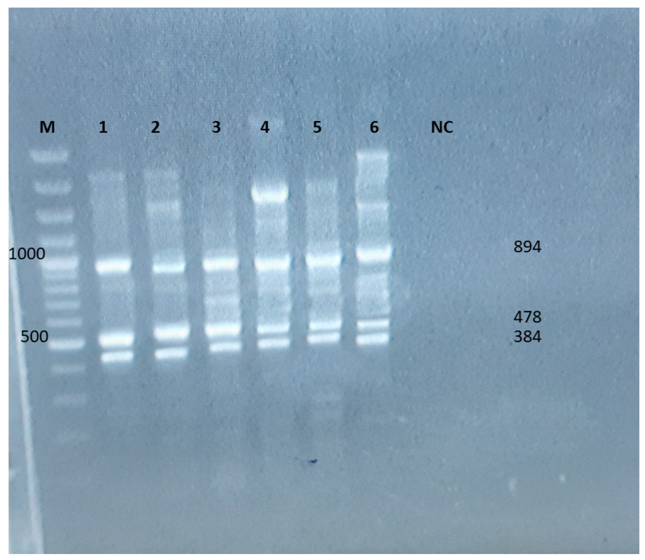
Following confirmation, Multiplex PCR was conducted to simultaneously detect key virulence genes (stx1, stx2, and eae) in all isolates. Expected product sizes were 894 bp for stx1, 478 bp for stx2, and 384 bp for eae. The stx1 and stx2 primers were adopted from [25], while the eae primers were obtained from [25]. The primer sequences are listed in Table 2. Multiplex PCR reagent concentrations were adapted from [25], with minor modifications based on preliminary optimization trials. Specifically, thermal cycling conditions were adjusted to account for the exclusion of the hlyA gene, which was included in the original protocol but not targeted in the present study. The optimized cycling conditions applied for the amplification of stx1, stx2, and eae genes are detailed in Table 3. To compare the overall prevalence of individual virulence genes between sources, Fisher’s exact test was performed.

2.4. Antibiotic Susceptibility Test

All confirmed E. coli isolates from both sources were selected for antibiotic resistance screening using the Kirby–Bauer disc diffusion method. The bacterial inoculum was adjusted to a 0.5 McFarland standard (approximately 1.5 × 10⁸ CFU/mL) by visual comparison to the McFarland standard, following CLSI guidelines [26]. Each isolate was cultured on Mueller–Hinton agar (MHA) plates, using a sterile cotton swab, and antibiotic susceptibility was tested against nine antibiotics: ampicillin–sulbactam (SAM-10/10), cefoxitin (FOX-30), tetracycline (TE-30), trimethoprim (TMP-5), norfloxacin (NOR-10), and nalidixic acid (NA-30). After 24 h of incubation, the zone of inhibition surrounding each disk was measured in millimeters using a caliper. The interpretation of antibiotic resistance was based on the standards outlined by the Clinical and Laboratory Standards Institute [26].

3. Results

3.1. Prevalence of E. coli in Samples

Out of 50 meat samples, 26 (52%) tested positive for E. coli. Similarly, 23 (46%) of the 50 environmental samples were positive for E. coli (Figure 1). Statistical analysis using Fisher’s exact test revealed no significant difference in E. coli prevalence between meat and environmental samples (p = 0.6893, two-sided).

3.2. Molecular Detection of Virulence Genes

Isolates were subjected to multiplex PCR, followed by gel electrophoresis. PCR products were detected by visualizing the bands under UV light using a Gel Documentation System (Figure 2). As shown in Figure 2, all samples, regardless of their origin, exhibited the presence of these virulence genes (stx1, stx2, and eae), indicating that E. coli virulence genes are prevalent across both poultry meat and poultry farm environment isolates. Faint non-specific bands were observed in some lanes, such as an upper band in lane 4. These bands are likely due to minor non-specific primer binding or residual genomic DNA. However, they did not interfere with the identification or interpretation of the specific target bands, which were clearly distinguishable based on their expected sizes and positions relative to the DNA ladder.

3.3. Comparative Analysis of Virulence Genes Detected in Poultry Meat and Poultry Farm Environment Isolates

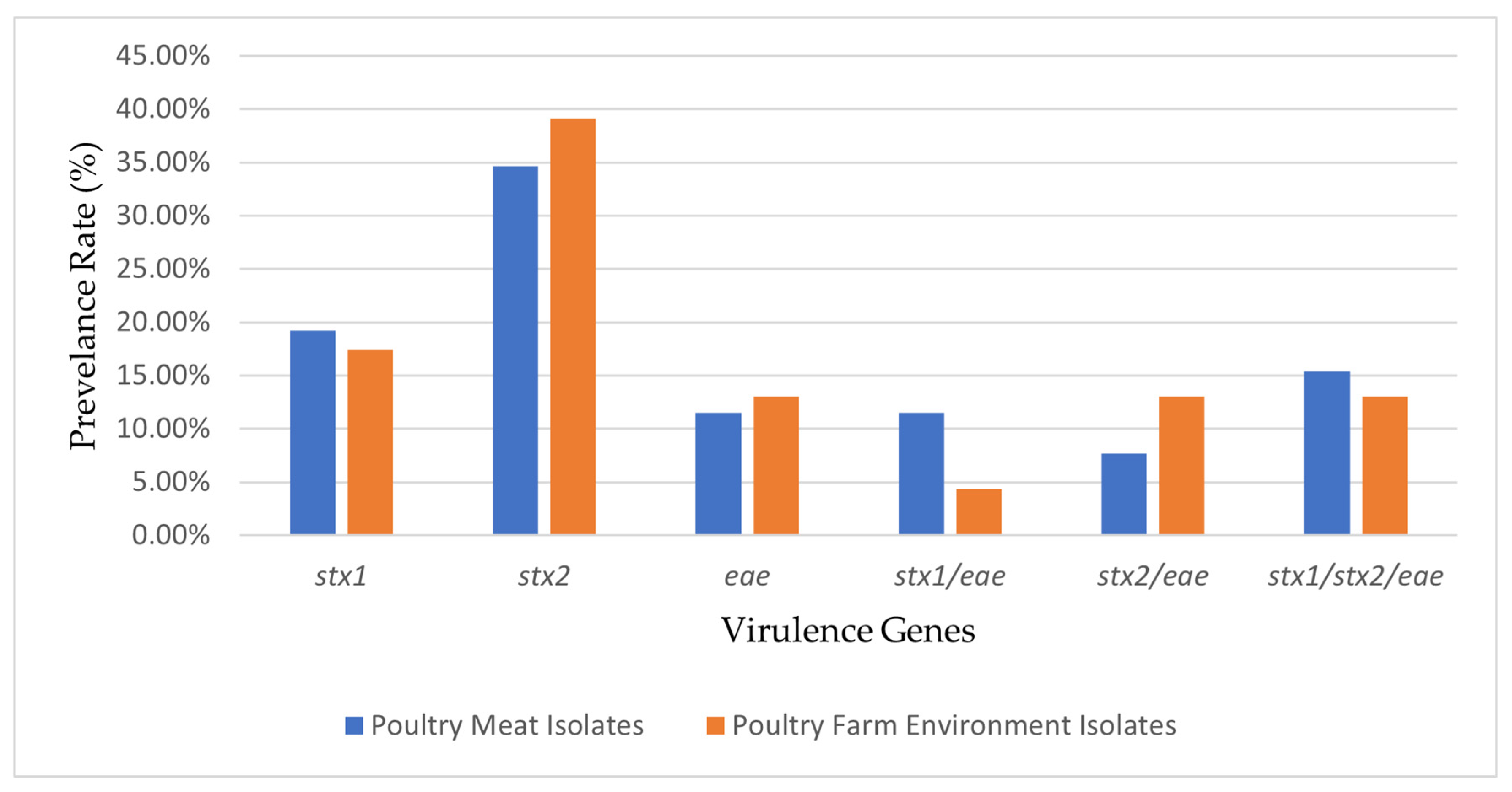
The distribution of virulence gene combinations detected in E. coli isolates from meat and poultry farm environment samples is summarized in Figure 3 and Table S1. Six distinct gene combinations were identified across both sources, including isolates harboring single genes and those with multiple gene combinations. The most prevalent gene detected was stx2, present alone or in combination, followed by stx1 and eae. No statistically significant differences were observed for stx1 (p = 0.562), stx2 (p = 0.770), or eae (p = 1.000) between meat and environment isolates. Similarly, comparison of specific virulence gene combinations revealed no significant differences for stx1/eae (p = 0.612), stx2/eae (p = 0.655), or stx1/stx2/eae (p = 1.000) between the two sources.

3.4. Antimicrobial Resistance Pattern

Phenotypic AMR analysis of E. coli isolates revealed complete resistance to cefoxitin (FOX-30) among both poultry meat (100%, 26/26) and poultry environment (100%, 23/23) isolates. Similarly, full resistance to trimethoprim (TMP-5) was observed in both meat (57.1%, 15/26) and environmental isolates (60.9%, 14/23), with additional intermediate resistance detected in 19.2% (5/26) of meat isolates and 17.4% (4/23) of environmental isolates. For ampicillin–sulbactam (SAM 10/10), resistance was observed in 42.3% (11/26) of meat isolates and 52.2% (12/23) of environmental isolates, with intermediate resistance present in 30.8% (8/26) of meat isolates and 26.1% (6/23) of environmental isolates. Tetracycline (TE-30) resistance was relatively low, detected in 15.4% (4/26) of meat isolates and 17.4% (4/23) of environmental isolates. The majority of isolates from both sources remained susceptible to TE-30, with 84.6% (22/26) of meat isolates and 82.6% (19/23) of environmental isolates classified as sensitive. Complete susceptibility to norfloxacin (NOR-10) was observed among meat isolates (100%, 26/26), while 95.7% (22/23) of environmental isolates were also susceptible, with one intermediate isolate detected.
For nalidixic acid (NA-30), resistance was higher among environmental isolates (30.4%, 7/23) compared to meat isolates (23.1%, 6/26). Intermediate resistance to NA-30 was observed in 26.9% (7/26) of meat isolates and 21.7% (5/23) of environmental isolates, while 50% (13/26) of meat and 47.8% (11/23) of environmental isolates remained susceptible.
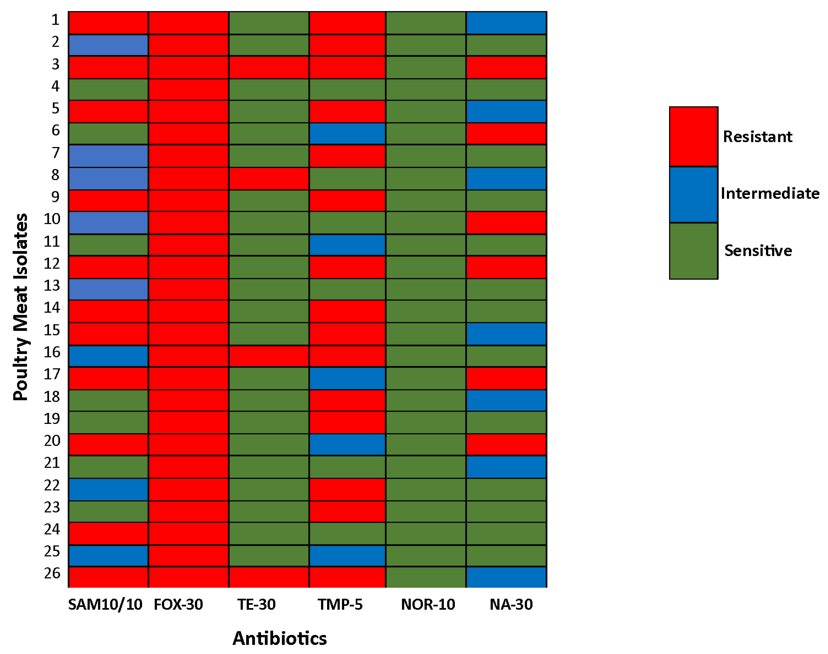
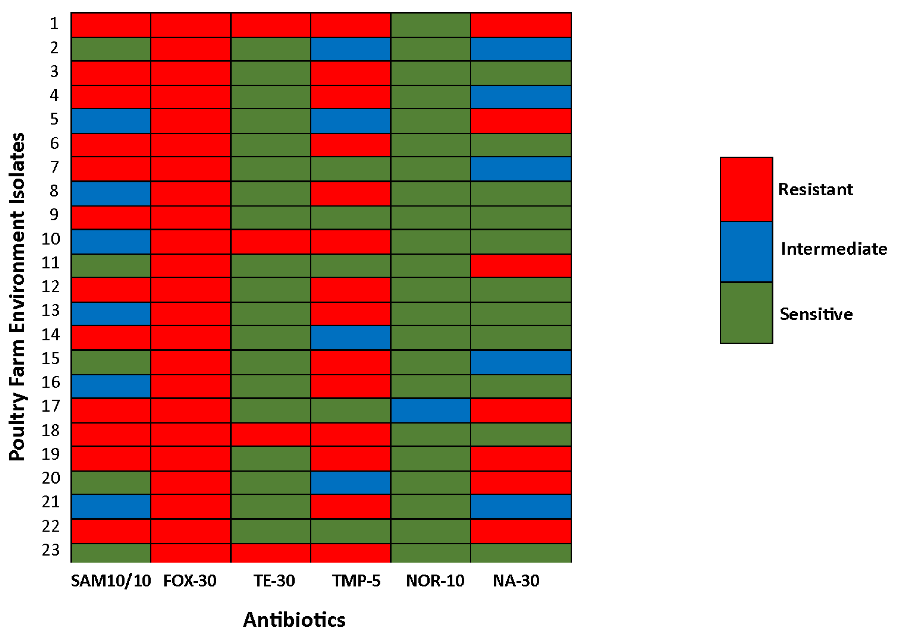
Heatmaps illustrating the antibiotic susceptibility profiles of E. coli isolates from poultry meat and poultry farm environments are presented in Figure 4 and Figure 5, respectively.

3.5. Comparative Analysis of AMR Patterns Between Poultry Meat and Poultry Farm Environment Isolates

The distribution of resistant, intermediate, and sensitive E. coli isolates for each antibiotic is summarized in Table 3. Although variations in AMR prevalence were observed between meat and environmental isolates, statistical analysis using the Kruskal–Wallis test revealed no significant differences for any of the antibiotics tested. The test statistics were as follows: ampicillin–sulbactam (SAM-10/10), H = 3.714, p = 0.2000; cefoxitin (FOX-30), H = 4.800, p = 0.2000; tetracycline (TE-30), H = 4.848, p = 0.0667; trimethoprim (TMP-5), H = 4.191, p = 0.1333; norfloxacin (NOR-10), H = 4.194, p = 0.2000; and nalidixic acid (NA-30), H = 3.603, p = 0.3333. All p-values exceeded the threshold for statistical significance (p > 0.05), indicating no significant differences in AMR patterns between isolates from meat and poultry environment sources. Figure 6 and Table S2 provide a comparative stacked column chart illustrating the AMR distribution across poultry meat and poultry farm environment isolates.

4. Discussion

This study offers important baseline data on the prevalence, virulence, and antimicrobial resistance of E. coli in poultry meat and farm environments in Pakistan, using standard biochemical confirmation, 16S rRNA PCR, and multiplex PCR targeting stx1, stx2, and eae genes. A key limitation is the exclusion of the hlyA gene from the virulence panel and the lack of sequencing, which was constrained by our budget. Additionally, the sample size was limited due to the seller’s reluctance to participate, potentially affecting the broader applicability of the findings. Future studies should include more comprehensive gene targets, larger sample sizes, and genomic analysis.
Given that chicken meat is a widely consumed, economical source of nutrition, its safety is compromised by bacterial contaminants like E. coli, including STEC strains and antibiotic-resistant variants; this study was conducted to assess such risks in poultry meat and farm environments, isolating and characterizing E. coli harboring virulent genes and exhibiting antibiotic resistance from poultry meat and poultry farm environmental samples. This study found a higher prevalence of E. coli in meat samples (52%) compared to environmental samples (46%), suggesting that contamination may be amplified during processing, handling, or storage stages post-harvest. E. coli contamination was detected in raw and ready-to-eat (RTE) meats, where improper handling, prolonged exposure at points of sale, and poor storage conditions significantly increase microbial load [27]. In a study by Zhao et al. (2001), the prevalence of E. coli in retail meats varied, with the highest occurrence in chicken samples (39%) and the lowest in turkey samples (12%). According to Zhao et al. (2012), E. coli was detected in 69.5% of retail meat samples, with the highest prevalence found in chicken breast (83.5%), followed by ground turkey (82.0%), ground beef (68.9%), and pork chops (44%) [28]. Cross-contamination is a potential contributor to the spread of antibiotic resistance, as the use of shared processing and slaughter facilities can facilitate the transfer of resistant strains between organisms and across different environments. Additionally, environmental sources can act as reservoirs of multidrug-resistant bacteria, reintroducing pathogens into the food chain through handling or fecal–oral transmission [29]. The prevalence of E. coli during the resting period across three farms ranged from 7.8 to 53.8%, while during the growing period, it increased significantly, ranging from 45 to 75%. This suggests that antimicrobial resistance patterns may be influenced by the production stage and the level of biosecurity practiced [30]. However, in the same study, even the farm that practiced strong disinfection management showed a statistically significant presence of E. coli during both the resting and growing periods. Notably, during the growing period, a high E. coli load was detected on equipment such as nipple drinkers and pan feeders, suggesting possible contamination from raw materials despite good sanitation practices. Variations in E. coli recovery rates between studies are likely influenced by factors such as sample transport and storage conditions, laboratory methods, and overall farm management practices, including biosecurity and sanitation measures [31]. This variation may be attributed to the common presence of E. coli in both animal production systems and food processing environments [32].
The universal resistance to cefoxitin observed in our isolates is particularly concerning, as it may indicate the presence of ESBL- or AmpC-producing E. coli strains capable of compromising treatment efficacy in humans. Similar resistance patterns have been reported in environmental E. coli isolates in Canada, highlighting the potential for cross-sectoral transmission of resistant bacteria [33]. Similarly, Amato et al. [34] reported an 18.9% higher proportion of E. coli isolates resistant to cefoxitin—a cephamycin antibiotic—in watersheds with the highest estimated manure exposure from poultry operations compared to watersheds with no manure exposure. Tang et al. [35] reported that 39.2% of E. coli isolates from chicken feces and 36.6% from chilled chicken meat in Zhejiang, China, exhibited resistance to three or more antimicrobial agents. Both sources showed high resistance to ampicillin and trimethoprim. These values indicate that AMR is comparable between both sources, with isolates from poultry farms exhibiting slightly higher resistance. The findings of this study align with the present research, as antibiotic resistance is comparable between both sources, with a slightly higher resistance percentage observed in isolates from poultry farms. According to a study by Reza et al. (2009), resistance among 50 E. coli strains from poultry sources was highest to penicillin (88%), ciprofloxacin (82%), and rifampicin (80%). Resistance to kanamycin, streptomycin, cefixime, erythromycin, ampicillin, tetracycline, chloramphenicol, and neomycin ranged from 76 down to 20% [36]. In Pakistan, there is currently no national surveillance system for monitoring AMR in E. coli isolates from poultry. While many countries have restricted or banned the use of certain antibiotic classes in animal agriculture, approximately 60% of all antibiotics produced globally are still used in livestock, including poultry [37]. Foysal et al. (2023) [38] reported that the frequency of antimicrobial use on farms is positively correlated with increased levels of antimicrobial resistance, indicating that more frequent administration of antimicrobials drives higher resistance rates. Notably, resistance tended to be lower when antimicrobials were used solely for therapeutic purposes. Conversely, prophylactic use of antimicrobials was associated with increased resistance, underscoring the risks of preventive antimicrobial administration in promoting resistance development. E. coli is particularly concerning due to its long-recognized ability to exchange genetic material with other related bacteria through horizontal gene transfer, facilitating the rapid spread of resistance traits [39]. Tetracycline resistance has been reported even in poultry flocks where the antibiotic was not used, suggesting that resistance genes are widespread in the environment [40]. This may result from horizontal gene transfer and cross-contamination via other animals, contaminated farm settings, water sources, or human activity, with such genes capable of persisting for extended periods even in the absence of antibiotic exposure [41]. The lack of significant differences in resistance profiles between meat and farm isolates highlights the likelihood of antibiotic selection pressure occurring early in the production cycle, with resistant strains persisting through to slaughter. Recent research reveals a global rise in infections caused by multidrug-resistant (MDR) ExPEC strains, especially foodborne and uropathogenic E. coli (UPEC), raising major public health concerns [42]. This study found that poultry-associated ExPEC pathotypes—including UPEC, sepsis-associated (SEPEC), neonatal meningitis-associated (NMEC), and avian pathogenic (APEC) pathotypes—exhibit resistance to a broad spectrum of 10 to 25 antimicrobial agents. Notably, overlapping ExPEC pathotypes showed extensive multidrug resistance patterns commonly including frontline and last-resort antibiotics used for urinary tract infections, sepsis, and meningitis [43]. These findings highlight the widespread MDR in poultry environments, likely due to antimicrobial overuse and the circulation of resistant clones, underscoring an urgent One Health challenge. The widespread and uncontrolled use of antimicrobial agents has likely contributed to the rise in multidrug-resistant bacteria, which may eventually outcompete and replace susceptible microorganisms in environments heavily exposed to antibiotics [40]. Additionally, the transfer of AMR genes from farmed animals to humans was validated in this study, which is also implicated in the current research. Given these results, it is imperative to monitor AMR at various stages of production and establish the traceability of AMR throughout the food production chain.
The detection of stx1, stx2, and eae genes in isolates from both meat and farm sources indicates the presence of Shiga toxin-producing E. coli, which poses a significant zoonotic risk [44]. Their presence in both food and environmental sources suggests that contamination may originate early in the production chain and persist through to the final product, underscoring the need for interventions at multiple points. Interestingly, Zhao et al. (2001) did not detect any Shiga toxin genes in the 82 E. coli isolates tested from chicken samples. This most likely suggests that the isolates were part of the normal intestinal microbiota commonly found in animals and frequently encountered in food production and processing environments [32]. Tayh et al. [45] reported that 34.7% of E. coli isolates from the cecum of chickens intended for slaughter carried a combination of STEC virulence genes (stx1, stx2, ehxA, and eaeA), as detected by PCR. The presence of these multiple virulence factors suggests increased potential for pathogenicity and highlights the public health risk associated with poultry-derived E. coli strains. Positive STEC isolates were found to carry only the stx2 gene in several studies [10,34]. In raw meat samples, the stx2 gene was detected more frequently (14.7%) than the stx1 gene (6.8%) [46]. In a different study, Borges et al. [47] investigated E. coli isolates from backyard chickens and commercial broilers to assess whether they shared sequence types, AMR profiles, and resistant genes with human extraintestinal pathogenic E. coli (ExPEC). The study compared 111 E. coli isolates from poultry backyard chickens and commercial broilers with 149 ExPEC isolates obtained from human patients with urinary tract infections or bloodstream infections (BSIs). The findings revealed that both human and poultry E. coli shared several genotypic and phenotypic traits, suggesting the potential for E. coli transmission from poultry to humans. Transmission routes, including direct contact, ingestion, or environmental contamination, were identified as possible modes for the spread of E. coli [48]. The study highlighted environmental factors as a likely transmission channel, which aligns with the findings of the present study. However, further research is needed to clarify the pathways and mechanisms of transmission.
A study by Sarowska et al. [49] compared the characteristics and pathogenic potential of E. coli isolates from retail meat, poultry farms, and human patients with UTI symptoms. The findings revealed that the highest degree of similarity in virulence profiles was observed between isolates recovered from retail meat and poultry farms. Furthermore, the AST results indicated similar resistance patterns across all three groups, with common resistance observed to ampicillin (AMP), tetracycline (TE), trimethoprim–sulfamethoxazole (SXT), and ciprofloxacin (CIP). These findings align with the results of the present study, as the virulence profiles of poultry meat and poultry farm environmental isolates were comparable, with no significant differences observed. Similarly, the AST findings also showed consistency between the two studies.

5. Conclusions

Based on the present study, it is concluded that in developing countries like Pakistan, non-hygienic practices during poultry slaughter and improper handling of slaughtered meat contribute to contamination with E. coli. This contamination can originate from the live bird itself, the unhygienic slaughtering process, or the environment in which the meat is processed. The virulent genes Stx1, Stx2, and eae are responsible for causing disease in humans. This study revealed that the virulent genes and antibiotic resistance patterns found in E. coli isolates from both poultry meat and farm environments were similar, suggesting the potential transfer of these traits across the food chain. Actionable interventions should involve routine farm hygiene audits, the strict implementation of biosecurity protocols, and the establishment of national surveillance systems to monitor AMR trends in zoonotic pathogens. Enforcing responsible antibiotic use policies in animal agriculture—including limiting antibiotics both as feed additives and for treatment—and promoting the use of regulated slaughterhouses could significantly reduce cross-contamination. These integrated One Health strategies are essential to limiting the transmission of antimicrobial-resistant E. coli and ensuring the health of animals, humans, and the environment.

Supplementary Materials

The following supporting information can be downloaded at: https://www.mdpi.com/article/10.3390/dna5030042/s1, Table S1: Prevalence of virulence genes detected in E. coli isolates from poultry meat samples (n = 26), and poultry farm environment samples (n = 23). Values are presented as percentages, with the number of isolates carrying each gene or gene combination shown in parentheses. Gene combinations refer to isolates simultaneously carrying multiple virulence genes; Table S2: Provides the distribution of antimicrobial resistance patterns among E. coli isolates recovered from poultry meat (n = 26) and poultry farm environment (n = 23) samples. Values are presented as percentages, with corresponding isolate counts shown in parentheses. Antibiotic abbreviations: SAM-10/10 = Ampicillin-Sulbactam; FOX-30 = Cefoxitin; TE-30 = Tetracycline; TMP-5 = Trimethoprim; NOR-10 = Norfloxacin; NA-30 = Nalidixic Acid.

Author Contributions

Conceptualization, A.F. and S.A.; methodology, A.F. and S.A.; investigation and formal analysis, A.F. and S.A.; validation: S.A.; writing—original draft, A.F.; elaborating the research questions, analyzing the data, formal analysis, software, and writing and reviewing the article; A.F., S.A. and R.R.; supervision and funding acquisition, S.A. All authors have read and agreed to the published version of the manuscript.

Funding

This research received no external funding.

Institutional Review Board Statement

Not applicable.

Informed Consent Statement

Not applicable.

Data Availability Statement

Data are available upon reasonable request from the corresponding author.

Conflicts of Interest

The authors declare no conflicts of interest.

References

  1. Raut, R.; Maharjan, P.; Fouladkhah, A.C. Practical Preventive Considerations for Reducing the Public Health Burden of Poultry-Related Salmonellosis. Int. J. Environ. Res. Public Health 2023, 20, 6654. [Google Scholar] [CrossRef] [PubMed]
  2. Hussain, M.; Hussain, J.; Usman, M.; Naseem, M.T.; Saleem, M.M.; Hashmi, S.G.M.D.; Latif, H.R.A.; Saleem, K.; Ahmad, S. Poultry Consumption and Perceptions in Tehsil Shakargarh, Punjab, Pakistan: Implications for Public Health during COVID-19. Heliyon 2024, 10, e29403. [Google Scholar] [CrossRef]
  3. Aslam, H.B.; Alarcon, P.; Yaqub, T.; Iqbal, M.; Häsler, B. A Value Chain Approach to Characterize the Chicken Sub-Sector in Pakistan. Front. Vet. Sci. 2020, 7, 361. [Google Scholar] [CrossRef]
  4. Khan, M.M.; Mushtaq, M.A.; Suleman, M.; Ahmed, U.; Ashraf, M.F.; Aslam, R.; Mohsin, M.; Rödiger, S.; Sarwar, Y.; Schierack, P.; et al. Fecal Microbiota Landscape of Commercial Poultry Farms in Faisalabad, Pakistan: A 16S rRNA Gene-Based Metagenomics Study. Poult. Sci. 2025, 104, 105089. [Google Scholar] [CrossRef]
  5. Feng, S.; Rao, A.; Pradhan, A.K. Chapter 3—Bacterial Contamination and Control in Food Products. In Antimicrobial Food Packaging, 2nd ed.; Barros-Velázquez, J., Ed.; Academic Press: San Diego, CA, USA, 2025; pp. 41–55. ISBN 978-0-323-90747-7. [Google Scholar]
  6. Ovuru, K.F.; Izah, S.C.; Ogidi, O.I.; Imarhiagbe, O.; Ogwu, M.C. Slaughterhouse Facilities in Developing Nations: Sanitation and Hygiene Practices, Microbial Contaminants and Sustainable Management System. Food Sci. Biotechnol. 2024, 33, 519–537. [Google Scholar] [CrossRef]
  7. Noorzai, A.Q.; Abro, S.H.; Abro, R.; Kalhoro, D.H.; Dasti, M.I.; Alhimaidi, A.R.; Ammari, A.A.; Abdel-Maksoud, M.A.; Kiani, B.H.; Jamali, F.H. Bacterial Assessment, Thermal Resilience and Antibiotic Resistance Profiles of Bacterial Contaminants in Retail Fish and Meat. LWT 2025, 217, 117462. [Google Scholar] [CrossRef]
  8. Kovačević, Z.; Čabarkapa, I.; Šarić, L.; Pajić, M.; Tomanić, D.; Kokić, B.; Božić, D.D. Natural Solutions to Antimicrobial Resistance: The Role of Essential Oils in Poultry Meat Preservation with Focus on Gram-Negative Bacteria. Foods 2024, 13, 3905. [Google Scholar] [CrossRef]
  9. Morshdy, A.; Mohamed, F.; El-tahlawy, A.; Mahmoud, A.; Basiony, S.; Mohamed, R.; Shosha, A.; Darwish, W.S.; Ali, E.-S. Prevalence of E. coli in The Retailed Chicken Meat Products with Protective Trials Using Carica Papaya and Moringa Oleifera Extracts. Egypt. J. Vet. Sci. 2025, 56, 1499–1506. [Google Scholar] [CrossRef]
  10. Duc, H.M.; Ha, C.T.T.; Hoa, T.T.K.; Hung, L.V.; Thang, N.V.; Son, H.M. Prevalence, Molecular Characterization, and Antimicrobial Resistance Profiles of Shiga Toxin-Producing Escherichia coli Isolated from Raw Beef, Pork, and Chicken Meat in Vietnam. Foods 2024, 13, 2059. [Google Scholar] [CrossRef]
  11. Shaikh, S.R.; Siddiqui, M.F.M.F.; MP, S. Importance of Colibacillosis in Poultry. Res. Perspect. Microbiol. Biotechnol. 2024, 1, 54–72. [Google Scholar] [CrossRef]
  12. Douwes, J.; Thorne, P.; Pearce, N.; Heederik, D. Bioaerosol Health Effects and Exposure Assessment: Progress and Prospects. Ann. Occup. Hyg. 2003, 47, 187–200. [Google Scholar] [CrossRef]
  13. Bruyand, M.; Mariani-Kurkdjian, P.; Gouali, M.; de Valk, H.; King, L.A.; Le Hello, S.; Bonacorsi, S.; Loirat, C. Hemolytic Uremic Syndrome Due to Shiga Toxin-Producing Escherichia coli Infection. Médecine Mal. Infect. 2018, 48, 167–174. [Google Scholar] [CrossRef]
  14. Proulx, F.; Turgeon, J.P.; Litalien, C.; Mariscalco, M.M.; Robitaille, P.; Seidman, E. Inflammatory Mediators in Escherichia coli O157:H7 Hemorrhagic Colitis and Hemolytic-Uremic Syndrome. Pediatr. Infect. Dis. J. 1998, 17, 899. [Google Scholar] [CrossRef]
  15. Farooq, M.; Smoglica, C.; Ruffini, F.; Soldati, L.; Marsilio, F.; Di Francesco, C.E. Antibiotic Resistance Genes Occurrence in Conventional and Antibiotic-Free Poultry Farming, Italy. Animals 2022, 12, 2310. [Google Scholar] [CrossRef]
  16. Vidovic, N.; Vidovic, S. Antimicrobial Resistance and Food Animals: Influence of Livestock Environment on the Emergence and Dissemination of Antimicrobial Resistance. Antibiotics 2020, 9, 52. [Google Scholar] [CrossRef]
  17. Osman, K.M.; Kappell, A.D.; Elhadidy, M.; ElMougy, F.; El-Ghany, W.A.A.; Orabi, A.; Mubarak, A.S.; Dawoud, T.M.; Hemeg, H.A.; Moussa, I.M.I.; et al. Poultry Hatcheries as Potential Reservoirs for Antimicrobial-Resistant Escherichia coli: A Risk to Public Health and Food Safety. Sci. Rep. 2018, 8, 5859. [Google Scholar] [CrossRef]
  18. Ali, N.H.; Farooqui, A.; Khan, A.; Khan, A.Y.; Kazmi, S.U. Microbial Contamination of Raw Meat and Its Environment in Retail Shops in Karachi, Pakistan. J. Infect. Dev. Ctries. 2010, 4, 382–388. [Google Scholar] [CrossRef]
  19. Soomro, A.H.; Khaskheli, M.; Bhutto, M.B.; Shah, G.; Memon, A.; Dewani, P. Prevalence and Antimicrobial Resistance of Salmonella Serovars Isolated from Poultry Meat in Hyderabad, Pakistan. Turk. J. Vet. Anim. Sci. 2010, 34, 455–460. [Google Scholar] [CrossRef]
  20. Miranda, J.M.; Vázquez, B.I.; Fente, C.A.; Barros-Velázquez, J.; Cepeda, A.; Franco Abuín, C.M. Antimicrobial Resistance in Escherichia coli Strains Isolated from Organic and Conventional Pork Meat: A Comparative Survey. Eur. Food Res. Technol. 2008, 226, 371–375. [Google Scholar] [CrossRef]
  21. Davis, G.S.; Waits, K.; Nordstrom, L.; Grande, H.; Weaver, B.; Papp, K.; Horwinski, J.; Koch, B.; Hungate, B.A.; Liu, C.M.; et al. Antibiotic-Resistant Escherichia coli from Retail Poultry Meat with Different Antibiotic Use Claims. BMC Microbiol. 2018, 18, 174. [Google Scholar] [CrossRef]
  22. Ugwu, I.C.; Wilson, F.; Ugwu, C.C. Multi-Drug Resistant Escherichia coli from Poultry: A Potential Source of Antimicrobial Resistance Spread in Poultry Farm Environment. Int. J. Enteric Pathog. 2022, 10, 8–12. [Google Scholar] [CrossRef]
  23. Dunislawska, A.; Pietrzak, E.; Bełdowska, A.; Siwek, M. Health in Poultry- Immunity and Microbiome with Regard to a Concept of One Health. Phys. Sci. Rev. 2024, 9, 477–495. [Google Scholar] [CrossRef]
  24. Elemuwa, C.O.; Raimi, M.O.; Geraldine, U.; Elemuwa, T.C.A. Decoding E. coli O157: H7: Insights from DNA Sequencing and Phylogenetic Analysis for Enhanced Public Health Strategies. JMIR Prepr. 2024, 26, 2024. [Google Scholar]
  25. Blanco, M.; Blanco, J.E.; Mora, A.; Rey, J.; Alonso, J.M.; Hermoso, M.; Hermoso, J.; Alonso, M.P.; Dahbi, G.; González, E.A.; et al. Serotypes, Virulence Genes, and Intimin Types of Shiga Toxin (Verotoxin)-Producing Escherichia coli Isolates from Healthy Sheep in Spain. J. Clin. Microbiol. 2003, 41, 1351–1356. [Google Scholar] [CrossRef]
  26. CLSI. Performance Standards for Antimicrobial Susceptibility Testing, 30th ed.; CLSI Supplement M100; Clinical and Laboratory Standards Institute: Wayne, PA, USA, 2020. [Google Scholar]
  27. Abass, A.; Adzitey, F.; Huda, N. Escherichia coli of Ready-to-Eat (RTE) Meats Origin Showed Resistance to Antibiotics Used by Farmers. Antibiotics 2020, 9, 869. [Google Scholar] [CrossRef]
  28. Zhao, S.; Blickenstaff, K.; Bodeis-Jones, S.; Gaines, S.A.; Tong, E.; McDermott, P.F. Comparison of the Prevalences and Antimicrobial Resistances of Escherichia coli Isolates from Different Retail Meats in the United States, 2002 to 2008. Appl. Environ. Microbiol. 2012, 78, 1701–1707. [Google Scholar] [CrossRef]
  29. Muloi, D.; Ward, M.J.; Pedersen, A.B.; Fèvre, E.M.; Woolhouse, M.E.J.; van Bunnik, B.A.D. Are Food Animals Responsible for Transfer of Antimicrobial-Resistant Escherichia coli or Their Resistance Determinants to Human Populations? A Systematic Review. Foodborne Pathog. Dis. 2018, 15, 467–474. [Google Scholar] [CrossRef]
  30. Thongratsakul, S.; Amavisit, P.; Poolkhet, C. Antimicrobial Resistance in Poultry Farming: A Look Back at Environmental Escherichia coli Isolated from Poultry Farms during the Growing and Resting Periods. Vet. Med. Int. 2023, 2023, 8354235. [Google Scholar] [CrossRef]
  31. Pereira, A.; Sidjabat, H.E.; Davis, S.; Vong Da Silva, P.G.; Alves, A.; Dos Santos, C.; Jong, J.B.D.C.; Da Conceição, F.; Felipe, N.D.J.; Ximenes, A.; et al. Prevalence of Antimicrobial Resistance in Escherichia coli and Salmonella Species Isolates from Chickens in Live Bird Markets and Boot Swabs from Layer Farms in Timor-Leste. Antibiotics 2024, 13, 120. [Google Scholar] [CrossRef]
  32. Zhao, C.; Ge, B.; De Villena, J.; Sudler, R.; Yeh, E.; Zhao, S.; White, D.G.; Wagner, D.; Meng, J. Prevalence of Campylobacter Spp., Escherichia coli, and Salmonella Serovars in Retail Chicken, Turkey, Pork, and Beef from the Greater Washington, D.C., Area. Appl. Environ. Microbiol. 2001, 67, 5431–5436. [Google Scholar] [CrossRef]
  33. Mataseje, L.F.; Neumann, N.; Crago, B.; Baudry, P.; Zhanel, G.G.; Louie, M.; Mulvey, M.R. Characterization of Cefoxitin-Resistant Escherichia coli Isolates from Recreational Beaches and Private Drinking Water in Canada between 2004 and 2006. Antimicrob. Agents Chemother. 2009, 53, 3126–3130. [Google Scholar] [CrossRef]
  34. Amato, H.K.; Wong, N.M.; Pelc, C.; Taylor, K.; Price, L.B.; Altabet, M.; Jordan, T.E.; Graham, J.P. Effects of Concentrated Poultry Operations and Cropland Manure Application on Antibiotic Resistant Escherichia coli and Nutrient Pollution in Chesapeake Bay Watersheds. Sci. Total Environ. 2020, 735, 139401. [Google Scholar] [CrossRef]
  35. Similar Antimicrobial Resistance of Escherichia coli Strains Isolated from Retail Chickens and Poultry Farms|Foodborne Pathogens and Disease. Available online: https://www.liebertpub.com/doi/abs/10.1089/fpd.2021.0019?casa_token=dEBC39c-jDsAAAAA:_nz-r-8YdelEOoK7VhA8wknLhpv3kMA5khiJ58Uc-XX3VE0co-TQl1oAYOnrAl1EK25PipvzsBM (accessed on 8 May 2025).
  36. Akond, M.A.; Alam, S.; Hassan, S.M.R.; Shirin, M. Reza Antibiotic Resistance of Escherichia coli Isolated From Poultry and Poultry Environment of Bangladesh. Am. J. Environ. Sci. 2009, 5, 47–52. [Google Scholar] [CrossRef]
  37. Van Boeckel, T.P.; Gandra, S.; Ashok, A.; Caudron, Q.; Grenfell, B.T.; Levin, S.A.; Laxminarayan, R. Global Antibiotic Consumption 2000 to 2010: An Analysis of National Pharmaceutical Sales Data. Lancet Infect. Dis. 2014, 14, 742–750. [Google Scholar] [CrossRef]
  38. Foysal, M.; Imam, T.; Das, S.B.; Gibson, J.S.; Mahmud, R.; Gupta, S.D.; Fournié, G.; Hoque, M.A.; Henning, J. Association between Antimicrobial Usage and Resistance on Commercial Broiler and Layer Farms in Bangladesh. Front. Vet. Sci. 2024, 11, 1435111. [Google Scholar] [CrossRef]
  39. Agyare, C.; Etsiapa Boamah, V.; Ngofi Zumbi, C.; Boateng Osei, F. Antibiotic Use in Poultry Production and Its Effects on Bacterial Resistance. In Antimicrobial Resistance—A Global Threat; Kumar, Y., Ed.; IntechOpen: London, UK, 2019; ISBN 978-1-78985-783-2. [Google Scholar]
  40. Van Den Bogaard, A. Epidemiology of Resistance to Antibiotics Links between Animals and Humans. Int. J. Antimicrob. Agents 2000, 14, 327–335. [Google Scholar] [CrossRef]
  41. Parvin, M.S.; Talukder, S.; Ali, M.Y.; Chowdhury, E.H.; Rahman, M.T.; Islam, M.T. Antimicrobial Resistance Pattern of Escherichia coli Isolated from Frozen Chicken Meat in Bangladesh. Pathogens 2020, 9, 420. [Google Scholar] [CrossRef]
  42. Meena, P.R.; Priyanka, P.; Rana, A.; Raj, D.; Singh, A.P. Alarming Level of Single or Multidrug Resistance in Poultry Environments-associated Extraintestinal Pathogenic Escherichia coli Pathotypes with Potential to Affect the One Health. Environ. Microbiol. Rep. 2022, 14, 400–411. [Google Scholar] [CrossRef]
  43. Denamur, E.; Clermont, O.; Bonacorsi, S.; Gordon, D. The Population Genetics of Pathogenic Escherichia coli. Nat. Rev. Microbiol. 2021, 19, 37–54. [Google Scholar] [CrossRef]
  44. Hughes, L.A.; Bennett, M.; Coffey, P.; Elliott, J.; Jones, T.R.; Jones, R.C.; Lahuerta-Marin, A.; McNIFFE, K.; Norman, D.; Williams, N.J.; et al. Risk Factors for the Occurrence of Escherichia Coli Virulence Genes Eae, Stx1 and Stx2 in Wild Bird Populations. Epidemiol. Infect. 2009, 137, 1574–1582. [Google Scholar] [CrossRef]
  45. Tayh, G.; Nsibi, F.; Chemli, K.; Daâloul-Jedidi, M.; Abbes, O.; Messadi, L. Occurrence, Antibiotic Resistance and Molecular Characterisation of Shiga Toxin-Producing Escherichia coli Isolated from Broiler Chickens in Tunisia. Br. Poult. Sci. 2024, 65, 751–761. [Google Scholar] [CrossRef]
  46. Hoang Minh, S.; Kimura, E.; Hoang Minh, D.; Honjoh, K.; Miyamoto, T. Virulence Characteristics of Shiga Toxin-producing Escherichia coli from Raw Meats and Clinical Samples. Microbiol. Immunol. 2015, 59, 114–122. [Google Scholar] [CrossRef]
  47. Escherichia coli from Commercial Broiler and Backyard Chickens Share Sequence Types, Antimicrobial Resistance Profiles, and Resistance Genes with Human Extraintestinal Pathogenic Escherichia coli|Foodborne Pathogens and Disease. Available online: https://www.liebertpub.com/doi/abs/10.1089/fpd.2019.2680?casa_token=Le6y0gcEVYcAAAAA:JbSmbRejQUlu_oBGqQn7349V5oqO3TEOhd2Naba7I3KgoSMoiEHSAKX7IK-hzbVqqzx8YPXTouE (accessed on 8 May 2025).
  48. Verweyen, H.M.; Karch, H.; Brandis, M.; Zimmerhackl, L.B. Enterohemorrhagic Escherichia coli Infections: Following Transmission Routes. Pediatr. Nephrol. 2000, 14, 73–83. [Google Scholar] [CrossRef]
  49. Sarowska, J.; Olszak, T.; Jama-Kmiecik, A.; Frej-Madrzak, M.; Futoma-Koloch, B.; Gawel, A.; Drulis-Kawa, Z.; Choroszy-Krol, I. Comparative Characteristics and Pathogenic Potential of Escherichia coli Isolates Originating from Poultry Farms, Retail Meat, and Human Urinary Tract Infection. Life 2022, 12, 845. [Google Scholar] [CrossRef]
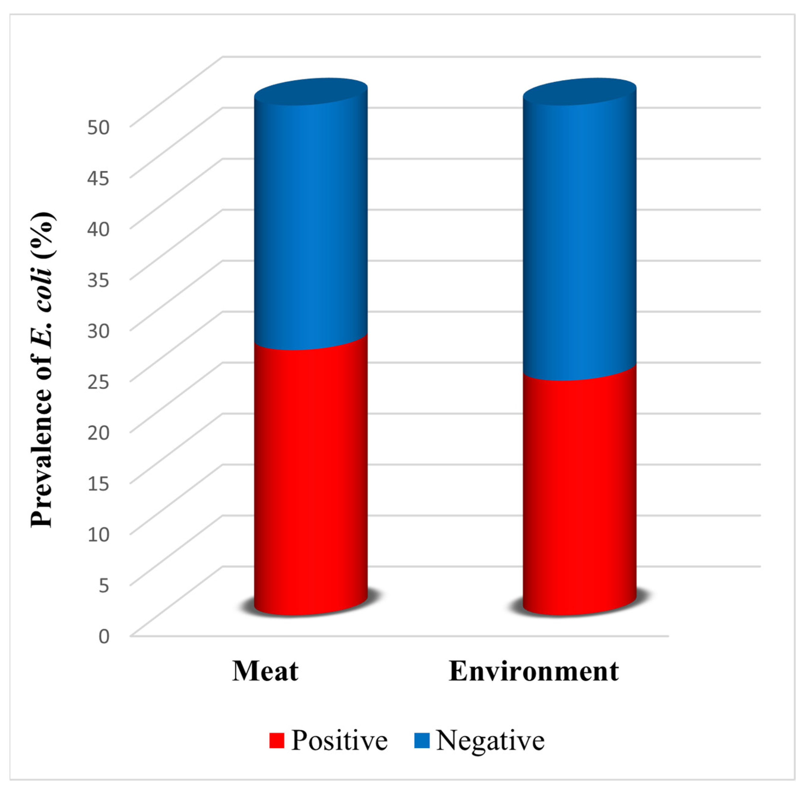
Figure 1. Stacked column chart showing the prevalence of E. coli in poultry meat and poultry farm environment samples (n = 50 per group). The chart displays the proportion of positive samples (red) and negative samples (blue) for each source.
Dna 05 00042 g001
Figure 2. Agarose gel electrophoresis image showing multiplex PCR amplification of E. coli virulence genes (stx1, stx2, and eae). ‘M’ indicates the marker (DNA ladder). Lanes 1–2: poultry litter isolates; lanes 3–4: poultry farm environment swab isolates; lanes 5–6: poultry meat isolates. NC: negative control. The stx1 gene has a base pair (bp) length of 894 bp, the stx2 gene’s length is 478 bp, and the eae gene’s length is 384 bp. The faint upper band observed in lane 4 may represent non-specific amplification and does not correspond to any of the target gene products.
Dna 05 00042 g002
Figure 3. Comparative prevalence of virulence genes detected in E. coli isolates from poultry meat (n = 26) and poultry farm environment (n = 23) isolates. The chart illustrates the percentage of isolates positive for each virulence gene or gene combination, including stx1, stx2, eae, stx1/eae, stx2/eae, and stx1/stx2/eae. Blue bars represent meat isolates; orange bars represent environmental isolates. No statistically significant differences were observed between sources for individual genes or gene combinations (Fisher’s exact test, p > 0.05).
Dna 05 00042 g003
Figure 4. Heatmap illustrating the AMR profiles of E. coli isolates recovered from poultry meat samples. The color gradient represents susceptibility patterns, where red indicates resistant isolates, blue indicates intermediate susceptibility, and green indicates susceptible isolates for each antibiotic tested. Antibiotic abbreviations: SAM-10/10 = ampicillin–sulbactam; FOX-30 = cefoxitin; TE-30 = tetracycline; TMP-5 = trimethoprim; NOR-10 = norfloxacin; NA-30 = nalidixic acid.
Dna 05 00042 g004
Figure 5. Heatmap illustrating the AMR profiles of E. coli isolates recovered from poultry farm environment samples. The color gradient represents susceptibility patterns, where red indicates resistant isolates, blue indicates intermediate susceptibility, and green indicates susceptible isolates for each antibiotic tested. Antibiotic abbreviations: SAM-10/10 = ampicillin–sulbactam; FOX-30 = cefoxitin; TE-30 = tetracycline; TMP-5 = trimethoprim; NOR-10 = norfloxacin; NA-30 = nalidixic acid.
Dna 05 00042 g005
Figure 6. Comparative stacked column chart illustrating the antimicrobial resistance patterns of E. coli isolates recovered from poultry meat and poultry farm environment samples. The chart displays the proportion of resistant (red), intermediate (blue), and sensitive (green) isolates for each antibiotic tested, facilitating visual comparison of antimicrobial susceptibility patterns between the two sources. Antibiotic abbreviations: SAM 10/10: ampicillin–sulbactam; FOX-30: cefoxitin; TE-30: tetracycline; TMP-5: trimethoprim; NOR-10: norfloxacin; NA-30: nalidixic acid.
Dna 05 00042 g006
Table 1. Biochemical test results for E. coli, including positive and negative controls.
Biochemical
Test
Reagents
Added
E. coliOriginal
Control
Positive ControlNegative
Control
IndoleKovac’s reagent+YellowRed ringYellow
MRMR indicator+YellowRedYellow
VPBarritt’s reagentCopperRedNo color change
CitrateBromothymol indicator+GreenBlueGreen
CatalaseH2O2+ClearBubbleNo bubble
Lactose fermentation test-+Red color with no gas production in the Durham tubeThe yellow color along with gas production in the Durham tubeRed color, along with no gas production in the Durham tube
Table 2. Primers, oligonucleotide sequences, and amplicon sizes for detection of stx1, stx2, and eae virulence genes.
PrimerOligonucleotide Sequence (5′-3′)Amplicon Size (bp)Reference
stx1F=CAGTTAATGTGGTGGCGAAG894[25]
R=CTGCTAATAGTTCTGCGCATC
stx2F=CTTCGGTATCCTATTCCCGG478[24]
R=GGATGCATCTCTGGTCATTG
eaeF=GACCCGGCACAAGCATAAGC384[25]
R=CCACCTGCAGCAACAAGAGG
Table 3. Multiplex PCR cycling conditions.
ConditionsTemperature and Time
Initial Denaturation95 °C for 8 min
Denaturation94 °C for 1 min
Annealing55 °C for 45 s
Extension72 °C for 1 min
Final Extension72 °C for 10 min
Disclaimer/Publisher’s Note: The statements, opinions and data contained in all publications are solely those of the individual author(s) and contributor(s) and not of MDPI and/or the editor(s). MDPI and/or the editor(s) disclaim responsibility for any injury to people or property resulting from any ideas, methods, instructions or products referred to in the content.