Article

Mutational Characterization of Astrocytoma, IDH-Mutant, CNS WHO Grade III in the AACR GENIE Database

1
Division of Hematology and Oncology, Department of Internal Medicine, Creighton University Medical Center, Omaha, NE 68124, USA
2
Department of Hematology and Oncology, Creighton University School of Medicine, Phoenix, AZ 85012, USA
*
Author to whom correspondence should be addressed.
Submission received: 11 May 2025 / Revised: 17 July 2025 / Accepted: 12 August 2025 / Published: 4 September 2025

Abstract

Background/Objectives: Astrocytoma, IDH-mutant, CNS WHO grade 3, is a diffuse glioma with poor prognosis, molecularly defined by IDH mutations and frequently co-occurring TP53 and ATRX alterations. This study aimed to delineate the genomic landscape and identify clinically relevant molecular features of astrocytoma, IDH-mutant, CNS WHO grade 3 using this resource. Methods: Patients in the American Association for Cancer Research Project Genomics Evidence Neoplasia Information Exchange (AACR Project GENIE) database were selected based on histological diagnosis of “anaplastic astrocytoma”, confirmed IDH1/2 mutation, and exclusion of CDKN2A/B homozygous deletions. We analyzed frequencies of somatic mutations, copy number alterations (CNAs), structural variants (SVs), assessed co-occurrence/exclusivity patterns, and explored associations with available demographic and limited survival data. Results: The most common somatic mutations were in IDH1 (98.0%), TP53 (94.8%), and ATRX (55.2%). The observed ATRX mutation frequency was lower than some historical reports (e.g., ~86%). Other recurrent alterations included phosphatidylinositol-4,5-bisphosphate 3-kinase catalytic subunit alpha (PIK3CA) (6.9%), Notch receptor 1 (NOTCH1) (6.9%), and platelet-derived growth factor receptor alpha (PDGFRA) (mutations 4.3%; CNAs also observed). Conclusions: This study provides a comprehensive genomic characterization of astrocytoma, IDH-mutant, CNS WHO grade 3 using the AACR GENIE database, confirming core mutational signatures while also highlighting potential variations in alteration frequencies, such as for ATRX. The findings establish a valuable real-world genomic benchmark for this tumor type, while promoting the need for continued data integration with robust clinical outcomes to identify actionable prognostic and therapeutic targets.

1. Introduction

Astrocytoma, isocitrate dehydrogenase (IDH)-mutant, CNS WHO grade 3 (IDH-mut A3), previously known as anaplastic astrocytoma before the WHO classification in 2021, is a grade III glioma composed of astrocytes with a median age of onset of 41 years [1,2]. Survival is poor, with a median overall survival of 5 years [2,3], and younger patients fare significantly better than older patients [3]. IDH-mut A3 is relatively rare, with an incidence of only 0.48 per 100,000 persons per year [4]. The frequency in males is slightly higher than in females [2].
Historically, the diagnosis of anaplastic astrocytoma was based on histology, characterized by increased cellularity, mitotic activity, presence of glial markers, and nuclear atypia [2]. Important exclusion criteria included lack of neuronal markers to distinguish from tumors of neuronal origin such as gangliomas, and necrosis or microvascularization to distinguish from glioblastomas [2,5]. However, advances in understanding genetic changes have led to a molecular based diagnostic approach [2]. Astrocytoma, IDH-mutant now has several essential criteria, including being a diffusely infiltrating glioma, having an IDH1/2 hotspot mutation, and absence of combined whole arm deletions of 1p and 19q. In addition, loss of ATRX expression or ATRX mutation supports the diagnosis and obviates the need for 1p/19q status assessment (a 1p/19q codeletion characterizes an oligodendroglioma pathology) [6]. Pathology laboratories that do not have access to the required technology to perform these molecular characterizations must rely on previous WHO guidelines for classification of IDH-mut A3 (previously anaplastic astrocytoma), which was a wholly histological approach.
IDH-mut A3 has a distinct tumorigenesis dependent upon the IDH mutation. With a gain of function mutation, IDH will convert alpha-ketoglutarate (2OG) into the oncometabolite (R)-2-hydroxyglutarate (2-HG) [7]. The accumulation of 2-HG then leads to the competitive inhibition of 2OG-dependent enzymes, such as the 2OG-dependent oxygenases that are important in histone and DNA methylation [7]. This perturbation leads to a hyper methylated state known as the glioma CpG island methylator phenotype (G-CIMP) [7,8]. This phenotype leads to the epigenetic silencing of tumor suppressor genes and promotes de-differentiation [9,10]. Additional mutations in tumor protein p53 (TP53) and ATP-dependent helicase ATRX (ATRX) form a triple-mutation signature very common in IDH-mut A3 [11]. Mutations in ATRX impair chromatin remodeling near the telomeres, which destabilizes telomere structure and causes increased reliance upon alternative lengthening of telomeres (ALT) for telomere maintenance, while mutations in TP53 decrease DNA maintenance and repair [12].
Given enough time, IDH-mut A3 will progress into astrocytoma, IDH-mutant CNS WHO grade 4 (IDH-mut A4), previously known as secondary glioblastoma, IDH-mutant in the 2016 WHO classification. The updated classification reflects the observation that grade 4 IDH-mutant astrocytomas have different responses to treatment and a different mechanism of gliomagenesis than their glioblastoma IDH-wild-type counterparts [10]. Unlike IDH-wild-type glioblastomas which commonly have promoter mutations in telomerase reverse transcriptase (TERT) and epidermal growth factor receptor (EGFR) amplification [11], IDH-mut A3 evolves into a IDH-mut A4 through homozygous deletions in CDKN2A/B [13]. The prognosis with CDKN2A/B homozygous deletions is so poor that WHO classifies any IDH-mut A3 with deletions in CDKN2A/B as IDH-mut A4 even in cases without necrosis or neovascularization [1]. Nevertheless, IDH-mut A4 tumors generally have longer survival than IDH wild-type glioblastomas [13].
Treatment for IDH-mut A3 is a multimodal approach, including surgical resection, radiotherapy, and alkylating chemotherapy, most often Temozolomide [14,15]. Surgery provides both a diagnostic and therapeutic benefit [15]. Occurrence of IDH-mut A3 in pediatric patients is rare, with all instances generally occurring in older adolescents [16]. In this scenario treatment is very similar to the adult case [16]. For patients with advanced secondary glioblastoma, the electric field therapy is added as a possible consideration [14].
While previous studies have characterized the molecular landscape of IDH-mutant astrocytomas using various datasets, the AACR Project GENIE database [17], a large, contemporary, and regularly updated international consortium, has not yet been leveraged for a comprehensive analysis of this specific tumor grade. This study aims to fill this gap by characterizing the mutational landscape of IDH-mutant WHO grade 3 astrocytomas (IDH-mut A3) using the information stored in the AACR GENIE database. Notably, because the GENIE database was established prior to the 2021 WHO reclassification, tumors are still annotated under the outdated term “anaplastic astrocytoma”, and cannot be readily filtered by updated molecular criteria. Accordingly, we also aim to generate a practical and up-to-date reference of the mutational landscape of IDH-mut A3 based on modern diagnostic definitions.

2. Materials and Methods

This investigation utilized data procured from the publicly accessible, de-identified American Association of Cancer Research (AACR) Project GENIE database. The cBioPortal for Cancer Genomics platform (https://genie.cbioportal.org/), accessed 1 May 2025 was employed for data retrieval and subsequent analysis, encompassing the determination of frequencies for gene mutations, copy number alterations, and structural variants, all of which are displayed on a per-sample basis. cBioPortal uses the mutation calls provided by each contributing publication, with little additional filtering except for standardization of mutation annotations using Genome Nexus [18,19,20]. cBioPortal does filter out non-synonymous mutations, including Silent, Intron, IGR, 3′UTR, 5′UTR, 3′Flank and 5′Flank mutations, with the exception for mutations in the promoter region of the TERT gene. These genomic alterations were stratified according to demographic variables, including sex, ethnicity, primary race, and biological sample type. Statistical analyses were conducted using R/RStudio (R Foundation for Statistical Computing, Vienna, Austria) version 4.4.2, with the cbioportalR, tidyR, knitr, survival, dplyr, GenomeInfoDb, IRanges, S4Vectors, BiocGenerics, and data.table packages [21,22,23,24,25,26,27,28,29,30,31,32,33,34]. To address multiple hypothesis testing, the Benjamini–Hochberg procedure was applied to control the False Discovery Rate (FDR). Differences in demographic distributions among cohorts were evaluated using chi-squared tests, comparing proportions across categorical variables. Amplifications as given in the cBioPortal CNA genes table were defined as genomic regions with a score of 2 with the copy-number analysis algorithm (e.g., GISTIC) employed by cBioPortal, representing a high level increase in the copy number of a specific gene region. Deep deletions (homozygous deletions) were define as genomic regions with a score of −2.
Due to the diagnostic requirement for both molecular and histopathological criteria in identifying IDH-mutant CNS WHO grade III astrocytomas, and given that histological slides are not available in the AACR GENIE database, we relied on the pre-annotated histological classifications provided. Specifically, we included cases labeled as anaplastic astrocytoma, as this designation most closely corresponds to IDH-mutant WHO grade III astrocytomas in the 2021 WHO CNS classification system. This initial filter identified 678 patients. We then refined the cohort by selecting only tumors with a confirmed mutation in either IDH1 or IDH2. To avoid inclusion of higher-grade tumors, we excluded cases with homozygous deletions in CDKN2A or CDKN2B. Additionally, tumors with 1p/19q co-deletion were excluded, based on the methodology described by Williams et al. [35] and using genomic coordinates obtained from the UCSC Genome Browser [36]. We also excluded samples with both a gain in chromosome 7 and loss in chromosome 10 (+7/−10), as to further exclude possible glioblastoma multiforme tumors that were incorrectly classified as astrocytoma [37]. After applying these criteria, the final study cohort consisted of 344 patients and 348 tumor samples.
Survival analyses were performed by generating Kaplan–Meier curves for each gene interrogated within the cohort. For each gene, two curves were constructed: one representing patients harboring a mutation in the specified gene, and the other representing patients with the wild-type allele. Statistical significance between survival distributions was assessed using log-rank tests (from the survival package, version 3.7.0) for the top 10 most frequently mutated genes in the cohort. Because there GENIE database does not provide a specific variable for time of diagnosis, we used the patient’s age at the time of sample sequencing as a proxy for time of diagnosis. Survival time was calculated as the interval between this proxy for diagnosis and the time of death or last follow-up. Importantly, time of death is provided in GENIE as an age in years (measured from birth), not as an interval from diagnosis. Accordingly, survival time was derived by subtracting the age at sequencing from the age at death or last contact. To account for individuals with multiple samples, mutation counts for each gene were aggregated across all samples from a given patient. The overall frequency of gene alteration on a per-patient basis was determined by classifying a gene as altered if its total count of detected alterations across all samples for that patient was greater than one.

3. Results

There were 344 patients from the database that were diagnosed with astrocytoma, IDH-mutant, CNS WHO grade III, with a total of 348 samples (Table 1). Sex, ethnicity, and race were collected on a per-patient basis, while age and sample type were collected on a per-sample basis. There were more male patients (201, 58.4%) than female patients (142, 41.3%) and only two patients with an unknown sex (1, 0.3%). The cohort had no pediatric sample as defined by <18 years of age at sequencing, with 347 adult samples (99.7%) and 1 sample with no age information (0.3%). White was the most common race (260, 75.6%), followed by Asian (24, 7.0%), Other (17, 4.9%), Black (11, 3.2%), and Pacific Islander (1, 0.3%), with an additional 29 patients having no race information (8.4%). In terms of Hispanic ethnicity, most were non-Hispanic (273, 79.4%), with a remainder having no information (51, 14.8%) and the rest being Spanish/Hispanic (20, 5.8%). A total of 303 (87.1%) of tumors were classified as primary, 40 (11.5%) classified as metastatic, and 5 (1.4%) missing this information.
A total of 348 samples were profiled for somatic mutations (Table 2). IDH1 was the most common somatic mutation among the IDH-mut A3 samples (n = 341, 98.0%), followed by TP53 (n = 330, 94.8%), ATRX (n = 192, 55.2%), NOTCH1 (n = 24, 6.9%), PIK3CA (n = 24, 6.9%), SWI/SNF-related, matrix-associated, actin-dependent regulator of chromatin subfamily a, member 4 (SMARCA4) (n = 19, 5.5%), FAT atypical cadherin 1 (FAT1) (n = 18, 5.2%), lysine methyltransferase 2D (KMT2D) (n = 18, 5.2%), PDGFRA (n = 16, 4.3%), and protein kinase, DNA-activated, catalytic subunit (PRKDC) (n = 15, 4.2%). ATR serine/threonine kinase (ATR), BCOR, phosphoinositide-3-kinase regulatory subunit 1 (PIK3R1), ARID1A, and neurofibromin 1 (NF1) were the 5 next most common somatic mutations, all with frequencies of greater than 3%.
A total of 200 samples were profiled for copy number alterations (Table 3), with variable number of profiles for each gene. The most frequent amplifications were found in CCND2 (n = 8, 4.0%), cyclin-dependent kinase 4 (CDK4) (n = 8, 4.0%), PDGFRA (n = 7, 3.5%), MYCN (n = 5, 2.5%), and KRAS (n = 5, 2.5%) Common homozygous deletions were found in ATRX (n = 6, 3.0%), CDKN1B (n = 3, 1.5%), EPHA7 (n = 3, 1.5%), PDCD1 (n = 3, 1.5%), and TERT (n = 3, 1.5%).
A total of 245 samples were profiled for structural variants (Table 4). There were 4 instances of structural variations in KMT2D with 1 duplication, 1 translocation, and 2 unspecified. There were 3 instances of structural variations in ATRX, with them all being unspecified. There were two instances of structural variations in SDHA, one inversion and the other unspecified. Finally, there were 2 unspecified structural variations in ABL1 and 1 unspecified structural variation in BRCA1. Figure 1 shows an oncoprint of structural variants, along with copy number variations and most common mutations.

3.1. Genetic Differences by Race and Sex

Removing the unknown sex category, chi-squared tests were performed to assess for sex-based differences in gene frequency, copy number alterations, and structural variants. PTCH1 was found to be enriched in females (8, 7.77% vs. 3, 1.85%, p = 0.026). However, significance was lost upon Benjamini–Hochberg multiple hypothesis test correction. CREBBP was also found to be enriched in females (7, 6.80% vs. 2, 1.23%, p = 0.030). No significant sex-based differences were found in copy number alterations or structural variants. APC showed enrichment in Spanish/Hispanics compared to non-Spanish/non-Hispanics (p = 0.00214), however this significance was lost after Benjamini–Hochberg multiple hypothesis test correction. No other gene mutations, structural variations, or copy number alterations exhibited significant differences in frequency across two-way chi squared comparison tests.

3.2. Co-Occurrence and Exclusivity

The 15 most frequently mutated genes in the cohort were evaluated for co-occurrence and exclusivity patterns using the query function in cBioPortal, resulting in 105 pairs tested for co-occurrence and mutual exclusivity. Significant co-occurrence was found between FAT1 and PDGFRA (4 mutations found in both out of 19 mutations found in either, p = 0.016), NOTCH1 and NF1 (3 mutations found in both out of 19 mutations found in either, p = 0.025), KMT2D and PRKDC (3 mutations found in both out of 13 mutations found in either, p = 0.043), FAT1 and ARID1A (3 mutations found in both out of 18 mutations found in either, p =0.043), KMT2D and BCOR (3 mutations found in both out of 20 mutations found in either, p = 0.044), and TP53 and ATRX (159 mutations found in both out of 189 found in either, p = 0.050). The only significant mutual exclusivity was found in TP53 and ARID1A (9 found in both out of 179 found in either, p = 0.040). In addition, all prior associations lost significance after multiple hypothesis test correction.

3.3. Primary vs. Metastatic Samples

The cohort consisted of 40 samples from metastatic sites, and 303 from primary sites. In this analysis, a gene was considered enriched if the proportion of metastatic samples (as pre-annotated in the GENIE database) harboring a mutation in that gene was significantly greater than the proportion observed in primary samples, based on statistical comparison in the two-sided Fisher exact test. Many genes were enriched in metastatic samples compared to primary samples, including CDKN2A (6 vs. 2, p = 4.2 × 10⁻⁵, KMT2C (3 vs. 1, p = 1.3 × 10⁻³), RICTOR (4 vs. 1, p = 1.4 × 10⁻³), BRD4 (4 vs. 1, p = 1.8 × 10⁻³), and CREBBP (5 vs. 3, p = 2.1 × 10⁻³). However, only CDKN2A (somatic mutation, not deletion) remained significant after multiple hypothesis test correction.

3.4. Mutations with Differential Effects on Lifespan

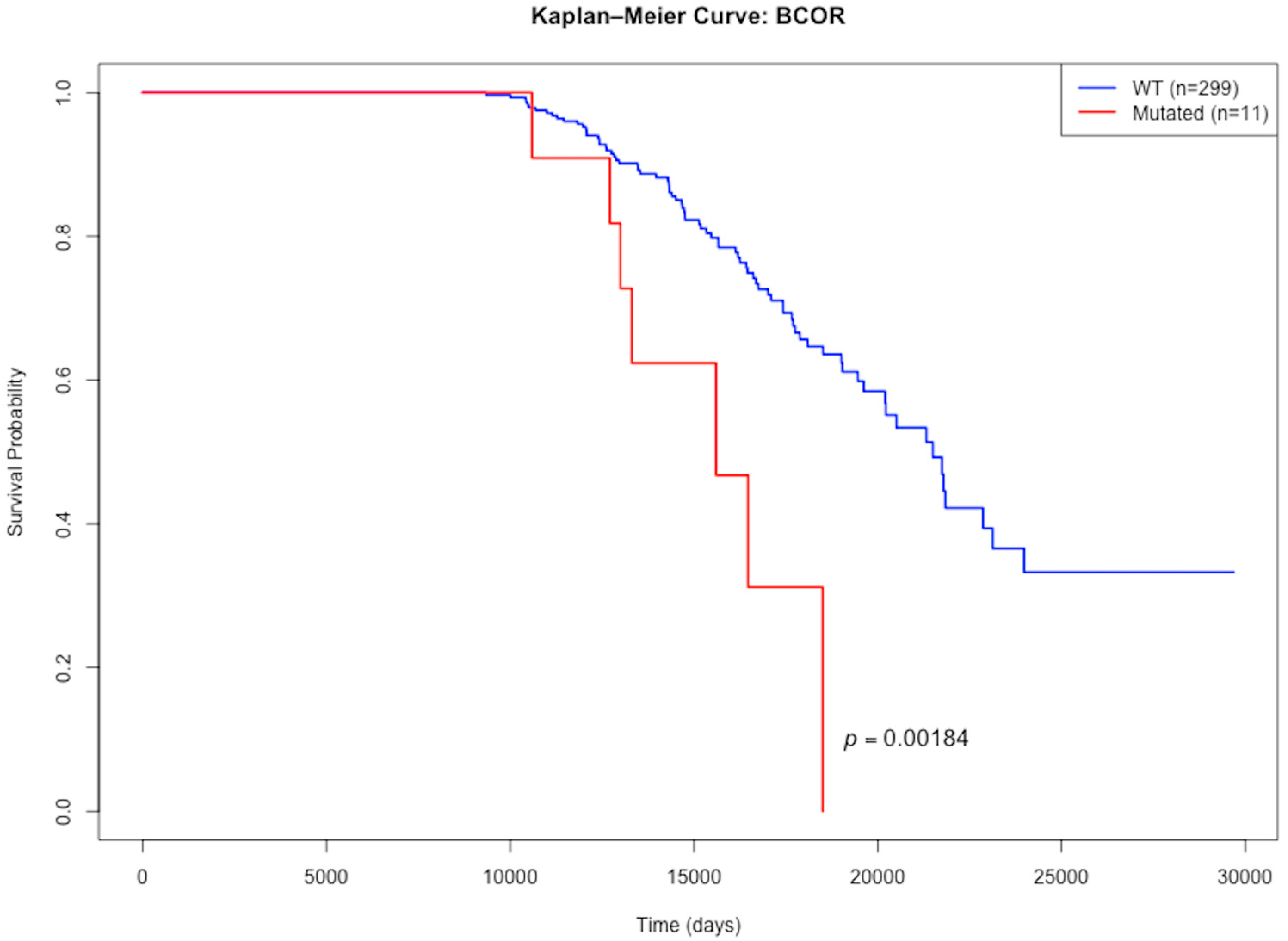
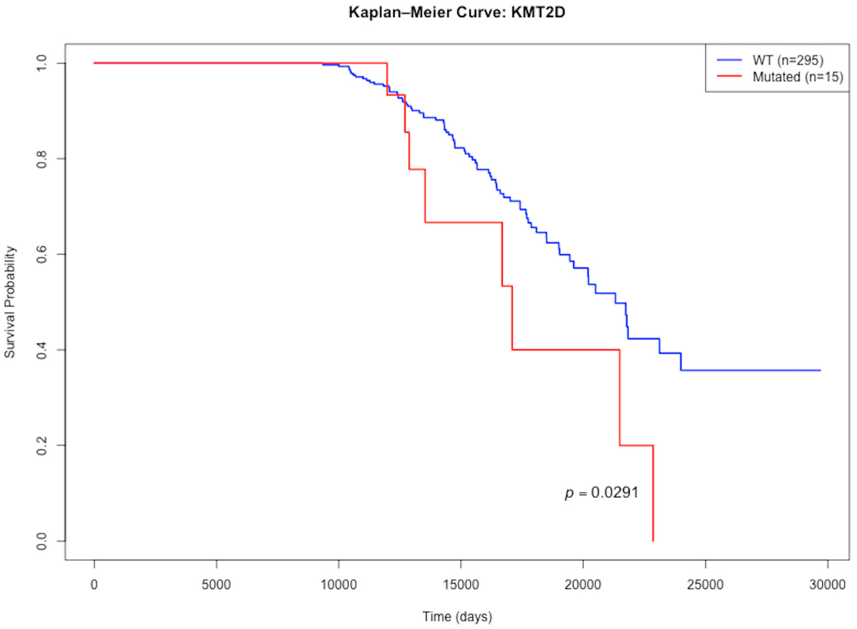
After filtering for required patient-level survival information, 310 patients were included in the survival analysis. Two genes with significantly decreased median patient survival time compared to non-mutated genes were BCOR and KMT2D (see Table 5, Figure 2 and Figure 3).

4. Discussion

Analysis of the cohort comprising 344 patients diagnosed with astrocytoma, IDH-mutant, CNS WHO Grade III, identified a primary mutational signature consistent with established literature, alongside several additional somatic alterations of potential significance. Per the inclusion criteria, all patients exhibited mutations in either IDH1 or IDH2. Mutations in TP53 (94.8%) and ATRX (55.2%) were the second and third most prevalent alterations, respectively, a finding consistent with the characteristic triple-mutation phenotype observed in this astrocytoma subtype [11]. However, the observed ATRX mutation frequency of 55.2% was notably lower than the previously reported frequency of 86% in comparable cohorts [38]. This discrepancy could stem from differences in patient selection criteria between studies, variations in the sensitivity of sequencing panels or bioinformatic pipelines used for ATRX mutation detection, or the specific inclusion criteria for ‘comparable cohorts’ in prior literature.
Mutations were also found in other genes, but at much lower frequencies compared to IDH1/2, TP53, and ATRX. PIK3CA and NOTCH1 were the next most frequently mutated genes, both with frequencies of 6.9%, respectively. Similar instances of PIK3CA and NOTCH1 frequency have been described, however we found no significant difference in prognosis between PIK3CA mutant and wild-type patients, unlike what has been described in the literature [38,39].
We report both mutations (4.9%) and amplifications (3.5%) in PDGFRA. PDGFRA has been reported as an important driver and therapeutic target in high grade glioma with a total mutation and amplification frequency of 15% [40]. However, even when reverting to the more-permissive histology classification of anaplastic astrocytoma by not excluding the cases of CDKN2A/B homozygous deletions and not requiring IDH1/2 mutations (thereby including the higher grade IV gliomas according to WHO classification), the frequency of PDGFRA mutations and amplifications did not markedly change.
We also found co-occurrence and mutual exclusivity patterns. TP53 and ATRX were found to co-occur, which matched the expected IDH1-TP53-ATRX triple mutation signature found in many IDH-mutant tumors [11]. Mutual exclusivity was found between TP53 and ARID1A, whose pattern has also been seen in ovarian cancer [41]. It has been shown that p53 interacts with ARID1A/BRG1, and this complex binds to the CDKN1A and SMAD3 promoter regions [41]. Mutations in either TP53 or ARID1A could thus lead to similar downstream effects, explaining this mutual exclusivity trend.
Overall, the molecular profile of IDH-mut A3 had the proper characteristics to exclude diagnosis of other cancers. For instance, mutations in TERT had a frequency of 3.2%, which aligns with its literature-reported expected frequency in IDH-mut A3 [42]. Much higher frequencies of TERT mutation are associated with an oligodendroglioma, not an astrocytoma. We also had low rates of EGFR amplification, which would be more characteristic of glioblastoma [43]. No instances of 1p/19q co-deletion also excluded the possibility of oligodendroglioma.
We also observed a higher ratio of male patients (n = 201) to female patients (n = 142) of 1.56. This higher incidence of male patients with glioma has been described, with it being hypothesized that estrogen plays a protective role against gliomagenesis, whereas androgen exposure increases risk [44]. Despite this difference in frequency, we did not find any significant differences in frequency across sex, race, or ethnicity that persisted after FDR correction.

Limitations

This investigation is subject to several significant limitations that warrant acknowledgment. Firstly, the analytical scope was confined to genomic data, specifically DNA sequence alterations and copy number variations. The absence of integrated transcriptomic, epigenomic (e.g., methylation arrays), proteomic, or microRNA expression data inherently limits the capacity to draw comprehensive conclusions regarding the functional consequences of the observed genomic alterations and the resultant tumor phenotypes. The lack of immunohistochemistry data also makes it more difficult to infer ATRX loss in the diagnostic algorithm.
Secondly, sample size constraints, particularly for specific sub-analyses, represent a critical limitation. Although the AACR Project GENIE database is extensive, encompassing over 196,000 patients and 229,000 samples, the application of stringent inclusion criteria significantly reduced the study cohort. Initial filtering for “anaplastic astrocytoma” yielded 659 patients. Subsequent refinement, mandating IDH1/2 mutation status and excluding cases with CDKN2A/B homozygous deletions, further reduced the patient number to 491. A more substantial attrition was observed for survival analyses, where incomplete follow-up or missing date parameters necessary for constructing survival intervals restricted the analyzable cohort to 86 patients. Such diminished sample sizes, especially within the survival analysis cohort, reduce statistical power, increase the margin of error for estimates, and may preclude the detection of true, albeit modest, associations. Furthermore, these limited numbers hampered robust stratification of mutation frequencies by demographic variables such as race and ethnicity, particularly for numerically smaller groups (e.g., Native American, Pacific Islander populations), thereby limiting the generalizability of findings related to these subgroups.
Thirdly, the retrospective nature of this study, utilizing pre-existing database information, introduces potential biases. Data were aggregated from multiple institutions, which, despite standardization efforts within Project GENIE, may harbor inter-institutional variability in diagnostic practices, pathological interpretation (especially for historical samples predating current WHO classifications), sequencing methodologies, and clinical data abstraction completeness. This heterogeneity could introduce unmeasured confounding variables. Furthermore, segmented copy number data, which were used to exclude 1p/19q samples, are not a required aspect for submission to the GENIE database, which means that some samples may have been included that have the IDH-A3 excluding characteristic of a 1p/19q co-deletion, but was impossible to detect due to the data not being submitted.
Fourthly, the absence of comprehensive clinical data, most notably the lack of detailed treatment regimens (e.g., specific chemotherapeutic agents, radiotherapy protocols, extent of surgical resection, and subsequent lines of therapy), severely curtails the ability to assess the impact of therapeutic interventions on survival outcomes or to adjust for their confounding effects on genotype–phenotype correlations. Other potentially influential clinical variables, such as patient performance status or detailed comorbidity profiles, were also not uniformly available.
Fifthly, while Project GENIE provides valuable genomic information, the specific gene panels or sequencing depths utilized by contributing institutions may vary. This could lead to differential sensitivity in detecting certain types of mutations (e.g., low-frequency subclones, complex structural variants, or alterations in non-exonic regions not comprehensively covered by all panels).
Finally, this study is descriptive and correlative; it does not involve functional validation of the identified mutations or their putative biological roles. Therefore, causality cannot be inferred from the observed associations. The interpretations presented are contingent upon the existing literature and bioinformatic predictions, requiring subsequent experimental investigation for definitive confirmation.

5. Conclusions

This genomic characterization of astrocytoma, IDH-mutant, CNS WHO grade 3 using the AACR GENIE database confirms key mutational patterns and demonstrates the resource’s value for studying this tumor type, provided that correct selection criteria are applied to match the updated WHO classification criteria.

Author Contributions

Conceptualization, E.T. and B.H.; methodology, E.T. and B.H.; software, E.T.; validation, E.T. and N.L. and B.H.; formal analysis, E.T.; investigation, E.T.; resources, P.S.; data curation, E.T.; writing—original draft preparation, E.T.; writing—review and editing, E.T. and N.L. and B.H.; visualization, B.H.; supervision, B.H. and P.S.; project administration, P.S.; All authors have read and agreed to the published version of the manuscript.

Funding

This research received no external funding.

Institutional Review Board Statement

Ethical review and approval were waived for this study because the AACR Project GENIE is a publicly available cancer genomic database containing de-identified patient data, which minimizes potential risks to human subjects and eliminates the need for individual participant consent.

Informed Consent Statement

Patient consent was waived due to the nature of the study, which used only de-identified data from the publicly available AACR Project GENIE database, and this does not require individual informed consent.

Data Availability Statement

The data presented in this study are available from the AACR GENIE Database at https://genie.cbioportal.org/ (accessed on 23 April 2025).

Acknowledgments

The authors would like to acknowledge the American Association for Cancer Research and its financial and material support in the development of the AACR Project GENIE registry, as well as members of the consortium for their commitment to data sharing. Interpretations are the responsibility of the authors.

Conflicts of Interest

The authors declare no conflicts of interest.

Abbreviations

The following abbreviations are used in this manuscript:
GENIEAACR Project Genomics Evidence Neoplasia Information Exchange
AACRAmerican Association for Cancer Research
2-HG(R)-2-hydroxyglutarate
2OGAlpha-ketoglutarate
IDH-mut A3Astrocytoma, IDH-mutant, CNS WHO grade 3
ALTAlternative lengthening of telomeres
ATRATR serine/threonine kinase
ATRXATP-dependent helicase ATRX
AXIN2Axin 2
BCORBCL6 corepressor
BRAFB-Raf Proto-oncogene, serine/threonine kinase
BRD4Bromodomain-containing protein 4
CDK4Cyclin-dependent kinase 4
CDKN2ACyclin-dependent kinase inhibitor 2A
CNSCentral nervous system
CREBBPCREB binding protein
EGFREpidermal growth factor receptor
FAT1FAT atypical cadherin 1
FDRFalse discovery rate
G-CIMPGlioma CpG island methylator phenotype
GLI1GLI family zinc finger 1
IDHIsocitrate dehydrogenase
KITKIT proto-oncogene, receptor tyrosine kinase
KMT2DLysine methyltransferase 2D
MAP2K2Mitogen-activated protein kinase 2
MLH1MutL homolog 1
MTAPMethylthioadenosine phosphorylase
MTORMechanistic target of rapamycin kinase
NF1Neurofibromin 1
NOTCH1Notch receptor 1
PDGFRAPlatelet-derived growth factor receptor alpha
PIK3CAPhosphatidylinositol-4,5-bisphosphate 3-kinase catalytic subunit alpha
PIK3R1Phosphoinositide-3-kinase regulatory subunit 1
PRKDCProtein kinase, DNA-activated, catalytic subunit
RETRet proto-oncogene
RICTORRPTOR independent companion of MTOR complex 2
ROS1ROS proto-oncogene 1, receptor tyrosine kinase
SDHASuccinate dehydrogenase complex flavoprotein subunit A
SMARCA4SWI/SNF-related, matrix-associated, actin-dependent regulator of chromatin, subfamily a, member 4
SMOSmoothened, frizzled class receptor
SVStructural variant
TERTTelomerase reverse transcriptase
TET1Tet methylcytosine dioxygenase 1
TP53Tumor protein p53
WHOWorld Health Organization

References

  1. Louis, D.N.; Perry, A.; Wesseling, P.; Brat, D.J.; Cree, I.A.; Figarella-Branger, D.; Hawkins, C.; Ng, H.K.; Pfister, S.M.; Reifenberger, G.; et al. The 2021 WHO Classification of Tumors of the Central Nervous System: A Summary. Neuro Oncol. 2021, 23, 1231–1251. [Google Scholar] [CrossRef]
  2. Alshiekh Nasany, R.; de la Fuente, M.I. Therapies for IDH-Mutant Gliomas. Curr. Neurol. Neurosci. Rep. 2023, 23, 225–233. [Google Scholar] [CrossRef]
  3. Anvari, K.; Seilanian Toussi, M.; Shahidsales, S.; Motlagh, F.; Reza Ehsaee, M.; Afshari, F. Treatment Outcomes and Prognostic Factors in Adult Astrocytoma: In North East of Iran. Iran. J. Cancer Prev. 2016, 9, e4099. [Google Scholar] [CrossRef] [PubMed]
  4. Smoll, N.R.; Hamilton, B. Incidence and Relative Survival of Anaplastic Astrocytomas. Neuro Oncol. 2014, 16, 1400–1407. [Google Scholar] [CrossRef] [PubMed]
  5. Louis, D.N.; Ohgaki, H.; Wiestler, O.D.; Cavenee, W.K.; Burger, P.C.; Jouvet, A.; Scheithauer, B.W.; Kleihues, P. The 2007 WHO Classification of Tumours of the Central Nervous System. Acta Neuropathol. 2007, 114, 97–109. [Google Scholar] [CrossRef]
  6. Brandner, S.; McAleenan, A.; Jones, H.E.; Kernohan, A.; Robinson, T.; Schmidt, L.; Dawson, S.; Kelly, C.; Leal, E.S.; Faulkner, C.L.; et al. Diagnostic accuracy of 1p/19q codeletion tests in oligodendroglioma: A comprehensive meta-analysis based on a Cochrane systematic review. Neuropathol. Appl. Neurobiol. 2022, 48, e12790. [Google Scholar] [CrossRef]
  7. Lin, M.D.; Tsai, A.C.-Y.; Abdullah, K.G.; McBrayer, S.K.; Shi, D.D. Treatment of IDH-Mutant Glioma in the INDIGO Era. npj Precis. Oncol. 2024, 8, 149. [Google Scholar] [CrossRef]
  8. Turcan, S.; Rohle, D.; Goenka, A.; Walsh, L.A.; Fang, F.; Yilmaz, E.; Campos, C.; Fabius, A.W.M.; Lu, C.; Ward, P.S.; et al. IDH1 Mutation Is Sufficient to Establish the Glioma Hypermethylator Phenotype. Nature 2012, 483, 479–483. [Google Scholar] [CrossRef] [PubMed]
  9. Malta, T.M.; de Souza, C.F.; Sabedot, T.S.; Silva, T.C.; Mosella, M.S.; Kalkanis, S.N.; Snyder, J.; Castro, A.V.B.; Noushmehr, H. Glioma CpG Island Methylator Phenotype (G-CIMP): Biological and Clinical Implications. Neuro Oncol. 2018, 20, 608–620. [Google Scholar] [CrossRef]
  10. Han, S.; Liu, Y.; Cai, S.J.; Qian, M.; Ding, J.; Larion, M.; Gilbert, M.R.; Yang, C. IDH Mutation in Glioma: Molecular Mechanisms and Potential Therapeutic Targets. Br. J. Cancer 2020, 122, 1580–1589. [Google Scholar] [CrossRef]
  11. Byun, Y.H.; Park, C.-K. Classification and Diagnosis of Adult Glioma: A Scoping Review. Brain Neurorehabilit. 2022, 15, e23. [Google Scholar] [CrossRef]
  12. Haase, S.; Garcia-Fabiani, M.B.; Carney, S.; Altshuler, D.; Núñez, F.J.; Méndez, F.M.; Núñez, F.; Lowenstein, P.R.; Castro, M.G. Mutant ATRX: Uncovering a New Therapeutic Target for Glioma. Expert Opin. Ther. Targets 2018, 22, 599–613. [Google Scholar] [CrossRef]
  13. Katzendobler, S.; Niedermeyer, S.; Blobner, J.; Trumm, C.; Harter, P.N.; von Baumgarten, L.; Stoecklein, V.M.; Tonn, J.-C.; Weller, M.; Thon, N.; et al. Determinants of Long-Term Survival in Patients with IDH-Mutant Gliomas. J. Neurooncol 2024, 170, 655–664. [Google Scholar] [CrossRef]
  14. Mohile, N.A.; Messersmith, H.; Gatson, N.T.; Hottinger, A.F.; Lassman, A.; Morton, J.; Ney, D.; Nghiemphu, P.L.; Olar, A.; Olson, J.; et al. Therapy for Diffuse Astrocytic and Oligodendroglial Tumors in Adults: ASCO-SNO Guideline. J. Clin. Oncol. 2022, 40, 403–426. [Google Scholar] [CrossRef]
  15. Weller, M.; van den Bent, M.; Preusser, M.; Le Rhun, E.; Tonn, J.C.; Minniti, G.; Bendszus, M.; Balana, C.; Chinot, O.; Dirven, L.; et al. EANO Guidelines on the Diagnosis and Treatment of Diffuse Gliomas of Adulthood. Nat. Rev. Clin. Oncol. 2021, 18, 170–186. [Google Scholar] [CrossRef]
  16. Childhood Astrocytomas and Other Gliomas Treatment (PDQ®)-NCI. Available online: https://www.cancer.gov/types/brain/hp/child-astrocytoma-glioma-treatment-pdq (accessed on 8 August 2025).
  17. AACR Project GENIE Consortium; André, F.; Arnedos, M.; Baras, A.S.; Baselga, J.; Bedard, P.L.; Berger, M.F.; Bierkens, M.; Calvo, F.; Cerami, E. AACR Project GENIE: Powering Precision Medicine through an International Consortium. Cancer Discov. 2017, 7, 818–831. [Google Scholar] [CrossRef] [PubMed]
  18. Cerami, E.; Gao, J.; Dogrusoz, U.; Gross, B.E.; Sumer, S.O.; Aksoy, B.A.; Jacobsen, A.; Byrne, C.J.; Heuer, M.L.; Larsson, E.; et al. The cBio cancer genomics portal: An open platform for exploring multidimensional cancer genomics data. Cancer Discov. 2012, 2, 401–404. [Google Scholar] [CrossRef]
  19. de Bruijn, I.; Kundra, R.; Mastrogiacomo, B.; Tran, T.N.; Sikina, L.; Mazor, T.; Li, X.; Ochoa, A.; Zhao, G.; Lai, B.; et al. Analysis and Visualization of Longitudinal Genomic and Clinical Data from the AACR Project GENIE Biopharma Collaborative in cBioPortal. Cancer Res. 2023, 83, 3861–3867. [Google Scholar] [CrossRef]
  20. Gao, J.; Aksoy, B.A.; Dogrusoz, U.; Dresdner, G.; Gross, B.; Sumer, S.O.; Sun, Y.; Jacobsen, A.; Sinha, R.; Larsson, E.; et al. Integrative analysis of complex cancer genomics and clinical profiles using the cBioPortal. Sci. Signal. 2013, 6, pl1. [Google Scholar] [CrossRef] [PubMed]
  21. Reuss, D.E. Updates on the WHO diagnosis of IDH-mutant glioma. J. Neurooncol. 2023, 162, 461–469. [Google Scholar] [CrossRef] [PubMed]
  22. Whiting, K. Cbioportalr: Browse and Query Clinical and Genomic Data from Cbioportal, R package version 1.1.1. 2024. Available online: https://CRAN.R-project.org/package=cbioportalR (accessed on 1 May 2025).
  23. Wickham, H.; Vaughan, D.; Girlich, M. Tidyr: Tidy Messy Data, R package version 1.3.1. 2024. Available online: https://CRAN.R-project.org/package=tidyr (accessed on 1 May 2025).
  24. Xie, Y. Knitr: A General-Purpose Package for Dynamic Report Generation in R, R Package Version 1.49. 2024. Available online: https://yihui.org/knitr/ (accessed on 1 May 2025).
  25. Xie, Y. Dynamic Documents with R and Knitr, 2nd ed.; Chapman and Hall/CRC: Boca Raton, FL, USA, 2015; ISBN 978-1498716963. Available online: https://github.com/yihui/knitr-book (accessed on 1 May 2025).
  26. Xie, Y. knitr: A Comprehensive Tool for Reproducible Research in R. In Implementing Reproducible Computational Research; Stodden, V., Leisch, F., Peng, R.D., Eds.; Chapman and Hall/CRC: Boca Raton, FL, USA, 2014; ISBN 978-1466561595. [Google Scholar]
  27. Therneau, T. A Package for Survival Analysis in R, R Package Version 3.7-0. 2024. Available online: https://CRAN.R-project.org/package=survival (accessed on 1 May 2025).
  28. Terry, M.T.; Patricia, M. Grambsch. Modeling Survival Data: Extending the Cox Model; Springer: New York, NY, USA, 2000; ISBN 0-387-98784-3. [Google Scholar]
  29. Wickham, H.; François, R.; Henry, L.; Müller, K.; Vaughan, D. Dplyr: A Grammar of Data Manipulation, R package version 1.1.4. 2023. Available online: https://CRAN.R-project.org/package=dplyr (accessed on 1 May 2025).
  30. Arora, S.; Morgan, M.; Carlson, M.; Pagès, H. GenomeInfoDb: Utilities for Manipulating Chromosome Names, Including Modifying Them to Follow a Particular Naming Style, R package version 1.42.3. 2025. Available online: https://bioconductor.org/packages/GenomeInfoDb (accessed on 1 May 2025).
  31. Lawrence, M.; Huber, W.; Pagès, H.; Aboyoun, P.; Carlson, M.; Gentleman, R.; Morgan, M.; Carey, V. Software for Computing and Annotating Genomic Ranges. PLoS Comput. Biol. 2013, 9, e1003118. [Google Scholar] [CrossRef] [PubMed]
  32. Pagès, H.; Lawrence, M.; Aboyoun, P. S4Vectors: Foundation of Vector-Like and List-Like Containers in Bioconductor, R Package Version 0.44.0. 2024. Available online: https://bioconductor.org/packages/S4Vectors (accessed on 8 August 2025).
  33. Huber, W.; Carey, J.V.; Gentleman, R.; Anders, S.; Carlson, M.; Carvalho, S.B.; Bravo, C.H.; Davis, S.; Gatto, L.; Girke, T.; et al. Orchestrating high-throughput genomic analysis with Bioconductor. Nat. Methods 2015, 12, 115–121. [Google Scholar] [CrossRef] [PubMed]
  34. Barrett, T.; Dowle, M.; Srinivasan, A.; Gorecki, J.; Chirico, M.; Hocking, T.; Schwendinger, B. Data.Table: Extension of ‘data.frame’, R Package Version 1.16.2. 2024. Available online: https://CRAN.R-project.org/package=data.table (accessed on 8 August 2025).
  35. Williams, E.A.; Sharaf, R.; Decker, B.; Werth, A.J.; Toma, H.; Montesion, M.; Sokol, E.S.; Pavlick, D.C.; Shah, N.; Williams, K.J.; et al. CDKN2C-Null Leiomyosarcoma: A Novel, Genomically Distinct Class of TP53/RB1–Wild-Type Tumor with Frequent CIC Genomic Alterations and 1p/19q-Codeletion. JCO Precis. Oncol. 2020, 4, 955–971. [Google Scholar] [CrossRef]
  36. Perez, G.; Barber, G.P.; Benet-Pages, A.; Casper, J.; Clawson, H.; Diekhans, M.; Fischer, C.; Gonzalez, J.N.; Hinrichs, A.S.; Lee, C.M.; et al. The UCSC Genome Browser database: 2025 update. Nucleic Acids Res. 2025, 53, D1243–D1249. [Google Scholar] [CrossRef]
  37. Stichel, D.; Ebrahimi, A.; Reuss, D.; Schrimpf, D.; Ono, T.; Shirahata, M.; Reifenberger, G.; Weller, M.; Hänggi, D.; Wick, W.; et al. Distribution of EGFR amplification, combined chromosome 7 gain and chromosome 10 loss, and TERT promoter mutation in brain tumors and their potential for the reclassification of IDHwt astrocytoma to glioblastoma. Acta Neuropathol. 2018, 136, 793–803. [Google Scholar] [CrossRef]
  38. The Cancer Genome Atlas Research Network. Comprehensive, Integrative Genomic Analysis of Diffuse Lower-Grade Gliomas. N. Engl. J. Med. 2015, 372, 2481–2498. [Google Scholar] [CrossRef]
  39. Li, Z.; Deng, Z.; Liu, F.; Li, C.; Yang, K.; Gong, X.; Feng, S.; Zeng, Y.; Zhou, H.; Fan, F.; et al. Clinical Sequencing Reveals Diagnostic, Therapeutic, and Prognostic Biomarkers for Adult-Type Diffuse Gliomas. Heliyon 2024, 10, e37712. [Google Scholar] [CrossRef] [PubMed]
  40. Mayr, L.; Neyazi, S.; Schwark, K.; Trissal, M.; Beck, A.; Labelle, J.; Eder, S.K.; Weiler-Wichtl, L.; Marques, J.G.; de Biagi-Junior, C.A.O.; et al. Effective Targeting of PDGFRA-Altered High-Grade Glioma with Avapritinib. Cancer Cell 2025, 43, 740–756.e8. [Google Scholar] [CrossRef]
  41. Guan, B.; Wang, T.-L.; Shih, I.-M. ARID1A, a Factor That Promotes Formation of SWI/SNF-Mediated Chromatin Remodeling, Is a Tumor Suppressor in Gynecologic Cancers. Cancer Res. 2011, 71, 6718–6727. [Google Scholar] [CrossRef]
  42. Lee, K.; Kim, S.-I.; Kim, E.E.; Shim, Y.-M.; Won, J.-K.; Park, C.-K.; Choi, S.H.; Yun, H.; Lee, H.; Park, S.-H. Genomic Profiles of IDH-Mutant Gliomas: MYCN-Amplified IDH-Mutant Astrocytoma Had the Worst Prognosis. Sci. Rep. 2023, 13, 6761. [Google Scholar] [CrossRef]
  43. Hatanpaa, K.J.; Burma, S.; Zhao, D.; Habib, A.A. Epidermal Growth Factor Receptor in Glioma: Signal Transduction, Neuropathology, Imaging, and Radioresistance. Neoplasia 2010, 12, 675–684. [Google Scholar] [CrossRef] [PubMed]
  44. Yin, J.; Liu, G.; Zhang, Y.; Zhou, Y.; Pan, Y.; Zhang, Q.; Yu, R.; Gao, S. Gender Differences in Gliomas: From Epidemiological Trends to Changes at the Hormonal and Molecular Levels. Cancer Lett. 2024, 598, 217114. [Google Scholar] [CrossRef] [PubMed]
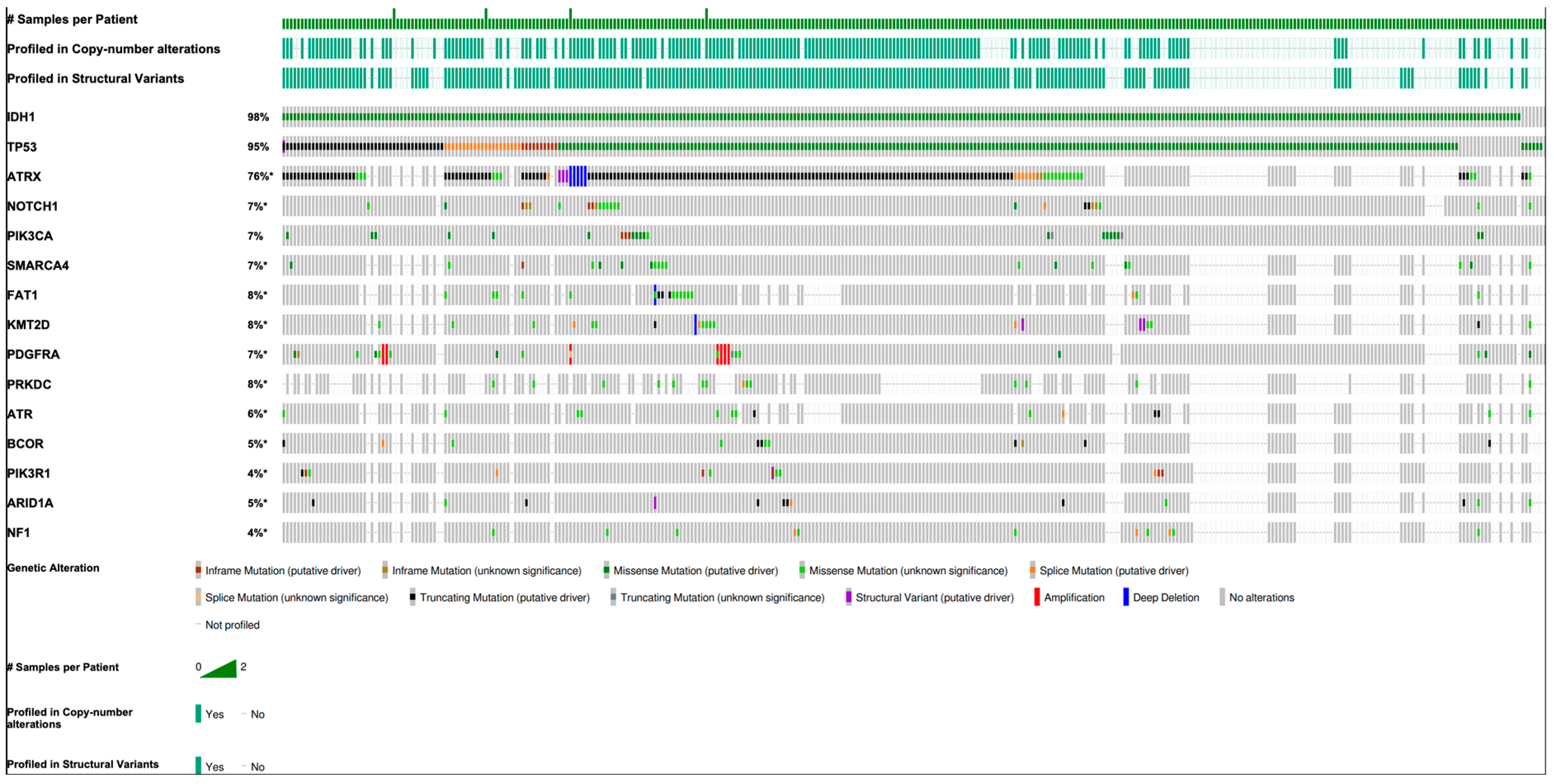
Figure 1. Recurrent genetic alterations in astrocytoma, IDH-mutant, CNS WHO grade III. An asterisk (*) indicates that frequency percentages were calculated based on the total number of samples profiled for that specific gene, rather than the total number of mutations. This explains any differences between these values and those reported in the main text, where frequencies were calculated using the total number of samples in the cohort. The “#” symbol denotes the absolute number of cases.
Dna 05 00043 g001
Figure 2. Kaplan–Meier survival curve of wild-type vs. BCOR mutation in astrocytoma, IDH-mutant, CNS III in AACR Project GENIE.
Dna 05 00043 g002
Figure 3. Kaplan–Meier survival curve of wild-type vs. KMT2D mutation in astrocytoma, IDH-mutant, CNS III in AACR Project GENIE.
Dna 05 00043 g003
Table 1. Astrocytoma, IDH-mutant, CNS WHO grade III patient and sample demographics.
DemographicsCategoryN (%)
SexMale201 (58.4)
Female142 (41.3)
Unknown1 (0.3)
Age categoryAdult347 (99.7)
Unknown1 (0.3)
Pediatric0 (0)
EthnicityNon-Hispanic273 (79.4)
Unknown/Not Collected51 (14.8)
Spanish/Hispanic20 (5.8)
RaceWhite260 (75.6)
Unknown/Not Collected29 (8.4)
Asian24 (7.0)
Other17 (4.9)
Black11 (3.2)
Pacific Islander1 (0.3)
Sample TypePrimary303 (87.1)
Metastatic40 (11.5)
Not Collected/Unspecified5 (1.4)
Table 2. Most common somatic mutations.
GeneN (%)Function
IDH1341 (98.0)Converts alpha-ketoglutarate (2OG) into the oncometabolite (R)-2-hydroxyglutarate
IDH28 (2.3)
TP53330 (94.8)Tumor suppressor
ATRX192 (55.2)Chromatin remodeler
NOTCH124 (6.9)Cell surface receptor in NOTCH pathway
PIK3CA24 (6.9)PI3K/AKT/mTOR signaling pathway
SMARCA419 (5.5)Chromatin remodeler
FAT118 (5.2)Tumor suppressor
KMT2D18 (5.2)Histone methyltransferase
PDGFRA16 (4.3)AKT1/MAP signaling pathway
PRKDC15 (4.2)Non-homologous end joining DNA repair
ATR14 (4.0)DNA damage sensor
BCOR12 (3.4)Transcriptional corepressor
PIK3R113 (3.7)Regulatory subunit of PI3K
ARID1A12 (3.4)Chromatin remodeler
NF111 (3.2)Tumor suppressor of Ras pathway
Table 3. Copy Number Alterations (out of 200) in Astrocytoma, IDH-mutant, CNS III in AACR Project GENIE.
TypeGene NameN (%)Function
Amplification CCND28 (4.0)Cyclin D2 cell cycle regulator
CDK48 (4.0)Cyclin-dependent kinase 4 cell cycle regulator
PDGFRA7 (3.5)AKT1/MAP signaling pathway
MYCN5 (2.5)Cellular proliferation transcription factor
KRAS5 (2.5)Oncogene GTPase
Homozygous Deletion ATRX6 (3.0)Chromatin remodeler
CDKN1B3 (1.5)Cell cycle inhibitor
EPHA73 (1.5)Receptor tyrosine kinase
PDCD13 (1.5)Immune checkpoint receptor
TERT3 (1.5)Telomerase reverse transcriptase
Table 4. Structural Variants (out of 245) in Astrocytoma, IDH-mutant, CNS III in AACR Project GENIE. NA represents a structural variant detected in cBioPortal that was not classified into the deletion, inversion, duplication, or translocation category.
Gene Name NDeletionsInversionsDuplicationsTranslocationNA
KMT2D400112
ATRX300003
SDHA201001
ABL1200002
BRCA1100001
Table 5. Effects of mutations on Survival in Astrocytoma, IDH-mutant, CNS III in AACR Project GENIE.
GeneEffect on Lifespan Median Lifespan MutatedMedian Lifespan WTN_MutatedN_WTp Value
BCORLessens15,59521,488112990.00184
KMT2DLessens17,09421,310152950.0291
Disclaimer/Publisher’s Note: The statements, opinions and data contained in all publications are solely those of the individual author(s) and contributor(s) and not of MDPI and/or the editor(s). MDPI and/or the editor(s) disclaim responsibility for any injury to people or property resulting from any ideas, methods, instructions or products referred to in the content.