Review

The Role of DNA in Neural Development and Cognitive Function

by
Tharsius Raja William Raja
1,2,†,
Janakiraman Pillai Udaiyappan
3,† and
Michael Pillay
1,*
1
Department of Life and Consumer Sciences, College of Agriculture and Environmental Sciences, University of South Africa, Florida Campus, Johannesburg 1709, South Africa
2
PG and Research Department of Biotechnology, Bishop Heber College (Autonomous), Tiruchirappalli 620017, Tamil Nadu, India
3
Department of Biological Sciences, Southern Methodist University, Dallas, TX 75205, USA
*
Author to whom correspondence should be addressed.
These authors contributed equally to this work.
Submission received: 14 April 2025 / Revised: 1 June 2025 / Accepted: 24 July 2025 / Published: 1 August 2025

Abstract

DNA connects the domains of genetic regulation and environmental interactions and plays a crucial role in neural development and cognitive function. The complex roles of genetic and epigenetic processes in brain development, synaptic plasticity, and higher-order cognitive abilities were reviewed in this study. Neural progenitors are formed and differentiated according to genetic instructions, whereas epigenetic changes, such as DNA methylation, dynamically control gene expression in response to external stimuli. These processes shape behavior and cognitive resilience by influencing neural identity, synaptic efficiency, and adaptation. This review also examines how DNA damage and repair mechanisms affect the integrity of neurons, which are essential for memory and learning. It also emphasizes how genetic predispositions and environmental factors interact to determine a person’s susceptibility to neurodegenerative disorders, such as Parkinson’s and Alzheimer’s diseases. Developments in gene-editing technologies, such as CRISPR, and non-viral delivery techniques provide encouraging treatment avenues for neurodegenerative disorders. This review highlights the fundamental role of DNA in coordinating the intricate interactions between molecular and environmental factors that underlie brain function and diseases.

Graphical Abstract

1. Introduction

Neural development in adolescents is characterized by notable brain structure and functional alterations that affect social cognition and behavior. Identity formation, changing social aspirations, and increased sensitivity to peer environments are the characteristics of this time. The interaction of neurodevelopmental processes with larger social settings shapes adolescents’ self-perception and ability to negotiate intricate social networks. Clarifying the interaction between cerebral processes and social experiences during this essential developmental stage requires an understanding of these interactions [1].
The vast network of neurons that comprise the brain undergoes important developmental phases from embryo to adulthood. The evolution of structural and functional connections during these periods makes cognitive capacity more sophisticated. Basic brain circuits are formed throughout early development, and significant synapse pruning and myelination occur to maximize network efficiency [2]. Sporns et al. indicated that understanding these developmental processes is essential for clarifying how brain construction promotes behaviors and cognitive processes throughout life [3].
Neural circuit formation and connectivity are influenced by genetic variables that are essential for brain development. Certain genes control the synthesis of proteins that are necessary for synaptic plasticity, neuronal development, and differentiation. Variations in these genes might result in different neurodevelopmental outcomes such as cognitive capacity and susceptibility to conditions such as autism and schizophrenia [4]. Genetic predispositions have a major influence on how people react to environmental stressors during critical developmental windows, highlighting the role of genetics in determining the shape and function of the brain [5].
Neurodevelopmental disorders (NDDs) constitute a diverse array of conditions that emerge from disturbances in the early processes of brain development, encompassing neuronal proliferation, migration, differentiation, synaptogenesis, and synaptic pruning [6]. NDDs include autism spectrum disorder (ASD), attention-deficit/hyperactivity disorder (ADHD), and intellectual disability (ID) and frequently arise from intricate interactions between genetic predispositions and environmental influences during critical developmental windows [7]. Notwithstanding the genetic heterogeneity present among NDDs, accumulating evidence suggests the existence of common molecular pathways such as perturbations in chromatin remodeling, synaptic functionality, and mammalian target of rapamycin (mTOR) signaling implicated in their etiology [8]. It is noteworthy that the susceptibility of particular genes during developmental phases is exacerbated by a cumulative mutational load, thereby reinforcing the two-hit model hypothesis, which posits that an initial genetic insult can be intensified by subsequent genetic events [9]. Investigations utilizing animal models have indicated that modifications in these pathways may induce phenotypic and behavioral anomalies analogous to those witnessed in human NDDs, underscoring the necessity of elucidating neurodevelopmental trajectories to pinpoint opportunities for therapeutic intervention [10].
Epigenetic mechanisms alter gene expression in response to environmental stimuli without changing the DNA sequence; they are also essential for brain development. Experiences during pregnancy and after delivery, such as maternal stress or diet, might affect these alterations and have long-term consequences for brain plasticity and cognitive processes. Histone and DNA methylation are two examples of epigenetic changes that may influence the effect of early-life trauma on neurodevelopmental trajectories [11]. Clarifying the roles of genetic and epigenetic factors in brain development and related behavioral outcomes requires an understanding of how these factors interact [12].
This review aims to investigate the crucial role of DNA in brain development and cognitive function emphasizing the complex interactions between genetic and epigenetic mechanisms. It seeks to clarify how genetic sequences and epigenetic changes affect higher-order cognitive capacities, synaptic plasticity, and neurodevelopmental processes. This review offers a thorough understanding of the contribution of DNA to brain shape, function, and behavior across developmental stages by combining knowledge of genetic predispositions, environmental impacts, and molecular mechanisms.
DNA plays a critical role in cerebral development and cognitive processes through both genetic sequences and epigenetic modifications. Gene expression is dynamically regulated by processes such as DNA methylation and hydroxymethylation, which affect synaptic plasticity, neurogenesis, and brain stem cell proliferation [13]. Moreover, transcripts essential for neural identity and network plasticity are altered by RNA editing, indicating a relationship between DNA alterations and higher-order cognitive processes [14]. This complex genetic–epigenetic conversation highlights the significance of brain development and function [15].

2. Role of Genes in Brain Formation

The genes that orchestrate regional patterning, neural induction, progenitor identity, neuronal differentiation, and subtype specification play a critical and highly coordinated role in the formation of both the architecture and functional properties of the brain. Homeobox genes, specifically Otx1 and Otx2, are pivotal in the early stages of brain patterning, as they regulate the induction of the anterior neural plate and the formation of the midbrain–hindbrain boundary. The presence of loss-of-function mutations in these genes can lead to significant neural tube defects, underscoring their essential role in brain morphogenesis [11]. Furthermore, the Otx genes exemplify the evolutionary conservation of mechanisms underlying brain development across various species, contributing to processes of neural specification and differentiation [12].

2.1. Genetic Instructions for Neural Progenitor Identity

In addition to regional patterning, a cascade of transcription factors intricately regulates neural progenitors’ proliferation and fate determination. Basic helix–loop–helix (bHLH) transcription factors, including Neurogenin (Ngn1/2) and Mash1 (Ascl1), initiate neuronal development programs within ectodermal cells. These genes assimilate spatial and temporal signals to ensure accurate lineage commitment. In vertebrates, they govern the commitment of neural progenitors to neuronal fates, thereby influencing both differentiation and lineage determination [13,14].

2.2. Genes and Neuronal Differentiation

After neural induction, the process of differentiation is modulated by downstream effectors such as NeuroD and Math3, which facilitate the exit from the cell cycle and promote the identity of neuronal subtypes. These signaling cascades guarantee appropriate specialization, with the formation of sensory neurons contingent upon the sequential activation of Ngn1 and NeuroD [15].

2.3. Genetic Regulation of Neuronal Subtype Specification

The genetic regulation of neuronal subtype specification encompasses complex interactions among cofactors. For example, Ngn2 collaborates with Olig2 during the development of spinal motor neurons, while the expression of Mash1 in designated brain regions contributes to the differentiation of GABAergic interneurons [16,17]. These interactions illuminate the context-dependent characteristics of neural specification.

2.4. Genes Involved in Synaptic Plasticity and Neurodegeneration

Genes such as BDNF, COMT, and APOE exert their effects beyond developmental stages, extending into the realms of synaptic plasticity and the risk of neurodegenerative diseases. BDNF is instrumental in regulating synaptic connectivity and neurogenesis, COMT is involved in modulating prefrontal dopamine levels that affect executive functions, and APOE isoforms play a significant role in lipid metabolism and the pathways involved in amyloid-beta clearance within the brain [18,19,20]. Collectively, these genes illustrate the intricate molecular relationships that impact brain development, cognitive capabilities, and susceptibility to disorders, including Alzheimer’s disease (Table 1).

3. Epigenetic Regulation

3.1. DNA Methylation and Histone Modifications

By changing chromatin structure, histone modifications, such as acetylation and methylation, and DNA methylation, which add methyl groups to cytosine residues, control gene expression [16]. Histone alterations are dynamic and affect transcriptional activity, whereas DNA methylation results in fixed gene silencing [17]. Several epigenetic processes cooperate to reinforce repressive chromatin states. DNA methylation can activate histone-modifying enzymes such as histone deacetylases (HDACs) [18]. Aberrant gene silencing in diseases such as cancer, cellular differentiation, and developmental programming depends on the interaction between these mechanisms.
One essential epigenetic process that controls gene expression during brain development is DNA methylation. DNA methyltransferases (DNMTs) help add methyl groups to cytosines, primarily at the CpG sites. This alteration represses gene transcription by changing chromatin structure and inhibiting transcription factor binding [19]. Dynamic variations in DNA methylation patterns characterize early brain development and influence circuit creation and neuronal differentiation.
Stress and diet are two environmental factors that affect DNA methylation during brain development. The modulation of DNMT activity caused by the nutritional availability of methyl donors such as folate and choline affects neural gene expression and plasticity [20]. Stress exposure during pregnancy can modify stress response pathways in adulthood by causing long-lasting methylation alterations in glucocorticoid receptor gene promoters [21].
DNA methylation, histone modifications, chromatin structure, active/repressed genomic areas, and the function of enhancers, TSS, and polymerase II in transcription are all examples of the epigenetic control of gene expression. Figure 1 explains the process of epigenetic regulation in detail. DNA methylation works in tandem with other epigenetic mechanisms, such as histone changes, to create and preserve neural identity. The methyl-CpG-binding protein MeCP2 plays a critical role by attracting corepressor complexes and maintaining repressive chromatin states [22]. Neurodevelopmental diseases such as Rett syndrome are associated with the dysregulation of these processes, underscoring their importance in normal brain maturation and function [23].
Brain development can be severely hampered by nutritional problems during fetal and early postnatal life; the effects vary greatly depending on the nutrient type, timing, dosage, and length of shortage. Neurodevelopmental activities like myelination, synaptogenesis, neurotransmitter synthesis, and energy metabolism depend on essential nutrients like protein, iron, zinc, copper, iodine, choline, folate, and long-chain polyunsaturated fatty acids (LC-PUFAs). For example, protein–energy malnutrition impairs cognitive results by reducing neuronal development and complexity, particularly in the cortex and hippocampus [24]. Myelination and neurotransmitter systems are altered by iron deprivation, leading to neurobehavioral abnormalities and impaired recognition memory [25,26]. Short-term memory is hampered, autonomic function is disturbed, and the hippocampus and cerebellum are affected by zinc deprivation [27]. Despite being uncommon, copper deficiency has a lasting impact on motor coordination and cerebellar development [28]. Visual–cognitive development and synaptic membrane function depend on LC-PUFAs like DHA [29]. Particularly during crucial developmental windows, these nutritional deficiencies can result in both global and region-specific brain dysfunctions. If left untreated, they may induce irreversible neurodevelopmental abnormalities [24].

3.2. Role of Histone Modifications in Neural Lineage Differentiation and Synaptic Plasticity

Histone alterations dynamically control gene expression and are essential for brain lineage differentiation. Histone acetyltransferases (HATs), such as CBP and p300, catalyze acetylation, which loosens the chromatin structure and stimulates transcription. HDACs restrict gene expression by deacetylation. CBP’s function of CBP in activating genes such as NeuroD and α1-tubulin demonstrates the importance of this balance during neural stem cell differentiation into neurons and glial cells [30].
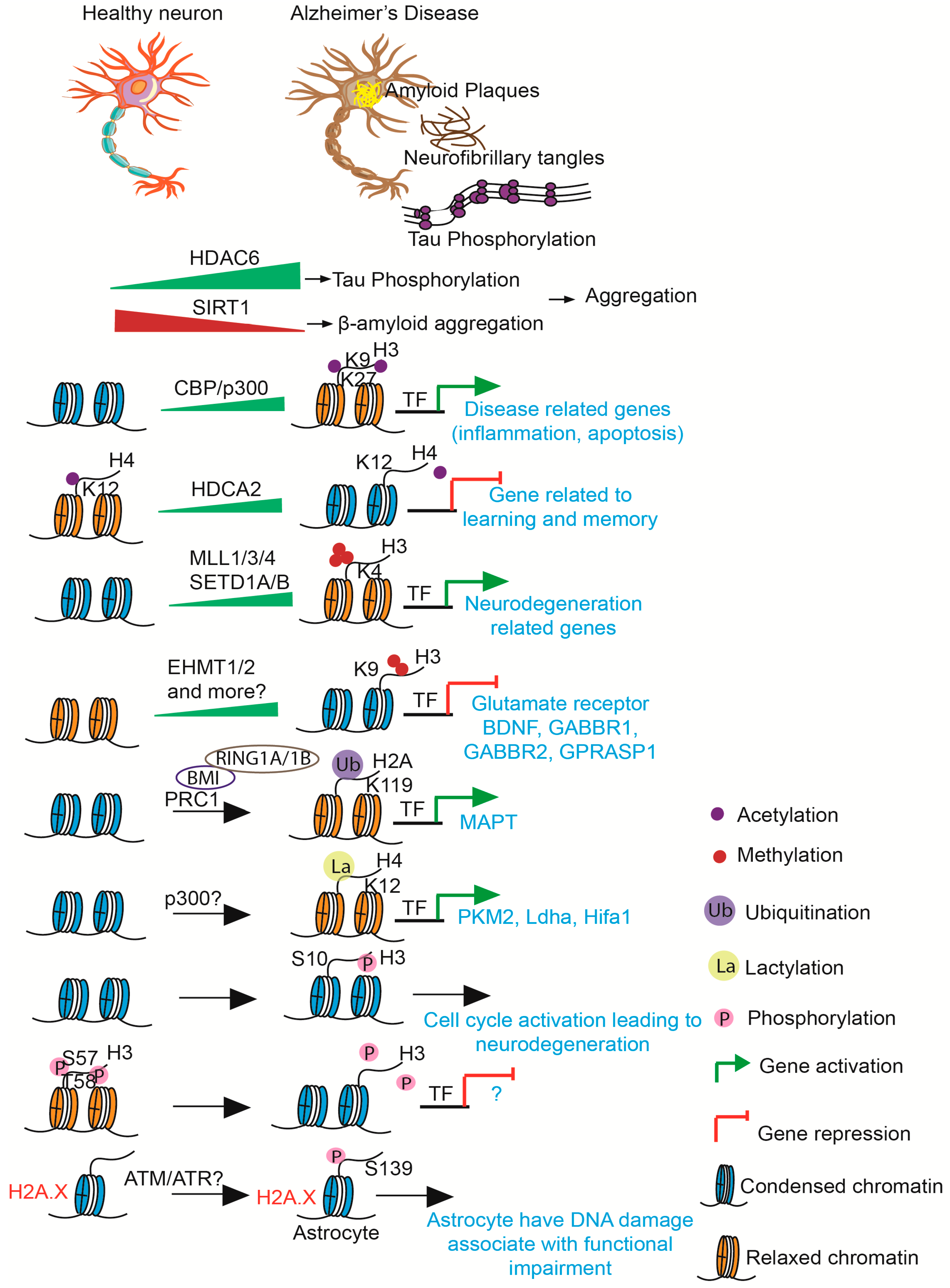
Histone modifications in synaptic plasticity influence gene expression, which is dependent on neural activity and is crucial for memory and learning. Increased transcription of genes involved in synaptic strengthening has been associated with histone H3 acetylation (H3K9ac) and methylation of lysine 4 (H3K4me3) [31]. Dysregulation of these markers affects memory-related gene expression and synaptic function, as observed (Figure 2) in Alzheimer’s disease models [32].
In neurogenesis and synaptic plasticity, precise spatiotemporal gene regulation is ensured by the interaction between histone modifications. Neurological illnesses can result from changes in this regulatory network, underscoring the potential for treatment. Preclinical research on neurological illnesses has shown the potential of blocking HDAC2 or increasing CBP activity [33].

3.3. Repetitive DNA Elements

Transposable elements (TEs) and simple sequence repeats (SSRs) are repetitive DNA elements that constitute sizable components of the human genome. By adding regulatory elements, such as enhancers and alternative promoters, TEs, which comprise approximately 45% of the euchromatic genome, contribute to genomic evolution. Tandem repeats (SSRs) make up approximately 3% of the genome and are primarily located in heterochromatic areas [34].
These components affect gene regulation and chromatin shape. TEs are the most abundant in distal promoter regions, where nucleosomes firmly bind and form a compact chromatin environment. SSRs peak close to Transcription Start Sites, encouraging an open chromatin shape that is advantageous for the assembly of the transcriptional machinery [34]. Their epigenetic functions in gene regulation are highlighted by these dynamic interactions.
Tissue-specific gene expression and functional diversity are affected by repetitive DNA motifs. SSR-enriched promoters are linked to more complex regulatory networks, whereas genes with TE-rich promoters exhibit more varied expression patterns. These results highlight their importance in determining genomic construction and regulatory complexity from evolutionary and functional standpoints [35].

3.4. Long Interspersed Nuclear Elements (L1) and Neural Progenitor Cells

The preservation of neural progenitor cells (NPCs) is greatly aided by long interspersed nuclear elements (L1). To preserve chromatin accessibility in NPCs, L1 elements control histone modifications, such as H3K9me3, which are coordinated by histone methyltransferases such as SETDB1 [36]. These epigenetic changes ensure the precise regulation of gene expression necessary for NPC survival and differentiation.
L1 elements serve as chromatin-looping scaffolds in NPCs, which helps organize the functional domains of the genome. The suppression of L1 by SETDB1 guarantees steady transcription of cell cycle genes and prevents chromatin misfolding. Their regulatory significance is highlighted by the fact that dysregulation of L1, frequently characterized by decreased H3K9me3, results in aberrant chromatin accessibility and changes in NPC proliferation [37]. In NPCs, L1 elements regulate genomic integrity. They affect the fate and function of neural progenitors by altering chromatin construction. This surprising function of L1 highlights its dual importance in neurogenesis and the larger epigenetic context of brain development [37].

4. DNA Damage and Cellular Response

Both endogenous and external chemicals frequently induce DNA damage in prokaryotic and eukaryotic cells. Following the detection of DNA damage by sensor proteins such as the MRN complex, transducers such as ATM and ATR are activated, which phosphorylate downstream effectors to control the damage. To stop errors from spreading, cells either repair the DNA or initiate apoptosis depending on the degree of damage [38].
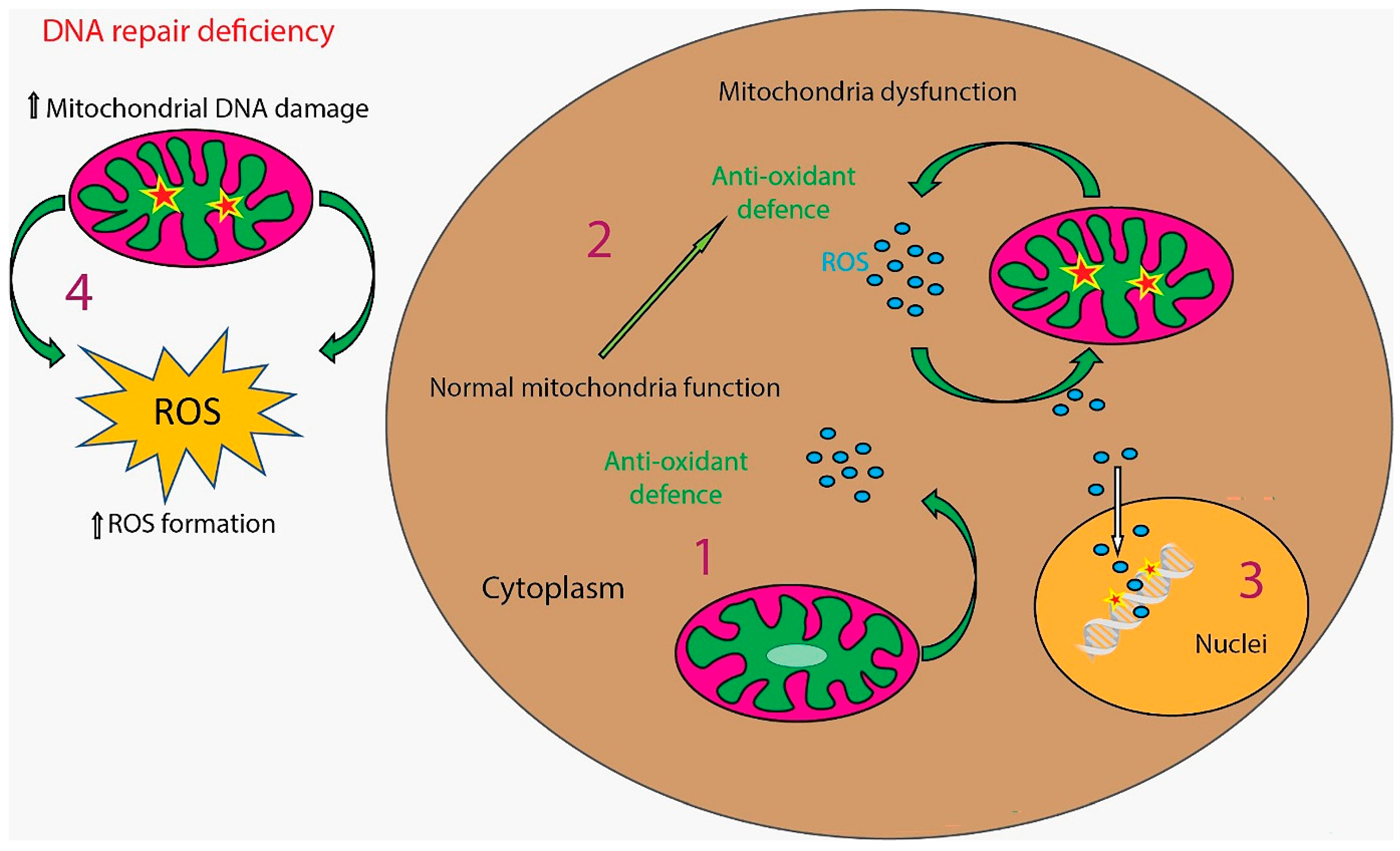
One of the main causes of DNA damage is oxidative stress, caused by an imbalance in reactive oxygen species (ROS). Although ROS are byproducts of regular cellular metabolism, they can oxidize DNA and cause mutations (Figure 3). One important method for repairing oxidative DNA damage and preserving genomic integrity in the face of oxidative stress is the base excision repair (BER) pathway [39].
Nucleotide excision repair (NER) is a second crucial DNA repair mechanism that handles large and helix-distorting DNA damage, in addition to base excision repair (BER), which mainly treats minor oxidative DNA damage. NER is especially crucial for eliminating oxidative stress-induced cyclopurine deoxynucleosides (cdPu) and UV-induced lesions including cyclobutane pyrimidine dimers (T^T) and 6–4 photoproducts. Notably, the NER pathway has subpathways such as transcription-coupled NER (TC-NER), which involves the CSA and CSB proteins, and global genome NER. Under oxidative stress, these NER components have been demonstrated to move from the nucleus to the mitochondria, where they interact with BER-associated proteins such as mtOGG1, indicating a collaborative involvement in preserving the integrity of mitochondrial DNA [40]. The significance of both BER and NER in the whole cellular response to oxidative DNA damage and aging-related mitochondrial dysfunction is highlighted by this partnership.
Double-strand breaks (DSBs) are the most dangerous DNA lesions and, if left untreated, can result in chromosomal instability and cell death. Non-homologous end joining (NHEJ) and homologous recombination (HR) are the two methods used to repair DSB. The MRN complex is essential for recognizing and processing DSBs and activating ATM, which is essential for initiating the DNA damage response [41].
DNA damage disrupts processes that maintain brain memory and adaptability and has a substantial impact on neural plasticity and cognitive abilities. DNA damage caused by oxidative stress reduces synaptic plasticity and hippocampal neurogenesis, which are both critical for memory and learning. Genes linked to brain plasticity and cognitive resilience are regulated by DNA methylation and histone modifications, which are affected by external variables like exercise [42]. These processes emphasize the importance of preserving DNA integrity for the best possible cognitive function and flexibility.
To maintain integrity and functionality, neurons use repair processes in response to DNA damage. Non-homologous end joining (NHEJ) and homologous recombination (HR) fix double-strand breaks, and base excision repair (BER) addresses oxidative damage. ATM and DNA-PK are important proteins that aid in damage sensing and repair coordination. Telomere preservation is especially important because alterations can cause DNA damage in post-mitotic neurons without causing apoptosis [43].
DNA repair mechanisms are essential for both memory and learning. Neuronal apoptosis resulting from impairments in BER or NHEJ may affect synaptic plasticity and hippocampal function, all of which are critical for cognitive function. TRF2 and other telomere-associated proteins affect neurogenesis-related gene expression and chromatin remodeling. Neurodegenerative diseases are associated with disrupted DNA repair, underscoring the need for strong repair systems to promote cognitive resilience [44].
The previous study focuses on strand bias in gene repair largely in the context of proliferating cells, where both transcription and DNA replication contribute to repair asymmetry. Transcription-coupled nucleotide excision repair (TC-NER) is the primary mechanism for strand-specific repair in post-mitotic neurons, which lack DNA replication. Because RNA polymerase II stalls at damage sites, this mechanism preferentially eliminates DNA lesions from the transcribed (sense) strand. This leads to the recruitment of important repair factors such as CSA, CSB, and the TFIIH complex [44,45]. This method highlights the significance of cell-type-specific DNA repair pathways and offers a convincing explanation for the preferential repair of the sense strand seen in neuronal cells.

5. Cognitive Functions and Gene Expression

5.1. Influence of Genetic Factors on Cognition

Memory, concentration, and problem-solving skills are among the cognitive talents greatly influenced by genetics. Genes such as KIBRA have been associated with memory function and Alzheimer’s disease susceptibility in genome-wide association studies (GWAS) [46]. This method reveals how genetic variants affect cognitive performance by combining genomic data with brain imaging. By combining neuroimaging and genotyping, imaging genetics offers a quantitative understanding of cognitive traits and sheds light on the links between genes, the brain, and behavior. The identification of genetic influences on cognition is improved by quantitative trait locus (QTL) studies. Researchers can identify genes influencing brain function and forebrain development by employing quantifiable cognitive qualities such as working memory. Functional magnetic resonance imaging (fMRI) has been used to identify genes associated with cognitive inefficiency in schizophrenia [47].

5.2. Genetic Effects on Learning and Memory

Certain genes have a major impact on cognitive functions such as memory and learning. Through dopamine transmission in the prefrontal cortex, the COMT polymorphism is associated with executive and memory processing. Enhancing memory recall and task efficiency has been validated using functional neuroimaging [48]. Similarly, BDNF influences hippocampal function and plasticity, which in turn affects declarative memory processes. Variations in this gene result in notable changes in memory performance [49].
Gene-specific effects on brain activity have been further revealed through developments in imaging genetics. Variations in COMT and BDNF interact with brain circuits and have varying effects on memory encoding and learning. GWAS have broadened this understanding by connecting cognitive qualities to more general genetic markers [50]. Understanding the specific functions of newly discovered genes can help with interventions aimed at improving cognitive function and treating memory-related conditions.

5.3. Genetic and Environmental Contributions to Cognitive Functions

Environmental variables and genetic predispositions interact to shape cognitive capacities. Twin and adoption studies have demonstrated that 50–70% of cognitive variation is due to genetic influences; however, environmental factors, including socioeconomic status (SES), also play a substantial role [51]. According to research, high-SES households, where children have more access to stimulating environments, may amplify the effects of genetics on cognition [52]. The reciprocal nature of genetic and environmental interactions throughout cognitive development is highlighted by this gene–environment association. Environmental factors have the power to either boost or restrict hereditary cognitive capacity. They have less access to enriching experiences, and children from low-SES homes frequently have reduced heritability of cognitive qualities. The advantageous circumstances foster positive feedback loops that enable genetically influenced talents to flourish, whereby early abilities result in enhanced learning opportunities and additional cognitive development [53]. The genetic and environmental factors that affect cognitive functions are listed in Table 2.

6. Pathophysiological Implications

Alzheimer’s disease, Parkinson’s disease, and Amyotrophic Lateral Sclerosis (ALS) are examples of neurodegenerative diseases (NDs), which are characterized by a gradual loss of neurons. The worldwide health burden of NDs is made worse by aging, a major risk factor [55]. The pathogenesis of ND revolves around oxidative stress, neuroinflammation, and abnormalities in axonal transport (AT). While AT abnormalities affect neuronal homeostasis and lead to neurodegeneration, inflammation and oxidative stress frequently create a vicious loop that exacerbates axonal damage. Therapeutic options to decrease the evolution of the disease may be available if AT-related deficits are identified early. This emphasizes the necessity of biomarkers and focused treatments to successfully alleviate these problems [55].
Chronic psychological stress, which is frequently brought on by the fast-paced nature of contemporary living, causes the hypothalamic–pituitary–adrenal (HPA) axis to be continuously activated, which leads to an excess of cortisol being secreted. Long-term increases in cortisol levels have been linked to neurodegenerative diseases via many interconnected pathways. Important brain areas including the substantia nigra and hippocampus, which are especially susceptible because of their high density of glucocorticoid receptors, atrophy as a result of excessive glucocorticoid exposure. Increased oxidative stress, mitochondrial dysfunction, and poor metabolic regulation are linked to this neurodegeneration; these factors all jeopardize oxygen use and neural homeostasis. The bioenergetic deficiencies that follow point to a decrease in neural oxygenation, which in turn reduces cellular viability and function. Moreover, it has been demonstrated that glucocorticoids worsen neuroinflammation, encourage the buildup of amyloid-β, and hinder the breakdown of tau proteins, all of which hasten cognitive loss and contribute to the pathophysiology of conditions like Parkinson’s and Alzheimer’s diseases. These results highlight the harmful effects of long-term cortisol dysregulation on neuronal integrity and function, which may be caused by cellularly altered oxygen homeostasis [56].
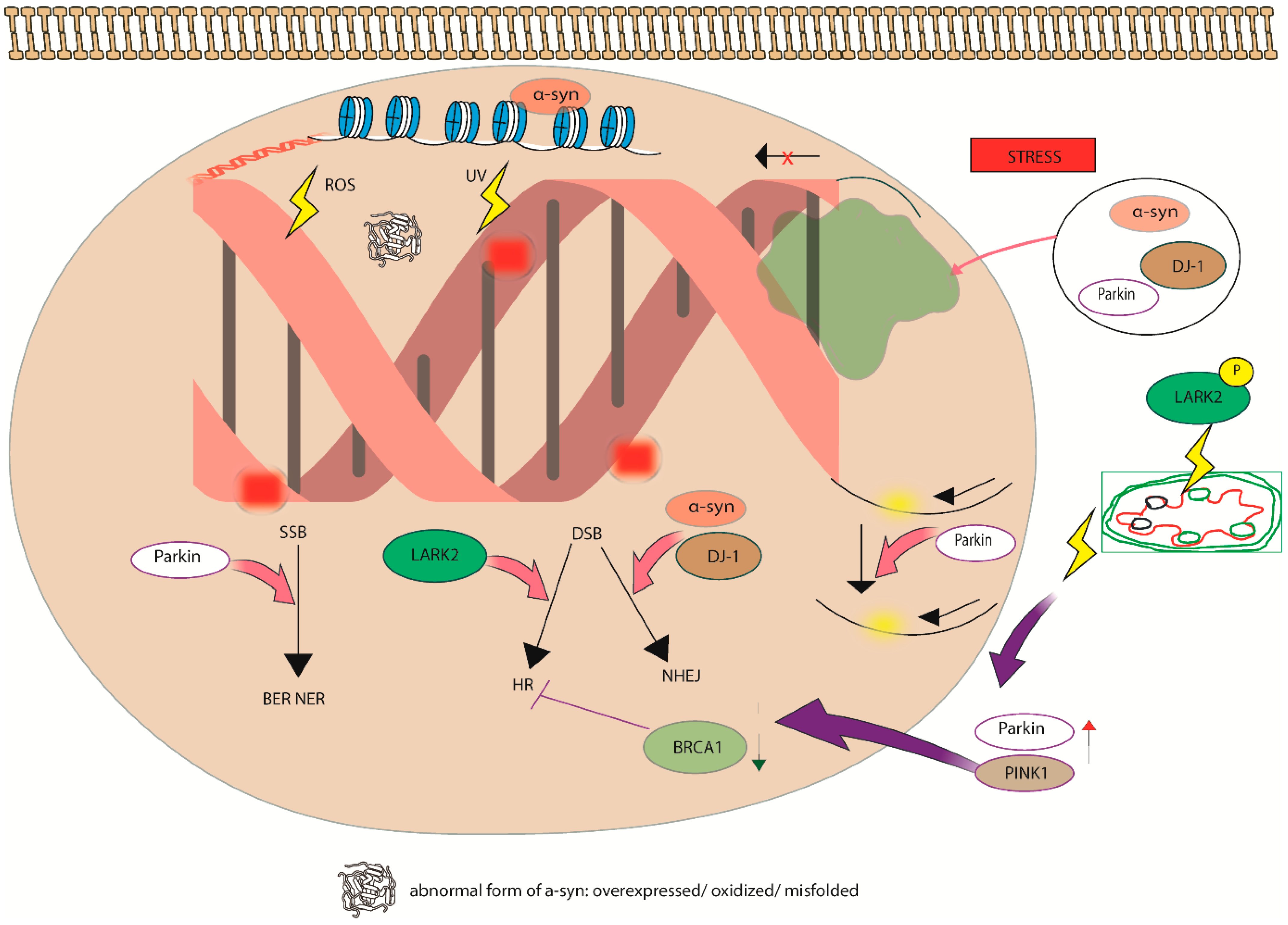
In addition to Alzheimer’s disease (AD), Parkinson’s disease (PD) and Amyotrophic Lateral Sclerosis (ALS) represent substantial neurodegenerative disorders that exert profound clinical and societal repercussions. PD ranks as the second most prevalent neurodegenerative disorder after AD and is characterized by both motor manifestations (such as tremor, rigidity, and bradykinesia) and non-motor manifestations, which encompass cognitive impairments and mood disorders. At the molecular dimension, PD is distinguished by the aggregation of α-synuclein, mitochondrial dysfunction, oxidative stress, and the degeneration of dopaminergic neurons in the substantia nigra (Figure 4).
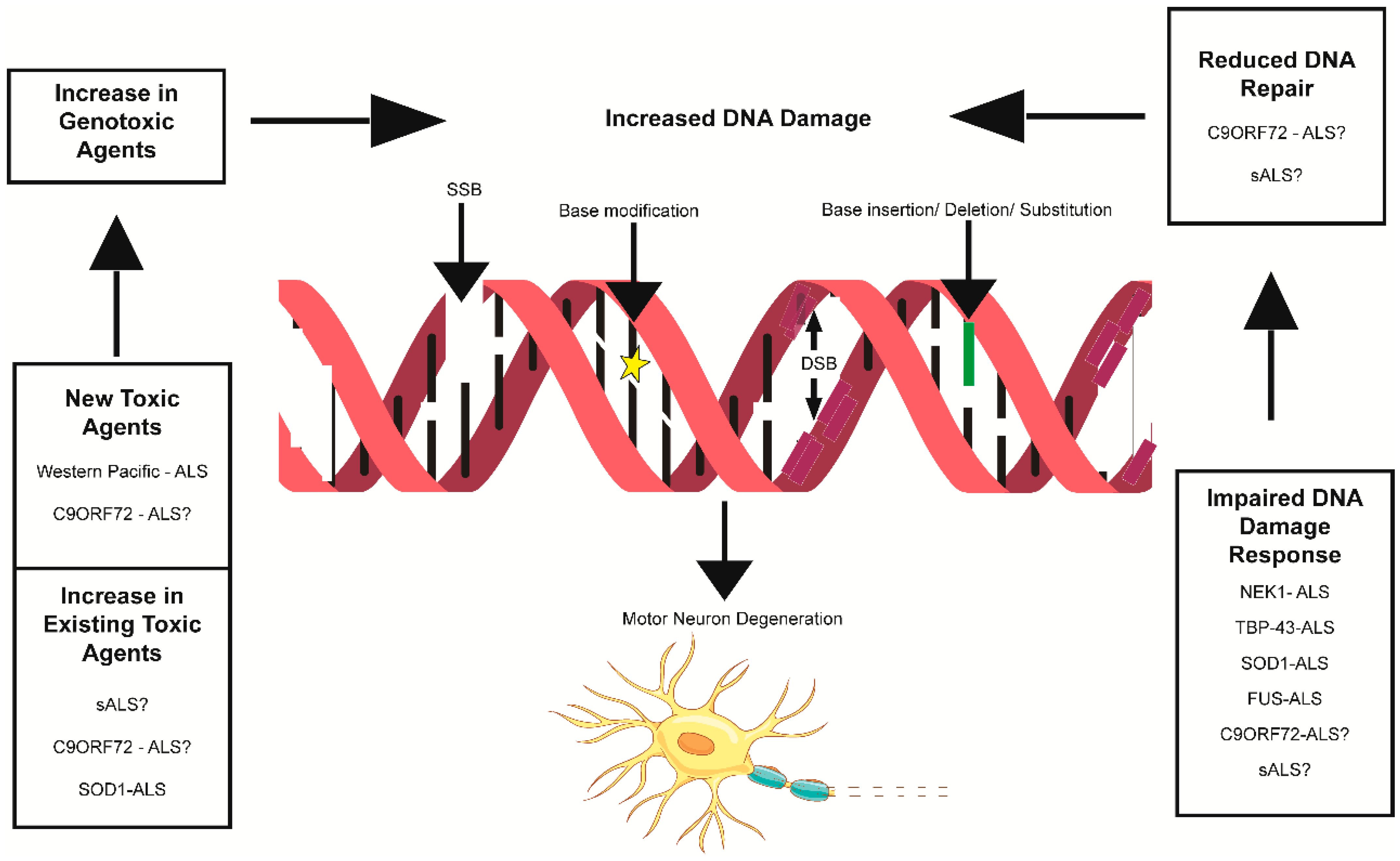
Recent investigations have concentrated on the genetic foundations of PD, illuminating mutations in genes such as LRRK2, PARK7, PINK1, and SNCA that affect disease pathogenesis and serve as potential therapeutic targets [57]. Correspondingly, ALS is a profoundly debilitating neurodegenerative disorder marked by the progressive degeneration of upper and lower motor neurons, culminating in muscle weakness, atrophy, and respiratory failure [58]. Multiple mechanisms have been implicated in the pathophysiology of ALS, including protein aggregation (e.g., TDP-43), excitotoxicity, oxidative stress, and neuroinflammation [59]. Genetic mutations in SOD1, C9ORF72, TARDBP, and FUS play a pivotal role in familial variants of ALS. Although current therapeutic strategies for PD and ALS primarily address symptomatic relief, ongoing research into disease-modifying approaches, such as gene therapy, stem cell transplantation, and targeted molecular inhibitors, provides optimism for more efficacious interventions in the future [59]. A more integrative comprehension of PD and ALS in conjunction with AD is imperative for the development of comprehensive neurotherapeutic modalities (Figure 5).
Environmental pollution, deleterious dietary practices characterized by the ingestion of highly processed foods, and insufficient physical activity constitute substantial contributing elements to the escalation of neurodegenerative diseases. Environmental toxins, particularly atmospheric pollutants such as particulate matter (PM2.5), nitrogen oxides, and volatile organic compounds are recognized for their capacity to provoke oxidative stress, neuroinflammatory responses, and disruption of the blood–brain barrier, all of which culminate in neuronal injury and cognitive deterioration [59,60,61]. Prolonged exposure to heavy metals and pesticides intensifies these adverse effects, leading to clinical manifestations akin to Alzheimer’s and Parkinson’s diseases [62]. The ingestion of processed foods, frequently characterized by elevated levels of saturated fats and diminished nutritional value, has been associated with the accumulation of β-amyloid and oxidative stress, which are pivotal pathogenic characteristics of Alzheimer’s disease [63]. Furthermore, longitudinal dietary investigations have demonstrated that adherence to a Western dietary model heightens the susceptibility to Parkinson’s disease, whereas a diet abundant in fruits, vegetables, fish, and whole grains (termed the “prudent” dietary pattern) is inversely correlated with its incidence [64]. A sedentary lifestyle, a prevalent feature of contemporary existence, additionally exacerbates neurodegeneration by hindering neurogenesis and amplifying inflammatory and oxidative stress pathways; while this aspect was not the primary focus of the present manuscript, it is extensively corroborated within the neurological literature. Consequently, multifaceted environmental and lifestyle stressors collaborate synergistically to propagate neurodegenerative processes [65].
Neurological conditions like Alzheimer’s disease are increasingly associated with abnormal DNA alterations, particularly in methylation patterns [66]. When interrupted, DNA methylation, which is essential for gene control, might result in compromised brain processes. Neurodegenerative diseases are linked to alterations in 5-methylcytosine (5 mC) and its derivatives, such as 5-hydroxymethylcytosine (5 hmC) [67]. Neuronal function and plasticity may be impacted by aberrant hypermethylation or hypomethylation in specific gene areas, which could accelerate the course of the disease [68]. These revelations highlight the possibility of medicinal approaches that target DNA methylation mechanisms.
People with metabolically unfavorable phenotypes, which generally include diseases such as diabetes, hypertension, and dyslipidemia, are at a significantly increased risk of stroke. In particular, compared to those with metabolically healthy normal weight, individuals with metabolically unhealthy normal weight (MUNW), metabolically unhealthy overweight (MUOW), or metabolically unhealthy obesity (MUO) all had higher hazard ratios for stroke. For example, the MUO group had the highest risk (HR = 1.99, 95% CI: 1.66–2.40) and the MUOW group had the highest risk (HR = 1.94, 95% CI: 1.58–2.40), indicating that obesity significantly raises the risk of stroke when combined with metabolic diseases such as diabetes. These results imply that both obesity and diabetes can lead to brain injury through a higher risk of stroke, and they corroborate the growing understanding that metabolic dysfunction, including diabetes, is a greater predictor of cerebrovascular events than body weight alone [69].
Susceptibility to neurodegenerative illnesses, such as Parkinson’s and Alzheimer’s, diseases, is mostly influenced by genetic factors. The initiation and development of the diseases are influenced by variations in important genes such as APOE and PSEN1, with the APOE4 variant markedly raising the risk of Alzheimer’s disease [70]. Similarly, Parkinson’s disease susceptibility is associated with mutations in the GBA and LRRK2 genes. Numerous loci linked to these disorders have been found by GWAS, indicating their polygenic nature [71]. Knowing genetic contributions helps with focused therapy development as well as risk prediction. The pathophysiology of neurodegenerative diseases is summarized in Table 3.
A lot of the research supports the neurotoxic effects of alcohol, tobacco, and lipid peroxidation products on neuronal populations. Hippocampal neurogenesis has been demonstrated to be severely hampered by alcohol use, especially in adolescent brains where binge-like alcohol exposure drastically lowers neural stem cell survival and proliferation. Alcohol exposure lowered neurogenesis by 28–33% (as measured by doublecortin expression) and BrdU-labeled cell proliferation by 21% in an adolescent rat model that mimicked alcohol use disorder. This was followed by a 50% decrease in new cell survival. Similarly, smoking tobacco and, more especially, being exposed to nicotine have complicated consequences on the nervous system. Chronic nicotine use is also linked to changes in neural stem cell function, proliferation, and differentiation, which may hinder hippocampus neurogenesis, particularly during withdrawal phases, despite some studies suggesting potential neuroprotective roles of nicotine evidenced by inverse correlations with neurodegenerative diseases like Parkinson’s and Alzheimer’s diseases [72]. Recovery after nerve damage may also be made more difficult by tobacco’s immunomodulatory effects, which may affect peripheral nerve regeneration and, consequently, neural repair mechanisms [73]. When combined, these findings demonstrate the wider effects of such exposures on the integrity of the nervous system and bolster the reviewer’s claim that alcohol and tobacco are major causes of neuronal loss, either by promoting neurodegeneration or preventing neurogenesis.

7. Future Directions

Although improvements in gene therapy have opened up new avenues for the treatment of neurological conditions, obstacles such as blood–brain barrier permeability, ineffective targeted delivery, and low long-term gene expression still exist. New non-viral approaches that improve precision and address safety issues include CRISPR-based systems and personalized nanoparticles [74]. Overcoming the variability of etiology and improving vector systems for wider central nervous system (CNS) application are necessary to expand therapeutic approaches for complicated disorders like Parkinson’s and Alzheimer’s diseases. Further research is required to maximize the clinical translation of these technologies and transform current medicines.
It is not yet known how exactly activity-regulated gene expression contributes to synaptic plasticity and memory storage, despite being essential for brain development and cognitive processes. The molecular routes that connect DNA alterations like methylation and activity-dependent transcription to the improvement of brain circuits are among the current gaps. Further investigation is also needed into the function of DNA enhancers in cell-specific transcriptional responses and how they affect behavioral adaptation. To understand how DNA dynamics underlie cognitive functions and neurodevelopmental problems, advances in single-cell epigenomics and imaging technologies are crucial.

8. Conclusions

Integrating genetic and epigenetic pathways to control neuronal differentiation, synaptic plasticity, and cognitive resilience, DNA is essential to brain development and cognitive function. Neural plasticity and reactions to stress or nutrition are influenced by epigenetic mechanisms such as DNA methylation, which dynamically modify gene expression in response to environmental stimuli. While genetic variants contribute to individual variances and the risk of neurodegenerative illnesses like Alzheimer’s and Parkinson’s diseases, DNA repair processes preserve neuronal integrity, which is essential for memory and learning. Although issues like delivery precision still exist, therapeutic potential is held by advances in gene-editing technology.

Author Contributions

T.R.W.R. and J.P.U. were responsible for composing the initial draft of the manuscript and contributed significantly to its conceptual framework and design. M.P. undertook the editing of the manuscript and approved the final version. All authors have read and agreed to the published version of the manuscript.

Funding

This research received no external funding.

Institutional Review Board Statement

Not applicable.

Informed Consent Statement

Not applicable.

Data Availability Statement

This paper does not contain any experimental data.

Acknowledgments

The authors are thankful to the Vice Chancellor, University of South Africa, Florida Campus, South Africa, and to the Vice Chancellor, Southern Methodist University, Dallas TX USA for their support. A. Jennifer Christy and A. Juliat Josephine contributed to the formulation of several concepts within the manuscript. Figures were drawn using the Adobe Illustrator 29.0 2025 application.

Conflicts of Interest

The authors have declared no conflicts of interest.

Abbreviations

The following abbreviations are used in this manuscript:
ADAlzheimer’s disease
ALSAmyotrophic Lateral Sclerosis
APOEApolipoprotein E
ATaxonal transport
BDNFBrain-Derived Neurotrophic Factor
BERbase excision repair
bHLHbasic helix–loop–helix
CBPCREB Binding Protein
CNScentral nervous system
COMTCatechol O Methyltransferase
DNMTsDNA methyltransferases
DSBsdouble-strand breaks
fMRIfunctional magnetic resonance imaging
GWASgenome-wide association studies
HATshistone acetyltransferases
HDACshistone deacetylases
HRhomologous recombination
HMTshistone methyltransferases
L1long interspersed nuclear elements
MeCP2methyl-CpG-binding protein 2
MRNMRE11 RAD50 NBS1 Complex
NDsneurodegenerative diseases
NgnNeurogenin
NHEJnon-homologous end joining
NPCsneural progenitor cells
PFCprefrontal cortex
Pol IIRNA polymerase II
PSEN1Presenilin 1
QTLquantitative trait locus
ROSreactive oxygen species
SESsocioeconomic status
SSRssimple sequence repeats
TEstransposable elements
TSSTranscription Start Site
mCmethylcytosine
hmChydroxymethylcytosine

References

  1. Capella, J.; Telzer, E.H. A Framework for Integrating Neural Development and Social Networks in Adolescence. Dev. Cogn. Neurosci. 2024, 69, 101442. [Google Scholar] [CrossRef]
  2. Pan, Y.; Monje, M. Activity Shapes Neural Circuit Form and Function: A Historical Perspective. J. Neurosci. 2020, 40, 944–954. [Google Scholar] [CrossRef] [PubMed]
  3. Sporns, O.; Chialvo, D.R.; Kaiser, M.; Hilgetag, C.C. Organization, Development and Function of Complex Brain Networks. Trends Cogn. Sci. 2004, 8, 418–425. [Google Scholar] [CrossRef] [PubMed]
  4. Cattane, N.; Richetto, J.; Cattaneo, A. Prenatal Exposure to Environmental Insults and Enhanced Risk of Developing Schizophrenia and Autism Spectrum Disorder: Focus on Biological Pathways and Epigenetic Mechanisms. Neurosci. Biobehav. Rev. 2020, 117, 253–278. [Google Scholar] [CrossRef] [PubMed]
  5. Oldenburg, K.S.; O’Shea, T.M.; Fry, R.C. Genetic and Epigenetic Factors and Early Life Inflammation as Predictors of Neurodevelopmental Outcomes. Semin. Fetal Neonatal Med. 2020, 25, 101115. [Google Scholar] [CrossRef]
  6. Silbereis, J.C.; Pochareddy, S.; Zhu, Y.; Li, M.; Sestan, N. The Cellular and Molecular Landscapes of the Developing Human Central Nervous System. Neuron 2016, 89, 248–268. [Google Scholar] [CrossRef]
  7. Gilmore, J.H.; Knickmeyer, R.C.; Gao, W. Imaging Structural and Functional Brain Development in Early Childhood. Nat. Rev. Neurosci. 2018, 19, 123–137. [Google Scholar] [CrossRef]
  8. Bourgeron, T. From the Genetic Architecture to Synaptic Plasticity in Autism Spectrum Disorder. Nat. Rev. Neurosci. 2015, 16, 551–563. [Google Scholar] [CrossRef]
  9. Pescosolido, M.F.; Gamsiz, E.D.; Nagpal, S.; Morrow, E.M. Distribution of Disease-Associated Copy Number Variants Across Distinct Disorders of Cognitive Development. J. Am. Acad. Child Adolesc. Psychiatry 2013, 52, 414–430.e14. [Google Scholar] [CrossRef]
  10. Choi, G.B.; Yim, Y.S.; Wong, H.; Kim, S.; Kim, H.; Kim, S.V.; Hoeffer, C.A.; Littman, D.R.; Huh, J.R. The Maternal Interleukin-17a Pathway in Mice Promotes Autism-like Phenotypes in Offspring. Science 2016, 351, 933–939. [Google Scholar] [CrossRef]
  11. Schrott, R.; Song, A.; Ladd-Acosta, C. Epigenetics as a Biomarker for Early-Life Environmental Exposure. Curr. Environ. Health Rep. 2022, 9, 604–624. [Google Scholar] [CrossRef]
  12. Gartstein, M.A.; Skinner, M.K. Prenatal Influences on Temperament Development: The Role of Environmental Epigenetics. Dev. Psychopathol. 2018, 30, 1269–1303. [Google Scholar] [CrossRef]
  13. Xie, J.; Xie, L.; Wei, H.; Li, X.-J.; Lin, L. Dynamic Regulation of DNA Methylation and Brain Functions. Biology 2023, 12, 152. [Google Scholar] [CrossRef] [PubMed]
  14. Yamada, T.; Valnegri, P.; Yang, Y. Higher-Order Genome Organization in the Control of Neuronal Identity and Neural Circuit Plasticity. In Transcriptional Regulation by Neuronal Activity: To the Nucleus and Back; Springer Nature: Cham, Switzerland, 2024; pp. 251–274. [Google Scholar]
  15. Ali, H.H.; Fazil, H. Epigenetic Factors Affecting Children with Autism Spectrum Disorder: Perspectives of Therapeutic Staff. Remit. Rev. 2024, 9, 1678–1695. [Google Scholar]
  16. Acharjee, S.; Chauhan, S.; Pal, R.; Tomar, R.S. Mechanisms of DNA Methylation and Histone Modifications. Prog. Mol. Biol. Transl. Sci. 2023, 197, 51–92. [Google Scholar] [CrossRef]
  17. Bure, I.V.; Nemtsova, M.V.; Kuznetsova, E.B. Histone Modifications and Non-Coding RNAs: Mutual Epigenetic Regulation and Role in Pathogenesis. Int. J. Mol. Sci. 2022, 23, 5801. [Google Scholar] [CrossRef]
  18. Gagnidze, K.; Pfaff, D.W. Epigenetic Mechanisms: DNA Methylation and Histone Protein Modification. In Neuroscience in the 21st Century: From Basic to Clinical; Springer International Publishing: Cham, Switzerland, 2022; pp. 2677–2716. [Google Scholar]
  19. Lyko, F. The DNA Methyltransferase Family: A Versatile Toolkit for Epigenetic Regulation. Nat. Rev. Genet. 2018, 19, 81–92. [Google Scholar] [CrossRef] [PubMed]
  20. Allison, J.; Kaliszewska, A.; Uceda, S.; Reiriz, M.; Arias, N. Targeting DNA Methylation in the Adult Brain through Diet. Nutrients 2021, 13, 3979. [Google Scholar] [CrossRef] [PubMed]
  21. Vaiserman, A.M. Epigenetic Programming by Early-Life Stress: Evidence from Human Populations. Dev. Dyn. 2015, 244, 254–265. [Google Scholar] [CrossRef]
  22. Leong, W.Y.; Lim, Z.H.; Korzh, V.; Pietri, T.; Goh, E.L.K. Methyl-CpG Binding Protein 2 (Mecp2) Regulates Sensory Function Through Sema5b and Robo2. Front. Cell. Neurosci. 2015, 9, 481. [Google Scholar] [CrossRef]
  23. Good, K.V.; Vincent, J.B.; Ausió, J. MeCP2: The Genetic Driver of Rett Syndrome Epigenetics. Front. Genet. 2021, 12, 620859. [Google Scholar] [CrossRef]
  24. Georgieff, M.K. Nutrition and the Developing Brain: Nutrient Priorities and Measurement. Am. J. Clin. Nutr. 2007, 85, 614S–620S. [Google Scholar] [CrossRef]
  25. Rao, R.; Tkac, I.; Townsend, E.L.; Gruetter, R.; Georgieff, M.K. Perinatal Iron Deficiency Alters the Neurochemical Profile of the Developing Rat Hippocampus. J. Nutr. 2003, 133, 3215–3221. [Google Scholar] [CrossRef]
  26. Beard, J.L.; Connor, J.R. Iron Status and Neural Functioning. Annu. Rev. Nutr. 2003, 23, 41–58. [Google Scholar] [CrossRef]
  27. Golub, M.; Takeuchi, P.; Keen, C.; Gershwin, M.; Hendrickx, A.; Lonnerdal, B. Modulation of Behavioral Performance of Prepubertal Monkeys by Moderate Dietary Zinc Deprivation. Am. J. Clin. Nutr. 1994, 60, 238–243. [Google Scholar] [CrossRef]
  28. Penland, J.G.; Prohaska, J.R. Abnormal Motor Function Persists Following Recovery from Perinatal Copper Deficiency in Rats. J. Nutr. 2004, 134, 1984–1988. [Google Scholar] [CrossRef]
  29. SanGiovanni, J.P.; Parra-Cabrera, S.; Colditz, G.A.; Berkey, C.S.; Dwyer, J.T. Meta-Analysis of Dietary Essential Fatty Acids and Long-Chain Polyunsaturated Fatty Acids as They Relate to Visual Resolution Acuity in Healthy Preterm Infants. Pediatrics 2000, 105, 1292–1298. [Google Scholar] [CrossRef]
  30. Park, J.; Lee, K.; Kim, K.; Yi, S.-J. The Role of Histone Modifications: From Neurodevelopment to Neurodiseases. Signal Transduct. Target. Ther. 2022, 7, 217. [Google Scholar] [CrossRef] [PubMed]
  31. Morelli, G.; Della Valle, F.; Orlando, V. Epigenetics and Brain Plasticity: Back to Function. In Neurobiological and Psychological Aspects of Brain Recovery; Springer International Publishing: Cham, Switzerland, 2023; pp. 237–252. [Google Scholar]
  32. Santana, D.A.; Smith, M.d.A.C.; Chen, E.S. Histone Modifications in Alzheimer’s Disease. Genes 2023, 14, 347. [Google Scholar] [CrossRef] [PubMed]
  33. Kumar, V.; Kundu, S.; Singh, A.; Singh, S. Understanding the Role of Histone Deacetylase and Their Inhibitors in Neurodegenerative Disorders: Current Targets and Future Perspective. Curr. Neuropharmacol. 2022, 20, 158–178. [Google Scholar] [CrossRef] [PubMed]
  34. Pappalardo, X.G.; Barra, V. Losing DNA Methylation at Repetitive Elements and Breaking Bad. Epigenetics Chromatin 2021, 14, 25. [Google Scholar] [CrossRef]
  35. Huda, A.; Mariño-Ramírez, L.; Landsman, D.; Jordan, I.K. Repetitive DNA Elements, Nucleosome Binding and Human Gene Expression. Gene 2009, 436, 12–22. [Google Scholar] [CrossRef]
  36. Reichard, J.; Zimmer-Bensch, G. The Epigenome in Neurodevelopmental Disorders. Front. Neurosci. 2021, 15, 776809. [Google Scholar] [CrossRef]
  37. Jiang, Y.; Sun, D.; Zhu, Y.; Peng, W.; Weng, J.; Dong, S.; Li, J.; Chen, Q.; Ge, C.; Liao, L.; et al. Epigenetic Modulation of Short Interspersed Nuclear Elements Activity Influences Neural Precursor Cell Proliferation. Res. Sq. 2023. [Google Scholar] [CrossRef]
  38. Shadfar, S.; Brocardo, M.; Atkin, J.D. The Complex Mechanisms by Which Neurons Die Following DNA Damage in Neurodegenerative Diseases. Int. J. Mol. Sci. 2022, 23, 2484. [Google Scholar] [CrossRef] [PubMed]
  39. Gohil, D.; Sarker, A.H.; Roy, R. Base Excision Repair: Mechanisms and Impact in Biology, Disease, and Medicine. Int. J. Mol. Sci. 2023, 24, 14186. [Google Scholar] [CrossRef] [PubMed]
  40. Bohr, V.A.; Dianov, G.L. Oxidative DNA Damage Processing in Nuclear and Mitochondrial DNA. Biochimie 1999, 81, 155–160. [Google Scholar] [CrossRef]
  41. Qiu, S.; Huang, J. MRN Complex Is an Essential Effector of DNA Damage Repair. J. Zhejiang Univ. Sci. B 2021, 22, 31–37. [Google Scholar] [CrossRef]
  42. Zhang, W.; Lukacsovich, D.; Young, J.I.; Gomez, L.; Schmidt, M.A.; Martin, E.R.; Kunkle, B.W.; Chen, X.; O’Shea, D.M.; Galvin, J.E.; et al. DNA Methylation Signature of a Lifestyle-Based Resilience Index for Cognitive Health. Res. Sq. 2024. [Google Scholar] [CrossRef]
  43. Shao, J.; Wang, J.; Li, B.; Liu, C. Potential Roles of Telomeres and Telomerase in Neurodegenerative Diseases. Ageing Neurodegener. Dis. 2024, 4, 1. [Google Scholar] [CrossRef]
  44. Cordeiro, A.; Gomes, C.; Bicker, J.; Fortuna, A. Aging and Cognitive Resilience: Molecular Mechanisms as New Potential Therapeutic Targets. Drug Discov. Today 2024, 29, 104093. [Google Scholar] [CrossRef] [PubMed]
  45. Hanawalt, P.C.; Spivak, G. Transcription-Coupled DNA Repair: Two Decades of Progress and Surprises. Nat. Rev. Mol. Cell Biol. 2008, 9, 958–970. [Google Scholar] [CrossRef]
  46. AlKhatib, M.I.; Maslat, A.O.; Ali, E.A.-H.; Al-Saqqar, T.; Khalil, R. A Cross-Sectional Study on the Association of KIBRA Genetic Polymorphism with Episodic Memory in North Jordanian Adults. Open Access Maced. J. Med. Sci. 2023, 11, 200–209. [Google Scholar] [CrossRef]
  47. Voineskos, A.N.; Hawco, C.; Neufeld, N.H.; Turner, J.A.; Ameis, S.H.; Anticevic, A.; Buchanan, R.W.; Cadenhead, K.; Dazzan, P.; Dickie, E.W.; et al. Functional Magnetic Resonance Imaging in Schizophrenia: Current Evidence, Methodological Advances, Limitations and Future Directions. World Psychiatry 2024, 23, 26–51. [Google Scholar] [CrossRef]
  48. Rosli, N.R.; Anuar, T.S.; Adenan, M.I.; Janor, R.M.; Ahmad, R.; Teh, L.K.; Salleh, M.Z.; Bakar, S.H.A.; James, R.J. Association of COMT Polymorphism and Academic Achievement among Female Undergraduate Students. Sci. Lett. 2021, 15, 90–101. [Google Scholar] [CrossRef]
  49. Goldberg, T.E.; Weinberger, D.R. Genes and the Parsing of Cognitive Processes. Trends Cogn. Sci. 2004, 8, 325–335. [Google Scholar] [CrossRef]
  50. Uffelmann, E.; Huang, Q.Q.; Munung, N.S.; De Vries, J.; Okada, Y.; Martin, A.R.; Martin, H.C.; Lappalainen, T.; Posthuma, D. Genome-Wide Association Studies. Nat. Rev. Methods Primers 2021, 1, 59. [Google Scholar] [CrossRef]
  51. Spengler, M.; Gottschling, J.; Hahn, E.; Tucker-Drob, E.M.; Harzer, C.; Spinath, F.M. Does the Heritability of Cognitive Abilities Vary as a Function of Parental Education? Evidence from a German Twin Sample. PLoS ONE 2018, 13, e0196597. [Google Scholar] [CrossRef]
  52. Ghirardi, G.; Gil-Hernández, C.J.; Bernardi, F.; Van Bergen, E.; Demange, P. Interaction of Family SES with Children’s Genetic Propensity for Cognitive and Noncognitive Skills: No Evidence of the Scarr-Rowe Hypothesis for Educational Outcomes. Res. Soc. Stratif. Mobil. 2024, 92, 100960. [Google Scholar] [CrossRef] [PubMed]
  53. Hart, E.R.; Bailey, D.H.; Luo, S.; Sengupta, P.; Watts, T.W. Fadeout and Persistence of Intervention Impacts on Social-Emotional and Cognitive Skills in Children and Adolescents: A Meta-Analytic Review of Randomized Controlled Trials. Psychol. Bull. 2024, 150, 1207–1236. [Google Scholar] [CrossRef] [PubMed]
  54. Nouspikel, T. DNA Repair in Mammalian Cells: Nucleotide Excision Repair: Variations on Versatility. Cell. Mol. Life Sci. 2009, 66, 994–1009. [Google Scholar] [CrossRef]
  55. Tesco, G.; Lomoio, S. Pathophysiology of Neurodegenerative Diseases: An Interplay among Axonal Transport Failure, Oxidative Stress, and Inflammation? Semin. Immunol. 2022, 59, 101628. [Google Scholar] [CrossRef]
  56. Knezevic, E.; Nenic, K.; Milanovic, V.; Knezevic, N.N. The Role of Cortisol in Chronic Stress, Neurodegenerative Diseases, and Psychological Disorders. Cells 2023, 12, 2726. [Google Scholar] [CrossRef] [PubMed]
  57. Kalia, L.V.; Lang, A.E. Parkinson’s Disease. Lancet 2015, 386, 896–912. [Google Scholar] [CrossRef]
  58. Hardiman, O.; Al-Chalabi, A.; Chio, A.; Corr, E.M.; Logroscino, G.; Robberecht, W.; Shaw, P.J.; Simmons, Z.; Van Den Berg, L.H. Amyotrophic Lateral Sclerosis. Nat. Rev. Dis. Primers 2017, 3, 17071. [Google Scholar] [CrossRef]
  59. Taylor, J.P.; Brown, R.H.; Cleveland, D.W. Decoding ALS: From Genes to Mechanism. Nature 2016, 539, 197–206. [Google Scholar] [CrossRef]
  60. Block, M.L.; Calderón-Garcidueñas, L. Air Pollution: Mechanisms of Neuroinflammation and CNS Disease. Trends Neurosci. 2009, 32, 506–516. [Google Scholar] [CrossRef] [PubMed]
  61. Zar, H.J.; Ferkol, T.W. The Global Burden of Respiratory Disease-Impact on Child Health: The Global Burden of Respiratory Disease. Pediatr. Pulmonol. 2014, 49, 430–434. [Google Scholar] [CrossRef]
  62. Parrón, T.; Requena, M.; Hernández, A.F.; Alarcón, R. Association between Environmental Exposure to Pesticides and Neurodegenerative Diseases. Toxicol. Appl. Pharmacol. 2011, 256, 379–385. [Google Scholar] [CrossRef] [PubMed]
  63. Chin-Chan, M.; Navarro-Yepes, J.; Quintanilla-Vega, B. Environmental Pollutants as Risk Factors for Neurodegenerative Disorders: Alzheimer and Parkinson Diseases. Front. Cell. Neurosci. 2015, 9, 124. [Google Scholar] [CrossRef]
  64. Gao, X.; Chen, H.; Fung, T.T.; Logroscino, G.; Schwarzschild, M.A.; Hu, F.B.; Ascherio, A. Prospective Study of Dietary Pattern and Risk of Parkinson Disease. Am. J. Clin. Nutr. 2007, 86, 1486–1494. [Google Scholar] [CrossRef] [PubMed]
  65. Nabi, M.; Tabassum, N. Role of Environmental Toxicants on Neurodegenerative Disorders. Front. Toxicol. 2022, 4, 837579. [Google Scholar] [CrossRef]
  66. Kaur, G.; Rathod, S.S.S.; Ghoneim, M.M.; Alshehri, S.; Ahmad, J.; Mishra, A.; Alhakamy, N.A. DNA Methylation: A Promising Approach in Management of Alzheimer’s Disease and Other Neurodegenerative Disorders. Biology 2022, 11, 90. [Google Scholar] [CrossRef]
  67. Zheng, K.; Lyu, Z.; Chen, J.; Chen, G. 5-Hydroxymethylcytosine: Far Beyond the Intermediate of DNA Demethylation. Int. J. Mol. Sci. 2024, 25, 11780. [Google Scholar] [CrossRef]
  68. Xu, C.; Fu, X.; Qin, H.; Yao, K. Traversing the Epigenetic Landscape: DNA Methylation from Retina to Brain in Development and Disease. Front. Cell. Neurosci. 2024, 18, 1499719. [Google Scholar] [CrossRef]
  69. Meng, M.; Guo, Y.; Kuang, Z.; Liu, L.; Cai, Y.; Ni, X. Risk of Stroke Among Different Metabolic Obesity Phenotypes: A Systematic Review and Meta-Analysis. Front. Cardiovasc. Med. 2022, 9, 844550. [Google Scholar] [CrossRef]
  70. Jackson, R.J.; Hyman, B.T.; Serrano-Pozo, A. Multifaceted Roles of APOE in Alzheimer Disease. Nat. Rev. Neurol. 2024, 20, 457–474. [Google Scholar] [CrossRef]
  71. DeBroff, J.; Omer, N.; Cohen, B.; Giladi, N.; Kestenbaum, M.; Shirvan, J.C.; Cedarbaum, J.M.; Gana-Weisz, M.; Goldstein, O.; Orr-Urtreger, A.; et al. The Influence of GBA and LRRK2 on Mood Disorders in Parkinson’s Disease. Mov. Disord. Clin. Pract. 2023, 10, 606–616. [Google Scholar] [CrossRef]
  72. Brooks, A.C.; Henderson, B.J. Systematic Review of Nicotine Exposure’s Effects on Neural Stem and Progenitor Cells. Brain Sci. 2021, 11, 172. [Google Scholar] [CrossRef] [PubMed]
  73. Rodriguez-Fontan, F.; Reeves, B.; Tuaño, K.; Colakoglu, S.; D’ Agostino, L.; Banegas, R. Tobacco Use and Neurogenesis: A Theoretical Review of Pathophysiological Mechanism Affecting the Outcome of Peripheral Nerve Regeneration. J. Orthop. 2020, 22, 59–63. [Google Scholar] [CrossRef] [PubMed]
  74. Salman, A.; Kantor, A.; McClements, M.E.; Marfany, G.; Trigueros, S.; MacLaren, R.E. Non-Viral Delivery of CRISPR/Cas Cargo to the Retina Using Nanoparticles: Current Possibilities, Challenges, and Limitations. Pharmaceutics 2022, 14, 1842. [Google Scholar] [CrossRef] [PubMed]
Figure 1. The nucleus-to-nucleosome epigenome outline: An active gene (green) with a nucleosome-free Transcription Start Site (TSS) makes contact with Pol II through a looping distal enhancer (blue). Heterochromatic regions (red) include repressed genes and peripheral chromatin. DNA methylation and hydroxymethylation patterns, as well as significant histone H3 and H4 modifications, are used to identify active, transcribed, or repressed regions.
Dna 05 00037 g001
Figure 2. Alzheimer’s disease-related epigenetic dysregulation and imbalance in histone modifications: Alzheimer’s disease (AD) is characterized by epigenetic dysregulation. Increased H3K9ac and H3K27ac via CBP/p300, decreased H3K12ac because of increased HDAC2, and upregulated HMTs for H3K4me3 and H3K9me2 are examples of altered histone modifications. Increased H2AK119ub, H3K12 lactylation, H2A.X phosphorylation, decreased SIRT1, and elevated HDAC6 are further AD-associated alterations that contribute to tau and Aβ pathogenesis.
Dna 05 00037 g002
Figure 3. Mechanisms of DNA damage response and repair in neuronal activity and cognitive resilience: The figure shows how excessive synthesis of reactive oxygen species (ROS) due to mitochondrial malfunction overwhelms antioxidant defenses and results in oxidative stress. This leads to damage to nuclear and mitochondrial DNA. When there is a lack of DNA repair, mitochondrial DNA damage rises, which intensifies the generation of ROS and starts a vicious cycle that damages neuronal function and cognitive resilience.
Dna 05 00037 g003
Figure 4. Molecular mechanisms contributing to the pathogenesis of Parkinson’s disease: This diagram delineates the multifaceted molecular mechanisms implicated in the pathogenesis of Parkinson’s disease (PD). Prominent aspects encompass the aggregation of α-synuclein, which culminates in the formation of Lewy bodies, mitochondrial dysfunction that leads to compromised ATP synthesis alongside an elevation in reactive oxygen species (ROS), and neuroinflammation orchestrated by activated microglia. Genetic alterations in pivotal genes such as LRRK2, PINK1, PARKIN, and SNCA perturb mitochondrial quality control, proteasomal degradation, and autophagic pathways. Moreover, the diagram accentuates the degeneration of dopaminergic neurons within the substantia nigra and the resultant disturbance of motor control pathways, which fundamentally underly the hallmark symptoms of PD. The interplay of genetic predisposition, environmental toxins, oxidative stress, and neuroimmune responses emphasizes the intricacy of PD etiology and its progression.
Dna 05 00037 g004
Figure 5. Schematic depiction of the pathogenic mechanisms that underlie Amyotrophic Lateral Sclerosis (ALS): The illustration delineates the principal molecular and cellular processes involved in the pathogenesis of ALS. Notable components encompass the aggregation of misfolded proteins, such as SOD1 and TDP-43, mitochondrial dysfunction, glutamate excitotoxicity, oxidative stress, compromised axonal transport, and neuroinflammation. These processes collectively contribute to the degeneration of motor neurons within the central nervous system, specifically in the brain and spinal cord. Disruptions in RNA metabolism, faulty protein clearance mechanisms, and immune responses mediated by activated microglia and astrocytes further intensify the progression of the disease. The figure additionally emphasizes the genetic mutations and environmental factors that play roles in the initiation and advancement of ALS.
Dna 05 00037 g005
Table 1. Important genes associated with brain development and their functions.
Aspects of Brain DevelopmentGenes InvolvedFunctionsReferences
Neural Development and Regional PatterningOtx1, Otx2Anterior neural structure induction, central nervous system patterning, and location of midbrain–hindbrain boundary.[11,12]
Identification of Neural ProgenitorsNeurogenin (Ngn1, Ngn2), Mash1 (Ascl1)Define regional progenitor identification, start neural fate programs, and trigger neural commitment.[13,14]
Differentiation of NeuronsNeuroD, Math3, Ngn1Encourage subtype specification, activate differentiation programs, and facilitate the development of sensory neurons.[15]
Subtype Specification of NeuronsNgn2 + Olig2, Mash1 (Ascl1)Use region-specific gene synergy to generate motor and GABAergic neurons.[16,17]
Genes That Affect Brain Development and FunctionBDNF, COMT, APOECOMT controls the metabolism of dopamine; BDNF enhances synaptic strength and survival; and lipid transport and the risk of Alzheimer’s disease: APOE.[18,19,20]
Table 2. Environmental and genetic factors influencing cognitive functions.
Aspects of Brain DevelopmentGenes InvolvedFunctionsReferences
The Impact of Genetics on CognitionKIBRAConnected to memory and Alzheimer’s disease risk; examined with imaging genetics and GWAS.[43]
Imaging-Based Gene IdentificationGenetic imaging and QTL researchRelationships between gene variations, brain anatomies, and cognitive characteristics based on genotyping and neuroimaging data.[44]
Genetic Influences on Memory and LearningCOMT, BDNFIn the prefrontal cortex (PFC), COMT modulates dopamine, while BDNF affects memory function and hippocampus development.[45,54]
Brain Activity Dependent on GenesCOMT, BDNF variationsInteract with brain circuits to influence learning and memory encoding; GWAS and imaging genetics are used to study this.[46]
Environmental and Genetic InteractionsGenetic predispositions + SES (socioeconomic status)SES influences genetic expression, which affects cognitive outcomes; 50–70% of cognition is heritable.[47,48]
SES and Cognitive HeritabilitySocial and economic aspectsLow SES restricts cognitive growth through constrained surroundings, while high SES increases genetic potential.[49]
Table 3. Pathophysiological, epigenetic, and genetic aspects of neurodegenerative diseases.
AspectsKey Points/GenesFindings/ImplicationsReferences
ND General PathophysiologyAxonal transport (AT), neuroinflammation, and oxidative stressDisturb neuronal homeostasis; AT abnormalities contribute to neurodegeneration; early identification may facilitate therapies.[50]
Changes in Epigenetics in NDsMethylation of DNA (5 mC, 5 hmC)Establish progenitor identity, initiate neural development, and ascertain neuronal lineage.[51,52,53]
Subtype Specification of NeuronsPSEN1, APOE (particularly APOE4)Variants affect the start and development of disease and increase susceptibility.[55]
Parkinson’s Disease Genetic RiskLRRK2 and GBAVariants raise vulnerability and have an impact on the onset and progression of disease.[56]
Disclaimer/Publisher’s Note: The statements, opinions and data contained in all publications are solely those of the individual author(s) and contributor(s) and not of MDPI and/or the editor(s). MDPI and/or the editor(s) disclaim responsibility for any injury to people or property resulting from any ideas, methods, instructions or products referred to in the content.