Review

Chemical Versus Enzymatic Nucleic Acid Modifications and Genomic Stability

by
Jonathan R. Cortez
and
Marie E. Migaud
*
Department of Pharmacology, Mitchell Cancer Institute, F. P. Whiddon College of Medicine, University of South Alabama, Mobile, AL 36688, USA
*
Author to whom correspondence should be addressed.
Submission received: 13 February 2025 / Revised: 5 March 2025 / Accepted: 17 March 2025 / Published: 9 April 2025
(This article belongs to the Special Issue Epigenetics and Environmental Exposures)

Abstract

:
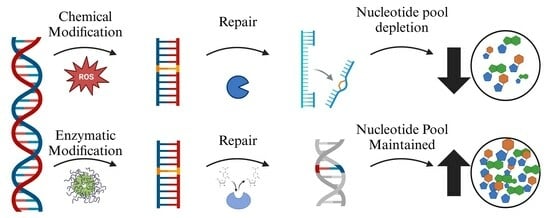
DNA damage and repair have been central themes in cellular biology research. Broadly, DNA damage is understood as modifications to canonical nucleotides that disrupt their function during transcription and replication. A deeper biochemical understanding of DNA damage is essential, as the genome governs all cellular processes. We can classify DNA damage according to whether the modifications to the nucleic acid scaffold are chemically or enzymatically initiated. This distinction is important because chemical modifications are often irreversible, sometimes sparse, and difficult to detect or control spatially and replicate systematically. This can result in genomic damage or modifications to nucleotides in the nucleotide pool, which is less commonly studied. In contrast, enzymatic modifications are typically induced by the cell for specific purposes and are under strong regulatory control. Enzymatic DNA modifications also present a degree of sequence specificity and are often reversible. However, both types of DNA modifications contribute to cellular aging when poorly repaired and, as a result, remain incompletely understood. This review hopes to gather less studied mechanisms in nucleotide modifications and show research gaps in our current understanding of nucleotide biology. By examining the implications of these mechanisms on DNA modifications, in the nucleotide pool and genome, we may gain insights into innovative strategies for mitigating the effects of cellular aging.

Graphical Abstract

1. Introduction

The central dogma of molecular biology describes the flow of genetic information from DNA to RNA to protein. This process relies on the five canonical nucleobases: adenine, thymine, guanine, cytosine, and uracil, which form the building blocks of DNA and RNA. The specific sequence of these nucleotide bases determines the identity of the ultimate gene product, whether a protein or a non-coding RNA. However, genomic instability and mutations retained during DNA replication can lead to errors in transcription and translation, disrupting cellular function.
Genomic errors can arise from endogenously modified deoxynucleotides and nucleoside triphosphates used in DNA and RNA synthesis and modifications made directly in the nucleic acid polymers. Typically, the incorporation of noncanonical nucleotides—those that deviate from the established nucleotide pool—is prevented by a set of sanitizing enzymes [1]. These enzymes work by hydrolyzing the triphosphate forms of noncanonical nucleotides into their mononucleotide, nucleoside, or nucleobase forms, effectively rendering them inert and enabling their disposal as waste products [1]. This process is essential for maintaining genomic integrity, as the accumulation of modified nucleotides compromises cellular homeostasis. When unregulated, there are several methods by which cellular and genomic instability can be induced. For example, dysregulation of individual deoxynucleotide pools can lead to frameshift mutations. These occur when a deoxyribonucleoside triphosphate (dNTP) is available in excess and is incorrectly incorporated into DNA by DNA polymerase during replication or repair, causing the DNA strand to slip and resulting in a frameshift [2]. These nucleotide pool imbalances can also promote mismatch mutations when DNA polymerase fails to correct the error [3]. These errors can remain innocuous as these mutations accumulate until enough collect to affect the cell. Cells often induce senescence to mitigate the mutagenic consequences of nucleotide pool dysregulation, halting the cell cycle [4]. Understanding the mechanisms that drive these imbalances could help prevent cancer by protecting proto-oncogenes from mutation and limiting excessive cellular senescence.
While many reactions involving nucleic acids and nucleotides are enzymatically reversible, this review focuses on the modifications that are considered irreversible. In other words, this review focuses on processes that, once initiated, cannot be reversed; instead, the modified nucleobase must be removed through the degradation of the nucleosidic unit. Interestingly, several enzymes involved in nucleoside degradation appear to have broad substrate specificity, suggesting that a wider range of modified nucleotides might serve as substrates for DNA and RNA polymerases than is currently recognized [5].
Understanding the chemical processes that generate noncanonical nucleoside triphosphates and the biochemical regulators preventing their accumulation in nucleic acids will have significant implications in nucleic acid research, cancer biology, and cellular aging, as these are independent of the widely investigated DNA damage repair pathways. By highlighting the occurrence of random chemical modifications and the prominence of noncanonical nucleobases in nucleic acids, we highlight the importance of the enzymatic mechanisms that generate and degrade the noncanonical NTPs [1,5,6]. The enzymes responsible for this cleaning and preventive genomic maintenance operation are often perceived as housekeeping gene products, warranting little attention. Insights into these processes may advance our knowledge of basic cellular mechanisms and unlock new therapeutic strategies for age-related diseases and cancer [7].

1.1. The Fate of Chemically Modified Nucleic Acids and Repair Processes

Chemical modifications to nucleotides can occur spontaneously through both endogenous and exogenous chemical agents [1,6,8]. Since this process occurs without enzymatic involvement and is not inherently regulated, no specific mechanisms exist to control these modifications. This includes the lack of direct reversal of nucleotide alterations within nucleic acid polymers. For this, cells rely on indirect DNA repair mechanisms that involve excising the modified nucleobase or oligonucleotide and replacing it with an undamaged unit. Two main DNA repair pathways address chemically modified bases: nucleotide excision repair (NER) and base excision repair (BER) [9,10,11,12]. NER repairs bulky DNA lesions like thymine dimers, while BER fixes smaller modifications like oxidized nucleotides [9,12,13,14].
Both repair pathways involve excision processes but differ in the extent of damage that they address. In NER, a section of 24–30 nucleotides surrounding the damage is excised, followed by re-synthesis using the undamaged strand as a template [10,13,14]. This process requires the coordinated action of several enzymes, including helicases and nucleases [13,14]. In contrast, BER begins with the action of a DNA glycosylase enzyme, which cleaves the damaged nucleobase from the sugar–phosphate backbone, creating an abasic site [9,12,15]. This site is then processed by additional enzymes, including AP endonuclease, which removes the sugar, and DNA polymerase, which fills in the gap [16,17]. Finally, the repair is completed by DNA ligase, which seals the newly synthesized nucleotide into place [16,17]. There is little research on the fate of the modified base cleaved and released by these processes. The modified bases are likely marked as waste material and removed from the nucleobase pool by phase 1 and 2 metabolism (e.g., guanine and uric acid). However, some damaged bases (e.g., hypoxanthine) may also re-enter the nucleotide pool. The latter would result in modified nucleotides being available for incorporation into DNA. Unfortunately, there is a lack of research defining the fate of modified bases after BER. If bases can re-enter the nucleotide pool through the salvage pathway, they would disbalance the nucleotide pool and may induce further DNA lesions.

1.2. Detection of Modified 2′Deoxy Nucleotides In Vivo

One issue with studying nucleotide modifications is finding them in vivo. A classic method for identifying nucleotides is high-performance liquid chromatography (HPLC) coupled with UV or mass spectroscopy (MS) [18,19]. These methods function by first digesting nucleotide polymers with enzyme mixtures. This results in a mixture of single nucleotides that can be separated on HPLC by different methods [19]. This can then be directly run from the HPLC into a mass spectrometer to determine the mass of each fraction. Combining the mass with the retention time from the HPLC methods, one can definitively identify the nucleotides in each fraction [18,19]. Although HPLC-coupled MS is an effective method for identifying and quantifying nucleotide modifications, recent studies are employing Oxford nanopore sequencing to identify nucleotide modifications [20,21]. Oxford nanopore sequencing is a DNA and RNA sequencing system that utilizes a small protein embedded in a membrane to sequence nucleotides. This is accomplished using an electrical current to pull nucleotide polymers through the pore [21]. As the nucleotides pass through the pore, the electrical current is disrupted, and that disruption can be measured and recorded. Each nucleotide will make a unique disruption pattern that can be used to determine its identity [21]. This method was utilized by Liu et al. to measure certain nucleotide modifications from long reads of DNA [20]. This group has developed a program called NanoMod, which uses raw sequencing data from Oxford nanopore long-read sequencing to identify modified nucleotides [20]. This program cannot distinguish all modifications (such as 5 mC vs. 4 mA) from each other [20]. Although nanopore is a powerful tool, there is still some work to be done to optimize it for sequence DNA modifications, and many researchers are actively working on this project.

2. Chemical Modifications: Uncontrolled, Unregulated, and Devoid of Protein Inducers

2.1. Oxidation of Nucleotides

The central dogma of molecular biology relies on nucleotides to carry genetic information, which is read in triplets to determine the order of amino acids in proteins [22]. In DNA, these nucleotides form base pairs, with adenine pairing with thymine, and guanine pairing with cytosine. In RNA, uracil replaces thymine, but the triplet codon system remains the same. However, chemical nucleotide modifications, particularly oxidation and deamination, can disrupt the traditional Watson–Crick base-pairing mechanism.
One of the most studied forms of nucleotide modification is oxidation by reactive oxygen species (ROS) (Figure 1A) [1,22]. ROS are naturally produced in cells as by-products of metabolic processes and can cause a range of molecular outcomes [23]. One significant oxidation product is 8-oxo-7,8-dihydroguanine 2′-deoxy-nucleoside triphosphate (8-oxodGTP). 8-OxodGTP can be generated from 2′-deoxy-guanosine triphosphate by direct oxidation of 2′-deoxyguanine nucleotides or putatively from the modified nucleobase (8-oxo guanine) via the purine salvage pathway [24,25]. 8-OxodGTP can be incorporated opposite cytosine or adenine during DNA replication [26,27]. Interestingly, the incorporation opposite to adenine is more favored, leading to A-T to G-C transversion mutations, which contribute to genomic instability [27]. These mutations highlight the critical role of sanitizing enzymes, which hydrolyze and eliminate noncanonical nucleotides like 8-oxodGTP [5,28]. These sanitizing enzymes prevent the accumulation of mutagenic species in the DNA and help maintain genomic integrity. In high-ROS environments, the inability of DNA polymerase to distinguish between canonical and oxidized nucleotides can lead to replication errors and, ultimately, cell death [1,8,25,29]. This suggests that DNA polymerase may have limited ability to discriminate between structural nucleotide analogs. This inability to accurately discriminate potentially leads to the incorporation of previously unrecognized endogenous noncanonical nucleotides.
Even a small amount of modified guanosine triphosphate in the mitochondrial nucleotidic pool—such as when 0.06% of available guanosine triphosphate is converted to 8-oxodGTP—can overcome the mitochondrial DNA polymerase fidelity. This occurs because the exonuclease activity of mitochondrial DNA polymerase (pol γ) fails to recognize 8-oxodGTP as an error, resulting in faulty DNA replication [30]. This becomes more concerning when one realizes how common 8-oxodGTP formation is in cells. Current research suggests that, on average, human lymphocytes contain 2.66 residues of 8-oxodGTP per 106 residues of deoxyguanosine but can contain up to 4.24 residues of 8-oxodGTP per 106 residues of deoxyguanosine [31,32]. This is more than 10,000 8-oxodGTP residues per cell [32]. These studies together indicate a greater need for methods to reduce the abundance of oxidized nucleotides, specifically 8-oxodGTP.
Other oxidation products include 2-hydroxy-2′-deoxyadenosine triphosphate (2-OH-dATP) (Figure 1A), 5-hydroxy 2′-deoxycytidine triphosphate (5-OH-dCTP), and 5-formyl 2′-deoxyuridine triphosphate (5-CHO-dUTP) [33,34]. Interestingly, 2-OH-dATP can be incorporated into DNA with similar efficiency to that of 8-oxodGTP and can pair with guanine to cause CG-to-AT transversion mutations, which is the opposite of the mutation caused by 8-oxodGTP [34,35]. Furthermore, 2-OH-dATP is more mutagenic than 8-oxodGTP in E. coli [33]. On the other hand, 5-OH-dCTP and 5-CHO-dUTP do not appear to cause significant DNA damage. This variability in mutagenic potential among different oxidation products underscores the complexity of oxidative damage and its impact on DNA stability. Given the frequency of ROS generation in cellular compartments, each nucleic acid-containing organelle must have mechanisms that remove damaged nucleotides, both free nucleotides and those incorporated into DNA and RNA polymers [36]. Removing these damaged derivatives is essential to preventing the accumulation of mutations and ensuring proteostasis and transcriptomic balance.
Although the oxidation of nucleotides can result in mutagenic effects and nucleotide pool depletion, this process may also offer secondary benefits to cells. Reactive oxygen species (ROS), a natural by-product of cellular metabolism, are typically present in excess despite mechanisms that reduce their levels (e.g., SOD, catalase, and glutathione). If left unchecked, the ROS surplus leads to the chemical oxidation of proteins and lipids, which, in both cases, is challenging for the cell to manage [37,38], as this process is often non-salvageable and rarely reversible. Therefore, the capacity of nucleotides to neutralize excess ROS might be advantageous in protecting structural and functional macromolecules from early demise, since, once modified, these nucleotides can be degraded and discarded. In contrast, oxidized lipids are difficult to replace and often give rise to more toxic compounds, while modified proteins might lose their function or lack the capacity for proteolysis and accumulate [39,40]. In this context, the nucleotide pool may serve as a protective mechanism, shielding the cell from more harmful oxidative by-products. Future research in anti-cellular aging may benefit from a more global approach to reducing macromolecular damage and seek to optimize nucleotide biosynthetic and degradation pathways rather than solely attempting to reduce nucleotide oxidation.

2.2. Halogenation of Nucleotides

In addition to oxidation, halogenation of aromatics and, more specifically, nucleobases in nucleotides is emerging as a novel form of DNA damage. Halogenation-induced damage has been documented in studies of DNA, RNA, and polynucleotides exposed to hypochlorite, suggesting that halogenation may play a role in genetic instability under certain physiological conditions. Hypohalous acids react with aromatic amino acids in proteins and purine and pyrimidine bases in nucleic acids like DNA (e.g., Figure 1B shows the chlorination of guanosine). This halogenation occurs more specifically in inflamed tissues where leukocytes produce these hypohalous acids (HOCl and HOBr) [41].
Much remains unknown about the consequences of this endogenous chemical modification of nucleobases. For instance, although cytosine can be halogenated, it is still unclear whether endogenous halogenation has mutagenic consequences. However, 5-chlorocytosine nucleoside triphosphate acts as a substrate for DNA polymerase, which has epigenetic consequences by altering the cytosine methylation patterns in DNA [42]. Furthermore, 5-chlorocytosine has recently been revealed to have these mutagenic properties that offer a strong mechanistic link between chronic inflammation and cancer [43,44]. Since other bases in DNA and RNA are modified by hypohalous acids, these modifications can potentially lead to additional forms of genetic damage or alterations, which warrant substantial investigations.

2.3. Chemical Alkylation of Nucleobases Bases and DNA Crosslinks

Alkylation is a common form of DNA damage caused by adding exogenous alkyl groups to the heteroatoms on nucleotide bases. This modification typically occurs when a carbon center undergoes a nucleophilic attack from a lone paired electron-rich atom, most commonly oxygen or nitrogen of the nucleobase. The process can occur by addition on a carbonyl or proceed via an SN1 or SN2 mechanism, with the SN1 pathway generally being more damaging [45,46]. In the SN1 mechanism, alkyl groups are added to an oxygen or nitrogen atom, while the SN2 pathway, a milder pathway, preferentially yields alkylation on nitrogen atoms. Naturally occurring alkylators can be found in the environment, foods, cosmetics, and tobacco smoke, while others form during cellular metabolism (e.g., nitrosamines and methylglyoxal) [47,48]. Since alkylation mechanisms have intrinsic specificity, this specificity can be optimized when creating DNA alkylators for specific purposes, such as anticancer agents. Most alkylating anticancer agents have been developed to maximize alkylation events that follow an SN1 mechanism [49]. Regardless of the mechanism, the attachment of alkyl groups to nucleophilic atoms within the DNA bases alters their structure, potentially leading to mutations [45].
A wide range of endogenous and exogenous chemicals can induce alkylation damage. Endogenous alkylating agents include gut bacteria’s metabolic products, by-products of lipid peroxidation, and reactions with cellular methyl donors such as S-adenosylmethionine, a common cofactor in cellular methylation reactions [50]. Since exogenous alkylating agents are present almost everywhere (e.g., tobacco smoke, gasoline, and certain foods) [50,51], the variation in the abundance and nature of these alkylation products adds another layer of complexity, as different products may cause distinct and yet potentially synergistic DNA damage, each requiring specific repair mechanisms. For example, small alkylation adducts, such as O6-methylguanine, can be repaired by enzymes like O6-methylguanine-DNA methyltransferase (MGMT) or AlkB-homolog (ALKBH) demethylases [52]. However, more complex alkylation products often require more extensive repair mechanisms, such as nucleotide excision repair (NER) or base excision repair (BER), to remove and replace the damaged bases [46].
One common form of complex alkylating damage is DNA crosslinks. Crosslinks can be classified as intrastrand or interstrand, depending on whether the alkylated bases are located on the same or across opposing strands. Intrastrand crosslinks cause replication stalling, but specific polymerases can bypass these lesions, making them less damaging than interstrand crosslinks [53]. In contrast, interstrand crosslinks prevent the separation of the DNA strands, blocking the replication and transcription processes. This leads to severe genomic instability if the damage is not repaired [53]. Unfortunately, many by-products of lipid peroxidation induce interstrand crosslinks. The most prominent ROS-promoted cross-linkers are acrolein, crotonaldehyde, and β-unsaturated aldehydes [54,55]. Repairing these DNA crosslinks requires specialized mechanisms. The effective repair of these crosslinking lesions is critical for maintaining genomic integrity, particularly in rapidly dividing cells. DNA crosslinks are repaired by the Fanconi anemia pathway when the replication fork stalls at a crosslink [56]. Once the replication fork stalls at a DNA crosslink, repair proteins are activated by ubiquitination, then nucleases can remove the crosslinked bases from one strand of DNA, and the resulting DBS is repaired by RAD51-dependent homologous recombination [56,57].
The diverse array of alkylation products complicates efforts to predict all possible outcomes of this damage. Nevertheless, alkylation generally leads to distortions in the DNA backbone, which can interfere with canonical DNA processing, including replication impairment and mismatch repair [58]. Another common consequence of alkylating agents is spontaneous depurination, which occurs with N7-methyl guanine (7meG) [49]. This results in an apurinic (AP) site, leading to a polymerase stall and single- or double-strand breaks [59]. In addition to these forms of damage, some naturally occurring alkylators can also induce the formation of crosslinks between DNA strands or DNA and proteins, further impeding the repair process [60]. Although these natural chemicals are toxic and hazardous, they have provided fantastic opportunities as therapeutics in cancer [60]. Currently, there are seven classes of alkylating agents used as cancer therapies. These classes stem from the parent molecule’s reactive group. They are as follows: nitrogen mustards and oxazaphosphorines, ethylene imines, nitrosoureas, alkyl sulfonates, triazenes and hydrazines, platinum derivatives (although acting via coordination chemistry rather than covalent bond formation), and tetrahydroisoquinolines [60].
One undefined area of research is the ability of these alkylating agents to affect free nucleosides and nucleotides. Research investigating how alkylating agents affect the nucleotide pool levels and their maintenance is dwindling. Much of the existing literature on this subject originated in the 1980s with Arecco et al., who suggested that alkylating agents react with free nucleotides and deoxynucleotides, affecting the cellular nucleotide pools [61]. They demonstrated this phenomenon by manipulating the nucleotide pool levels and treating cells with N-methyl-N-nitrosourea (MNU). Their work revealed that expanding the nucleotide pool in cells exposed to MNU increases mutagenic events, which indirectly suggested that the nucleotide pool is a target for alkylating agents and enables genomic evasion mechanisms [61]. This study is unique in showing the ability of an alkylating agent to modify intracellular free nucleotides and providing evidence that changes in the levels of nucleic acid building blocks could drive adaptation through mutations. A substantial knowledge gap exists as more endogenous and environmental toxins and chemotherapies are revealed as DNA modifiers through alkylation. The recent advances in nucleic acid research and molecular biology could easily address this shortfall.

2.4. Formation of Thymine Dimers

Ultraviolet (UV) light is a well-known environmental factor that causes DNA damage. One of the primary mechanisms through which UV light induces mutations is by generating reactive oxygen species (ROS) in irradiated cells [62]. These ROS can lead to the oxidation of nucleotides, as previously described. However, UV light also directly causes a specific form of DNA damage: cyclobutane pyrimidine dimers (CPD) formation. Thymine dimers are preferred, although mixed cytosine–thymine dimers occur [63]. The least frequent cytosine can also dimerize with an adjacent cytosine [63,64]. Thymine dimers occur when adjacent thymine bases in a DNA strand form covalent bonds (Figure 1), typically between the C5 and C6 carbon atoms of the pyrimidine rings [65]. This dimerization process is catalyzed by UV light with a wavelength of 254 nm or higher [66]. While shorter wavelengths of UV light can also induce thymine dimers, UV light with an emission of 290 nm is most effective at penetrating deeper into epithelial tissues, where it can trigger carcinogenic mutations [66]. CPDs occur in a 15 times higher concentration in CpG islands [67,68]. This specificity has been ascribed to the overall DNA scaffold.
A consequence of CPD formation incorporating cytosine is the spontaneous deamination of cytosine [67]. This event can result in the transversion of cytosine into thymine, altering the genetic code. It is important to recognize this event, as CPD formation and cytosine deamination are not regulated or induced by enzymes and, therefore, cannot be regulated.
Structures determine function and stability in biology; CPD formation is no exception. The cyclobutane rings in CPD can be broken by UV light at the same excitation wavelength that formed them [69]. This process depends on how the adjacent scaffold, and thus its sequence, strains the CPD. If the CPD forms and is surrounded by stable scaffolds, it will remain until enzymatically removed. However, irradiation can reverse the cyclization and free the nucleobases [69] if the sequence is strained.
CPDs are unique to DNA and RNA polymers, requiring the nucleobases to be stacked near each other. The majority of prokaryotes and eukaryotes carry the photolyase gene, which encodes the protein that breaks these dimers. Unfortunately, placental mammals lack an active form of such photolyase enzyme [70]. Therefore, in humans, thymine dimers must be removed by complex mechanisms, primarily through the nucleotide excision repair (NER) pathway, which recognizes and removes these bulky adducts from the DNA strand [11].

3. Enzyme-Driven Modification

Enzymes play a crucial role in the reversible and irreversible modification of nucleotides, particularly in processes like DNA repair and signaling. One typical example of irreversible modification occurs in purine metabolism. The purine biosynthesis pathway can produce noncanonical nucleotides like inosine and xanthosine phosphates, which arise from the deamination of adenine and guanine [71]. Once converted to deoxyribonucleoside triphosphates, these modified nucleotides can be incorporated into DNA, potentially causing mutations if not readily removed from circulation or correctly repaired once incorporated [72].

3.1. Deaminated Ribonucleotides vs. Ribonucleotide Deamination

Another consequence of elevated oxidative stress is the dysregulation of the levels of deaminated nucleotides and nucleosides [33,73,74]. The deamination process differs from oxidation in replacing an amine functional group (the enamino group) with a hydroxyl group, which equates to acquiring keto functionality (Figure 2A,B). This later event dramatically alters the base-pairing properties of the modified nucleobase [26,74]. Chemical deamination typically occurs in DNA polymers, affecting base pairing during replication and transcription [75]. If the deaminated bases remain in the DNA, they promote mispairing in transcription until they are excised. Chemical deamination can lead to mutations and genomic instability when its products persist at high concentrations. Together, hypoxanthine incorporation into nucleic acids introduces substitution mutations that not only affect genomic stability but also may alter gene product functions through transcriptomic errors. Furthermore, hypoxanthine in DNA can alter the recognition sites of DNA-binding proteins, which may deregulate gene expression [76].
Another method for nucleotide deamination is through enzyme activity. Unlike chemical deamination, enzymatic deamination occurs mainly in the nucleotide pool as a means to regulate the pool concentration. Enzymatic deamination occurs on pyrimidine and purine nucleosides, producing uracil-, hypoxanthine-, and xanthine-derived nucleosides. For example, adenosine and 2′deoxy adenosine can spontaneously deaminate into inosine and 2′deoxy inosine [72,76]. Because of the structural similarities between inosine and guanosine, cells will pair a cytosine with inosine during replication and transcription, creating a transition mutation [72,76]. If unrepaired, this would create a buildup of mutations that alter the genetic code. Unfortunately, the spontaneous deamination of nucleobases within DNA polymers results in the unregulated release of deaminated nucleobase upon repair [74]. However, if unrepaired, this chemical deamination can be mutagenic [74]. The nucleobase salvage pathways are critical to limit the accumulation of these entities and facilitate their recycling to canonical nucleotides [27]. However, whether it is through direct deamination of DNA polymers or DNA polymerase use of deaminated nucleotides available from the recycled deaminated nucleobase pools, an unregulated increasing abundance of deaminated nucleotides, deoxynucleotides, and nucleobases during replication, transcription, or translation will have deleterious cellular outcomes [27,77].
In contrast, enzymatic deamination is highly regulated, occurring in the deoxynucleosidic and nucleosidic pools and RNA and DNA polymers [78,79,80]. Notably, the enzymatic deamination process is reversible and therefore tightly regulated. Given the stringent regulation of these enzymes, it is crucial to consider the potential consequences of their activity for DNA integrity. As such, errors occurring in DNA are more likely to arise if DNA polymerases incorporate deaminated nucleotides during replication.
Purine synthetic metabolism is regulated through two fundamentally distinct pathways: the de novo purine biosynthesis (DNPB) pathway and the purine salvage pathway (PSP). The eleven-step DNPB pathway begins with 5′-phosphoriboside pyrophosphate (PRPP) forming inosine monophosphate (IMP), which can then be converted into adenosine monophosphate (AMP) via guanosine triphosphate (GTP) [71]. AMP is converted into adenosine di- and triphosphates or dephosphorylated and deaminated to form inosine by adenosine deaminase (ADA) or fatty acid metabolism–immunity nexus (FAMIN) [76,78]. On the other hand, the PSP functions by recycling adenine and hypoxanthine to generate additional AMP and IMP via PRPP [71]. Together, these pathways regulate the nucleotide pool, balancing the metabolite pools necessary for cellular functions, including bioenergetics, signaling, and replication.
One prominent deamination product is inosine, formed when a hydroxyl moiety replaces the amine group (NH2) on the C4 position of adenosine. Inosine can pair with cytosine like guanosine, potentially causing substitution mutations in DNA. Hypoxanthine incorporation into DNA can occur through two mechanisms. First, free deoxyadenosine can be deaminated by ADA [76]. Deamination of deoxyadenosine is considered a form of DNA damage, as it can lead to a point mutation where an A-T base pair is converted to a G-C base pair if not repaired. Notably, ADAR, the RNA-specific adenosine deaminase, modifies RNA, furthering the importance of this process in nucleic acid regulation processes [76,81]. Once formed, inosine and deoxy inosine are converted enzymatically into hypoxanthine by purine nucleoside phosphorylase (PNP) or Lacc1, also known as FAMIN [77]. Once hypoxanthine is formed, it can be converted into inosine monophosphate (IMP) by hypoxanthine phosphoribosyl transferase (HRPT) and deoxy inosine monophosphate (dIMP) and subsequently into inosine triphosphate (ITP) or deoxy inosine triphosphate (dITP) through an unknown mechanism [77].
In addition to performing controlled deamination in response to signaling effectors, enzymatic deamination is of primary importance for the immune cells, as inosine acts as a regulatory ligand of adenosine receptors and a major contributor to the purine nucleoside salvage pathways. As a result, immune diseases are associated with aberrant deamination pathways when the enzymes responsible for the optimal turnover of nucleosides and nucleotides are underperforming. One classic example is adenosine deaminase (ADA) mutations, which can cause autoimmune disorders [82]. In humans, severe combined immunodeficiency (SCID) results from insufficient ADA activity, accumulating 2′-deoxyadenosine at high concentrations [82]. This buildup is harmful and disrupts the immune system, preventing its full maturation and resulting in the development of SCID. In ADA deficiency, the adenosine levels increase in the extracellular space, leading to the over-activation of adenosine receptors, which also suppresses T-cell function. Thankfully, SCID can be effectively prevented by replacing ADA through gene therapy [82,83]. Another key deaminase that plays a critical role in the immune response and development is FAMIN. FAMIN is a multifunctional enzyme identified in 2016 and is continuously becoming more important in nucleotide pool regulation and immune cell development [84]. Recently, Cader et al. demonstrated the multifunctionality of FAMIN by using purified enzymes mixed with labeled and unlabeled nucleotides. By running the products from this reaction on LC-MS, they showed deaminated products (adenosine to inosine) and purine nucleoside phosphorylase activity (adenosine to adenine) [78]. This group also showed that FAMIN exerts phosphorylase activity on inosine and guanosine to produce ribose 1-phosphate and the respective bases [78].
Along with those two functions, FAMIN is also able to cleave S-methyl-50-thioadenosine into adenine and create an active S-methyl-50-thioribose-1-phosphate (SAM) [78]. FAMIN’s diverse activity has many important functions. FAMIN appears to regulate M1 and M2 macrophage function, and loss of or reduction in FAMIN reduces both macrophages’ activity [84,85]. The loss or inhibition of FAMIN also impairs ROS production, bacterial killing, NOD2- and Toll-like receptor (TLR)-dependent signaling, cytokine production, and many other immune functions [78,84,86]. Together, this shows the importance of deaminases for immune cell function and may suggest that deaminases, specifically FAMIN, may be an important target for immune-based therapies.
These examples highlight the key difference between chemical and enzymatic dysregulation. Although undesirable, chemical deamination has limited effects when it occurs in small amounts and can be repaired, and the deaminated nucleobase can be recycled. In contrast, debilitating diseases emerge when functional mutations occur in the enzymes responsible for maintaining the nucleotide pools. Problems arise when chemical initiators promote excessive deamination, and the resulting DNA lesions accumulate, leading to genomic instability and imbalance in the nucleotide pools [72,76,87].

3.2. Signaling Alkylation: A Reversible Process Under Tight Regulation

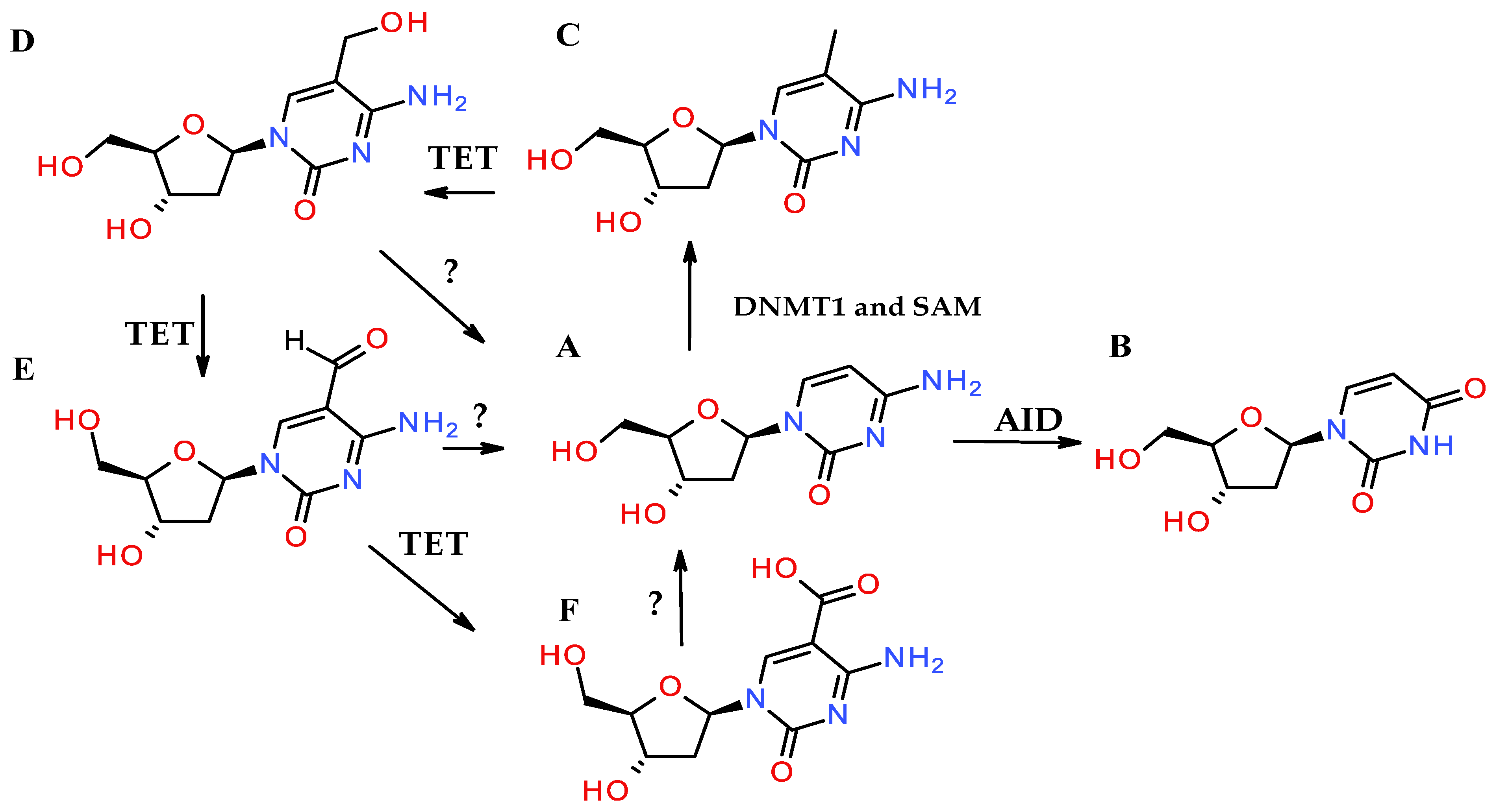
DNA methylation is one of the most common forms of enzyme-catalyzed epigenetic modification. It is commonly understood to be the process by which a methyl group is added to a specific nucleobase in a specific nucleic acid sequence by biochemical means, making it distinct from alkylation by mechanism [88]. The most common form of this process adds a methyl group to the carbon 5 of cytosine (Figure 2A,C), typically within CpG scaffolds of nucleic acids [89]. DNA methyltransferases (DNMTs) catalyze this modification, transferring the methyl group from S-adenosylmethionine (SAM) to its target nucleobase (Figure 2C) [90,91,92,93]. The most common example is cytosine conversion to 5-methyl cytosine in DNA and RNA [94,95]. This conversion starts with a nucleophilic attack of the C6 of cytosine by Cys1226 of DNMT1 [94]. This intermediate then interacts with SAM, which transfers a methyl group onto the C5 of cytosine, resulting in the β-elimination of H5 and 5-methyl-cytosine formation [94]. There are five members in the DNMT family, i.e., DNMT1, DNMT2, DNMT3A, DNMT3B, and DNMT3L, the last of which does not have enzymatic activity [91,93,95]. Although DNA methylation is often associated with gene silencing, it can also play roles in gene activation, depending on the context [96].
DNA methylation plays a crucial role in embryonic development, as DNA methyltransferases (DNMTs) are highly expressed during this stage. The importance of DNA methylation is particularly evident in cases where specific DNMTs, such as DNMT3a and DNMT3b, are knocked out, resulting in death at different stages depending on which DNMT is knocked out [97]. While the expression of DNMTs typically decreases over time in most tissues, it remains notably higher in the brain, highlighting the critical role of DNA methylation in brain function. This pattern underscores the essential involvement of DNA methylation in both development and neuronal activity [88].
While DNMT expression is essential for embryonic development and brain function, the overactivation of DNMTs plays a critical role in cancer development. Enzymatic methylation, particularly at CpG islands, is strongly associated with gene silencing. Furthermore, excessive activation of DNMTs can lead to the silencing of genes involved in crucial DNA repair mechanisms, contributing to cancer progression [91]. DNA methylation by DNMTs is linked to the silencing of genes required for base excision repair (BER), nucleotide excision repair (NER), mismatch repair, and other repair pathways, including that involving O6-methylguanine-DNA methyltransferase (MGMT) [91,98,99,100]. In essence, the overactivation of DNMTs silences these vital genes, inhibiting the function of their protein products and compromising the cell’s ability to repair DNA damage, which can drive tumorigenesis. It is currently highly debated if there is an enzyme that can directly demethylate 5-methylcytosine. Because of the stability of carbon–carbon bonds, it is commonly accepted that this modification is irreversible by direct means [101]. The most direct way to “remove” 5-methylcytosine is to deaminate it with activation-induced deaminase (AID), forming thymine [101]. The deamination of 5-methylcytosine does result in the effective loss of the methylation pattern and the loss of cytosine. Another way to remove the methyl group is by modifying the methyl group to create a new functionality on the heteroatom, allowing the base to be removed [101,102]. One well-characterized mechanism of losing a carbon-bound methyl group in DNA polymers is converting the methyl group to a hydroxymethyl group. This mechanism involves the conversion of 5-methylcytosine to 5-hydroxymethylcytosine (5hmC) via ten–eleven translocation (TET) enzymes (Figure 2D) [101,102,103]. Further modifications of 5-methylcytosine by Tet family enzymes can convert it into other derivatives, such as 5-formyl cytosine (5fC) (Figure 2E) and 5-carboxyl cytosine (5caC) (Figure 2F), which can then be excised by thymine DNA glycosylase (TDG) and base excision repair (BER) or be directly demethylated by unknown mechanisms commonly suggested to involve a group of enzymes [90,104]. Although enzymatic demethylation of 5fC and 5caC is likely, chemical processes may also drive this demethylation. This chemical removal of the formyl and carboxyl groups of 5fC and 5caC is an understudied process lacking a proper mechanism.
Nonetheless, the loss of these groups has been observed in enzyme-free conditions [105,106]. The rate at which these processes occur in PBS is slow (k25 = 5.0 × 10⁻¹¹ s−1; ΔH‡ = 27 kcal/mol for 5caC) [106]. Although these processes may be too slow for biological relevance, Schiesser et al. showed an increase in their rate in a thiol-dependent manner [105]. They showed a 28% increase in the rate of conversion of 5cadC to dC under physiological conditions in 48 h [105]. Although thiols did not affect the rate of conversion of 5fdC and 5-hydroxymethyl 2′-deoxy-cytosine to 2′-deoxy-cytosine, the authors still showed some conversion in DNA polymers. This highlights the importance of considering chemical mechanisms in cells. Although cells commonly rely on enzymes to complete a task, certain chemical agents can catalyze the same reactions. Notably, these chemical agents are less regulated than enzymes, which could result in internal degradation that is hard to account for.
One question that is brought up during this process is how the intermediates to demethylation affect DNA (5hmC, 5fC, etc.). Some people suggest that 5hmC and its downstream products have no epigenetic effect on cells [102]. This would indicate that converting 5-methylcytosine into 5hmC is virtually the same as removing the methyl group altogether. However, recent advances in bioinformatics and biotechnology have allowed researchers to dive deeper into 5hmC formation and location and challenge this viewpoint. Researchers are coupling next-generation sequencing techniques (e.g., nanopore, PacBio sequencing) with methods to capture sequences rich in 5-methylcytosine and 5hmC to help formulate a function for the latter. These methods indicate that 5hmC has the opposite effect to that of 5-methyl cytosine. 5hmC appeared to be increased in certain cells during replication and differentiation, mainly neurons and pancreatic cells [107]. There also appeared to be an increase in transcripts correlating with 5hmC composition [107]. Together, this information implies that 5hmC increases gene transcripts, increasing protein or non-coding RNA production.

3.3. The Aging Process and DNA Damage

The accumulation of DNA damage over time is a central factor in the aging process, as proposed by the free-radical theory of aging [108]. According to this hypothesis, DNA damage induced by reactive oxygen species (ROS) contributes to cellular dysfunction and aging. ROS can directly oxidize DNA or lead to the incorporation of modified nucleotides, which ultimately results in mutations and genomic instability. This theory’s important aspect is the role of mitochondrial DNA (mtDNA) in aging [109]. Since mitochondria are a significant source of ROS, damage to mtDNA is thought to be a major contributor to age-related cellular decline [109].
A well-established mechanism of cellular aging involves oxidative stress induction of cellular senescence. Oxidative stress depletes the nucleotide pool, particularly the dGTP pool, leading to increased replication errors [110]. In response, the cell activates replication arrest (senescence) to prevent the transmission of damaged genetic material to daughter cells. Current research strongly suggests that double-strand breaks are a major causative factor in senescence induction, representing one pathway through which oxidative damage leads to cellular senescence [111]. Although this pathway remains underexplored, it may also serve as a mechanism by which other nucleotide modifications induce senescence. Notably, nucleotide pool depletion results in ribonucleotide reductase (RNR) inhibition, which correlates with reduced DNA repair capacity [110]. While these mechanisms have been extensively studied in oxidative stress, they remain poorly understood in other cases, such as alkylating damage.

4. Conclusions

Addressing the current knowledge gap could yield pivotal insights into the molecular processes driving physiological changes, loss of biochemical homeostasis, and cellular aging. Currently, the need for genetic amplification limits the extent to which chemical modifications, especially random chemical modifications, can be detected. By predicting modifications that can occur chemically and biochemically in nucleic acids, one can expand the chemical toolbox to generate new nucleic acid templates. Such templates could then be used to train the next generation of DNA sequencers and enable higher sensitivity and accuracy. As such, expanding the scope of research to encompass a broader spectrum of nucleotide modifications is essential. A deeper understanding of these modifications, including well-characterized and emerging variants, is critical for deciphering the complex (chemical or enzymatic) mechanisms underlying mutagenic events and senescence. A comprehensive investigation of the modified DNA landscape—spanning both nuclear and mitochondrial genomes—may unveil novel pathways contributing to cellular aging and age-related diseases.

Funding

The work was supported through the Internal Grant Program of the F. P. Whiddon College of Medicine, University of South Alabama, and the Translational Research Institute through NASA Cooperative Agreement NNX16AO69A.

Conflicts of Interest

The authors declare no conflict of interest.

References

  1. Broderick, K.; Moutaoufik, M.T.; Aly, K.A.; Babu, M. Sanitation enzymes: Exquisite surveillance of the noncanonical nucleotide pool to safeguard the genetic blueprint. Semin. Cancer Biol. 2023, 94, 11–20. [Google Scholar] [CrossRef] [PubMed]
  2. Bebenek, K.; Roberts, J.D.; Kunkel, T.A. The effects of dNTP pool imbalances on frameshift fidelity during DNA replication. J. Biol. Chem. 1992, 267, 3589–3596. [Google Scholar] [CrossRef] [PubMed]
  3. Watt, D.L.; Buckland, R.J.; Lujan, S.A.; Kunkel, T.A.; Chabes, A. Genome-wide analysis of the specificity and mechanisms of replication infidelity driven by imbalanced dNTP pools. Nucleic Acids Res. 2016, 44, 1669–1680. [Google Scholar] [CrossRef] [PubMed]
  4. Mannava, S.; Moparthy, K.C.; Wheeler, L.J.; Natarajan, V.; Zucker, S.N.; Fink, E.E.; Im, M.; Flanagan, S.; Burhans, W.C.; Zeitouni, N.C.; et al. Depletion of Deoxyribonucleotide Pools Is an Endogenous Source of DNA Damage in Cells Undergoing Oncogene-Induced Senescence. Am. J. Pathol. 2013, 182, 142–151. [Google Scholar] [CrossRef]
  5. Galperin, M.Y.; Moroz, O.V.; Wilson, K.S.; Murzin, A.G. House cleaning, a part of good housekeeping. Mol. Microbiol. 2006, 59, 5–19. [Google Scholar] [CrossRef]
  6. Nagy, G.N.; Leveles, I.; Vértessy, B.G. Preventive DNArepair by sanitizing the cellular (deoxy)nucleoside triphosphate pool. FEBS J. 2014, 281, 4207–4223. [Google Scholar] [CrossRef]
  7. Bilyard, M.K.; Becker, S.; Balasubramanian, S. Natural, modified DNA bases. Curr. Opin. Chem. Biol. 2020, 57, 1–7. [Google Scholar] [CrossRef]
  8. Gad, H.; Koolmeister, T.; Jemth, A.-S.; Eshtad, S.; Jacques, S.A.; Ström, C.E.; Svensson, L.M.; Schultz, N.; Lundbäck, T.; Einarsdottir, B.O.; et al. MTH1 inhibition eradicates cancer by preventing sanitation of the dNTP pool. Nature 2014, 508, 215–221. [Google Scholar] [CrossRef]
  9. Gohil, D.; Sarker, A.H.; Roy, R. Base Excision Repair: Mechanisms and Impact in Biology, Disease, and Medicine. Int. J. Mol. Sci. 2023, 24, 14186. [Google Scholar] [CrossRef]
  10. Kemp, M.G.; Reardon, J.T.; Lindsey-Boltz, L.A.; Sancar, A. Mechanism of release and fate of excised oligonucleotides during nucleotide excision repair. J. Biol. Chem. 2012, 287, 22889–22899. [Google Scholar] [CrossRef]
  11. Iyama, T.; Wilson, D.M. DNA repair mechanisms in dividing and non-dividing cells. DNA Repair 2013, 12, 620–636. [Google Scholar] [CrossRef] [PubMed]
  12. Krokan, H.E.; Bjoras, M. Base Excision Repair. Cold Spring Harb. Perspect. Biol. 2013, 5, a012583. [Google Scholar] [CrossRef] [PubMed]
  13. Krasikova, Y.; Rechkunova, N.; Lavrik, O. Nucleotide Excision Repair: From Molecular Defects to Neurological Abnormalities. Int. J. Mol. Sci. 2021, 22, 6220. [Google Scholar] [CrossRef] [PubMed]
  14. Kumar, N.; Raja, S.; Van Houten, B. The involvement of nucleotide excision repair proteins in the removal of oxidative DNA damage. Nucleic Acids Res. 2020, 48, 11227–11243. [Google Scholar] [CrossRef]
  15. Jacobs, A.L.; Schar, P. DNA glycosylases: In DNA repair and beyond. Chromosoma 2012, 121, 1–20. [Google Scholar] [CrossRef]
  16. Krokan, H.E.; Nilsen, H.; Skorpen, F.; Otterlei, M.; Slupphaug, G. Base excision repair of DNA in mammalian cells. FEBS Lett. 2000, 476, 73–77. [Google Scholar] [CrossRef]
  17. Fortini, P.; Pascucci, B.; Parlanti, E.; D’Errico, M.; Simonelli, V.; Dogliotti, E. The base excision repair: Mechanisms and its relevance for cancer susceptibility. Biochimie 2003, 85, 1053–1071. [Google Scholar] [CrossRef]
  18. Varma, S.J.; Calvani, E.; Gruning, N.M.; Messner, C.B.; Grayson, N.; Capuano, F.; Mulleder, M.; Ralser, M. Global analysis of cytosine and adenine DNA modifications across the tree of life. Elife 2022, 11, e81002. [Google Scholar] [CrossRef]
  19. Marchante-Gayon, J.M.; Nicolas Carcelen, J.; Potes Rodriguez, H.; Pineda-Cevallos, D.; Rodas Sanchez, L.; Gonzalez-Gago, A.; Rodriguez-Gonzalez, P.; Garcia Alonso, J.I. Quantification of modified nucleotides and nucleosides by isotope dilution mass spectrometry. Mass. Spectrom. Rev. 2024, 43, 998–1018. [Google Scholar] [CrossRef]
  20. Liu, Q.; Georgieva, D.C.; Egli, D.; Wang, K. NanoMod: A computational tool to detect DNA modifications using Nanopore long-read sequencing data. BMC Genom. 2019, 20, 78. [Google Scholar] [CrossRef]
  21. Liu, Q.; Fang, L.; Yu, G.; Wang, D.; Xiao, C.-L.; Wang, K. Detection of DNA base modifications by deep recurrent neural network on Oxford Nanopore sequencing data. Nat. Commun. 2019, 10, 2449. [Google Scholar] [CrossRef] [PubMed]
  22. Saier, M.H. Understanding the Genetic Code. J. Bacteriol. 2019, 201, 10-1128. [Google Scholar] [CrossRef] [PubMed]
  23. Hayyan, M.; Hashim, M.A.; AlNashef, I.M. Superoxide Ion: Generation and Chemical Implications. Chem. Rev. 2016, 116, 3029–3085. [Google Scholar] [CrossRef] [PubMed]
  24. Jun, Y.W.; Kant, M.; Coskun, E.; Kato, T.A.; Jaruga, P.; Palafox, E.; Dizdaroglu, M.; Kool, E.T. Possible Genetic Risks from Heat-Damaged DNA in Food. ACS Cent. Sci. 2023, 9, 1170–1179. [Google Scholar] [CrossRef]
  25. Henderson, P.T.; Evans, M.D.; Cooke, M.S. Salvage of oxidized guanine derivatives in the (2’-deoxy)ribonucleotide pool as source of mutations in DNA. Mutat. Res. 2010, 703, 11–17. [Google Scholar] [CrossRef]
  26. Kamiya, H. Mutagenic potentials of damaged nucleic acids produced by reactive oxygen/nitrogen species: Approaches using synthetic oligonucleotides and nucleotides: Survey and Summary. Nucleic Acids Res. 2003, 31, 517–531. [Google Scholar] [CrossRef]
  27. Rudd, S.G.; Valerie, N.C.K.; Helleday, T. Pathways controlling dNTP pools to maintain genome stability. DNA Repair 2016, 44, 193–204. [Google Scholar] [CrossRef]
  28. Pai, C.C.; Kearsey, S.E. A Critical Balance: dNTPs and the Maintenance of Genome Stability. Genes 2017, 8, 57. [Google Scholar] [CrossRef]
  29. Kent, T.; Rusanov, T.D.; Hoang, T.M.; Velema, W.A.; Krueger, A.T.; Copeland, W.C.; Kool, E.T.; Pomerantz, R.T. DNA polymerase theta specializes in incorporating synthetic expanded-size (xDNA) nucleotides. Nucleic Acids Res. 2016, 44, 9381–9392. [Google Scholar] [CrossRef]
  30. Longley, M.J.; Nguyen, D.; Kunkel, T.A.; Copeland, W.C. The Fidelity of Human DNA Polymerase γ with and without Exonucleolytic Proofreading and the p55 Accessory Subunit. J. Biol. Chem. 2001, 276, 38555–38562. [Google Scholar] [CrossRef]
  31. Gedik, C.M.; Collins, A. Establishing the background level of base oxidation in human lymphocyte DNA: Results of an interlaboratory validation study. FASEB J. 2005, 19, 82–84. [Google Scholar] [CrossRef] [PubMed]
  32. Ohno, M.; Miura, T.; Furuichi, M.; Tominaga, Y.; Tsuchimoto, D.; Sakumi, K.; Nakabeppu, Y. A genome-wide distribution of 8-oxoguanine correlates with the preferred regions for recombination and single nucleotide polymorphism in the human genome. Genome Res. 2006, 16, 567–575. [Google Scholar] [CrossRef] [PubMed]
  33. Kamiya, H. 2-Hydroxy-dATP is incorporated opposite G by Escherichia coli DNA polymerase III resulting in high mutagenicity. Nucleic Acids Res. 2000, 28, 1640–1646. [Google Scholar] [CrossRef] [PubMed]
  34. Kamiya, H.; Kasai, H. Formation of 2-hydroxydeoxyadenosine triphosphate, an oxidatively damaged nucleotide, and its incorporation by DNA polymerases. Steady-state kinetics of the incorporation. J. Biol. Chem. 1995, 270, 19446–19450. [Google Scholar] [CrossRef]
  35. Satou, K. Mutagenic effects of 2-hydroxy-dATP on replication in a HeLa extract: Induction of substitution and deletion mutations. Nucleic Acids Res. 2003, 31, 2570–2575. [Google Scholar] [CrossRef]
  36. Juan, C.A.; Perez de la Lastra, J.M.; Plou, F.J.; Perez-Lebena, E. The Chemistry of Reactive Oxygen Species (ROS) Revisited: Outlining Their Role in Biological Macromolecules (DNA, Lipids and Proteins) and Induced Pathologies. Int. J. Mol. Sci. 2021, 22, 4642. [Google Scholar] [CrossRef]
  37. Ramana, K.V.; Srivastava, S.; Singhal, S.S. Lipid Peroxidation Products in Human Health and Disease. Oxidative Med. Cell. Longev. 2013, 2013, 583438. [Google Scholar] [CrossRef]
  38. Maddu, N. Diseases Related to Types of Free Radicals; IntechOpen: London, UK, 2019. [Google Scholar]
  39. Kanner, J. Dietary advanced lipid oxidation endproducts are risk factors to human health. Mol. Nutr. Food Res. 2007, 51, 1094–1101. [Google Scholar] [CrossRef]
  40. Davies, M.J. Protein oxidation and peroxidation. Biochem. J. 2016, 473, 805–825. [Google Scholar] [CrossRef]
  41. Hawkins, C.L.; Davies, M.J. Hypochlorite-induced damage to DNA, RNA, and polynucleotides: Formation of chloramines and nitrogen-centered radicals. Chem. Res. Toxicol. 2002, 15, 83–92. [Google Scholar] [CrossRef]
  42. Lao, V.V.; Herring, J.L.; Kim, C.H.; Darwanto, A.; Soto, U.; Sowers, L.C. Incorporation of 5-chlorocytosine into mammalian DNA results in heritable gene silencing and altered cytosine methylation patterns. Carcinogenesis 2009, 30, 886–893. [Google Scholar] [CrossRef] [PubMed]
  43. Fedeles, B.I.; Freudenthal, B.D.; Yau, E.; Singh, V.; Chang, S.C.; Li, D.; Delaney, J.C.; Wilson, S.H.; Essigmann, J.M. Intrinsic mutagenic properties of 5-chlorocytosine: A mechanistic connection between chronic inflammation and cancer. Proc. Natl. Acad. Sci. USA 2015, 112, E4571–E4580. [Google Scholar] [CrossRef] [PubMed]
  44. Chancharoen, M.; Yang, Z.; Dalvie, E.D.; Gubina, N.; Ruchirawat, M.; Croy, R.G.; Fedeles, B.I.; Essigmann, J.M. 5-Chloro-2′-deoxycytidine Induces a Distinctive High-Resolution Mutational Spectrum of Transition Mutations In Vivo. Chem. Res. Toxicol. 2024, 37, 486–496. [Google Scholar] [CrossRef] [PubMed]
  45. Loechler, E.L. A violation of the Swain-Scott principle, and not SN1 versus SN2 reaction mechanisms, explains why carcinogenic alkylating agents can form different proportions of adducts at oxygen versus nitrogen in DNA. Chem. Res. Toxicol. 1994, 7, 277–280. [Google Scholar] [CrossRef]
  46. Drablos, F.; Feyzi, E.; Aas, P.A.; Vaagbo, C.B.; Kavli, B.; Bratlie, M.S.; Pena-Diaz, J.; Otterlei, M.; Slupphaug, G.; Krokan, H.E. Alkylation damage in DNA and RNA--repair mechanisms and medical significance. DNA Repair 2004, 3, 1389–1407. [Google Scholar] [CrossRef]
  47. Frischmann, M.; Bidmon, C.; Angerer, J.; Pischetsrieder, M. Identification of DNA adducts of methylglyoxal. Chem. Res. Toxicol. 2005, 18, 1586–1592. [Google Scholar] [CrossRef]
  48. Donald, W.K.; Pollock, R.E.; Weichselbaum, R.R.; Bast, R.C.; Gansler, T.S.; Holland, J.F.; Frei, E. Alkylating Agents. In Holland-Frei Cancer Medicine, 6th ed.; Holland-Frei, Ed.; BC Decker: Hamilton, ON, USA, 2003. [Google Scholar]
  49. Fu, D.; Calvo, J.A.; Samson, L.D. Balancing repair and tolerance of DNA damage caused by alkylating agents. Nat. Rev. Cancer 2012, 12, 104–120. [Google Scholar] [CrossRef]
  50. Klapacz, J.; Pottenger, L.H.; Engelward, B.P.; Heinen, C.D.; Johnson, G.E.; Clewell, R.A.; Carmichael, P.L.; Adeleye, Y.; Andersen, M.E. Contributions of DNA repair and damage response pathways to the non-linear genotoxic responses of alkylating agents. Mutat. Res. Rev. Mutat. Res. 2016, 767, 77–91. [Google Scholar] [CrossRef]
  51. Hamilton, J.T.; McRoberts, W.C.; Keppler, F.; Kalin, R.M.; Harper, D.B. Chloride methylation by plant pectin: An efficient environmentally significant process. Science 2003, 301, 206–209. [Google Scholar] [CrossRef]
  52. Soll, J.M.; Sobol, R.W.; Mosammaparast, N. Regulation of DNA Alkylation Damage Repair: Lessons and Therapeutic Opportunities. Trends Biochem. Sci. 2017, 42, 206–218. [Google Scholar] [CrossRef]
  53. Deans, A.J.; West, S.C. DNA interstrand crosslink repair and cancer. Nat. Rev. Cancer 2011, 11, 467–480. [Google Scholar] [CrossRef] [PubMed]
  54. Stone, M.P.; Cho, Y.J.; Huang, H.; Kim, H.Y.; Kozekov, I.D.; Kozekova, A.; Wang, H.; Minko, I.G.; Lloyd, R.S.; Harris, T.M.; et al. Interstrand DNA cross-links induced by α,β-unsaturated aldehydes derived from lipid peroxidation and environmental sources. Acc. Chem. Res. 2008, 41, 793–804. [Google Scholar] [CrossRef] [PubMed]
  55. Kozekov, I.D.; Nechev, L.V.; Moseley, M.S.; Harris, C.M.; Rizzo, C.J.; Stone, M.P.; Harris, T.M. DNA interchain cross-links formed by acrolein and crotonaldehyde. J. Am. Chem. Soc. 2003, 125, 50–61. [Google Scholar] [CrossRef] [PubMed]
  56. Housh, K.; Jha, J.S.; Haldar, T.; Amin, S.B.M.; Islam, T.; Wallace, A.; Gomina, A.; Guo, X.; Nel, C.; Wyatt, J.W.; et al. Formation and repair of unavoidable, endogenous interstrand cross-links in cellular DNA. DNA Repair 2021, 98, 103029. [Google Scholar] [CrossRef]
  57. Price, N.E.; Gates, K.S. Novel Processes Associated with the Repair of Interstrand Cross-Links Derived from Abasic Sites in Duplex DNA: Roles for the Base Excision Repair Glycosylase NEIL3 and the SRAP Protein HMCES. Chem. Res. Toxicol. 2024, 37, 199–207. [Google Scholar] [CrossRef]
  58. Donnellan, L.; Simpson, B.; Dhillon, V.S.; Costabile, M.; Fenech, M.; Deo, P. Methylglyoxal induces chromosomal instability and mitotic dysfunction in lymphocytes. Mutagenesis 2021, 36, 339–348. [Google Scholar] [CrossRef]
  59. Boiteux, S.; Guillet, M. Abasic sites in DNA: Repair and biological consequences in Saccharomyces cerevisiae. DNA Repair 2004, 3, 1–12. [Google Scholar] [CrossRef]
  60. Puyo, S.; Montaudon, D.; Pourquier, P. From old alkylating agents to new minor groove binders. Crit. Rev. Oncol. Hematol. 2014, 89, 43–61. [Google Scholar] [CrossRef]
  61. Arecco, A.; Mun, B.J.; Mathews, C.K. Deoxyribonucleotide pools as targets for mutagenesis by N-methyl-N-nitrosourea. Mutat. Res. Rev. Mutat. Res. 1988, 200, 165–175. [Google Scholar]
  62. Ikehata, H.; Ono, T. The Mechanisms of UV Mutagenesis. J. Radiat. Res. 2011, 52, 115–125. [Google Scholar] [CrossRef]
  63. Mitchell, D.L.; Jen, J.; Cleaver, J.E. Sequence specificity of cyclobutane pyrimidine dimers in DNA treated with solar (ultraviolet B) radiation. Nucleic Acids Res. 1992, 20, 225–229. [Google Scholar] [CrossRef] [PubMed]
  64. Premi, S.; Han, L.; Mehta, S.; Knight, J.; Zhao, D.; Palmatier, M.A.; Kornacker, K.; Brash, D.E. Genomic sites hypersensitive to ultraviolet radiation. Proc. Natl. Acad. Sci. USA 2019, 116, 24196–24205. [Google Scholar] [CrossRef] [PubMed]
  65. Rauer, C.; Nogueira, J.J.; Marquetand, P.; Gonzalez, L. Cyclobutane Thymine Photodimerization Mechanism Revealed by Nonadiabatic Molecular Dynamics. J. Am. Chem. Soc. 2016, 138, 15911–15916. [Google Scholar] [CrossRef] [PubMed]
  66. Pathak, M.A.; Kramer, D.M.; Gungerich, U. Formation of thymine dimers in mammalian skin by ultraviolet radiation in vivo. Photochem. Photobiol. 1972, 15, 177–185. [Google Scholar] [CrossRef]
  67. Cannistraro, V.J.; Taylor, J.-S. Acceleration of 5-Methylcytosine Deamination in Cyclobutane Dimers by G and Its Implications for UV-Induced C-to-T Mutation Hotspots. J. Mol. Biol. 2009, 392, 1145–1157. [Google Scholar] [CrossRef]
  68. Tommasi, S.; Denissenko, M.F.; Pfeifer, G.P. Sunlight induces pyrimidine dimers preferentially at 5-methylcytosine bases. Cancer Res. 1997, 57, 4727–4730. [Google Scholar]
  69. Moirangthem, R.; Gamage, M.N.; Rokita, S.E. Dynamic accumulation of cyclobutane pyrimidine dimers and its response to changes in DNA conformation. Nucleic Acids Res. 2023, 51, 5341–5350. [Google Scholar] [CrossRef]
  70. Beukers, R.; Eker, A.P.; Lohman, P.H. 50 years thymine dimer. DNA Repair 2008, 7, 530–543. [Google Scholar] [CrossRef]
  71. Gessner, P.; Lum, J.; Frenguelli, B.G. The mammalian purine salvage pathway as an exploitable route for cerebral bioenergetic support after brain injury. Neuropharmacology 2023, 224, 109370. [Google Scholar] [CrossRef]
  72. Zhang, Q.; Tretyakova, N. Incorporation of inosine into DNA by human polymerase eta (Polη): Kinetics of nucleotide misincorporation and structural basis for the mutagenicity. Biochem. J. 2023, 480, 1479–1483. [Google Scholar] [CrossRef]
  73. Shapiro, R.; Pohl, S.H. The reaction of ribonucleotides with nitrous acid. Side products and kinetics. Biochemistry 1968, 7, 448–455. [Google Scholar] [CrossRef] [PubMed]
  74. Kow, Y.W. Repair of deaminated bases in DNA. Free Radic. Biol. Med. 2002, 33, 886–893. [Google Scholar] [CrossRef] [PubMed]
  75. Ventura, I.; Russo, M.T.; De Luca, G.; Bignami, M. Oxidized purine nucleotides, genome instability and neurodegeneration. Mutat. Res. 2010, 703, 59–65. [Google Scholar] [CrossRef] [PubMed]
  76. Alseth, I.; Dalhus, B.; Bjoras, M. Inosine in DNA and RNA. Curr. Opin. Genet. Dev. 2014, 26, 116–123. [Google Scholar] [CrossRef]
  77. Sakumi, K.; Abolhassani, N.; Behmanesh, M.; Iyama, T.; Tsuchimoto, D.; Nakabeppu, Y. ITPA protein, an enzyme that eliminates deaminated purine nucleoside triphosphates in cells. Mutat. Res. 2010, 703, 43–50. [Google Scholar] [CrossRef]
  78. Cader, M.Z.; De Almeida Rodrigues, R.P.; West, J.A.; Sewell, G.W.; Md-Ibrahim, M.N.; Reikine, S.; Sirago, G.; Unger, L.W.; Iglesias-Romero, A.B.; Ramshorn, K.; et al. FAMIN Is a Multifunctional Purine Enzyme Enabling the Purine Nucleotide Cycle. Cell 2020, 180, 278–295.e223. [Google Scholar] [CrossRef]
  79. Iyer, L.M.; Zhang, D.; Rogozin, I.B.; Aravind, L. Evolution of the deaminase fold and multiple origins of eukaryotic editing and mutagenic nucleic acid deaminases from bacterial toxin systems. Nucleic Acids Res. 2011, 39, 9473–9497. [Google Scholar] [CrossRef]
  80. Gaded, V.; Anand, R. Nucleobase deaminases: A potential enzyme system for new therapies. RSC Adv. 2018, 8, 23567–23577. [Google Scholar] [CrossRef]
  81. Bass, B.L.; Nishikura, K.; Keller, W.; Seeburg, P.H.; Emeson, R.B.; O’Connell, M.A.; Samuel, C.E.; Herbert, A. A standardized nomenclature for adenosine deaminases that act on RNA. RNA 1997, 3, 947–949. [Google Scholar]
  82. Gao, Z.W.; Wang, X.; Zhang, H.Z.; Lin, F.; Liu, C.; Dong, K. The roles of adenosine deaminase in autoimmune diseases. Autoimmun. Rev. 2021, 20, 102709. [Google Scholar] [CrossRef]
  83. Parkman, R.; Weinberg, K.; Crooks, G.; Nolta, J.; Kapoor, N.; Kohn, D. Gene therapy for adenosine deaminase deficiency. Annu. Rev. Med. 2000, 51, 33–47. [Google Scholar] [CrossRef] [PubMed]
  84. Cader, M.Z.; Boroviak, K.; Zhang, Q.; Assadi, G.; Kempster, S.L.; Sewell, G.W.; Saveljeva, S.; Ashcroft, J.W.; Clare, S.; Mukhopadhyay, S.; et al. C13orf31 (FAMIN) is a central regulator of immunometabolic function. Nat. Immunol. 2016, 17, 1046–1056. [Google Scholar] [CrossRef]
  85. O’Neill, L.A.J.; Pearce, E.J. Immunometabolism governs dendritic cell and macrophage function. J. Exp. Med. 2016, 213, 15–23. [Google Scholar] [CrossRef] [PubMed]
  86. Skon-Hegg, C.; Zhang, J.; Wu, X.; Sagolla, M.; Ota, N.; Wuster, A.; Tom, J.; Doran, E.; Ramamoorthi, N.; Caplazi, P.; et al. LACC1 Regulates TNF and IL-17 in Mouse Models of Arthritis and Inflammation. J. Immunol. 2019, 202, 183–193. [Google Scholar] [CrossRef] [PubMed]
  87. Yla-Herttuala, S. ADA-SCID Gene Therapy Endorsed By European Medicines Agency For Marketing Authorization. Mol. Ther. 2016, 24, 1013–1014. [Google Scholar] [CrossRef]
  88. Cui, D.; Xu, X. DNA Methyltransferases, DNA Methylation, and Age-Associated Cognitive Function. Int. J. Mol. Sci. 2018, 19, 1315. [Google Scholar] [CrossRef]
  89. Moore, L.D.; Le, T.; Fan, G. DNA methylation and its basic function. Neuropsychopharmacology 2013, 38, 23–38. [Google Scholar] [CrossRef]
  90. Chen, Z.X.; Riggs, A.D. DNA methylation and demethylation in mammals. J. Biol. Chem. 2011, 286, 18347–18353. [Google Scholar] [CrossRef]
  91. Jin, B.; Robertson, K.D. DNA methyltransferases, DNA damage repair, and cancer. Adv. Exp. Med. Biol. 2013, 754, 3–29. [Google Scholar] [CrossRef]
  92. Bird, J.G.; Basu, U.; Kuster, D.; Ramachandran, A.; Grudzien-Nogalska, E.; Towheed, A.; Wallace, D.C.; Kiledjian, M.; Temiakov, D.; Patel, S.S.; et al. Highly efficient 5′ capping of mitochondrial RNA with NAD+ and NADH by yeast and human mitochondrial RNA polymerase. eLife 2018, 7, e42179. [Google Scholar] [CrossRef]
  93. Kareta, M.S.; Botello, Z.M.; Ennis, J.J.; Chou, C.; Chedin, F. Reconstitution and mechanism of the stimulation of de novo methylation by human DNMT3L. J. Biol. Chem. 2006, 281, 25893–25902. [Google Scholar] [CrossRef] [PubMed]
  94. Du, Q.; Wang, Z.; Schramm, V.L. Human DNMT1 transition state structure. Proc. Natl. Acad. Sci. USA 2016, 113, 2916–2921. [Google Scholar] [CrossRef] [PubMed]
  95. Jeltsch, A.; Ehrenhofer-Murray, A.; Jurkowski, T.P.; Lyko, F.; Reuter, G.; Ankri, S.; Nellen, W.; Schaefer, M.; Helm, M. Mechanism and biological role of Dnmt2 in Nucleic Acid Methylation. RNA Biol. 2017, 14, 1108–1123. [Google Scholar] [CrossRef]
  96. Mohn, F.; Weber, M.; Rebhan, M.; Roloff, T.C.; Richter, J.; Stadler, M.B.; Bibel, M.; Schubeler, D. Lineage-specific polycomb targets and de novo DNA methylation define restriction and potential of neuronal progenitors. Mol. Cell 2008, 30, 755–766. [Google Scholar] [CrossRef] [PubMed]
  97. Okano, M.; Bell, D.W.; Haber, D.A.; Li, E. DNA methyltransferases Dnmt3a and Dnmt3b are essential for de novo methylation and mammalian development. Cell 1999, 99, 247–257. [Google Scholar] [CrossRef]
  98. Peng, B.; Hurt, E.M.; Hodge, D.R.; Thomas, S.B.; Farrar, W.L. DNA hypermethylation and partial gene silencing of human thymine-DNA glycosylase in multiple myeloma cell lines. Epigenetics 2006, 1, 138–145. [Google Scholar] [CrossRef]
  99. Riggs, A.D.; Xiong, Z. Methylation and epigenetic fidelity. Proc. Natl. Acad. Sci. USA 2004, 101, 4–5. [Google Scholar] [CrossRef]
  100. Howard, J.H.; Frolov, A.; Tzeng, C.W.; Stewart, A.; Midzak, A.; Majmundar, A.; Godwin, A.; Heslin, M.; Bellacosa, A.; Arnoletti, J.P. Epigenetic downregulation of the DNA repair gene MED1/MBD4 in colorectal and ovarian cancer. Cancer Biol. Ther. 2009, 8, 94–100. [Google Scholar] [CrossRef]
  101. Bochtler, M.; Kolano, A.; Xu, G.L. DNA demethylation pathways: Additional players and regulators. BioEssays 2017, 39, 1–13. [Google Scholar] [CrossRef]
  102. Carey, N.; Marques, C.J.; Reik, W. DNA demethylases: A new epigenetic frontier in drug discovery. Drug Discov. Today 2011, 16, 683–690. [Google Scholar] [CrossRef]
  103. Tahiliani, M.; Koh, K.P.; Shen, Y.; Pastor, W.A.; Bandukwala, H.; Brudno, Y.; Agarwal, S.; Iyer, L.M.; Liu, D.R.; Aravind, L.; et al. Conversion of 5-Methylcytosine to 5-Hydroxymethylcytosine in Mammalian DNA by MLL Partner TET1. Science 2009, 324, 930–935. [Google Scholar] [CrossRef] [PubMed]
  104. Iwan, K.; Rahimoff, R.; Kirchner, A.; Spada, F.; Schroder, A.S.; Kosmatchev, O.; Ferizaj, S.; Steinbacher, J.; Parsa, E.; Muller, M.; et al. 5-Formylcytosine to cytosine conversion by C-C bond cleavage in vivo. Nat. Chem. Biol. 2018, 14, 72–78. [Google Scholar] [CrossRef]
  105. Schiesser, S.; Pfaffeneder, T.; Sadeghian, K.; Hackner, B.; Steigenberger, B.; Schroder, A.S.; Steinbacher, J.; Kashiwazaki, G.; Hofner, G.; Wanner, K.T.; et al. Deamination, oxidation, and C-C bond cleavage reactivity of 5-hydroxymethylcytosine, 5-formylcytosine, and 5-carboxycytosine. J. Am. Chem. Soc. 2013, 135, 14593–14599. [Google Scholar] [CrossRef] [PubMed]
  106. Lewis, C.A.; Shen, L.; Yang, W.; Wolfenden, R. Three Pyrimidine Decarboxylations in the Absence of a Catalyst. Biochemistry 2017, 56, 1498–1503. [Google Scholar] [CrossRef] [PubMed]
  107. Lee, S.-M. Detecting DNA hydroxymethylation: Exploring its role in genome regulation. BMB Rep. 2024, 57, 135–142. [Google Scholar] [CrossRef]
  108. Harman, D. The aging process. Proc. Natl. Acad. Sci. USA 1981, 78, 7124–7128. [Google Scholar] [CrossRef]
  109. Bratic, A.; Larsson, N.-G. The role of mitochondria in aging. J. Clin. Investig. 2013, 123, 951–957. [Google Scholar] [CrossRef]
  110. Rai, P. Oxidation in the nucleotide pool, the DNA damage response and cellular senescence: Defective bricks build a defective house. Mutat. Res. 2010, 703, 71–81. [Google Scholar] [CrossRef]
  111. White, R.R.; Vijg, J. Do DNA Double-Strand Breaks Drive Aging? Mol. Cell 2016, 63, 729–738. [Google Scholar] [CrossRef]
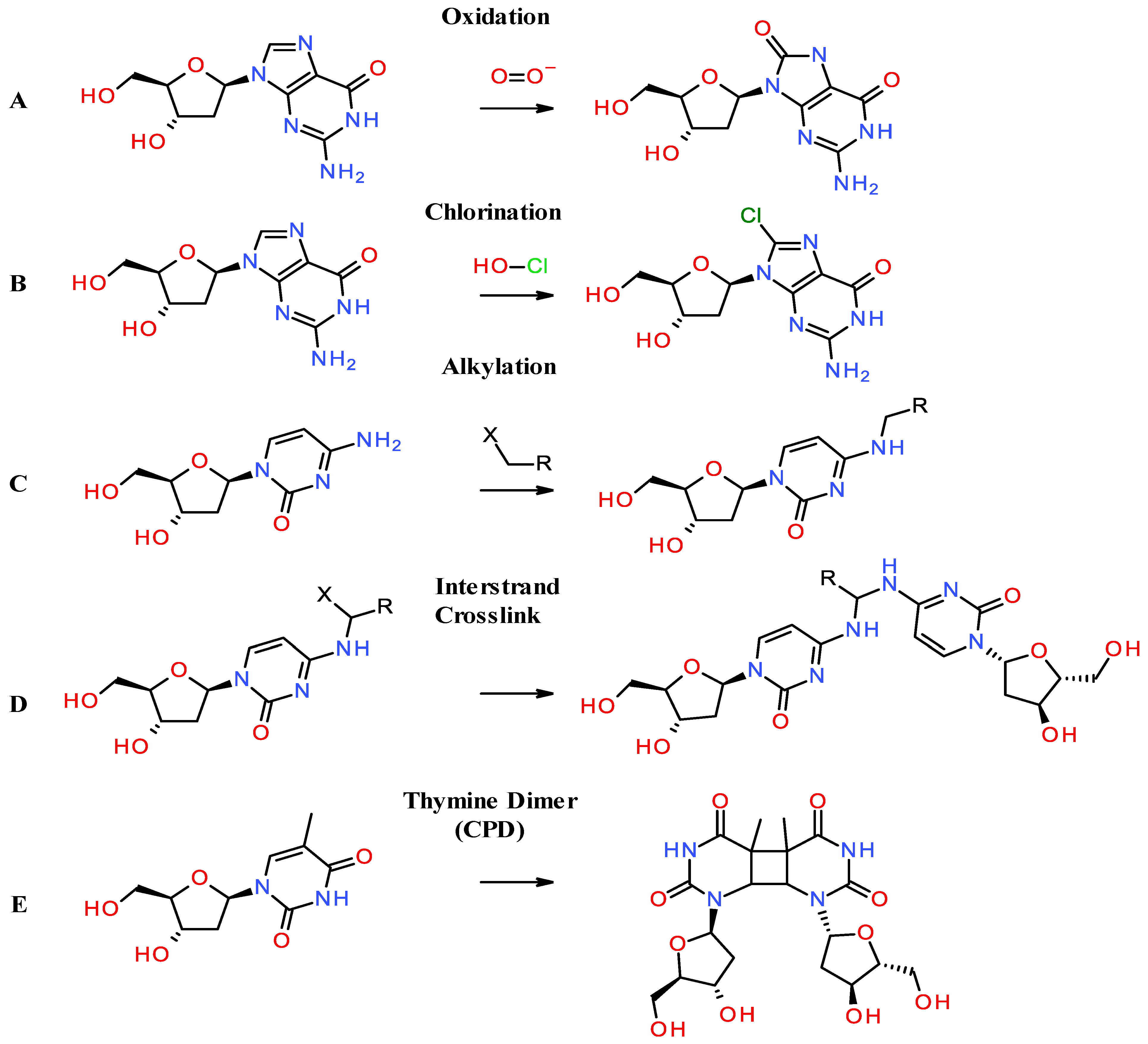
Figure 1. General chemical mechanisms leading to 2′-deoxy nucleoside modifications in vivo and in vitro. (A) Oxidation of 2′-deoxy adenosine by superoxide. (B) halogenation of 2′-deoxy adenosine by hypochlorous acid. (C) Alkylation of cytosine by an alkyl halide. (D) Interstrand or interstrand crosslink of cytosines catalyzed by an alkylated cytosine with a halogen. (E) Thymine dimer induced by UV exposure.
Dna 05 00019 g001
Figure 2. Methylation and hydroxymethylation catalyzed by DNMT1, SAM, and the TET family of proteins. (A) 2′Deoxy-cytosine. (B) 2′Deoxy-uracil formed by deaminating cytosine catalyzed by AID. (C) 5-Methyl-2′deoxy-cytosine catalyzed by DNMT1 and SAM. (D) 5-Hydroxymethyl-2′deoxy-cytosine catalyzed by TET proteins. (E) 5-Formyl-2′deoxy-cytosine catalyzed by TET proteins. (F) 5-Carboxy2′deoxy-cytosine catalyzed by TET proteins.
Dna 05 00019 g002
Disclaimer/Publisher’s Note: The statements, opinions and data contained in all publications are solely those of the individual author(s) and contributor(s) and not of MDPI and/or the editor(s). MDPI and/or the editor(s) disclaim responsibility for any injury to people or property resulting from any ideas, methods, instructions or products referred to in the content.