Review

DNA as a Double-Coding Device for Information Conversion and Organization of a Self-Referential Unity

1
School of Natural Sciences and Biotechnology, Agricultural University of Georgia, 0159 Tbilisi, Georgia
2
INSA-Lyon, CNRS, UMR5240, Microbiologie, Adaptation, Pathogénie, Université Lyon 1, F-69622 Villeurbanne, France
3
MRC Laboratory of Molecular Biology, Francis Crick Avenue, Cambridge Biomedical Campus, Cambridge CB2 0QH, UK
*
Author to whom correspondence should be addressed.
DNA 2024, 4(4), 473-493; https://doi.org/10.3390/dna4040032
Submission received: 12 October 2024 / Revised: 6 November 2024 / Accepted: 14 November 2024 / Published: 19 November 2024

Abstract

:
Living systems are capable on the one hand of eliciting a coordinated response to changing environments (also known as adaptation), and on the other hand, they are capable of reproducing themselves. Notably, adaptation to environmental change requires the monitoring of the surroundings, while reproduction requires monitoring oneself. These two tasks appear separate and make use of different sources of information. Yet, both the process of adaptation as well as that of reproduction are inextricably coupled to alterations in genomic DNA expression, while a cell behaves as an indivisible unity in which apparently independent processes and mechanisms are both integrated and coordinated. We argue that at the most basic level, this integration is enabled by the unique property of the DNA to act as a double coding device harboring two logically distinct types of information. We review biological systems of different complexities and infer that the inter-conversion of these two distinct types of DNA information represents a fundamental self-referential device underlying both systemic integration and coordinated adaptive responses.

1. Introduction

The distinctive organizational hallmark of living systems is their ability to self-reproduce. For that matter, living systems are regarded as ‘autopoietic’ self-referential systems implying the capacity to monitor oneself, that is, to perpetually assess their status quo [1,2]. At the same time, living systems are capable of monitoring their surroundings and eliciting a functionally coordinated adaptive response to environmental change (Figure 1). The capacity of ‘monitoring oneself’ assumes that the system divides itself, as it were, into two parts, that which monitors and that which is monitored. Furthermore, monitoring oneself and monitoring the environment appear as separate tasks utilizing different sources of information. In multicellular eukaryotes, the relative independence of the information used to control cellular reproduction and functional specialization is apparent in the separation of the processes of proliferation and differentiation [3] observed in most cells, especially during development.
In unicellular organisms such as bacteria, alteration of, e.g., the cell motility in response to environmental signals (chemotaxis) and the process of cell division are also regulated independently. For example, the control of bacterial cell density (quorum sensing) is executed by autocrine/paracrine signaling pathways involving autoinducer molecules [4], whereas bacterial chemotaxis is induced by environmental factors including, e.g., those produced by plants [5,6,7]. In the Caulobacter system, cell differentiation can be decoupled from DNA replication [8], and the processes of cell division and differentiation are distributed between the two morphologically and developmentally distinct daughter cells [9]. However, both in eukaryotes and prokaryotes, the information underpinning the apparently independent processes of reproduction and adaptation is encoded in the very same DNA genome being reflected in, and largely governed by, the genetic control mechanisms. Furthermore, even if cellular reproduction and the adaptive environmental response may utilize different information sources and independent regulation mechanisms, it is obvious that the intrinsic organization of a cell endows it with the capacity to behave as a whole, indivisible unity in which apparently independent processes and mechanisms are both coordinated and integrated [3,7,10]. Importantly, a coordinated switch in gene expression during the transition between bacterial motile and biofilm lifestyles appears to involve a change in chromosome structure [11]. Additionally, the switching between alternative gene expression programs both during the growth cycle and in response to various stress impacts involves coordinated alterations of DNA topology, coherently modulating the gene expression in extended chromosomal domains [12,13]. So then, assuming that both the genetic expression and the structural dynamics of the genomic DNA polymer are intimately involved in this integration process, the central question to address is the nature of the coordinating device.

2. DNA Is a Source of Two Distinct Types of Information

The still largely underappreciated characteristic of the double helical DNA polymer is that it is a source of two logically distinct types of information. One is the well-known linear genetic code, which is discontinuous (digital), being embodied in discrete triplets of base pairs—the codons (Figure 2). The other source of information stored in the DNA is of a continuous (analog) nature, being embodied in the juxtaposition of distinct base steps, which partly overlap [14,15,16,17,18].
Since in contrast to the codons, the base steps are overlapping (that is, each first base of any base step is the second base of a previous step and each second base of any base step is the first base of a following step), it is exactly this latter feature, which confers the characteristic of continuity to the DNA analog ‘code’. Importantly, various base steps are characterized by distinct stacking/melting energy levels (Table 1) and can also adopt different preferential conformations [15,16,17,18] (Figure 3).
The contiguous base steps favoring various local conformations can determine the 3D configuration and average trajectory of the DNA [15,16,18,19]. Importantly, the base stacking and, accordingly, the conformation of DNA base steps, can be modulated by environmental conditions inducing alterations in the DNA twist eventually affecting the DNA helical repeat and the torque accommodated by the double helix. The configuration of DNA depends both on the sequence organization and average superhelical density [20,21,22], as well as on the size of the affected topological domain [23].
In the bacterium E. coli, the DNA superhelical density varies as a function of cellular energy charge, which depends on, and changes with, the environmental conditions. The level of negative DNA superhelicity varies as a function of the ATP/ADP ratio, primarily because ATP is utilized by DNA gyrase, an enzyme introducing negative supercoils into the DNA [24,25,26,27]. Both the ATP/ADP ratio as well as the gyrase activity increase on nutritional shift-up, e.g., when the starved bacterial cells are inoculated in fresh growth medium. However, at this stage, there is also a more direct effect on the DNA topology (namely, on the DNA twist) mediated by the changing ionic composition [28,29,30,31,32,33]. In other words, changing environmental conditions altering the DNA superhelicity eventually stabilize distinct DNA structures depending on the sequence organization and size of the affected domain. Ultimately, the sensing of available metabolic energy and ionic composition by DNA would select the configuration and topology optimally adapted to a given environmental impact. In turn, alterations in the DNA configuration are relevant to gene expression, as the DNA binding ligands, architectural proteins, and enzymes (e.g., the transcription and replication machinery) show preferences for a particular DNA topology [34,35,36,37,38,39,40,41,42,43,44]. The DNA binding proteins can in turn stabilize different DNA deformations such as bending, over- or under-twisting, wrapping, looping, and bridging, as well as can constrain DNA supercoils [45,46,47,48,49]. All these effects are pertinent, as the various assembled nucleoprotein complexes modulate the genetic expression. Furthermore, when translocating along the DNA template, the transcription and replication machineries directionally modulate the DNA superhelicity, inducing positive supercoils ahead and negative supercoils in their wake [50]. Diffusion of these induced free supercoils can distinctly affect the activity of neighboring genes in the genome [51,52,53]. In addition, this topological differentiation of DNA on opposite sides of the moving DNA translocases has the potential to spatially organize the binding of regulatory proteins recognizing distinct DNA supercoil structures [23].
Thus, in principle, coordination of different genetic programs (e.g., those governing self-reproduction and those for adaptive responses) could be achieved simply by arranging the genomic DNA analog information in such a way as to couple the emergence of distinct 3D DNA structures and particular DNA topologies to different internal and external impacts on the one hand and, on the other hand, to employ these distinct structures for selective and coordinated readout of the digital (genetic) code optimizing the expression of traits apt for coping with the given demands. The expression of different genetic programs in response to both environmental and internal signals would then be integrated and coordinated by variation of a single, continuous tunable parameter sensitive to both internal alterations and environmental change. Conceivably, the superhelicity of DNA serving as an interface between the external and internal milieu is the most plausible contender for the role of the pivotal variable adjusting the dynamics of DNA analog information (i.e., the genomic DNA configuration) and the pattern of gene expression in response to both internal and external signals. Indeed, in bacteria, the induction of distinctly different patterns of gene transcription coupled to the activation of disparate genetic functions has been observed in response to the directional modulation of DNA superhelical density by environmental stress or topoisomerase poisons and inhibitors as well as in response to topoisomerase gene mutations [12,13,54,55,56,57,58,59].
In bacteria, the role of DNA topology in coordinating the genetic adaptive response with various environmental cues is well documented [13,60,61]. Additionally, in experimental evolution studies, the modulation of global regulatory networks and DNA topology were identified as the main internal factors subject to the process of selection [62,63]. During the bacterial growth cycle, the successive stages of cell reproduction (also known as the exponential growth phase) and maintenance (that is, the stationary phase) are long known to be associated with distinct—high and low, respectively—negative superhelical densities of the DNA [64]. Notably, the spatial separation of relatively G/C-rich and relatively A/T-rich sequences, organized respectively around the oriC and ter poles of the E. coli chromosome, allows for the temporal separation of gene expression at the two chromosomal poles due to growth phase-dependent changes in the superhelicity. Indeed, the chromosomal oriC pole is not only G/C-rich relative to the ter pole but is also enriched for gyrase binding sites [54,65,66]. On nutritional shift-up, the increase in negative superhelicity at this chromosomal pole is reinforced by the production of negative supercoils trailing both the translocating replisomes and the trains of RNA polymerase σ70 holoenzyme molecules transcribing the numerous strong ribosomal RNA operons, all of which are directionally oriented from oriC towards the terminus of chromosomal replication [65]. The vegetative σ70 RNA polymerase and the stationary phase σS holoenzymes respectively prefer highly supercoiled and relaxed DNA templates and, accordingly, are activated in succession during the growth cycle [34,36,67,68]. The frequency distributions of the σ70 and σS binding sites form correspondingly decreasing and increasing spatial gradients along the chromosomal oriC-ter axis [66]. Thus, the oriC pole (the Ori macrodomain and the flanking left and right non-structured domains) of the E. coli chromosome is enriched for σ70 binding sites and transcribed by σ70 RNA polymerase both earlier and more actively than the ter pole enriched for σS binding sites, giving rise to early gene products underpinning fast growth and replication [12,66,69]. Furthermore, the anabolic and catabolic genes are respectively enriched at the oriC and ter poles of the E. coli chromosome [70]. As a result, anabolic pathways are activated early during the reproduction stage under conditions of high negative superhelicity, and catabolic pathways are activated later under conditions of low negative superhelicity characteristic of the maintenance stage [12,56]. Thus, during the bacterial growth cycle, the temporal separation of anabolic (reproductive) and catabolic (maintenance) gene expression ‘subprograms’ is achieved by strategic spatial organization of the DNA analog information (such as the oriC-ter gradients of the DNA thermodynamic stability and the relative frequencies of the gyrase, σ70, and σS binding sites) coordinated with the asymmetric enrichment of anabolic and catabolic genes around the chromosomal poles (Figure 4).
In addition to the enrichment of anabolic and catabolic genes around opposite chromosomal poles, the order of cognate regulatory genes along the oriC-ter axis is also correlated with their successive expression during the growth cycle [66]. The genes for reproduction stage regulators are located in the vicinity of the oriC pole, whereas the genes for maintenance function regulators are positioned closer to the ter pole of the chromosome (Figure 5).
Therefore, these regulators are also expressed sequentially: first, because as already mentioned, on the commencement of growth, the oriC pole is activated earlier than the ter pole, and second, because the iterative rounds of chromosomal replication initiation at oriC increase the copy numbers of early regulatory genes located in its vicinity relative to that of the genes of maintenance regulators located closer to the chromosomal replication terminus [12,66,73,74,75]. Most important among these sequentially expressed regulators, in addition to the DNA topoisomerases and RNA polymerase sigma factors, are the highly abundant nucleoid-associated proteins (NAPs), which potentially form spatiotemporal gradients (Figure 5) interacting with cognate binding sites spatially organized in the genome [66,76,77,78,79,80]. Importantly, the abundant NAPs bind DNA with different affinities depending on its 3D structure and are capable of constraining supercoils and stabilizing topological domains and thus of partitioning and storing the superhelical energy [81,82,83], which can be used to do work, e.g., to separate the DNA strands and facilitate transcription initiation.
Similar organizational logic applies to the operation of the aerobic/anaerobic switch during the bacterial growth cycle. The atp operon responsible for ATP production under aerobic growth conditions is located in close vicinity of oriC, while the fnr gene, encoding the major DNA binding regulator of anaerobic growth, is located in the vicinity of ter. Accordingly, the arcA and arcB genes encoding the two-component system responsible for gene regulation under conditions of microaerobiosis are located in between atp and fnr (Figure 6A). Thus, again, these regulatory genes are spatially ordered in the genome according to their sequential requirement during growth, while the expression of aerobic and anaerobic gene groups appears to be correlated with the gradual alteration of oxygen partial pressure (Figure 6B). As with nutritional shift-up, topoisomerase activities are involved in the regulation of DNA supercoiling during aerobic–anaerobic transitions in E. coli [84], whereby the growth under high-oxygen conditions is correlated with the high negative superhelicity of plasmid DNA [85].

3. Coupling of Logically Distinct Types of Information

The pivotal question is how the coupling of DNA analog information (i.e., the spatial distribution of DNA torsional energy, which is a continuous variable) with the digital information (i.e., the selective expression of unique genes manifesting a discontinuous pattern) is accomplished in the genome. More compellingly, how do the DNA analog information and digital code communicate with each other? In bacteria, it has been shown that variations in the G/C content of the promoter sequence context [55,89] as well as the peculiar sequence organization of the different promoter elements such as, e.g., the deviation of the −35 hexamer from the consensus sequence, as well as the G/C-richness and/or extension of the discriminator sequence and the length of the spacer between the −10 and −35 hexamers confer the ability to distinctly respond to alterations in the DNA superhelical density [90,91,92,93,94,95]. Additionally, the sequences located upstream of the core promoter and characterized by anisotropic bending modulate the response to DNA superhelicity [91,96,97,98]. Thus, a simple way to produce a coordinated transcriptional response to the changes in supercoiling would be to put all the functionally relevant genes under the control of promoters with similar sequence organization, and indeed, that is the case for many stringently regulated genes (that is, the genes down-regulated by the alarmone ppGpp; see below) including the stable RNA (transfer and ribosomal RNA) operons [99]. However, several studies identified supercoiling-dependent, spatially extending coherent gene expression patterns in the bacterial genome that cannot be readily expounded by the promoter sequence similarity scenario [12,13,54,80,82,100]. Various explanations have been proposed for the organization of such extended topological domains including coherent domains of gene expression (also known as CODOs) [12,13,71,101], but the issue remains controversial [71,102,103,104]. Importantly, CODOs were found to harbor distinct genetic functions [12,13,71] consistent with the spatial coupling of the DNA analog information and the digital code in the genome.

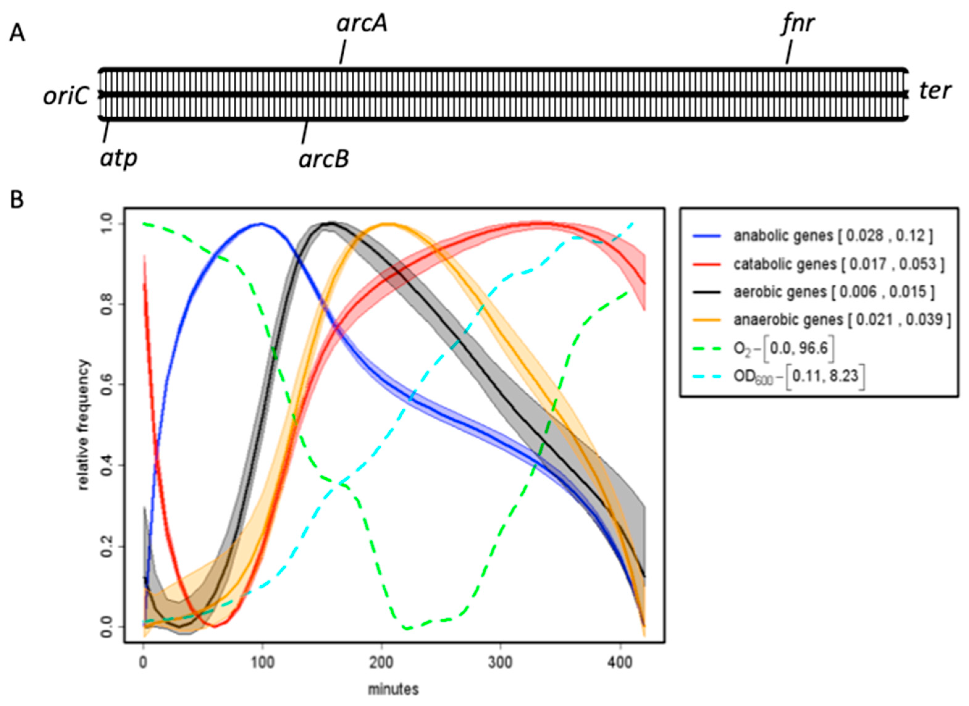
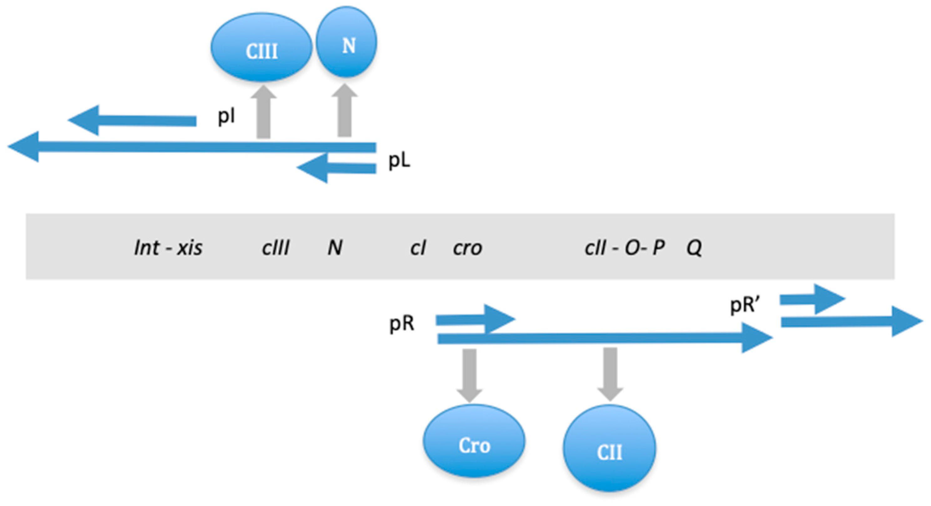
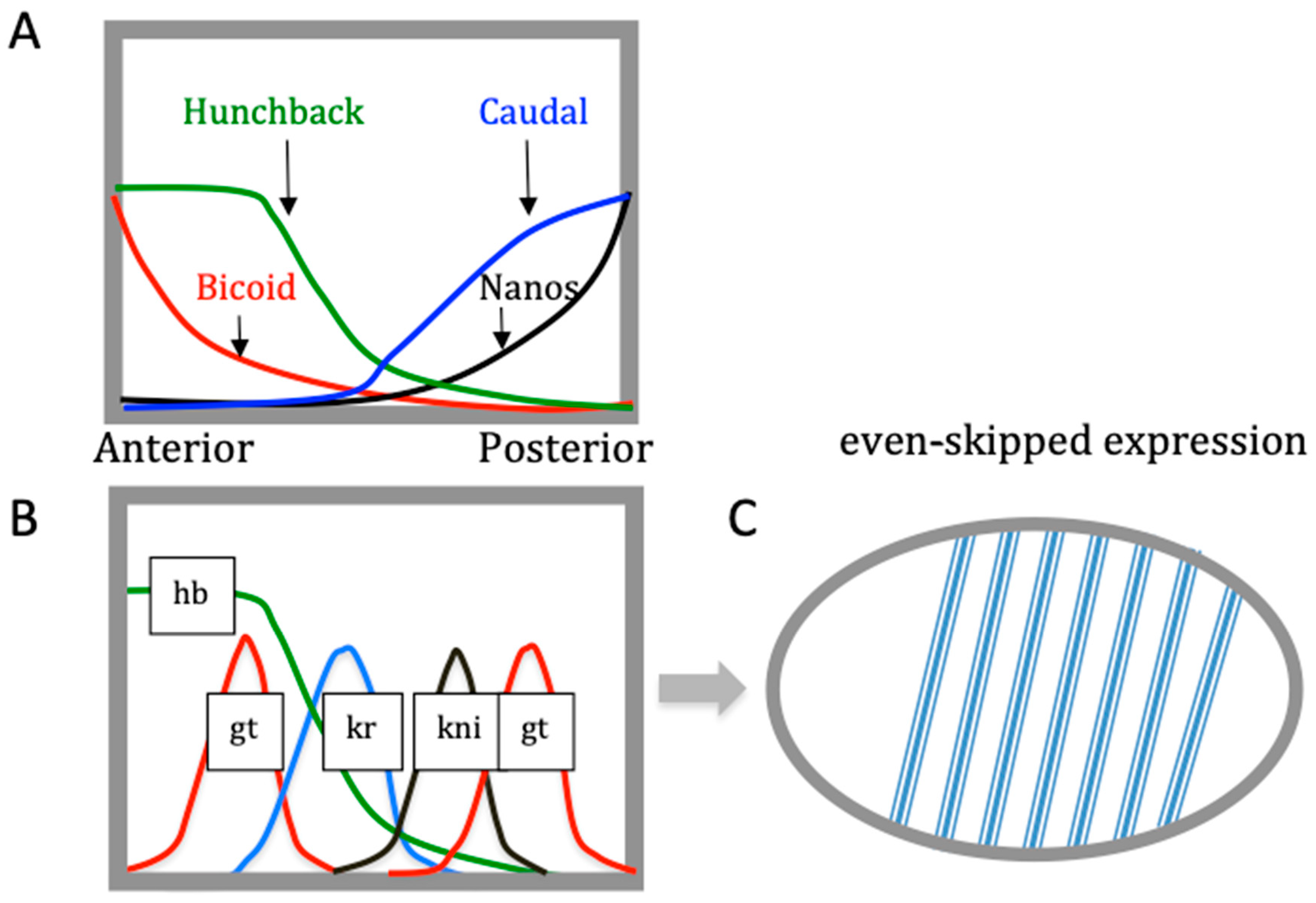
4. Switching Between Alternative Gene Expression Programs

In bacteria, global alterations in gene expression can be induced not only by alterations in DNA superhelicity but also by small intracellular effectors, such as the nucleotide guanosine tetraphosphate (ppGpp). In metazoan cells, the role of ppGpp is less clear [105], while in bacteria, it serves as an alarmone, reprograming the cell physiology by interacting directly with the transcription and translation machinery [106,107]. However, ppGpp also interferes with replication initiation by modulating the DNA topology at oriC [108]. For that matter, ppGpp, the production of which is sharply induced during a shortage of nutritional resources, appears to act as a switch curtailing cell reproduction and promoting the establishment of the maintenance program.
This ppGpp-dependent switch in gene expression occurs on exhaustion of nutritional resources at the later stage of growth. At this stage, ribosome production and gyrase activity subside, whereas the sharply increased ppGpp concentration facilitates the compositional change in the transcription machinery, e.g., the partial substitution of σ70 by stationary phase σS factor in the RNAP holoenzyme, and, for that matter, ppGpp also switches the supercoiling preferences of the polymerase. While RNA polymerase is a direct target of ppGpp, the ppGpp sensitivity of the σ70 holoenzyme in vitro can be attenuated by increased DNA superhelicity [109]. In addition, ppGpp appears to stabilize the so-called ‘tight’ conformer of the σ70 holoenzyme at the expense of the ‘ratcheted’ conformer favoring supercoiled DNA (Malcolm Buckle, G.M. and A.T., manuscript in preparation). Furthermore, the composition of the abundant NAPs changes at this stage such that overall, the intracellular milieu and the bacterial chromatin composition facilitate the transcription of more relaxed and relatively A/T-rich DNA around the terminus of replication (the Ter macrodomain), which is enriched for genes involved in maintenance functions. The ppGpp-facilitated growth phase-dependent substitution of σ70 by the stationary phase σS factor in the RNAP holoenzyme is associated with switching between the reproduction and maintenance programs and thus resembles the genetic switch between the alternative growth pathways of temperate bacterial phages such as phage λ. The λ switch is also sensitive to both the cell density and metabolic state [110,111], as well as to the supercoiling level of the DNA [112,113].
In these two systems, despite the huge difference in complexity, there is a notable organizational similarity manifest in the conversion of distinct information types occurring during the establishment of both the bacterial switch between the reproduction and maintenance programs and the λ phage switch between the lytic and lysogenic pathways. In the latter case, the system is much simpler, and ultimately, the switch boils down to competition between two DNA binding transcriptional regulators (the Cro and CI repressors) for binding specific operator sites in the λ regulatory region. However, in both systems, first, the information of a continuous (analog) type is produced and then converted into information of a discontinuous (digital) type.
In the E. coli system, as mentioned above, analog information is manifest in the oriC-ter skew of DNA binding site frequencies for DNA gyrase, σ70, and σS, interacting with the changing ratio of the RNAP σ70 and σS holoenzymes, the spatiotemporal gradient of the chromosomal superhelical density, and the temporal concentration gradients evident in various growth phase-dependent levels and combinations of NAPs (Table 2). These DNA architectural proteins form distinct spatiotemporal patterns of regulatory nucleoprotein complexes in the genome [69,114,115,116]. The NAPs compete for the stabilization of alternative supercoil structures (Figure 7) but can also cooperate depending on the DNA sequence organization [47,117]. The mutations of NAP genes alter both the gene expression patterns and DNA topology, consistent with the notion that NAPs coordinate the growth phase-dependent chromosome structure and function [11,13,56,65,118,119,120].
In the case of bacteriophage λ, the analog information is manifest as the continual bidirectional extension of transcription initiated from the divergent pR and pL promoters located in the λ control region, producing on extension distinct sets of regulatory proteins, including those involved in sensing physiological conditions (e.g., CII) and eventually, by modulating the CI/Cro repressor ratio, favoring either the lytic or lysogenic pathway (Figure 8). Continual transcription is both contingent on and also results in the formation of a spatiotemporal pattern of regulatory nucleoprotein complexes in the λ genome.
In E. coli, the switch between the reproduction and maintenance stages is primarily dictated by the energy status, which in turn depends on environmental conditions. Notwithstanding the difficulty of considering a phage an organism, it not only can reproduce itself (albeit hijacking the cellular components and machinery) but also responds to environmental conditions. For example, the λ phage may prefer lysogenic to lytic growth under conditions of starvation, perhaps since starving cells cannot provide components supporting efficient lytic growth [110]. Starving bacterial cells produce low amounts of proteases, which, at high concentrations observed in rich medium, destroy the phage CII protein required for the activation of the phage cI and int genes essential for establishing lysogeny. The CI repressor produced under conditions of high CII activity inhibits transcription from the divergent pR and pL promoters in the λ regulatory region and thus turns off the expression of all the phage genes except that of its own. However, if CII is rapidly degraded, no CI repressor is synthesized, the Cro repressor occupies the λ regulatory region instead of CI, and lytic growth ensues. So, while the phage senses the energy status of the cell, this latter is ultimately translated into specific nucleoprotein complexes competing for binding at the λ regulatory region and acting as a switch between alternative developmental pathways. Furthermore, the regulation of the lysogenic/lytic switch by CI repressor appears sensitive to DNA supercoiling [112,113], as is the RNAP σ70/σS holoenzyme switch in E. coli [36]. A similar relationship of DNA topology-dependent competitive binding at the overlapping DNA sites has been observed between the early and late NAPs, FIS and Lrp, respectively, involved in the control of the type 1 fimbrial genetic switch in E. coli [122].
Ptashne [110] suggested that the regulatory sequences initiated at the λ control region essentially generate a ‘cascade’ along each pathway, sequentially turning on and off groups of genes. In this cascade, one regulatory protein turns on or off a block of genes, which includes another regulatory gene, the product of which in turn regulates another block of genes and so on. Here, the regulatory cascade is established by protein binding only at a few DNA sites on the phage genome. This type of regulation is made possible due to the peculiar spatial organization of functionally related genes in the genome, as they are grouped together and also transcribed in the same direction. Thus, while emphasizing the role of cascades, this mode of regulation also implicates the spatial gene organization and the directional extension of transcription—properties considered here as belonging to the analog (continuous) information type—in contrast to the ‘cascade control’, which essentially turns the genes on or off and therefore provides purely digital information.
Despite the differences in complexity and details, in both the bacterial and phage systems, there is a discernible common organizational design: initial utilization of analog information (spatial oriC-ter gradients of DNA binding sites interacting with temporal gradients of regulatory proteins in the former, and gradually extending transcription starting from the divergent pR and pL promoters in the latter) and its subsequent conversion into digital information (the differing nucleoprotein complexes producing specific gene expression patterns sustaining either reproduction or maintenance in the former, and the distinct sets of regulatory proteins underpinning either the lytic or lysogenic pathway in the latter).

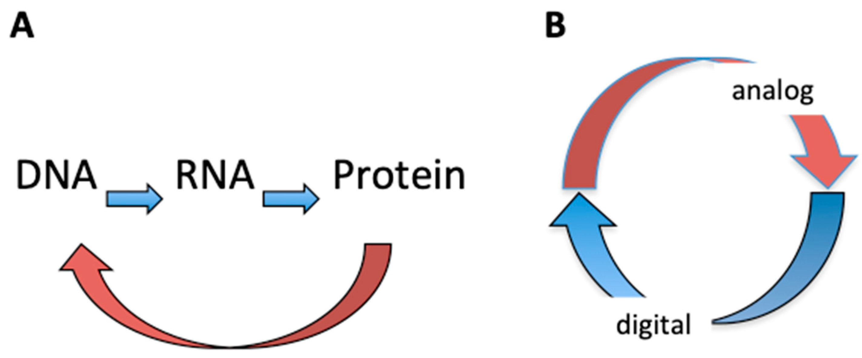
5. Analog/Digital Information Conversion Operates as a Regulatory Device in Living Systems of Diverse Structural Complexity

Given the similarity of the underlying regulatory design in the bacterial and phage systems, the pertinent question is whether this mode of information conversion occurs also in more complex multicellular organisms. Indeed, over three decades ago, Ptashne [110] drew parallels between the processes of gene regulation in phage λ and higher organisms, in particular the process of Drosophila embryogenesis, where the formation of the pattern of stripes expressing the segmentation gene even-skipped (eve) depends on the sequential turning on and off of transcriptional regulators, a form of cascade control similar to that of the λ life cycle. Actually, during Drosophila embryogenesis, notwithstanding the role of the digital on or off type ‘cascade control’, the importance of analog information in the pattern formation is most conspicuous.
During Drosophila embryonic development, the maternal gene messages are strategically deposited at opposite—anterior and posterior—poles of the embryo, such that the translated proteins diffuse from the poles forming spatial concentration gradients along the anterior–posterior axis (Figure 9). These overlapping concentration gradients lead to spatially determined, locally fixed ratios of transcriptional regulators and thus establish boundaries of target gene expression. The spatially determined threshold concentrations of transcriptional regulators lead to the sequential activation of the various segmentation genes, eventually producing a distinct pattern of seven stripes expressing the even-skipped pair-rule gene, which is essential for the segmentation of the embryo [123,124]. So, here we have a clear case of the conversion of analog information (continuous protein concentration gradients) into digital information (specific pattern of seven discrete eve stripes). This conversion of protein concentration gradients into a particular pattern of stripes is enabled by the existence of seven distinct enhancers of the eve gene (one for each stripe), each of which binds different combinations of regulatory proteins depending on their spatially determined threshold concentrations established along the anterior–posterior axis. Thus, seven distinct enhancers binding different combinations of regulatory proteins generate alternative nucleoprotein complexes independently activating eve expression—albeit in a spatially defined manner—and producing the specific pattern of seven stripes.
At this stage of development (syncytial blastoderm), the Drosophila embryo is not yet cellularized and contains about 1500 nuclei evenly distributed underneath the membrane, while each stripe extends over six nuclei on average [110]. The Drosophila genome is about 180 Mb in size, so a single row of nuclei expressing even-skipped gene would contain about 1 Gb of DNA. In contrast, the E. coli genome is 4.6 Mb in size, and that of phage λ is about 48.5 Kb. The Drosophila embryo is (longitudinally) about 500 times the size of an E. coli cell. Thus, concerning the spatial extension of implicated gradients, there is a difference in orders of magnitude. Furthermore, in the case of phage λ and E. coli, the gradients (directionally elongating transcripts in the former and the putative sigma factor and NAP gradients in the latter) extend over single genomes (albeit differing in size by two orders of magnitude), whereas in the case of Drosophila, the protein concentration gradients extend over more than a thousand spatially arranged genomes (nuclei).
Finally, in the phage and bacterial genomes, the regulatory proteins have relatively easy access to DNA binding sites, whereas the binding of cognate regulatory sites is obstructed in Drosophila nuclei by the tight packaging of the DNA in chromatin. Furthermore, in phage λ, the spatial extension of genomic transcription produces distinct sets of proteins that are put to work in temporal succession. The temporal gradients of NAPs and sigma factors do not coexist in a single bacterial cell but are successively established in the progeny. In contrast, the opposite concentration gradients of Bicoid and Nanos extending from the poles and responsible for the formation of the anterior and posterior structures coexist in a single Drosophila embryo.
Notwithstanding the abovementioned differences, there are also remarkable similarities between these systems. During Drosophila embryonic development, as well as during the E. coli growth cycle, the transcriptional and metabolic programs appear tightly correlated [56,125,126]. The regulation of the activity of DNA topoisomerases is associated with both the E. coli growth cycle and Drosophila embryogenesis [27,127]. Additionally, the Drosophila development time, the phase transition during the E. coli growth cycle, and the λ phage lytic/lysogeny decision all respond to nutritional supply [110,128,129]. However, the most important similarity between the systems is the phenomenon of the conversion of continuous data (analog information) into discrete data (digital information). First, in all three systems, there is a directional dispersion of analog information (gradients of proteins in Drosophila and E. coli and continually elongating transcripts in phage λ) from spatially localized sources (anterior and posterior poles in Drosophila, proximities of the chromosomal oriC and ter poles in E. coli, and divergent pR and pL promoters in the regulatory region of phage λ). Second, in all three systems, the conversion of analog into digital information is manifest in the formation of distinct nucleoprotein complexes involving DNA binding proteins interacting with DNA sites spatially organized in corresponding genomes or in the embryo. In the latter case, this interaction is facilitated and fine-tuned by ATP-driven chromatin remodelers [130]. Finally, and more generally, assuming that the initial gradients possess higher entropy than the ensuing discrete patterns of DNA-protein interactions, we may also assume an energy-driven decrease in entropy associated with the pattern-making in all three systems.
Thus, despite the substantial differences in size, complexity, and structural detail between these three living systems (although the phage system can barely qualify as such), in all cases, we have a spatiotemporally organized gene regulation program. The temporally organized regulatory cascades alone cannot provide for the unity of the living system—a cascade has a beginning and an end—yet it does not necessarily close onto itself, while as mentioned above, from the systems-theoretical perspective, the living system constitutes a self-referential circuit. What is assumed here is the closure of the system onto itself, and this organization of unity implicates spatial coordinates [131,132,133]. A relevant example of the integration of cell division and differentiation by coordinating the temporal gene expression and spatial organization of gene products and protein gradients has been provided in studies of the Caulobacter crescentus system [9,134,135]. All three systems discussed here have a similar organization embodied in the conversion of two distinct information types manifesting a coordinated unity (Figure 10). In this view, a phage acquires the properties of a living system primarily by tapping into its intrinsic organization, namely, appropriating the device of analog/digital information conversion.

6. Conclusions

The central dogma of molecular biology states that genetic information flows only in one direction, from DNA, to RNA, to protein, or from RNA directly to protein. This theory, highlighting the unidirectional flow of genetic information, does not consider the DNA analog information and the crosstalk between the DNA and DNA binding proteins—essentially a feedback loop (Figure 10A, brown curved arrow). It thus cannot account for the main organizational hallmark of living systems manifesting a self-referential circuit. We argue here that this latter organizational feature is inherent in the structure of the DNA double helix, representing a basic device for interconverting information. This conversion of information is made possible by the existence of two logically different—digital and analog—information types stored in the DNA. While the digital genetic information encodes all the DNA binding proteins and enzymes, the DNA appears to ‘read itself’ via DNA–protein interactions. These interactions are informative as they lead to the modulation of gene expression according to nascent external and/or internal signals. The coordinated DNA ‘self-readout’ mediated by DNA binding proteins is based on a strategic spatial organization of regulatory DNA binding sites and regulated genes in the genome (or the spatial organization of nuclei in the case of Drosophila embryos). This spatial organization in turn is determinative for the successive formation of distinct regulatory nucleoprotein complexes and the emergence of temporal regulatory ‘cascades.’ Thus, the DNA genome can generate spatiotemporally coordinated patterns of activated and repressed genes in response to both internal and external signals. We infer that the genomic DNA, acting as a double-coding device, represents the integrative interface where ‘that which monitors and that which is monitored’ meet to generate a coordinated self-referential unity (Figure 10B) responding to both the internal and external signals as an indivisible whole.
Finally, we note that the concept of information conversion emphasizes the importance of therapeutic approaches focused on the development of novel drugs targeting global regulators such as NAPs and DNA topoisomerases [136,137,138], as well as particular sequences and/or structural features of the DNA [139,140,141]. Research along these lines may eventually pave the way to the discovery of new approaches for modulating the developmental plasticity—the capacity of the genotype to produce different phenotypes—and thus restricting high phenotypic variation and the potential of the emergence of abnormal developmental trajectories [142,143]. This approach may also prove useful for the assessment of the relative contribution of genes and the environment to the development of disease [144,145] as well as for the understanding of the evolution of gene regulation [146].

Author Contributions

Conceptualization, G.M., W.N., S.R. and A.T.; methodology, G.M., W.N., S.R. and A.T.; formal analysis, G.M., W.N., S.R. and A.T.; writing—original draft preparation, G.M., W.N., S.R. and A.T.; writing—review and editing, G.M., W.N., S.R. and A.T.; visualization, G.M., W.N., S.R. and A.T.; supervision, G.M., W.N., S.R. and A.T. All authors have read and agreed to the published version of the manuscript.

Funding

This research received no external funding.

Data Availability Statement

No new data were created in this study.

Conflicts of Interest

The authors declare no conflicts of interest.

References

  1. Kaufman, L.H. Self reference and recursive forms. J. Social. Biol. Struct. 1987, 10, 53–72. [Google Scholar] [CrossRef]
  2. Razeto-Barry, P. Autopoiesis 40 years later. A review and a reformulation. Orig. Life Evol. Biosph. 2012, 42, 543–567. [Google Scholar] [CrossRef] [PubMed]
  3. Ruijtenberg, S.; van den Heuvel, S. Coordinating cell proliferation and differentiation: Antagonism between cell cycle regulators and cell type-specific gene expression. Cell Cycle 2016, 15, 196–212. [Google Scholar] [CrossRef] [PubMed] [PubMed Central]
  4. Papenfort, K.; Bassler, B.L. Quorum sensing signal-response systems in Gram-negative bacteria. Nat. Rev. Microbiol. 2016, 14, 576–588. [Google Scholar] [CrossRef] [PubMed] [PubMed Central]
  5. Antunez-Lamas, M.; Cabrera, E.; Lopez-Solanilla, E.; Solano, R.; González-Melendi, P.; Chico, J.M.; Toth, I.; Birch, P.; Pritchard, L.; Liu, H.; et al. Bacterial chemoattraction towards jasmonate plays a role in the entry of Dickeya dadantii through wounded tissues. Mol. Microbiol. 2009, 74, 662–671. [Google Scholar] [CrossRef] [PubMed]
  6. Río-Álvarez, I.; Muñoz-Gómez, C.; Navas-Vásquez, M.; Martínez-García, P.M.; Antúnez-Lamas, M.; Rodríguez-Palenzuela, P.; López-Solanilla, E. Role of Dickeya dadantii 3937 chemoreceptors in the entry to Arabidopsis leaves through wounds. Mol Plant Pathol. 2015, 16, 685–698. [Google Scholar] [CrossRef] [PubMed] [PubMed Central]
  7. Jiang, X.; Zghidi-Abouzid, O.; Oger-Desfeux, C.; Hommais, F.; Greliche, N.; Muskhelishvili, G.; Nasser, W.; Reverchon, S. Global transcriptional response of Dickeya dadantii to environmental stimuli relevant to the plant infection. Environ. Microbiol. 2016, 18, 3651–3672. [Google Scholar] [CrossRef] [PubMed]
  8. Hallgren, J.; Koonce, K.; Felletti, M.; Mortier, J.; Turco, E.; Jonas, K. Phosphate starvation decouples cell differentiation from DNA replication control in the dimorphic bacterium Caulobacter crescentus. PLoS Genet. 2023, 19, e1010882. [Google Scholar] [CrossRef] [PubMed] [PubMed Central]
  9. Barrows, J.M.; Goley, E.D. Synchronized Swarmers and Sticky Stalks: Caulobacter crescentus as a Model for Bacterial Cell Biology. J. Bacteriol. 2023, 205, e0038422. [Google Scholar] [CrossRef] [PubMed] [PubMed Central]
  10. Dudin, O.; Geiselmann, J.; Ogasawara, H.; Ishihama, A.; Lacour, S. Repression of flagellar genes in exponential phase by CsgD and CpxR, two crucial modulators of Escherichia coli biofilm formation. J. Bacteriol. 2014, 196, 707–715. [Google Scholar] [CrossRef] [PubMed] [PubMed Central]
  11. Scolari, V.F.; Bassetti, B.; Sclavi, B.; Lagomarsino, M.C. Gene clusters reflecting macrodomain structure respond to nucleoid perturbations. Mol. Biosyst. 2011, 7, 878–888. [Google Scholar] [CrossRef] [PubMed]
  12. Sobetzko, P.; Glinkowska, M.; Travers, A.; Muskhelishvili, G. DNA thermodynamic stability and supercoil dynamics determine the gene expression program during the bacterial growth cycle. Mol. Biosyst. 2013, 9, 1643–1651. [Google Scholar] [CrossRef] [PubMed]
  13. Jiang, X.; Sobetzko, P.; Nasser, W.; Reverchon, S.; Muskhelishvili, G. Chromosomal “stress-response” domains govern the spatiotemporal expression of the bacterial virulence program. mBio 2015, 6, e00353-15. [Google Scholar] [CrossRef] [PubMed] [PubMed Central]
  14. SantaLucia, J., Jr. A unified view of polymer, dumbbell, and oligonucleotide DNA nearest-neighbor thermodynamics. Proc. Natl. Acad. Sci. USA 1998, 95, 1460–1465. [Google Scholar] [CrossRef] [PubMed] [PubMed Central]
  15. Packer, M.J.; Dauncey, M.P.; Hunter, C.A. Sequence-dependent DNA structure: Dinucleotide conformational maps. J. Mol. Biol. 2000, 295, 71–83. [Google Scholar] [CrossRef] [PubMed]
  16. Young, R.T.; Czapla, L.; Wefers, Z.O.; Cohen, B.M.; Olson, W.K. Revisiting DNA Sequence-Dependent Deformability in High-Resolution Structures: Effects of Flanking Base Pairs on Dinucleotide Morphology and Global Chain Configuration. Life 2022, 12, 759. [Google Scholar] [CrossRef] [PubMed] [PubMed Central]
  17. El Hassan, M.A.; Calladine, C.R. The assessment of the geometry of dinucleotide steps in double-helical DNA; a new local calculation scheme. J. Mol. Biol. 1995, 251, 648–664. [Google Scholar] [CrossRef] [PubMed]
  18. Calladine, C.R.; Drew, H.R.; Luisi, B.F.; Travers, A.A. Understanding DNA. The Molecule & How It Works, 3rd ed.; Elsevier Academic Press: Amsterdam, The Netherlands, 2004. [Google Scholar]
  19. Palecek, E. Local supercoil-stabilized DNA structures. Crit. Rev. Biochem. Mol. Biol. 1991, 26, 151–226. [Google Scholar] [CrossRef] [PubMed]
  20. Irobalieva, R.N.; Fogg, J.M.; Catanese, D.J., Jr.; Sutthibutpong, T.; Chen, M.; Barker, A.K.; Ludtke, S.J.; Harris, S.A.; Schmid, M.F.; Chiu, W.; et al. Structural diversity of supercoiled DNA. Nat. Commun. 2015, 6, 8440. [Google Scholar] [CrossRef] [PubMed] [PubMed Central]
  21. Wang, Q.; Irobalieva, R.N.; Chiu, W.; Schmid, M.F.; Fogg, J.M.; Zechiedrich, L.; Pettitt, B.M. Influence of DNA sequence on the structure of minicircles under torsional stress. Nucleic Acids Res. 2017, 45, 7633–7642. [Google Scholar] [CrossRef] [PubMed] [PubMed Central]
  22. Pyne, A.L.B.; Noy, A.; Main, K.H.S.; Velasco-Berrelleza, V.; Piperakis, M.M.; Mitchenall, L.A.; Cugliandolo, F.M.; Beton, J.G.; Stevenson, C.E.M.; Hoogenboom, B.W.; et al. Base-pair resolution analysis of the effect of supercoiling on DNA flexibility and major groove recognition by triplex-forming oligonucleotides. Nat. Commun. 2021, 12, 1053. [Google Scholar] [CrossRef] [PubMed] [PubMed Central]
  23. Muskhelishvili, G.; Travers, A. The regulatory role of DNA supercoiling in nucleoprotein complex assembly and genetic activity. Biophys. Rev. 2016, 8 (Suppl. S1), 5–22. [Google Scholar] [CrossRef] [PubMed] [PubMed Central]
  24. Hsieh, L.S.; Burger, R.M.; Drlica, K. Bacterial DNA supercoiling and [ATP]/[ADP]. Changes associated with a transition to anaerobic growth. J. Mol. Biol. 1991, 219, 443–450. [Google Scholar] [CrossRef] [PubMed]
  25. Drlica, K. Control of bacterial DNA supercoiling. Mol. Microbiol. 1992, 6, 425–433. [Google Scholar] [CrossRef] [PubMed]
  26. Van Workum, M.; van Dooren, S.J.; Oldenburg, N.; Molenaar, D.; Jensen, P.R.; Snoep, J.L.; Westerhoff, H.V. DNA supercoiling depends on the phosphorylation potential in Escherichia coli. Mol. Microbiol. 1996, 20, 351–360. [Google Scholar] [CrossRef] [PubMed]
  27. Snoep, J.L.; van der Weijden, C.C.; Andersen, H.W.; Westerhoff, H.V.; Jensen, P.R. DNA supercoiling in Escherichia coli is under tight and subtle homeostatic control, involving gene-expression and metabolic regulation of both topoisomerase I and DNA gyrase. Eur. J. Biochem. 2002, 269, 1662–1669. [Google Scholar] [CrossRef] [PubMed]
  28. Schultz, S.G.; Solomon, A.K. Cation transport in Escherichia coli. I. Intracellular Na and K concentrations and net cation movement. J. Gen. Physiol. 1961, 45, 355–369. [Google Scholar] [CrossRef] [PubMed]
  29. Hempfling, W.P.; Höfer, M.; Harris, E.J.; Pressman, B.C. Correlation between changes in metabolite concentrations and rate of ion transport following glucose addition to Escherichia coli B. Biochim. Biophys. Acta 1967, 141, 391–400. [Google Scholar] [CrossRef] [PubMed]
  30. Anderson, P.; Bauer, W. Supercoiling in closed circular DNA: Dependence upon ion type and concentration. Biochemistry 1978, 17, 594–601. [Google Scholar] [CrossRef] [PubMed]
  31. Vologodskii, A.V.; Cozzarelli, N.R. Conformational and thermodynamic properties of supercoiled DNA. Annu. Rev. Biophys. Biomol. Struct. 1994, 23, 609–643. [Google Scholar] [CrossRef] [PubMed]
  32. Rybenkov, V.V.; Vologodskii, A.V.; Cozzarelli, N.R. The effect of ionic conditions on DNA helical repeat, effective diameter and free energy of supercoiling. Nucleic Acids Res. 1997, 25, 1412–1418. [Google Scholar] [CrossRef] [PubMed] [PubMed Central]
  33. Xu, Y.C.; Bremer, H. Winding of the DNA helix by divalent metal ions. Nucleic Acids Res. 1997, 25, 4067–4071. [Google Scholar] [CrossRef] [PubMed] [PubMed Central]
  34. Kusano, S.; Ding, Q.; Fujita, N.; Ishihama, A. Promoter selectivity of Escherichia coli RNA polymerase E sigma 70 and E sigma 38 holoenzymes. Effect of DNA supercoiling. J. Biol. Chem. 1996, 271, 1998–2004. [Google Scholar] [CrossRef] [PubMed]
  35. Schneider, R.; Travers, A.; Muskhelishvili, G. FIS modulates growth phase-dependent topological transitions of DNA in Escherichia coli. Mol. Microbiol. 1997, 26, 519–530. [Google Scholar] [CrossRef] [PubMed]
  36. Bordes, P.; Conter, A.; Morales, V.; Bouvier, J.; Kolb, A.; Gutierrez, C. DNA supercoiling contributes to disconnect sigmaS accumulation from sigmaS-dependent transcription in Escherichia coli. Mol. Microbiol. 2003, 48, 561–571. [Google Scholar] [CrossRef] [PubMed]
  37. Fulcrand, G.; Dages, S.; Zhi, X.; Chapagain, P.; Gerstman, B.S.; Dunlap, D.; Leng, F. DNA supercoiling, a critical signal regulating the basal expression of the lac operon in Escherichia coli. Sci. Rep. 2016, 6, 19243. [Google Scholar] [CrossRef]
  38. Gerganova, V.; Maurer, S.; Stoliar, L.; Japaridze, A.; Dietler, G.; Nasser, W.; Kutateladze, T.; Travers, A.; Muskhelishvili, G. Upstream binding of idling RNA polymerase modulates transcription initiation from a nearby promoter. J. Biol. Chem. 2015, 290, 8095–8109. [Google Scholar] [CrossRef] [PubMed] [PubMed Central]
  39. Japaridze, A.; Muskhelishvili, G.; Benedetti, F.; Gavriilidou, A.F.; Zenobi, R.; De Los Rios, P.; Longo, G.; Dietler, G. Hyperplectonemes: A Higher Order Compact and Dynamic DNA Self-Organization. Nano Lett. 2017, 17, 1938–1948. [Google Scholar] [CrossRef] [PubMed]
  40. Guo, M.S.; Haakonsen, D.L.; Zeng, W.; Schumacher, M.A.; Laub, M.T. A Bacterial Chromosome Structuring Protein Binds Overtwisted DNA to Stimulate Type II Topoisomerases and Enable DNA Replication. Cell 2018, 175, 583–597.e23. [Google Scholar] [CrossRef] [PubMed] [PubMed Central]
  41. Tarry, M.J.; Harmel, C.; Taylor, J.A.; Marczynski, G.T.; Schmeing, T.M. Structures of GapR reveal a central channel which could accommodate B-DNA. Sci. Rep. 2019, 9, 16679. [Google Scholar] [CrossRef] [PubMed] [PubMed Central]
  42. Huang, Q.; Duan, B.; Qu, Z.; Fan, S.; Xia, B. The DNA Recognition Motif of GapR Has an Intrinsic DNA Binding Preference towards AT-rich DNA. Molecules. 2021, 26, 5776. [Google Scholar] [CrossRef] [PubMed] [PubMed Central]
  43. Xu, W.; Yan, Y.; Artsimovitch, I.; Dunlap, D.; Finzi, L. Positive supercoiling favors transcription elongation through lac repressor-mediated DNA loops. Nucleic Acids Res. 2022, 50, 2826–2835. [Google Scholar] [CrossRef] [PubMed] [PubMed Central]
  44. Visser, B.J.; Sharma, S.; Chen, P.J.; McMullin, A.B.; Bates, M.L.; Bates, D. Psoralen mapping reveals a bacterial genome supercoiling landscape dominated by transcription. Nucleic Acids Res. 2022, 50, 4436–4449. [Google Scholar] [CrossRef] [PubMed] [PubMed Central]
  45. Schneider, R.; Lurz, R.; Lüder, G.; Tolksdorf, C.; Travers, A.; Muskhelishvili, G. An architectural role of the Escherichia coli chromatin protein FIS in organising DNA. Nucleic Acids Res. 2001, 29, 5107–5114. [Google Scholar] [CrossRef] [PubMed] [PubMed Central]
  46. Guo, F.; Adhya, S. Spiral structure of Escherichia coli HUalphabeta provides foundation for DNA supercoiling. Proc. Natl. Acad. Sci. USA 2007, 104, 4309–4314. [Google Scholar] [CrossRef] [PubMed] [PubMed Central]
  47. Maurer, S.; Fritz, J.; Muskhelishvili, G. A systematic in vitro study of nucleoprotein complexes formed by bacterial nucleoid-associated proteins revealing novel types of DNA organization. J. Mol. Biol. 2009, 387, 1261–1276. [Google Scholar] [CrossRef] [PubMed]
  48. Dame, R.T.; Kalmykowa, O.J.; Grainger, D.C. Chromosomal macrodomains and associated proteins: Implications for DNA organization and replication in gram negative bacteria. PLoS Genet. 2011, 7, e1002123. [Google Scholar] [CrossRef] [PubMed] [PubMed Central]
  49. Verma, S.C.; Qian, Z.; Adhya, S.L. Architecture of the Escherichia coli nucleoid. PLoS Genet. 2019, 15, e1008456. [Google Scholar] [CrossRef] [PubMed] [PubMed Central]
  50. Liu, L.F.; Wang, J.C. Supercoiling of the DNA template during transcription. Proc. Natl. Acad. Sci. USA 1987, 84, 7024–7027. [Google Scholar] [CrossRef] [PubMed] [PubMed Central]
  51. Sobetzko, P. Transcription-coupled DNA supercoiling dictates the chromosomal arrangement of bacterial genes. Nucleic Acids Res. 2016, 44, 1514–1524. [Google Scholar] [CrossRef] [PubMed] [PubMed Central]
  52. Dages, S.; Dages, K.; Zhi, X.; Leng, F. Inhibition of the gyrA promoter by transcription-coupled DNA supercoiling in Escherichia coli. Sci. Rep. 2018, 8, 14759. [Google Scholar] [CrossRef] [PubMed] [PubMed Central]
  53. El Houdaigui, B.; Forquet, R.; Hindré, T.; Schneider, D.; Nasser, W.; Reverchon, S.; Meyer, S. Bacterial genome architecture shapes global transcriptional regulation by DNA supercoiling. Nucleic Acids Res. 2019, 47, 5648–5657. [Google Scholar] [CrossRef] [PubMed] [PubMed Central]
  54. Jeong, K.S.; Ahn, J.; Khodursky, A.B. Spatial patterns of transcriptional activity in the chromosome of Escherichia coli. Genome Biol. 2004, 5, R86. [Google Scholar] [CrossRef] [PubMed] [PubMed Central]
  55. Peter, B.J.; Arsuaga, J.; Breier, A.M.; Khodursky, A.B.; Brown, P.O.; Cozzarelli, N.R. Genomic transcriptional response to loss of chromosomal supercoiling in Escherichia coli. Genome Biol. 2004, 5, R87. [Google Scholar] [CrossRef] [PubMed] [PubMed Central]
  56. Blot, N.; Mavathur, R.; Geertz, M.; Travers, A.; Muskhelishvili, G. Homeostatic regulation of supercoiling sensitivity coordinates transcription of the bacterial genome. EMBO Rep. 2006, 7, 710–715. [Google Scholar] [CrossRef] [PubMed] [PubMed Central]
  57. Ferrándiz, M.J.; Martín-Galiano, A.J.; Arnanz, C.; Camacho-Soguero, I.; Tirado-Vélez, J.M.; de la Campa, A.G. An increase in negative supercoiling in bacteria reveals topology-reacting gene clusters and a homeostatic response mediated by the DNA topoisomerase I gene. Nucleic Acids Res. 2016, 44, 7292–7303. [Google Scholar] [CrossRef] [PubMed] [PubMed Central]
  58. Behle, A.; Dietsch, M.; Goldschmidt, L.; Murugathas, W.; Berwanger, L.C.; Burmester, J.; Yao, L.; Brandt, D.; Busche, T.; Kalinowski, J.; et al. Manipulation of topoisomerase expression inhibits cell division but not growth and reveals a distinctive promoter structure in Synechocystis. Nucleic Acids Res. 2022, 50, 12790–12808. [Google Scholar] [CrossRef] [PubMed] [PubMed Central]
  59. Pineau, M.; Martis, B.S.; Forquet, R.; Baude, J.; Villard, C.; Grand, L.; Popowycz, F.; Soulère, L.; Hommais, F.; Nasser, W.; et al. What is a supercoiling-sensitive gene? Insights from topoisomerase I inhibition in the Gram-negative bacterium Dickeya dadantii. Nucleic Acids Res. 2022, 50, 9149–9161. [Google Scholar] [CrossRef] [PubMed] [PubMed Central]
  60. Dorman, C.J.; Ni Bhriain, N.; Higgins, C.F. DNA supercoiling and environmental regulation of virulence gene expression in Shigella flexneri. Nature 1990, 344, 789–792. [Google Scholar] [CrossRef] [PubMed]
  61. Hsieh, L.S.; Rouviere-Yaniv, J.; Drlica, K. Bacterial DNA supercoiling and [ATP]/[ADP] ratio: Changes associated with salt shock. J. Bacteriol. 1991, 173, 3914–3917. [Google Scholar] [CrossRef] [PubMed] [PubMed Central]
  62. Crozat, E.; Winkworth, C.; Gaffé, J.; Hallin, P.F.; Riley, M.A.; Lenski, R.E.; Schneider, D. Parallel genetic and phenotypic evolution of DNA superhelicity in experimental populations of Escherichia coli. Mol. Biol. Evol. 2010, 27, 2113–2128. [Google Scholar] [CrossRef] [PubMed]
  63. Hindré, T.; Knibbe, C.; Beslon, G.; Schneider, D. New insights into bacterial adaptation through in vivo and in silico experimental evolution. Nat. Rev. Microbiol. 2012, 10, 352–365. [Google Scholar] [CrossRef] [PubMed]
  64. Balke, V.L.; Gralla, J.D. Changes in the linking number of supercoiled DNA accompany growth transitions in Escherichia coli. J. Bacteriol. 1987, 169, 4499–4506. [Google Scholar] [CrossRef] [PubMed] [PubMed Central]
  65. Berger, M.; Farcas, A.; Geertz, M.; Zhelyazkova, P.; Brix, K.; Travers, A.; Muskhelishvili, G. Coordination of genomic structure and transcription by the main bacterial nucleoid-associated protein HU. EMBO Rep. 2010, 11, 59–64. [Google Scholar] [CrossRef] [PubMed] [PubMed Central]
  66. Sobetzko, P.; Travers, A.; Muskhelishvili, G. Gene order and chromosome dynamics coordinate spatiotemporal gene expression during the bacterial growth cycle. Proc. Natl. Acad. Sci. USA 2012, 109, E42–E50. [Google Scholar] [CrossRef] [PubMed] [PubMed Central]
  67. Hengge-Aronis, R. Stationary phase gene regulation: What makes an Escherichia coli promoter sigmaS-selective? Curr. Opin. Microbiol. 2002, 5, 591–595. [Google Scholar] [CrossRef] [PubMed]
  68. Klauck, E.; Typas, A.; Hengge, R. The sigmaS subunit of RNA polymerase as a signal integrator and network master regulator in the general stress response in Escherichia coli. Sci. Prog. 2007, 90 Pt 2–3, 103–127. [Google Scholar] [CrossRef] [PubMed] [PubMed Central]
  69. Cameron, A.D.S.; Dillon, S.C.; Kröger, C.; Beran, L.; Dorman, C.J. Broad-scale redistribution of mRNA abundance and transcriptional machinery in response to growth rate in Salmonella enterica serovar Typhimurium. Microb. Genom. 2017, 3, e000127. [Google Scholar] [CrossRef] [PubMed] [PubMed Central]
  70. Nigatu, D.; Henkel, W.; Sobetzko, P.; Muskhelishvili, G. Relationship between digital information and thermodynamic stability in bacterial genomes. EURASIP J. Bioinform. Syst. Biol. 2016, 2016, 4. [Google Scholar] [CrossRef] [PubMed] [PubMed Central]
  71. Meyer, S.; Reverchon, S.; Nasser, W.; Muskhelishvili, G. Chromosomal organization of transcription: In a nutshell. Curr. Genet. 2018, 64, 555–565. [Google Scholar] [CrossRef] [PubMed]
  72. Muskhelishvili, G.; Sobetzko, P.; Geertz, M.; Berger, M. General organisational principles of the transcriptional regulation system: A tree or a circle? Mol. Biosyst. 2010, 6, 662–676. [Google Scholar] [CrossRef] [PubMed]
  73. Sousa, C.; de Lorenzo, V.; Cebolla, A. Modulation of gene expression through chromosomal positioning in Escherichia coli. Microbiology 1997, 143 Pt 6, 2071–2078. [Google Scholar] [CrossRef] [PubMed]
  74. Teufel, M.; Henkel, W.; Sobetzko, P. The role of replication-induced chromosomal copy numbers in spatio-temporal gene regulation and evolutionary chromosome plasticity. Front. Microbiol. 2023, 14, 1119878. [Google Scholar] [CrossRef] [PubMed] [PubMed Central]
  75. Pountain, A.W.; Jiang, P.; Yao, T.; Homaee, E.; Guan, Y.; McDonald, K.J.C.; Podkowik, M.; Shopsin, B.; Torres, V.J.; Golding, I.; et al. Transcription-replication interactions reveal bacterial genome regulation. Nature 2024, 626, 661–669. [Google Scholar] [CrossRef] [PubMed] [PubMed Central]
  76. Lang, B.; Blot, N.; Bouffartigues, E.; Buckle, M.; Geertz, M.; Gualerzi, C.O.; Mavathur, R.; Muskhelishvili, G.; Pon, C.L.; Rimsky, S.; et al. High-affinity DNA binding sites for H-NS provide a molecular basis for selective silencing within proteobacterial genomes. Nucleic Acids Res. 2007, 35, 6330–6337. [Google Scholar] [CrossRef] [PubMed] [PubMed Central]
  77. Montero Llopis, P.; Jackson, A.F.; Sliusarenko, O.; Surovtsev, I.; Heinritz, J.; Emonet, T.; Jacobs-Wagner, C. Spatial organization of the flow of genetic information in bacteria. Nature 2010, 466, 77–81. [Google Scholar] [CrossRef] [PubMed] [PubMed Central]
  78. Kuhlman, T.E.; Cox, E.C. Gene location and DNA density determine transcription factor distributions in Escherichia coli. Mol. Syst. Biol. 2012, 8, 610. [Google Scholar] [CrossRef] [PubMed] [PubMed Central]
  79. Antipov, S.S.; Tutukina, M.N.; Preobrazhenskaya, E.V.; Kondrashov, F.A.; Patrushev, M.V.; Toshchakov, S.V.; Dominova, I.; Shvyreva, U.S.; Vrublevskaya, V.V.; Morenkov, O.S.; et al. The nucleoid protein Dps binds genomic DNA of Escherichia coli in a non-random manner. PLoS ONE 2017, 12, e0182800. [Google Scholar] [CrossRef] [PubMed] [PubMed Central]
  80. Reverchon, S.; Meyer, S.; Forquet, R.; Hommais, F.; Muskhelishvili, G.; Nasser, W. The nucleoid-associated protein IHF acts as a ‘transcriptional domainin’ protein coordinating the bacterial virulence traits with global transcription. Nucleic Acids Res. 2021, 49, 776–790. [Google Scholar] [CrossRef] [PubMed] [PubMed Central]
  81. Hardy, C.D.; Cozzarelli, N.R. A genetic selection for supercoiling mutants of Escherichia coli reveals proteins implicated in chromosome structure. Mol. Microbiol. 2005, 57, 1636–1652. [Google Scholar] [CrossRef] [PubMed]
  82. Martín-Galiano, A.J.; Ferrándiz, M.J.; de la Campa, A.G. Bridging Chromosomal Architecture and Pathophysiology of Streptococcus pneumoniae. Genome Biol. Evol. 2017, 9, 350–361. [Google Scholar] [CrossRef] [PubMed] [PubMed Central]
  83. Yan, Y.; Xu, W.; Kumar, S.; Zhang, A.; Leng, F.; Dunlap, D.; Finzi, L. Negative DNA supercoiling makes protein-mediated looping deterministic and ergodic within the bacterial doubling time. Nucleic Acids Res. 2021, 49, 11550–11559. [Google Scholar] [CrossRef] [PubMed] [PubMed Central]
  84. Cortassa, S.; Aon, M.A. Altered topoisomerase activities may be involved in the regulation of DNA supercoiling in aerobic-anaerobic transitions in Escherichia coli. Mol. Cell Biochem. 1993, 126, 115–124. [Google Scholar] [CrossRef] [PubMed]
  85. Jaén, K.E.; Sigala, J.C.; Olivares-Hernández, R.; Niehaus, K.; Lara, A.R. Heterogeneous oxygen availability affects the titer and topology but not the fidelity of plasmid DNA produced by Escherichia coli. BMC Biotechnol. 2017, 17, 60. [Google Scholar] [CrossRef] [PubMed] [PubMed Central]
  86. Alexeeva, S.; Hellingwerf, K.J.; Teixeira de Mattos, M.J. Requirement of ArcA for redox regulation in Escherichia coli under microaerobic but not anaerobic or aerobic conditions. J. Bacteriol. 2003, 185, 204–209. [Google Scholar] [CrossRef]
  87. Levanon, S.S.; San, K.Y.; Bennett, G.N. Effect of oxygen on the Escherichia coli ArcA and FNR regulation systems and metabolic responses. Biotechnol. Bioeng. 2005, 89, 556–564. [Google Scholar] [CrossRef]
  88. Mika, F.; Hengge, R. A two-component phosphotransfer network involving ArcB, ArcA, and RssB coordinates synthesis and proteolysis of sigmaS (RpoS) in E. coli. Genes Dev. 2005, 19, 2770–2781. [Google Scholar] [CrossRef]
  89. Muskhelishvili, G.; Sobetzko, P.; Mehandziska, S.; Travers, A. Composition of Transcription Machinery and Its Crosstalk with Nucleoid-Associated Proteins and Global Transcription Factors. Biomolecules 2021, 11, 924. [Google Scholar] [CrossRef] [PubMed] [PubMed Central]
  90. Borowiec, J.A.; Gralla, J.D. All three elements of the lac ps promoter mediate its transcriptional response to DNA supercoiling. J. Mol. Biol. 1987, 195, 89–97. [Google Scholar] [CrossRef] [PubMed]
  91. Auner, H.; Buckle, M.; Deufel, A.; Kutateladze, T.; Lazarus, L.; Mavathur, R.; Muskhelishvili, G.; Pemberton, I.; Schneider, R.; Travers, A. Mechanism of transcriptional activation by FIS: Role of core promoter structure and DNA topology. J. Mol. Biol. 2003, 331, 331–344. [Google Scholar] [CrossRef] [PubMed]
  92. Travers, A.; Muskhelishvili, G. DNA supercoiling—A global transcriptional regulator for enterobacterial growth? Nat. Rev. Microbiol. 2005, 3, 157–169. [Google Scholar] [CrossRef] [PubMed]
  93. Forquet, R.; Pineau, M.; Nasser, W.; Reverchon, S.; Meyer, S. Role of the Discriminator Sequence in the Supercoiling Sensitivity of Bacterial Promoters. mSystems 2021, 6, e0097821. [Google Scholar] [CrossRef] [PubMed] [PubMed Central]
  94. Forquet, R.; Nasser, W.; Reverchon, S.; Meyer, S. Quantitative contribution of the spacer length in the supercoiling-sensitivity of bacterial promoters. Nucleic Acids Res. 2022, 50, 7287–7297. [Google Scholar] [CrossRef] [PubMed] [PubMed Central]
  95. Klein, C.A.; Teufel, M.; Weile, C.J.; Sobetzko, P. The bacterial promoter spacer modulates promoter strength and timing by length, TG-motifs and DNA supercoiling sensitivity. Sci. Rep. 2021, 11, 24399. [Google Scholar] [CrossRef] [PubMed] [PubMed Central]
  96. Rochman, M.; Aviv, M.; Glaser, G.; Muskhelishvili, G. Promoter protection by a transcription factor acting as a local topological homeostat. EMBO Rep. 2002, 3, 355–360. [Google Scholar] [CrossRef] [PubMed] [PubMed Central]
  97. Olivares-Zavaleta, N.; Jáuregui, R.; Merino, E. Genome analysis of Escherichia coli promoter sequences evidences that DNA static curvature plays a more important role in gene transcription than has previously been anticipated. Genomics 2006, 87, 329–337. [Google Scholar] [CrossRef] [PubMed]
  98. Balas, D.; Fernández-Moreira, E.; De La Campa, A.G. Molecular characterization of the gene encoding the DNA gyrase A subunit of Streptococcus pneumoniae. J. Bacteriol. 1998, 180, 2854–2861. [Google Scholar] [CrossRef] [PubMed] [PubMed Central]
  99. Hirvonen, C.A.; Ross, W.; Wozniak, C.E.; Marasco, E.; Anthony, J.R.; Aiyar, S.E.; Newburn, V.H.; Gourse, R.L. Contributions of UP elements and the transcription factor FIS to expression from the seven rrn P1 promoters in Escherichia coli. J. Bacteriol. 2001, 183, 6305–6314. [Google Scholar] [CrossRef] [PubMed] [PubMed Central]
  100. Vijayan, V.; Zuzow, R.; O’Shea, E.K. Oscillations in supercoiling drive circadian gene expression in cyanobacteria. Proc. Natl. Acad. Sci. USA 2009, 106, 22564–22568. [Google Scholar] [CrossRef] [PubMed] [PubMed Central]
  101. Muskhelishvili, G.; Forquet, R.; Reverchon, S.; Meyer, S.; Nasser, W. Coherent Domains of Transcription Coordinate Gene Expression During Bacterial Growth and Adaptation. Microorganisms 2019, 7, 694. [Google Scholar] [CrossRef] [PubMed] [PubMed Central]
  102. Le, T.B.; Imakaev, M.V.; Mirny, L.A.; Laub, M.T. High-resolution mapping of the spatial organization of a bacterial chromosome. Science 2013, 342, 731–734. [Google Scholar] [CrossRef] [PubMed] [PubMed Central]
  103. Le, T.B.; Laub, M.T. Transcription rate and transcript length drive formation of chromosomal interaction domain boundaries. EMBO J. 2016, 35, 1582–1595. [Google Scholar] [CrossRef] [PubMed] [PubMed Central]
  104. Booker, B.M.; Deng, S.; Higgins, N.P. DNA topology of highly transcribed operons in Salmonella enterica serovar Typhimurium. Mol. Microbiol. 2010, 78, 1348–1364. [Google Scholar] [CrossRef] [PubMed]
  105. Ito, D.; Kawamura, H.; Oikawa, A.; Ihara, Y.; Shibata, T.; Nakamura, N.; Asano, T.; Kawabata, S.I.; Suzuki, T.; Masuda, S. ppGpp functions as an alarmone in metazoa. Commun. Biol. 2020, 3, 671. [Google Scholar] [CrossRef] [PubMed] [PubMed Central]
  106. Potrykus, K.; Cashel, M. (p)ppGpp: Still magical? Annu. Rev. Microbiol. 2008, 62, 35–51. [Google Scholar] [CrossRef] [PubMed]
  107. Gonzalez, D.; Collier, J. Effects of (p)ppGpp on the progression of the cell cycle of Caulobacter crescentus. J. Bacteriol. 2014, 196, 2514–2525. [Google Scholar] [CrossRef] [PubMed] [PubMed Central]
  108. Kraemer, J.A.; Sanderlin, A.G.; Laub, M.T. The Stringent Response Inhibits DNA Replication Initiation in E. coli by Modulating Supercoiling of oriC. mBio 2019, 10, e01330-19. [Google Scholar] [CrossRef] [PubMed]
  109. Cashel, M. Inhibition of RNA polymerase by ppGpp, a nucleotide accumulated during the stringent response to aminoacid starvation in E. coli. Cold Spring Harb. Symp. Quant. Biol. 1970, 35, 407–413. [Google Scholar] [CrossRef]
  110. Ptashne, M. A Genetic Switch, 2nd ed.; Blackwell Scientific Publications & Cell Press: Hoboken, NY, USA, 1992. [Google Scholar]
  111. Laganenka, L.; Sander, T.; Lagonenko, A.; Chen, Y.; Link, H.; Sourjik, V. 2019. Quorum Sensing and Metabolic State of the Host Control Lysogeny-Lysis Switch of Bacteriophage T1. MBio 2019, 10, e01884-19. [Google Scholar] [CrossRef]
  112. Norregaard, K.; Andersson, M.; Sneppen, K.; Nielsen, P.E.; Brown, S.; Oddershede, L.B. Effect of supercoiling on the λ switch. Bacteriophage 2014, 4, e27517. [Google Scholar] [CrossRef] [PubMed] [PubMed Central]
  113. Ding, Y.; Manzo, C.; Fulcrand, G.; Leng, F.; Dunlap, D.; Finzi, L. DNA supercoiling: A regulatory signal for the λ repressor. Proc. Natl. Acad. Sci. USA 2014, 111, 15402–15407. [Google Scholar] [CrossRef] [PubMed] [PubMed Central]
  114. Kahramanoglou, C.; Prieto, A.I.; Khedkar, S.; Haase, B.; Gupta, A.; Benes, V.; Fraser, G.M.; Luscombe, N.M.; Seshasayee, A.S. Genomics of DNA cytosine methylation in Escherichia coli reveals its role in stationary phase transcription. Nat. Commun. 2012, 3, 886. [Google Scholar] [CrossRef] [PubMed]
  115. Kahramanoglou, C.; Seshasayee, A.S.; Prieto, A.I.; Ibberson, D.; Schmidt, S.; Zimmermann, J.; Benes, V.; Fraser, G.M.; Luscombe, N.M. Direct and indirect effects of H-NS and Fison global gene expression control in Escherichia coli. Nucleic Acids Res. 2011, 39, 2073–2091. [Google Scholar] [CrossRef] [PubMed] [PubMed Central]
  116. Prieto, A.I.; Kahramanoglou, C.; Ali, R.M.; Fraser, G.M.; Seshasayee, A.S.; Luscombe, N.M. Genomic analysis of DNA binding and gene regulation by homologous nucleoid-associated proteins IHF and HU in Escherichia coli K12. Nucleic Acids Res. 2012, 40, 3524–3537. [Google Scholar] [CrossRef] [PubMed] [PubMed Central]
  117. Japaridze, A.; Yang, W.; Dekker, C.; Nasser, W.; Muskhelishvili, G. DNA sequence-directed cooperation between nucleoid-associated proteins. iScience 2021, 24, 102408. [Google Scholar] [CrossRef] [PubMed] [PubMed Central]
  118. Hommais, F.; Krin, E.; Laurent-Winter, C.; Soutourina, O.; Malpertuy, A.; Le Caer, J.P.; Danchin, A.; Bertin, P. Large-scale monitoring of pleiotropic regulation of gene expression by the prokaryotic nucleoid-associated protein, H-NS. Mol. Microbiol. 2001, 40, 20–36. [Google Scholar] [CrossRef] [PubMed]
  119. Berger, M.; Gerganova, V.; Berger, P.; Rapiteanu, R.; Lisicovas, V.; Dobrindt, U. Genes on a Wire: The Nucleoid-Associated Protein HU Insulates Transcription Units in Escherichia coli. Sci. Rep. 2016, 6, 31512. [Google Scholar] [CrossRef] [PubMed] [PubMed Central]
  120. Rashid, F.M.; Dame, R.T. 2024: A “nucleoid space” odyssey featuring H-NS. Bioessays 2024, 26, e2400098. [Google Scholar] [CrossRef] [PubMed]
  121. Ali Azam, T.; Iwata, A.; Nishimura, A.; Ueda, S.; Ishihama, A. Growth phase-dependent variation in protein composition of the Escherichia coli nucleoid. J. Bacteriol. 1999, 181, 6361–6370. [Google Scholar] [CrossRef] [PubMed] [PubMed Central]
  122. Conway, C.; Beckett, M.C.; Dorman, C.J. The DNA relaxation-dependent OFF-to-ON biasing of the type 1 fimbrial genetic switch requires the Fis nucleoid-associated protein. Microbiology 2023, 169, 001283. [Google Scholar] [CrossRef] [PubMed] [PubMed Central]
  123. Fujioka, M.; Jaynes, J.B.; Goto, T. Early even-skipped stripes act as morphogenetic gradients at the single cell level to establish engrailed expression. Development 1995, 121, 4371–4382. [Google Scholar] [CrossRef] [PubMed] [PubMed Central]
  124. Nakamura, Y.; Tomonari, S.; Kawamoto, K.; Yamashita, T.; Watanabe, T.; Ishimaru, Y.; Noji, S.; Mito, T. Evolutionarily conserved function of the even-skipped ortholog in insects revealed by gene knock-out analyses in Gryllus bimaculatus. Dev. Biol. 2022, 485, 1–8. [Google Scholar] [CrossRef] [PubMed]
  125. Sonnenschein, N.; Geertz, M.; Muskhelishvili, G.; Hütt, M.T. Analog regulation of metabolic demand. BMC Syst. Biol. 2011, 5, 40. [Google Scholar] [CrossRef] [PubMed] [PubMed Central]
  126. Pérez-Mojica, J.E.; Enders, L.; Walsh, J.; Lau, K.H.; Lempradl, A. Continuous transcriptome analysis reveals novel patterns of early gene expression in Drosophila embryos. Cell Genom. 2023, 3, 100265. [Google Scholar] [CrossRef] [PubMed] [PubMed Central]
  127. Gemkow, M.J.; Dichter, J.; Arndt-Jovin, D.J. Developmental regulation of DNA-topoisomerases during Drosophila embryogenesis. Exp. Cell Res. 2001, 262, 114–121. [Google Scholar] [CrossRef] [PubMed]
  128. Marr, A.G. Growth rate of Escherichia coli. Microbiol. Rev. 1991, 55, 316–333. [Google Scholar] [CrossRef] [PubMed] [PubMed Central]
  129. Koyama, T.; Mirth, C.K. Unravelling the diversity of mechanisms through which nutrition regulates body size in insects. Curr. Opin. Insect Sci. 2018, 25, 1–8. [Google Scholar] [CrossRef] [PubMed]
  130. Moshkin, Y.M.; Chalkley, G.E.; Kan, T.W.; Reddy, B.A.; Ozgur, Z.; van Ijcken, W.F.; Dekkers, D.H.; Demmers, J.A.; Travers, A.A.; Verrijzer, C.P. Remodelers organize cellular chromatin by counteracting intrinsic histone-DNA sequence preferences in a class-specific manner. Mol. Cell Biol. 2012, 32, 675–688. [Google Scholar] [CrossRef] [PubMed] [PubMed Central]
  131. Kosmidis, K.; Hütt, M.T. The E. coli transcriptional regulatory network and its spatial embedding. Eur. Phys. J. E Soft Matter. 2019, 42, 30. [Google Scholar] [CrossRef] [PubMed]
  132. Kosmidis, K.; Jablonski, K.P.; Muskhelishvili, G.; Hütt, M.T. Chromosomal origin of replication coordinates logically distinct types of bacterial genetic regulation. NPJ Syst. Biol. Appl. 2020, 6, 5. [Google Scholar] [CrossRef] [PubMed] [PubMed Central]
  133. Ricci, D.P.; Melfi, M.D.; Lasker, K.; Dill, D.L.; McAdams, H.H.; Shapiro, L. Cell cycle progression in Caulobacter requires a nucleoid-associated protein with high AT sequence recognition. Proc. Natl. Acad. Sci. USA 2016, 113, E5952–E5961. [Google Scholar] [CrossRef] [PubMed] [PubMed Central]
  134. Biondi, E.G.; Reisinger, S.J.; Skerker, J.M.; Arif, M.; Perchuk, B.S.; Ryan, K.R.; Laub, M.T. Regulation of the bacterial cell cycle by an integrated genetic circuit. Nature 2006, 444, 899–904. [Google Scholar] [CrossRef] [PubMed]
  135. Laub, M.T.; Shapiro, L.; McAdams, H.H. Systems biology of Caulobacter. Annu. Rev. Genet. 2007, 41, 429–441. [Google Scholar] [CrossRef] [PubMed]
  136. Bhowmick, T.; Ghosh, S.; Dixit, K.; Ganesan, V.; Ramagopal, U.A.; Dey, D.; Sarma, S.P.; Ramakumar, S.; Nagaraja, V. Targeting Mycobacterium tuberculosis nucleoid-associated protein HU with structure-based inhibitors. Nat. Commun. 2014, 5, 4124. [Google Scholar] [CrossRef] [PubMed]
  137. Suarez, M.A.; Valencia, J.; Cadena, C.C.; Maiti, R.; Datta, C.; Puerto, G.; Isaza, J.H.; San Juan, H.; Nagaraja, V.; Guzman, J.D. Diarylethenes Display In Vitro Anti-TB Activity and Are Efficient Hits Targeting the Mycobacterium tuberculosis HU Protein. Molecules 2017, 22, 1245. [Google Scholar] [CrossRef] [PubMed] [PubMed Central]
  138. Sitarek, P.; Merecz-Sadowska, A.; Sikora, J.; Dudzic, M.; Wiertek-Płoszaj, N.; Picot, L.; Śliwiński, T.; Kowalczyk, T. Flavonoids and their derivatives as DNA topoisomerase inhibitors with anti-cancer activity in various cell models: Exploring a novel mode of action. Pharmacol. Res. 2024, 209, 107457. [Google Scholar] [CrossRef] [PubMed]
  139. Paul, A.; Nanjunda, R.; Wilson, W.D. Binding to the DNA Minor Groove by Heterocyclic Dications: From AT Specific to GC Recognition Compounds. Curr. Protoc. 2023, 3, e729. [Google Scholar] [CrossRef] [PubMed]
  140. Guo, P.; Farahat, A.A.; Paul, A.; Kumar, A.; Boykin, D.W.; Wilson, W.D. Extending the σ-Hole Motif for Sequence-Specific Recognition of the DNA Minor Groove. Biochemistry 2020, 59, 1756–1768. [Google Scholar] [CrossRef] [PubMed] [PubMed Central]
  141. Paul, A.; Guo, P.; Boykin, D.W.; Wilson, W.D. A New Generation of Minor-Groove-Binding-Heterocyclic Diamidines That Recognize G·C Base Pairs in an AT Sequence Context. Molecules 2019, 24, 946. [Google Scholar] [CrossRef] [PubMed] [PubMed Central]
  142. Metcalfe, N.B. How important is hidden phenotypic plasticity arising from alternative but converging developmental trajectories, and what limits it? J. Exp. Biol. 2024, 227 (Suppl. S1), jeb246010. [Google Scholar] [CrossRef] [PubMed] [PubMed Central]
  143. Moreno-Gámez, S.; Kiviet, D.J.; Vulin, C.; Schlegel, S.; Schlegel, K.; van Doorn, G.S.; Ackermann, M. Wide lag time distributions break a trade-off between reproduction and survival in bacteria. Proc. Natl. Acad. Sci. USA 2020, 117, 18729–18736. [Google Scholar] [CrossRef] [PubMed] [PubMed Central]
  144. Hales, C.N.; Barker, D.J. The thrifty phenotype hypothesis. Br. Med. Bull. 2001, 60, 5–20. [Google Scholar] [CrossRef] [PubMed]
  145. Neel, J.V. Diabetes mellitus: A “thrifty” genotype rendered detrimental by “progress”? 1962. Bull World Health Organ. 1999, 77, 694–703; discussion 692–693. [Google Scholar] [PubMed] [PubMed Central]
  146. Westman, C.A.; Goldbach, L.; Wagner, A. The adaptive Landscapes of Three Global Escherichia coli Transcriptional Regulators. bioRxiv 2024. bioRxiv:11.623025. [Google Scholar] [CrossRef]
Figure 1. The self-referential organization of living system is represented by an arrow, which closes on itself (left panel). The self-pointing arrow is a symbol for the condition in which the system divides itself into that which monitors and that which is monitored [1]. The system thus constitutes an isolated, operationally closed circuit. The environment (the space outside of the circle circumference) is marked as ‘E’ (right panel). The environmental impact on the system is indicated by the black arrow crossing the circle circumference from outside to inside. Unless this environmental impact is deteriorating, the operational closure of the system is retained.
Dna 04 00032 g001
Figure 2. Relation between the digital genetic code and the analog information stored in overlapping base steps of the DNA. Two deliberately chosen triplets coding, e.g., for lysine and serine are indicated as ‘digital code’. Indicated below as analog ‘code’ are the five consecutive overlapping base steps harbored in these two codons. The free stacking/melting energies of the base steps [14] are indicated underneath (see also Table 1).
Dna 04 00032 g002
Figure 3. Pyrimidine–purine (YR) base steps are flexible and can adopt various conformations. Purine and pyrimidine bases are indicated by large and small rectangles, respectively. The asterisk in the upper panel indicates the potential steric clash between large purine bases, which is avoided by the positive slide of the base pairs. In the lower panel, this base step has a negative slide and positive roll, and this alternative configuration is stabilized by increased cross-chain stacking interactions between the large purine bases (vertically striated region between the large rectangles). The minor groove side of the bases is shaded (after [18]).
Dna 04 00032 g003
Figure 4. Organization of the gene expression program during the bacterial (E. coli) growth cycle. The circular bacterial chromosome is indicated on the top in folded form, with two arms aligned along the oriC-ter axis. Indicated below, all arranged along the oriC-ter axis are (A) the spatial gradient of the DNA average negative stacking/melting free energy (approx. the G/C content); (B) the frequency distribution of gyrase binding sites; (C) the frequency distribution of the σ70 and σS binding sites; (D) the spatial organization of anabolic and catabolic genes; (E) the spatiotemporal gradient of negative superhelical density (−σ), which changes both temporally with growth phase (from ~−0.068 on shift-up to ~−0.043 in stationary phase [56]) as well as forms a spatial gradient along the oriC-ter axis of the chromosome [44,66]. Thus, (AD) show the distribution of variables in space, whereas (E) indicates the distribution of superhelical density both in space and in time. This spatiotemporal gradient is proposed to coordinate the expression of the anabolic and catabolic genes during the bacterial growth cycle [66,71].
Dna 04 00032 g004
Figure 5. Switch between the reproduction and maintenance programs in E. coli. (A) Growth phase-dependent expression of the NAP and sigma factor genes The different expression curves were normalized to [0;1] to compare them in one plot. Minimum and maximum values are indicated in brackets in the legend. Abscissa—time in minutes after inoculation of cells in fresh growth medium. The Escherichia coli CSH50 overnight (16 h) cultures were inoculated at an initial OD600 of 0.1 in rich double yeast-tryptone (dYT) medium and grown in a fermenter under constant pH 7.4 and high aeration (5 L air per min) at 37 °C for 7 h (420 min). Samples for RNA-seq were taken at 1, 2, 3, 5, and 7 h after inoculation. (Graph, courtesy of Patrick Sobetzko). (B) The circular chromosomes are depicted with the Ori and Ter poles indicated. The early regulatory genes are indicated in blue (left panel), and the late regulatory genes are indicated in red (right panel). The original position of these genes on the circular chromosome is approximated. Note that the reproduction and maintenance regulators are located around the opposite poles of the chromosome. Colored areas indicate the putative spatiotemporal concentration gradients of regulators. Connecting lines indicate the crosstalk between regulatory genes [72]. The ‘alarmone’ ppGpp produced during a shortage of nutritional resources acts as a switch from the reproduction to maintenance program. Conversely, a high ATP/ADP ratio (established, e.g., on nutritional shift-up) favors the commencement of reproduction.
Dna 04 00032 g005
Figure 6. Chromosomal order of regulators and temporal pattern of regulated gene expression. (A) Spatial ordering of aerobic/anaerobic growth regulatory genes on the E. coli chromosome along the oriC–ter axis. Genes on the clockwise (right) replichore are indicated on the upper bar, and genes on anti-clockwise (left) replichore are indicated on the lower bar. The atp operon encodes ATP synthase. arcA/arcB encode a two-component system active under microaerobic conditions [86,87]. ArcA also represses rpoS encoding the stationary phase sigma factor [88]. fnr has a dominant role under more strictly anaerobic conditions [86]. (B) Temporal dynamics of expression of various gene classes. The Escherichia coli CSH50 overnight (16 h) cultures were inoculated at an initial OD600 of 0.1 in rich double yeast-tryptone (dYT) medium and grown in a fermenter under constant pH 7.4 and high aeration (5 L air per min) at 37 °C for 7 h. Samples for RNA-seq were taken at 1, 2, 3, 5, and 7 h after inoculation. The different curves were normalized to [0;1] to compare them in one plot. The envelopes of the curves indicate the standard deviation at 10% random remapping of the expression patterns to genes. Minimum and maximum values are indicated in brackets in the legend. Expression values (anabolic, catabolic, aerobic, anaerobic) in brackets are normalized to the expression of all genes. The optical density and partial oxygen pressure are indicated, respectively, by the dashed light blue and green lines. Note the correlation between the maximal expression of anaerobic genes and minimal partial oxygen pressure. (Graph, courtesy of Patrick Sobetzko).
Dna 04 00032 g006
Figure 7. Competition between the bacterial NAPs, HU, and H-NS, which stabilize alternative supercoil structures on binding DNA. Distinct supercoil structures stabilized by HU and H-NS are indicated by white arrows. HU stabilizes more open toroidal coils, whereas H-NS stabilizes tightly interwound, stiff plectonemic DNA structures (AFM image, courtesy of Sebastian Maurer).
Dna 04 00032 g007
Figure 8. Regulation of the lysis–lysogeny decision in λ phage development. Gene and early transcription map of λ is shown (simplified). Genes are indicated in the shaded rectangle. The early transcripts produced from the pL and pR promoters are shown as blue arrows. The immediate early gene (short arrows) products are N and Cro. On extension of the transcripts known as ‘delayed early transcription’ (long arrows underneath), the CIII and CII proteins are produced. The N and Cro proteins support lytic development, whereas the CIII and CII proteins support lysogeny. Note that the synthesis of transcripts of different lengths (i.e., the generation of analog information) results in the production of distinct sets of specific proteins (digital information). O and P are DNA replication genes involved in lytic growth. Q protein turns on the late genes for the production of phage tails and heads. cI, cII, cIII, and int genes are involved in the establishment of lysogeny. The xis gene (together with int) is involved in the excision of the integrated prophage. Cro and CI are repressor proteins competing for binding at the operator sites in the λ regulatory region. Critical for the active production of CI repressor is the CII protein, the stability of which in turn is sensitive to physiological conditions.
Dna 04 00032 g008
Figure 9. Generation of the anterior–posterior pattern of even-skipped (eve pair-rule gene) expression initiated by gradients of the Drosophila maternal effect genes. (A) The Bicoid protein gradient extends from anterior to posterior, while the Nanos protein gradient extends from posterior to anterior. Nanos inhibits the translation of the hunchback message (in the posterior), while Bicoid prevents the translation of the caudal message (in the anterior). This inhibition results in opposing Caudal and Hunchback gradients. (B) Spatial distribution of Bicoid-responsive segmentation (gap) gene expression such as giant (gt), krueppel (kr), and knirps (kni). The gap gene products mutually repress each other’s expression, resulting in spatially defined domains of gap gene expression along the anterior–posterior axis of the embryo, which ultimately define the pattern of even-skipped (a pair-rule gene) expression in seven stripes (C). Each stripe has an independent transcriptional control system consisting of different constellations of transcription factors (specific combinations of activators) interacting with seven different enhancer sequences, all of which can activate even-skipped gene expression.
Dna 04 00032 g009
Figure 10. Organization of information flow and inter-conversion in the living system. (A) Depicted is the central dogma of molecular biology: digital genetic information flows only in one direction, from DNA, to RNA, to protein (blue arrows). However, the DNA analog information is recognized directly by the DNA binding proteins stabilizing various 3D conformations of the DNA (curved brown arrow) and thus modulating the content of digital information. (B) Systems-theoretical model for the inter-conversion of DNA information. The terms ‘analog’ and ‘digital’ refer to two distinct information types stored in the genomic DNA polymer. DNA analog information implies both static (sequence organization viz. arrangement of base steps) and dynamic parameters (DNA 3D configuration and superhelicity). Digital information implies the differential gene expression patterns and the gene interaction networks (including regulatory cascades) emerging thereof. Analog information provides an integrative sensory interface for both internal and external signals as well as a regulatory context for digital code expression, whereas the latter provides information for reproduction and the maintenance of the former. Together, the inter-converting DNA ‘codes’ form a coordinated self-referential circuit responding to both the internal and external signals as an indivisible unity.
Dna 04 00032 g010
Table 1. Free stacking/melting energy of the ten DNA base steps [14].
Base Stepskcal/mol
AA/TT−1.02
AT/AT −0.73
TA/TA −0.60
CA/TG −1.38
GT/AC −1.43
CT/AG−1.16
GA/CT−1.46
CG/CG −2.09
GC/GC −2.28
GG/CC−1.77
Table 2. Estimated number and concentrations of the most abundant nucleoid-associated proteins in E. coli [121]. Approximate numbers for RNA polymerase and lac repressor are shown for comparison.
ProteinExponential PhaseEarly Stationary Phase
No./CellConcn (μM)No./CellConcn (μM)
Dps80007120,000100
FIS60,00050Not detectable
H-NS20,0001715,00013
HU55,0004525,00020
IHF10,000850,00041
StpA25,0002815,00017
Totals 155 191
LacI (LacR)10
RNAP4000–6000
Disclaimer/Publisher’s Note: The statements, opinions and data contained in all publications are solely those of the individual author(s) and contributor(s) and not of MDPI and/or the editor(s). MDPI and/or the editor(s) disclaim responsibility for any injury to people or property resulting from any ideas, methods, instructions or products referred to in the content.

