


Abstract
Background/Objectives: Traumatic brain injury in elderly patients is a significant public health concern, particularly for those on antithrombotic therapy. A clearer understanding of how different antithrombotic agents affect the likelihood of intracranial hemorrhage in elderly patients with TBI is needed to guide clinical management. Therefore, the objective of this study was to assess the effect of preinjury antithrombotic agents on the incidence of intracranial hemorrhage in elderly patients with traumatic brain injury. Methods: The design was a retrospective cohort study set in a regional Australian hospital emergency department. The study evaluated elderly patients (≥65 years) with head injury cases identified from the integrated electronic medical record using SNOMED codes. Data on patient demographics, antithrombotic use, computed tomography imaging, and outcomes were collected. Results: A total of 152 elderly TBI patients were included in the study. Of these patients, 90.1% had falls leading to TBI. Among the patients, 30.3% were on antiplatelet agents, 23% were on direct oral anticoagulants, 7.2% were on vitamin K antagonists, and 39.5% were not on any antithrombotic agents. Intracranial hemorrhage was found in 26.5% of patients, with both direct oral anticoagulants (aOR 4.87, 95% CI 1.42–16.67, p < 0.01) and vitamin K antagonists (aOR 4.95, 95% CI 1.04–23.55, p < 0.04) demonstrating statistically significant associations with increased odds of ICH. Conclusions: Both vitamin K antagonists and direct oral anticoagulants were associated with a higher odds of intracranial hemorrhage in elderly patients with TBI, while antiplatelet therapy did not show this effect.
1. Introduction
Traumatic brain injury (TBI) is defined as an alteration in brain function or other evidence of brain pathology caused by an external force [1]. The severity of TBI is commonly classified into mild, moderate and severe based on the Glasgow Coma Scale (GCS) [2]. TBI in the geriatric population is an important public health issue in Australia due to the high incidence of accidental falls [3]. This poses a significant burden on the Australian Healthcare system due to the rapidly growing elderly population (aged ≥ 65 years) [4].
A major challenge in the management of TBI in the elderly is the prevalent preinjury use of antithrombotic therapy, comprising antiplatelet and anticoagulant drugs. These agents are commonly prescribed to elderly patients to prevent thromboembolic events associated with conditions such as cerebrovascular accidents (CVA), coronary artery disease (CAD), atrial fibrillation (AF) and deep vein thrombosis (DVT) [5]. Aspirin and clopidogrel are the most prescribed antiplatelet medications. Warfarin, a vitamin K antagonist (VKA), was traditionally the most widely prescribed anticoagulant [6]. However, in recent decades, direct oral anticoagulants (DOACs) such as direct thrombin inhibitors (e.g., dabigatran) and direct factor Xa inhibitors (e.g., apixaban and rivaroxaban) have become preferred due to advantages including no required laboratory monitoring with chronic use and fewer polypharmacy interactions [7]. DOACs have also been demonstrated to have better safety profiles for spontaneous bleeding compared to warfarin. The potential downside of broader use is the difficulty in reversing the anticoagulant effects of DOACs immediately in cases of trauma or emergency operation [8].
The most serious complication associated with antithrombotic use is intracranial hemorrhage (ICH) [9]. This may be spontaneous or due to trauma, with anticoagulated patients being more likely to experience spontaneous ICH compared to the general population [9]. Additionally, ICH occurs more frequently in mild TBI in older adults [10]. This may contribute to higher mortality rates seen in elderly patients with mild TBI compared to younger patients with similar injuries [11]. Therefore, close monitoring of elderly patients on antithrombotic therapy with mild TBI is advisable, as early identification and management of traumatic ICH in this population may enhance post-injury quality of life [12].
Historically, anticoagulant therapy has been regarded as more likely to increase the incidence of ICH and mortality from TBI than antiplatelet therapy [13]. For TBI patients on anticoagulants, the findings are inconsistent between VKAs and DOACs [14,15,16,17]. There is a paucity of data on the prevalence of specific antithrombotic agents among regional hospital patients who sustain a TBI, as well as their associated clinical outcomes. Therefore, this study aims to describe and compare outcomes of elderly patients taking preinjury antithrombotic agents who presented to a regional hospital emergency department (ED) with TBI.
2. Materials and Methods
2.1. Study Design and Data Source
We conducted a retrospective cohort study, analyzing data from patients presenting to the Mackay Base Hospital (MBH) ED from 1 January 2021 to 31 December 2021. MBH services the Mackay regional area of north Queensland, a Modified Monash remoteness category 2 (MM2) location [18]. The Townsville Hospital and Health Service Human Research Ethics Committee (HREC/2022/QTHS/88219) approved the study.
We used the Systematised Nomenclature of Medicine—Clinical Terms, Australian release (SNOMED CT-AU) codes relevant to head injury to identify potential TBI presentations [19]. We extracted relevant ED presentations from FirstNet (Cerner, Kansas City, MO, USA) data housed in the integrated electronic medical record (ieMR). Medical case notes from ieMR were screened for inclusion and exclusion criteria for each identified patient. Inclusion criteria were patients with a diagnosed TBI in the medical record and age ≥ 65 years. Exclusion criteria were patients whose injury did not meet the TBI definition, who did not have a documented GCS score, and whose mechanism of injury could not be confirmed.
2.2. Variables and Measurements
The primary outcome was the presence of traumatic ICH, determined through review of radiologist-reported CT brain imaging performed at the index presentation. This included acute subdural, extradural, subarachnoid, and intraparenchymal hemorrhages. Only acute traumatic ICH were classified as positive outcomes. Acute hemorrhage was defined based on radiological reporting consistent with recent bleeding. Chronic hemorrhages without features of acute bleeding were not included. Injury severity was assessed using GCS scores and patient symptoms upon presentation. Patients were categorized into a priori defined groups as moderate to severe TBI (GCS ≤ 12) or mild TBI (GCS ≥ 13) [2]. Secondary outcomes were ED length of stay (LOS) and patient disposition.
Demographic variables, including age, gender, and country of birth, were collected. Mechanisms of TBI were classified as falls, blunt injuries, sports injuries, and assaults. Patient treatment information included ED LOS, computed tomography (CT) imaging, the category of antithrombotic drugs prescribed, and patient disposition. Where CT imaging was not undertaken, the patient record was interrogated for details of the clinical rationale and the use of the clinical decision rule used in this facility, the Canadian CT head rule. Sample size calculation was not performed as this was a retrospective cohort study, time-limited to one year.
2.3. Data Analysis
We used Stata version 16.0 (StataCorp LLC., College Station, TX, USA) to analyze the data. The distribution of ICH by patient characteristics, causes of injury, TBI classification, category of drugs prescribed and length of stay in the ED are presented as counts and proportions. Wilcoxon rank-sum tests and Chi-square tests were employed to evaluate differences in continuous and categorical variables, respectively. Binary logistic regression was used to estimate crude odds ratios (cOR) and corresponding 95% confidence intervals (CIs). We performed multivariable regression analyses, adjusting for key covariates such as sex, mechanism of injury, and TBI severity. Statistical significance was determined by a two-sided p-value of less than 0.05. The STROBE guidelines for reporting observational studies were used [20].
3. Results
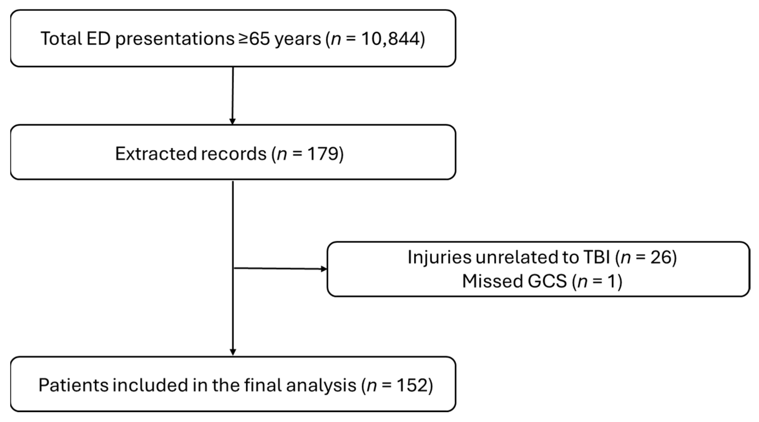
152 elderly patients (age ≥ 65 years) with TBI presenting to the ED were included (Figure 1). Table 1 presents patients’ demographics and clinical characteristics. The median age (IQR) for patients was 81 (73–86) years. The most common cause of injury for the cohort was falls (90.1%).
Figure 1.
Patient selection flow diagram.
Table 1.
Characteristics of TBI patients by incidence of haemorrhagic events.
In this study, 46 (30.3%) of the TBI patients were on antiplatelet therapy, 35 (23.0%) on DOACs, 11 (7.2%) on VKAs, and 60 (39.5%) were not on any antithrombotic agents. ICH incidence for the overall study population was 35 (23.0%). The distribution of ICH cases was 10/46 (21.7%) for patients taking antiplatelets, 10/35 (28.6%) for patients on DOACs, 5/11 (45.5%) for patients on VKAs, and 10/60 (16.7%) for patients on no antithrombotic therapeutics. No patients in our sample were recorded as being on a combination of therapies.
The median (IQR) LOS for the total study population was 5.3 (3.8–8.2) h (Table 1). The median length of stay for patients with ICH was significantly higher than for the patients without ICH (8.2 h vs. 4.5 h, p < 0.001). 69.7% of patient presentations did not meet the National Emergency Access Target (NEAT) stipulating that patients should be admitted, discharged, or transferred within 4 h of presentation [21].
Patients who had CT assessment demonstrated higher odds of a stay in the ED longer than 4 h compared to those with no CT (OR: 6.90, 95% CI: 2.52–18.87, p < 0.001). CT imaging was performed in 132/152 patients (86.8%). Imaging rates were 94.3% in patients receiving DOACs, 90.9% in those receiving VKAs, 84.8% in those on antiplatelet therapy, and 85.0% in patients not receiving antithrombotic agents. The clinical decisions documented in the medical record for not undertaking CT imaging included that the findings would not change management (as in patients with chronic neurodegenerative disease or who were not candidates for neurosurgery), or the patient was not on antithrombotic agents and was deemed a low risk of ICH.
In our unadjusted analysis, GCS classification < 12 (cOR 5.56, 95% CI: 1.56–19.76, p < 0.01) and VKA therapy (cOR 4.07, 95 CI%: 1.09–15.17, p < 0.04) were associated with ICH (Table 2). After adjusting for sex, mechanism of injury, and severity of TBI, the use of DOACs and VKAs was found to increase the odds of ICH significantly (aOR 4.87, 95% CI: 1.42–16.67, p < 0.01) and (aOR 4.95, 95% CI: 1.04–23.55, p < 0.04), respectively (Table 3).
Table 2.
Unadjusted association of patient characteristics with traumatic haemorrhagic event.
Table 3.
Adjusted association of prescribed drugs with traumatic haemorrhagic events.
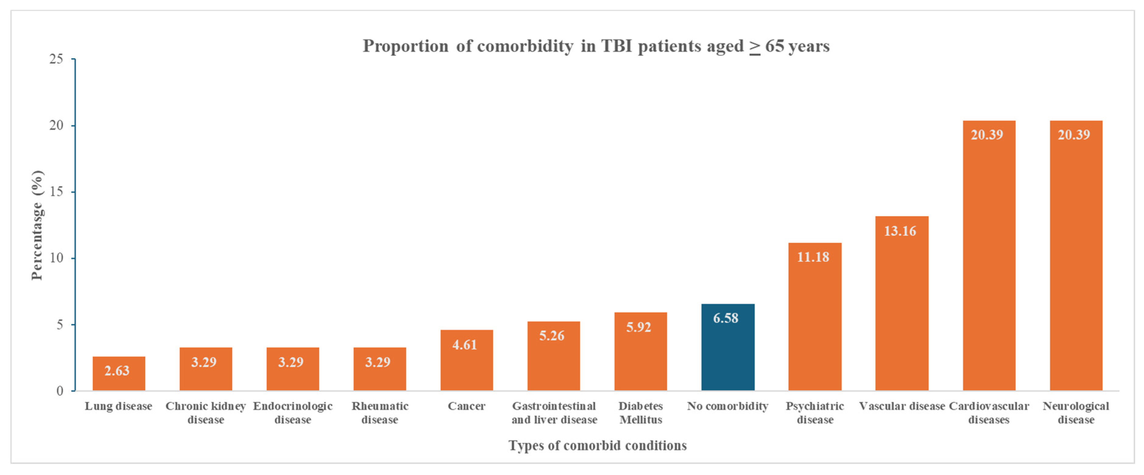
We found that only 6.6% of TBI patients were free of chronic co-morbid conditions. Cardiovascular (20.39%) and neurological diseases (20.39%) were the two most frequent co-morbid conditions in elderly TBI patients (Figure 2).
Figure 2.
Proportion of comorbidity in TBI patients 65 years and above presenting to the regional emergency department. Orange refers to comorbidities, blue refers to no comorbidities.
4. Discussion
Our study found that anticoagulants were associated with increased odds of ICH in elderly TBI patients, whereas antiplatelet therapy did not demonstrate such an effect. This conclusion aligns with findings from retrospective studies reporting anticoagulants, but not antiplatelets, were associated with increased ICH risk or injury evolution [14,22,23]. However, Vedin et al. have presented contrasting evidence from a retrospective study including 1938 TBI patients with fall from standing height, suggesting that antiplatelets rather than anticoagulants were independently linked to traumatic ICH, especially in older patients [24]. A meta-analysis by Fuller, including seven studies of patients on DOACs, reported a pooled adverse outcome risk of 4%. However, the overall quality of the body of evidence was low, resulting from imprecision, indirectness, and risk of bias [25]. These differing conclusions and the lack of high-level evidence point to the need for prospective studies exploring the effects of these drug classes.
With consideration to anticoagulants, the literature suggests that DOACs confer less risk of ICH than VKAs. A recent systematic review and meta-analysis evaluating 28 studies concluded that patients on DOACs were at lower risk of ICH after mild TBI compared to patients on VKAs [17]. Our study found increased odds of ICH from both DOACs and VKAs but was not powered to differentiate risk between these classes of agents. Individual studies with large patient cohorts also suggest that DOACs may be safer than VKAs, with lower rates of hemorrhage progression and improved functional outcomes [16,26]. These studies argue that DOACs could be a preferable option for elderly patients prone to falls, offering a reduced risk of ICH following blunt trauma without the same high rates of progression seen with VKAs. This variability could be explained by differences in study design, patient selection, or the timing of anticoagulant administration.
The association of VKAs and DOACs with ICH following TBI in this study should promote caution in the use of these drugs in elderly patients with atrial fibrillation. In managing this risk, the HAS-BLED score can be used to estimate the likelihood of major bleeding for patients on anticoagulation to assess risk-benefit in atrial fibrillation care. Patients with higher HAS-BLED scores have coexisting conditions that amplify the bleeding risk, making this tool particularly valuable for elderly individuals who are already predisposed to falls and other trauma. Further similar studies, including the HAS-BLED score as part of the initial assessment, could highlight the validity of using such scores prior to commencing DOACs or VKAs [27].
Patients on VKAs with elevated INR levels, even if their initial brain CT scan is normal, remain at considerable risk for developing delayed ICH, and guidelines recommend that patients be admitted for observation to monitor for delayed complications [28,29]. While some studies suggest that routine follow-up CT scans may not be necessary for anticoagulated patients with a negative initial scan and no signs of neurological deterioration, the overall risk of ICH following mild head injury in this patient group remains uncertain. Future prospective research that investigates drug type, injury severity, and the role of advanced diagnostic biomarkers [30] will be important in the detection of acute and delayed ICH in elderly patients using antithrombotic agents.
The NEAT stipulates that patients should be admitted, discharged, or transferred from Australian EDs within 4 h of presentation [21]. In our study, we observed that 69.7% of patient presentations exceeded this target, with a significantly higher proportion among patients with intracranial hemorrhage (94.3% vs. 62.4%, p < 0.001). Delays in meeting NEAT may be attributed to various factors, including awaiting procedures such as CT scan, bed block due to high occupancy rates, or delays in specialist consultations [21].
A key challenge in managing elderly patients with mild TBI in ED is determining when a CT scan is necessary, while also aiming to optimize resource use. The Canadian CT Head Rule (CCHR) is used for mild TBI in Australian healthcare facilities and has shown 99–100% sensitivity in detecting patients needing a neurosurgical intervention, with low specificity (39–51%) [13]. However, usage of preinjury antithrombotic agents is one of the exclusion criteria for CCHR. Clinical guidelines state that patients aged 65 years or greater with coagulopathy undergo a CT [29,31]. Our study found that 13.2% of patients aged ≥ 65 years with TBI did not receive a CT scan, and this discrepancy suggests non-compliance with the guidelines. However, when interrogating the medical records for these patients, the reasons documented for withholding CT scans included clinical judgments that imaging would not alter management (e.g., in patients with neurodegenerative conditions such as late-stage dementia) or patients not requiring neurosurgical intervention specified on an advance care directive. This finding highlights the importance of balancing guideline implementation, resource optimization, and individual patient circumstances.
This study did not explore the impact of combination therapy, and therefore, no specific conclusions can be drawn about its effects within our cohort. Overall, while our findings are consistent with much of the literature regarding the risks associated with VKAs, further research is needed to clarify the role of DOACs, antiplatelets and combination therapy, particularly in diverse elderly populations with varying comorbidities and TBI severities.
There are limitations identified in this current study. Coagulation parameters were not included in the analysis. Consequently, we were unable to determine the intensity of anticoagulation at presentation or assess whether supratherapeutic anticoagulation contributed to the observed risk of ICH. This limitation is particularly relevant for patients receiving VKAs. Another limitation is that detailed comorbidity data were not collected or incorporated into the multivariable model. Elderly patients frequently have multiple chronic conditions, which may influence both baseline neurological status and clinical presentation following head injury. Pre-existing cognitive impairment or neurological disease may affect GCS independently of injury severity. Although 13.2% of patients did not undergo CT imaging, imaging rates were highest among anticoagulated patients. Therefore, any under-ascertainment of ICH would be more likely in lower-risk cohorts, potentially biasing associations toward the null rather than exaggerating risk estimates. Another limitation of this study is the relatively small sample size, particularly within individual antithrombotic subgroups. There were 35 ICH events available for multivariable modeling, and given the number of covariates included, the events-per-variable ratio was modest. This may increase the risk of model overfitting and instability of adjusted estimates, as reflected by the wide confidence intervals observed, especially for VKAs. Accordingly, the magnitude of the observed associations should be interpreted cautiously. Additionally, residual confounding could influence the estimates, as certain variables might not have been included in the adjusted analysis due to data limitations. These findings represent associations rather than evidence of causality and require confirmation in larger, adequately powered cohorts. This is a retrospective study; therefore the classification of antithrombotic agent groups was based on self-reported data and the completeness of medical records, which may introduce potential biases or inaccuracies. Incomplete data, such as the precise timing and adherence to antithrombotic therapy, potentially affect our findings. Furthermore, the short follow-up period precludes the identification of delayed complications, such as late-onset ICH. Despite these limitations, our findings from this regional center allow valuable comparisons with international data.
Author Contributions
Conceptualization, M.H.; methodology, M.H.; formal analysis, T.S.M.; investigation, Z.K.H.W.; data curation, T.S.M.; writing—original draft preparation, Z.K.H.W.; writing—review and editing, M.H., T.S.M., E.R. and N.T.; supervision, M.H. All authors have read and agreed to the published version of the manuscript.
Funding
This research received no external funding.
Institutional Review Board Statement
The study was conducted in accordance with the Declaration of Helsinki and approved by the Townsville Hospital and Health Service Human Research Ethics Committee (HREC/2022/QTHS/88219) on 25 July 2022.
Informed Consent Statement
Patient consent was waived due to retrospective collection of patient data.
Data Availability Statement
The datasets analyzed through this study are available from the corresponding author upon reasonable request.
Conflicts of Interest
The authors declare no conflicts of interest.
References
- Menon, D.K.; Schwab, K.; Wright, D.W.; Maas, A.I. Position statement: Definition of traumatic brain injury. Arch. Phys. Med. Rehabil. 2010, 91, 1637–1640. [Google Scholar] [CrossRef] [Scilit]
- Salottolo, K.; Levy, A.S.; Slone, D.S.; Mains, C.W.; Bar-Or, D. The effect of age on Glasgow Coma Scale score in patients with traumatic brain injury. JAMA Surg. 2014, 149, 727–734. [Google Scholar] [CrossRef] [Scilit]
- Gardner, R.C.; Dams-O’Connor, K.; Morrissey, M.R.; Manley, G.T. Geriatric traumatic brain injury: Epidemiology, outcomes, knowledge gaps, and future directions. J. Neurotraum. 2018, 35, 889–906. [Google Scholar] [CrossRef] [Scilit]
- Bath, S.; Dinh, M.M.; Casley, S.; Sarrami, P. Predictors of mortality in older patients with isolated severe head injury: A data linkage study from New South Wales, Australia. Aust. Health Rev. 2021, 46, 107–114. [Google Scholar] [CrossRef] [Scilit]
- Pan, L.; Hu, J. Effect of prior anticoagulation therapy on outcomes of traumatic brain injury: A systematic review and meta-analysis. Exp. Ther. Med. 2024, 27, 160. [Google Scholar] [CrossRef] [Scilit]
- Riva, N.; Ageno, W. Pros and Cons of Vitamin K Antagonists and Non–Vitamin K Antagonist Oral Anticoagulants. Semin. Thromb. Hemost. 2015, 41, 178–187. [Google Scholar] [CrossRef] [Scilit] [PubMed]
- Nederpelt, C.J.; Naar, L.; Meier, K.; van Wijck, S.F.; Krijnen, P.; Velmahos, G.C.; Kaafarani, H.M.A.; Rosenthal, M.G.; Schipper, I.B. Treatment and outcomes of anticoagulated geriatric trauma patients with traumatic intracranial hemorrhage after falls. Eur. J. Trauma Emerg. Surg. 2022, 48, 4297–4304. [Google Scholar] [CrossRef] [Scilit] [PubMed]
- Hecht, J.P.; LaDuke, Z.J.; Cain-Nielsen, A.H.; Hemmila, M.R.; Wahl, W.L. Effect of preinjury oral anticoagulants on outcomes following traumatic brain injury from falls in older adults. Pharmacother. J. Hum. Pharmacol. Drug Ther. 2020, 40, 604–613. [Google Scholar] [CrossRef] [Scilit] [PubMed]
- Milling, T.J., Jr.; Ziebell, C.M. A review of oral anticoagulants, old and new, in major bleeding and the need for urgent surgery. Trends Cardiovasc. Med. 2020, 30, 86–90. [Google Scholar] [CrossRef] [Scilit]
- Savioli, G.; Ceresa, I.F.; Ciceri, L.; Sciutti, F.; Belliato, M.; Iotti, G.A.; Luzzi, S.; Del Maestro, M.; Mezzini, G.; Lafe, E.; et al. Mild head trauma in elderly patients: Experience of an emergency department. Heliyon 2020, 6, e04226. [Google Scholar] [CrossRef] [Scilit]
- Susman, M.; DiRusso, S.M.; Sullivan, T.; Risucci, D.; Nealon, P.; Cuff, S.; Haider, A.; Benzil, D. Traumatic Brain Injury in the Elderly: Increased Mortality and Worse Functional Outcome At Discharge Despite Lower Injury Severity. J. Trauma Acute Care Surg. 2002, 53, 219–224. [Google Scholar] [CrossRef] [Scilit] [PubMed]
- Papa, L.; Mendes, M.E.; Braga, C.F. Mild traumatic brain injury among the geriatric population. Curr. Transl. Geriatr. Exp. Gerontol. Rep. 2012, 1, 135–142. [Google Scholar] [CrossRef] [Scilit]
- Stiell, I.G.; Wells, G.A.; Vandemheen, K.; Clement, C.; Lesiuk, H.; Laupacis, A.; McKnight, R.D.; Verbeek, R.; Brison, R.; Cass, D.; et al. The Canadian CT Head Rule for patients with minor head injury. Lancet 2001, 357, 1391–1396. [Google Scholar] [CrossRef] [Scilit]
- Batey, M.; Hecht, J.; Callahan, C.; Wahl, W. Direct oral anticoagulants do not worsen traumatic brain injury after low-level falls in the elderly. Surgery 2018, 164, 814–819. [Google Scholar] [CrossRef] [Scilit]
- Bhogadi, S.K.; Alizai, Q.; Colosimo, C.; Spencer, A.L.; Stewart, C.; Nelson, A.; Ditillo, M.; Castanon, L.; Magnotti, L.J.; Joseph, B.; et al. Not all traumatic brain injury patients on preinjury anticoagulation are the same. Am. J. Surg. 2023, 226, 785–789. [Google Scholar] [CrossRef] [Scilit]
- Scotti, P.; Séguin, C.; Lo, B.W.; De Guise, E.; Troquet, J.-M.; Marcoux, J. Antithrombotic agents and traumatic brain injury in the elderly population: Hemorrhage patterns and outcomes. J. Neurosurg. 2019, 133, 486–495. [Google Scholar] [CrossRef] [Scilit]
- Karamian, A.; Seifi, A.; Karamian, A.; Lucke-Wold, B. Incidence of intracranial bleeding in mild traumatic brain injury patients taking oral anticoagulants: A systematic review and meta-analysis. J. Neurol. 2024, 271, 3849–3868. [Google Scholar] [CrossRef] [Scilit]
- Hiskens, M.I.; Mengistu, T.S.; Hovinga, B.; Thornton, N.; Smith, K.B.; Mitchell, G. Epidemiology and management of traumatic brain injury in a regional Queensland Emergency Department. Australas. Emerg. Care 2023, 26, 314–320. [Google Scholar] [CrossRef] [Scilit]
- Hansen, D.P.; Kemp, M.L.; Mills, S.R.; Mercer, M.A.; Frosdick, P.A.; Lawley, M.J. Developing a national emergency department data reference set based on SNOMED CT. Med. J. Aust. 2011, 194, S8–S10. [Google Scholar] [CrossRef] [Scilit] [PubMed]
- Von Elm, E.; Altman, D.G.; Egger, M.; Pocock, S.J.; Gøtzsche, P.C.; Vandenbroucke, J.P. The Strengthening the Reporting of Observational Studies in Epidemiology (STROBE) statement: Guidelines for reporting observational studies. Lancet 2007, 370, 1453–1457. [Google Scholar] [CrossRef] [Scilit] [PubMed]
- Sullivan, C.M.; Staib, A.; Flores, J.; Aggarwal, L.; Scanlon, A.; Martin, J.H.; Scott, I.A. Aiming to be NEAT: Safely improving and sustaining access to emergency care in a tertiary referral hospital. Aust. Health Rev. 2014, 38, 564–574. [Google Scholar] [CrossRef] [Scilit] [PubMed]
- Svedung Wettervik, T.; Lenell, S.; Enblad, P.; Lewén, A. Pre-injury antithrombotic agents predict intracranial hemorrhagic progression, but not worse clinical outcome in severe traumatic brain injury. Acta Neurochir. 2021, 163, 1403–1413. [Google Scholar] [CrossRef] [Scilit] [PubMed]
- Riccardi, A.; Spinola, B.; Minuto, P.; Ghinatti, M.; Guiddo, G.; Malerba, M.; Lerza, R. Intracranial complications after minor head injury (MHI) in patients taking vitamin K antagonists (VKA) or direct oral anticoagulants (DOACs). Am. J. Emerg. Med. 2017, 35, 1317–1319. [Google Scholar] [CrossRef] [Scilit] [PubMed]
- Vedin, T.; Lundager Forberg, J.; Anefjäll, E.; Lehtinen, R.; Faisal, M.; Edelhamre, M. Antiplatelet therapy contributes to a higher risk of traumatic intracranial hemorrhage compared to anticoagulation therapy in ground-level falls: A single-center retrospective study. Eur. J. Trauma Emerg. Surg. 2022, 48, 4909–4917. [Google Scholar] [CrossRef] [Scilit]
- Fuller, G.W.; Evans, R.; Preston, L.; Woods, H.B.; Mason, S. Should adults with mild head injury who are receiving direct oral anticoagulants undergo computed tomography scanning? A systematic review. Ann. Emerg. Med. 2019, 73, 66–75. [Google Scholar] [CrossRef] [Scilit]
- Santing, J.A.L.; Van den Brand, C.L.; Jellema, K. Traumatic Brain Injury in Patients Receiving Direct Oral Anticoagulants. J. Emerg. Med. 2021, 60, 285–291. [Google Scholar] [CrossRef] [Scilit]
- Zhu, W.; He, W.; Guo, L.; Wang, X.; Hong, K. The HAS-BLED score for predicting major bleeding risk in anticoagulated patients with atrial fibrillation: A systematic review and meta-analysis. Clin. Cardiol. 2015, 38, 555–561. [Google Scholar] [CrossRef] [Scilit]
- National Institute of Health and Care Excellence. Head injury: Triage, assessment, investigation and early management of head injury in children, young people and adults. Arch. Dis. Child.-Educ. Pract. 2015, 100, 97–100. [Google Scholar] [CrossRef] [Scilit]
- Barbosa, R.R.; Jawa, R.; Watters, J.M.; Knight, J.C.; Kerwin, A.J.; Winston, E.S.; Barraco, R.D.; Tucker, B.; Bardes, J.M.; Rowell, S.E.; et al. Evaluation and management of mild traumatic brain injury: An Eastern Association for the Surgery of Trauma practice management guideline. J. Trauma Acute Care Surg. 2012, 73, S307–S314. [Google Scholar] [CrossRef] [Scilit]
- Hiskens, M.I.; Schneiders, A.G.; Angoa-Perez, M.; Vella, R.K.; Fenning, A.S. Blood biomarkers for assessment of mild traumatic brain injury and chronic traumatic encephalopathy. Biomarkers 2020, 25, 213–227. [Google Scholar] [CrossRef] [Scilit]
- Jagoda, A.S.; Bazarian, J.J.; Bruns, J.J., Jr.; Cantrill, S.V.; Gean, A.D.; Howard, P.K.; Ghajar, J.; Riggio, S.; Wright, D.W.; Wears, R.L.; et al. Clinical policy: Neuroimaging and decisionmaking in adult mild traumatic brain injury in the acute setting. J. Emerg. Nurs. 2009, 35, e5–e40. [Google Scholar] [CrossRef] [Scilit] [PubMed]

Disclaimer/Publisher’s Note: The statements, opinions and data contained in all publications are solely those of the individual author(s) and contributor(s) and not of MDPI and/or the editor(s). MDPI and/or the editor(s) disclaim responsibility for any injury to people or property resulting from any ideas, methods, instructions or products referred to in the content. |
© 2026 by the authors. Licensee MDPI, Basel, Switzerland. This article is an open access article distributed under the terms and conditions of the Creative Commons Attribution (CC BY) license.