Review

Emerging and Current Biologics for the Treatment of Intracranial Aneurysms

1
School of Medicine, University of Utah, Salt Lake City, UT 84132, USA
2
School of Medicine, University of Florida, Gainesville, FL 32608, USA
3
Department of Neurosurgery, University of Florida, 1505 SW Archer Rd, Gainesville, FL 32608, USA
*
Author to whom correspondence should be addressed.
Biologics 2024, 4(4), 364-375; https://doi.org/10.3390/biologics4040022
Submission received: 30 July 2024 / Revised: 19 September 2024 / Accepted: 20 September 2024 / Published: 26 September 2024

Abstract

:
The integration of biologics in endovascularly treated intracranial aneurysms is a significant area of focus in an evolving field. By presenting the clinical relevance, pathogenesis, management (historical and current), and emerging biologics themselves, this work provides a broad overview of the current landscape of the biologics under current investigation. Growth factors, cytokines, and biologic-coated coils are compared and described as modalities to increase healing, aneurysm occlusion, and long-term recovery. These emerging biologics may increase the efficacy and durability of less invasive endovascular methods and potentially change standard practice with continued exploration.

1. Introduction

Intracranial aneurysms (IAs), also known as cerebral aneurysms, occur when a blood vessel wall in the brain weakens, leading to a bulging defect with a propensity for rupture, functional disability, and mortality [1]. A foundational knowledge of arterial structure is needed to understand IA formation and classification. Arteries have three layers: the tunica intima, tunica media, and adventitia [2]. The tunica intima mainly consists of endothelial cells with the basement membrane, and it is separated from the tunica media by the internal elastic lamina [2]. The tunica media comprises smooth muscle cells and an extracellular matrix separated from the adventitia by the external elastic lamina [2]. IAs are classified based on size and morphology. There are generally three accepted categories: small IAs, large IAs, and giant IAs. Small IAs are typically less than 5 mm, large IAs are 10 mm to 25 mm, and giant IAs are larger than 25 mm [3]. Medium IAs are ill-defined in the literature and often included in small or large IA classifications [3].
The primary morphologies of IAs are saccular, fusiform, and mycotic types [1]. Saccular IAs represent the most prevalent aneurysmal subtype by far. Saccular IAs occur due to collagen deficiency in the internal elastic lamina and the breakdown of the tunica media [1]. This ultimately creates an outpouching of only tunica intima and adventitia to produce the aneurysmal sac [1]. Fusiform IAs are distinctively identified by open communication between the true lumen of the artery and the pseudo-lumen that exists because of the disruption of the internal elastic lamina [4]. Fusiform IAs rarely rupture [4]. Dissecting aneurysms result from a longitudinal tear of the tunica intima and an expansion of the arterial wall without the inclusion of all layers [5]. In most cases, the false lumen created by the dissecting aneurysm is made of the tunica adventitia [5]. Mycotic IAs are subsequent to infection; the aneurysm can develop after bacteriemia or direct vessel wall invasion [6].
The present review will broadly describe the clinical relevance, pathogenesis, and historical treatment of IAs. We will then describe emerging biologics for the treatment of IAs and look to future directions.

2. Clinical Relevance

According to research by Vlak et al., the prevalence of unruptured intracranial aneurysms is estimated at 3.2% (95% confidence interval (CI) 1.9–5.2), with an average patient age of 50 [7]. This population comprises an equal distribution of genders, with 50% men and 50% women [7]. Data for this estimation were gathered from 21 diverse countries [7]. In addition, according to a clinical article written by Lawton et al., the incidence rate of aneurysmal subarachnoid hemorrhage is more common in women, with the highest incidence in those who are 50 years or older [8]. In a recent study by Kamp et al., the incidence of an aneurysm rupturing post-growth was approximated at 2.9% (95% CI, 0.9–4.9) at six months of follow-up, 4.3% (95% CI, 1.9–6.7) at one year of follow up, and 6.0% (95% CI, 2.9–9.1) at two years of follow up [9]. According to their study, IAs are of extreme clinical importance due to the danger they pose to patients, even with clinical follow-up, due to the potential for hemorrhage [9]. They found that the most reliable predictors of rupture incidence were the size and shape of the aneurysms, even though the risk varies greatly and is contingent on the time of diagnosis [9]. If the aneurysm is not clinically diagnosed and managed, the average risk of rupture increases from 1.4% in the first year to 3.4% for the 5-year risk [10]. It is also clinically essential to note underlying health conditions such as hypertension and a history of subarachnoid hemorrhage [10]. Clinicians must remain mindful of patients’ individualized risk factors for aneurysms, as well as any underlying conditions that may heighten their susceptibility to hemorrhage. Moreover, considering the specific characteristics of the aneurysm can offer valuable insights as predictors for spontaneous rupture.

3. Pathogenesis

Hemodynamics plays a significant role in the growth of IAs [11]. Although the exact mechanism is unclear, aneurysm growth is hypothesized to occur due to the interaction between the hemodynamic environment at these sites and the endothelial cells of the arterial wall [11]. IAs tend to grow at arterial bends and bifurcations; for example, the torturous internal carotid artery and the anterior cerebral artery bifurcation are common sites of IAs [12]. Interestingly, the greater the bifurcation angle, the more likely it is for the aneurysm to form [12]. Deflection or the sudden alteration of flow directions can induce hemodynamic stress in these sites [13]. Aneurysm formation is the mechanism that relieves that hemodynamic stress [13]. One of the main contributing hemodynamic factors is the wall shear stress (WSS), which is the force exerted by the vessel wall on the fluid per unit area in the direction of the local tangent plane [13]. Greater WSS is often observed at the aneurysm initiation site. Other hemodynamic processes are at play, such as total pressure, dynamic pressure, vorticity, and strain rate, all of which combine to induce aneurysm formation [13].
Typically, the rupture of IAs is a spontaneous process, but any trigger that would cause a rapid increase in blood pressure could lead to the rupture event and subarachnoid hemorrhage [8]. Often, sudden, severe headaches are the typical presentation of these patients in the ER [8]. Rupture could also lead to a sudden loss of consciousness that is either brief or prolonged [8]. Another presentation of ruptured IAs is sentinel headaches [14]. These happen due to a small leak out of the aneurysm occurring before the full rupture event, causing the subarachnoid hemorrhage [14]. They typically occur two weeks before the event [14].

4. Management History

The history of intracranial aneurysm treatment extends as far back as the 1930s, with the first surgical management performed by Dr. Dott in 1933, where he wrapped the aneurysm to contain it (Figure 1) [15]. The first introduction to obliterative clipping was introduced by Dr. Walter Dandy in 1938, which was then developed into a safer technique by Dr. Yasargil and Dr. Fox in 1975 using a microscopically assisted approach with safer access to the circle of Willis [16]. The first endoscope-assisted clip surgery was done by Dr. Fischer in 1994, followed by the first balloon-assisted coiling and stent-assisted coiling by Dr. Moret and Dr. Higashida, respectively, in 1997. Flow diverter technology was first introduced in 2007 [17]. As technology advanced and funding increased to support research in the area of intracranial aneurysm management, some of the present treatment techniques for managing IAs are surgical clipping, endovascular coiling, stent-assisted coiling, flow diversions, and flow disruptors [17,18].

5. Clinical Presentation

Unruptured IAs are often asymptomatic, especially when measuring less than 7 mm, and are often detected incidentally by neuroimaging ordered for other indications [19]. With advancements in noninvasive imaging modalities such as computed tomography angiography (CTA) and magnetic resonance angiography (MRA), the detection of unruptured IAs has increased significantly [19]. When unruptured IAs are symptomatic, clinical presentation is often vague and nonspecific, with symptoms like headache, dizziness, nausea, and visual disturbances [19]. Symptoms are usually dependent on the location of the aneurysm; for example, a posterior communicating artery (PCA) aneurysm may present with oculomotor palsy due to the proximity of the artery to the third cranial nerve [19]. Alternatively, ruptured intracranial aneurysms are associated with the classic “thunderclap” headache seen in patients with subarachnoid hemorrhage (SAH) [19].

6. Diagnostic Techniques

Initial diagnostics include the use of CTA for aneurysm detection. This imaging modality uses intravenous administration of contrast dye to visualize vessels in a 3-dimensional space. The sensitivity of CTA ranges from 77% to 97%, decreasing to 40%–91% if the aneurysm measures smaller than 3 mm, and the specificity ranges from 87% to 100% [20,21]. MRA is another imaging modality commonly used to identify IAs and can be completed with or without contrast enhancement to visualize cerebral vasculature [21,22]. The sensitivity of MRA is 74%–98%, and similarly to CTA, it decreases to 40% when the aneurysm measures less than 3 mm [21,22]. However, the specificity for detecting aneurysms measuring 3 mm or more is 100% [21,22]. The gold standard for detecting IAs is diagnostic cerebral angiography due to its ability to provide a detailed evaluation of aneurysms and their adjacent vessels [23,24,25]. The sensitivity of cerebral angiography in detecting aneurysms smaller than 3 mm is high, making it effective in detecting aneurysms in cases of high clinical suspicion when other imaging modalities exhibit normal findings [19,23]. While this imaging modality is most effective in detecting IAs, there are higher risks associated with cerebral angiography when compared to CTA or MRA, including neurologic complications, femoral artery injury, groin hematoma, and contrast-induced nephropathy [19,26]. Lastly, a recent focus on perfusion-weighted imaging to identify flow characteristics of vascular malformations has identified the benefits of understanding cerebral blood flow, cerebral blood volume, and microvessel density to help inform treatment protocols [27].

7. Current Management

7.1. Medical Management

The management of IAs depends on many factors, including the patient’s overall health and age, the size and location of the aneurysm, its growth rate and rupture risk, and risks associated with less conservative treatments [19]. Treatment of IAs should be determined by comparing rupture risk with the risk of treatment complications. Initial recommendations for patients with unruptured IAs include counseling patients on the importance of blood pressure control to normotensive levels and smoking cessation [19]. Additionally, conservative management is recommended for patients whose risk of treatment complications exceeds their 5-year rupture risk, as well as for older patients who pose a higher risk for non-conservative treatment complications [19]. Conservative treatment includes observation and serial imaging at regular intervals to evaluate aneurysmal growth rate and progression [19]. Patients with higher rupture risk should receive follow-up imaging at shorter intervals [19].
The role of antiplatelets in the medical management of IAs has previously been debated, though there has been evidence that long-term aspirin demonstrates a reduced risk of rupture without worsening the outcomes of patients with SAH [19,28]. While antiplatelets have been deemed safe in the treatment of unruptured IAs, anticoagulants warrant careful consideration due to the increased risk of morbidity and mortality in patients with SAH [28]. Anticoagulants may be safe in patients with small, unruptured IAs but are not recommended in patients with symptomatic, high-risk aneurysms [19]. This emphasizes the importance of individualized decision-making when deciding on treatment regimens for patients with IA.

7.2. Surgical Treatment

Appropriate treatment for aneurysm occlusion is multifactorial, and decisions should be made based on factors such as patient age, the size and location of the aneurysm, cerebrovascular circulation, and inherent risks of complications [19]. Studies have consistently shown that older patients tend to have worse outcomes than younger patients when treated with open surgical aneurysm clipping [19,28]. Additionally, size and location are important factors when deciding on conventional treatment. Larger aneurysms carry a higher risk of intraoperative rupture, leading to increased morbidity and mortality [29]. Similarly, aneurysms in the posterior circulation are associated with increased surgical risk compared to anterior circulation aneurysms [28,30]. Additional factors that warrant consideration include the presence of a thrombus or calcification and the width of the aneurysm neck, as these factors increase the complexity of the procedure and come with additional post-operative risks [28].
Surgical treatment for IAs is characterized by open surgical procedures performed to achieve complete aneurysm occlusion, including microsurgical clipping and intracranial bypass techniques [17]. Microsurgical clipping is the mainstay of IA treatment, and the decision to treat via this method depends heavily on the size and location of the aneurysm [17]. The surgical procedure involves performing an open craniotomy, exposing the aneurysm and parent artery, gaining proximal control, dissecting perforating arteries away from the aneurysm, and applying a microsurgical clip around the neck of the aneurysm, occluding blood flow from entering the aneurysm and restoring normal blood flow through the parent artery [17,19,28,31]. Li et al. conducted a meta-analysis evaluating the recurrence rate in patients who underwent transluminal embolization versus open surgical clipping and found that recurrence rates were significantly higher in the embolized patients, evidencing microsurgical clipping as the more durable treatment option [32].
While open clipping provides a more durable option for aneurysmal occlusion, larger, more complex aneurysms may not be as amenable to microsurgical clip reconstruction [33]. An intracranial bypass may be a more feasible treatment choice for large fusiform or multilobulated aneurysms [33]. This procedure involves numerous techniques, including extracranial-to-intracranial and intracranial-to-intracranial, depending on the location of the aneurysm and the surgical approach [33]. The procedure includes excising the aneurysm en bloc and anastomosing the vessels to restore cerebral blood flow [19,31,33,34].

7.3. Endovascular Treatment

Endovascular treatment of intracranial aneurysms was revolutionized by the development of detachable platinum coils, which marked the first step toward the evolution of endovascular coiling [35]. While these platinum coils became widespread, the technique came with challenges. The first challenge to this technique was the structure of some aneurysms related to size, shape, and neck width. Large-neck aneurysms were complex to coil successfully, leading to insufficient packing density and increased retreatment rates [31,36]. However, while retreatment rates proved to be higher among patients who underwent endovascular coiling, it is important to note that researchers found a decreased morbidity and mortality rate among patients who received endovascular treatment compared to open surgical clipping [32,35]. The second challenge was because endovascular coiling was not as durable as open surgical clipping [31]. These challenges led to the evolution and development of new embolization techniques such as balloon-assisted coiling, stent-assisted coiling, flow diversion/disruption, and biologic-coated coils [19,28,31,36,37].
Balloon-assisted coiling involves the temporary inflation of a non-detachable balloon in the parent vessel at the base of the aneurysm to facilitate the placement of coils within the aneurysm [17,36]. The balloon is subsequently deflated and removed, leaving the coils in place. Stent-assisted coiling is most used for wide neck, giant, and fusiform aneurysms due to their complexity [17]. Similar to balloon-assisted coiling, a stent is deployed before coiling the aneurysm; however, the stent will remain in place permanently to prevent coil migration and maintain normal flow through the parent artery [17,31]. Flow-diverting stents are useful in treating wide neck and fusiform aneurysms and are deployed without coils. The mechanism by which these stents work involves the redirection of flow through the parent artery, preventing flow toward the inner lumen of the aneurysm and blocking turbulent flow [17]. This creates blood stasis in the outpouching of the aneurysm, promoting thrombosis and occlusion over time [17].
While there has been widespread evolution and development of new coiling techniques, the durability of endovascular treatment continues to be a point of debate when compared to the open surgical clipping of IAs. The development of novel techniques has been aimed at uncovering successful methods for obtaining better aneurysm occlusion, lower morbidity and mortality, and decreased retreatment rates. One such method includes the use of biologic-coated coils to facilitate the initiation of the immune response within the aneurysm neck. This leads to subsequent resolution of inflammation, which promotes tissue healing and prevents aneurysm recanalization.

8. Emerging Biologics for the Treatment of Intracranial Aneurysms

Current endovascular treatment of IAs has been shown to carry a decreased risk of morbidity and mortality. However, there is a high rate of recurrence through this minimally invasive approach [37]. The utilization of detachable bare platinum coils revolutionized the endovascular treatment of IAs through mechanical occlusion and subsequent thrombosis [35]. Modern therapies have since expanded to include the modification of coils using biologics, an emerging technique with promising therapeutic outcomes due to its role in promoting tissue regeneration, endothelization, and aneurysmal healing [37,38]. Various FDA-approved biologically active coils are currently used in the treatment of unruptured and ruptured IAs, including hydrogel- and poly(lactic-co-glycolic acid) (PLGA)-coated coils [39], which have proven to be successful in increasing the durability of endovascular aneurysm treatment through mechanical and biochemical processes, respectively [40,41]. Additionally, cytokine- and chemokine-coated coils have been introduced, capitalizing on the biochemical processes involved in normalizing and achieving homeostasis, further amplifying the tissue regeneration process, promoting aneurysmal healing, and decreasing the recurrence rate [37,38,42].

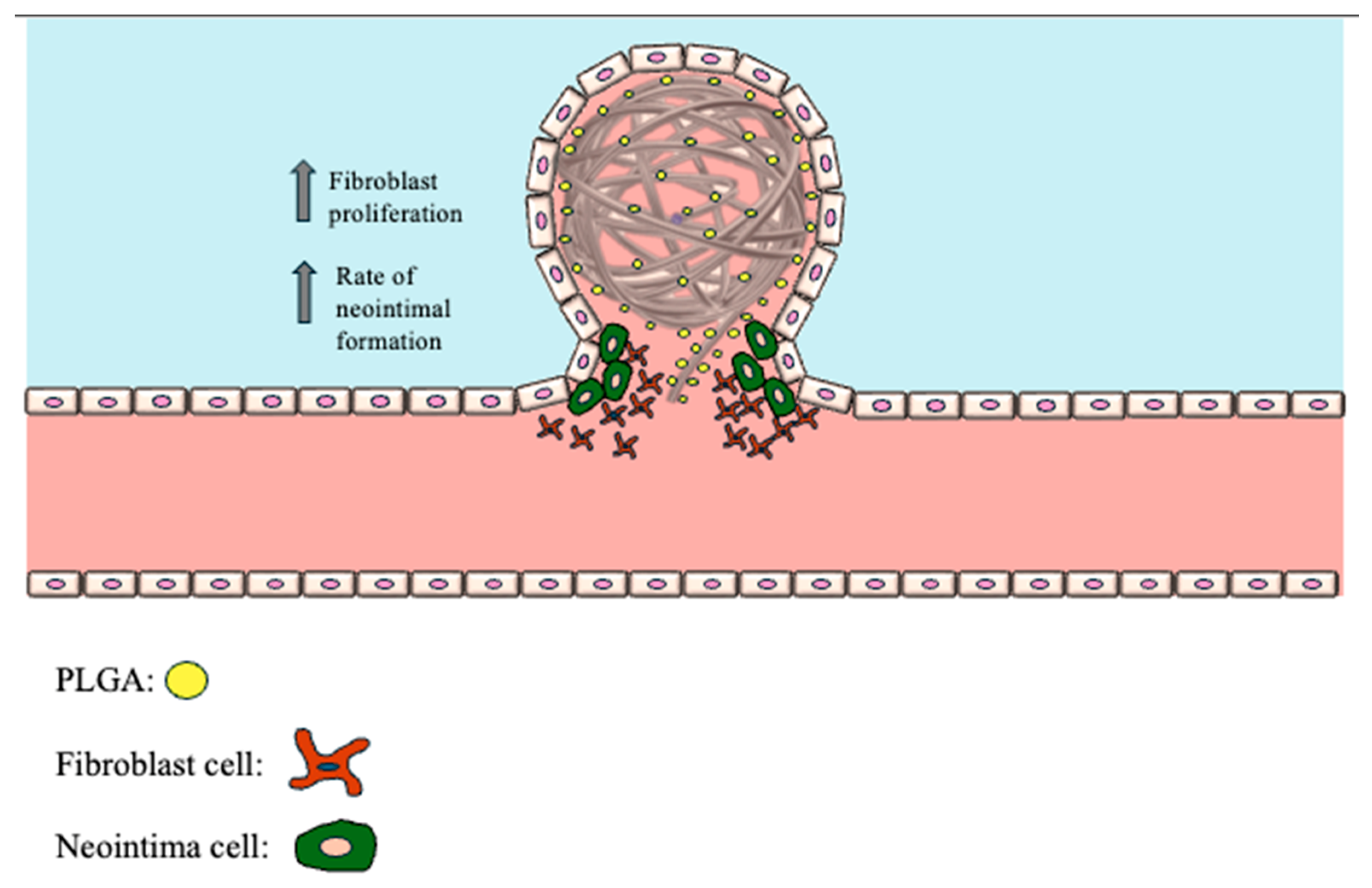
8.1. PLGA-Coated Coils

PLGA-coated coils are composed of polylactic acid and polyglycolic acid copolymers, with the ratio of lactic acid to glycolic acid adjusted to control the body’s degradation rate (Figure 2) [40]. This biologic increases the fibrosis and neointima formation rate without causing significant stenosis within the parent artery [40]. Additionally, the tissue regeneration response is enhanced across the neck of the aneurysm [17], with studies showing that aneurysms coiled with PLGA-coated coils showed the highest likelihood of obtaining a modified Raymond scale grade I occlusion immediately following surgery compared to other coated coils [41]. However, these findings were not statistically different from those of other coated coils at follow-up [41]. This was also true for recurrence and retreatment rates [40]. While the ability to achieve immediate post-operative occlusion was higher with PLGA-coated coils, significant drawbacks were noted with the first iteration of coils. First, providers reported that these coils possessed physical qualities of stiffness and increased friction, making it challenging to maximize the packing density within the aneurysm [40,43]. Second, the degradation of the coated material caused defects in the coils within the aneurysm, leading to compromised occlusion over time [40,43]. The second iteration of these coils incorporated smooth surfaces and decreased PGLA material coating to address these shortcomings; however, there is mixed literature on the benefits of these coils compared to bare platinum coils [40].

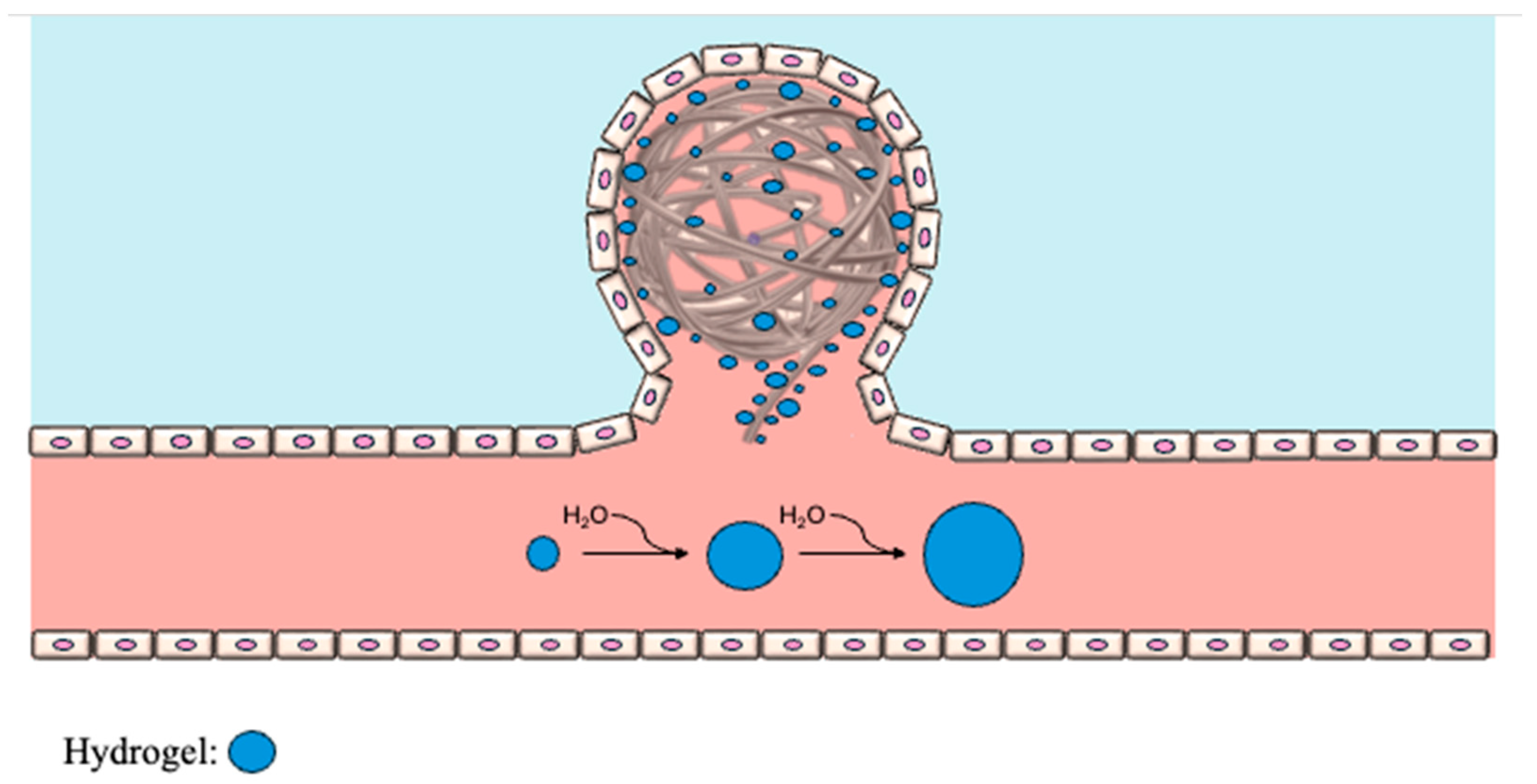
8.2. Hydrogel-Coated Coils

Bare platinum coils modified with coated hydrogels are implemented in treating IAs due to their ability to swell and achieve near-full occlusion within the neck of the aneurysm (Figure 3). Hydrogel-coated coils do not incite an inflammatory response, unlike the PLGA-coated coils [40]. Instead, the mechanism involves mechanical occlusion by expanding and reducing dead space within the body of the aneurysm due to the hydrophilic nature of the gels [17]. Two studies demonstrated that the immediate postoperative modified Raymond scale was not statistically different than PLGA-coated coils [41,44]. However, at 6-month follow-up, there was a significantly higher percentage of Raymond scale grade I patients treated with hydrogel-coated coils compared to patients with PLGA-coated coils [41,44]. This finding was consistent with another study by Nickele et al., who found that 49% of aneurysms treated with hydrogel-coated coils were classified as Raymond scale grade I immediately after treatment, with that number rising to 83% by the 6-month follow-up [45]. Given the improved occlusion shown with hydrogel-coated coils at follow-up compared to PLGA-coated coils, studies looked at these methods to assess for long-term occlusion and thrombosis to gauge recurrence [40]. It was found that hydrogels offered an advantage over other bioactive coils due to their composition possessing resistance to thrombolytic processes over time [40]. However, studies found that the rate of aneurysms treated with hydrogel-coated coils with improved occlusion at the 6-month follow-up plateaued around the 18-month follow-up mark, showing no significant difference in efficacy compared to bare platinum and PLGA-coated coils [40,41,45].

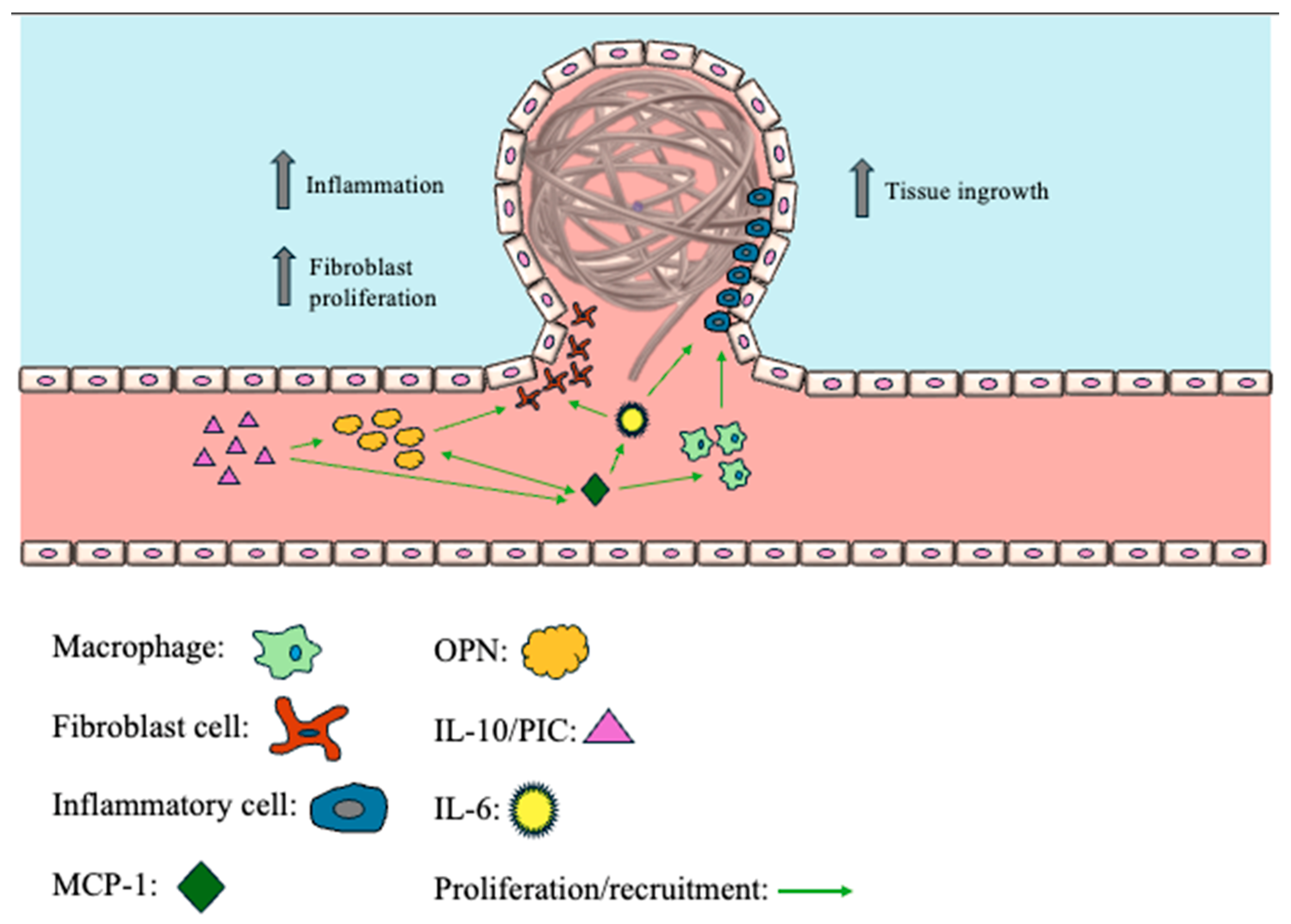
8.3. Monocyte Chemoattractant Protein-1 (MCP-1)

MCP-1 is a chemokine released in response to proinflammatory cytokines following tissue injury, playing a significant role in aneurysm wall healing and stabilization [46]. MCP-1 expression incites a macrophage-dependent proinflammatory state, leading to the recruitment of inflammatory cells and the initiation of inflammatory processes within the walls of IAs (Figure 4) [37]. This has a downstream anti-inflammatory effect that promotes the resolution of inflammation and encourages the maturation of the tissue at the aneurysm site. One study piloted the use of dual-coated catheters and found an increase in tissue ingrowth in catheters coated with bioactive polymers, such as PLGA or CG910, combined with MCP-1 [37]. Hourani et al. found that the systemic release of MCP-1 combined with PLGA-coated coils did not demonstrate an increase in aneurysmal healing [47]. Instead, the local delivery of MCP-1 successfully initiated site-specific healing of IAs, further insinuating the critical role of MCP-1 in the aneurysm healing pathway [47]. Another study demonstrated the downstream mediators of MCP-1 in tissue healing, showing an increased expression of IL-6 and OPN in MCP-1-treated aneurysms, essential components in the tissue ingrowth process [38].

8.4. Osteopontin (OPN)

Fibroblast invasion and thrombus formation are two key events necessary for proper aneurysm healing, sparking a growing interest in developing coil-coating materials with fibroblastic and thrombolytic properties. OPN is a chemokine-like protein that recruits and regulates fibroblast expression, pivotal in cell migration, adhesion, and bone regeneration (Figure 4) [37,42]. In aneurysms that have undergone successful coiling, there was a notable increase in OPN expression, proposing another promising candidate for bioactive endovascular coiling [37]. This protein was utilized in multiple studies assessing its efficacy in promoting aneurysmal tissue ingrowth and healing due to its known role in the downstream mediation of the MCP-1 pathway without dependency on MCP-1 [37]. Kadirvel et al. conducted coil embolization in rabbit models to compare the gene expression patterns of well-healed and poorly healed aneurysms [48]. This study found an increased expression of adhesion molecules, primarily OPN, in densely packed aneurysms compared to loosely packed aneurysms, highlighting the importance of maximizing coil packing density [48]. Another study found that OPN can act on the tissue healing pathway independently of MCP-1, whereas MCP-1-mediated treatment depended on OPN expression, demonstrating the utility of OPN-releasing coils in initiating tissue healing and fibrosis [38]. Laurent et al. found that OPN expression was associated with significantly increased tissue ingrowth compared to non-bioactive coils following dual coating with CG910 and OPN, highlighting the necessity for fibroblastic techniques [37].

8.5. Interleukin-6/Interleukin-10 (IL-6/IL-10)

Mild inflammation is thought to aid in tissue healing and repair, and cytokines play an essential role in the inflammatory processes responsible for vascular inflammation by regulating the proliferation and differentiation of various associated cell types [49]. IL-10 is a proinflammatory cytokine with anti-inflammatory qualities used in the endovascular treatment of IAs. Similarly, IL-6 plays a role in intra-aneurysmal healing by recruiting fibroblasts and endothelial cells, suggesting its potential as a therapeutic approach to endovascular coiling (Figure 4). Chen et al. performed a comparative study using a rat carotid aneurysm model to assess the efficacy of coating PLGA coils with OPN, IL-10, or matrix metallopeptidase-9 (MMP-9) to promote cerebral aneurysm healing [42]. They found that tissue ingrowth significantly increased with the PLGA/OPN-coated coils at 7 days, 20 days, and 60 days; however, the PLGA/IL-10-coated coils only showed significant tissue ingrowth at 60 days [42]. Even so, the OPN and IL-10-coated coils showed significantly increased tissue ingrowth compared to the PLGA/MMP-9 and PLGA-only coils [42]. Another study used murine carotid aneurysm models with cytokine-releasing coils containing MCP-1, IL-6, and OPN to evaluate their effects on intra-aneurysmal tissue healing [38]. This study demonstrated a relationship between IL-6 and MCP-1, validating that local administration of IL-6 could not induce a tissue response independently of MCP-1 and highlighting its role as an important downstream mediator of this pathway [38].

8.6. Priming the System

The inflammatory model of injury, repair, and remodeling is a well-known tissue healing pathway, previously demonstrated with tissue repair and remodeling following myocardial infarction [50]. In treating IAs, an intraluminal scaffold of biochemical cascades must be surgically introduced to the aneurysm to initiate the inflammatory response and promote tissue regeneration and repair [38]. MCP-1 is a known monocyte-recruiting chemokine that has been shown to mediate this process through other downstream mediators like IL-6 and OPN [38,42]. The development of bioactive endovascular coils utilizing MCP-1-mediated wound healing has proven to be a promising alternative to bare platinum coils, showing improved aneurysmal healing and enhanced occlusion over time [51]. The process by which MCP-1 is responsible for tissue repair and remodeling involves a cascade of cell recruitment and proliferation, starting with an influx of neutrophils and T-cells, followed by M1 (proinflammatory) and M2 (anti-inflammatory) macrophages [52]. Macrophage infiltration is characterized by an early M1, or destructive phase, followed by an M2, or reparative phase [52]. The next step in this pathway involves fibroblast, smooth muscle, and endothelial cell proliferation, mediators of aneurysm tissue ingrowth leading to complete durable aneurysm treatment and healing [37,51]. Establishing the biochemical pathways needed to initiate the inflammatory process and promote tissue healing is a step toward achieving optimal occlusion of IAs and reducing endovascular retreatment rates.

9. Future Directions

Various studies have utilized biologics in aneurysm coiling to promote inflammatory healing in hopes of achieving better occlusion and reducing retreatment rates of endovascular treatment [38,39,42,46,47,52]. These studies have shown that this emerging technique has the potential to offer a superior alternative to standard coiling; however, aside from PLGA- and hydrogel-coated coils, most of these studies were performed on animal test subjects, and their efficacy has not been widely demonstrated on human cerebrovasculature. Additionally, further understanding of the various targets of cytokine and chemokine therapies is needed to administer safe and effective treatment. One of the biggest challenges is the large number of chemokines and chemokine receptors and the difficulty in identifying which compounds will deliver inflammation-mediated healing [46]. MCP-1, OPN, and Interleukins are all promising mediators, but identifying additional biological therapies capable of stimulating aneurysmal healing is a complex process, and future research is needed to identify potential treatment targets [51]. There is currently limited literature on the utility of growth factors in promoting endothelial cell proliferation and vessel healing, potentially enhancing the aneurysmal healing process. Future research is needed on the safety and efficacy of biologics in aneurysm treatment to introduce novel techniques into endovascular coiling and improve patient outcomes. Furthermore, comprehensive studies assessing the impact of these treatments on morbidity and mortality rates, as well as postoperative complications, are essential to validate any potential claims that biologics represents a superior treatment option compared to conventional endovascular coiling or open surgical treatment. The clinical implementation of these biological agents alongside current treatment modalities, such as the Contour Neurovascular system, is of utmost importance [53]. Future studies should focus on these treatment methods’ practical, widespread implementation.

10. Conclusions

The treatment of intracranial aneurysms requires a multifaceted approach. Open surgical clipping and endovascular techniques have advantages and disadvantages, necessitating appropriate modality selection for individual patients. Emerging biologics in the endovascular treatment of IAs may lead to increased efficacy and durability of these less invasive methods. While still in its infancy, the integration of biochemically leveraged surgical techniques has the potential to change standard practice and deserves continued exploration.

Author Contributions

S.A.T.: data curation, writing—original draft preparation, writing—review and editing; M.-R.O.: data curation, writing—original draft preparation; R.R.: data curation, writing—original draft preparation; A.G.H.: data curation, writing—original draft preparation; B.L.-W.: conceptualization, data curation, writing—review and editing, supervision, project administration. All authors have read and agreed to the published version of the manuscript.

Funding

This research received no external funding.

Institutional Review Board Statement

Not applicable.

Data Availability Statement

Not applicable.

Conflicts of Interest

The authors declare no conflicts of interest.

References

  1. Keedy, A. An overview of intracranial aneurysms. Mcgill J. Med. 2006, 9, 141–146. [Google Scholar] [CrossRef] [PubMed]
  2. Mercadante, A.A.; Raja, A. Anatomy, Arteries. In StatPearls; StatPearls Publishing LLC.: Treasure Island, FL, USA, 2024. [Google Scholar]
  3. Merritt, W.C.; Berns, H.F.; Ducruet, A.F.; Becker, T.A. Definitions of intracranial aneurysm size and morphology: A call for standardization. Surg. Neurol. Int. 2021, 12, 506. [Google Scholar] [CrossRef] [PubMed]
  4. Barletta, E.; Ricci, R.; Silva, R.G.; Gaspar, R.M.L.; Araújo, J.M.; Neves, M.F.; de Aquino, J.B.; Belsuzarri, T.B. Fusiform aneurysms: A review from its pathogenesis to treatment options. Surg. Neurol. Int. 2018, 9, 189. [Google Scholar] [CrossRef] [PubMed]
  5. Fatterpekar, G.; Naidich, T.P.; Som, P.M. The Teaching Files: Brain and Spine; Elsevier/Saunders: Philadelphia, PA, USA, 2011. [Google Scholar]
  6. Majeed, H.; Ahmad, F. Mycotic Aneurysm. In StatPearls; StatPearls Publishing LLC.: Treasure Island, FL, USA, 2024. [Google Scholar]
  7. Vlak, M.H.; Algra, A.; Brandenburg, R.; Rinkel, G.J. Prevalence of unruptured intracranial aneurysms, with emphasis on sex, age, comorbidity, country, and time period: A systematic review and meta-analysis. Lancet Neurol. 2011, 10, 626–636. [Google Scholar] [CrossRef]
  8. Lawton, M.T.; Vates, G.E. Subarachnoid Hemorrhage. N. Engl. J. Med. 2017, 377, 257–266. [Google Scholar] [CrossRef] [PubMed]
  9. van der Kamp, L.T.; Rinkel, G.J.E.; Verbaan, D.; Berg, R.v.D.; Vandertop, W.P.; Murayama, Y.; Ishibashi, T.; Lindgren, A.; Koivisto, T.; Teo, M.; et al. Risk of Rupture after Intracranial Aneurysm Growth. JAMA Neurol. 2021, 78, 1228–1235. [Google Scholar] [CrossRef]
  10. Greving, J.P.; Wermer, M.J.H.; Brown, R.D., Jr.; Morita, A.; Juvela, S.; Yonekura, M.; Ishibashi, T.; Torner, J.C.; Nakayama, T.; Rinkel, G.J.E.; et al. Development of the PHASES score for prediction of risk of rupture of intracranial aneurysms: A pooled analysis of six prospective cohort studies. Lancet Neurol. 2014, 13, 59–66. [Google Scholar] [CrossRef]
  11. Sforza, D.M.; Putman, C.M.; Cebral, J.R. Computational fluid dynamics in brain aneurysms. Int. J. Numer. Method Biomed. Eng. 2012, 28, 801–808. [Google Scholar] [CrossRef]
  12. Guo, H.; Yang, S.-T.; Wang, J.-W.; Li, H.; Gao, B.-L.; Li, C.-H. High hemodynamic stresses induce aneurysms at internal carotid artery bends. Medicine 2023, 102, e34587. [Google Scholar] [CrossRef]
  13. Katritsis, D.; Kaiktsis, L.; Chaniotis, A.; Pantos, J.; Efstathopoulos, E.P.; Marmarelis, V. Wall shear stress: Theoretical considerations and methods of measurement. Prog. Cardiovasc. Dis. 2007, 49, 307–329. [Google Scholar] [CrossRef]
  14. de Falco, F.A. Sentinel headache. Neurol. Sci. 2004, 25 (Suppl. 3), S215–S217. [Google Scholar] [CrossRef] [PubMed]
  15. Dott, N.M. Intracranial Aneurysms: Cerebral Arterio-Radiography: Surgical Treatment. Edinb. Med. J. 1933, 40, T219–T240. [Google Scholar] [PubMed]
  16. Yasargil, M.G.; Fox, J.L. The microsurgical approach to intracranial aneurysms. Surg. Neurol. 1975, 3, 7–14. [Google Scholar] [PubMed]
  17. Lee, K.S.; Zhang, J.J.Y.; Nguyen, V.; Han, J.; Johnson, J.N.; Kirollos, R.; Teo, M. The evolution of intracranial aneurysm treatment techniques and future directions. Neurosurg. Rev. 2022, 45, 1–25. [Google Scholar] [CrossRef]
  18. Loewenstein, J.E.; Gayle, S.C.; Duffis, E.J.; Prestigiacomo, C.J.; Gandhi, C.D. The natural history and treatment options for unruptured intracranial aneurysms. Int. J. Vasc. Med. 2012, 2012, 898052. [Google Scholar] [CrossRef]
  19. Tawk, R.G.; Hasan, T.F.; D’souza, C.E.; Peel, J.B.; Freeman, W.D. Diagnosis and Treatment of Unruptured Intracranial Aneurysms and Aneurysmal Subarachnoid Hemorrhage. Mayo Clin. Proc. 2021, 96, 1970–2000. [Google Scholar] [CrossRef]
  20. Chappell, E.T.; Moure, F.C.; Good, M.C. Comparison of computed tomographic angiography with digital subtraction angiography in the diagnosis of cerebral aneurysms: A meta-analysis. Neurosurgery 2003, 52, 624–631; discussion 630–631. [Google Scholar] [CrossRef]
  21. White, P.M.; Wardlaw, J.M.; Easton, V. Can noninvasive imaging accurately depict intracranial aneurysms? A systematic review. Radiology 2000, 217, 361–370. [Google Scholar] [CrossRef]
  22. Sailer, A.M.; Wagemans, B.A.; Nelemans, P.J.; de Graaf, R.; van Zwam, W.H. Diagnosing intracranial aneurysms with MR angiography: Systematic review and meta-analysis. Stroke 2014, 45, 119–126. [Google Scholar] [CrossRef]
  23. Yoon, D.Y.; Lim, K.J.; Choi, C.S.; Cho, B.M.; Oh, S.M.; Chang, S.K. Detection and characterization of intracranial aneurysms with 16-channel multidetector row CT angiography: A prospective comparison of volume-rendered images and digital subtraction angiography. AJNR Am. J. Neuroradiol. 2007, 28, 60–67. [Google Scholar]
  24. Teksam, M.; McKinney, A.; Casey, S.; Asis, M.; Kieffer, S.; Truwit, C.L. Multi-section CT angiography for detection of cerebral aneurysms. AJNR Am. J. Neuroradiol. 2004, 25, 1485–1492. [Google Scholar] [PubMed]
  25. Kucukay, F.; Okten, R.S.; Tekiner, A.; Dagli, M.; Gocek, C.; Bayar, M.A.; Cumhur, T. Three-dimensional volume rendering digital subtraction angiography in comparison with two-dimensional digital subtraction angiography and rotational angiography for detecting aneurysms and their morphological properties in patients with subarachnoid hemorrhage. Eur. J. Radiol. 2012, 81, 2794–2800. [Google Scholar] [CrossRef] [PubMed]
  26. Rinkel, G.J. Intracranial aneurysm screening: Indications and advice for practice. Lancet Neurol. 2005, 4, 122–128. [Google Scholar] [CrossRef]
  27. De Simone, M.; Fontanella, M.M.; Choucha, A.; Schaller, K.; Machi, P.; Lanzino, G.; Bijlenga, P.; Kurz, F.T.; Lövblad, K.-O.; De Maria, L. Current and Future Applications of Arterial Spin Labeling MRI in Cerebral Arteriovenous Malformations. Biomedicines 2024, 12, 753. [Google Scholar] [CrossRef]
  28. Flemming, K.D.; Lanzino, G. Management of Unruptured Intracranial Aneurysms and Cerebrovascular Malformations. Continuum 2017, 23, 181–210. [Google Scholar] [CrossRef]
  29. Muirhead, W.R.; Grover, P.J.; Toma, A.K.; Stoyanov, D.; Marcus, H.J.; Murphy, M. Adverse intraoperative events during surgical repair of ruptured cerebral aneurysms: A systematic review. Neurosurg. Rev. 2021, 44, 1273–1285. [Google Scholar] [CrossRef]
  30. Wiebers, D.O.; International Study of Unruptured Intracranial Aneurysms Investigators. Unruptured intracranial aneurysms: Natural history, clinical outcome, and risks of surgical and endovascular treatment. Lancet 2003, 362, 103–110. [Google Scholar] [CrossRef] [PubMed]
  31. Zhao, J.; Lin, H.; Summers, R.; Yang, M.; Cousins, B.G.; Tsui, J. Current Treatment Strategies for Intracranial Aneurysms: An Overview. Angiology 2018, 69, 17–30. [Google Scholar] [CrossRef]
  32. Li, H.; Pan, R.; Wang, H.; Rong, X.; Yin, Z.; Milgrom, D.P.; Shi, X.; Tang, Y.; Peng, Y. Clipping versus coiling for ruptured intracranial aneurysms: A systematic review and meta-analysis. Stroke 2013, 44, 29–37. [Google Scholar] [CrossRef]
  33. Sanai, N.; Zador, Z.; Lawton, M.T. Bypass surgery for complex brain aneurysms: An assessment of intracranial-intracranial bypass. Neurosurgery 2009, 65, 670–683; discussion 683. [Google Scholar] [CrossRef]
  34. Koch, M.J.; Stapleton, C.J.; Charbel, F.T.; Russin, J.; Amin-Hanjani, S. Intracranial-Intracranial Bypass for Aneurysms: Quantitative Intraoperative Assessment of Flow Preservation. Oper. Neurosurg. 2022, 22, 337–342. [Google Scholar] [CrossRef] [PubMed]
  35. Guglielmi, G.; Viñuela, F.; Dion, J.; Duckwiler, G. Electrothrombosis of saccular aneurysms via endovascular approach. Part 2: Preliminary clinical experience. J. Neurosurg. 1991, 75, 8–14. [Google Scholar] [CrossRef] [PubMed]
  36. Pierot, L.; Wakhloo, A.K. Endovascular treatment of intracranial aneurysms: Current status. Stroke 2013, 44, 2046–2054. [Google Scholar] [CrossRef]
  37. Laurent, D.; Lucke-Wold, B.; Dodd, W.S.; Martinez, M.; Chowdhury, M.A.B.; Hosaka, K.; Motwani, K.; Hoh, B. Combination release of chemokines from coated coils to target aneurysm healing. J. NeuroInterventional Surg. 2023, 15, 689–694. [Google Scholar] [CrossRef]
  38. Hosaka, K.; Rojas, K.; Fazal, H.Z.; Schneider, M.B.; Shores, J.; Federico, V.; McCord, M.; Lin, L.; Hoh, B. Monocyte Chemotactic Protein-1-Interleukin-6-Osteopontin Pathway of Intra-Aneurysmal Tissue Healing. Stroke 2017, 48, 1052–1060. [Google Scholar] [CrossRef] [PubMed]
  39. Hudson, J.S.; Hoyne, D.S.; Hasan, D.M. Inflammation and human cerebral aneurysms: Current and future treatment prospects. Future Neurol. 2013, 8, 663–676. [Google Scholar] [CrossRef]
  40. Vance, A.; Welch, B.G. The utility of bioactive coils in the embolization of aneurysms. Neurol. Res. 2014, 36, 356–362. [Google Scholar] [CrossRef] [PubMed]
  41. Zhang, J.; Jiang, G.; Song, Z.; Cheng, W.; Wu, W.; Chen, Z.; Wang, Z.; You, W.; Chen, G. Efficacy and Safety of Different Bioactive Coils in Intracranial Aneurysm Interventional Treatment, a Systematic Review and Bayesian Network Meta-Analysis. Brain Sci. 2022, 12, 1062. [Google Scholar] [CrossRef]
  42. Chen, J.; Yang, L.; Chen, Y.; Zhang, G.; Fan, Z. Controlled release of osteopontin and interleukin-10 from modified endovascular coil promote cerebral aneurysm healing. J. Neurol. Sci. 2016, 360, 13–17. [Google Scholar] [CrossRef]
  43. Kim, S.; Nowicki, K.W.; Ye, S.; Jang, K.; Elsisy, M.; Ibrahim, M.; Chun, Y.; Gross, B.A.; Friedlander, R.M.; Wagner, W.R. Bioabsorbable, elastomer-coated magnesium alloy coils for treating saccular cerebrovascular aneurysms. Biomaterials 2022, 290, 121857. [Google Scholar] [CrossRef]
  44. Abi-Aad, K.R.; Rahme, R.J.; Patra, D.P.; Turcotte, E.L.; Richter, K.R.; Merrill, S.A.; Syal, A.; Neville, M.R.; Hudson, M.; Garcia, J.O.; et al. Clinical outcomes of first- and second-generation hydrogel coils compared with bare platinum coils: A systematic literature review. Neurosurg. Rev. 2022, 45, 1873–1882. [Google Scholar] [CrossRef] [PubMed]
  45. Nickele, C.; Oravec, C.S.; Morris, S.D.; Hoit, D.; Elijovich, L.; Arthur, A.S. Long-Term Follow-up of Aneurysms Treated with Hydrogel-Coated Coils Shows Progressive Thrombosis and Improvement in Raymond-Roy Classification. Oper. Neurosurg. 2022, 22, 239–243. [Google Scholar] [CrossRef]
  46. Deshmane, S.L.; Kremlev, S.; Amini, S.; Sawaya, B.E. Monocyte chemoattractant protein-1 (MCP-1): An overview. J. Interferon Cytokine Res. 2009, 29, 313–326. [Google Scholar] [CrossRef] [PubMed]
  47. Hourani, S.; Motwani, K.; Wajima, D.; Fazal, H.; Jones, C.H.; Doré, S.; Hosaka, K.; Hoh, B.L. Local Delivery Is Critical for Monocyte Chemotactic Protein-1 Mediated Site-Specific Murine Aneurysm Healing. Front. Neurol. 2018, 9, 158. [Google Scholar] [CrossRef]
  48. Kadirvel, R.; Ding, Y.-H.; Dai, D.; Lewis, D.A.; Kallmes, D.F. Differential gene expression in well-healed and poorly healed experimental aneurysms after coil treatment. Radiology 2010, 257, 418–426. [Google Scholar] [CrossRef]
  49. Girn, H.; Orsi, N.; Homer-Vanniasinkam, S. An overview of cytokine interactions in atherosclerosis and implications for peripheral arterial disease. Vasc. Med. 2007, 12, 299–309. [Google Scholar] [CrossRef] [PubMed]
  50. Frangogiannis, N.G. The inflammatory response in myocardial injury, repair, and remodelling. Nat. Rev. Cardiol. 2014, 11, 255–265. [Google Scholar] [CrossRef]
  51. Hoh, B.L.; Hosaka, K.; Downes, D.P.; Nowicki, K.W.; Fernandez, C.E.; Batich, C.D.; Scott, E.W. Monocyte chemotactic protein-1 promotes inflammatory vascular repair of murine carotid aneurysms via a macrophage inflammatory protein-1α and macrophage inflammatory protein-2-dependent pathway. Circulation 2011, 124, 2243–2252. [Google Scholar] [CrossRef]
  52. Hoh, B.L.; Fazal, H.Z.; Hourani, S.; Li, M.; Lin, L.; Hosaka, K. Temporal cascade of inflammatory cytokines and cell-type populations in monocyte chemotactic protein-1 (MCP-1)-mediated aneurysm healing. J. NeuroInterventional Surg. 2018, 10, 301–305. [Google Scholar] [CrossRef]
  53. Ghozy, S.; Lashin, B.I.; Elfil, M.; Bilgin, C.; Kobeissi, H.; Shehata, M.; Kadirvel, R.; Kallmes, D.F. The safety and effectiveness of the Contour Neurovascular System for the treatment of wide-necked aneurysms: A systematic review and meta-analysis of early experience. Interv. Neuroradiol. 2022, 15910199221139546. [Google Scholar] [CrossRef]
Figure 1. Timeline of IA management history.
Biologics 04 00022 g001
Figure 2. PLGA-coated coil. PLGA: Polylactic/polyglycolic acids copolymer nanoparticles.
Biologics 04 00022 g002
Figure 3. Hydrogel-coated coil demonstrating the expansion of the gels to occlude the aneurysm mechanically.
Biologics 04 00022 g003
Figure 4. Simplified tissue healing cascade demonstrating inflammatory effects of MCP-1, IL-6, IL-10, and osteopontin, along with increased proliferation of fibroblast cells. MCP-1: Monocyte Chemoattractant Protein-1; OPN: osteopontin; IL: interleukin; PIC: proinflammatory cell.
Biologics 04 00022 g004
Disclaimer/Publisher’s Note: The statements, opinions and data contained in all publications are solely those of the individual author(s) and contributor(s) and not of MDPI and/or the editor(s). MDPI and/or the editor(s) disclaim responsibility for any injury to people or property resulting from any ideas, methods, instructions or products referred to in the content.

