Entry

Improving Compliance with Medical Treatment Using Eye Drop Aids

1
Department of Ophthalmology, Tel Aviv Sourasky Medical Center and Sackler School of Medicine, Tel Aviv University, Tel Aviv 6423906, Israel
2
Department of Ophthalmology, Shaare Zedek Medical Center and Hebrew University of Jerusalem, Jerusalem 9103102, Israel
*
Author to whom correspondence should be addressed.
Encyclopedia 2023, 3(3), 919-927; https://doi.org/10.3390/encyclopedia3030065
Submission received: 11 May 2023 / Revised: 15 June 2023 / Accepted: 10 July 2023 / Published: 23 July 2023
(This article belongs to the Collection Encyclopedia of Glaucoma)

Definition

:
Achieving optimal treatment outcomes in glaucoma requires patients to adhere to their medication regimens. Possible barriers to patients’ cooperation include the misunderstanding of a treatment’s importance or errors in applying instructions, forgetfulness, financial constraints and others. Due to the fact that glaucoma usually causes no apparent symptoms or pain, on the one hand, and the significant inconvenience that the eye drops used for glaucoma treatment can cause due to local irritation, on the other, patient compliance is a challenge. To address this challenge, we require strategies for improving adherence to glaucoma treatment. The importance of proper eye drop administration techniques cannot be overstated, particularly for vulnerable populations such as the elderly, the sick and the visually handicapped. Studies have shown that failure to comply with glaucoma treatment is a significant factor affecting disease progression, emphasizing the need for interventions that improve patient compliance. Educational interventions, medication reminders and the use of assistive devices such as eye drop aids have been shown to improve adherence to glaucoma treatment. By promoting strategies that can be used to enhance treatment adherence, healthcare providers can ensure that glaucoma patients receive the full benefits of their treatment plans, reducing the risk of disease progression. Many patients struggle with the complexity of their treatment regimens and the challenges of administering eye drops. This entry provides a comprehensive overview of the different barriers to patient adherence to glaucoma eye drop treatment, emphasizing the difficulties associated with eye drop instillation. This entry examines a range of eye drop aids available to patients, evaluating their modes of action, benefits, drawbacks and effectiveness in improving patient compliance. By providing detailed information on the barriers to adherence and the range of eye drop aids available, this entry aims to support healthcare providers in helping glaucoma patients to achieve better treatment adherence and outcomes.

1. Introduction

Primary open-angle glaucoma is a chronic and progressive optic neuropathy characterized by a gradual loss of vision, which often goes unnoticed until the later stages of the disease. The proposed explanations for optic neuropathy include direct pressure on nerve fiber layer axons, local pressure on optic nerve axons at the lamina cribrosa (the “mechanical theory”) and vascular insufficiency of the optic nerve head (the “vascular theory”). The fact that intra-ocular pressure is the most important risk factor in glaucoma supports the mechanical theory. Normotensive glaucoma is associated with other diseases characterized by vascular dysregulation (migraine, Raynaud’s phenomenon and sleep apnea), which supports the vascular theory of optic neuropathy in glaucoma. Based on these data, one may assume that the optic neuropathy of glaucoma is the result of both mechanisms, whereas the lower the pressure is, the more dominant the vascular component is, and the higher the pressure is, the more dominant the mechanical component is. Recent studies suggested that glaucoma might even be an autoimmune disease—this theory is supported by immune responses to heat shock protein in some glaucoma patients and in animal models of the disease [1]. One of the most significant challenges in glaucoma is the fact that, in many cases, it is diagnosed late in the course of the disease, when the damage is severe. The reason for that is that glaucoma does not cause pain, discomfort or functional disturbances in its early stages, leading many patients to delay seeking medical treatment. Glaucoma is one of the leading causes of blindness worldwide, and if left untreated, it can result in permanent vision loss and blindness [2,3]. The most important risk factors for glaucoma are intra-ocular pressure, age, family history, corneal thickness and myopia. Intra-ocular pressure is unique, as it is the only risk factor that can be manipulated through treatment. The lowering of intra-ocular pressure is the only proven treatment for improving the prognosis of open-angle glaucoma (including normal-tension glaucoma) and preventing the further deterioration of optic neuropathy [3,4].
As in the case of other chronic medical conditions, treatment is warranted for the rest of the patient’s life after the diagnosis of the disease. If the chosen treatment is eye drops, this encompasses the instillation of at least one drop from a single bottle daily. In most cases, this requires three drops from two different bottles. No “penalty” that the patient can experience will result from not applying the drops, and the drops usually cause some degree of local discomfort. For this reason, compliance with glaucoma treatment is a challenge, and its promotion is mandatory for improving the prognosis of glaucoma. These difficulties in patients’ adherence to chronic glaucoma treatment are a major challenge for the treating ophthalmologist. Moreover, healthcare providers should recognize that prescribing fewer eye drop instillations for a patient can increase the likelihood of treatment compliance [5,6].
The pharmacokinetics of eye drops necessitates their repetitive and scheduled application, and inaccuracy in timely instillation results in untreated periods in which pressure is high and the optic nerve is damaged. Failure to adhere to timely instillation may result from forgetfulness, an inability to understand the risk–benefit aspects of treatment, reluctance due to the side effects of treatment or difficulties in medication supply.
Lack of compliance in glaucoma patients is a well-known problem that is believed to account, at least in part, for medical treatment failures in glaucoma [5,7,8,9,10]. Age is considered one of the foremost risk factors for the development of glaucoma, as individuals affected by this disease are typically older [2]. Unfortunately, this population is also more likely to experience challenges in adhering to their medication regimens. In older age, people may lack the strength needed to pinch the bottle, experience cognitive problems affecting their ability to remember the drops’ schedule or be using many other medications, which leads to confusion and impedes their ability to understand risk–benefit issues and act according to them. Difficulties in instilling the drops due to general medical and cognitive status pose as an additional barrier to compliance [11,12,13,14]. Achieving high compliance rates among glaucoma patients is critical for successful treatment improvement and for decreasing preventable future visual disturbances and blindness.
When measuring intra-ocular pressure at a glaucoma patient visit, we must bear in mind that the measured pressure is a kind of a “snapshot” reflecting the minute value. Intra-ocular pressure is well-known to fluctuate, and neuronal damage is caused whenever the pressure is too high for the nerve. This means that even if we find a normal value at a given time, the pressure may be higher in other parts of the day and cause further damage to the optic nerve. In addition, the timing of the follow-up visit may influence the patient’s compliance—patients tend to remember to take their drops better soon after a visit and when facing a visit scheduled close ahead. This may obscure the actual intra-ocular pressure value throughout the year and “hide” non-compliance.
In view of the limited value of intra-ocular pressure as a reliable marker of successful treatment and disease control, more reliable measures of disease control are needed. Two general groups of disease markers are used for this purpose: anatomic and functional.
The anatomic refers to the characteristic appearance of the optic nerve head in the disease. Nerve fiber atrophy creates nerve “cupping” (=enlargement of the central part of the nerve head containing no nerve fibers). This cupping can act as a measure for disease severity if it is stable when the disease is under control and worsens with the disease progressing. Cupping is not influenced by pressure fluctuation and provides an estimation of the possible added damage following the former examination and its severity. Cupping can be estimated clinically or measured via Optical Coherence Tomography (OCT). The main problem with progress estimation based on cupping is its loss in value in severe disease, in which cupping is total or near-total.
The functional refers to visual field testing. Glaucoma causes some characteristic changes in the visual field (e.g., nasal step, arcuate scotomata, paracentral scotomas). Following visual field status and changes can provide an idea of disease control; if the visual field is stable, the disease is under control. If the visual field has deteriorated, the disease is worsening. Just like cupping, changes in visual field are not influenced by moment pressure or pressure fluctuation, and they can effectively summarize the relevant general behavior of the disease between the two tests under comparison.
As mentioned, cupping estimation is limited in advanced disease. Visual field defects are usually not found in early disease contexts. For this reason, cupping is considered a good marker of disease control in early glaucoma, while visual field status is a better marker of advanced disease.

2. Data

2.1. Eye Drops: The Challenges

Eye drops are the most common method for intra-ocular pressure lowering in glaucoma. Each kind of eye drop used has to be instilled 1–2 times a day for each eye. Aside from the technical aspects involved in instillation, as discussed below, one has to store the drops properly and carry them whenever one travels or leaves home. Furthermore, patients’ successful and independent use of eye drops can be challenging, and failure to use them correctly can lead to non-compliance and treatment inefficacy [15,16,17].
Patients may fail to comply with their medication regimens due to the challenges associated with the accurate instillation of eye drops or concerns about potential harm during self-administration, including bottle-tip-related trauma or contamination [15,18,19]. It has been shown that up to 25% of patients miss doses because of these difficulties [20]. Successful eye drop administration is a skill that one learns and improves upon through practice. The prevalence of glaucoma is higher in the elderly population, for whom additional comorbidities such as hand tremors, arthritis, a poor coordination and peripheral neuropathy conditions may pose additional difficulties for successful eye drop administration [15,21,22]. Non-adherence to glaucoma treatment can lead to inadequate intra-ocular pressure reduction, which can cause disease progression, visual impairment and the need for more aggressive treatment.
Several primary issues can significantly affect eye drop treatment in glaucoma patients. Firstly, patients may show an inability to produce sufficient and powerful force on the bottle in order to allow the drop to drip out. Secondly, there may be difficulties in placing and positioning the bottle, which may cause a loss of drops. Furthermore, the loss of drops due to insufficient pressure on the bottle or its misplacement can lead to an injury, secondary to accidental contact of the tip of the bottle with the patient’s eye. Table 1 presents a summary of the main problems limiting patient’s adherence to the treatment schedule.
The overcoming of technical obstacles for the proper instillation of eye drops among glaucoma patients, such as hand tremors and poor cognitive states, can be achieved through specialized devices and patient education, which can make instillation simpler and safer, improve treatment outcomes and reduce the risk of further vision loss.

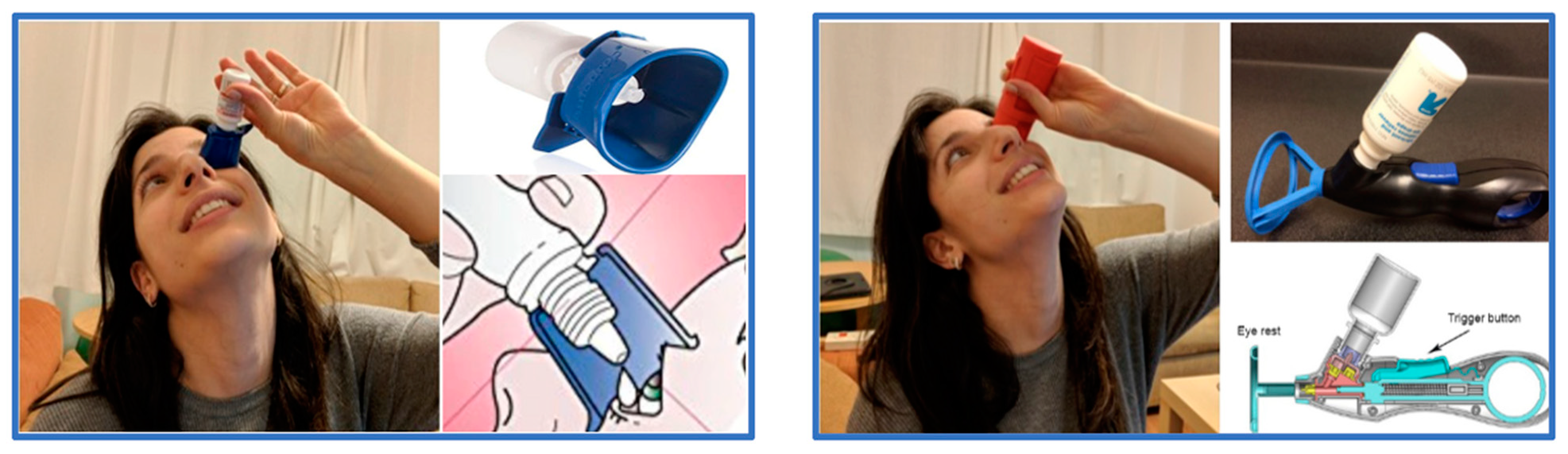
2.2. Possible Solution: Drop Aids

Drop aids aim to ease the technical issues related to proper eye drop instillation. These include the strengthening of the bottle pinch, the proper placement of the bottle over the eye and the prevention of bottle tip contact with the cornea. The use of drop aids can simplify the instillation of glaucoma medication and help to overcome technical difficulties that commonly result in non-compliance, thereby promoting adherence to therapy and potentially improving the effectiveness of glaucoma treatment.
In terms of compatibility with eye drop bottles, both Autodrop (Owen Mumford Ltd., Woodstock, UK) and Opticare (Cameron Graham Limited, Huddersfield, UK) are highly suitable. Autodrop allows for the easy attachment of the eye drop bottle, which is securely clipped into place. Additionally, the device’s body keeps the lower eyelid in an open position, preventing blinking. A small pinhole in the device facilitates administration by directing the gaze upward and away from the falling drops. Conversely, the Opticare device encloses the eye drop bottle within its structure, providing better grip. The device is then positioned on the eye, and the eyepiece keeps the upper eyelid open, thus overcoming the blinking reflex and facilitating the accurate administration of eye drops. Figure 1 presents examples of drops aid devices.
Several studies have examined the impacts of using eye drop aid devices on various factors that can affect the efficacy and safety of glaucoma treatment, such as the amount of force required to expel a drop, the accuracy of drop placement, the risk of bottle tip contact with the cornea and the number of drops required for adequate medication delivery, providing valuable insights into the potential benefits of these devices in promoting effective and safe medication administration among glaucoma patients [16,18,21,22,24,25,26,27,28]. The results of these reports are presented in Table 2.

3. Application

The reports on drop aids as a potential solution for improving compliance with medical treatments of glaucoma have identified four key problem categories that need to be taken into consideration for the successful instillation of eye drops: the force needed to expel a drop, drop placement, bottle tip contact and contamination risk, and the number of drops expelled.

3.1. Force Needed to Expel a Drop

An evaluation of the force required to expel a drop from various glaucoma medication bottles revealed significant variability between different products, and further analysis of the impact of drop aids on expelling force demonstrated varying levels of effectiveness across different types of aids, with some aids proving to be more effective than others in reducing the force needed to expel a drop [20,32]. In rheumatic arthritis patients, the use of Opticare (Cameron Graham Limited, Huddersfield, UK) allowed for better pinch power to be exerted, improving the ease of use. According to the authors, when using the conventional bottle, half of the patients encountered difficulties when attempting to instill their eye drops, whereas one-third experienced such difficulties when using Opticare. Furthermore, this device proved particularly effective in improving patient independence, with 13% of individuals who previously required the assistance of their spouse for eye drop administration being able to independently self-administer their drops using the device [14].

3.2. Drop Placement

The accurate placement of glaucoma eye drops in the conjunctival sac is essential for optimal therapeutic outcomes, but many patients have trouble achieving this. The use of an upright eye drop bottle (EG Gilero, Durham, NC, USA), eye drop (Vangouard Design, Sao Paulo, Brazil) mirror hat device (noncommercial, developed by John Beck) or inverted-funnel-shaped eye drop guide (Merck Frosst Canada, Kirkland, QC, Canada) did not improve the placement of drops in comparison to instillation without a drop aid, while that of a black-tipped bottle (using black tape developed by Sterion, Minneapolis, MN, USA, with a Timolol eye drop bottle developed by Apotex NK, Aukland, New Zealand), Opticare (Cameron Graham Limited, Huddersfield, UK), Xal-ease (Pfizer ophthalmics, New York, NY, USA) and easydrop (Quoteforce, UK) improved drop placement in the conjunctival sac [17,22,26].

3.3. Bottle Tip Contact and Contamination Risk

One of the major concerns for patients undergoing topical treatment is the potential for contamination or trauma caused by contact between the eye drop bottle tip and the eye surface. Contact between the eye drop bottle and the cornea can cause sloughing of the corneal epithelium (corneal erosion), a painful situation which needs time to heal. However, this need is not met because of the recurrent application of drops, and this may cause chronic damage to the cornea. Another possible result of bottle tip contact is contamination, with possible significant damage to the cornea. Despite 92.3% of patients with ocular diseases reporting no issues with eye drop administration, only 21.9% (with a 15 mL bottle) and 31% (with a 2.5 mL bottle) were able to successfully instill a single drop without the bottle tip making contact with the eye. This highlights the need for increased awareness and education, along with drop aid assistance, in regard to proper eye drop administration techniques in order to minimize the risk of contamination or injury [33].
Consistent with the challenges of accurate drop placement, several studies have reported that less than half of glaucoma patients are able to instill eye drops without the tip of the bottle coming into contact with the eye [22,33]. Further emphasizing the importance of this issue, bottle tip contact was reported as a cause of ocular infection [34,35] and inflammation [36]. Moreover, contamination caused by bottle tips when using a conventional bottle was reported in 46–61% of glaucoma patients [22,23]. Consistent with the potential benefits of using drop aids for reducing bottle tip contact, reports that compared contact between instillation with and without drop aids demonstrated a decrease in the incidence of contact with the eye when using an eye drop aid, underscoring the potential of these devices to enhance medication adherence and reduce the risk of ocular injury or infection associated with conventional medication administration techniques [24,25,26,30].

3.4. The Number of Drops Expelled

The various issues related to conventional medication administration techniques, such as inaccurate drop placement, excess force on the bottle and insecurity regarding the drops’ arrival into the eye, cause excess drops to be expelled. The instillation of excessive drops increases the risk of adverse events such as local skin irritation from the spillage of drops onto the periocular skin and systemic side effects. Furthermore, it may lead to the wastage of medication and requires patients to visit the pharmacy more frequently in order to purchase new medication bottles. It has been shown that nearly 50% of glaucoma patients use two or more drops in each instillation [37]. In addition, it is stated in the literature that medication waste increases the cost of treatment and has a negative effect on compliance [38]. Therefore, when using drop aids, a decrease in the number of drops used was expected. While upright eye drop bottles (EG Gilero, Durham, NC, USA) and Opticare (Cameron Graham Limited, Huddersfield, UK) were found to reduce the number of drops [24,27], Xal-ease (Pfizer Ophthalmics, New York, NY, USA) and the mirror hat device (noncommercial, developed by John Beck) did not show an effect on the number of drops used [25,26]. In a previous report, the numbers of drops instilled were compared between drop bottles, Autodrop and Opticare [23]. According to this report, Autodrop’s use was associated with more drops used compared to the use of bottles without aids and Opticare. This report stated that one of the problems in bottle instillation is the need for multiple drops due to the wastage of drops that land on the cheeks and lids. The capacity for reducing the number of drops with a drop aid depends on skilled use, which takes time to develop. This report on the increasing number of drops probably reflects a need for additional training in the use of aids, as the research reported here referred to a single event of drop placement after two sessions of training. In real life, a patient who uses the aid on a regular basis has much more training gained through everyday use and is expected to master the act much better than is possible after two training sessions. Once the patient masters the use of the aid, they should be able to properly direct the drops, waste less drops and, accordingly, use less drops.

4. Conclusions and Prospect

The key benefit of using drop aids lies in their capacity to effectively prevent bottle tip contact with the eye, thereby reducing the risk of ocular inflammation and infection associated with conventional medication administration techniques, ultimately promoting greater patient safety and medication adherence. Among the devices reviewed in the present report, the Xal-ease, mirror hat and upright drop bottle seem to show the best effects in preventing bottle tip contact with the cornea. Moreover, drop aids may decrease the force needed to insert the bottle and the number of drops used for each drop’s ejection. Opticare was found to have a favorable effect on the force needed to expel a drop. Drop placement improves with some drop aid devices and remains unchanged for others.
Drop aids have been developed to facilitate the instillation of eye drops by providing patients with a more accurate means of placing the drops into the conjunctival sac and controlling the force applied to the bottle, which helps to prevent excessive dripping and reduce the risk of ocular infection or erosion resulting from bottle tip contact with the eye. The improved convenience and safety of drop administration support patient confidence and, in turn, may increase compliance and positively affect disease prognosis. Several reports on the advantages of drop aids have been published, focusing on both specific aids and comparative research. No comprehensive, prospective study which includes all the available drop aids has been published. The obvious need for an efficient aid and the large number of products call for large-scale research including all the relevant characteristics of drop aid performance in order to provide recommendations on the most efficient products.

Author Contributions

Writing—original draft preparation, A.B.; writing—review and editing, M.G., N.S. and A.A. supervision, A.A. All authors have read and agreed to the published version of the manuscript.

Funding

This research received no external funding.

Institutional Review Board Statement

Not applicable.

Informed Consent Statement

Not applicable.

Data Availability Statement

Not applicable.

Conflicts of Interest

The authors declare no conflict of interest.

References

  1. Wakefield, D.; Wildner, G. Is glaucoma an autoimmune disease? Clin. Transl. Immunol. 2020, 9, e1180. [Google Scholar] [CrossRef] [PubMed]
  2. Tham, Y.C.; Li, X.; Wong, T.Y.; Quigley, H.A.; Aung, T.; Cheng, C.Y. Global prevalence of glaucoma and projections of glaucoma burden through 2040: A systematic review and meta-analysis. Ophthalmology 2014, 121, 2081–2090. [Google Scholar] [CrossRef] [PubMed]
  3. Mokhles, P.; Schouten, J.S.; Beckers, H.J.; Azuara-Blanco, A.; Tuulonen, A.; Webers, C.A. Glaucoma blindness at the end of life. Acta Ophthalmol. 2017, 95, 10–11. [Google Scholar] [CrossRef] [PubMed]
  4. Heijl, A.; Leske, M.C.; Bengtsson, B.; Hyman, L.; Bengtsson, B.; Hussein, M.; Early Manifest Glaucoma Trial Group. Reduction of intraocular pressure and glaucoma progression: Results from the Early Manifest Glaucoma Trial. Arch. Ophthalmol. Chic. Ill 1960 2002, 120, 1268–1279. [Google Scholar] [CrossRef] [PubMed]
  5. Tsai, J.C. A comprehensive perspective on patient adherence to topical glaucoma therapy. Ophthalmology 2009, 116 (Suppl. 11), S30–S36. [Google Scholar] [CrossRef] [PubMed]
  6. Buchan, J.; Siddiqui, S.; Gilmour, D. Once daily drop regimes help reduce involuntary non-compliance. Graefes Arch. Clin. Exp. Ophthalmol. Albrecht. Von Graefes Arch. Für. Klin. Exp. Ophthalmol. 2007, 245, 327–328. [Google Scholar] [CrossRef]
  7. Newman-Casey, P.A.; Salman, M.; Lee, P.P.; Gatwood, J.D. Cost-Utility Analysis of Glaucoma Medication Adherence. Ophthalmology 2020, 127, 589–598. [Google Scholar] [CrossRef]
  8. Sanchez, F.G.; Mansberger, S.L.; Newman-Casey, P.A. Predicting Adherence with the Glaucoma Treatment Compliance Assessment Tool. J. Glaucoma 2020, 29, 1017–1024. [Google Scholar] [CrossRef]
  9. Boland, M.V.; Chang, D.S.; Frazier, T.; Plyler, R.; Friedman, D.S. Electronic monitoring to assess adherence with once-daily glaucoma medications and risk factors for nonadherence: The automated dosing reminder study. JAMA Ophthalmol. 2014, 132, 838–844. [Google Scholar] [CrossRef] [Green Version]
  10. Friedman, D.S.; Okeke, C.O.; Jampel, H.D.; Ying, G.-S.; Plyler, R.J.; Jiang, Y.; Quigley, H.A. Risk factors for poor adherence to eyedrops in electronically monitored patients with glaucoma. Ophthalmology 2009, 116, 1097–1105. [Google Scholar] [CrossRef]
  11. Broadway, D.C.; Cate, H. Pharmacotherapy and Adherence Issues in Treating Elderly Patients with Glaucoma. Drugs Aging 2015, 32, 569–581. [Google Scholar] [CrossRef]
  12. Aleem, A.; Amin, F.; Asim, M.H.; Farooq, N.; Arshad, S.; Raziq, M. Impact of pharmacist-led interventions in improving adherence to glaucoma medications in the geriatric population. Eur. J. Hosp. Pharm. Sci. Pract. 2021, 28 (Suppl. 2), e191–e196. [Google Scholar] [CrossRef]
  13. Osman, E.A.; Alqarni, B.A.M.; AlHasani, S.S.H.; Al Harbi, S.S.S.; Gikandi, P.W.; Mousa, A. Compliance of Glaucoma Patients to Ocular Hypotensive Medications Among the Saudi Population. J. Ocul. Pharmacol. Ther. Off J. Assoc. Ocul. Pharmacol. Ther. 2016, 32, 50–54. [Google Scholar] [CrossRef] [PubMed]
  14. Gurwitz, J.H.; Glynn, R.J.; Monane, M.; Everitt, D.E.; Gilden, D.; Smith, N.; Avorn, J. Treatment for glaucoma: Adherence by the elderly. Am. J. Public Health 1993, 83, 711–716. [Google Scholar] [CrossRef] [PubMed] [Green Version]
  15. Winfield, A.J.; Jessiman, D.; Williams, A.; Esakowitz, L. A study of the causes of non-compliance by patients prescribed eyedrops. Br. J. Ophthalmol. 1990, 74, 477–480. [Google Scholar] [CrossRef] [PubMed] [Green Version]
  16. Konstas, A.G.; Maskaleris, G.; Gratsonidis, S.; Sardelli, C. Compliance and viewpoint of glaucoma patients in Greece. Eye Lond. Engl. 2000, 14 Pt 5, 752–756. [Google Scholar] [CrossRef] [PubMed] [Green Version]
  17. Kholdebarin, R.; Campbell, R.J.; Jin, Y.P.; Buys, Y.M. Multicenter study of compliance and drop administration in glaucoma. Can. J. Ophthalmol. 2008, 43, 454–461. [Google Scholar] [CrossRef]
  18. Davies, I.; Williams, A.M.; Muir, K.W. Aids for eye drop administration. Surv. Ophthalmol. 2017, 62, 332–345. [Google Scholar] [CrossRef]
  19. Parkkari, M.; Latvala, T.; Ropo, A. Handling Test of Eye Drop Dispenser—Comparison of Unit-Dose Pipettes with Conventional Eye Drop Bottles. J. Ocul. Pharmacol. Ther. 2010, 26, 273–276. [Google Scholar] [CrossRef]
  20. Connor, A.J.; Severn, P.S. Force requirements in topical medicine use-the squeezability factor. Eye Lond. Engl. 2011, 25, 466–469. [Google Scholar] [CrossRef] [Green Version]
  21. Wisher, S.J. An assistive eye-drop mold. Am. J. Occup. Ther. Off Publ. Am. Occup. Ther. Assoc. 1991, 45, 751–752. [Google Scholar] [CrossRef] [PubMed] [Green Version]
  22. Dietlein, T.S.; Jordan, J.F.; Lüke, C.; Schild, A.; Dinslage, S.; Krieglstein, G.K. Self-application of single-use eyedrop containers in an elderly population: Comparisons with standard eyedrop bottle and with younger patients. Acta Ophthalmol. 2008, 86, 856–859. [Google Scholar] [CrossRef] [PubMed]
  23. Brand, G.; Hecht, I.; Burgansky-Eliash, Z.; Haim, L.N.B.; Leadbetter, D.; Spierer, O.; Achiron, A. Comparison of the Usability of Eye Drop Aids and the Conventional Bottle. J. Clin. Med. 2021, 10, 5658. [Google Scholar] [CrossRef]
  24. Davies, I.J.; Brown, N.H.; Wen, J.C.; Stinnett, S.S.; Kubelick, K.; Patel, R.P.; Benokraitis, K.L.; Greene, L.; Cheek, C.; Muir, K.W. An upright eyedrop bottle: Accuracy, usage of excess drops, and contamination compared to a conventional bottle. Clin. Ophthalmol. Auckl. N. Z. 2016, 10, 1411–1417. [Google Scholar] [CrossRef] [Green Version]
  25. Gomes, B.F.; Lordello, M.; Celli, L.F.; Santhiago, M.R.; Moraes, H.V. Comparison of Eyedrop Instillation Technique with and without a Delivery Device in Inexperienced Patients. Eur. J. Ophthalmol. 2016, 26, 594–597. [Google Scholar] [CrossRef] [PubMed]
  26. Strungaru, M.H.; Peck, J.; Compeau, E.C.; Trope, G.E.; Buys, Y.M. Mirror-hat device as a drop delivery aid: A pilot study. Can. J. Ophthalmol. 2014, 49, 333–338. [Google Scholar] [CrossRef]
  27. Averns, H.; Hall, J.; Webley, M. Role of opticare eye drop delivery system in patients with rheumatoid arthritis. J. Rheumatol. 1999, 26, 2615–2618. [Google Scholar]
  28. Zhu, C.Q.; Sadlak, N.; Fiorello, M.G.; Lee, D.; Desai, M. A Comparison of Patient Acceptance of 3 Eye Drop Instillation Aids. J. Glaucoma 2021, 30, 725–731. [Google Scholar] [CrossRef]
  29. Salyani, A.; Birt, C. Evaluation of an eye drop guide to aid self-administration by patients experienced with topical use of glaucoma medication. Can. J. Ophthalmol. 2005, 40, 170–174. [Google Scholar] [CrossRef]
  30. Sharma, R.; Singhal, D.; Shashni, A.; Agarwal, E.; Wadhwani, M.; Dada, T. Comparison of Eye Drop Instillation Before and After Use of Drop Application Strips in Glaucoma Patients on Chronic Topical Therapy. J. Glaucoma 2016, 25, e438–e440. [Google Scholar] [CrossRef] [PubMed]
  31. Goldet, G.; Howick, J. Understanding GRADE: An introduction. J. Evid-Based Med. 2013, 6, 50–54. [Google Scholar] [CrossRef] [PubMed]
  32. Mathiowetz, V.; Kashman, N.; Volland, G.; Weber, K.; Dowe, M.; Rogers, S. Grip and pinch strength: Normative data for adults. Arch. Phys. Med. Rehabil. 1985, 66, 69–74. [Google Scholar] [PubMed]
  33. Stone, J.L.; Robin, A.L.; Novack, G.D.; Covert, D.W.; Cagle, G.D. An Objective Evaluation of Eyedrop Instillation in Patients with Glaucoma. Arch. Ophthalmol. 2009, 127, 732–736. [Google Scholar] [CrossRef] [PubMed]
  34. Alfonso, E.; Kenyon, K.R.; Ormerod, L.D.; Stevens, R.; Wagoner, M.D.; Albert, D.M. Pseudomonas Corneoscleritis. Am. J. Ophthalmol. 1987, 103, 90–98. [Google Scholar] [CrossRef]
  35. Templeton, W.C.; Eiferman, R.A.; Snyder, J.W.; Melo, J.C.; Raff, M.J. Serratia Keratitis Transmitted by Contaminated Eyedroppers. Am. J. Ophthalmol. 1982, 93, 723–726. [Google Scholar] [CrossRef]
  36. Solomon, A.; Chowers, I.; Raiskup, F.; Siganos, C.S.; Frucht-Pery, J. Inadvertent conjunctival trauma related to contact with drug container tips: A masquerade syndrome. Ophthalmology 2003, 110, 796–800. [Google Scholar] [CrossRef]
  37. Gupta, R.; Patil, B.; Shah, B.M.; Bali, S.J.; Mishra, S.K.; Dada, T. Evaluating eye drop instillation technique in glaucoma patients. J. Glaucoma 2012, 21, 189–192. [Google Scholar] [CrossRef]
  38. Jin, J.; Sklar, G.E.; Min Sen Oh, V.; Chuen Li, S. Factors affecting therapeutic compliance: A review from the patient’s perspective. Ther. Clin. Risk Manag. 2008, 4, 269–286. [Google Scholar]
Figure 1. Examples of drop aid devices: Opticare (Right); Autodrop (Left). Adapted from [23].
Encyclopedia 03 00065 g001
Table 1. Summary of the main problems limiting patients’ adherence to the treatment schedule.
Expected ChallengeRisk Factors for Difficulty
Difficulty in bottle pinchingLack of physical power, neuropathy
Difficulty in bottle placementTremor, rigidity, poor coordination, fear of corneal touch
Difficulty in timingReduced mental ability, poor memory
Local side effectsConjunctival reaction, discomfort, dryness
Systemic side effectsCompromised pulmonary function, cardiac
Systemic medicationConfusion about the overall medical treatment regimen
Table 2. A summary of the reports and their findings.
PublicationTypeNo of PatientPatientsProductConclusionsStrength/Quality of Evidence (*)
Salyami et al. [29]Prospective, comparative93POAG patientsEye drop guideAid use lessens compliancehigh
Davies et al. [24]Prospective, comparative40POAG patientsUpright eye-drop bottleNo change in placement, fewer drops, less touchhigh
Davies et al. [18]Meta-analysis14 publications
1194 patients
14 different aidsBetter pinch power, better coordinationvery high
Gomes et al. [25]Prospective23naiveXal-easeNo reduction in drops, less touch and no difference in general uselow
Strungaru et al. [26]Prospective, comparative30POAGMirror hat deviceNo change in time, number of drops and placement. Less touch and better vision of dropsmedium
Averns et al. [27]Prospective, comparative30RA patientsOpticareBetter pinch power. Less Fewer dropsmedium
Connor et al. [20]Comparison of physical properties nonenoneXal-ease, Opticare, Eyot, Opticare Increased power is needed in the first three cases, decreased in the lastN/A
Zhu et al. [28]Prospective, comparative39POAG patientsAutodrop, autosqueeze, simplitouchFewer drops missed in Autodrop, increased patient satisfactionlow
Sharma et al. [30]Prospective, comparative72POAG patientsApplication stripsLess eye contact, fewer drops missedmedium
(*) The report’s strength and the quality of evidence presented were evaluated based on the GRADE system for evaluating studies [31].
Disclaimer/Publisher’s Note: The statements, opinions and data contained in all publications are solely those of the individual author(s) and contributor(s) and not of MDPI and/or the editor(s). MDPI and/or the editor(s) disclaim responsibility for any injury to people or property resulting from any ideas, methods, instructions or products referred to in the content.

