Review

Excitotoxicity and Neurological Post-COVID-19 Syndrome: Exploring Possible Connections of Pathophysiological Mechanisms


by
Rodrigo Portes Ureshino
1,2,*,
Larissa Augusta de Sousa
1,2,
Rafaela Brito Oliveira
1,2,
Giulia Alves Saullo
1,2,
Pedro Henrique Zonaro
1,2,
Louise Newson
3,
Carla Máximo Prado
4 and
Roberta Sessa Stilhano
5
1
Department of Biological Sciences, Universidade Federal de São Paulo, Diadema 09972-270, SP, Brazil
2
Laboratory of Molecular and Translational Endocrinology, Escola Paulista de Medicina, Universidade Federal de São Paulo, São Paulo 04039-002, SP, Brazil
3
Newson Global Ltd., Stratford-Upon-Avon CV37 6HB, UK
4
Department of Biosciences, Universidade Federal de São Paulo, Santos 11015-020, SP, Brazil
5
Department of Physiological Sciences, Faculdade de Ciências Médicas da Santa Casa de São Paulo, São Paulo 01224-001, SP, Brazil
*
Author to whom correspondence should be addressed.
COVID 2026, 6(5), 85; https://doi.org/10.3390/covid6050085
Submission received: 9 February 2026 / Revised: 18 April 2026 / Accepted: 14 May 2026 / Published: 19 May 2026
(This article belongs to the Special Issue Exploring Neuropathology in the Post-COVID-19 Era)

Abstract

Excitotoxicity is one of the factors that participates in neurodegeneration, impairing neuronal and glial cells’ function, and leading to the development of chronic neurodegenerative diseases. The main mechanism of action lies in the overstimulation of excitatory receptors, especially the NMDA (N-methyl-D-aspartic acid) receptor, by glutamate, which promotes a massive influx of Ca2+ that is not sufficiently buffered by the intracellular machinery, or not released by mechanisms such as Ca2+ ATPase and plasma membrane Ca2+/Na+ exchanger promoting, among other toxic effects, mitochondrial damage and an increase in reactive oxygen species (ROS). Notably, many cases reported of long COVID-19 describe significant brain alterations and neuropsychiatric disorders, including delirium, depression, etc., and patients required increased use of antidepressant or anxiolytic drugs, for example. In addition, emerging evidence links neurodegeneration as a potential long-term sequelae associated with an increased number of patients with cognitive disorders. This review analyzes data from the literature regarding brain alterations associated with post-COVID-19 syndrome and explores a potential link to the excitotoxicity pathways, due to its participation in neurodegeneration by homeostatic failure, and it is clearly present in various brain conditions, such as Alzheimer’s and Parkinson’s diseases.

1. Glutamate: From Neurophysiological “Master Key” Neurotransmitter to Excitotoxicity and Neurological Alterations

Glutamate is the most abundant excitatory amino acid in the Central Nervous System (CNS) and plays a dual role, both in neuronal transmission and, in excess, in triggering cell death. Firstly, as an excitatory neurotransmitter, glutamate released in the synaptic cleft activates its receptors and promotes the generation of an action potential on the membrane, promoting a rapid depolarization of the cell membrane. This results in a high and transient intracellular cytosolic Ca2+ peak, with AMPA (β-amino-3-hydroxy-5-methylisoxazole) and kainate receptors as prominent constituents in this action due to their rapid kinetics and desensitization in millisecond intervals. Subsequently, the NMDA (N-methyl-D-aspartic acid) receptor (NMDAR) requires the co-agonist glycine [1] to be activated; then, at −70 mV resting membrane potential, there is the removal of the voltage-dependent Mg2+ blockade for the NMDA channel, which allows the influx of Ca2+ [2]. However, in the case of high glutamate concentration in the synapse, NMDAR overstimulation occurs, which can lead to increased cytosolic Ca2+ concentrations to toxic levels [3]. Following the glutamate release, the massive and prolonged influx of Ca2+ accumulates in the mitochondria, which in turn elevates reactive oxygen species (ROS) production and participates in the transition permeability by keeping the mitochondrial permeability transition pore (mPTP) open to release pro-apoptotic proteins stored in this organelle [4]. It has been found that in many neurodegenerative diseases, such as Parkinson’s disease, cell death occurs and excitotoxicity is present [5,6]. Furthermore, in the context of neurological symptoms associated with COVID-19, elevated Ca2+-activated enzymes such as proteases, kinases, and endonucleases have been proposed to be involved in neuronal deficits [7].
Mechanistically, in addition to its well-known action as a synaptic neurotransmitter, glutamate signaling is highly relevant in neuronal plasticity through the modulation of long-term potentiation (LTP), thus participating in the memory consolidation process [8]. It has been shown that Ca2+ ions may be related to hippocampal cell apoptosis in animal models of chronic sleep restriction (a reactive response to a psychiatric alteration, for example) during aging [9]. It is worth highlighting that astrocytes play a very important role in synaptic plasticity, through the control of neurotransmitter reuptake and recycling [10]. Glutamate activates metabotropic receptors: Group I (mGluR1 and mGluR5 receptors—GPCR, which increase PLC activity, resulting in increased intracellular Ca2+), Group II (mGluR2 and mGluR3 receptors—GPCR, which reduce adenylyl cyclase activity, resulting in reduced cAMP), and Group III (mGluR4, mGluR6-8 receptors—GPCR, which reduce adenylyl cyclase activity, resulting in reduced cAMP); and ionotropic receptors. These neurons express receptors on their membranes, which are composed of NMDAR (NR1, NR2A-D) and AMPA (GluR1-4) receptors and Kainate (GluR5-7, KA1-2) receptors, whose expression is found differentially in brain structures. NR1 expression in the brain is widespread, but NR2 expression is denser in some brain structures [11]. Related to the aging process, in the striatum of elderly animals, Ca2+ homeostasis is altered in response to glutamate stimulation [12]. Other cells also take part in excitotoxic damage. For example, astrocytes use the excitatory amino acid transporter 2 (EAAT2) to reuptake the excess of glutamate released in the synaptic cleft [13,14], and its expression is regulated by NF-κB [15]. Regarding this protein, the neuroinflammation process (as discussed below) is mechanistically linked to NMDAR-mediated excitotoxicity, involving the p53 and c-myc mRNA expression [16], and also the inhibition of NF-κB results in neuroprotective actions caused by glutamate excitotoxicity. The astrocyte function dysregulation can also lead to an increase in extracellular glutamate [17], and also with the reactivity of astrocytes in brain damage processes [18]. Interestingly, this process comes with the activation of microglia [19], which, in turn, can convert kynurenine into quinolinic acid (NMDAR agonist), which could contribute to exacerbating the excitotoxicity [20].

2. Consequences of Unbalance of Mitochondrial Ca2+ Homeostasis in Excitotoxicity

Mitochondria are the main cellular energy power source, also playing a role in the β-oxidation of fatty acids, in the regulation of cell death pathways and in Ca2+ homeostasis; in the context of this review and neurodegenerative processes, as mentioned above, when a massive influx of Ca2+ via NMDAR over-stimulation occurs, it results in a disturbance of mitochondrial Ca2+ homeostasis. As such, due to the wide variety of cellular processes in which mitochondria are involved, mitochondrial dysfunction is a common feature of neurodegenerative diseases and is related to excitotoxicity [21]. Notably, 90% of intracellular ROS are produced in mitochondria as a consequence of the activity of the electron transport chain (ETC) and oxidative phosphorylation complexes. The main electron leakage occurs at the level of respiratory complexes I and III (for review [22]). As a result, increased radical production during aging can trigger apoptosis. Consequently, aged brains show increased mPTP opening and increased release of pro-apoptotic factors into the cytosol (for review [23]). As mentioned earlier, the massive influx of Ca2+ into cells via the NMDAR can lead to apoptosis by increasing ROS generation. For example, in Parkinson’s disease, oxidative stress also plays an important role in the degeneration of dopaminergic neurons, including mitochondrial dysfunction and excitotoxicity [24]. Another 2005 study showed that mutant Huntingtin (mHtt) expression in astrocytes was toxic to healthy neurons, which may occur due to the inability of these astrocytes to protect neurons from glutamate toxicity, leading to excitotoxicity [25]. Notably, in the basal ganglia affected in both neurodegenerative diseases, the striatum has a high density of glutamate receptors with afferent fibers from the neocortex, hippocampus, amygdala, and thalamus [26].
An interesting finding is that, during the aging process, the state 3 (ADP-dependent) of respiration in the hippocampus is substantially decreased, likewise the activity measured in complexes I and IV of the ETC [27]. This finding is interesting because, during oxidative phosphorylation, electron escape can occur in complexes I and III of the respiratory chain, resulting in the reduction in molecular oxygen to the superoxide radical (O2) in the mitochondrial matrix. Furthermore, ROS can lead to dysfunction in Ca2+ transport mechanisms [28], forming an alternation or sequence of effects that can cause serious cellular damage. Furthermore, to combat oxidative stress, cells intrinsically have some antioxidant systems, such as glutathione peroxidase, and the failure of these systems can lead to cell damage, sometimes as excitotoxicity [29].
Mitochondrial Ca2+ plays an important role in the activation of citric acid cycle dehydrogenases and in ATP production [30], but, on the other hand, it can increase the production of ROS. Mitochondrial location is a contributing factor in determining its oxidative imbalance and, consequently, peroxidation by oxygen radicals [31]. Therefore, impaired mitochondrial Ca2+ homeostasis, in part due to massive Ca2+ influx via NMDAR, can make cells more vulnerable to cell death.

3. Neurological Sequelae of SARS-CoV-2 Infection

3.1. CNS Infection

For SARS-CoV-2 infection of host cells, first, the Spike protein’s receptor-binding domain (RBD) binds to the angiotensin-converting enzyme 2 (ACE2) receptors [32]. This enzyme is found in cellular receptors in lung tissue, nasal mucosa, and oral mucosa. Furthermore, it is found in astrocytes, oligodendrocytes, and neurons, and is widely expressed throughout the brain, including the substantia nigra, ventricles, the middle temporal gyrus located in the temporal lobe, the olfactory bulb, and the posterior cingulate cortex [33]. Regarding the invasiveness of the CNS, it has been shown that cells that form the blood–brain barrier (BBB) also express ACE2, such as endothelial cells and pericytes (for review [34]). One proposed mechanism for CNS invasion by SARS-CoV-2 is transcellular migration, which involves the virus binding to its receptors, ACE2, basigin (BSG), or neuropilin-1 (NRP-1), on endothelial cells of the cerebral microvasculature, subsequently crossing them by transcytosis [35].
Furthermore, SARS-CoV-2 infection can promote the hyperactivation of glial cells, which may result in the disruption of the tight junctions of the BBB [36] and then increased permeability, which contributes to the virus’s access to the CNS [37]. In addition to this mechanism, infected immune cells transport the virus across the endothelial cells of the BBB to the CNS via an ICAM-1-dependent mechanism, whose expression is increased by TNF-α. This interaction promotes the activation of matrix metalloproteinases, especially MMP-9, contributing both to the leakage of these cells and to the degradation of membrane components [38]. Subsequently, leakage across the barrier can lead to the entry of pro-inflammatory cytokines, promoting a neuroinflammatory state [34].
SARS-CoV-2 can use the olfactory epithelium to gain access to the CNS [39]. SARS-CoV-2 appears to exploit this neuromucosal interface as an entry point into the CNS [39]. For comparison, using MHV-CoV (murine hepatitis virus) via intranasal inoculation, Singh et al. found the virus in CNS structures such as the limbic system and the brainstem, which could use the olfactory nerve for CNS access [40], as noticed with SARS-CoV-2 in the rhesus monkey [41,42].
Finally, retrograde transport along cranial nerves such as the optic, trigeminal, and vagus nerves is another route of SARS-CoV-2 brain infection [43]. Although human data are limited, rodent studies have shown ACE2 expression in the vagus nerve complex, suggesting a possible route of SARS-CoV-2 dissemination to the brainstem [44]. Alternatively, Chen et al. demonstrated that, once on the ocular surface, SARS-CoV-2 can access the brain via the nasolacrimal duct [43].

3.2. Neurological Sequelae and Post-COVID-19

The findings related to the SARS-CoV-2 infection and its long-term consequences are still unclear; however, evidence below offers a perspective on possible neurological sequelae. During the COVID-19 period, patients without a prior neuropsychiatric history presented with attention difficulties, insomnia, fatigue, hysteria, and delusions, occasionally leading to severe behavioral changes such as suicide attempts [45,46,47,48,49]. Patients admitted to intensive care units (ICUs) presented with agitation (69%) and signs of corticospinal tract injury (67%), suggesting a definite association between COVID-19 and encephalopathy [50]. A cohort study involving over 100,000 infected patients estimated that, one year following acute SARS-CoV-2 infection, individuals exhibited an increased risk of a range of neurological sequelae, spanning from encephalitis or encephalopathy to syndromes such as Guillain-Barré and cognitive disorders, among others. The hazard ratio for any neurological sequela was estimated at 1.42 compared to the study’s control groups. These elevated risks were observed even in individuals who did not require hospitalization during the acute phase, reinforcing the systemic and persistent impact of SARS-CoV-2 on the nervous system [51]. Another study found that 13.5% of COVID-19 patients developed neurological disorders [52], and also dementia, hallucinations, seizures, and encephalopathy [51,53,54,55]. Therefore, the neurological sequelae of COVID-19 have been defined by multiple terms and are part of post-COVID-19 syndrome, or long COVID [56,57,58], as it affects many individuals after several years [59], which can also be a consequence of inflammation caused by SARS-CoV-2 infection [60].
Furthermore, many COVID-19 survivors have reported persistent symptoms and/or the development of long-term symptoms after infection. The manifestations and impact of neurological diseases related to SARS-CoV-2 require clinical, diagnostic, and epidemiological studies [61]. Emerging evidence suggests that patients with COVID-19 also present neurological symptoms and complications [50,62,63], such as memory loss [64,65]. Several structural neurological abnormalities can persist for a long time after acute COVID-19 infection, which is part of the long spectrum of COVID-19 [57,66,67].
Among the studies analyzed in this review, Bianchetti et al. presented a comprehensive summary of the clinical manifestations of COVID-19 in patients with dementia. Among 82 patients diagnosed with dementia, the most common initial symptoms of COVID-19 were delirium (67%) and worsening of functional status (56.1%). On the other hand, typical COVID-19 symptoms, such as fever (47.6%), dyspnea (43.6%), and cough (13.4%), were less common in patients with dementia [68], which may suggest a neurological vulnerability to viral infections. Furthermore, comprehensive neuropsychological assessments identified neurocognitive disorders (NCDs) in approximately 59.6% of participants, with 50% presenting severe NCDs. In the cognitive domain, learning difficulties, memory, and executive function impairments are frequently observed, affecting 60.7% of individuals with NCDs. Other reported cognitive impairments include complex attention deficit (51.6%), language disorders (35.5%), and perceptual-motor impairment (29.0%) [69]. Overall, 53% of patients presented at least one domain of cognitive impairment two months after resolution of COVID-19. Individuals with COVID-19 presented higher volumes of hyperintensity in white matter, which were associated with worse baseline memory function and total cardiovascular risk factors. In fact, COVID-19 results in hyperinflammation and associated coagulopathy, since one of the primary mechanisms of brain injury is microvascular disorder. Evidence indicates that the endothelial basal lamina gets thinner; there is fibrinogen leakage, and microhemorrhages within the brainstem and olfactory bulbs [70]. Lower delta waves on the electroencephalogram at baseline have been suggested as predictors of worse cognitive function at follow-up [71]. Since the integrity of subcortical white matter is essential for maintaining normal cognitive function [72]. In summary, the neurological landscape of COVID-19 is characterized by a complex interplay between multiple neuroinvasion pathways, including hematogenous dissemination associated with the blood–brain barrier permeability and retrograde axonal transport.

3.3. Brain Cell Alterations Due to COVID

Since many cell types are infected by SARS-CoV-2, including neurons [73], studies on the physiology of the CNS and how neuronal cells infected by SARS-CoV-2 are affected are of great scientific relevance, as is understanding the viral impact on these cells. In this sense, a post-mortem study of COVID-19 patients found the presence of hypertrophic astrocytes and activated microglia. The same study reported the presence of activated microglia adjacent to neurons, suggesting neuronophagia in the olfactory bulb, the dorsal motor nucleus of the vagus nerve, the substantia nigra, and the pre-Bötzinger complex of the spinal cord [70]. In addition to aberrantly activated microglia, COVID-19 significantly reduced the complexity, length, and total number of astrocytic processes and significantly increased the size of astrocyte cell bodies in infected patients [74]. Notably, mild respiratory infections with SARS-CoV-2 activated microglia in the subcortical white matter of a mouse model, leading to the loss of oligodendrocyte precursors and mature oligodendrocytes, followed by the loss of myelinated axons, thus impairing the structure and function of the neuronal network [75]. These findings underscore that the pathophysiology of CNS involvement in COVID-19 is a multifaceted interplay between vascular integrity, glial reactivity, and neuronal viability.
Astrocytes may play a key role in the neuropathology of COVID-19, being involved in virus dissemination in the CNS, immune responses, and neuronal function [76]. Another study analyzed the long-term sequelae of COVID-19 in a murine model induced by murine hepatitis coronavirus type 1 (MHV-1). It observed alterations in astrocytes, with increased size, reactive microglia, hyperphosphorylation of TDP-43 and tau, and decreased synaptic protein synaptophysin-1, suggesting a possible long-term impact of SARS-CoV-2 infection on compromised neuronal integrity [77]. Astrocytes are essential for neurotransmitter recycling, a critical function in preserving synaptic transmission and neuronal excitability. They play a crucial role in regulating glutamate levels in the brain [13,14]. Changes related to these cells can impair the recycling of these neurotransmitters, which could compromise metabolic processes important for the proper functioning of the central nervous system in the long term.
The cellular changes resulting from neurological infection caused by SARS-CoV-2 can encompass not only astrocytic hypertrophy, excessive microglial activation, and neurophagic events in neuronal tissues, but also disturbances in mitochondrial function within CNS cells. The SARS-CoV-2 spike protein can induce mitochondrial damage in the brain’s endothelial cells, impairing their respiratory activity and contributing to the heterogeneous neurological manifestations observed in individuals with COVID-19. In addition, fragments of the viral spike protein exhibit toxic amyloidogenic properties and can co-aggregate with Aβ, potentially exacerbating the pathology of Alzheimer’s disease. This interaction can promote subsequent events such as Aβ amyloidosis, tauopathy, neuroinflammation, and dysregulated immune responses, ultimately contributing to long COVID-19 [78]. Therefore, the structural degradation of the microvascular basement membrane, coupled with the morphological and functional remodeling of astrocytes and microglia, establishes a dysfunctional microvasculature-glia environment. This gliovascular disruption not only accounts for the acute encephalopathy and clinical sequelae observed in hospitalized patients but also provides a mechanistic foundation for the persistent cognitive deficits and neurodegenerative-like features of long COVID-19.

4. Dysregulation of Ca2+ Mobilization and Neuronal Death: A Possible Association with Post-COVID-19 Syndrome

Cellular signaling and homeostasis are driven by the activation/deactivation of various enzymes, with ion mobilization playing a crucial role in these processes. Ca2+ is a “key” mediator in neural transmission, maintaining the resting potential (−80 mV in neurons) and establishing electrophysiological balance with K+ and Na+. It is not surprising that this ion may play a role in viral infection, since the cleavage of the S protein exposes a fusion peptide, and experiments suggest that Ca2+ plays a role in this fusogenic activity through a Ca2+ binding site with conserved glutamic acid and aspartic acid residues [79]. Therefore, alterations in Ca2+ levels in host cells can alter cellular physiology, as well as facilitate viral entry and promote coronavirus infection (for review [80]). As an example, Simpson et al. suggest that some virus structures contain a flexible surface loop that binds to Ca2+, which may aid virus entry and infection [81].
Interestingly, some viruses increase the mobilization of Ca2+ from the ER, such as Poliovirus [82] and HSV [83]. Other viruses, such as the Japanese encephalitis virus (JEV), can activate NMDAR and promote encephalitis [84]. The lack of research on SARS-CoV-2-related excitotoxicity in these cases needs to be addressed, as some other viruses exhibit this pattern related to cell death and Ca2+ overload. It is worth noting that a possible strategy to be investigated could favor the use of Ca2+ channel blockers [80,85], but this issue remains controversial. A study with a large number of patients found that Ca2+ channel blockers, alone or in combination with other medications (statins, angiotensin-converting enzyme inhibitors), reduced mortality from COVID-19 [86]. On the other hand, another meta-analysis indicated that these compounds were not associated with the outcome of COVID-19 mortality, while subgroup analysis showed that Ca2+ channel blockers are associated with a decrease in mortality in hypertensive patients with COVID-19 [87].
The elderly are the most affected by this disease and have the highest mortality rate from COVID-19. In this sense, since aging is the main known factor for the development of neurodegenerative diseases, there may be a risk of comorbidity, given that many studies have shown that COVID-19 infection is associated with the worsening, or even acceleration, of symptoms during the progression of the most common neurodegenerative diseases (e.g., Parkinson’s disease and Alzheimer’s disease) [88,89,90,91,92]. On the other hand, comorbidity in patients with Alzheimer’s disease and Parkinson’s disease has been reported by [93,94,95,96], who presumably are the most fragile and require medical support (sometimes in hospitals), being infected by SARS-CoV-2.
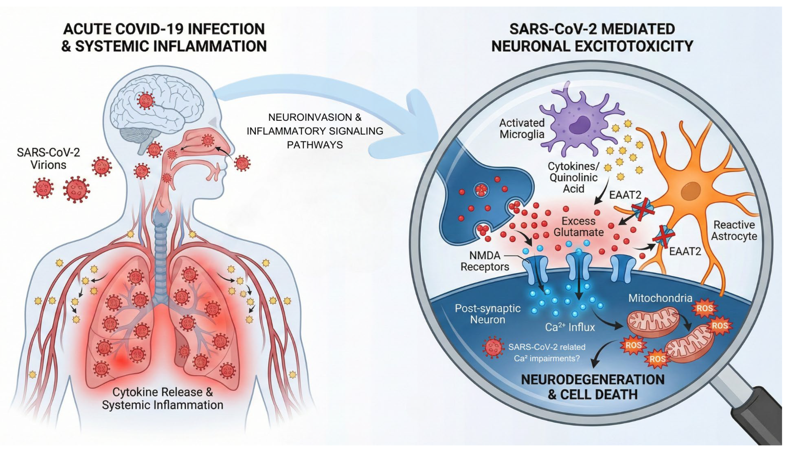
Literature data correlate alterations in Ca2+ signaling and apoptosis in Alzheimer’s disease [97,98] and in other neurodegenerative diseases [99], mainly associated with the aging process. In line with this ionic mobilization, as discussed earlier, astrogliosis and activated microglia, which are related to neurodegeneration and brain aging, play a role in excitotoxicity [100]. Furthermore, along with hypertrophic astrocytes, activated microglia were found in COVID-19 patients, related to microvascular injury [70], which may be involved in neuropathology [76,101]. In addition, a characterization of astrocytes was made by [74], configuring an altered phenotype. Therefore, it is well known that reactive astrocytes are associated with a worse prognosis, considering neurodegenerative processes [17]. Regarding alterations in the CNS and neuronal connectivity (e.g., synaptogenesis), Ca2+ is crucial for LTP, as mentioned earlier, and it is important to investigate the consequences of this absent binding. It is important to highlight that Fernández-Castañeda et al. observed notable alterations in the CNS (such as elevated cytokines, impaired hippocampal neurogenesis, etc.) in a mouse model infected with SARS-CoV-2 [102]. In addition, other harmful factors, such as elevated ROS, excitotoxicity, and inflammation, contribute to the development of neurological disorders, and long COVID-19 may be associated with neurodegeneration (for review [103]). Figure 1 summarizes the proposed mechanism for the possible association between neuroCOVID and excitotoxicity.

5. Conclusions

Much evidence suggests a possible association between neuroCOVID and intrinsic cascades of events that occur under neuroinflammation and disruption of ionic homeostasis, particularly through the excitotoxicity in neurodegenerative conditions. In this sense, Ca2+ signaling is essential for a myriad of processes involving cell survival and neuroplasticity (such as LTP), and can even activate proteases, kinases, phosphatases, endonucleases, etc., some of them related to degradative processes (such as calpains). In addition, massive cytoplasmic Ca2+ entry leads to mitochondrial overloading, impairing ATP function or raising ROS levels, which, in some cases, can be unbalanced by SARS-CoV-2-mediated microvascular alterations and inflammation, also involving microglia and astrocytes, with the participation of NMDAR. These cellular alterations suggest that impaired neurotransmitter homeostasis and mitochondrial failure are important drivers of neurological impairment following SARS-CoV-2 infection.

Author Contributions

Conceptualization, R.P.U.; methodology, L.A.d.S.; validation, R.P.U., L.A.d.S., R.B.O., G.A.S., P.H.Z., L.N., C.M.P., R.S.S.; formal analysis, R.P.U., L.A.d.S.; investigation, R.P.U., L.A.d.S.; resources, R.P.U., L.A.d.S., R.B.O., G.A.S., P.H.Z.; data curation, R.P.U., L.A.d.S.; writing—original draft preparation, R.P.U., L.A.d.S.; writing—review and editing, R.P.U., L.A.d.S., R.B.O., G.A.S.; visualization, R.P.U., L.A.d.S., R.B.O., G.A.S., P.H.Z., L.N., C.M.P., R.S.S.; supervision, R.P.U., C.M.P., R.S.S.; project administration, R.P.U., C.M.P., R.S.S.; funding acquisition, R.P.U., C.M.P., R.S.S. All authors have read and agreed to the published version of the manuscript.

Funding

The authors thank to the Fundação de Amparo à Pesquisa do Estado de São Paulo—FAPESP: 20796-2 (R.P.U.), 2020/04709-8 (R.P.U.), 2024/04261-8 (R.P.U.), 2025/05035-4 (P.H.Z.), 2019/10922-9 (R.S.S.), 2021/00168-5 (R.S.S.), 2020/13480-4 (C.M.P.); Coordenação de Aperfeiçoamento de Pessoal de Nível Superior (CAPES) code 001 (R.B.O., G.A.S., L.A.d.S.); Conselho Nacional de Desenvolvimento Científico e Tecnológico (CNPq) 306397/2023-4 (R.P.U.), 305843/2025-7 (C.M.P.).

Data Availability Statement

Data and materials can be provided by the authors on request.

Acknowledgments

The authors thank the Federal University of São Paulo and the Santa Casa Medical School of São Paulo. During the final revision of this manuscript, the authors used Google’s large language model [Gemini, Advanced version] to refine the language and grammar of the text by using a “Light Proofread” Prompt. The authors have reviewed and edited the output and take full responsibility for the content of this publication.

Conflicts of Interest

Author Louise Newson was employed by the company Newson Global Ltd. The remaining authors declare that the research was conducted in the absence of any commercial or financial relationships that could be construed as a potential conflict of interest.

References

  1. Kleckner, N.W.; Dingledine, R. Requirement for glycine in activation of NMDA-receptors expressed in Xenopus oocytes. Science 1988, 241, 835–837. [Google Scholar] [CrossRef] [PubMed]
  2. Nowak, L.; Bregestovski, P.; Ascher, P.; Herbet, A.; Prochiantz, A. Magnesium gates glutamate-activated channels in mouse central neurones. Nature 1984, 307, 462–465. [Google Scholar] [CrossRef]
  3. Rajdev, S.; Reynolds, I.J. Glutamate-induced intracellular calcium changes and neurotoxicity in cortical neurons in vitro: Effect of chemical ischemia. Neuroscience 1994, 62, 667–679. [Google Scholar] [CrossRef] [PubMed]
  4. Nicholls, D.; Attwell, D. The release and uptake of excitatory amino acids. Trends Pharmacol. Sci. 1990, 11, 462–468. [Google Scholar] [CrossRef] [PubMed]
  5. Coyle, J.T.; Puttfarcken, P. Oxidative stress, glutamate, and neurodegenerative disorders. Science 1993, 262, 689–695. [Google Scholar] [CrossRef]
  6. Beal, M.F. Excitotoxicity and nitric oxide in Parkinson’s disease pathogenesis. Ann. Neurol. 1998, 44, S110–S114. [Google Scholar] [CrossRef]
  7. Zhang, Y.Y.; Ren, K.D.; Luo, X.J.; Peng, J. COVID-19-induced neurological symptoms: Focus on the role of metal ions. Inflammopharmacology 2023, 31, 611–631. [Google Scholar] [CrossRef]
  8. Bennett, M.R. The concept of long term potentiation of transmission at synapses. Prog. Neurobiol. 2000, 60, 109–137. [Google Scholar] [CrossRef]
  9. de Souza, L.; Smaili, S.S.; Ureshino, R.P.; Sinigaglia-Coimbra, R.; Andersen, M.L.; Lopes, G.S.; Tufik, S. Effect of chronic sleep restriction and aging on calcium signaling and apoptosis in the hippocampus of young and aged animals. Prog. Neuropsychopharmacol. Biol. Psychiatry 2012, 39, 23–30. [Google Scholar] [CrossRef]
  10. De Pitta, M.; Brunel, N.; Volterra, A. Astrocytes: Orchestrating synaptic plasticity? Neuroscience 2016, 323, 43–61. [Google Scholar] [CrossRef]
  11. Nakanishi, S. Metabotropic glutamate receptors: Synaptic transmission, modulation, and plasticity. Neuron 1994, 13, 1031–1037. [Google Scholar] [CrossRef]
  12. Ureshino, R.P.; Bertoncini, C.R.; Fernandes, M.J.; Abdalla, F.M.; Porto, C.S.; Hsu, Y.T.; Lopes, G.S.; Smaili, S.S. Alterations in calcium signaling and a decrease in Bcl-2 expression: Possible correlation with apoptosis in aged striatum. J. Neurosci. Res. 2010, 88, 438–447. [Google Scholar] [CrossRef]
  13. Magistretti, P.J. Role of glutamate in neuron-glia metabolic coupling. Am. J. Clin. Nutr. 2009, 90, 875S–880S. [Google Scholar] [CrossRef]
  14. Belanger, M.; Magistretti, P.J. The role of astroglia in neuroprotection. Dialogues Clin. Neurosci. 2009, 11, 281–295. [Google Scholar] [CrossRef] [PubMed]
  15. Kim, K.; Lee, S.G.; Kegelman, T.P.; Su, Z.Z.; Das, S.K.; Dash, R.; Dasgupta, S.; Barral, P.M.; Hedvat, M.; Diaz, P.; et al. Role of excitatory amino acid transporter-2 (EAAT2) and glutamate in neurodegeneration: Opportunities for developing novel therapeutics. J. Cell. Physiol. 2011, 226, 2484–2493. [Google Scholar] [CrossRef]
  16. Qin, Z.H.; Chen, R.W.; Wang, Y.; Nakai, M.; Chuang, D.M.; Chase, T.N. Nuclear factor kappaB nuclear translocation upregulates c-Myc and p53 expression during NMDA receptor-mediated apoptosis in rat striatum. J. Neurosci. 1999, 19, 4023–4033. [Google Scholar] [CrossRef]
  17. Verkhratsky, A.; Parpura, V. Astrogliopathology in neurological, neurodevelopmental and psychiatric disorders. Neurobiol. Dis. 2016, 85, 254–261. [Google Scholar] [CrossRef]
  18. Zhou, X.; Liang, J.; Wang, J.; Fei, Z.; Qin, G.; Zhang, D.; Zhou, J.; Chen, L. Up-regulation of astrocyte excitatory amino acid transporter 2 alleviates central sensitization in a rat model of chronic migraine. J. Neurochem. 2020, 155, 370–389. [Google Scholar] [CrossRef]
  19. Villarreal, A.; Vidos, C.; Monteverde Busso, M.; Cieri, M.B.; Ramos, A.J. Pathological Neuroinflammatory Conversion of Reactive Astrocytes Is Induced by Microglia and Involves Chromatin Remodeling. Front. Pharmacol. 2021, 12, 689346. [Google Scholar] [CrossRef] [PubMed]
  20. Roman, M.; Irwin, M.R. Novel neuroimmunologic therapeutics in depression: A clinical perspective on what we know so far. Brain Behav. Immun. 2020, 83, 7–21. [Google Scholar] [CrossRef] [PubMed]
  21. Smaili, S.S.; Ureshino, R.P.; Rodrigues, L.; Rocha, K.K.; Carvalho, J.T.; Oseki, K.T.; Bincoletto, C.; Lopes, G.S.; Hirata, H. The role of mitochondrial function in glutamate-dependent metabolism in neuronal cells. Curr. Pharm. Des. 2011, 17, 3865–3877. [Google Scholar] [CrossRef] [PubMed]
  22. Jastroch, M.; Divakaruni, A.S.; Mookerjee, S.; Treberg, J.R.; Brand, M.D. Mitochondrial proton and electron leaks. Essays Biochem. 2010, 47, 53–67. [Google Scholar] [CrossRef]
  23. Kent, A.C.; El Baradie, K.B.Y.; Hamrick, M.W. Targeting the Mitochondrial Permeability Transition Pore to Prevent Age-Associated Cell Damage and Neurodegeneration. Oxidative Med. Cell. Longev. 2021, 2021, 6626484. [Google Scholar] [CrossRef]
  24. Blesa, J.; Trigo-Damas, I.; Quiroga-Varela, A.; Jackson-Lewis, V.R. Oxidative stress and Parkinson’s disease. Front. Neuroanat. 2015, 9, 91. [Google Scholar] [CrossRef]
  25. Shin, J.Y.; Fang, Z.H.; Yu, Z.X.; Wang, C.E.; Li, S.H.; Li, X.J. Expression of mutant huntingtin in glial cells contributes to neuronal excitotoxicity. J. Cell Biol. 2005, 171, 1001–1012. [Google Scholar] [CrossRef]
  26. Albin, R.L.; Makowiec, R.L.; Hollingsworth, Z.R.; Dure, L.S.t.; Penney, J.B.; Young, A.B. Excitatory amino acid binding sites in the basal ganglia of the rat: A quantitative autoradiographic study. Neuroscience 1992, 46, 35–48. [Google Scholar] [CrossRef]
  27. Navarro, A.; Lopez-Cepero, J.M.; Bandez, M.J.; Sanchez-Pino, M.J.; Gomez, C.; Cadenas, E.; Boveris, A. Hippocampal mitochondrial dysfunction in rat aging. Am. J. Physiol. Regul. Integr. Comp. Physiol. 2008, 294, R501–R509. [Google Scholar] [CrossRef]
  28. Kourie, J.I. Interaction of reactive oxygen species with ion transport mechanisms. Am. J. Physiol. 1998, 275, C1–C24. [Google Scholar] [CrossRef]
  29. Blaylock, R.L.; Maroon, J. Immunoexcitotoxicity as a central mechanism in chronic traumatic encephalopathy—A unifying hypothesis. Surg. Neurol. Int. 2011, 2, 107. [Google Scholar] [CrossRef] [PubMed]
  30. Hajnoczky, G.; Robb-Gaspers, L.D.; Seitz, M.B.; Thomas, A.P. Decoding of cytosolic calcium oscillations in the mitochondria. Cell 1995, 82, 415–424. [Google Scholar] [CrossRef] [PubMed]
  31. Drew, B.; Leeuwenburgh, C. Ageing and subcellular distribution of mitochondria: Role of mitochondrial DNA deletions and energy production. Acta Physiol. Scand. 2004, 182, 333–341. [Google Scholar] [CrossRef]
  32. Wiersinga, W.J.; Rhodes, A.; Cheng, A.C.; Peacock, S.J.; Prescott, H.C. Pathophysiology, Transmission, Diagnosis, and Treatment of Coronavirus Disease 2019 (COVID-19): A Review. JAMA 2020, 324, 782–793. [Google Scholar] [CrossRef]
  33. Divani, A.A.; Andalib, S.; Biller, J.; Di Napoli, M.; Moghimi, N.; Rubinos, C.A.; Nobleza, C.O.; Sylaja, P.N.; Toledano, M.; Lattanzi, S.; et al. Central Nervous System Manifestations Associated with COVID-19. Curr. Neurol. Neurosci. Rep. 2020, 20, 60. [Google Scholar] [CrossRef] [PubMed]
  34. Johansson, A.; Mohamed, M.S.; Moulin, T.C.; Schioth, H.B. Neurological manifestations of COVID-19: A comprehensive literature review and discussion of mechanisms. J. Neuroimmunol. 2021, 358, 577658. [Google Scholar] [CrossRef]
  35. Shehata, G.A.; Lord, K.C.; Grudzinski, M.C.; Elsayed, M.; Abdelnaby, R.; Elshabrawy, H.A. Neurological Complications of COVID-19: Underlying Mechanisms and Management. Int. J. Mol. Sci. 2021, 22, 4081. [Google Scholar] [CrossRef]
  36. Proust, A.; Queval, C.J.; Harvey, R.; Adams, L.; Bennett, M.; Wilkinson, R.J. Differential effects of SARS-CoV-2 variants on central nervous system cells and blood-brain barrier functions. J. Neuroinflammation 2023, 20, 184. [Google Scholar] [CrossRef] [PubMed]
  37. Zhang, L.; Zhou, L.; Bao, L.; Liu, J.; Zhu, H.; Lv, Q.; Liu, R.; Chen, W.; Tong, W.; Wei, Q.; et al. SARS-CoV-2 crosses the blood-brain barrier accompanied with basement membrane disruption without tight junctions alteration. Signal Transduct. Target. Ther. 2021, 6, 337. [Google Scholar] [CrossRef] [PubMed]
  38. Iadecola, C.; Anrather, J.; Kamel, H. Effects of COVID-19 on the Nervous System. Cell 2020, 183, 16–27.e1. [Google Scholar] [CrossRef]
  39. Meinhardt, J.; Radke, J.; Dittmayer, C.; Franz, J.; Thomas, C.; Mothes, R.; Laue, M.; Schneider, J.; Brunink, S.; Greuel, S.; et al. Olfactory transmucosal SARS-CoV-2 invasion as a port of central nervous system entry in individuals with COVID-19. Nat. Neurosci. 2021, 24, 168–175. [Google Scholar] [CrossRef]
  40. Singh, M.; Khan, R.S.; Dine, K.; Das Sarma, J.; Shindler, K.S. Intracranial Inoculation Is More Potent Than Intranasal Inoculation for Inducing Optic Neuritis in the Mouse Hepatitis Virus-Induced Model of Multiple Sclerosis. Front. Cell. Infect. Microbiol. 2018, 8, 311. [Google Scholar] [CrossRef]
  41. Jiao, L.; Yang, Y.; Yu, W.; Zhao, Y.; Long, H.; Gao, J.; Ding, K.; Ma, C.; Li, J.; Zhao, S.; et al. The olfactory route is a potential way for SARS-CoV-2 to invade the central nervous system of rhesus monkeys. Signal Transduct. Target. Ther. 2021, 6, 169. [Google Scholar] [CrossRef]
  42. van Riel, D.; Verdijk, R.; Kuiken, T. The olfactory nerve: A shortcut for influenza and other viral diseases into the central nervous system. J. Pathol. 2015, 235, 277–287. [Google Scholar] [CrossRef]
  43. Chen, Y.; Yang, W.; Chen, F.; Cui, L. COVID-19 and cognitive impairment: Neuroinvasive and blood–brain barrier dysfunction. J. Neuroinflammation 2022, 19, 222. [Google Scholar] [CrossRef] [PubMed]
  44. Ruchaya, P.J.; Speretta, G.F.; Blanch, G.T.; Li, H.; Sumners, C.; Menani, J.V.; Colombari, E.; Colombari, D.S. Overexpression of AT2R in the solitary-vagal complex improves baroreflex in the spontaneously hypertensive rat. Neuropeptides 2016, 60, 29–36. [Google Scholar] [CrossRef]
  45. Becker, S.P.; Breaux, R.; Cusick, C.N.; Dvorsky, M.R.; Marsh, N.P.; Sciberras, E.; Langberg, J.M. Remote Learning During COVID-19: Examining School Practices, Service Continuation, and Difficulties for Adolescents With and Without Attention-Deficit/Hyperactivity Disorder. J. Adolesc. Health 2020, 67, 769–777. [Google Scholar] [CrossRef] [PubMed]
  46. Ceban, F.; Ling, S.; Lui, L.M.W.; Lee, Y.; Gill, H.; Teopiz, K.M.; Rodrigues, N.B.; Subramaniapillai, M.; Di Vincenzo, J.D.; Cao, B.; et al. Fatigue and cognitive impairment in Post-COVID-19 Syndrome: A systematic review and meta-analysis. Brain Behav. Immun. 2022, 101, 93–135. [Google Scholar] [CrossRef] [PubMed]
  47. Farooq, S.; Tunmore, J.; Wajid Ali, M.; Ayub, M. Suicide, self-harm and suicidal ideation during COVID-19: A systematic review. Psychiatry Res. 2021, 306, 114228. [Google Scholar] [CrossRef]
  48. Fricchione, G.L.; Paul, A.B.; Chemali, Z.; Kritzer, M.D. Case 34-2022: A 57-Year-Old Woman with Covid-19 and Delusions. N. Engl. J. Med. 2022, 387, 1795–1803. [Google Scholar] [CrossRef]
  49. Pappa, S.; Ntella, V.; Giannakas, T.; Giannakoulis, V.G.; Papoutsi, E.; Katsaounou, P. Prevalence of depression, anxiety, and insomnia among healthcare workers during the COVID-19 pandemic: A systematic review and meta-analysis. Brain Behav. Immun. 2020, 88, 901–907. [Google Scholar] [CrossRef]
  50. Helms, J.; Kremer, S.; Merdji, H.; Clere-Jehl, R.; Schenck, M.; Kummerlen, C.; Collange, O.; Boulay, C.; Fafi-Kremer, S.; Ohana, M.; et al. Neurologic Features in Severe SARS-CoV-2 Infection. N. Engl. J. Med. 2020, 382, 2268–2270. [Google Scholar] [CrossRef]
  51. Xu, E.; Xie, Y.; Al-Aly, Z. Long-term neurologic outcomes of COVID-19. Nat. Med. 2022, 28, 2406–2415. [Google Scholar] [CrossRef]
  52. Frontera, J.A.; Sabadia, S.; Lalchan, R.; Fang, T.; Flusty, B.; Millar-Vernetti, P.; Snyder, T.; Berger, S.; Yang, D.; Granger, A.; et al. A Prospective Study of Neurologic Disorders in Hospitalized Patients with COVID-19 in New York City. Neurology 2021, 96, e575–e586. [Google Scholar] [CrossRef]
  53. Poloni, T.E.; Medici, V.; Moretti, M.; Visona, S.D.; Cirrincione, A.; Carlos, A.F.; Davin, A.; Gagliardi, S.; Pansarasa, O.; Cereda, C.; et al. COVID-19-related neuropathology and microglial activation in elderly with and without dementia. Brain Pathol. 2021, 31, e12997. [Google Scholar] [CrossRef]
  54. Stein, L.K.; Mayman, N.A.; Dhamoon, M.S.; Fifi, J.T. The emerging association between COVID-19 and acute stroke. Trends Neurosci. 2021, 44, 527–537. [Google Scholar] [CrossRef] [PubMed]
  55. Uginet, M.; Breville, G.; Assal, F.; Lovblad, K.O.; Vargas, M.I.; Pugin, J.; Serratrice, J.; Herrmann, F.R.; Lalive, P.H.; Allali, G. COVID-19 encephalopathy: Clinical and neurobiological features. J. Med. Virol. 2021, 93, 4374–4381. [Google Scholar] [CrossRef]
  56. Nalbandian, A.; Sehgal, K.; Gupta, A.; Madhavan, M.V.; McGroder, C.; Stevens, J.S.; Cook, J.R.; Nordvig, A.S.; Shalev, D.; Sehrawat, T.S.; et al. Post-acute COVID-19 syndrome. Nat. Med. 2021, 27, 601–615. [Google Scholar] [CrossRef]
  57. Sudre, C.H.; Murray, B.; Varsavsky, T.; Graham, M.S.; Penfold, R.S.; Bowyer, R.C.; Pujol, J.C.; Klaser, K.; Antonelli, M.; Canas, L.S.; et al. Attributes and predictors of long COVID. Nat. Med. 2021, 27, 626–631. [Google Scholar] [CrossRef]
  58. Al-Aly, Z.; Xie, Y.; Bowe, B. High-dimensional characterization of post-acute sequelae of COVID-19. Nature 2021, 594, 259–264. [Google Scholar] [CrossRef] [PubMed]
  59. Cai, M.; Xie, Y.; Topol, E.J.; Al-Aly, Z. Three-year outcomes of post-acute sequelae of COVID-19. Nat. Med. 2024, 30, 1564–1573. [Google Scholar] [CrossRef]
  60. Heneka, M.T.; Golenbock, D.; Latz, E.; Morgan, D.; Brown, R. Immediate and long-term consequences of COVID-19 infections for the development of neurological disease. Alzheimers Res. Ther. 2020, 12, 69. [Google Scholar] [CrossRef] [PubMed]
  61. Ellul, M.A.; Benjamin, L.; Singh, B.; Lant, S.; Michael, B.D.; Easton, A.; Kneen, R.; Defres, S.; Sejvar, J.; Solomon, T. Neurological associations of COVID-19. Lancet Neurol. 2020, 19, 767–783. [Google Scholar] [CrossRef]
  62. Jaywant, A.; Vanderlind, W.M.; Alexopoulos, G.S.; Fridman, C.B.; Perlis, R.H.; Gunning, F.M. Frequency and profile of objective cognitive deficits in hospitalized patients recovering from COVID-19. Neuropsychopharmacology 2021, 46, 2235–2240. [Google Scholar] [CrossRef] [PubMed]
  63. Mendez, R.; Balanza-Martinez, V.; Luperdi, S.C.; Estrada, I.; Latorre, A.; Gonzalez-Jimenez, P.; Bouzas, L.; Yepez, K.; Ferrando, A.; Reyes, S.; et al. Long-term neuropsychiatric outcomes in COVID-19 survivors: A 1-year longitudinal study. J. Intern. Med. 2022, 291, 247–251. [Google Scholar] [CrossRef]
  64. Paterson, R.W.; Brown, R.L.; Benjamin, L.; Nortley, R.; Wiethoff, S.; Bharucha, T.; Jayaseelan, D.L.; Kumar, G.; Raftopoulos, R.E.; Zambreanu, L.; et al. The emerging spectrum of COVID-19 neurology: Clinical, radiological and laboratory findings. Brain 2020, 143, 3104–3120. [Google Scholar] [CrossRef]
  65. Beghi, E.; Giussani, G.; Westenberg, E.; Allegri, R.; Garcia-Azorin, D.; Guekht, A.; Frontera, J.; Kivipelto, M.; Mangialasche, F.; Mukaetova-Ladinska, E.B.; et al. Acute and post-acute neurological manifestations of COVID-19: Present findings, critical appraisal, and future directions. J. Neurol. 2022, 269, 2265–2274. [Google Scholar] [CrossRef]
  66. Davis, H.E.; Assaf, G.S.; McCorkell, L.; Wei, H.; Low, R.J.; Re’em, Y.; Redfield, S.; Austin, J.P.; Akrami, A. Characterizing long COVID in an international cohort: 7 months of symptoms and their impact. EClinicalMedicine 2021, 38, 101019. [Google Scholar] [CrossRef]
  67. Global Burden of Disease Long COVID Collaborators; Wulf Hanson, S.; Abbafati, C.; Aerts, J.G.; Al-Aly, Z.; Ashbaugh, C.; Ballouz, T.; Blyuss, O.; Bobkova, P.; Bonsel, G.; et al. Estimated Global Proportions of Individuals with Persistent Fatigue, Cognitive, and Respiratory Symptom Clusters Following Symptomatic COVID-19 in 2020 and 2021. JAMA 2022, 328, 1604–1615. [Google Scholar] [CrossRef] [PubMed]
  68. Bianchetti, A.; Rozzini, R.; Guerini, F.; Boffelli, S.; Ranieri, P.; Minelli, G.; Bianchetti, L.; Trabucchi, M. Clinical Presentation of COVID19 in Dementia Patients. J. Nutr. Health Aging 2020, 24, 560–562. [Google Scholar] [CrossRef] [PubMed]
  69. Schild, A.K.; Goereci, Y.; Scharfenberg, D.; Klein, K.; Lulling, J.; Meiberth, D.; Schweitzer, F.; Sturmer, S.; Zeyen, P.; Sahin, D.; et al. Multidomain cognitive impairment in non-hospitalized patients with the post-COVID-19 syndrome: Results from a prospective monocentric cohort. J. Neurol. 2023, 270, 1215–1223. [Google Scholar] [CrossRef]
  70. Lee, M.H.; Perl, D.P.; Nair, G.; Li, W.; Maric, D.; Murray, H.; Dodd, S.J.; Koretsky, A.P.; Watts, J.A.; Cheung, V.; et al. Microvascular Injury in the Brains of Patients with COVID-19. N. Engl. J. Med. 2021, 384, 481–483. [Google Scholar] [CrossRef]
  71. Cecchetti, G.; Agosta, F.; Canu, E.; Basaia, S.; Barbieri, A.; Cardamone, R.; Bernasconi, M.P.; Castelnovo, V.; Cividini, C.; Cursi, M.; et al. Cognitive, EEG, and MRI features of COVID-19 survivors: A 10-month study. J. Neurol. 2022, 269, 3400–3412. [Google Scholar] [CrossRef]
  72. Bennett, I.J.; Madden, D.J. Disconnected aging: Cerebral white matter integrity and age-related differences in cognition. Neuroscience 2014, 276, 187–205. [Google Scholar] [CrossRef]
  73. Bartolomeo, C.S.; Lemes, R.M.R.; Morais, R.L.; Pereria, G.C.; Nunes, T.A.; Costa, A.J.; de Barros Maciel, R.M.; Braconi, C.T.; Maricato, J.T.; Janini, L.M.R.; et al. SARS-CoV-2 infection and replication kinetics in different human cell types: The role of autophagy, cellular metabolism and ACE2 expression. Life Sci. 2022, 308, 120930. [Google Scholar] [CrossRef]
  74. Bayat, A.H.; Azimi, H.; Hassani Moghaddam, M.; Ebrahimi, V.; Fathi, M.; Vakili, K.; Mahmoudiasl, G.R.; Forouzesh, M.; Boroujeni, M.E.; Nariman, Z.; et al. COVID-19 causes neuronal degeneration and reduces neurogenesis in human hippocampus. Apoptosis 2022, 27, 852–868. [Google Scholar] [CrossRef] [PubMed]
  75. Venkataramani, V.; Winkler, F. Cognitive Deficits in Long COVID-19. N. Engl. J. Med. 2022, 387, 1813–1815. [Google Scholar] [CrossRef] [PubMed]
  76. Tavcar, P.; Potokar, M.; Kolenc, M.; Korva, M.; Avsic-Zupanc, T.; Zorec, R.; Jorgacevski, J. Neurotropic Viruses, Astrocytes, and COVID-19. Front. Cell. Neurosci. 2021, 15, 662578. [Google Scholar] [CrossRef]
  77. Paidas, M.J.; Cosio, D.S.; Ali, S.; Kenyon, N.S.; Jayakumar, A.R. Long-Term Sequelae of COVID-19 in Experimental Mice. Mol. Neurobiol. 2022, 59, 5970–5986. [Google Scholar] [CrossRef]
  78. Cao, S.; Song, Z.; Rong, J.; Andrikopoulos, N.; Liang, X.; Wang, Y.; Peng, G.; Ding, F.; Ke, P.C. Spike Protein Fragments Promote Alzheimer’s Amyloidogenesis. ACS Appl. Mater. Interfaces 2023, 15, 40317–40329. [Google Scholar] [CrossRef]
  79. Straus, M.R.; Tang, T.; Lai, A.L.; Flegel, A.; Bidon, M.; Freed, J.H.; Daniel, S.; Whittaker, G.R. Ca(2+) Ions Promote Fusion of Middle East Respiratory Syndrome Coronavirus with Host Cells and Increase Infectivity. J. Virol. 2020, 94, e00426-20. [Google Scholar] [CrossRef]
  80. Danta, C.C. Calcium Channel Blockers: A Possible Potential Therapeutic Strategy for the Treatment of Alzheimer’s Dementia Patients with SARS-CoV-2 Infection. ACS Chem. Neurosci. 2020, 11, 2145–2148. [Google Scholar] [CrossRef]
  81. Simpson, A.A.; Chandrasekar, V.; Hebert, B.; Sullivan, G.M.; Rossmann, M.G.; Parrish, C.R. Host range and variability of calcium binding by surface loops in the capsids of canine and feline parvoviruses. J. Mol. Biol. 2000, 300, 597–610. [Google Scholar] [CrossRef]
  82. Brisac, C.; Teoule, F.; Autret, A.; Pelletier, I.; Colbere-Garapin, F.; Brenner, C.; Lemaire, C.; Blondel, B. Calcium flux between the endoplasmic reticulum and mitochondrion contributes to poliovirus-induced apoptosis. J. Virol. 2010, 84, 12226–12235. [Google Scholar] [CrossRef]
  83. Cheshenko, N.; Del Rosario, B.; Woda, C.; Marcellino, D.; Satlin, L.M.; Herold, B.C. Herpes simplex virus triggers activation of calcium-signaling pathways. J. Cell Biol. 2003, 163, 283–293. [Google Scholar] [CrossRef] [PubMed]
  84. Chen, Z.; Wang, X.; Ashraf, U.; Zheng, B.; Ye, J.; Zhou, D.; Zhang, H.; Song, Y.; Chen, H.; Zhao, S.; et al. Activation of neuronal N-methyl-D-aspartate receptor plays a pivotal role in Japanese encephalitis virus-induced neuronal cell damage. J. Neuroinflammation 2018, 15, 238. [Google Scholar] [CrossRef]
  85. Jayaseelan, V.P.; Paramasivam, A. Repurposing calcium channel blockers as antiviral drugs. J. Cell Commun. Signal. 2020, 14, 467–468. [Google Scholar] [CrossRef] [PubMed]
  86. Rosenthal, N.; Cao, Z.; Gundrum, J.; Sianis, J.; Safo, S. Risk Factors Associated with In-Hospital Mortality in a US National Sample of Patients with COVID-19. JAMA Netw. Open 2020, 3, e2029058. [Google Scholar] [CrossRef]
  87. Alsagaff, M.Y.; Mulia, E.P.B.; Maghfirah, I.; Luke, K.; Nugraha, D.; Rachmi, D.A.; Septianda, I.; A’Yun, M.Q. Association of calcium channel blocker use with clinical outcome of COVID-19: A meta-analysis. Diabetes Metab. Syndr. 2021, 15, 102210. [Google Scholar] [CrossRef]
  88. Brown, E.G.; Chahine, L.M.; Goldman, S.M.; Korell, M.; Mann, E.; Kinel, D.R.; Arnedo, V.; Marek, K.L.; Tanner, C.M. The Effect of the COVID-19 Pandemic on People with Parkinson’s Disease. J. Park. Dis. 2020, 10, 1365–1377. [Google Scholar] [CrossRef] [PubMed]
  89. Ineichen, C.; Baumann-Vogel, H.; Sitzler, M.; Waldvogel, D.; Baumann, C.R. Worsened Parkinson’s Disease Progression: Impact of the COVID-19 Pandemic. J. Park. Dis. 2021, 11, 1579–1583. [Google Scholar] [CrossRef]
  90. Boutoleau-Bretonniere, C.; Pouclet-Courtemanche, H.; Gillet, A.; Bernard, A.; Deruet, A.L.; Gouraud, I.; Mazoue, A.; Lamy, E.; Rocher, L.; Kapogiannis, D.; et al. The Effects of Confinement on Neuropsychiatric Symptoms in Alzheimer’s Disease During the COVID-19 Crisis. J. Alzheimers Dis. 2020, 76, 41–47. [Google Scholar] [CrossRef] [PubMed]
  91. Barguilla, A.; Fernandez-Lebrero, A.; Estragues-Gazquez, I.; Garcia-Escobar, G.; Navalpotro-Gomez, I.; Manero, R.M.; Puente-Periz, V.; Roquer, J.; Puig-Pijoan, A. Effects of COVID-19 Pandemic Confinement in Patients with Cognitive Impairment. Front. Neurol. 2020, 11, 589901. [Google Scholar] [CrossRef]
  92. Chen, Z.C.; Liu, S.; Gan, J.; Ma, L.; Du, X.; Zhu, H.; Han, J.; Xu, J.; Wu, H.; Fei, M.; et al. The Impact of the COVID-19 Pandemic and Lockdown on Mild Cognitive Impairment, Alzheimer’s Disease and Dementia with Lewy Bodies in China: A 1-Year Follow-up Study. Front. Psychiatry 2021, 12, 711658. [Google Scholar] [CrossRef]
  93. Fedeli, U.; Casotto, V.; Barbiellini Amidei, C.; Saia, M.; Tiozzo, S.N.; Basso, C.; Schievano, E. Parkinson’s disease related mortality: Long-term trends and impact of COVID-19 pandemic waves. Park. Relat. Disord. 2022, 98, 75–77. [Google Scholar] [CrossRef] [PubMed]
  94. Zenesini, C.; Vignatelli, L.; Belotti, L.M.B.; Baccari, F.; Calandra-Buonaura, G.; Cortelli, P.; Descovich, C.; Giannini, G.; Guaraldi, P.; Guarino, M.; et al. Risk of SARS-CoV-2 infection, hospitalization and death for COVID-19 in people with Parkinson’s disease or parkinsonism over a 15-month period: A cohort study. Eur. J. Neurol. 2022, 29, 3205–3217. [Google Scholar] [CrossRef]
  95. Xia, X.; Wang, Y.; Zheng, J. COVID-19 and Alzheimer’s disease: How one crisis worsens the other. Transl. Neurodegener. 2021, 10, 15. [Google Scholar] [CrossRef]
  96. Atkins, J.L.; Masoli, J.A.H.; Delgado, J.; Pilling, L.C.; Kuo, C.L.; Kuchel, G.A.; Melzer, D. Preexisting Comorbidities Predicting COVID-19 and Mortality in the UK Biobank Community Cohort. J. Gerontol. A Biol. Sci. Med. Sci. 2020, 75, 2224–2230. [Google Scholar] [CrossRef]
  97. Demuro, A.; Parker, I.; Stutzmann, G.E. Calcium signaling and amyloid toxicity in Alzheimer disease. J. Biol. Chem. 2010, 285, 12463–12468. [Google Scholar] [CrossRef] [PubMed]
  98. Oseki, K.T.; Monteforte, P.T.; Pereira, G.J.; Hirata, H.; Ureshino, R.P.; Bincoletto, C.; Hsu, Y.T.; Smaili, S.S. Apoptosis induced by Abeta25-35 peptide is Ca2+-IP3 signaling-dependent in murine astrocytes. Eur. J. Neurosci. 2014, 40, 2471–2478. [Google Scholar] [CrossRef] [PubMed]
  99. Ureshino, R.P.; Erustes, A.G.; Bassani, T.B.; Wachilewski, P.; Guarache, G.C.; Nascimento, A.C.; Costa, A.J.; Smaili, S.S.; Pereira, G. The Interplay between Ca2+ Signaling Pathways and Neurodegeneration. Int. J. Mol. Sci. 2019, 20, 6004. [Google Scholar] [CrossRef]
  100. Castillo-Ruiz, M.M.; Campuzano, O.; Acarin, L.; Castellano, B.; Gonzalez, B. Delayed neurodegeneration and early astrogliosis after excitotoxicity to the aged brain. Exp. Gerontol. 2007, 42, 343–354. [Google Scholar] [CrossRef]
  101. Gerhard, A. Does Microglial Activation Lead to Cognitive Changes After COVID-19 Infection? JAMA Psychiatry 2023, 80, 767. [Google Scholar] [CrossRef]
  102. Fernandez-Castaneda, A.; Lu, P.; Geraghty, A.C.; Song, E.; Lee, M.H.; Wood, J.; O’Dea, M.R.; Dutton, S.; Shamardani, K.; Nwangwu, K.; et al. Mild respiratory COVID can cause multi-lineage neural cell and myelin dysregulation. Cell 2022, 185, 2452–2468.e16. [Google Scholar] [CrossRef] [PubMed]
  103. Ureshino, R.P.; Stilhano, R.S.; Prado, C.M.; Newson, L. Post-COVID-19 Neuropathology and Perspectives of Protective Roles of Estrogens. Covid 2025, 5, 9. [Google Scholar] [CrossRef]
Figure 1. Integrative model of SARS-CoV-2-induced neuronal excitotoxicity and the pathophysi-ology of post-COVID-19 syndrome. This diagram illustrates the pathophysiological cascade extending from systemic infection to molecular neurodegeneration. Left Panel—Macroscale context: Exposure to SARS-CoV-2 triggers an acute respiratory infection and a subsequent systemic inflammatory response. Transition: These systemic signals, along with neuroinvasion pathways, activate neuroinflammatory signaling pathways within the Central Nervous System (CNS). Right Panel—Microscale mechanism: The magnified tripartite synapse highlights the disruption of glutamate homeostasis. Activated microglia release pro-inflammatory cytokines and neurotoxic metabolites, such as quinolinic acid (an NMDAR agonist), while reactive astrocytes exhibit impaired glutamate clearance due to the downregulation or dysfunction of glutamate transporters (EAAT2/GLT-1). This results in excess synaptic glutamate, leading to the overactivation of NMDA receptors (NMDAR). The consequent massive influx of Ca2+—indicated as “SARS-CoV-2-related Ca2+ impairments”—triggers intracellular cascades, including mitochondrial dysfunction and increased production of reactive oxygen species (ROS). Collectively, these mechanisms culminate in synaptic loss, neuronal death, and the clinical manifestations of neurological post-COVID-19 syndrome, such as cognitive impairment and delirium.
Covid 06 00085 g001
Disclaimer/Publisher’s Note: The statements, opinions and data contained in all publications are solely those of the individual author(s) and contributor(s) and not of MDPI and/or the editor(s). MDPI and/or the editor(s) disclaim responsibility for any injury to people or property resulting from any ideas, methods, instructions or products referred to in the content.