1. Introduction
In recent years, vaccines against COVID-19, the disease caused by the severe acute respiratory syndrome coronavirus 2 (SARS-CoV-2), have played a significant global role in reducing deaths and severe illness associated with infection, as well as reducing transmission. According to the World Health Organization (WHO), the widespread vaccination of people against SARS-CoV-2 has resulted in far fewer people contracting the disease and being hospitalized, thereby alleviating the burden on healthcare systems. Moreover, vaccination programs have been vital in helping societies resume normal activities and economies reopen around the world. Although the benefits of vaccination far outweigh the risks, vaccines can cause serious adverse reactions that affect various organ systems. The most common short-term symptoms include migraines, fever, exhaustion, and reactions at the injection site [
1]. Cardiovascular problems have also been reported, including myocarditis, pericarditis, thrombotic events and arrhythmias [
2]. A growing body of literature indicates that neurological manifestations can follow vaccination against SARS-CoV-2 [
3,
4].
Although non-ocular adverse events have been well documented, ocular side effects have received far less attention. These manifestations are likely to affect the eyelids, cornea and ocular surface, as well as the retina, uvea, optic nerve and blood vessels [
4,
5,
6,
7,
8,
9]. While the pandemic is no longer a public health concern, we believe it is important to recall that vaccines can lead to ocular manifestations, bearing in mind that new, updated versions of the vaccine are currently becoming available.
We present here the case of a patient who presented with unilateral choroidal effusion with secondary angle-closure due to severe panuveitis four weeks after receiving the third dose of the anti-SARS-CoV2 vaccine (COMIRNATY®, BioNTech–Pfizer).
2. Case Report
An 87-year-old woman with no significant medical history was referred to our ophthalmology ward due to a progressive decrease in visual acuity and severe pain in her right eye over the past four weeks. The medical history was not contributory except for the third dose of the anti-SARS-CoV-2 vaccine (COMIRNATY®, BioNTech–Pfizer) undergone four weeks prior to the onset of symptoms. She had undergone bilateral cataract surgery several years prior.
Her best-corrected visual acuity was hand movements in the right eye and 20/20 in the left eye. Her intraocular pressure was 34 mmHg and 16 mmHg, respectively. A slit lamp examination of her right eye revealed palpebral edema and chemosis, temporal scleral thickening and hyperemia. The cornea was edematous, with endothelial precipitates. The anterior chamber was shallow with a closed angle and grade 1+ cells and 1+ flare according to the Standardization of Uveitis Nomenclature (SUN) criteria [
10]. Reactive mydriasis was present, defined as pupillary dilation with preserved light reactivity, suggestive of early optic nerve dysfunction [
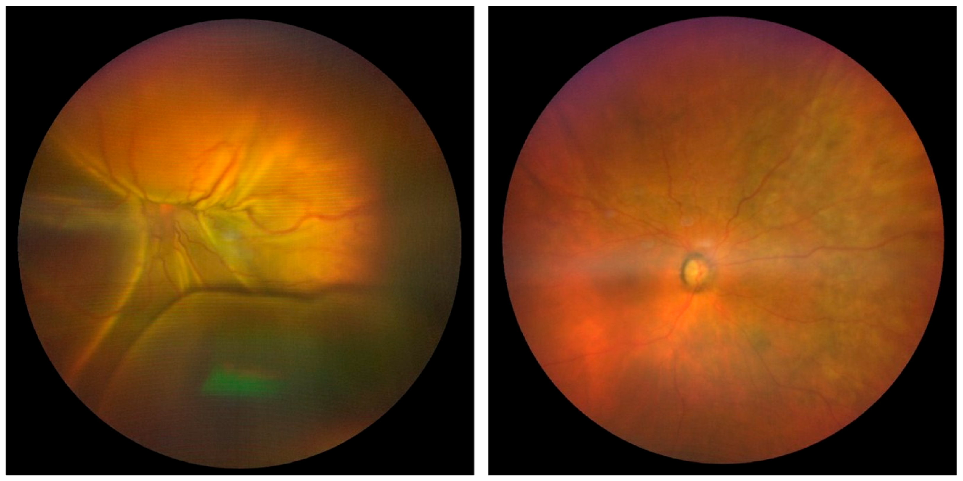
11]. Mild vitreous inflammation was present, with grade 0.5+ vitreous cells, and the dilated fundus revealed total choroidal detachment with limited exudative retinal detachment (
Figure 1, left). The examination of the left eye was unremarkable.
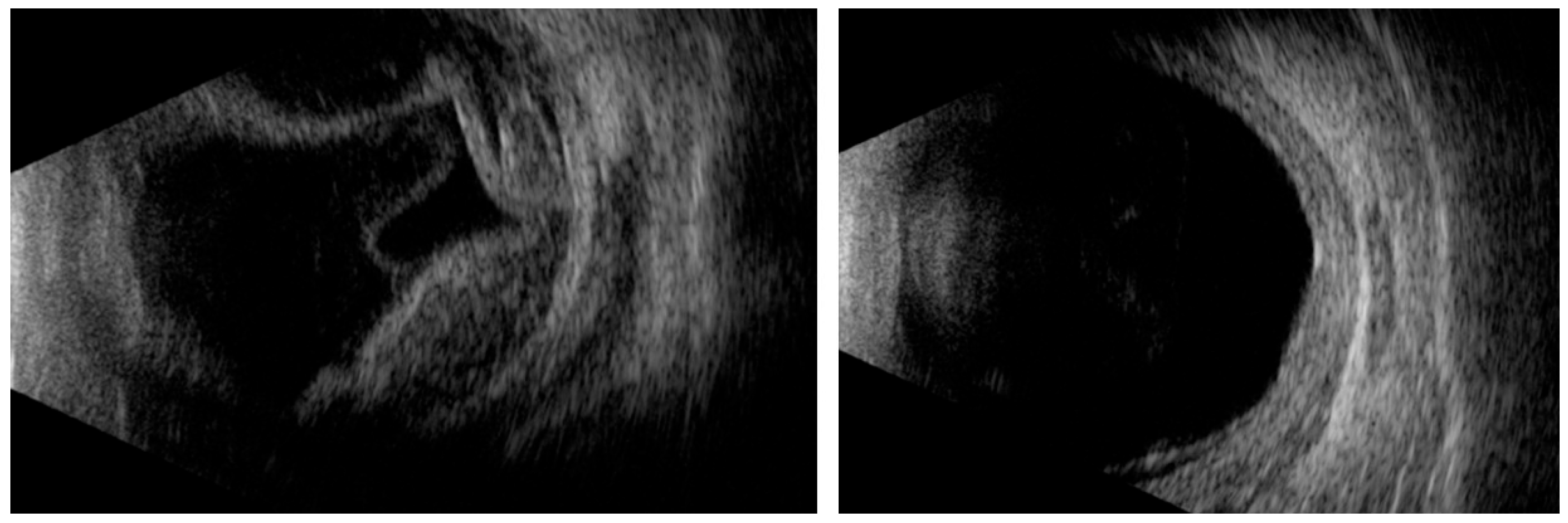
B-mode ultrasonography revealed total choroidal detachment with limited exudative retinal detachment (
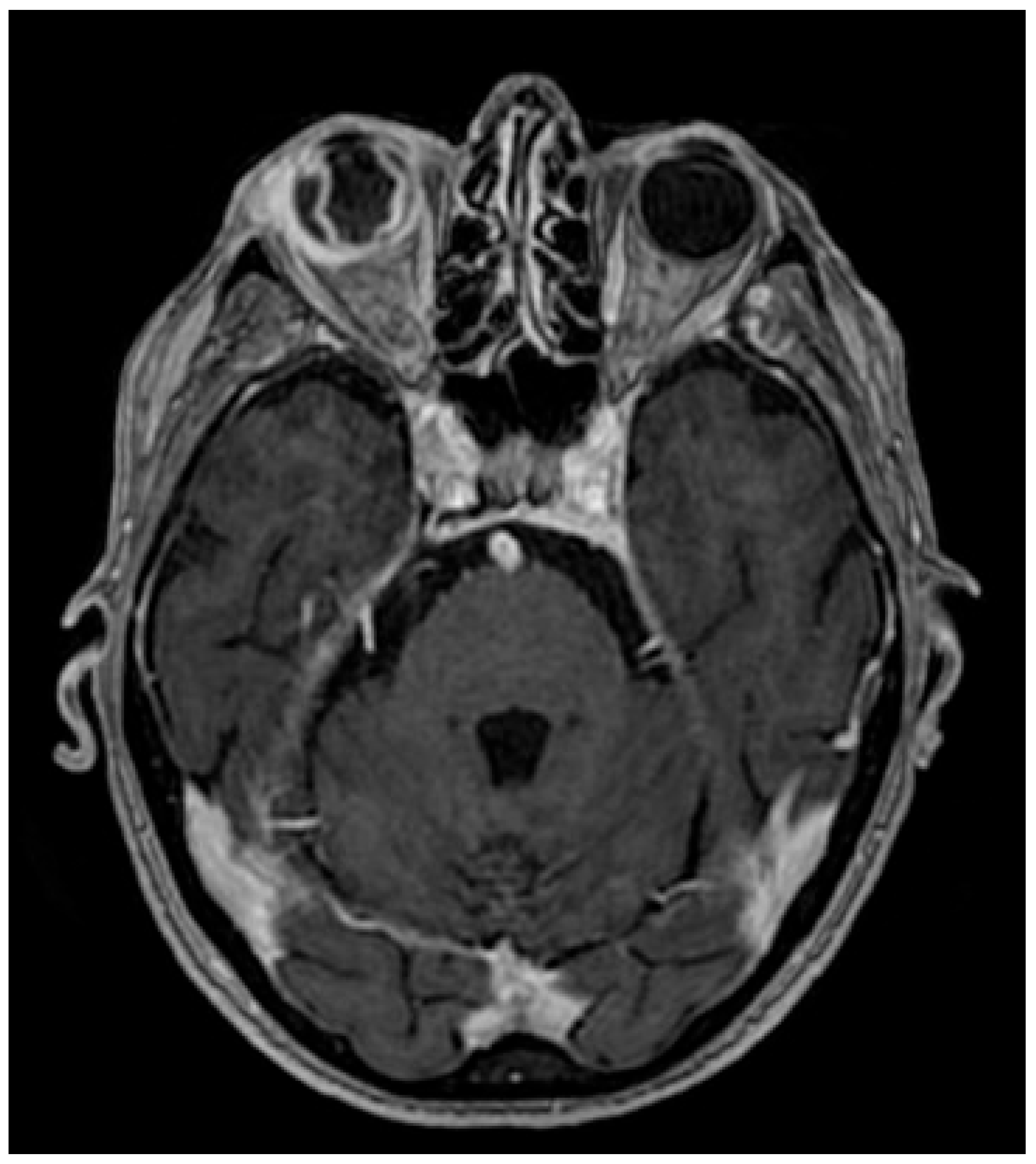
Figure 2, left). Magnetic resonance imaging (MRI) showed an inflammatory enhancement of the sclera, choroid, extraocular muscles, lacrimal gland, and orbital fat (
Figure 3). All biological tests were normal, including the infectious and autoimmune tests, notably the anti-neutrophil cytoplasm antibody (ANCA) and IgG4 serum levels. In addition, the internist’s evaluation and positron emission tomography/computed tomography (PET/CT) scan were inconclusive. A biopsy of the right sclera revealed fibrous tissue and nonspecific chronic inflammatory changes.
We concluded that the patient had unilateral choroidal effusion with secondary angle-closure due to panuveitis. She was treated with antihypertensive, anti-inflammatory, and cycloplegic eye drops. Six weeks later, the best-corrected visual acuity (BCVA) of the right eye had improved to 20/100 and the intraocular pressure had normalized. The anterior segment appeared deep and quiet. The choroidal and retinal detachments were regressing, with mild residual optic disc pallor (
Figure 1, right).
After an extensive evaluation revealed no plausible cause, and given that the patient had not begun any new treatments, we considered that her ocular condition was likely associated with the third vaccine dose, according to the Naranjo Adverse Drug Reaction Probability Scale [
12]. This scale was developed by Narango et al. at the University of Toronto to standardize the assessment of causality for all adverse drug reactions (ADRs), including those associated with vaccines.
3. Discussion
Previous reports have documented an association between vaccinations and various types of ocular inflammation. Numerous vaccines have been linked to anterior and intermediate uveitis, Vogt–Koyanagi–Harada syndrome, and multiple evanescent white dot syndrome (MEWDS). Examples include vaccines for the influenza and hepatitis B viruses [
13,
14]. To date, several ocular adverse effects of anti-SARS-CoV-2 vaccines have been reported in the scientific literature, involving anterior uveitis, panuveitis, posterior uveitis, and others [
15]. Although the underlying pathological mechanism is not fully understood, these ocular side effects are believed to result from the body’s humoral and cellular immune responses to the vaccine [
4,
16]. As reported by Mushtaq et al., the majority of vaccine reactions peak within six weeks after vaccination [
17]. In our case, the reaction peaked at four weeks. According to Abrishami et al., ophthalmological manifestations such as eyelid edema, purpuric eyelid lesions, and uveitis are more frequently associated with mRNA-based vaccines. The latter was used in our case [
18].
One remaining question concerns the strictly unilateral presentation. Theoretically, systemic immune activation following mRNA vaccination would expose both eyes to the same inflammatory stimuli. However, asymmetric ocular involvement is not uncommon in immune-mediated eye diseases [
19]. Subclinical differences between the two eyes, such as variations in vascular permeability, blood–ocular barrier integrity, or the local immune microenvironment, may make one eye more susceptible to inflammation [
20]. In our patient, prior bilateral cataract surgery may also have contributed to subtle differences in ocular vulnerability [
21].
Our case highlights the importance of a complete ophthalmic examination in patients with ocular symptoms after vaccination. Ophthalmologists must be aware of the potential risk of ocular manifestations following anti-SARS-CoV-2 vaccination. The European Medicines Agency (EMA) carefully monitors all side effects of different vaccines. As vaccinations against SARS-CoV-2 continue, potential adverse events affecting the eyes should be notified to EMA in detail to raise awareness among medical professionals, although a definitive causal relationship cannot typically be established. Furthermore, as SARS-CoV-2 becomes endemic, establishing an international registry dedicated to compiling rare ocular adverse drug reactions occurring after anti-SARS-CoV-2 vaccination could improve our understanding of these uncommon events.
This case report has limitations that are inherent to many single-case observations and should be considered when interpreting the findings. An additional limitation concerns the incomplete documentation of the patient’s vaccination history. Although it is known that the patient received a third dose of the Pfizer–BioNTech mRNA vaccine, precise information regarding the interval between doses and the exact timing of prior injections was not available. The chronological relationship between antigen exposure and symptom onset is a critical element when evaluating potential immune-mediated adverse events, as shorter intervals or recent booster doses could theoretically enhance immune reactivity through primed humoral and cellular responses. The absence of these data limits the ability to fully assess temporal plausibility and immunological context. While a causal relationship between anti-SARS-CoV-2 vaccination and the reported ophthalmic abnormalities remains unproven, biological mechanisms analogous to those reported after other types of vaccination support the plausibility of such a link. Although the pandemic is no longer considered a public health emergency, it remains important to recognize that vaccines may be associated with ocular adverse events. This is especially true as new, updated versions of the SARS-CoV-2 vaccine become available.
4. Conclusions
Several ocular inflammatory complications can occur following SARS-CoV-2 vaccination. In this case, the patient experienced unilateral choroidal effusion with secondary angle-closure due to severe panuveitis, presumably caused by the anti-SARS-CoV2 vaccine (COMIRNATY®, BioNTech–Pfizer).
Author Contributions
P.B., E.A., P.S., L.P. and A.K. made substantial contributions to the acquisition, analysis and interpretation of data. P.B. and E.A. contributed to the conception and design of the work. P.B. and E.A. prepared the publication draft, which was revised critically for intellectual content by P.S., L.P. and A.K. All authors have read and agreed to the published version of the manuscript.
Funding
This research received no external funding.
Institutional Review Board Statement
This case report was conducted in accordance with the principles of the Declaration of Helsinki. All protected patient health information was collected and evaluated in a HIPAA-compliant manner. The publication of this case report has been approved by the Institutional Review Board Comité d’Ethique Hospitalo-Facultaire Saint-Luc—UCLouvain, Brussels, Belgium.
Informed Consent Statement
Written informed consent was obtained from the patient before publication of all photographs and images included herein.
Data Availability Statement
The original contributions presented in this study are included in the article. Further inquiries can be directed to the corresponding author.
Conflicts of Interest
The authors declared no conflicts of interest regarding the research, authorship, or publication of this article.
Abbreviations
The following abbreviations are used in this manuscript:
| ADR | Adverse drug reaction |
| ANCA | Anti-neutrophil cytoplasm antibody |
| BCVA | Best-corrected visual acuity |
| CT | Computed tomography |
| EMA | European Medicines Agency |
| MRI | Magnetic resonance imaging |
| PET | Positron emission tomography |
| SARS-CoV2 | Severe acute respiratory syndrome coronavirus 2 |
| WHO | World Health Organization |
References
- Yaamika, H.; Muralidas, D.; Elumalai, K. Review of adverse events associated with COVID-19 vaccines, highlighting their frequencies and reported cases. J. Taibah Univ. Med. Sci. 2023, 18, 1646–1661. [Google Scholar] [CrossRef] [Scilit] [PubMed]
- Hana, D.; Patel, K.; Roman, S.; Gattas, B.; Sofka, S. Clinical Cardiovascular Adverse Events Reported Post-COVID-19 Vaccination: Are They a Real Risk? Curr. Probl. Cardiol. 2022, 47, 101077. [Google Scholar] [CrossRef] [Scilit] [PubMed]
- Khatami, S.S.; Revheim, M.E.; Høilund-Carlsen, P.F.; Alavi, A.; Ghorbani Shirkouhi, S.; Andalib, S. Central nervous system manifestations following vaccination against COVID-19. Brain Behav. Immun. Health 2024, 38, 100788. [Google Scholar] [CrossRef] [Scilit] [PubMed]
- Ng, X.L.; Betzler, B.K.; Testi, I.; Ho, S.L.; Tien, M.; Ngo, W.K.; Zierhut, M.; Chee, S.P.; Gupta, V.; Pavesio, C.E.; et al. Ocular Adverse Events After COVID-19 Vaccination. Ocul. Immunol. Inflamm. 2021, 29, 1216–1224. [Google Scholar] [CrossRef] [Scilit]
- Lee, Y.K.; Huang, Y.H. Ocular Manifestations after Receiving COVID-19 Vaccine: A Systematic Review. Vaccines 2021, 9, 1404. [Google Scholar] [CrossRef] [Scilit]
- Yeung, M.; Su, C.K.; Au, S.C.L. Vaccine-related retinal artery occlusion in adults: A review of the current literature. J. Stroke Cerebrovasc. Dis. 2023, 32, 106694. [Google Scholar] [CrossRef] [Scilit]
- Mahendradas, P.; Mishra, S.B.; Sangoram, R.; Srinivasan, S.; Kawali, A.; Patil, A.; Shetty, R. Ocular manifestations following COVID-19 vaccination. J. Ophthalmic Inflamm. Infect. 2023, 13, 44. [Google Scholar] [CrossRef] [Scilit]
- Au, S.C.L. Comments on: After the Storm: Ophthalmic Manifestations of COVID-19 Vaccines. Indian. J. Ophthalmol. 2022, 70, 3737–3738. [Google Scholar] [CrossRef] [Scilit]
- Rallis, K.I.; Fausto, R.; Ting, D.S.J.; Al-Aqaba, M.A.; Said, D.G.; Dua, H.S. Manifestation of Herpetic Eye Disease After COVID-19 Vaccine: A UK Case Series. Ocul. Immunol. Inflamm. 2022, 30, 1136–1141. [Google Scholar] [CrossRef] [Scilit]
- Jabs, D.A.; Nussenblatt, R.B.; Rosenbaum, J.T.; Standardization of Uveitis Nomenclature (SUN) Working Group. Standardization of Uveitis Nomenclature for Reporting Clinical Data. Results of the First International Workshop. Am. J. Ophthalmol. 2005, 140, 509–516. [Google Scholar]
- Leon, L.R.; Marinaro, C. Previous ischaemic optic neuropathy in haemodialysis. Nefrologia 2009, 29, 503–671. [Google Scholar]
- Naranjo, C.A.; Busto, U.; Sellers, E.M.; Sandor, P.; Ruiz, I.; Roberts, E.A.; Janecek, E.; Domecq, C.; Greenblatt, D.J. A method for estimating the probability of adverse drug reactions. Clin. Pharmacol. Ther. 1981, 30, 239–245. [Google Scholar] [CrossRef] [Scilit]
- Abou-Samra, A.; Tarabishy, A.B. Multiple Evanescent White Dot Syndrome Following Intradermal Influenza Vaccination. Ocul. Immunol. Inflamm. 2019, 27, 528–530. [Google Scholar] [CrossRef] [Scilit]
- Sood, A.B.; O’Keefe, G.; Bui, D.; Jain, N. Vogt-Koyanagi-Harada Disease Associated with Hepatitis B Vaccination. Ocul. Immunol. Inflamm. 2019, 27, 524–527. [Google Scholar] [CrossRef] [Scilit]
- Bolletta, E.; Iannetta, D.; Mastrofilippo, V.; De Simone, L.; Gozzi, F.; Croci, S.; Bonacini, M.; Belloni, L.; Zerbini, A.; Adani, C.; et al. Uveitis and Other Ocular Complications Following COVID-19 Vaccination. J. Clin. Med. 2021, 10, 5960. [Google Scholar] [CrossRef] [Scilit]
- Renisi, G.; Lombardi, A.; Stanzione, M.; Invernizzi, A.; Bandera, A.; Gori, A. Anterior uveitis onset after bnt162b2 vaccination: Is this just a coincidence? Int. J. Infect. Dis. 2021, 110, 95–97. [Google Scholar] [CrossRef] [Scilit] [PubMed]
- Mushtaq, H.A.; Khedr, A.; Koritala, T.; Bartlett, B.N.; Jain, N.K.; Khan, S.A. A review of adverse effects of COVID-19 vaccines. Infez. Med. 2022, 30, 1–10. [Google Scholar] [CrossRef] [Scilit] [PubMed]
- Abrishami, M.; Hosseini, S.M.; Shoeibi, N.; Heidarzadeh, H.R. Unilateral Acute Central Serous Chorioretinopathy with Inactivated Coronavirus Disease 2019 Vaccination: A Case Report and Review of Literature. J. Curr. Ophthalmol. 2022, 34, 373–378. [Google Scholar] [CrossRef] [Scilit] [PubMed]
- Gritz, D.C.; Wong, I.G. Incidence and Prevalence of Uveitis in Northern California; the Northern California Epidemiology of Uveitis Study. Ophthalmology 2004, 111, 491–500. [Google Scholar] [CrossRef] [Scilit]
- Forrester, J.V.; Xu, H.; Lambe, T.; Cornall, R. Immune privilege or privileged immunity? Mucosal Immunol. 2008, 1, 372–381. [Google Scholar] [CrossRef] [Scilit]
- Miyake, K.; Asakura, M.; Kobayashi, H. Effect of intraocular lens fixation on the blood-aqueous barrier. Am. J. Ophthalmol. 1984, 98, 451–455. [Google Scholar] [CrossRef] [Scilit] [PubMed]
| Disclaimer/Publisher’s Note: The statements, opinions and data contained in all publications are solely those of the individual author(s) and contributor(s) and not of MDPI and/or the editor(s). MDPI and/or the editor(s) disclaim responsibility for any injury to people or property resulting from any ideas, methods, instructions or products referred to in the content. |