Review

Light-Activated Antimicrobial Agents and Biomaterials for Bacterial and Fungal Infections

by
Rostyslav Marunych
1,*,
Dorota Bartusik-Aebisher
2,
Barbara Smolak
3,
Klaudia Dynarowicz
2 and
David Aebisher
4,*
1
Doctoral School, Faculty of Medicine, University of Rzeszów, 35-310 Rzeszów, Poland
2
Department of Biochemistry and General Chemistry, Faculty of Medicine, University of Rzeszów, 35-310 Rzeszów, Poland
3
Department of Diagnostic Imaging and Nuclear Medicine, Faculty of Medicine, University of Rzeszów, 35-310 Rzeszów, Poland
4
Department of Photomedicine and Physical Chemistry, Faculty of Medicine, University of Rzeszów, 35-310 Rzeszów, Poland
*
Authors to whom correspondence should be addressed.
Micro 2026, 6(2), 45; https://doi.org/10.3390/micro6020045
Submission received: 7 May 2026 / Revised: 7 June 2026 / Accepted: 12 June 2026 / Published: 17 June 2026
(This article belongs to the Section Microscale Biology and Medicines)

Abstract

Photodynamic therapy (PDT) represents a promising non-antibiotic strategy for addressing bacterial and fungal infections, particularly in the context of increasing antimicrobial resistance and biofilm-associated disease. PDT is based on the light-induced activation of photosensitizers, leading to the generation of reactive oxygen species (ROS), including singlet oxygen (1O2), which induce oxidative damage to multiple microbial targets. Unlike conventional antimicrobial drugs that often act through specific molecular pathways, antimicrobial PDT produces simultaneous damage to membranes, proteins, nucleic acids, and extracellular biofilm components, thereby reducing the probability of resistance development. This review critically analyzes the cellular, biochemical, and biophysical determinants that govern PDT selectivity toward bacterial and fungal cells in comparison with mammalian host tissues. Particular attention is given to photosensitizer localization, membrane interactions, photobleaching, oxygen dependence, light penetration, and the balance between Type I and Type II photochemical mechanisms. The review provides a comparative overview of major molecular photosensitizer classes, including phenothiazines, porphyrins, chlorins, phthalocyanines, xanthene dyes, natural polyphenols, endogenous compounds, and advanced targeted photosensitizers. In addition, this review distinguishes molecular photosensitizers from nanotechnology-based platforms and delivery systems. Nanoparticles, polymeric carriers, hydrogels, and light-activated coatings are discussed not only as photosensitizer delivery tools, but also as systems that modulate aggregation, improve localization, enhance biofilm penetration, and enable surface-confined ROS generation. ROS are capable of causing phototoxic effects wherever they are located. Unless selectively accumulated by target organisms, there can be systemic phototoxicity. Overall, PDT should be regarded as a modular antimicrobial platform in which photosensitizer chemistry, formulation, light delivery, oxygen availability, and infection biology must be co-optimized. Although further studies are required to address clinical translation, regulatory complexity, material safety, and standardized treatment protocols, PDT offers a scientifically robust and clinically relevant approach that may complement conventional antibacterial and antifungal therapies, especially in localized, biofilm-associated, and device-related infections.

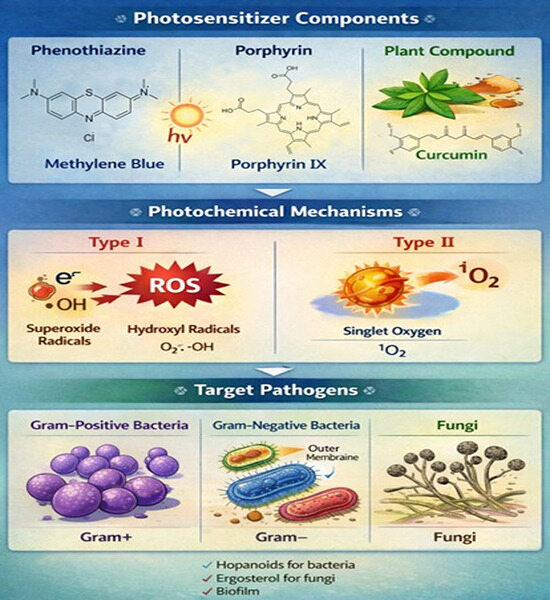
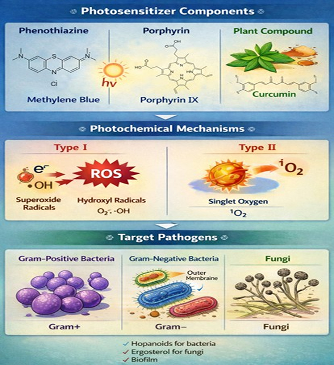
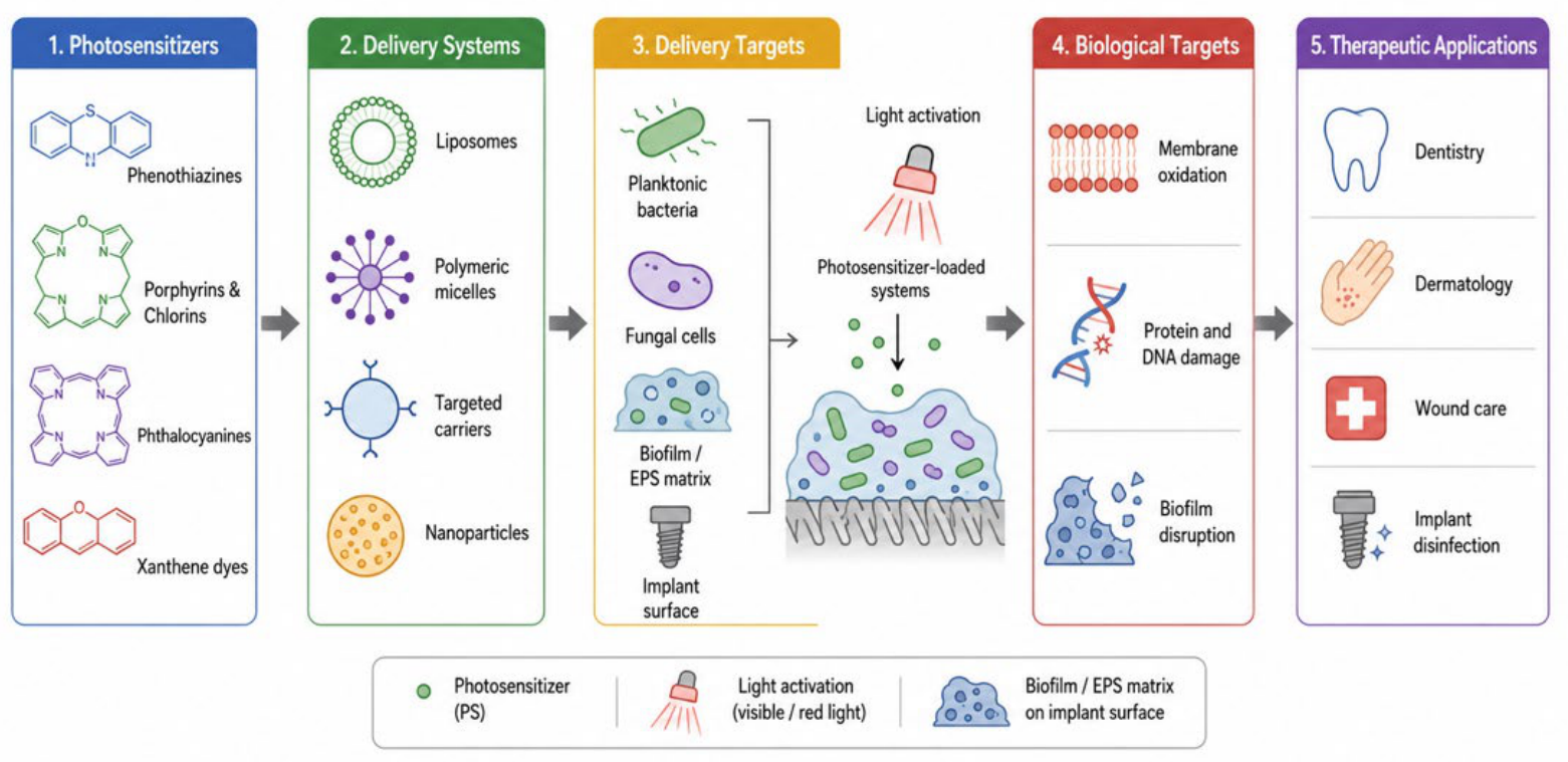
Graphical Abstract

1. Introduction

The rapidly growing resistance of microbial and fungal strains to conventional antimicrobials represents one of the most dangerous challenges facing modern medicine. The therapeutic efficiency of standard antibiotic and antifungal drugs has drastically decreased due to the continuous accumulation of genetic mutations, the upregulation of molecular resistance mechanisms, the formation of complex biofilms, metabolic cooperation among pathogens, and the capability of microorganisms to proliferate intracellularly [1,2,3]. These strict therapeutic limitations have urgently stimulated the development of alternative, non-antibiotic approaches capable of bypassing traditional drug-resistance pathways.
In this critical landscape, photodynamic therapy (PDT) has demonstrated a powerful, independent mechanism of action compared to classical antibiotic-based regimens, offering a radically different strategy to control bacterial and fungal populations during infectious processes. In contrast to conventional chemotherapeutic agents, PDT relies on fundamental photophysical and photochemical processes that generate short-lived, highly reactive oxygen species (ROS), which induce aggressive, multi-target oxidation across various cellular structures [4,5]. This multi-site mechanism of action guarantees a significantly lower probability of pathogens developing resistance compared to traditional drugs, which typically rely on a single, gene-dependent target.
The fundamental execution of PDT is highly sensitive to the local availability of three indispensable components: a photo-sensitizer (PS) molecule, light of an appropriate wavelength, and ambient molecular oxygen. Upon absorbing a photon, the ground-state singlet photosensitizer (S0) is promoted to a short-lived excited singlet state (S1). This excited state can undergo intersystem crossing to convert into a longer-lived excited triplet state (T1), which ultimately drives the downstream therapeutic photochemical reactions [6,7].
The excited triplet photosensitizer can then participate in two distinct photochemical pathways. Type I reactions involve direct electron or hydrogen transfer processes with neighboring substrates, generating localized reactive oxygen species such as superoxide anions, hydroxyl radicals, and hydrogen peroxide. Type II reactions involve direct energy transfer to ground-state molecular oxygen, resulting in the highly efficient generation of cytotoxic singlet oxygen (1O2).
Crucially, both singlet oxygen and Type I ROS possess extremely short lifespans and highly limited diffusion distances. These strict physical parameters restrict oxidative damage to the immediate microenvironment of the photosensitizer. Consequently, the primary determinants of antimicrobial PDT (aPDT) efficacy and selectivity are the intracellular localization, cellular uptake, structural retention, lifespan, and diffusion coefficient of the specific photosensitizer employed [8].
While PDT was initially pioneered for oncological applications [9], its successful translation into antimicrobial and antifungal fields relies entirely on fundamental biochemical, structural, and biophysical differences between mammalian host cells and parasitic microbial or fungal targets [10,11]. These unique cellular differences are critical for photosensitizer–cell interactions, determining membrane vulnerability, tolerance to oxidative stress, biofilm formation dynamics, and the ultimate cellular response to light activation [12].
Going beyond these fundamental interactions between ROS and cells—a topic that has already been discussed extensively in the literature—the modern state-of-the-art in this field focuses heavily on the engineering of functional translational biomaterials to combat nosocomial infections. In intensive care units (ICUs) and high-risk hospital departments, medical devices such as catheters, endotracheal tubes, and surgical implants serve as critical vectors for persistent bacterial and fungal biofilms.
To address this major clinical challenge, recent advancements have shifted toward the development of light-triggered polymer coatings permanently loaded with photosensitizers. Because these polymer coatings are permanently loaded with photosensitizers, they do not release free molecules into the surrounding tissue. Therefore, the specific targets of this PDT system are strictly the localized microorganisms that come into direct contact with the modified polymer surface (contact-killing mechanism). By restricting the photodynamic action exclusively to the device surface, this technology circumvents the depth-penetration limits of systemic PDT, offering a highly targeted, localized defense that prevents initial microbial colonization and biofilm formation on medical devices without systemic toxicity. These advanced anti-infective surfaces remain completely dormant under ambient conditions but undergo localized, on-demand photodynamic activation when exposed to specific wavelengths of light. By generating a continuous flux of surface-bound ROS, these smart polymeric matrices actively prevent microbial attachment, disrupt early-stage biofilm architectures, and prevent microbial colonization and biofilm formation on medical equipment in real-time without leaching toxic chemicals into the patient.
Ultimately, this review bridges the gap between fundamental cellular photochemistry and these cutting-edge medical device applications. By critically evaluating preclinical breakthroughs and the latest clinical data, this review explicitly demonstrates that in clinical settings, PDT serves as a unique, highly adaptive solution for antibacterial and antifungal treatment. Whether deployed as a targeted therapeutic modality or as light-activated polymer coatings, PDT represents a promising alternative to conventional antimicrobial therapy. Since the reactive oxygen species (ROS) generated during PDT are highly non-specific and can potentially affect any cell type, achieving selectivity is a critical clinical challenge.

1.1. Cellular Determinants of Differential Sensitivity to PDT

The main question of antimicrobial and antifungal PDT is why do pathogenic microorganisms demonstrate higher sensitivity to PDT in comparison to mammalian host cells? The answer to this question lies in the comparative structural and biochemical differences between these cell types [13].
Human and animal cells are eukaryotic. This means that they are enclosed by a single plasma membrane, built from a phospholipid bilayer enriched in cholesterol. In comparison with mammalian cells, bacteria and fungi have an additional external structure, known as the cell wall, which does not exist in mammalian cells. Bacterial and fungal cell walls are constructed from components such as peptidoglycan, lipopolysaccharides, chitin, and β-glucans, which influence photosensitizer binding, uptake, and sensitivity to PDT [14].
Negatively charged components are typical for bacterial cell walls. This fact facilitates electrostatic interactions with cationic photosensitizers. Gram-positive bacteria, which are usually covered with thick and relatively porous peptidoglycan layers, are more sensitive to PDT [14,15]. There is also an additional outer membrane in Gram-negative bacteria, which works as a permeability barrier. However, appropriately designed amphiphilic or cationic photosensitizers can penetrate the outer membrane [16].
Fungal cells are eukaryotic, although they exhibit different structural features than mammalian cells. They cover a rigid cell wall composed mostly of chitin and β-glucans. Their plasma membrane is enriched in ergosterol rather than cholesterol. These differences provide selective biochemical and biophysical targets, which can be useful as antifungal PDT [17].

1.2. Membrane Composition, Sterol Biology, and Oxidative Vulnerability

The presence of a cell wall is important for the effectivity of PDT, but the composition and organization of the cellular membrane are also crucial to determining PDT sensitivity. The critical biochemical difference between mammalian, fungi, and bacterial membranes on touch to PDT is the type of sterol included in the membrane. Cholesterol is typical for mammalian membranes—it enhances bilayer stability and provides partial resistance to oxidative lipid peroxidation. In contrast to mammalian, fungal membranes contain ergosterol. This differs structurally from cholesterol and is more easily subjected to oxidative damage induced by singlet oxygen and ROS [18]. Cholesterol or ergosterol are not typical for most bacteria; instead, many of them contain hopanoids or other lipids for membrane stabilization [19].
Highly specialized antioxidant and enzyme systems protect eukaryotic cellular membranes from oxidation. Within these systems, there is the glutathione peroxidase (GPx) family, which plays the central role. GPx4 (phospholipid hydroperoxide glutathione peroxidase) is a critical ‘gatekeeper’ enzyme that protects cells from lipid peroxidation within cellular membranes [20,21,22].
There are many polyunsaturated fatty acids (PUFAs) in cell membranes. PUFAs can undergo ROS-mediated oxidation. Lipid peroxidation is a chain reaction, which means that if it has started, it proceeds. During this process, toxic lipid hydroperoxides (LOOHs) are generated and destabilize the membrane architecture [23]. Without any control, this process culminates in ferroptosis, a regulated form of cell death that is provided by iron-dependent lipid peroxidation. Thus, redox homeostasis and oxidative stress defense systems maintain membrane integrity and architecture [24].
There are many highly evolved and redundant antioxidant network systems in mammalian cells. Some of them are superoxide dismutase, catalase, glutathione peroxidases, peroxiredoxins, and abundant low-molecular weight antioxidants [25]. These systems can reduce moderate ROS levels and partially mitigate PDT-induced oxidative stress in mammalian cells [26].
Compared to mammalian cells, bacteria and fungi demonstrate more limited or less redundant antioxidant defense systems [27,28]. As a result, when PDT generates an intense oxidative burst, which rapidly overwhelms the antioxidant defense systems of bacterial and fungi, this leads to irreversible damage to membranes, enzymes, and nucleic acids. This differential redox buffering capacity represents a central mechanistic determinant of the selectivity of PDT [28].

1.3. The Mechanistic Basis of Antimicrobial and Antifungal PDT

Taken together, the described structural, biochemical, and redox differences define the mechanistic basis of antimicrobial and antifungal PDT [29,30,31]. In case of aPDT, oxidative damage is multitarget and non-specific, simultaneously affecting everything simultaneously: from cell walls and membranes to proteins and nucleic acids [29,30,31].
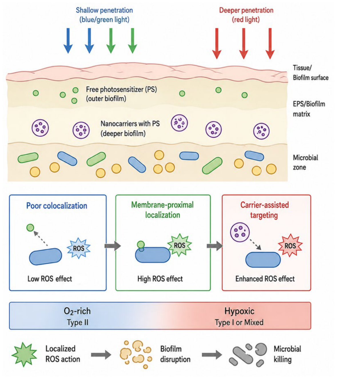
In addition, singlet oxygen has a short lifespan and usually diffuses only tens of nanometers (~125 nm in pure water, ≈10–20 nm, depending on microenvironment), so the damage occurs in the closest area of the photosensitizer [32,33,34]. From this point of view, PDT efficiency depends on the environment and the distance between the photosensitizer and the essential cellular structures.
Bacterial and fungal cell walls demonstrate a rigid yet porous architecture, which facilitates localized ROS generation at lethal concentrations. At the same time, the absence of robust antioxidant buffering in the case of fungi and bacteria amplifies damage. Due to total and multipurpose damage, PDT-induced cytotoxicity does not depend on specific metabolic pathways, enzymatic targets, or active cell division stage [35,36,37]. Consequently, PDT remains effective against different types of pathogenic cells: dormant cells, persisting populations, and biofilms. Furthermore, the absence of a specific enzymatic purpose could reduce the probability of resistance development (Table 1) [38].

1.4. Scope of the Review

The primary objective of this review was to conduct a systematic analysis of photodynamic therapy as an antimicrobial and antifungal approach. The main focus lies in integrating the principles of photophysics, photochemistry, cell biology, and biophysics. The main emphasis is placed on how fundamental cellular differences between mammalian, bacterial, and fungal cells contribute to the selectivity and efficacy. Special attention is directed toward the design of the photosensitizer, singlet oxygen, and ROS generation mechanisms. Also, the future perspectives of PDT as a resistance-free therapeutic strategy in antimicrobial and antifungal medicine are noted. Therefore, we start by examining classes of photosensitizers from phenothiazine photosensitizers, which include methylene blue, toluidine blue O, and new methylene blue.

2. Molecular Photosensitizers for Antibacterial and Antifungal PDT

2.1. Phenothiazine Photosensitizers

2.1.1. Phenothiazinium Dyes: Methylene Blue as a Translational Model

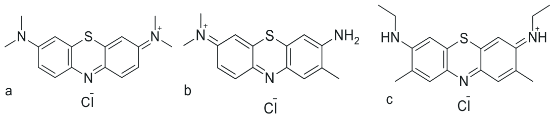
The first photosensitizer examined in this review is methylene blue (MB), a representative compound from the phenothiazinium family. Methylene blue (MB, Figure 1a) is one of the most widely investigated phenothiazinium photosensitizers and is commonly regarded as a reference compound in antimicrobial photodynamic therapy (aPDT).
From a biophysical perspective, MB is characterized by strong absorption in the red spectral region, with λ_max around 660–670 nm. This enables its efficient excitation using clinically accessible light sources. Importantly, red light penetrates biological tissues more effectively than blue light; therefore, MB is particularly suitable for the treatment of localized infections located beneath superficial tissue layers or within partially covered anatomical sites [55].
Upon photoexcitation, MB undergoes efficient intersystem crossing to the triplet excited state (T1), enabling both Type I and Type II photochemical pathways. As a result, MB can generate singlet oxygen (1O2) as well as superoxide-derived reactive oxygen species (ROS), which together contribute to its antimicrobial phototoxicity [5].
From a biochemical point of view, MB shows a high affinity for nucleic acids and negatively charged cellular structures. Gram-positive bacteria possess a strongly anionic surface due to the presence of teichoic acids in their thick peptidoglycan cell wall. These negatively charged components promote electrostatic interactions with the cationic MB molecule and favor its accumulation within the bacterial envelope [56].
In contrast, mammalian cells are surrounded by zwitterionic lipid membranes and a glycocalyx, which limit the nonspecific uptake of cationic dyes and reduce the risk of host tissue phototoxicity. In Gram-negative bacteria, MB uptake is more restricted because the outer membrane acts as an additional permeability barrier.
The primary targets of MB-mediated aPDT include membrane lipids, proteins, and nucleic acids. Oxidative damage to membrane lipids increases membrane permeability and disrupts the transmembrane potential. At the same time, intracellular ROS can oxidize essential metabolic proteins and damage nucleic acids. This multi-target mode of action explains the low probability of resistance development, even after repeated sublethal photodynamic exposure.
Accordingly, MB demonstrates high antimicrobial efficacy against clinically relevant and antibiotic-resistant strains, including methicillin-resistant Staphylococcus aureus (MRSA), Enterococcus spp., and multidrug-resistant Pseudomonas aeruginosa [57,58,59]. MB-mediated aPDT also shows significant antifungal activity, particularly against Candida albicans and non-albicans Candida species [60,61,62].
This antifungal effect is associated with electrostatic interactions between the cationic dye and negatively charged components of the fungal cell wall, including chitin and β-glucans. Subsequent light activation leads to oxidative damage of the ergosterol-rich plasma membrane, which causes the loss of membrane integrity and fungal cell death [63]. A comparative evaluation of antioxidant defenses indicates the following general hierarchy: mammalian cells > fungal cells > bacterial cells. This gradient helps explain why microbial cells are highly vulnerable to photodynamic inactivation, whereas mammalian cells are relatively less susceptible under controlled treatment conditions [4,64].
From the perspective of clinical translation, MB has several important advantages. It is inexpensive, widely available, pharmacologically well-characterized, and associated with relatively low dark toxicity. Moreover, it has already been approved for selected medical applications, which facilitates its consideration for translational aPDT approaches.
However, free molecular MB also has several limitations. These include photobleaching under prolonged irradiation, limited penetration into thick and mature biofilms, possible aggregation at higher concentrations, and dependence on local oxygen availability [65]. These limitations are particularly relevant in infected tissues and biofilm-associated infections, where oxygen gradients, extracellular polymeric substances, and heterogeneous photosensitizer distribution may reduce therapeutic efficacy.
To overcome these limitations, recent research has increasingly shifted from free photosensitizer solutions toward immobilized or carrier-associated MB systems. In particular, light-triggered polymer coatings and macromolecular networks loaded with MB have been developed to prevent or eradicate biofilms on medical devices used in intensive care units (ICUs) and other high-risk hospital settings.
For example, MB has been incorporated into acrylamide-based diblock copolymers, producing polymeric MB systems with improved mammalian biocompatibility while preserving red-light-activated antimicrobial activity against Escherichia coli, Pseudomonas aeruginosa, and Staphylococcus aureus [66]. In another strategy, “swell–encapsulation–shrink” methods have been used to incorporate MB, alone or together with gold nanoparticles, into commercial polyvinyl chloride (PVC) and silicone urinary catheters [67,68].
These photoactive polymer matrices are designed to retain the photosensitizer within the material and minimize leaching into the surrounding aqueous environment. Upon local red-light irradiation, including irradiation delivered through internal optical fibers, such systems generate surface-confined singlet oxygen and other ROS [69]. This localized oxidative activity can damage the extracellular polymeric substance (EPS) matrix, inhibit microbial adhesion, reduce early biofilm formation, and contribute to the self-disinfection of the device surface [70].
Thus, MB-mediated aPDT has evolved from a conventional soluble photosensitizer approach into a broader platform that includes surface-anchored and polymer-integrated photodynamic biomaterials. This transition is particularly important for the prevention of hospital-acquired device-associated infections, where local, light-controlled antimicrobial activity may offer advantages over conventional systemic antimicrobial therapy.
The second phenothiazinium compound discussed in this review is toluidine blue O, which contains structural features that influence hydrophobicity, membrane interaction, and antimicrobial photodynamic performance.

2.1.2. Toluidine Blue O: Lipophilicity, Membrane Binding, and Surface Engineering

Another phenothiazinium dye widely used in antimicrobial and antifungal photodynamic therapy (aPDT) is toluidine blue O (TBO, Figure 1b). TBO is structurally related to methylene blue, but it differs in substituent pattern and physicochemical behavior. It exhibits an absorption maximum in the red spectral region, with λ_max around 630–635 nm, which enables excitation with clinically accessible red light and supports its use in superficial and moderately deep localized infections [71].
Upon irradiation, TBO efficiently generates singlet oxygen (1O2), which is considered one of the main reactive species responsible for its antimicrobial activity [71]. Compared with MB, TBO shows a slightly different balance between hydrophilicity and lipophilicity. The presence of alkyl substituents increases its interaction with lipid-rich structures and influences cellular binding, membrane partitioning, and antimicrobial selectivity [72].
From a biophysical perspective, the high efficacy of TBO against Gram-positive bacteria is strongly associated with its cationic and amphiphilic character. Gram-positive bacteria possess a thick, porous peptidoglycan layer enriched with anionic teichoic acids. These negatively charged structures promote the electrostatic attraction of TBO and facilitate its accumulation within the bacterial cell envelope.
This localization is particularly important for PDT efficacy because singlet oxygen and other ROS have very short lifetimes and limited diffusion distances. When TBO is positioned close to the cytoplasmic membrane, light activation results in localized oxidative damage to membrane lipids, membrane-associated proteins, and cell-wall-associated enzymes. Consequently, TBO-mediated aPDT can compromise membrane integrity, disturb essential metabolic functions, and ultimately lead to bacterial cell death [71].
In antifungal PDT, TBO demonstrates relevant activity against pathogenic Candida species and dermatophytes [73,74]. The fungal cell wall, composed mainly of chitin, β-glucans, and mannoproteins, can act as a binding scaffold for cationic photosensitizers. This facilitates the retention of TBO near the plasma membrane, where illumination induces oxidative lipid peroxidation and damage to ergosterol-rich membrane domains.
At comparable irradiation conditions, TBO may show stronger antifungal phototoxicity than MB in selected experimental models. This effect is usually attributed to its favorable membrane interaction, amphiphilic character, and efficient localization close to lipid-rich fungal structures [75]. These properties explain why TBO has been widely investigated in dental disinfection, periodontal therapy, and endodontic canal sterilization [16].
Despite its advantages, TBO shares several limitations with other free molecular phenothiazinium photosensitizers. These include susceptibility to photobleaching, dependence on local oxygen availability, and limited passive diffusion into dense or mature biofilm architectures [76]. In biofilms, the extracellular polymeric substance (EPS) matrix can restrict photosensitizer penetration, reduce homogeneous distribution, and limit contact between the photosensitizer and microbial membranes.
To overcome these barriers, recent research has increasingly focused on immobilized and material-integrated TBO systems. In this approach, TBO is no longer used only as a freely diffusing topical dye, but also as a photoactive component of polymer coatings, cross-linked matrices, and antimicrobial surfaces intended for infection control in medical environments.
This strategy is particularly relevant for intensive care units (ICUs), surgical settings, and implant-associated applications, where medical devices can be rapidly colonized by bacterial and fungal biofilms. TBO has therefore been incorporated into or immobilized on polymeric materials such as polyurethane, silicone elastomers, and acrylic resins [77]. Such systems are designed to retain the photosensitizer within the material while preserving its ability to generate ROS upon illumination.
For example, TBO-functionalized polymer coatings have been developed using swell–encapsulation or related loading strategies for potential application on endotracheal tubes, catheters, and other medical device surfaces [78]. In these systems, the polymer matrix limits photosensitizer leaching while maintaining local photodynamic activity at the material–microorganism interface.
When activated by red light, including external irradiation or light delivered through optical fibers or waveguides, immobilized TBO can generate singlet oxygen directly at the device surface [79]. This localized ROS production can inhibit microbial adhesion, damage early EPS formation, and reduce the development of surface-associated biofilms. Importantly, because the oxidative stress is generated locally and only after irradiation, such coatings may reduce the need for the continuous release of antimicrobial drugs.
Thus, TBO-mediated aPDT is not limited to conventional liquid formulations used in oral or dental applications. Its integration into light-activated polymeric biomaterials expands its potential toward the prevention of hospital-acquired, device-associated infections. In this context, TBO represents both a classical molecular photosensitizer and a useful photoactive component for the design of self-disinfecting antimicrobial surfaces.
Another strategy for increasing membrane interaction is the structural modification of phenothiazinium dyes to enhance lipophilicity. This approach is represented by new methylene blue, which is discussed in the following section.

2.1.3. New Methylene Blue: Enhanced Lipophilicity and Biofilm Penetration

New methylene blue (NMB, Figure 1c) is a structurally modified phenothiazinium photosensitizer developed to improve lipophilicity and membrane partitioning compared with conventional methylene blue. This chemical modification significantly influences its biophysical behavior, increasing its ability to interact with lipid-rich cellular structures and penetrate microbial biofilms [72].
Like other phenothiazinium derivatives, NMB absorbs strongly in the red spectral region, with λ_max around 629–632 nm. This wavelength range allows effective excitation with red light, which penetrates biological tissues more efficiently than blue or green light. After photoexcitation, NMB undergoes intersystem crossing to the triplet excited state (T1), enabling the efficient generation of singlet oxygen (1O2), mainly through Type II photochemistry.
From a biochemical perspective, the increased hydrophobic character of NMB helps overcome one of the major limitations of antimicrobial therapy: the diffusion barrier created by the extracellular polymeric substance (EPS) matrix of mature biofilms [73,74]. Highly hydrophilic photosensitizers and many conventional antimicrobial agents may be retained or inactivated in the outer layers of the EPS matrix, resulting in incomplete pathogen eradication and the survival of tolerant persister cells.
Due to its balanced cationic charge and enhanced lipophilicity, NMB can diffuse more effectively into dense microbial aggregates than more hydrophilic phenothiazinium dyes. In vitro and in vivo studies have demonstrated strong NMB-mediated photodynamic activity against biofilm-forming pathogens, including Staphylococcus aureus and Pseudomonas aeruginosa [75].
After accumulation within biofilm structures and bacterial membranes, red-light irradiation activates NMB and induces localized oxidative damage. This process affects membrane lipids, membrane-associated proteins, and other essential cellular components, leading to loss of membrane integrity, biofilm disruption, and microbial cell death.
NMB has also shown promising activity against fungal biofilms, including Candida biofilms, which are often more resistant to conventional antifungal therapy than planktonic fungal cells [76]. In this context, NMB-mediated PDT may be particularly valuable because ROS can damage both fungal cells and the protective biofilm matrix.
From a translational perspective, NMB represents a useful compromise between high photodynamic efficacy, improved biofilm penetration, and favorable amphiphilic properties. Its molecular structure supports strong antimicrobial and antifungal effects while maintaining relatively low toxicity toward mammalian host cells under controlled irradiation conditions.
To address the limitations of free molecular photosensitizers, recent research has also explored the incorporation of NMB into light-triggered polymer coatings and hydrogel-based delivery systems [80]. This approach is particularly relevant for medical devices used in intensive care units (ICUs), operating rooms, and other high-risk clinical environments, where catheters, endotracheal tubes, and central venous lines are frequently colonized by bacterial and fungal biofilms.
NMB can be incorporated into polymeric matrices, including polyurethane- and silicone-based materials, using loading or swell–encapsulation strategies [81,82]. These systems are designed to retain the photosensitizer within the material, reduce uncontrolled leaching, and preserve local photodynamic activity at the device surface.
Upon stimulation with red light, including external irradiation or light delivered through optical fibers, NMB-containing polymer surfaces can generate singlet oxygen and other ROS at the device–fluid interface [83]. This localized oxidative activity may inhibit microbial adhesion, damage early EPS formation, and reduce the viability of cells embedded in developing biofilms.
Because of its improved biofilm penetration, NMB may offer advantages over more hydrophilic dyes in surface-associated infections. However, its efficacy depends on several parameters, including photosensitizer loading, polymer composition, irradiation dose, oxygen availability, and the maturity and composition of the biofilm [84]. Therefore, NMB-based coatings should be regarded as promising light-activated antimicrobial platforms rather than universal replacements for conventional antimicrobial materials.
Overall, NMB extends the phenothiazinium class from conventional soluble aPDT toward more advanced biofilm-oriented and surface-integrated photodynamic systems. This ability to combine membrane affinity, red-light activation, and biofilm penetration makes NMB an important representative of advanced phenothiazinium photosensitizers.
A similar principle of efficient photodynamic activity combined with selective microbial damage is also observed in macrocyclic porphyrins, which are discussed in the following section.

2.2. Porphyrin Photosensitizers

2.2.1. TMPyP: A Tetracationic Porphyrin Model for Antimicrobial PDT

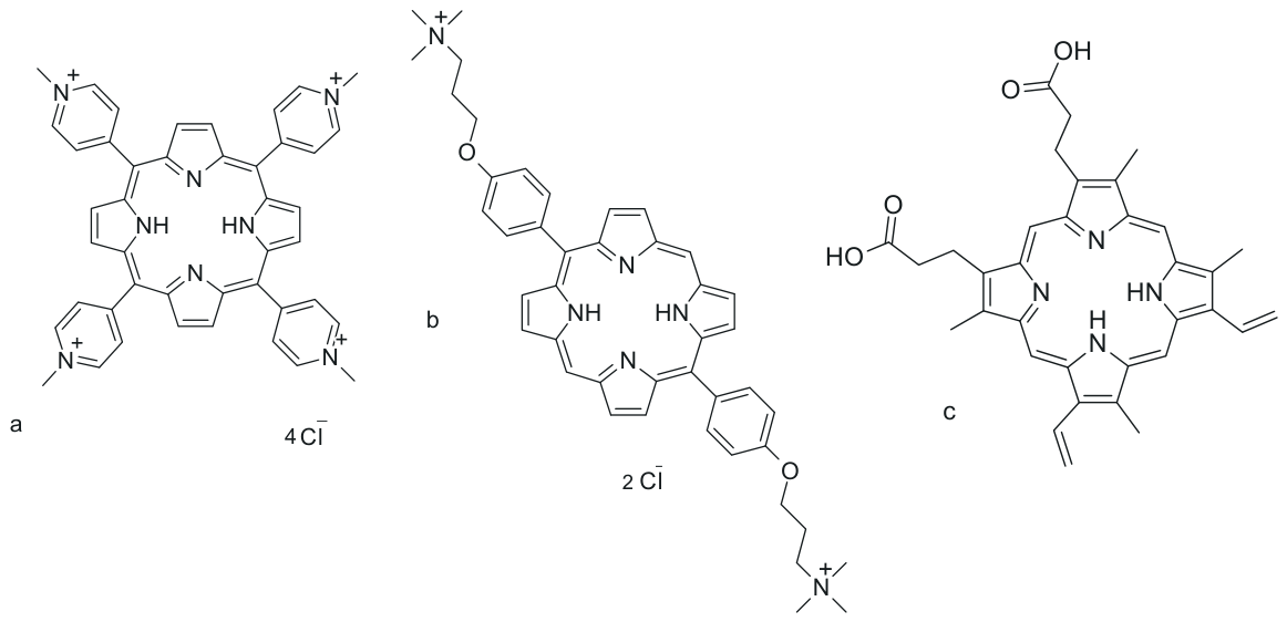
Another important class of photosensitizers used in antimicrobial photodynamic therapy (aPDT) is represented by cationic porphyrins. Among them, 5,10,15,20-tetrakis(4-N-methylpyridyl)porphyrin (TMPyP, Figure 2a) is one of the most widely investigated reference compounds [77].
From a photophysical perspective, TMPyP exhibits an intense Soret band in the blue spectral region, with λ_max around 420–430 nm. This absorption is accompanied by weaker Q bands extending into the green and red regions of the visible spectrum. As a result, TMPyP can be activated using blue light, white light, or broader-spectrum irradiation systems [77,78].
TMPyP is valued for its efficient triplet-state formation and predominant Type II photochemical behavior. Upon illumination, it generates singlet oxygen (1O2), which is one of the principal cytotoxic species responsible for microbial inactivation [78]. These properties make TMPyP a useful model compound for studying the relationship between porphyrin structure, charge distribution, and antimicrobial photodynamic efficacy.
From a biochemical perspective, the tetracationic structure of TMPyP is central to its antimicrobial activity. The four positively charged N-methylpyridyl substituents promote strong electrostatic interactions with negatively charged microbial surfaces [79]. This is particularly important for bacteria and fungi, whose cell envelopes contain anionic components that can retain cationic photosensitizers near the cell surface.
In Gram-negative bacteria, TMPyP can interact with lipopolysaccharides (LPSs), outer membrane proteins, and other negatively charged components of the outer envelope [79]. Although the outer membrane usually limits the uptake of many antimicrobial agents, cationic porphyrins can partially overcome this barrier through electrostatic binding and self-promoted uptake-like interactions [80]. This promotes photosensitizer accumulation at the bacterial surface and within the cell envelope.
After light activation, surface-associated TMPyP generates singlet oxygen in close proximity to lipid bilayers, periplasmic proteins, and membrane-associated enzymes. Because singlet oxygen has a short lifetime and limited diffusion distance, this localization is crucial for efficient oxidative damage [80]. The resulting injury to the bacterial envelope can compromise membrane integrity, disrupt essential enzymatic functions, and lead to irreversible cell death.
TMPyP also binds effectively to Gram-positive bacteria. Their thick but porous peptidoglycan layer and anionic teichoic acids provide favorable binding sites for tetracationic porphyrins [79,81]. In this case, TMPyP does not necessarily need to enter the cytoplasm to exert antimicrobial activity. Localization at or near the cell envelope may be sufficient to induce lethal oxidative damage after illumination [81].
This membrane- and envelope-associated mechanism is especially relevant because it is not strongly dependent on bacterial metabolic activity. Therefore, TMPyP-mediated aPDT may remain effective against slow-growing cells, dormant populations, and persister-like phenotypes that are often less susceptible to conventional metabolism-dependent antibiotics [81].
TMPyP-mediated aPDT has also been investigated against pathogenic Candida species. Fungal cells possess a thick carbohydrate-rich cell wall composed of β-glucans, chitin, and mannoproteins. These structures can serve as binding scaffolds for cationic porphyrins and support photosensitizer retention close to the underlying plasma membrane [82].
Upon illumination, TMPyP-induced singlet oxygen can oxidize ergosterol-rich fungal membranes, damage cell-wall-associated proteins, and disturb membrane permeability [82]. Because fungal cells generally have lower oxidative stress tolerance than mammalian host cells, this creates a photodynamic selectivity window. However, this selectivity depends strongly on photosensitizer concentration, irradiation dose, incubation time, and the local biological environment.
From a translational perspective, TMPyP represents an important structural model because it combines high cationic charge density with the favorable photophysical properties of the porphyrin macrocycle. These features make it particularly useful for overcoming, at least partially, the permeability barriers of Gram-negative bacteria and for targeting negatively charged microbial envelopes [77,80].
However, free TMPyP also has limitations. Its strong absorption in the blue region restricts tissue penetration compared with red- or near-infrared-absorbing photosensitizers. In addition, free porphyrins may undergo aggregation, nonspecific binding, and reduced activity in complex biological matrices or mature biofilms [78,81].
Recent research has therefore moved beyond free TMPyP solutions toward photoactive polymer coatings, hydrogels, and macromolecular materials containing cationic porphyrins [82]. Such systems are particularly relevant for medical devices used in intensive care units (ICUs), surgical settings, and implant-associated applications, where central venous catheters, urinary catheters, and endotracheal tubes are vulnerable to bacterial and fungal biofilm formation.
In these approaches, TMPyP or related porphyrins may be covalently grafted onto polymer chains or physically entrapped within polymeric matrices such as polyurethane, silicone-based materials, or hydrogel networks [82]. The purpose of this immobilization is to retain the photosensitizer at the material surface, reduce uncontrolled leaching into biological fluids, and generate ROS locally after illumination.
When activated by appropriate light, surface-associated TMPyP can generate singlet oxygen directly at the device–fluid or material–microorganism interface [82]. This localized ROS production may inhibit early bacterial adhesion, damage the extracellular polymeric substance (EPS) matrix, and reduce biofilm development. Because the oxidative mechanism attacks multiple cellular and extracellular targets simultaneously, the probability of resistance development remains low.
Thus, TMPyP should be considered not only as a classical soluble cationic porphyrin, but also as a useful structural platform for the design of photoactive antimicrobial coatings and light-triggered biomaterials. In this form, it contributes to a broader strategy in which aPDT is applied not only as a therapeutic intervention, but also as a preventive surface-based technology for reducing device-associated infections.
Another cationic porphyrin-derived antimicrobial compound with tailored amphiphilic properties is exeporfinium chloride (XF-73), which is discussed in the following section.

2.2.2. XF-73/Exeporfinium Chloride: A Membrane-Targeted Antimicrobial Porphyrin

In contrast to classical macrocyclic porphyrins such as TMPyP, exeporfinium chloride (XF-73, Figure 2b) represents a distinct dicationic synthetic porphyrin derivative developed specifically for antimicrobial applications [82]. While many conventional porphyrins were initially optimized for oncological photodynamic therapy, XF-73 was designed to enhance interactions with bacterial membranes while limiting mammalian cell toxicity.
XF-73 (exeporfinium chloride) is designed for the prevention of post-surgical staphylococcal infections, specifically targeting MRSA and mupirocin-resistant S. aureus in nasal decolonization. It is formulated for topical applications, including preventing infections in surgical wounds and treating superficial bacterial skin conditions. While XF-73 has a strong Soret absorption band at 420 nm, blue light scatters heavily in biological tissues. Because of this high scattering and low penetration depth, any cells located beneath the immediate surface or shaded by tissue structures remain protected from irradiation. This explains why this approach is limited to superficial treatments [83]. After photoexcitation, the molecule can populate the triplet excited state (T1), which enables the generation of singlet oxygen (1O2) through Type II photochemistry, and under appropriate conditions, reactive radical species through Type I pathways [83,84]. From a biochemical point of view, XF-73 demonstrates rapid binding to bacterial cell membranes, including membranes of multidrug-resistant pathogens such as methicillin-resistant Staphylococcus aureus (MRSA) [84]. Its dicationic and amphiphilic structure supports interaction with negatively charged bacterial surfaces and promotes partitioning close to the lipid bilayer.
This membrane-directed localization is central to the antimicrobial mechanism of XF-73. After light activation, ROS are generated in close proximity to membrane lipids and membrane-associated proteins. The resulting oxidative damage may cause lipid peroxidation, membrane depolarization, leakage of intracellular components, and rapid loss of bacterial viability. An important advantage of XF-73-mediated antimicrobial activity is that it can be effective under relatively short irradiation times and low light doses, depending on the experimental model. This is particularly relevant for topical and localized antimicrobial applications, where treatment time and light exposure must be compatible with clinical use [85].
XF-73 has also been investigated in the context of biofilm-associated infections. Its membrane-targeted mechanism may be advantageous in dense microbial communities, where conventional antibiotics often show reduced efficacy because of limited diffusion, extracellular polymeric substance (EPS) barriers, and the presence of slow-growing or tolerant cells. Repeated exposure studies suggest that XF-73 has a low tendency to induce resistance, which is consistent with the multitarget oxidative mechanism of photodynamic therapy. Because PDT damages several cellular structures simultaneously, pathogens are less likely to adapt through a single genetic or enzymatic resistance mechanism [86].
From a safety and translational perspective, XF-73 is attractive because of its localized mode of action and limited dark toxicity toward mammalian cells under controlled conditions [87]. These properties support its potential use in topical antimicrobial therapy and localized infection control.
To address the limitations of free molecular photosensitizers, recent research has explored the integration of XF-73 and related amphiphilic porphyrin derivatives into polymer coatings, hydrogels, and light-activated antimicrobial surfaces. Such systems are particularly relevant for medical devices used in intensive care units (ICUs), surgical settings, and implant-associated procedures, where device-associated infections remain a major clinical problem. In these material-based approaches, the amphiphilic structure of XF-73 can support its retention within polymeric matrices such as polyurethane, silicone-based elastomers, or hydrogel networks. The aim is to maintain local photosensitizer activity at the device surface while reducing uncontrolled leaching into biological fluids [87].
When illuminated with appropriate light, XF-73-containing polymeric surfaces may generate singlet oxygen and other ROS directly at the device–fluid interface. This localized photodynamic activity can reduce microbial adhesion, damage early biofilm structures, and limit the formation of mature biofilms on medical surfaces [86,87].
Therefore, XF-73 should be considered not only as a soluble antimicrobial photosensitizer, but also as a promising molecular scaffold for the design of surface-bound and polymer-integrated photodynamic biomaterials. However, the efficacy of such systems depends on photosensitizer loading, polymer composition, light dose, oxygen availability, and the biological complexity of the infection site. In contrast to the synthetic and purpose-designed structure of XF-73, the next compound discussed in this review, protoporphyrin IX (PpIX), is an endogenous macrocycle generated within living cells through the heme biosynthetic pathway.

2.2.3. Protoporphyrin IX: Endogenous Metabolic Pathways and Light-Triggered Activity

Protoporphyrin IX (PpIX, Figure 2c) is an endogenous macrocyclic photosensitizer and the immediate tetrapyrrolic precursor of heme in the heme biosynthetic pathway [88].
Its endogenous formation, particularly after the administration of precursors such as 5-aminolevulinic acid (ALA), is relevant mainly because it enables in situ photosensitizer generation and metabolism-dependent accumulation, rather than because endogenous origin alone guarantees superior biocompatibility. Indeed, many fully synthetic photosensitizers may also show favorable biocompatibility profiles, depending on their chemical structure, formulation, dose, and clearance. The clearest clinical proof of PpIX in healthy host tissues is post-treatment skin photosensitivity. Patients must avoid bright light because PpIX accumulates in the skin and vascular endothelial cells, causing standard phototoxic burns if exposed.
From a photophysical perspective, PpIX exhibits an intense Soret absorption band in the near-ultraviolet to blue spectral region, with λ_max around 405–410 nm [88]. This absorption is accompanied by weaker Q bands extending into the visible region. The strong Soret band enables efficient excitation with blue light, whereas the Q bands allow additional activation at longer visible wavelengths, although with lower absorption intensity.
After photon absorption, PpIX can populate the triplet excited state (T1), which enables the photochemical generation of reactive oxygen species [89]. PpIX-mediated PDT is mainly associated with Type II photochemistry, leading to the formation of singlet oxygen (1O2), although other ROS may also contribute depending on the local oxygen concentration and biological environment [89].
The clinical and biological relevance of PpIX-mediated aPDT is closely related to differences in porphyrin metabolism between host cells and microbial cells. In mammalian cells, ferrochelatase catalyzes the insertion of ferrous iron (Fe2+) into PpIX, converting it into heme [90]. This pathway limits the excessive accumulation of photoreactive PpIX in healthy host tissues and contributes to the relatively favorable safety profile of metabolism-driven PDT.
In contrast, several pathogenic microorganisms can accumulate PpIX or PpIX-related porphyrins more efficiently because of differences in heme biosynthesis, iron metabolism, or ferrochelatase activity [90]. This selective accumulation is particularly relevant when PpIX is generated endogenously after the administration of precursors such as 5-aminolevulinic acid (ALA). In such systems, the photosensitizer is formed in situ rather than delivered only as a preformed external molecule.
After light exposure, accumulated PpIX generates ROS close to cellular envelopes, membranes, and intracellular targets [91]. This can lead to lipid peroxidation, oxidation of membrane-associated proteins, disruption of cell-wall integrity, and damage to nucleic acids. Because these effects occur simultaneously at multiple sites, PpIX-mediated aPDT can reduce microbial viability without relying on a single metabolic target.
PpIX-mediated photodynamic activity has been reported against Gram-positive bacteria and fungal pathogens, including Candida species [91]. Its localization within lipid-rich regions and cellular envelopes supports membrane-associated oxidative damage. However, the effectiveness of PpIX-mediated aPDT depends strongly on precursor uptake, intracellular conversion to PpIX, oxygen availability, irradiation wavelength, and light dose.
As with many porphyrin-based photosensitizers, a practical limitation of PpIX is the mismatch between its strongest absorption band and the optical window most favorable for deeper tissue treatment. PpIX exhibits an intense Soret band in the near-ultraviolet/blue region, whereas its longer-wavelength Q bands are weaker. Therefore, PpIX-based antimicrobial applications are particularly relevant for settings in which light can be delivered directly to the target site, such as superficial, topical, mucosal, wound-related, or surface-associated infections, rather than for deeply located infections [88,89].
Beyond its role as a soluble or metabolism-generated photosensitizer, PpIX has also been investigated in photoactive biomaterials, including polymeric coatings and tissue-engineering scaffolds [92]. These systems aim to immobilize or retain PpIX within a material, reduce uncontrolled leaching, and enable local ROS generation after illumination.
In the context of light-accessible medical-device surfaces, PpIX-containing coatings may be useful for limiting microbial adhesion and early biofilm formation on surfaces such as catheters, endotracheal tubes, wound dressings, or implant-associated materials [92]. Here, “medical-device surfaces” refers to device-associated materials that can be illuminated during use, maintenance, dressing exchange, or perioperative handling. When illuminated with appropriate light, immobilized PpIX can generate singlet oxygen at the material–microorganism interface, leading to localized oxidative stress without the continuous release of conventional antimicrobial drugs.
The effectiveness of such PpIX-based coatings depends on the method of immobilization, photosensitizer loading, polymer hydrophobicity, oxygen diffusion through the material, and the intensity and penetration depth of the activating light. Therefore, these systems should be described as promising local photodynamic biomaterials, rather than as universally superior replacements for antibiotic-eluting materials.
Overall, PpIX represents a clinically relevant endogenous photosensitizer that links antimicrobial PDT with metabolism-driven selectivity, fluorescence-guided detection, and localized photodynamic inactivation. Its main advantages include endogenous formation, theranostic potential, and selective accumulation under appropriate biological conditions. Its main limitations include shallow blue-light penetration and dependence on metabolic conversion or local accumulation.
The next compound discussed in this review is deuteroporphyrin, a porphyrin derivative with structural modifications that influence hydrophobicity, metal coordination, and antimicrobial photodynamic performance.

2.2.4. Deuteroporphyrin: A Structural Porphyrin Model for Photodynamic Applications

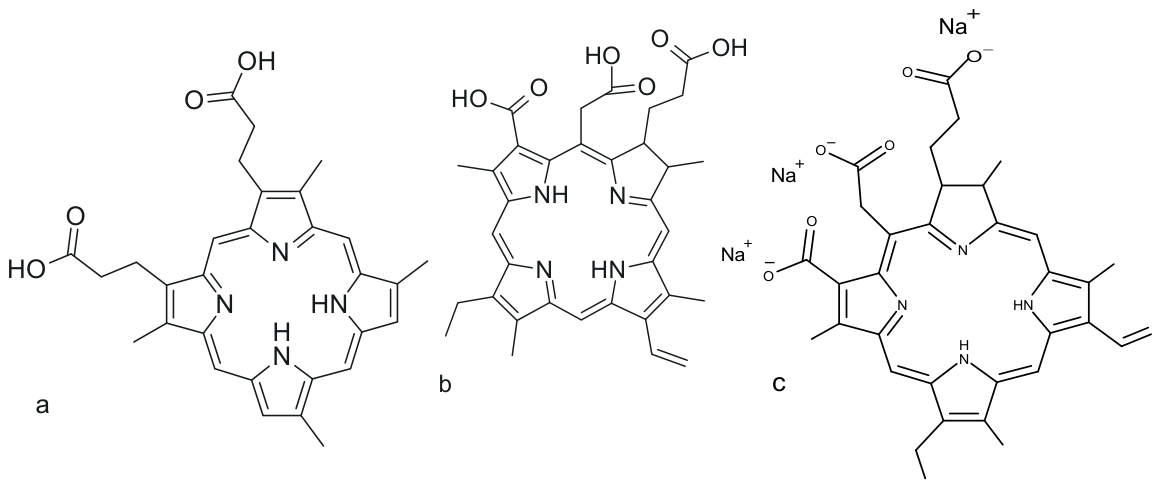
Deuteroporphyrin (Figure 3a) is a semisynthetic dicarboxylic tetrapyrrole derived from naturally occurring heme-related porphyrins [93]. Structurally, it differs from protoporphyrin IX by the absence of vinyl groups at the C2 and C4 positions of the porphyrin macrocycle. These structural modifications influence its hydrophobicity, aggregation behavior, and interaction with biological membranes.
From a photophysical perspective, deuteroporphyrin exhibits an intense Soret absorption band near 398 nm, accompanied by several Q bands extending through the visible region [93]. These Q bands extend into the red region, although, as is typical for porphyrins, their absorption intensity is lower than that of the Soret band. After photoexcitation, the molecule can undergo intersystem crossing to the triplet excited state (T1), which enables the generation of reactive oxygen species under appropriate irradiation conditions [94].
Deuteroporphyrin-mediated photodynamic activity is mainly associated with Type II photochemistry and singlet oxygen (1O2) generation [94]. This makes it a useful reference porphyrin for studying how macrocycle structure, peripheral substituents, hydrophobicity, and aggregation state influence photodynamic efficiency.
From a biochemical point of view, the dicarboxylic peripheral structure of deuteroporphyrin supports interactions with microbial cell envelopes and lipid-rich membrane domains [95]. Its amphiphilic character may facilitate partial partitioning into biological membranes, positioning the photosensitizer close to targets that are highly vulnerable to short-lived ROS.
In bacterial cells, light activation of membrane-associated deuteroporphyrin can induce oxidative damage to membrane lipids and membrane-associated proteins [95]. This may lead to altered membrane permeability, disruption of ion gradients, inhibition of transport processes, and progressive loss of cell viability.
A similar mechanism may occur in fungal cells, where deuteroporphyrin-mediated ROS can damage ergosterol-containing membranes and cell-wall-associated components [95]. However, compared with more widely used cationic porphyrins or clinically oriented chlorins, deuteroporphyrin is more often applied as an experimental or mechanistic reference compound than as a routine therapeutic photosensitizer.
Its value therefore lies mainly in structure–function studies. Deuteroporphyrin provides a useful example of how specific peripheral modifications of a porphyrin macrocycle, such as removal of the vinyl groups present in PpIX, may influence physicochemical properties relevant to photodynamic activity, including hydrophobicity, aggregation behavior, membrane interaction, and ROS-mediated antimicrobial effects [93,94]. However, conclusions about general structure–activity relationships within the porphyrin class require comparison with additional porphyrin derivatives rather than reliance on deuteroporphyrin alone.
Although porphyrin-containing coatings, hydrogels, and surface-functionalized biomaterials are being explored as material-based antimicrobial PDT platforms [96], the present section does not propose the routine coating of catheters, tubes, or dressings specifically with deuteroporphyrin. Rather, deuteroporphyrin is discussed here primarily as a photosensitizing porphyrin used in experimental studies and as a structural comparator to PpIX. Its carboxylic acid groups could, in principle, support conjugation to polymer chains or surface linkers, but any translation into device coatings would require direct evidence of immobilization stability, light-accessible ROS generation, antimicrobial efficacy, and biocompatibility under device-relevant conditions.
Such immobilization strategies are relevant for the development of light-activated antimicrobial surfaces on catheters, endotracheal tubes, wound dressings, or implant-associated materials [96]. In these systems, the goal is to generate ROS locally at the material–microorganism interface while reducing uncontrolled photosensitizer release into surrounding biological fluids.
When appropriately illuminated, surface-bound deuteroporphyrin can generate singlet oxygen near the colonized interface [96]. This localized oxidative activity may inhibit microbial adhesion, damage early extracellular polymeric substance (EPS) formation, and reduce biofilm development. However, the efficacy of such systems depends on the density of immobilized photosensitizer, polymer composition, oxygen diffusion, light penetration, and long-term photostability.
Importantly, immobilization can reduce photosensitizer aggregation if the molecules are spatially separated within the polymer network. This may help preserve singlet oxygen generation over repeated irradiation cycles. Nevertheless, excessive immobilization density may produce self-quenching, whereas insufficient loading may reduce antimicrobial activity. Therefore, material design must balance photosensitizer loading, accessibility to oxygen, and proximity to microbial targets.
Overall, deuteroporphyrin should be presented primarily as a structurally informative porphyrin derivative and a potential building block for photoactive antimicrobial materials, rather than as a broadly established standalone clinical antimicrobial photosensitizer. Its main strengths are photochemical predictability, chemical functionality for conjugation, and usefulness in structure–activity studies.
Another group of tetrapyrrolic photosensitizers with absorption shifted toward longer and more tissue-penetrating wavelengths is represented by chlorins. Among them, chlorin e6 is one of the most widely investigated compounds and is discussed in the following section.

2.3. Chlorins and Phthalocyanines

2.3.1. Chlorin e6: Red-Light Activation and Antimicrobial Phototoxicity

Chlorin e6 (Ce6, Figure 3b) is a second-generation macrocyclic photosensitizer derived from chlorophyll a degradation products [97]. It is one of the most widely investigated chlorin-based compounds in antimicrobial photodynamic therapy (aPDT), mainly because its photophysical properties are more favorable than those of many first-generation porphyrins. From a photophysical perspective, Ce6 exhibits strong absorption in the red spectral region, with λ_max around 660–670 nm, which is advantageous because red light penetrates biological tissues more effectively than blue or green light [97]. After red-light excitation, Ce6 can efficiently populate the triplet excited state (T1), supporting the generation of reactive oxygen species. Ce6-mediated PDT is predominantly associated with Type II photochemistry and the production of singlet oxygen (1O2), although Type I processes may also contribute depending on oxygen availability and the local biological environment [98].
From a biochemical point of view, Ce6 contains three peripheral carboxylic acid groups, which give the molecule a moderately amphiphilic and predominantly anionic character at physiological pH [99]. This distinguishes Ce6 from cationic phenothiazinium dyes and tetracationic porphyrins, whose antimicrobial activity is largely driven by electrostatic attraction to negatively charged microbial envelopes. Despite its anionic character, Ce6 can accumulate in microbial structures through amphiphilic interactions and partitioning into lipid-rich domains. This is particularly relevant for Gram-positive bacteria and fungal cells, where the cell envelope and membrane architecture support close photosensitizer localization near vulnerable oxidative targets. In fungal systems, Ce6 can associate with ergosterol-containing membranes, which are highly susceptible to oxidative lipid damage [100]. After illumination, membrane-associated Ce6 generates singlet oxygen close to the plasma membrane, leading to lipid peroxidation, altered membrane permeability, loss of membrane potential, and leakage of intracellular components.
Ce6-mediated aPDT has shown activity against both planktonic and biofilm forms of clinically relevant microorganisms, including Staphylococcus aureus, Enterococcus spp., Escherichia coli, and Candida species [100,101]. Its activity against biofilms is particularly important because biofilm-associated infections are often less responsive to conventional antimicrobial therapy. However, the activity of free Ce6 may be limited by aggregation, heterogeneous distribution, reduced penetration into mature extracellular polymeric substance (EPS) matrices, and local oxygen depletion [101]. These limitations are especially relevant in dense biofilms, where photosensitizer localization and oxygen diffusion strongly determine photodynamic efficacy.
From a translational perspective, Ce6 is attractive because it combines red-light activation, efficient ROS generation, and relatively low dark toxicity toward mammalian cells under controlled conditions [102]. Its longer-wavelength absorption is advantageous because red light is less strongly scattered and penetrates tissue more effectively than blue or near-UV light, although the achievable depth still depends on tissue type, optical properties, and irradiation parameters. To overcome the limitations of free Ce6, recent research has increasingly focused on Ce6-loaded or Ce6-functionalized delivery platforms, including polymer coatings, hydrogels, nanoparticles, and layer-by-layer assemblies.
In light-accessible antimicrobial material applications, Ce6 can be physically embedded or chemically linked to polymeric matrices such as polyurethane, silicone-based materials, hydrogel networks, or polyelectrolyte multilayers. The aim of this strategy is to generate photodynamic activity at the material–microorganism interface while limiting uncontrolled photosensitizer leaching into surrounding biological fluids.
When illuminated with red light, immobilized or matrix-associated Ce6 can generate singlet oxygen in the illuminated regions where Ce6, molecular oxygen, and light overlap, including at or near the material–microorganism interface. This localized ROS generation can inhibit microbial adhesion, damage early EPS formation, and reduce the viability of cells embedded in developing biofilms [101,102]. Overall, Ce6 represents one of the most important chlorin photosensitizers for antimicrobial and antifungal PDT. Its main advantages include red-light absorption, strong singlet oxygen generation, relatively low dark toxicity, and compatibility with advanced delivery systems. Its main limitations include aggregation, reduced efficacy under hypoxic or poorly oxygenated conditions, and reduced efficacy in poorly penetrable biofilms when used as a free molecular photosensitizer. Because molecular oxygen is required for Type II photochemistry, oxygen availability should be considered a treatment-limiting condition rather than an inherent disadvantage of Ce6 itself.
Another chlorin-based formulation with optimized pharmaceutical stability and clinical relevance is Radachlorin, a clinically used chlorin-based formulation composed of a mixture of chlorins and related structures, which is discussed in the following section. Rather than being a single chlorin molecule with entirely distinct photochemical principles, Radachlorin is distinguished mainly by its defined pharmaceutical formulation, composition, stability, and clinical use profile.

2.3.2. Radachlorin: A Clinically Relevant Chlorin-Based Formulation

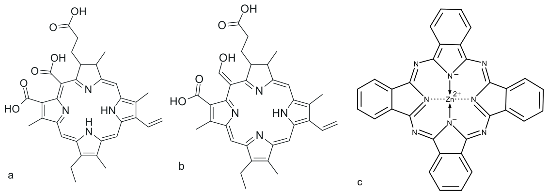
Radachlorin is a multi-component chlorin-based pharmaceutical formulation composed mainly of sodium salts of chlorin e6 (Figure 3c), chlorin p6 (Figure 4a), and purpurin 5 (Figure 4b) [103]. The photophysical behavior of these components is closely related to that of chlorin e6; however, their combined formulation improves aqueous solubility, stability, and pharmaceutical handling compared with many unformulated hydrophobic macrocycles [103,104]. Radachlorin absorbs strongly in the red spectral region, with λ_max around 660–670 nm, which allows for activation with tissue-penetrating red light [104]. After irradiation, its chlorin components populate the triplet excited state (T1), leading mainly to Type II photochemistry and the generation of singlet oxygen (1O2), although Type I radical pathways may also contribute under specific local conditions [104,105].
From a biochemical perspective, Radachlorin can associate with microbial envelopes and lipid-rich membrane domains because of the amphiphilic character of its chlorin components [105]. In antimicrobial photodynamic therapy (aPDT), this localization is important because ROS have short lifetimes and limited diffusion distances. After red-light activation, membrane-associated Radachlorin can induce lipid peroxidation, protein oxidation, loss of membrane integrity, and disruption of essential cellular functions. Experimental studies have shown activity against clinically relevant Gram-positive bacteria, including resistant Staphylococcus strains, as well as fungal pathogens such as Candida species [106]. In fungal cells, Radachlorin-mediated PDT may damage the cell wall, ergosterol-containing plasma membrane, and intracellular organelles, including mitochondria [106,107].
Radachlorin has also been investigated in the context of biofilm-associated infections, where conventional antimicrobial therapy is often limited by poor penetration, EPS-mediated protection, and the presence of slow-growing cells [107]. Its red-light activation and chlorin-based ROS generation make it relevant for localized infections and surface-associated microbial communities. However, as with other molecular photosensitizers, the efficacy of free Radachlorin can be reduced by heterogeneous distribution, oxygen limitation, and restricted diffusion into mature biofilm structures [107,108]. Therefore, formulation and delivery strategies remain essential for improving its antimicrobial and antifungal performance.
From a translational point of view, Radachlorin is especially interesting because chlorin-based PDT has already been clinically explored in oncological and other medical contexts, providing a useful safety and implementation background for further anti-infective development [108]. Its relatively low dark toxicity, compatibility with red-light lasers and LEDs, and improved solubility compared with many hydrophobic chlorins support its potential for localized antimicrobial PDT. Nevertheless, its use as an anti-infective agent still requires the optimization of dose, irradiation parameters, formulation stability, and target-specific delivery.
Recent material-based strategies have therefore explored Radachlorin and related chlorin systems in photoactive polymeric matrices, hydrogel coatings, and surface-functionalized antimicrobial materials [109]. In light-accessible antimicrobial material applications, chlorin photosensitizers can be physically entrapped, electrostatically associated, or chemically immobilized within polyurethane, silicone-based materials, hydrogel networks, or related polymeric platforms. Such systems aim to retain the photosensitizer at the illuminated material surface, reduce uncontrolled leaching, limit host exposure, and generate ROS locally after illumination. When activated with red light, Radachlorin-containing coatings generate singlet oxygen and other ROS in illuminated regions where the chlorin photosensitizer, molecular oxygen, and activating light overlap, including at or near the material–fluid or material–microorganism interface. This localized photodynamic activity can reduce microbial adhesion, damage early extracellular polymeric substance (EPS) formation, and suppress biofilm development [109]. Overall, Radachlorin represents a clinically relevant chlorin formulation that combines red-light activation, improved pharmaceutical properties, and potential compatibility with advanced delivery systems and photoactive biomaterials.
In line with the development of photosensitizers optimized for longer-wavelength tissue penetration, the next group discussed in this review is represented by phthalocyanines, synthetic macrocycles with absorption shifted further toward the red and near-infrared spectral regions.

2.3.3. Zinc Phthalocyanine (ZnPc): Red/NIR Absorption and Formulation-Dependent Activity

Zinc phthalocyanine (ZnPc, Figure 4c) belongs to the class of second-generation macrocyclic photosensitizers characterized by an extended, planar π-conjugated heteroaromatic ring system [110]. This expanded electronic structure shifts its main absorption band toward the red and near-infrared spectral region, with λ_max around 670–690 nm [110,111]. This spectral range is highly advantageous for photodynamic therapy because red and near-infrared light penetrate biological tissues more effectively than shorter wavelengths. The presence of the central Zn2+ ion supports efficient intersystem crossing and triplet-state formation (T1), which enables Type II photochemistry and the generation of singlet oxygen (1O2) [111].
From a biochemical and formulation perspective, the main limitation of ZnPc is its strong hydrophobicity and high tendency to self-aggregate in aqueous biological environments [112]. Aggregation through π–π stacking can cause self-quenching of excited states, reduce triplet-state formation, and decrease singlet oxygen production. Therefore, ZnPc usually requires formulation in liposomes, polymeric nanoparticles, micelles, or other carrier systems to maintain the photosensitizer in a monomeric and photodynamically active state [112,113]. Once appropriately delivered, ZnPc can partition into lipid-rich microbial membranes, where light activation generates ROS close to essential cellular targets. This leads to lipid peroxidation, oxidation of membrane-associated proteins, loss of membrane integrity, and reduced microbial viability [113].
ZnPc-mediated aPDT has shown activity against Gram-positive bacteria, selected Gram-negative bacteria, and fungal pathogens, particularly when suitable formulation strategies are used [114]. In fungal systems, ZnPc-induced oxidative damage is especially relevant for ergosterol-rich membranes, which are highly sensitive to lipid peroxidation. ZnPc formulations have also been investigated against biofilms, where carrier design can improve photosensitizer penetration, reduce aggregation, and enhance contact with cells embedded in the extracellular polymeric substance (EPS) matrix [114,115]. However, the final antimicrobial outcome depends strongly on ZnPc formulation, particle or carrier size, surface charge, oxygen availability, irradiation wavelength, and the maturity of the biofilm.
From a translational point of view, ZnPc has considerable potential because it combines strong red/NIR absorption, high photostability, and efficient ROS generation [116]. At the same time, its clinical and antimicrobial application depends on overcoming hydrophobicity, aggregation, and limited aqueous bioavailability. Recent material-based strategies therefore focus on incorporating ZnPc into polymeric coatings, hydrogel networks, elastomeric matrices, and nanostructured delivery systems [116,117]. In medical-device applications, ZnPc can be embedded within hydrophobic polymer domains or chemically linked to polymer chains, which helps retain the photosensitizer in the material and reduces uncontrolled leaching into biological fluids. When illuminated with red or near-infrared light, these ZnPc-containing surfaces can generate singlet oxygen at the device–fluid interface, limiting microbial adhesion, damaging early EPS formation, and reducing biofilm development [117]. Overall, ZnPc should be presented as a highly promising but formulation-dependent photosensitizer: its photophysical properties are excellent, but its biological performance depends on maintaining monomeric dispersion, efficient oxygen access, and close localization to microbial targets.
In line with the development of phthalocyanine frameworks designed to reduce aggregation while preserving macrocyclic photophysics, the next compound discussed in this review is cationic aluminum phthalocyanine (AlPcN), which contains a central aluminum core and positively charged peripheral groups that modify interfacial binding behavior.

2.3.4. Cationic Aluminum Phthalocyanines: Interfacial Electrostatics and Light-Activated

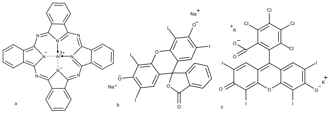
Cationic aluminum phthalocyanines (AlPcN, Figure 5a) represent an important second-generation subclass of macrocyclic photosensitizers designed to improve antimicrobial activity through surface-charge engineering [118]. The introduction of quaternary ammonium or pyridinium groups into the peripheral phthalocyanine structure gives these compounds a permanent positive charge, which promotes electrostatic interactions with negatively charged bacterial cell walls, fungal cell envelopes, and biofilm extracellular matrices [118,119]. This feature distinguishes cationic AlPcN derivatives from neutral or anionic phthalocyanines, which may show weaker microbial binding and reduced uptake. From a photophysical perspective, AlPcN retains the advantages of the extended phthalocyanine macrocycle, including strong absorption in the red and near-infrared spectral region, with λ_max around 680 nm [120]. This wavelength range is beneficial for antimicrobial PDT because red/NIR light penetrates biological tissues more effectively than blue or green light. The centrally coordinated Al3+ ion stabilizes the macrocyclic structure and supports efficient intersystem crossing to the triplet excited state (T1), enabling effective singlet oxygen (1O2) generation under illumination [120,121].
From a biochemical point of view, the cationic character of AlPcN supports rapid binding to microbial envelopes and increases local photosensitizer concentration at membrane-associated targets [121]. After light activation, AlPcN generates singlet oxygen and other reactive oxygen species close to bacterial membranes, cell-wall components, and membrane-associated proteins. This localized oxidative stress can induce lipid peroxidation, membrane depolarization, permeability changes, enzyme inactivation, and microbial cell death [122]. In fungal systems, AlPcN-mediated PDT can damage the carbohydrate-rich cell wall and the underlying ergosterol-containing plasma membrane, leading to loss of membrane integrity and reduced fungal viability [123,124,125]. The selectivity of this process is supported by differences between microbial and mammalian cell envelopes. Bacterial and fungal surfaces are generally more anionic than mammalian plasma membranes, while mammalian cells also possess more developed antioxidant defense systems. As a result, properly optimized AlPcN-mediated PDT can provide strong antimicrobial and antifungal activity with limited dark toxicity toward host cells [126,127].
AlPcN derivatives are particularly relevant for biofilm-associated infections because their positive charge may improve interaction with the negatively charged extracellular polymeric substance (EPS) matrix [128]. However, the same EPS matrix can also restrict penetration when the photosensitizer is too strongly sequestered in the outer biofilm layers. Therefore, effective AlPcN-based aPDT depends on the balance between electrostatic binding, diffusion, photosensitizer aggregation, oxygen availability, and light penetration [129]. To overcome these limitations, AlPcN has been investigated in delivery systems such as polymeric nanoparticles, liposomes, micelles, hydrogels, and surface-functionalized coatings [130,131,132]. These platforms can improve dispersion, reduce self-aggregation, protect the photosensitizer from premature deactivation, and increase contact between AlPcN and microbial targets.
Recent material-based approaches have also explored AlPcN incorporation into light-activated antimicrobial surfaces, including polymer coatings, layer-by-layer polyelectrolyte assemblies, hydrogel networks, and medical-device interfaces [133,134,135,136]. In these systems, AlPcN can be electrostatically assembled with negatively charged polymers or covalently linked to functionalized matrices such as polyurethane, silicone-based elastomers, or hydrogel scaffolds. The aim is to retain the photosensitizer at the material surface, reduce uncontrolled leaching into biological fluids, and generate ROS locally after illumination [136,137]. When activated with red or NIR light, immobilized AlPcN can produce singlet oxygen at the device–fluid interface, where it may inhibit microbial adhesion, damage early EPS formation, and reduce surface-associated biofilm development [138,139]. Overall, AlPcN should be considered a highly promising cationic phthalocyanine platform for antimicrobial and antifungal PDT. Its main advantages include red/NIR absorption, high singlet oxygen generation, strong microbial surface binding, and compatibility with coating or delivery technologies. Its main limitations include possible aggregation, oxygen dependence, and the need for careful optimization of charge density, loading, and material architecture [140].
Having completed the discussion of tetrapyrrolic macrocycles, the next section focuses on xanthene photosensitizers, with erythrosine B serving as the first representative compound.

2.4. Xanthene Photosensitizers

2.4.1. Erythrosine B: Iodinated Xanthene Photosensitizer for Superficial Antimicrobial PDT

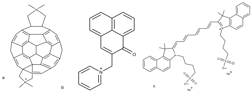
Erythrosine B (Figure 5b) is an iodinated xanthene dye that has historically been used as a food colorant, dental plaque-disclosing agent, and diagnostic stain. Beyond these conventional applications, it has attracted considerable attention as a photosensitizer for antimicrobial photodynamic therapy (aPDT), particularly in dental, dermatological, mucosal, and superficial infection models. Its clinical relevance results from the combination of low cost, broad availability, visible-light activation, relatively low dark toxicity, and efficient production of reactive oxygen species after illumination.
From a photophysical perspective, erythrosine B absorbs mainly in the green region of the visible spectrum, with an absorption maximum at approximately 520–530 nm [128,129]. This spectral profile makes it compatible with inexpensive and widely available green-light LEDs. Although green light penetrates tissue less deeply than red or near-infrared light, it is sufficient for many superficial antimicrobial applications, including oral biofilms, infected mucosal surfaces, dermatological lesions, and device-accessible sites. Therefore, erythrosine B is particularly suitable for topical or surface-confined PDT rather than deeply located infections.
The photodynamic activity of erythrosine B is strongly influenced by the presence of iodine atoms in the xanthene scaffold. These heavy atoms enhance spin–orbit coupling and promote efficient intersystem crossing from the excited singlet state to the triplet state. As a consequence, erythrosine B shows a high triplet-state yield and efficiently generates singlet oxygen through a predominantly Type II photochemical pathway [130,131]. Under hypoxic, reducing, or highly protein-rich microenvironments, Type I electron-transfer reactions may also contribute to the formation of radical species and other reactive oxygen species [132]. This dual photochemical potential is relevant for infected tissues and biofilms, where oxygen availability may be heterogeneous.
The antimicrobial effect of erythrosine B is largely determined by its localization close to microbial cell envelopes. Because singlet oxygen has a short lifetime and limited diffusion distance, oxidative damage occurs mainly in the immediate vicinity of the bound photosensitizer. Erythrosine B can associate with cell-surface components, membrane proteins, and extracellular biofilm structures. After illumination, locally generated singlet oxygen oxidizes membrane lipids, amino acid residues in proteins, and components of the extracellular polymeric substance matrix [133,134]. These reactions compromise membrane integrity, disturb enzymatic activity, increase permeability, and can lead to rapid microbial inactivation.
Erythrosine B has shown particularly strong activity against Gram-positive bacteria. This susceptibility is commonly attributed to the relatively porous peptidoglycan wall, favorable dye binding, and easier access of reactive oxygen species to membrane-associated targets [133,136]. In Gram-negative bacteria, the outer membrane can limit photosensitizer access; however, photodynamic inactivation may still be achieved when irradiation parameters, incubation time, formulation, or permeability-enhancing strategies are optimized. In this context, erythrosine B may be especially useful against oral and surface-associated mixed microbial communities, where direct illumination and local dye application are technically feasible.
Antifungal activity is also an important aspect of erythrosine B-mediated PDT. The dye has demonstrated photodynamic effects against Candida species, including biofilm-forming strains [138]. In fungal cells, the carbohydrate-rich cell wall can provide binding sites for the dye, while the underlying ergosterol-rich plasma membrane is highly vulnerable to singlet oxygen-mediated lipid peroxidation. Oxidative damage to ergosterol-containing membranes may result in altered permeability, protein oxidation, metabolic disruption, and loss of fungal viability. This is particularly relevant because Candida biofilms often exhibit reduced susceptibility to conventional antifungal agents.
The selectivity of erythrosine B-mediated aPDT depends on differences in photosensitizer binding, cellular uptake, membrane composition, and antioxidant defense capacity between microbial and mammalian cells [135,137]. Microbial cells generally show stronger surface binding and lower redox-buffering capacity, whereas mammalian cells possess more developed antioxidant systems and usually exhibit lower dye accumulation under controlled topical conditions. These differences contribute to a useful therapeutic window, although selectivity remains dose-, light-, oxygen-, and formulation-dependent.
An additional advantage of erythrosine B is its low dark toxicity, which supports its use in localized antimicrobial applications [139,140]. However, several limitations should also be considered. Its activation by green light restricts its use mainly to superficial or optically accessible infections. Moreover, its activity may be reduced in thick mature biofilms because of heterogeneous dye penetration, oxygen gradients, and shielding by extracellular polymeric substances. Photobleaching may also occur during prolonged illumination, reducing the amount of active photosensitizer available for ROS generation. Therefore, the therapeutic effect of erythrosine B depends strongly on photosensitizer concentration, incubation time, irradiation fluence, oxygen availability, and the structural organization of the target biofilm.
From a translational perspective, erythrosine B is attractive because it is inexpensive, chemically well-characterized, and already used in food, pharmaceutical, and dental contexts. These features may facilitate the further development of erythrosine B-based PDT for topical antimicrobial, antifungal, dental, and dermatological applications. In addition to free-dye formulations, erythrosine B can also be incorporated into hydrogels, polymeric films, dental materials, or surface coatings to improve local retention and enable controlled, light-triggered antimicrobial activity. Such material-based systems may be useful for preventing microbial colonization on oral, wound-related, or device-associated surfaces.
Overall, erythrosine B should be regarded as a practical xanthene photosensitizer for superficial and surface-confined antimicrobial PDT. Its main advantages include efficient singlet oxygen generation, low dark toxicity, compatibility with inexpensive green-light sources, and relevance for oral and topical infections. Its main limitations are limited tissue penetration at green wavelengths, possible photobleaching, and reduced efficacy in deeply located or mature biofilm-associated infections. The next representative of this class is Rose Bengal, another halogenated xanthene dye with a high singlet oxygen yield and broad antimicrobial activity.

2.4.2. Rose Bengal: Halogenated Xanthene Photosensitizer with High Singlet Oxygen Yield

Rose Bengal (Figure 5c) is a well-established and widely studied halogenated xanthene-based photosensitizer used in antimicrobial and antifungal photodynamic therapy. It demonstrates strong absorption in the green region of the visible spectrum, with the wavelength of maximum absorbance (λ_max) around 540–550 nm, and efficiently generates singlet oxygen (1O2) after light activation [141]. Although this absorption profile limits tissue penetration compared with red-light-activated photosensitizers, it remains suitable for localized, superficial, and surface-accessible antimicrobial applications. The high photodynamic efficiency of Rose Bengal is closely related to its molecular structure. The presence of multiple iodine and chlorine atoms enhances spin–orbit coupling through the heavy-atom effect, which promotes efficient intersystem crossing and triplet-state formation. As a result, a large fraction of absorbed photons is converted into triplet-state excitation, leading predominantly to Type II photochemistry and high singlet oxygen production [141].
From a microbiological perspective, Rose Bengal demonstrates broad-spectrum photodynamic activity against Gram-positive and Gram-negative bacteria, including methicillin-resistant Staphylococcus aureus (MRSA), Enterococcus faecalis, Escherichia coli, Pseudomonas aeruginosa, Bacillus subtilis, and Salmonella spp. [142]. It has also shown activity against fungal pathogens and enveloped viruses [143,144,145]. Its antimicrobial effect is mainly associated with the localized generation of singlet oxygen near the microbial envelope. In bacterial cells, Rose Bengal can associate with the cell envelope, where photoinduced 1O2 causes oxidative damage to membrane lipids, membrane-associated proteins, and cell-wall components. This leads to the disruption of membrane integrity, loss of cellular homeostasis, enzyme inactivation, and ultimately, microbial cell death [146,147,148]. In biofilm systems, Rose Bengal-mediated PDT may also oxidize extracellular polymeric substances, including proteins and polysaccharide-rich matrix components, thereby weakening biofilm architecture and increasing microbial susceptibility to oxidative damage [146,147,148].
In antifungal photodynamic therapy, Rose Bengal has shown efficacy against Candida albicans, non-albicans Candida species, and filamentous fungi [138]. Its antifungal activity is strongly associated with oxidative damage to the fungal cell wall and the ergosterol-containing plasma membrane. Because ergosterol-rich membranes are particularly sensitive to singlet oxygen-mediated lipid peroxidation, Rose Bengal irradiation can cause irreversible membrane damage, altered permeability, and loss of fungal viability [149]. In addition, fungal cells generally show lower antioxidant capacity and delayed detoxification compared with mammalian cells, which can contribute to selective phototoxicity under optimized treatment conditions [150].
Despite its high photodynamic efficacy, Rose Bengal also has important limitations. One of the main disadvantages is photobleaching, which occurs when the photosensitizer is progressively degraded during irradiation. This process can reduce the amount of active photosensitizer available for subsequent ROS generation and may limit efficacy during prolonged or repeated light exposure [151]. Rose Bengal may also show dark toxicity at elevated concentrations, which means that dose selection and irradiation parameters must be carefully optimized to preserve antimicrobial activity while minimizing host-cell damage [152]. Therefore, Rose Bengal is particularly valuable in localized and surface-accessible applications, where photosensitizer concentration, incubation time, oxygen availability, and light dose can be precisely controlled. Its green-light absorption should therefore be viewed as compatible with superficial antimicrobial PDT rather than as advantageous for deep-tissue treatment.
From a broader biophysical and biochemical perspective, Rose Bengal and erythrosine B illustrate the antimicrobial potential of xanthene photosensitizers with dominant Type II photochemistry. Their high singlet oxygen quantum efficiency, strong interaction with microbial envelopes, and ability to damage both planktonic cells and biofilm-associated structures make them attractive candidates for further development in antimicrobial and antifungal PDT [141]. However, their clinical translation requires the careful control of photobleaching, dark toxicity, tissue penetration limits of green light, and formulation stability.
The next compound discussed in this review is curcumin, a naturally occurring plant-derived polyphenol with antimicrobial photodynamic activity.

2.5. Natural Polyphenols and Endogenous Compounds in Antimicrobial and Antifungal Photodynamic Therapy

2.5.1. Curcumin: Natural Polyphenol with Blue-Light-Activated Antimicrobial Activity

Curcumin (Figure 6a) is a naturally occurring polyphenolic compound derived from Curcuma longa. Curcumin exhibits strong absorption in the blue region of the visible spectrum, with an absorption maximum (λmax) around 420–430 nm, which corresponds mainly to π–π* transitions within its conjugated diketone structure [153,154]. This absorption profile is a major practical limitation for PDT applications requiring tissue penetration, because curcumin shows little or no therapeutically useful absorption above approximately 430 nm. Compared with porphyrins, chlorins, and phthalocyanines, curcumin has a lower intrinsic triplet-state yield and weaker photophysical stability. Nevertheless, under short-wavelength blue-light irradiation, typically using wavelengths matching its absorption band, sufficient light dose, adequate oxygenation, and close contact between curcumin and microbial targets, it can generate reactive oxygen species and produce relevant antimicrobial photodynamic effects [155]. Therefore, curcumin-mediated aPDT should be considered mainly for superficial, topical, dental, or surface-accessible antimicrobial applications rather than for deeper tissue infections.
From a biochemical perspective, curcumin exhibits amphiphilic behavior and can interact with lipid-rich cellular structures [156]. In bacterial and fungal cells, curcumin is frequently associated with the cell wall and cytoplasmic membrane, where local ROS generation can directly damage essential envelope components [157]. Photoactivated curcumin induces the oxidation of membrane lipids and proteins, leading to impaired membrane-associated enzymatic functions, loss of membrane potential, increased permeability, and ultimately reduced cell viability [155,158]. This membrane-centered mechanism is particularly important because the short lifetime and limited diffusion distance of ROS require close spatial proximity between the photosensitizer and its cellular targets.
Curcumin-mediated PDT has demonstrated activity against both Gram-positive and Gram-negative bacteria under optimized experimental conditions, including Staphylococcus aureus and Enterococcus species [159,160]. In fungal systems, curcumin has shown notable activity against Candida albicans. Its interaction with fungal cell-wall structures facilitates localization near the ergosterol-rich plasma membrane, which is highly vulnerable to oxidative lipid damage [161]. Compared with mammalian cells, fungal cells generally possess a lower capacity to detoxify photoinduced ROS, which supports selective phototoxicity when curcumin concentration and light dose are properly controlled [162].
The main limitations of curcumin are its poor aqueous solubility, chemical instability, low photostability, and rapid photobleaching under continuous illumination and unfavorable blue-light absorption profile for applications requiring tissue penetration [155]. These features can reduce bioavailability, limit homogeneous distribution in biological environments, and decrease ROS generation during prolonged irradiation. Therefore, formulation strategies are essential for improving curcumin-mediated aPDT. These strategies include nanoparticle encapsulation, polymer conjugation, lipid-based carriers, liposomes, micelles, and carbon-based nanocarriers [159]. In this context, curcumin-loaded carbon nanoparticles have been proposed as an effective approach to improve curcumin dispersion, enhance delivery, and modify its light-triggered photodynamic activity [163,164]. However, formulation strategies improve delivery and local availability, but they do not eliminate the fundamental limitation associated with curcumin’s short-wavelength absorption.
From a translational perspective, curcumin is of interest mainly for localized antimicrobial PDT because of its natural origin, relatively low dark toxicity under controlled conditions, and extensive prior human exposure [163]. However, its successful application in antimicrobial and antifungal PDT depends strongly on formulation design, irradiation parameters, oxygen availability, and target localization. Properly formulated curcumin systems are especially promising for topical antimicrobial applications, including dentistry, dermatology, wound care, and localized antifungal therapy.
The next compound discussed in this review is hypericin, another plant-derived natural photosensitizer with strong membrane affinity and high photodynamic potency.

2.5.2. Hypericin: Plant-Derived Photosensitizer with Antimicrobial and Antifungal Potential

Hypericin (Figure 6b) is a naturally occurring naphthodianthrone derivative isolated from Hypericum perforatum (St. John’s wort). It is considered one of the most potent plant-derived photosensitizers because of its strong absorption in the visible region, with λ_max around 590–600 nm, and its highly efficient triplet-state formation [164]. Upon illumination, hypericin generates singlet oxygen and other reactive oxygen species, which are responsible for its strong photodynamic activity. Its rigid, extended polycyclic structure contributes to high photostability, reduced structural flexibility, and efficient light-induced ROS production, making it more photophysically robust than many other natural photosensitizers [164].
From a biochemical perspective, hypericin is highly lipophilic and displays strong affinity for lipid-rich biological membranes. This includes bacterial membranes, fungal plasma membranes, and envelopes of certain viruses [165,166,167]. Such membrane affinity is crucial for antimicrobial PDT because singlet oxygen has a short lifetime and limited diffusion distance. Therefore, the localization of hypericin close to microbial membranes allows ROS to be generated directly at vulnerable cellular interfaces. Under light irradiation, hypericin-mediated PDT induces oxidative damage to membrane lipids, membrane-associated proteins, and depending on cellular uptake, intracellular targets such as nucleic acids. These combined effects lead to membrane dysfunction, disruption of metabolic activity, and rapid microbial cell death [165,166,167].
In antifungal PDT, hypericin has shown strong activity against Candida species and filamentous fungi [168,169]. The dominant mechanism of fungal inactivation is associated with oxidative damage to ergosterol-rich membranes and impairment of mitochondrial function. Since fungal membranes contain ergosterol instead of cholesterol, they are particularly susceptible to lipid peroxidation induced by singlet oxygen and other ROS. Hypericin can also accumulate in fungal cell structures for a longer time than in mammalian cells, while fungal antioxidant systems are generally less efficient at neutralizing sudden photoinduced oxidative stress. These differences support selective phototoxicity toward fungal cells under properly optimized irradiation and concentration conditions [168,169,170,171].
Despite its excellent photodynamic potency, hypericin has several translational limitations. Its strong hydrophobicity causes poor aqueous solubility and a tendency to aggregate in biological media, which can reduce bioavailability and decrease effective ROS generation [170,171]. Another important concern is prolonged photosensitivity in host tissues, especially if hypericin is retained after treatment. These limitations have stimulated the development of formulation strategies, including liposomes, cyclodextrin complexes, polymeric carriers, and nanoparticle delivery systems [172]. Such approaches improve solubility, reduce aggregation, enhance delivery to microbial targets, and may limit off-target phototoxicity. Overall, hypericin represents a highly effective natural photosensitizer whose antimicrobial and antifungal potential depends strongly on formulation design, membrane localization, light dose, and controlled delivery.
From a translational perspective, hypericin illustrates how high triplet-state yield, strong photostability, and membrane affinity can be converted into potent antimicrobial and antifungal photodynamic effects. However, because of its hydrophobicity and possible prolonged tissue photosensitivity, hypericin is better considered as a formulation-dependent photosensitizer rather than a simple free molecular agent. Properly designed delivery systems may allow its use in topical antifungal therapy, wound-related infections, and biofilm-associated applications where localized illumination can be precisely controlled.

2.5.3. Riboflavin: Endogenous Vitamin-Based Photosensitizer for Local Antimicrobial Applications

Riboflavin, also known as vitamin B2 (Figure 6c), is an endogenous isoalloxazine derivative that plays an essential role in cellular metabolism as a precursor of flavin mononucleotide (FMN) and flavin adenine dinucleotide (FAD). Because it is naturally present in biological systems and has an established safety profile, riboflavin represents a distinct group of endogenous photosensitizers with proven clinical relevance. Photophysically, riboflavin absorbs mainly in the UV-blue region of the spectrum, with characteristic absorption bands around 224, 268, 373, and 440–450 nm [173]. After light absorption, it can undergo intersystem crossing to the triplet excited state, which enables both Type I and Type II photochemical pathways. As a result, illuminated riboflavin can generate singlet oxygen (1O2), superoxide, hydrogen peroxide, and other radical species that contribute to microbial inactivation [173].
From a biochemical perspective, riboflavin has low dark toxicity and is well-recognized as a naturally occurring vitamin and cellular metabolite [174]. In antimicrobial PDT, its phototoxicity is not based on strong intrinsic membrane binding alone, but rather on light-induced oxidative damage to multiple cellular targets. Riboflavin-mediated photodynamic reactions can oxidize nucleic acids, proteins, and membrane components, leading to impaired replication, enzyme dysfunction, membrane damage, and loss of microbial viability [175]. This broad oxidative mechanism is particularly useful in antimicrobial applications because it does not rely on one specific molecular target. Bacterial and fungal cells may be more vulnerable to this type of damage than mammalian cells because they often have lower antioxidant buffering capacity and more limited repair mechanisms under acute photooxidative stress [175].
Riboflavin-based photodynamic inactivation has particular translational importance because it is already widely used in sterilization and pathogen reduction in plasma and blood products [176]. This application provides strong practical evidence that riboflavin can be used safely as a light-activated antimicrobial agent under controlled conditions. In this context, riboflavin is especially valuable because it combines photochemical activity with high biocompatibility and minimal concerns regarding systemic toxicity. Its mechanism involves oxidative damage and the photochemical modification of nucleic acids and proteins, which helps reduce the viability of bacteria, viruses, and other contaminating pathogens in biological fluids [176].
In fungal systems, riboflavin-mediated PDT has shown activity against Candida species, particularly in planktonic cells and early biofilm stages [177]. Its efficacy against mature biofilms may be more limited than that of more hydrophobic or membrane-targeted photosensitizers, because riboflavin is relatively hydrophilic and absorbs mainly blue light, which has shallow tissue penetration. Nevertheless, its favorable safety profile makes it useful for applications where biocompatibility is more important than maximal photodynamic potency. These include blood sterilization, surface decontamination, oral applications, wound irrigation, and other localized treatments where blue-light exposure can be precisely controlled.
Due to its endogenous character, regulatory familiarity, and established clinical use, riboflavin occupies a unique position among photosensitizers [178]. Compared with synthetic porphyrins, chlorins, phthalocyanines, or phenothiazinium dyes, riboflavin generally shows lower photodynamic potency. However, this limitation is balanced by excellent biocompatibility, low dark toxicity, and broad acceptance in medical and biological applications. Therefore, riboflavin should be considered not as the strongest antimicrobial photosensitizer, but as one of the safest and most clinically familiar endogenous compounds for photodynamic pathogen reduction. The mechanisms presented herein for riboflavin are broad photodynamic principles that apply to multiple short-wavelength photosensitizers (including riboflavin), rather than being exclusive to one compound.
The next compound discussed in this review is fullerene C60, a predominantly synthetic carbon-based photosensitizer with distinct photophysical properties and strong potential for antimicrobial PDT.

2.6. Advanced and Targeted Photosensitizers

2.6.1. Cationic Fullerene (C60) Derivatives: Type I/Type II ROS Generation and Nanostructured Activity

Fullerene derivatives (C60, Figure 7a) represent a distinct class of carbon-based photosensitizers with photophysical properties that differ substantially from those of classical porphyrins, chlorins, phthalocyanines, and phenothiazinium dyes. Native C60 has a spherical carbon cage structure and absorbs light in the UV–visible spectral range. However, its absorption in the visible region is relatively weak and does not provide the same favorable red-light activation profile as chlorins, phthalocyanines, or other photosensitizers designed for deeper tissue PDT. One of its most important photophysical features is a near-unity triplet-state quantum yield, which makes it highly efficient in generating long-lived excited states after irradiation [179,180]. However, unmodified C60 is highly hydrophobic and poorly dispersed in aqueous biological media. This strongly limits its direct biological application and creates the need for chemical functionalization. Cationic substitution improves water dispersibility and promotes interaction with negatively charged bacterial cell walls, fungal cell envelopes, and microbial membranes [1].
From a biophysical perspective, cationic C60 derivatives can act as nanoscale light-harvesting systems. After photoexcitation, the long-lived triplet excited state can participate in both Type I and Type II photochemical pathways, leading to the formation of singlet oxygen (1O2), superoxide anions, hydroxyl radicals, and other reactive oxygen species [181]. This mixed photochemical behavior is particularly important in antimicrobial PDT because dense biofilms and infected tissues often contain oxygen gradients. Under partially hypoxic conditions, photosensitizers that rely exclusively on Type II singlet oxygen generation may lose efficacy. In contrast, C60 derivatives capable of Type I electron-transfer reactions may retain antimicrobial activity by producing radical species even when oxygen availability is limited. The rationale for considering fullerene derivatives in antimicrobial PDT is therefore not superior tissue-directed light absorption, but their high triplet-state yield, tunable functionalization, capacity for both Type I and Type II photochemistry, and ability to interact with microbial envelopes after appropriate chemical modification. Nevertheless, their unfavorable aqueous dispersibility, need for functionalization, and often suboptimal visible-light absorption profiles should be clearly recognized as major barriers to broader translational use.
From a biochemical point of view, cationic fullerenes show strong affinity for microbial envelopes because bacterial and fungal surfaces are generally negatively charged [182]. This electrostatic attraction promotes localization of the photosensitizer close to the cell wall, cytoplasmic membrane, and biofilm matrix. Since ROS have short lifetimes and limited diffusion distances, this spatial proximity is essential for effective photodynamic damage. In bacterial cells, fullerene-mediated PDT first affects the cell wall and cytoplasmic membrane, causing lipid peroxidation, membrane depolarization, and permeability disruption. Secondary oxidative injury may involve membrane-associated proteins, intracellular enzymes, and nucleic acids [182]. In fungal cells, oxidative damage to ergosterol-containing membranes and cell-wall polysaccharides can lead to irreversible loss of structural integrity and fungal viability [139].
Cationic fullerene derivatives are also relevant for biofilm-associated infections. Their nanoscale structure and tunable surface chemistry may support penetration into biofilm matrices and retention near embedded microbial cells. Moreover, their ability to generate both singlet oxygen and radical ROS can help damage not only microbial membranes, but also extracellular polymeric substances, including proteins, polysaccharides, and extracellular DNA. This is important because biofilm eradication requires the disruption of both living cells and the protective matrix that surrounds them. Compared with many molecular photosensitizers, fullerene derivatives also demonstrate relatively high photostability and limited photobleaching, which is advantageous during repeated or prolonged irradiation.
Selectivity is supported by several factors. Cationic fullerenes preferentially interact with negatively charged microbial surfaces, whereas mammalian cells usually show lower uptake of these highly charged derivatives and possess more developed antioxidant defense systems [182,183]. This creates a potentially favorable therapeutic window, particularly for localized antimicrobial applications. Nevertheless, selectivity depends strongly on the degree of functionalization, aggregation state, concentration, incubation time, irradiation dose, and the biological model used.
From a translational perspective, cationic fullerene derivatives have demonstrated promising activity in experimental infection models, including in vivo bacterial wound infection studies [184]. Their combination of high triplet-state yield, mixed Type I/Type II photochemistry, strong photostability, and low photobleaching makes them attractive candidates for next-generation antimicrobial and antifungal PDT. However, their clinical development still requires the careful evaluation of long-term biocompatibility, biodistribution, clearance, possible nanomaterial accumulation, and regulatory safety. Therefore, cationic C60 derivatives should be described as promising advanced photosensitizers and nano-enabled PDT agents, rather than as fully established clinical antimicrobials. Native and pristine fullerene C60 are primarily “activated” in the ultraviolet (UV) and short-wavelength blue-violet region, typically between 220 nm and 450 nm. As explicitly stated in our manuscript, unmodified C60 has relatively weak and suboptimal absorption in the visible spectrum and lacks the favorable red-light activation profile seen in classical porphyrins or phthalocyanines. However, we would like to clarify that the scientific rationale for utilizing fullerenes in antimicrobial PDT does not rely on superior light absorption. Instead, they are activated using UVA, blue, or broadband white light because they possess unique photochemical advantages.

2.6.2. P9 Peptide–Photosensitizer Conjugate: Targeted Antimicrobial PDT

Peptide–photosensitizer conjugates are attractive platforms for precision aPDT, especially in infections where nonspecific photosensitizer distribution limits therapeutic efficacy. A critical feature of this platform is its high selectivity for microbial cells over infected host tissues. This pathogen-directed selectivity is governed by the structural differences between microbial and mammalian membranes. The cationic sequence of P9 preferentially targets the highly anionic bacterial cell envelope (stabilized by lipopolysaccharides or teichoic acids) while displaying low affinity for the net-neutral zwitterionic outer leaflets of mammalian cells. Furthermore, the presence of cholesterol in mammalian membranes provides steric and mechanical resistance against peptide insertion. Experimental evaluations of structurally related AMP-photosensitizer conjugates confirm a broad therapeutic window, demonstrating negligible hemolytic activity and high viability (>90%) in human fibroblasts and keratinocytes at concentrations that induce up to a 6-log reduction in multidrug-resistant bacterial strains. In vivo wound models further validate this safety profile, showing robust localized bacterial clearance without inducing tissue necrosis or impairing the host’s natural healing cascade. From a translational perspective, these conjugates may offer high local activity. P9 represents a hybrid and rationally designed class of antimicrobial photosensitizers in which a cationic antimicrobial peptide is covalently linked to a photoactive chromophore. In this system, the peptide fragment is responsible for microbial recognition and selective membrane binding, whereas the chromophore acts as the light-activated photosensitizing unit. This design combines two mechanisms in one molecule: the natural affinity of cationic peptides for microbial envelopes and the ability of the photosensitizer to generate reactive oxygen species after irradiation [185].
From a biophysical perspective, the activity of P9 is based on electrostatic attraction between the positively charged peptide sequence and negatively charged bacterial surfaces. Bacterial membranes and cell walls contain anionic components, including teichoic acids in Gram-positive bacteria and lipopolysaccharides in Gram-negative bacteria. These structures favor peptide binding and bring the photosensitizer into close proximity to the microbial envelope [185]. This spatial localization is critical for antimicrobial PDT because singlet oxygen and other ROS have very short lifetimes and limited diffusion distances. Therefore, when the photosensitizer is positioned near the membrane, the generated ROS can immediately oxidize membrane lipids, membrane-associated proteins, and cell-wall components.
P9-mediated PDT has shown strong activity against both Gram-positive and Gram-negative bacteria, including multidrug-resistant strains [186]. Its mechanism does not require deep intracellular accumulation of the photosensitizer. Instead, membrane-associated localization is often sufficient to induce lethal oxidative damage. This is advantageous because it reduces dependence on active uptake, metabolic state, or intracellular transport pathways, which are frequently altered in resistant or dormant microbial cells. In fungal systems, peptide-mediated targeting can also promote accumulation of the photosensitizer at the cell surface, where irradiation causes rapid photoinactivation [187]. The fungal cell wall and plasma membrane become primary targets of ROS-mediated damage, leading to loss of barrier function and reduced cell viability.
An important advantage of P9-type constructs is their modularity. The peptide sequence, charge density, hydrophobicity, linker chemistry, and photosensitizer structure can be modified independently to optimize microbial selectivity, membrane affinity, solubility, and photodynamic potency. This makes peptide–photosensitizer conjugates attractive platforms for precision aPDT, especially in infections where nonspecific photosensitizer distribution limits therapeutic efficacy. From a translational perspective, these conjugates may offer high local activity with reduced off-target effects, because the photosensitizer is guided toward microbial membranes rather than passively distributed in surrounding tissue [188]. However, challenges remain, including synthesis complexity, peptide stability, proteolytic degradation, production cost, and the need to optimize pharmacokinetics in biological fluids. Despite these limitations, P9 illustrates how molecular targeting can transform aPDT from a broadly oxidative approach into a more selective, pathogen-directed strategy. An important advantage of P9-type constructs is their modularity. The peptide sequence, charge density, hydrophobicity, linker chemistry, and photosensitizer structure can be modified independently to optimize microbial selectivity, membrane affinity, solubility, and photodynamic potency. Crucially, this modular nature allows for the selection of specific activation wavelengths depending on the clinical target. While early-generation photosensitizers like SAPYR are restricted to blue-light activation (360–410 nm)—which severely limits their clinical utility to superficial infections due to poor tissue penetration—the P9 platform can be readily conjugated to long-wavelength, near-infrared (NIR) chromophores. By incorporating NIR-active agents such as indocyanine green (ICG) or phthalocyanines, which absorb light within the optical window of tissue (650–800 nm), the system can bypass the limitations of short-wavelength phototoxicity and achieve the depth of penetration required for deep-seated or systemic infections [188]. This makes peptide–photosensitizer conjugates attractive platforms for precision aPDT.

2.6.3. SAPYR: A Cationic Anthraquinone-Based Photosensitizer

SAPYR (Figure 7b) belongs to a newer class of anthraquinone-based photosensitizers designed specifically for antimicrobial photodynamic therapy. It absorbs in the UV and visible region, approximately 360–410 nm, and demonstrates efficient intersystem crossing after light excitation [189,190]. This enables both Type I and Type II photochemical pathways, which is particularly important for antimicrobial applications. Type II reactions generate singlet oxygen, whereas Type I pathways produce radical species through electron-transfer processes. Because infection sites and biofilms may contain regions with limited oxygen availability, this mixed photochemical behavior can support antimicrobial activity under conditions where purely Type II photosensitizers may be less effective.
The structure of SAPYR contains a pyridyl substituent that introduces cationic character and promotes interaction with negatively charged bacterial cells [190]. From a biochemical point of view, this favors accumulation at or near the bacterial envelope through electrostatic interactions. Once localized close to the membrane, SAPYR-mediated irradiation induces oxidative damage to membrane lipids, proteins, and nucleic acids [191]. The anthraquinone core is also capable of redox cycling, which can intensify ROS production and enhance the antimicrobial effect [192]. This dual activity, combining photochemical ROS generation with redox-active behavior, makes SAPYR mechanistically distinct from many classical photosensitizers that rely mainly on singlet oxygen.
SAPYR-mediated aPDT is particularly relevant for bacterial cells because its cationic and redox-active structure supports membrane interaction and oxidative stress at multiple cellular levels. Damage to the envelope can increase permeability and disturb membrane-associated metabolic processes, while intracellular or pericellular ROS may oxidize proteins and nucleic acids. This multi-target mechanism supports rapid microbial inactivation and reduces the likelihood of resistance development. In biofilm-related contexts, SAPYR may also contribute to oxidative weakening of extracellular polymeric substances, although its final efficacy depends on penetration depth, light dose, oxygen availability, and biofilm composition.
In antifungal PDT, SAPYR-mediated oxidative stress may damage the fungal cell wall and the underlying plasma membrane. The high oxidative burden can exceed the antioxidant buffering capacity of fungal cells, leading to membrane dysfunction and loss of viability. However, compared with bacterial models, the antifungal profile of SAPYR is less extensively characterized, and direct comparisons with mammalian cells remain limited. Therefore, its selectivity should be presented cautiously and interpreted as promising rather than fully established.
Overall, SAPYR remains an experimental but highly interesting antimicrobial photosensitizer. Its main strengths include a defined synthetic scaffold, cationic character, mixed Type I/Type II photochemistry, and redox-active anthraquinone chemistry. These properties make it useful not only as a candidate compound for aPDT, but also as a framework for studying structure–activity relationships in antimicrobial photodynamic therapy. Further studies are still needed to clarify its antifungal efficacy, dark toxicity, behavior in mature biofilms, formulation requirements, and safety profile in host tissues.
The next compound discussed in this review is indocyanine green, a clinically established near-infrared dye with combined photodynamic and photothermal antimicrobial potential.

2.6.4. Indocyanine Green Near-Infrared Activation and Photodynamic–Photothermal Effects

Indocyanine green (ICG, Figure 7c) is a clinically approved near-infrared fluorescent dye that has been widely used in diagnostic imaging, angiography, perfusion assessment, and fluorescence-guided procedures. In the context of antimicrobial and antifungal photodynamic therapy, ICG occupies a distinct position because it does not behave as a classical high-yield singlet oxygen photosensitizer. Compared with porphyrins, chlorins, phthalocyanines, or xanthenes, ICG generally exhibits a relatively low singlet oxygen quantum yield. Nevertheless, under appropriate irradiation conditions and at suitable local concentrations, it can produce a significant antimicrobial effect through the combined action of photodynamic and photothermal mechanisms [193,194].
From a photochemical point of view, ICG absorbs in the near-infrared spectral region, with absorption maxima reported around 710 nm and 780–810 nm, depending on concentration, solvent environment, aggregation state, and binding to biological macromolecules [195]. This near-infrared absorption is one of its most important advantages, because NIR light penetrates biological tissues more deeply than blue, green, or most red wavelengths. Therefore, ICG is particularly relevant for infections located in deeper tissue layers, within complex wound environments, or in biofilm-associated sites where optical access is limited. Upon irradiation, ICG dissipates absorbed energy through two major pathways: partial generation of reactive oxygen species and the conversion of light energy into localized heat. This dual photodynamic–photothermal behavior distinguishes ICG from many classical photosensitizers and may enhance antimicrobial efficacy, especially when microbial cells are embedded within protective biofilm matrices [195].
The photothermal component of ICG activity is especially important in antimicrobial applications. Local temperature elevation can destabilize microbial membranes, increase membrane permeability, weaken biofilm architecture, and enhance susceptibility to ROS-mediated damage. At the same time, ICG-mediated ROS generation can oxidize membrane lipids, proteins, and extracellular biofilm components. These two processes may act synergistically: photothermal stress compromises cell-envelope integrity, while photooxidative stress causes irreversible biochemical damage. This combined mechanism can be valuable against dense biofilms, mixed microbial communities, and infections where conventional antimicrobial agents show limited penetration or reduced activity [193,194,195].
From a biochemical perspective, ICG can associate with microbial cells and biofilm structures, although its localization and activity depend strongly on formulation, concentration, incubation time, and the surrounding biological environment [193]. In bacterial systems, ICG-mediated irradiation may damage the cytoplasmic membrane, disturb permeability barriers, and reduce cell viability. In fungal systems, the combined photodynamic and photothermal effect may target the cell wall and plasma membrane, including lipid-rich regions that are sensitive to oxidative and thermal stress. However, because ICG is not primarily a strongly cationic membrane-targeting photosensitizer, its antimicrobial efficacy can be improved by delivery systems, nanoparticles, polymeric carriers, or surface immobilization strategies that increase local retention at the infection site.
A major translational advantage of ICG is its established clinical use and known pharmacokinetic profile. In mammalian organisms, ICG is rapidly bound to plasma proteins, cleared mainly by the liver, and eliminated through biliary excretion. This contributes to its favorable safety profile when used under clinically controlled conditions [193]. Its regulatory familiarity, compatibility with clinically available NIR light sources, and extensive use in diagnostic medicine make it an attractive candidate for localized antimicrobial PDT, especially in settings where deeper light penetration is required. Nevertheless, its application in aPDT must account for several limitations, including concentration-dependent aggregation, photobleaching, thermal dose control, and the need to avoid excessive heat generation in surrounding host tissues.
From a translational perspective, ICG should be viewed as a clinically familiar dye with hybrid photodynamic and photothermal antimicrobial potential rather than as a conventional high-efficiency singlet oxygen photosensitizer. Its greatest value lies in applications where NIR activation, tissue penetration, and combined ROS/heat-mediated microbial damage are advantageous. These include infected wounds, biofilm-associated infections, periodontal or endodontic applications, implant-associated infections, and other localized conditions in which light delivery can be controlled. Future development of ICG-based antimicrobial therapy will likely depend on optimized formulations, controlled irradiation protocols, and integration with nanocarriers or surface-based delivery systems to improve local accumulation and reduce off-target effects.
The next section focuses on nanotechnology-based platforms and nanoformulations in antimicrobial and antifungal photodynamic therapy. These systems are designed to improve photosensitizer delivery, reduce aggregation, enhance biofilm penetration, modulate ROS generation, and increase the spatial precision of photodynamic action.

3. Nanotechnology-Based Techniques in Antimicrobial and Antifungal Photodynamic Therapy

3.1. The Rationale for Nanotechnology in Antimicrobial and Antifungal PDT

Despite the significant advantages of antimicrobial and antifungal photodynamic therapy, several physicochemical and biological limitations still restrict its broader clinical translation. The major advantage of PDT is its multi-target oxidative mechanism, which strongly reduces the probability of resistance development compared with conventional antibiotics. However, many molecular photosensitizers suffer from poor aqueous solubility, aggregation, low bioavailability, limited penetration into mature biofilms, insufficient retention at microbial targets, and nonspecific biodistribution [196]. These limitations are especially problematic in complex biological environments, where extracellular polymeric substances, proteins, host cells, and oxygen gradients can reduce the effective concentration of the active photosensitizer at the infection site. Nanotechnology-based platforms are therefore increasingly used not simply as additional “photosensitizer classes”, but as carriers, stabilizers, photophysical enhancers, surface-active antimicrobial systems, and biofilm-penetrating delivery tools. The purpose of this section is therefore to distinguish true photoactive nanomaterials from nanocarriers and auxiliary nanoplatforms that improve the delivery, localization, or performance of conventional photosensitizers.
At the nanoscale, carrier systems can modulate the aggregation state, spatial distribution, and local microenvironment of photosensitizers. This is particularly important because aggregation usually reduces triplet-state formation, shortens excited-state lifetimes, and decreases singlet oxygen generation. Nanoparticles, polymeric matrices, carbon-based nanostructures, and hybrid nanocomposites can spatially separate photosensitizer molecules, protect them from premature degradation, and increase local ROS generation after irradiation [13,196]. In such cases, the photosensitizer remains the primary ROS-generating species, whereas the nanoparticle or matrix acts as a delivery, stabilization, or localization platform. In addition, nanosystems can improve microbial targeting by exploiting differences in surface charge, membrane composition, biofilm architecture, and local pH. Because singlet oxygen has a very short lifetime and diffusion radius, usually within the nanometer range in biological environments, the photosensitizer or ROS-generating nanoplatform must be positioned close to microbial membranes, cell walls, or biofilm matrix components to achieve efficient oxidative damage [32,33,34]. Thus, the main value of nanotechnology in aPDT is not only the introduction of new photosensitizing structures, but also the precise spatial organization of light activation, ROS generation, and microbial target localization.
An important conceptual correction is that not all nanoparticles used in PDT should be called photosensitizers. Some nanoparticles are true photoactive ROS-generating systems, whereas others function mainly as carriers, optical enhancers, antimicrobial co-agents, oxygen modulators, or immobilization matrices. Strictly speaking, nanoparticles that do not generate ROS upon irradiation should not be described as photosensitizers or as independent PDT agents. Instead, they should be described as components of PDT systems only when they deliver, retain, enhance, or spatially organize a genuine photosensitizer.

3.2. Metallic and Metal Oxide Nanoparticles

Metallic and metal oxide nanoparticles are not generally selective photosensitizing agents in the same sense as molecular photosensitizers. In antimicrobial and antifungal PDT systems, they are more appropriately described as auxiliary nanomaterials that may act as photosensitizer carriers, photophysical enhancers, antimicrobial co-agents, magnetic delivery platforms, or redox-active components, depending on their composition and surface functionalization. Gold nanoparticles (AuNPs) are particularly important as photonic enhancers and photosensitizer carriers. Through localized surface plasmon resonance (LSPR), AuNPs can increase the local electromagnetic field near the particle surface, thereby improving the excitation efficiency of nearby photosensitizer molecules [197]. This can increase ROS generation, and in selected configurations, improve photostability by enhancing the effective absorption cross section of the photosensitizer [198]. In such systems, ROS generation usually arises from the molecular photosensitizer associated with the nanoparticle, rather than from AuNPs acting as independent selective photosensitizers. However, the effect is highly distance-dependent. If the photosensitizer is positioned too close to the gold surface, non-radiative energy transfer, Förster resonance energy transfer, or other quenching mechanisms may reduce fluorescence and ROS generation [199]. Therefore, rational spacer design and control of photosensitizer–metal distance are essential for achieving enhancement rather than quenching.
Silver nanoparticles (AgNPs) represent a different type of antimicrobial nanoplatform. They possess intrinsic antimicrobial activity related to silver ion release, membrane disruption, protein binding, and oxidative stress. When combined with photosensitizers, AgNPs can create synergistic systems in which silver-mediated toxicity and light-induced ROS generation reinforce each other [200]. However, AgNPs should not be described as selective photosensitizers unless direct light-triggered ROS generation and target selectivity are demonstrated. Their main role in many aPDT systems is as antimicrobial co-agents or carriers combined with conventional photosensitizers. In antifungal PDT, AgNP–photosensitizer hybrids may improve activity against Candida biofilms by increasing membrane stress, damaging fungal cell-wall components, and overwhelming antioxidant defenses [201]. However, these systems must be carefully optimized, because excessive silver release may increase dark toxicity and reduce selectivity.
Iron oxide nanoparticles add another level of functionality because they can be magnetically manipulated and may contribute to redox-based antimicrobial mechanisms. Photosensitizers immobilized on iron oxide nanoparticles can be retained or enriched at a defined, light-accessible target site by an external magnetic field before or during irradiation. In PDT, this would be useful only when magnetic localization can increase the local concentration or residence time of the photosensitizer-containing nanoplatform at a site that can also be illuminated. Here, selectivity is achieved primarily through physical or magnetic localization and photosensitizer delivery, not because iron oxide nanoparticles are inherently selective photosensitizers. In addition, iron oxide nanoparticles may participate in Fenton-like reactions, generating secondary redox stress that can amplify photodynamically produced ROS [202,203]. This is especially relevant in biofilms, where local hypoxia and heterogeneous oxygen distribution can limit purely Type II photochemistry. Nevertheless, iron oxide-based systems require a careful evaluation of dose, particle stability, iron release, and host-tissue compatibility. Thus, magnetic guidance should be viewed as a delivery/localization strategy rather than as a mechanism that directly improves light activation or ROS generation.
Overall, metallic and metal oxide nanoparticles should be discussed as functional components of PDT platforms rather than as a uniform class of selective photosensitizing agents. Their contribution depends on whether they enhance light absorption, carry a molecular photosensitizer, provide intrinsic antimicrobial activity, enable magnetic localization, or participate in secondary redox reactions.

3.3. Semiconductor and Upconversion Nanoparticles

Semiconductor nanoparticles, including titanium dioxide (TiO2) and zinc oxide (ZnO), are inorganic photoactive systems capable of ROS generation after light irradiation. Unlike classical molecular photosensitizers, which usually generate ROS through excited triplet states, semiconductor nanoparticles generate electron–hole pairs after photon absorption. The photogenerated holes can oxidize water or hydroxide ions to produce hydroxyl radicals, whereas electrons can reduce molecular oxygen to superoxide radicals [204]. This mechanism makes semiconductor nanoparticles attractive for antimicrobial PDT because they can generate strongly oxidative species without requiring the same molecular photochemistry as porphyrins, chlorins, or phthalocyanines [205].
TiO2 and ZnO have been widely investigated for antimicrobial coatings, wound-related applications, and surface disinfection. Their major advantage is chemical stability and strong ROS generation. Their major limitation is that many semiconductor systems require UV or near-UV light for efficient activation, which restricts tissue penetration and increases the risk of host-cell damage. To overcome this limitation, doping, surface modification, coupling with visible-light photosensitizers, and hybridization with carbon-based nanomaterials have been explored. These strategies aim to shift activation toward visible light, improve charge separation, reduce electron–hole recombination, and increase antimicrobial performance under clinically more acceptable irradiation conditions.
Upconversion nanoparticles (UCNPs) have been explored as an indirect excitation strategy for photosensitizers whose absorption bands do not overlap well with tissue-penetrating wavelengths. UCNPs can absorb near-infrared light and emit visible photons capable of activating conventional photosensitizers. This strategy can, in principle, convert near-infrared excitation into local visible emission capable of activating photosensitizers that normally require shorter wavelengths [206]. UCNP–photosensitizer systems are therefore not generally necessary when potent red- or near-infrared-absorbing photosensitizers are available and can be delivered effectively to the target site. Their rationale is more limited: they may be useful when there is a specific reason to use a short-wavelength-activated photosensitizer, when localized visible emission after near-infrared excitation is desired, or when the photosensitizer and UCNP can be engineered into a closely coupled system with efficient energy transfer. In antimicrobial and antifungal PDT, they can be designed to deliver visible emission locally in the immediate vicinity of microbial targets. However, UCNP systems also face challenges, including low upconversion efficiency, complex synthesis, possible rare-earth element concerns, and the need to optimize energy transfer between the nanoparticle and photosensitizer. Thus, UCNP-based PDT should be regarded as a specialized engineering approach rather than as a generally superior or necessary alternative to the direct activation of suitable long-wavelength photosensitizers.

3.4. Carbon-Based Nanostructures

Carbon-based nanostructures represent one of the most versatile groups of nano-enabled PDT platforms. This category includes fullerenes, graphene oxide [202], graphene quantum dots, carbon dots, carbon polymerized dots, and carbon nanotubes. These systems are highly diverse: some act as true photosensitizers, some serve as delivery scaffolds for molecular photosensitizers, and others function as antibacterial or antibiofouling components in polymeric composites. Functionalized fullerenes are particularly important because they can exhibit high triplet-state yields and mixed Type I/Type II photochemistry [203]. The biomedical relevance of fullerene C60 is strongly connected with its dual ability to generate or quench reactive oxygen species depending on functionalization, aggregation state, oxygen availability, and irradiation conditions [207]. This means that C60-based materials must be interpreted carefully: the same carbon cage can behave as a pro-oxidant photosensitizer under light activation or as a radical scavenger under different physicochemical conditions.
Cationic fullerene derivatives are especially useful in antimicrobial PDT because their positive charge promotes interaction with negatively charged bacterial and fungal envelopes. After photoexcitation, their long-lived triplet states can generate singlet oxygen, superoxide, and other radical species. The contribution of Type I pathways is particularly valuable in hypoxic biofilm regions, where oxygen concentration is too low for efficient Type II photochemistry alone [203,207]. Fullerene-based systems may therefore be advantageous for dense biofilms and infected tissues with oxygen gradients. At the same time, fullerene aggregation, hydrophobicity, and long-term biodistribution remain important translational issues, which explains why many C60 systems are now studied as immobilized or polymer-integrated materials rather than only as freely dispersed nanoparticles.
Graphene oxide (GO) and graphene-based composites provide large surface areas for photosensitizer immobilization and strong interactions with microbial cell envelopes. GO can interact with bacterial and fungal surfaces through electrostatic forces, hydrophobic interactions, and physical contact. In some systems, graphene oxide may also contribute to mechanical membrane stress, while simultaneously acting as a π–π stacking scaffold for aromatic photosensitizers [202]. This increases local photosensitizer density at the microbe–material interface and can amplify photodynamic effects. In fungal systems, GO-based platforms may interact with the porous fungal cell wall and improve the delivery of photosensitizers toward the underlying plasma membrane. However, the same strong adsorption capacity that makes GO useful as a carrier can also promote photosensitizer quenching if the distance and loading density are not optimized.
Carbon dots and carbon polymerized dots are particularly promising because they combine tunable fluorescence, nanoscale size, surface functionalizability, and in selected systems, light-triggered ROS generation. Their small size may improve penetration into biofilm structures, while their surface chemistry can be adjusted to increase interaction with bacterial or fungal cells [202]. A particularly relevant example is the development of hydrophobic riboflavin-based carbon polymerized dots encapsulated into polyurethane composites, which demonstrated highly efficient antibacterial activity and illustrates how a natural photosensitizer precursor can be transformed into a carbon-based antimicrobial material [208]. This type of work is important for the review because it shows that the field has moved beyond simple molecular photosensitizers toward engineered polymer–carbon hybrid systems. Recent studies on carbon polymerized dots/polyurethane and C60/polyurethane composite films further demonstrate that carbon nanostructures may be used not only as dispersed photosensitizers, but also as components of antibacterial and antibiofouling surfaces [209]. Importantly, these composite films are not proposed as alternative methods for high-volume reprocessing or standard chemical disinfection of hospital materials, which would be logistically impractical and overly effort-intensive in a clinical setting. Instead, they function as passive, continuous, self-disinfecting coatings designed specifically for high-touch clinical surfaces and medical devices (such as equipment interfaces or catheters). When embedded in polymer matrices, these carbon-based nanostructures require only ambient hospital lighting to operate. These composite films are particularly relevant because they generate antimicrobial effects at the material surface rather than relying on photosensitizer uptake by microbial cells.
To clarify the translational utility of these nanosystems, they must be explicitly differentiated by their mechanisms of selectivity, as not all nanoparticles interact with target tissues in the same manner. They can be broadly classified into three categories. (1) Non-selective nanocarriers (such as unmodified liposomes, solid lipid nanoparticles, or mesoporous silica). While these systems lack molecular targeting properties to actively distinguish between microbial and host cells, their primary therapeutic role is physicochemical and pharmacological. They molecularly isolate hydrophobic photosensitizers to prevent self-aggregation—which would otherwise quench triplet states and drastically reduce singlet oxygen yields—and shield the payload from rapid enzymatic degradation. Furthermore, they facilitate mechanical diffusion through the dense extracellular polymeric substance (EPS) matrix of mature biofilms, allowing the photosensitizer to penetrate deep into the infection architecture where free molecular dyes fail to accumulate. (2) Electrostatically selective nanoparticles and actively selective nanoplatforms. (3) Similarly, polyurethane films doped with carbon polymerized dots or fullerene C60 represent photoactive antimicrobial composite materials rather than freely diffusing molecular photosensitizers [209]. Carbon-based nanostructures, including carbon dots (CDs), fullerene C60, and graphene oxide (GO), have provided specific, robust evidence of antimicrobial efficacy. Functionalized carbon dots often act as independent photosensitizers; due to heteroatom doping (e.g., nitrogen or sulfur), they exhibit high intersystem crossing efficiency and generate lethal yields of both Type I and Type II ROS under irradiation, achieving a reduction against multi-drug resistant (MDR) S. aureus biofilms. Fullerene C60 utilizes its heavily conjugated π systems to act as excellent electron acceptors, driving potent Type I photodynamic mechanisms (generating superoxide and hydroxyl radicals) that remain highly active even in the hypoxic environments characteristic of deep-seated infections. Finally, graphene oxide sheets serve as ultra-high-surface-area platforms that not only prevent photosensitizer aggregation via π–π stacking, but also mechanically destabilize bacterial cell walls through physical membrane shearing, multiplying the overall oxidative damage. This distinction is important because nanoparticle-based PDT must be evaluated according to additional parameters.

3.5. Nano-Enabled Biofilm Disruption

Biofilms are among the most difficult targets in antimicrobial and antifungal PDT because they combine physical, chemical, and biological protection. The extracellular polymeric substance (EPS) matrix restricts photosensitizer diffusion, binds or neutralizes antimicrobial compounds, creates oxygen gradients, and shelters dormant or slow-growing cells. Nanotechnology-based approaches can address these limitations through several mechanisms: enhanced penetration, electrostatic targeting, matrix disruption, localized ROS amplification, and co-delivery of photosensitizers with antimicrobial or enzymatic agents [13]. However, the relationship between nanoparticles and biofilms is not always straightforward. Cationic systems may bind strongly to anionic EPS components such as extracellular DNA and acidic polysaccharides, which can improve retention but also trap the photosensitizer in outer biofilm layers and reduce penetration toward deeper cells.
Nano-enabled PDT can attack biofilms at both the cellular and matrix levels. ROS generated close to microbial membranes can kill embedded cells, whereas ROS generated near the EPS matrix can oxidize extracellular proteins, polysaccharides, and nucleic acids, weakening the structural organization of the biofilm [13]. Carbon-based polymer composites, including carbon polymerized dot/polyurethane and C60/polyurethane films, are particularly relevant because they act at the interface where microbial adhesion and early biofilm formation occur [209]. Instead of relying only on the diffusion of a soluble photosensitizer into an established biofilm, these materials create a photoactive antimicrobial surface that can reduce initial attachment and suppress biofilm development.
This surface-based mechanism is important for responding to the assumption that antimicrobial PDT is effective only when the photosensitizer enters the microbial cell or binds directly to its membrane. In nanoparticle- or polymer-based coatings, antimicrobial activity can also arise from immobilized ROS-generating systems that remain within the material. In such cases, the critical interaction occurs at the material–microorganism interface. Short-lived ROS produced at this interface can damage approaching planktonic cells, interfere with adhesion, and weaken early EPS formation. Therefore, nano-PDT and coating-based PDT expand the mechanistic framework of antimicrobial photodynamic therapy beyond intracellular uptake or membrane localization of free molecular photosensitizers.

3.6. Translational Considerations for Nanoparticle-Based PDT

Nanotechnology-based PDT systems provide important opportunities to improve antimicrobial and antifungal therapy, but they also introduce additional complexity. Their performance depends not only on the photosensitizer, but also on the particle size, shape, charge, hydrophobicity, porosity, aggregation state, surface functionalization, loading density, oxygen diffusion, ROS diffusion within the carrier, and interactions with proteins and extracellular matrices [196]. These parameters are especially important because ROS are short-lived and act only near their site of generation. If ROS are generated too deeply inside a carrier or are quenched before reaching microbial targets, antimicrobial efficacy may be reduced even when the nanoplatform shows strong photophysical activity in solution.
Another important issue is the formation of a protein corona in biological fluids. Adsorbed proteins can alter nanoparticle surface charge, mask targeting ligands, modify biodistribution, reduce microbial binding, and change cellular uptake. In addition, nanoparticles may raise concerns related to long-term biocompatibility, clearance, immunological response, manufacturing reproducibility, sterilization, and regulatory classification [196]. These concerns are particularly relevant for inorganic nanoparticles and persistent carbon-based materials. Therefore, although many nano-PDT systems demonstrate excellent preclinical activity, clinical translation will likely favor simple, reproducible, biodegradable, and well-characterized platforms.
The most promising direction is not to treat nanotechnology as a replacement for molecular photosensitizers, but as a way to solve specific limitations of molecular PDT. Nanocarriers can improve solubility and prevent aggregation; plasmonic and semiconductor systems can enhance or diversify ROS generation; carbon-based composites can provide antimicrobial and antibiofouling surfaces; and upconversion nanoparticles can improve activation depth. Studies on riboflavin-derived carbon polymerized dots in polyurethane composites and carbon polymerized dot/C60 polyurethane films show that antimicrobial PDT is increasingly moving toward material-integrated, surface-confined, and biofilm-oriented systems [208,209]. Therefore, nanotechnology-based PDT should be positioned as a targeted enhancement strategy for biofilms, device-associated infections, wound materials, and surface decontamination, rather than as a universal substitute for soluble molecular photosensitizers.

4. Delivery Systems and Light-Activated Biomaterials for Antimicrobial and Antifungal PDT

4.1. Importance of Controlled Photosensitizer Delivery

Effective antimicrobial and antifungal photodynamic therapy depends not only on the intrinsic photophysical properties of the photosensitizer, but also on its delivery, localization, retention, and spatial relationship with microbial targets. Although delivery systems are widely discussed in the broader PDT literature, their relevance in antimicrobial and antifungal PDT is particularly linked to microbial-envelope targeting, biofilm penetration, surface retention, and the control of photosensitizer leaching from local materials. For antimicrobial PDT to be effective, four elements must be coordinated: photosensitizer localization, light penetration, oxygen availability, and proximity between ROS generation and vulnerable microbial structures [210].
This is especially important because the main cytotoxic products of PDT, particularly singlet oxygen and short-lived radical species, act over extremely limited distances. If a photosensitizer is activated too far from the bacterial membrane, fungal cell wall, biofilm matrix, or device-associated microbial interface, a considerable fraction of ROS may be quenched before reaching the target. Conversely, when the photosensitizer is positioned at the cell envelope or at the material–microorganism interface, even modest ROS generation can produce strong biological effects. Delivery systems therefore act as spatial organizers of photochemistry. They define where the photosensitizer accumulates, how long it remains active, whether it aggregates, how easily oxygen reaches it, and whether ROS are generated in a biologically useful location [210]. This section therefore focuses only on delivery principles that are especially relevant to antimicrobial use: interaction with microbial surfaces, biofilm-associated diffusion barriers, retention at infected or device-associated sites, and localized ROS generation.
Advanced delivery systems address several limitations of free molecular photosensitizers. They can improve aqueous solubility, reduce aggregation, protect the photosensitizer from premature degradation, enhance penetration into biofilms, increase retention at infected sites, and reduce nonspecific host exposure. These effects are especially relevant for hydrophobic photosensitizers, such as chlorins, phthalocyanines, hypericin, curcumin, and fullerene derivatives, which often show promising photophysical properties in controlled systems but reduced antimicrobial performance without appropriate formulation or localization. Delivery systems can also exploit differences between microbial and mammalian cells, including surface charge, membrane composition, cell-wall architecture, local pH, enzymatic activity, and biofilm extracellular matrix structure [210]. In this sense, aPDT delivery platforms are discussed here mainly as tools for improving the local exposure of microbial targets to photosensitizer-generated ROS, rather than as a novel concept unique to PDT.
A key conceptual point is that delivery systems are not always designed to increase intracellular uptake. In many successful aPDT platforms, especially coatings, hydrogels, and surface-immobilized systems, the photosensitizer remains outside the microbial cell or is permanently retained within the material. Antimicrobial activity then results from localized ROS generation at the interface between the material and the microorganism. This approach is particularly important for light-accessible antimicrobial surfaces, wound dressings, implant-associated materials, and catheter-associated infection models, where the main therapeutic goal is not systemic distribution but the prevention of microbial adhesion and biofilm formation. Therefore, delivery-system-mediated PDT should be understood as a broad strategy that includes soluble carriers, nanocarriers, hydrogels, coatings, and immobilized photodynamic surfaces. To avoid repetition of general PDT literature, the following subsections emphasize only those delivery approaches that directly address antimicrobial constraints, including biofilm penetration, surface-associated infections, photosensitizer retention, and material–microorganism interfaces.

4.2. Liposomes and Lipid-Based Carriers

Liposomes are among the most widely studied delivery systems for PDT-active compounds because their bilayer structure resembles biological membranes and allows for simultaneous encapsulation or association of both hydrophilic and hydrophobic photosensitizers. Hydrophilic photosensitizers may be retained in the aqueous core, whereas hydrophobic photosensitizers can be incorporated into the lipid bilayer. This flexibility makes liposomes particularly useful for chlorins, phthalocyanines, xanthenes, porphyrins, hypericin, and curcumin, whose biological activity is frequently limited by aggregation, low solubility, or nonspecific distribution. In antimicrobial PDT, liposomes can improve photosensitizer delivery by increasing colloidal stability, reducing dark toxicity, and enhancing local interaction with microbial envelopes [29].
The surface properties of liposomes are especially important in aPDT. Cationic liposomes can interact electrostatically with negatively charged bacterial surfaces, including teichoic acids in Gram-positive bacteria and lipopolysaccharides in Gram-negative bacteria. They may also interact with fungal cell-wall components such as β-glucans, mannoproteins, and chitin-associated structures. This electrostatic attraction improves retention of the photosensitizer-loaded carrier near microbial cells, increasing the probability that ROS will be generated close to vulnerable targets. In contrast, neutral or anionic liposomes may show lower nonspecific binding and reduced toxicity toward host tissues, but they may also require additional targeting ligands to achieve comparable antimicrobial localization.
Lipid-based carriers are particularly valuable for antifungal PDT. Fungal cells possess a thick and porous cell wall that protects the plasma membrane but also permits interaction with nanoscale or colloidal delivery systems. After crossing or interacting with the chitin–β-glucan network, liposomal formulations can deliver photosensitizers toward the ergosterol-rich plasma membrane, which is highly susceptible to oxidative lipid damage. Encapsulated chlorins, phthalocyanines, and xanthene dyes often demonstrate improved photodynamic performance because liposomes reduce aggregation and help maintain the photosensitizer in a photoactive state [211]. This is critical for hydrophobic photosensitizers that otherwise undergo self-quenching in aqueous media.
Another advantage of liposomes is their ability to modulate pharmacokinetics and local release. Depending on lipid composition, charge, size, membrane rigidity, and cholesterol content, liposomes can be designed for rapid interaction with microbial membranes or for slower release in infected tissues. They may also be combined with stimuli-responsive components that respond to pH, enzymes, or oxidative conditions typical of infected microenvironments. However, liposomal systems are not without limitations. Their stability during storage, sensitivity to oxidation, possible leakage of photosensitizer, batch-to-batch variability, and cost of formulation may complicate translation. Despite these challenges, liposomes remain one of the most rational delivery systems for improving the solubility, membrane localization, and antimicrobial selectivity of molecular photosensitizers.

4.3. Polymeric Nanocarriers and Micelles

Polymeric nanocarriers and micelles represent another important group of delivery systems for antimicrobial and antifungal PDT. These platforms are especially useful for hydrophobic photosensitizers because amphiphilic block copolymers can self-assemble into nanoscale structures with hydrophobic cores and hydrophilic coronas. The hydrophobic core solubilizes poorly water-soluble photosensitizers, while the hydrophilic shell stabilizes the carrier in biological fluids and reduces uncontrolled aggregation [212]. This is particularly important for phthalocyanines, hypericin, curcumin, and other aromatic photosensitizers that tend to form π–π stacked aggregates in aqueous environments.
The main advantage of polymeric carriers is tunability. Their size, surface charge, degradation rate, hydrophobicity, release kinetics, and targeting properties can be adjusted by changing the polymer composition. In aPDT, this tunability allows for the design of carriers that accumulate near microbial cells, penetrate biofilm matrices, or release photosensitizers in response to infection-associated stimuli. For example, pH-responsive carriers may release their payload in acidic infected tissues, enzyme-responsive systems may respond to microbial enzymes, and redox-responsive carriers may react to oxidative or reducing conditions within biofilms [212]. These mechanisms can increase selectivity and reduce phototoxicity toward surrounding host tissues.
Polymeric micelles are also useful because they can prevent photosensitizer self-quenching. In free solution, hydrophobic photosensitizers often aggregate, which reduces the triplet-state yield and decreases singlet oxygen production. When photosensitizers are molecularly dispersed within a polymeric carrier, they may retain higher photochemical activity. However, carrier design must be balanced carefully. If the photosensitizer is trapped too deeply inside the hydrophobic core, ROS may be generated far from microbial targets or quenched before escaping the carrier. If the photosensitizer is too loosely bound, premature leakage may increase dark toxicity and reduce local efficacy. Therefore, the ideal polymeric nanocarrier should stabilize the photosensitizer while still allowing oxygen diffusion, light activation, and ROS action at biologically relevant interfaces.
Polymeric nanocarriers are particularly relevant for biofilm-associated infections. Biofilms create strong diffusion barriers through extracellular polymeric substances, extracellular DNA, polysaccharides, and proteins. Properly designed polymeric nanoparticles can improve penetration through this matrix, especially when their size and surface charge are optimized. Cationic carriers may bind strongly to anionic biofilm components, improving retention but sometimes limiting deep penetration. Neutral or zwitterionic carriers may penetrate more deeply but show weaker microbial binding. Thus, successful design requires a balance between mobility through the matrix and retention near target cells. This balance is one of the central challenges of delivery-system-mediated aPDT.
The scheme summarizes the main components required for antimicrobial and antifungal PDT, including molecular photosensitizer classes, delivery systems, biological targets, and potential therapeutic applications. Photosensitizers may be used as free molecules or incorporated into liposomes, polymeric micelles, targeted carriers, nanoparticles, hydrogels, or surface coatings. After light activation, these systems generate reactive oxygen species (ROS), leading to membrane oxidation, protein and nucleic acid damage, biofilm disruption, and microbial killing. The platform can be adapted for localized applications such as dentistry, dermatology, wound care, and implant or device disinfection (Figure 8).

4.4. Targeted and Ligand-Modified Delivery Systems

Targeted delivery systems introduce an additional level of selectivity by incorporating ligands that recognize microbial-specific structures. These structures include lipopolysaccharides in Gram-negative bacteria, peptidoglycan and teichoic acids in Gram-positive bacteria, and β-glucans, chitin, or mannoproteins in fungal cell walls. Ligands used for this purpose may include antibodies, lectins, antimicrobial peptides, carbohydrates, aptamers, and other recognition motifs [188]. The purpose of ligand modification is to increase photosensitizer concentration at microbial targets while reducing nonspecific distribution in host tissue.
Among these strategies, antimicrobial peptide–photosensitizer systems are particularly attractive because they combine membrane targeting with localized photochemistry. Cationic peptides naturally bind to negatively charged microbial membranes and cell walls. When a photosensitizer is attached to such a peptide or loaded into a peptide-functionalized carrier, the photodynamic effect is concentrated at the microbial envelope. This is advantageous because the microbial membrane is one of the most vulnerable targets in aPDT. Even if the photosensitizer does not enter the cytoplasm, membrane-localized ROS generation may be sufficient to cause lipid peroxidation, membrane depolarization, enzyme inactivation, leakage of cellular components, and cell death [213].
Ligand-modified systems are also valuable in antifungal PDT. Fungal cells are eukaryotic and therefore more similar to mammalian cells than bacteria are, which makes selectivity more challenging. However, fungal cell walls contain structures absent from mammalian cells, including β-glucans and chitin. Targeting these components can improve antifungal selectivity and increase local photosensitizer retention. Lectins, β-glucan-binding ligands, and mannoprotein-targeting strategies may help concentrate PDT activity at the fungal surface, where ROS can damage both the cell wall and the underlying ergosterol-rich membrane.
Aptamers represent another promising ligand class because they can be chemically synthesized, modified, and selected for high specificity. Compared with antibodies, aptamers may offer improved stability, lower immunogenicity, and more cost-effective production [188]. However, targeted systems also have limitations. Ligand accessibility may be reduced in dense biofilms, binding sites may vary between strains, and biological fluids may mask targeting motifs through protein adsorption. Furthermore, strong binding to the outer biofilm layers may reduce penetration into deeper regions. Therefore, ligand-modified PDT must be designed not only for molecular recognition, but also for transport through real infection environments.

4.5. Hydrogel and Surface-Immobilized Systems

Hydrogels and surface-immobilized photosensitizer systems are among the most important delivery strategies for modern antimicrobial and antifungal PDT. They are particularly relevant for wound care, dentistry, implant disinfection, catheter protection, and medical-device-associated infections. Unlike freely diffusing photosensitizers, hydrogels and coatings can localize photodynamic activity within a defined material volume or directly at a device surface. This creates a controlled therapeutic microenvironment in which photosensitizer distribution, water content, oxygen diffusion, tissue contact, and light exposure can be regulated [214]. In many cases, this approach is more clinically realistic than relying on passive diffusion of a soluble photosensitizer into complex biofilms or infected tissues.
Hydrogels are especially useful because they combine high water content, soft tissue compatibility, and the ability to maintain a moist wound environment. These properties are important in infected wounds, where excessive drying can impair healing, while uncontrolled fluid accumulation may support microbial proliferation. A hydrogel can act simultaneously as a wound dressing, photosensitizer reservoir, oxygen-permeable matrix, and local antimicrobial platform. Photosensitizers may be physically entrapped, covalently linked, electrostatically associated, or loaded within nanoparticles embedded inside the hydrogel. Depending on the design, the system may provide sustained release of the photosensitizer or retain it within the matrix for surface-confined ROS generation.
This distinction between release-based and immobilized PDT is crucial. In release-based hydrogels, the photosensitizer diffuses from the material into the infected tissue or biofilm before light activation. In immobilized systems, the photosensitizer remains attached to the material, and ROS are generated at the interface. Both strategies have advantages. Release-based systems may improve penetration into tissue and biofilm, whereas immobilized systems reduce systemic exposure, limit leaching, and provide repeated or on-demand antimicrobial activation. The best design depends on the clinical target: infected wounds may benefit from controlled release, whereas catheters and implants may benefit more from immobilized self-disinfecting surfaces.
Surface-immobilized systems are equally important for medical-device protection. Catheters, endotracheal tubes, orthopedic implants, dental materials, and wound-contacting devices are highly vulnerable to microbial colonization. Once bacteria or fungi attach to these surfaces, they can produce extracellular polymeric substances and form biofilms that are difficult to eradicate. Immobilized photosensitizers on device surfaces can generate ROS under illumination and reduce microbial adhesion, early biofilm formation, and surface contamination [215]. Importantly, this mechanism does not require the photosensitizer to enter microbial cells. The antimicrobial effect arises because short-lived ROS are generated directly at the material–microorganism interface, where planktonic cells attempt to adhere and where early EPS formation begins.
Carbon dot- and fullerene-containing polyurethane composite films represent a particularly relevant example of surface-confined antimicrobial materials. Carbon polymerized dots/polyurethane and C60/polyurethane films have demonstrated antibacterial and antibiofouling activity, showing that carbon-based photoactive or ROS-modulating structures can be embedded into polymeric materials to create functional antimicrobial surfaces [209]. This is conceptually important because it shifts PDT from a drug-like model to a materials-based model. Instead of asking only whether a photosensitizer enters a bacterium, the key question becomes whether a material can generate ROS at the correct interface, at the correct time, and at the sufficient intensity to prevent colonization.
Hydrogel and coating systems also help solve several limitations of free photosensitizers. First, they can prevent uncontrolled distribution and reduce dark toxicity. Second, they can limit photobleaching by stabilizing photosensitizers within protective matrices. Third, they can reduce aggregation by spatially separating photosensitizer molecules. Fourth, they can allow repeated activation, which is particularly valuable for devices exposed to repeated contamination. However, material design must also avoid excessive immobilization density, because high photosensitizer loading may cause self-quenching and reduced ROS generation. The material must also allow oxygen diffusion, because oxygen availability remains essential for most photodynamic mechanisms.
Overall, hydrogels and surface-immobilized systems represent one of the most clinically relevant directions in antimicrobial PDT. They are not merely delivery vehicles; they are functional therapeutic microenvironments. They define where the photosensitizer is located, how it interacts with the pathogen, whether ROS can reach the microbial target, and whether the system can be safely used in contact with host tissue. In this sense, hydrogels, wound dressings, and antimicrobial coatings provide a bridge between photochemistry, biomaterials science, infection control, and clinical translation.

4.6. Endogenous and Metabolism-Driven Delivery

An alternative delivery strategy is based on endogenous metabolic pathways rather than the direct administration of a fully formed photosensitizer. The most important example is the 5-aminolevulinic acid (ALA)-mediated generation of protoporphyrin IX (PpIX). After administration, ALA can enter cells and be converted through the heme biosynthetic pathway into PpIX, which then acts as an intracellular or membrane-associated photosensitizer after light activation [90]. This strategy differs from conventional delivery because the photosensitizer is produced in situ, and selectivity depends on metabolic differences between host and microbial cells.
In mammalian cells, PpIX can be converted into heme by ferrochelatase through the insertion of Fe2+. This process reduces the accumulation of photoreactive PpIX and limits prolonged phototoxicity. In contrast, several microbial cells may accumulate porphyrin intermediates because of differences in heme metabolism, iron availability, or enzymatic activity. For example, Staphylococcus aureus can show altered handling of porphyrin metabolism, leading to PpIX accumulation and enhanced photodynamic susceptibility [216]. This metabolic imbalance creates a therapeutic window in which microbial cells become more photosensitive than the surrounding host tissue.
Metabolism-driven PDT is attractive because it avoids some limitations of externally administered photosensitizers. Instead of depending entirely on passive diffusion or carrier-mediated delivery, the photosensitizer is generated within or near the target cell. However, this approach also has limitations. It depends on precursor uptake, metabolic conversion, oxygen availability, incubation time, and light penetration. It may also vary significantly between microbial species and strains. Therefore, endogenous photosensitizer generation should be viewed as a complementary strategy rather than a universal delivery solution.

4.7. Clinical and Regulatory Perspectives

Delivery systems have a profound influence on the safety, efficacy, and clinical applicability of antimicrobial and antifungal PDT. In many cases, the success of PDT depends less on discovering a completely new photosensitizer and more on delivering an existing one in the correct physical form. The same photosensitizer may be ineffective as an aggregated free molecule, moderately active in solution, highly active in a liposome, and clinically useful when immobilized within a wound dressing or device coating. Therefore, formulation is not simply an accessory to PDT; it is often the difference between photochemical potential and therapeutic performance.
From a regulatory perspective, simpler delivery systems may have a clearer translational path. Liposomes, polymeric carriers, hydrogels, and surface coatings may be easier to evaluate when they are reproducible, non-leaching, biocompatible, and based on well-characterized materials. More complex nanostructures, especially persistent inorganic or carbon-based particles, may face higher regulatory barriers because of concerns related to biodistribution, long-term accumulation, clearance, and manufacturing consistency [217].
The future of antimicrobial and antifungal PDT will likely depend on matching the delivery system to the clinical problem. Soluble photosensitizers may be sufficient for superficial infections, oral disinfection, or short-contact applications. Liposomes and micelles are more appropriate for hydrophobic molecules and membrane-targeted delivery. Polymeric nanocarriers are useful for controlled release and biofilm penetration. Hydrogels are particularly valuable for infected wounds, burns, ulcers, and tissue-contacting applications. Surface-immobilized systems are best suited for catheters, implants, dental materials, and hospital devices where the prevention of microbial attachment is the primary objective. Thus, delivery-optimized PDT provides a modular framework in which photosensitizer chemistry, material architecture, light delivery, and infection biology are co-designed [218]. However, recent composite materials such as graphene quantum dot–bacterial cellulose hydrogels and carbon dot- or fullerene-containing polyurethane films demonstrate that material-integrated antimicrobial PDT can be designed as a local technology with reduced systemic exposure [209,219].
In conclusion, delivery systems expand the conceptual scope of aPDT beyond the classical model of a freely diffusing molecular photosensitizer. They allow control over solubility, aggregation, localization, release, oxygen access, biofilm interaction, and host exposure. Most importantly, they enable antimicrobial activity even when the photosensitizer is not internalized by the microbial cell, as demonstrated by hydrogel and surface-immobilized systems. This is a crucial point for modern antimicrobial PDT: the therapeutic effect depends not only on the molecule, but on the architecture in which the molecule or ROS-generating platform is placed. The next section therefore integrates these molecular, nanoscale, and delivery-based strategies into a comparative analysis of antimicrobial and antifungal PDT.

5. Integrated Discussion: Mechanistic and Translational Determinants of Antimicrobial PDT

Antimicrobial and antifungal photodynamic therapy (aPDT) has emerged as a mechanistically distinct strategy for the inactivation of bacterial and fungal pathogens, particularly in the context of increasing antimicrobial resistance. In contrast to conventional antibiotics and antifungal drugs, which usually interfere with defined biochemical pathways or single molecular targets, aPDT is based on the light-triggered generation of reactive oxygen species (ROS), including singlet oxygen, superoxide, hydroxyl radicals, hydrogen peroxide, and other short-lived oxidants. These reactive species damage several cellular and extracellular targets simultaneously, including membranes, proteins, nucleic acids, enzymes, and biofilm matrix components [5,10,11,12]. This multi-target oxidative mechanism is one of the most important reasons why aPDT is associated with a low probability of resistance development.
The previous sections described molecular photosensitizers, nanoparticle-based platforms, and delivery systems separately. However, the antimicrobial outcome of PDT cannot be fully understood by considering photosensitizer chemistry alone. The efficacy of aPDT depends on the integrated relationship between photophysics, formulation, delivery architecture, light penetration, oxygen availability, microbial cell-wall structure, membrane composition, biofilm organization, and host-cell tolerance to oxidative stress. Therefore, this section combines the comparative analysis and discussion into one integrated framework. It also distinguishes between two major mechanistic models of aPDT: first, classical molecular PDT, where efficacy is mainly determined by cellular uptake or membrane localization of the photosensitizer; and second, material-confined PDT, where light-activated polymer coatings, hydrogels, or surface-immobilized systems generate ROS at the material–microorganism interface without requiring intracellular photosensitizer uptake.

5.1. Photophysical Determinants of Antimicrobial and Antifungal Efficacy

Among all photosensitizer classes discussed in this review, photophysical properties remain fundamental determinants of antimicrobial efficacy. The most important parameters include absorption wavelength, molar absorptivity, triplet-state quantum yield, triplet-state lifetime, photostability, photobleaching rate, and the relative contribution of Type I and Type II photochemical pathways [181,183]. Phenothiazinium dyes and xanthenes typically absorb in the blue-to-green or red spectral regions and often rely strongly on singlet oxygen-mediated Type II photochemistry. In contrast, porphyrins, chlorins, and phthalocyanines exhibit strong macrocyclic photophysics and can absorb in the red or near-infrared spectral range, which improves light penetration through biological tissues [97,110,119,120]. Fullerenes, SAPYR, and indocyanine green expand this framework because they can contribute mixed Type I/Type II activity or combined photodynamic–photothermal effects [181,189,190,191,192,193,194,195].
A high triplet-state yield and long triplet lifetime generally correlate with strong ROS generation. This is especially evident for porphyrins, chlorins, phthalocyanines, hypericin, and fullerene derivatives [78,94,110,164,179,180,181]. However, high ROS yield alone is not sufficient to guarantee antimicrobial efficacy. The generated ROS must reach biologically relevant targets before being quenched. Since singlet oxygen and many radical species have short lifetimes and limited diffusion distances, their biological effect depends strongly on the spatial relationship between the photosensitizer and microbial structures [32,33,34]. A photosensitizer with moderate ROS yield but excellent membrane localization may outperform a more efficient ROS generator that remains distant from the pathogen.
Photostability is another decisive parameter. Photobleaching reduces the concentration of active photosensitizer during irradiation and may limit antimicrobial performance during prolonged or repeated treatments [65,71,151,152,155]. Phthalocyanines, fullerenes, and hypericin are generally more photostable than more photolabile natural compounds such as curcumin or some xanthene dyes [155,164,188]. However, photobleaching is not always purely disadvantageous. In some topical applications, partial photodegradation may reduce post-treatment photosensitivity. Thus, the optimal degree of photostability depends on the intended clinical use: repeated device disinfection requires durable photoactivity, whereas short topical therapy may benefit from controlled photosensitizer clearance.
The infection microenvironment strongly influences which photochemical pathway is most effective. Type II photosensitizers, such as many xanthenes and chlorins, are highly efficient when oxygen is available, but their activity may decrease in hypoxic biofilms or poorly perfused infected tissues [147,217,218]. Type I-capable systems, including fullerene derivatives and anthraquinone-based compounds such as SAPYR, can maintain oxidative activity under reduced oxygen conditions by generating radical species through electron-transfer reactions [181,189,190,191,192]. Indocyanine green represents a further variation because its antimicrobial activity depends not only on ROS generation, but also on localized photothermal effects [193,194,195]. Therefore, the choice of photosensitizer should be matched to the oxygenation status, optical accessibility, depth, and biofilm architecture of the infection site.

5.2. Cellular and Structural Basis of Selectivity

The selectivity of aPDT is rooted in structural and biochemical differences between bacterial, fungal, and mammalian cells. Bacterial cells possess rigid cell walls containing negatively charged components, including peptidoglycan, teichoic acids, and lipopolysaccharides, depending on Gram status [41]. These anionic structures promote electrostatic interaction with cationic photosensitizers such as methylene blue, toluidine blue O, TMPyP, XF-73, cationic aluminum phthalocyanines, peptide–photosensitizer conjugates, and functionalized fullerenes [50,56,66,79,84,87,118,182,185]. As a result, many cationic photosensitizers accumulate at the microbial envelope, where ROS generation is most damaging.
Mammalian cells differ substantially from bacterial cells. They lack a rigid cell wall and are surrounded by a plasma membrane composed mainly of zwitterionic phospholipids and cholesterol. This membrane architecture reduces the nonspecific binding of many cationic photosensitizers and contributes to lower uptake under controlled conditions [45]. Mammalian cells also possess stronger antioxidant defenses, including superoxide dismutase, catalase, glutathione peroxidases, peroxiredoxins, and low-molecular-weight antioxidants [26]. These defenses can partially compensate for moderate ROS exposure, whereas bacterial and fungal cells often have more limited redox-buffering capacity [28,200].
Fungal cells occupy an intermediate position. They are eukaryotic and therefore share certain similarities with mammalian cells, but they also possess a thick cell wall composed mainly of chitin, β-glucans, and mannoproteins [4,43,188]. This wall can serve as a scaffold for photosensitizer binding and is itself a target for oxidative damage. Another key biochemical distinction is sterol composition: mammalian membranes contain cholesterol, whereas fungal membranes contain ergosterol, which is particularly susceptible to oxidative lipid peroxidation [18,19,219]. Therefore, photosensitizers that localize near fungal plasma membranes, including Rose Bengal, hypericin, Ce6, ZnPc, and AlPcN, can induce strong antifungal effects through oxidative damage to ergosterol-rich domains [138,149,164,169].
Together, these structural and biochemical differences create a therapeutic window. However, this window is conditional rather than absolute. Selectivity depends on photosensitizer concentration, incubation time, irradiation dose, oxygen availability, formulation, and the biological complexity of the infection site. Excessive photosensitizer concentration or light dose may damage host tissue, whereas insufficient localization or poor penetration may reduce antimicrobial efficacy. Therefore, aPDT selectivity should be understood as the result of coordinated photophysical, biochemical, and delivery-related factors. These chemical and translational differences among representative photosensitizers are summarized in Table 2. This table has been overhauled to address structural ambiguity by replacing a multi-dimensional matrix with a linear, translational-readiness framework, categorizing photosensitizers by developmental stage, activation wavelength, and application limitations. This revision clarifies the comparative clinical utility of compounds ranging from established biocompatible dyes to advanced targeted systems, directly addressing reviewer concerns regarding organization and missing technical data.
Figure 9 visually synthesizes the complex interplay between light physics, nanomedicine delivery, and microbial microenvironments that dictates the success or failure of antimicrobial photodynamic therapy (aPDT). This figure directly illustrates the core thermodynamic and biophysical arguments discussed throughout the manuscript.
The spatial organization of photosensitizer localization, microbial targets, and surface-confined ROS generation is illustrated in Figure 9.
Schematic illustration of how light penetration, photosensitizer localization, nanocarrier-assisted delivery, and oxygen availability influence reactive oxygen species (ROS) generation and antimicrobial activity within biofilms. Free photosensitizers mainly act in the outer biofilm layers, whereas carrier-assisted systems may improve delivery to deeper regions. Membrane-proximal localization enhances oxidative damage and contributes to biofilm disruption and microbial killing. The scheme is conceptual and intended for qualitative illustration.

5.3. Membrane Localization, Cellular Uptake, and the Classical Mechanistic Model of aPDT

A central assumption in many aPDT studies is that antibacterial and antifungal efficacy is strongest when the photosensitizer is either taken up by microbial cells or localized at the microbial membrane. This assumption is well-supported by the short lifetime and limited diffusion distance of ROS [32,33,34]. If singlet oxygen is generated far from the microbial envelope, it may be quenched before reaching essential cellular targets. In contrast, when the photosensitizer accumulates in the cell wall, outer membrane, cytoplasmic membrane, or fungal plasma membrane, ROS generation occurs directly at vulnerable structures and produces rapid biological damage.
Several examples support this model. Phenothiazinium dyes such as methylene blue, toluidine blue O, and new methylene blue interact with negatively charged microbial structures and damage membrane lipids, proteins, and nucleic acids after illumination [56,65,66,75,76]. Cationic porphyrins such as TMPyP bind efficiently to anionic bacterial envelopes and can exert lethal phototoxicity without necessarily entering the cytoplasm [78,79,80,81,82]. XF-73 was specifically designed for membrane-directed antimicrobial activity and shows strong efficacy against resistant bacterial strains [84,85,86,87]. Cationic aluminum phthalocyanines and functionalized fullerenes also rely strongly on electrostatic interaction with microbial envelopes, enabling localized ROS generation at the cell surface [118,121,182].
Membrane localization is also central to antifungal PDT. Fungal membranes contain ergosterol-rich domains that are vulnerable to singlet oxygen-mediated peroxidation [18,19,149,169]. Hypericin, Rose Bengal, ZnPc, Ce6, Radachlorin, and AlPcN all demonstrate antifungal activity associated with oxidative membrane damage, mitochondrial dysfunction, or cell-wall disruption [100,106,107,138,149,164,168,169]. In this classical model, the photosensitizer must be sufficiently close to the fungal wall or plasma membrane for ROS to produce irreversible damage.
However, cellular uptake is not always required. In many cases, membrane or cell-wall localization is enough. This is especially important for slow-growing cells, dormant populations, and persister-like phenotypes that are less susceptible to antibiotics targeting active metabolism [10,12]. Because PDT damages structural and chemical components rather than one specific metabolic pathway, membrane-centered phototoxicity can remain effective even when microbial replication is reduced.

5.4. Light-Activated Polymer Coatings and Hydrogels: A Complementary Mechanistic Model

Although the classical model of aPDT emphasizes photosensitizer uptake or membrane localization, light-activated polymer coatings and hydrogels demonstrate that antimicrobial activity can also occur without intracellular photosensitizer accumulation. In these systems, the photosensitizer or ROS-generating nanostructure may remain immobilized within a polymer, hydrogel, or surface coating. Upon illumination, ROS are generated at the material–microorganism interface, where microbial adhesion, early biofilm formation, and surface colonization occur [211,212].
This mechanism is fundamentally different from soluble molecular PDT. In free-solution PDT, the photosensitizer must diffuse through biological fluids, avoid nonspecific binding, reach the microorganism, and localize close to vulnerable targets. In surface-confined PDT, the therapeutic architecture is reversed: the photosensitizer remains fixed in the material, while microorganisms approach the photoactive surface. The site of ROS generation is therefore predetermined by the material design. This is particularly advantageous for medical devices, catheters, wound dressings, dental materials, and implants, where infection begins at a surface.
Hydrogels provide another version of this concept. They can act as hydrated, tissue-compatible reservoirs that either release photosensitizers into infected tissues or retain photosensitizers within the matrix for localized ROS generation [211]. In wound applications, hydrogels also preserve moisture, maintain contact with irregular tissue surfaces, and provide a controlled microenvironment for light exposure. Their antimicrobial effect may involve both the direct killing of planktonic cells and disruption of early biofilm formation. Importantly, a hydrogel does not need to deliver the photosensitizer into every microbial cell to be effective. If ROS are generated at the wound–material interface or in the immediate extracellular environment, microbial adhesion and local proliferation can be suppressed.
The relevance of this model is supported by hydrogel and surface-immobilized systems in which antimicrobial action is generated locally at the biomaterial interface. Graphene quantum dot–bacterial cellulose hydrogels, for example, demonstrate how hydrogel-based materials can combine tissue-supportive wound healing properties with antibacterial functionality [219]. Similarly, carbon polymerized dots/polyurethane and C60/polyurethane composite films demonstrate that photoactive or ROS-modulating carbon-based materials can be incorporated into polymeric surfaces to provide antibacterial and antibiofouling activity [209]. These examples show that aPDT can operate as a material-integrated technology, not only as a soluble drug-like therapy.
Polymer coatings and hydrogel systems therefore broaden the mechanistic framework of aPDT. They show that effective antimicrobial action may arise from at least three spatial configurations: intracellular photosensitizer accumulation, membrane-associated photosensitizer localization, and surface-confined ROS generation. These mechanisms are not mutually exclusive. In release-based hydrogels, photosensitizers may diffuse toward cells and act according to the classical uptake/localization model. In immobilized coatings, photosensitizers remain in the material and act through interface-confined oxidative stress. In hybrid systems, both processes may occur simultaneously.
The comparison is important for addressing the assumption that aPDT is effective only when the photosensitizer is internalized by microbial cells or localized directly at the microbial membrane. Results supporting the role of photosensitizer uptake or membrane localization are valid for soluble molecular photosensitizers and many nanoparticle-delivered systems. However, they do not fully explain the antimicrobial activity of light-triggered polymer coatings or hydrogels. In these materials, the decisive variable is not cellular uptake but the distance between the immobilized photosensitizer and the microorganism at the surface. If bacteria or fungi adhere closely to the illuminated coating, even short-lived ROS can damage membranes, cell walls, adhesins, extracellular polymeric substances, and early biofilm structures [201,209,212,219]. Therefore, material-confined PDT complements rather than contradicts the classical uptake-based model.

5.5. Mechanistic Convergence and Divergence Across Photosensitizer Classes

Despite major chemical differences among photosensitizers, antimicrobial and antifungal PDT converges at the level of oxidative injury. Most photosensitizers ultimately cause membrane disruption, enzyme inactivation, protein oxidation, nucleic acid damage, and biofilm matrix weakening [29]. Nevertheless, the route to these outcomes differs between photosensitizer classes.
Membrane-localized photosensitizers, including phenothiazines, phthalocyanines, hypericin, and fullerenes, primarily cause lipid peroxidation, membrane depolarization, and barrier failure [65,110,111,112,164,181,182]. Photosensitizers that associate with intracellular or organelle-like structures, such as some porphyrins and hypericin, may additionally affect nucleic acids, mitochondria-like fungal functions, or intracellular metabolic proteins [90,164,168,169,216]. Type I-capable photosensitizers, including fullerenes and SAPYR, can maintain activity under less ideal oxygen conditions, whereas strongly Type II systems, including many xanthenes, are more dependent on molecular oxygen [147,181,189,190,191,192,217,218].
Formulation changes these mechanisms further. A free photosensitizer may diffuse broadly and bind nonspecifically, whereas a nanoparticle or delivery platform can concentrate the same photosensitizer at a membrane, within a biofilm, or at a device interface [196,207]. Liposomes and polymeric carriers primarily improve solubility, reduce aggregation, and enhance delivery [29,208,209]. Nanoparticles may additionally provide photophysical enhancement, redox activity, magnetic targeting, plasmonic effects, or Type I radical generation [197,198,199,200,201,202,203,204,205,206]. Hydrogels and coatings localize photochemistry within a material and shift the antimicrobial mechanism toward interface-controlled ROS generation [209,211,212,219].
Thus, aPDT should not be understood as a single mechanism but as a family of related oxidative strategies. The unifying principle is localized ROS generation. The differentiating factor is where that ROS is generated: inside the cell, at the membrane, in the biofilm matrix, within a nanocarrier, or at a material surface. The main photophysical and photochemical differences between the discussed photosensitizer groups are summarized in Table 3.

5.6. Resistance, Biofilms, and Extracellular Matrix Disruption

Biofilm-associated infections are among the most difficult microbial states to treat with conventional antimicrobial therapy. Biofilms reduce drug penetration, protect dormant cells, create oxygen and nutrient gradients, and provide extracellular polymeric substances that bind or neutralize antimicrobial agents [10,12,13]. aPDT is well-suited for biofilm inactivation because ROS can damage both embedded cells and extracellular matrix components. Several photosensitizers, including new methylene blue, chlorin e6, cationic phthalocyanines, functionalized fullerenes, Radachlorin, hypericin, and ICG, have been discussed as biofilm-oriented aPDT agents [75,76,101,106,107,113,114,115,172,184,193,194,195].
The key advantage of PDT in biofilms is its ability to act independently of conventional metabolic targets. Antibiotics often fail against dormant or slow-growing cells because their mechanisms require active replication, protein synthesis, cell-wall turnover, or metabolic activity. In contrast, ROS can oxidize lipids, proteins, polysaccharides, and extracellular DNA regardless of the replication state of the cell [10,12]. This explains why PDT is often effective against persister-like populations and mature biofilms.
However, biofilms also reveal the limitations of free molecular PDT. Soluble photosensitizers can be trapped in outer EPS layers, quenched by matrix components, or prevented from reaching deeper cells [13]. Nanoparticles, liposomes, micelles, and polymeric carriers can improve biofilm penetration, but they must balance penetration with retention. Strong cationic charge improves binding but may increase sequestration in the outer matrix. Neutral or zwitterionic systems may penetrate better but bind less efficiently. This makes biofilm-oriented PDT a problem of transport, localization, and photochemistry at the same time.
Surface-immobilized coatings and hydrogels provide a different strategy. Instead of attempting to penetrate a mature biofilm after it has formed, they aim to prevent microbial adhesion or damage the early EPS matrix during the first stages of colonization [209,211,212,219]. This preventive mode is particularly relevant for medical devices. It also explains why coating-based PDT can be effective even when photosensitizers are not internalized by microbial cells. The decisive event is early contact between microorganisms and the illuminated surface. In this context, biofilm control is achieved not only by killing already embedded cells, but also by preventing the initial biological events that allow biofilms to become established.

5.7. Comparative Analysis of Soluble Molecular PDT, Nano-PDT, and Delivery-System-Mediated PDT

Soluble molecular photosensitizers remain essential to aPDT because they provide well-defined photochemistry, simpler characterization, and often clearer regulatory pathways. Examples include methylene blue, toluidine blue O, erythrosine B, Rose Bengal, riboflavin, Ce6, Radachlorin, and ICG [55,64,65,97,102,109,128,129,141,173,193]. Their advantages include known chemical structures, controllable dosing, compatibility with topical applications, and extensive experimental history. Their limitations include aggregation, photobleaching, poor biofilm penetration, nonspecific distribution, and oxygen dependence [65,71,151,152,155,196].
Nanoparticle-mediated PDT introduces an additional structural level. Nanoparticles can prevent aggregation, enhance light absorption, improve local retention, support Type I radical pathways, or provide additional antimicrobial mechanisms [196,197,198,199,200,201,202,203,204,205,206]. For example, gold nanoparticles can enhance photophysical excitation through plasmonic effects [197,198,199], silver nanoparticles provide intrinsic antimicrobial activity [200,201], iron oxide nanoparticles may contribute magnetic targeting and Fenton-type redox stress [202], carbon-based systems can provide photostable Type I/Type II behavior [203], and upconversion nanoparticles can enable the near-infrared activation of visible-light photosensitizers [206]. However, nano-PDT also raises concerns about long-term safety, clearance, protein corona formation, reproducibility, and regulatory complexity [196,203,204,214].
Delivery-system-mediated PDT occupies an intermediate position between free molecular PDT and complex nanomedicine. Liposomes, polymeric micelles, ligand-modified carriers, hydrogels, and coatings improve the presentation of the photosensitizer without always introducing independent photochemical activity [207,208,209,210,211,212]. Delivery systems can increase solubility, prevent aggregation, protect the photosensitizer from degradation, and enable controlled release. They also provide spatial and temporal control: the photosensitizer can accumulate before illumination, and light activation can be applied only when sufficient localization has occurred [32,33,34,207]. This improves reproducibility and may reduce off-target phototoxicity.
The most important conceptual distinction is that soluble molecular PDT usually depends on diffusion and microbial localization, whereas coatings and hydrogels can act through surface-confined ROS generation. Therefore, the apparent requirement for photosensitizer uptake is not universal. It is valid for many soluble and nanoparticle-delivered photosensitizers, but less relevant for immobilized systems. In device coatings, antimicrobial efficacy depends primarily on surface contact, ROS escape distance, oxygen diffusion, coating stability, prevention of photosensitizer leaching, and the capacity of the material to generate oxidative stress exactly where microbial colonization begins [209,211,212,219]. The influence of physical form, formulation strategy, and delivery architecture on aPDT efficacy is summarized in Table 4.

5.8. Integrated Perspective and Future Directions

The comparative analysis shows that no single photosensitizer class is universally superior for all antimicrobial and antifungal PDT applications. Instead, aPDT should be understood as a modular platform in which photosensitizer chemistry, formulation, light delivery, oxygen availability, infection biology, and material architecture are co-optimized [11]. Water-soluble photosensitizers are useful for simple topical applications. Hydrophobic photosensitizers often require formulation. Cationic systems are advantageous for membrane targeting. Type I-capable systems may be preferable in hypoxic biofilms. Red and near-infrared absorbers are better suited for deeper or less accessible infections. Hydrogels and coatings are optimal when the therapeutic target is a surface, wound, implant, or device interface.
Future progress will likely come not from simply expanding lists of molecular photosensitizers, but from designing application-specific PDT systems. For planktonic infections, soluble or liposomal photosensitizers may be sufficient. For mature biofilms, nanoformulations and matrix-penetrating carriers are more appropriate. For catheters, implants, wound dressings, and dental materials, light-triggered coatings and hydrogels may be more clinically relevant than freely diffusing drugs. For hypoxic infections, Type I-capable photosensitizers, fullerenes, anthraquinones, or photothermal systems may provide advantages. For antifungal therapy, membrane-targeted and ergosterol-directed oxidative damage remains particularly important.
The most important conclusion is that the original membrane-localization paradigm remains valid, but it must be expanded. In soluble molecular PDT, photosensitizer uptake or membrane association is often essential because ROS must be generated close to microbial structures. In surface-confined PDT, however, the photosensitizer may remain immobilized in a coating or hydrogel, and antimicrobial efficacy depends on local ROS generation at the material–microorganism interface. Thus, the decisive factor is not always cellular uptake; it is nanoscale proximity between ROS generation and the biological target.
By integrating molecular photosensitizers, nanoparticles, delivery platforms, hydrogels, and light-activated coatings, aPDT can evolve from a laboratory photochemical technique into a clinically adaptable antimicrobial technology. Its future lies in rational system design: selecting the correct photosensitizer, placing it in the correct formulation, activating it with the correct wavelength, and applying it to the correct infection model. This integrated strategy provides the strongest route toward clinically meaningful antibacterial and antifungal PDT.

6. Conclusions

Antimicrobial and antifungal photodynamic therapy has evolved from a predominantly molecular photochemical approach into a multiscale therapeutic framework that integrates photosensitizer chemistry, controlled delivery, nanotechnology, light management, and biomaterial engineering. Classical soluble photosensitizers remain highly relevant because they provide well-defined photochemical mechanisms, relatively simple dosing, and in several cases, established clinical or regulatory familiarity. However, their antimicrobial performance is often limited by poor aqueous solubility, aggregation, photobleaching, nonspecific biodistribution, limited retention at infection sites, and insufficient penetration into mature biofilms. These limitations are particularly important in bacterial and fungal infections, where the extracellular polymeric matrix, oxygen gradients, and heterogeneous microbial architecture can strongly reduce the effective concentration of the active photosensitizer at the target site.
The analysis presented in this review shows that the efficacy of aPDT cannot be attributed only to the intrinsic photophysical properties of a photosensitizer. Parameters such as absorption wavelength, triplet-state yield, singlet oxygen production, and photostability are essential, but they must be considered together with spatial localization, formulation, oxygen availability, and the biological structure of the infection. The strongest antimicrobial effects are usually obtained when reactive oxygen species are generated close to vulnerable microbial targets, especially the bacterial cell envelope, fungal cell wall, plasma membrane, or biofilm matrix. This explains why cationic, amphiphilic, membrane-associated, and targeted photosensitizers often show high antimicrobial and antifungal activity. Nevertheless, the review also demonstrates that intracellular uptake is not an absolute requirement for effective aPDT. In light-activated polymer coatings, hydrogels, and surface-immobilized systems, the photosensitizer may remain confined within the material, while ROS are generated locally at the material–microorganism interface. This expands the classical mechanism of aPDT from cell-centered phototoxicity toward interface-centered antimicrobial action.
Nanoparticle-mediated and delivery-system-mediated PDT provide complementary solutions to the limitations of free molecular photosensitizers. Nanoparticles can enhance photophysical efficiency, reduce aggregation, improve biofilm penetration, increase local retention, and introduce additional antimicrobial functions such as plasmonic enhancement, redox activity, magnetic targeting, photothermal effects, or mixed Type I/Type II ROS generation. These properties are particularly valuable in hypoxic, biofilm-associated, or treatment-refractory infections. Delivery systems, including liposomes, polymeric micelles, ligand-modified carriers, hydrogels, and surface coatings, improve photosensitizer solubility, localization, stability, and release while often maintaining a more direct path toward clinical translation. In this context, hydrogels and polymer coatings are especially important because they enable localized and repeated photodynamic action without requiring systemic photosensitizer exposure.
A central conclusion of this review is that the future of aPDT does not lie in replacing molecular photosensitizers with nanoparticles or delivery systems, but in rationally integrating these components. Molecular photosensitizers provide the photochemical core; nanotechnology and delivery systems define where, when, and how this photochemistry is expressed. The most promising strategies will therefore be modular and indication-specific. Superficial infections may be treated effectively with soluble or liposomal photosensitizers. Mature biofilms may require nanoparticle-assisted or matrix-penetrating formulations. Device-associated infections may benefit most from light-activated coatings and self-disinfecting biomaterials. Wound infections may be addressed using hydrogel-based platforms that combine antimicrobial activity with tissue-compatible healing environments. Deep or poorly oxygenated infections may require red/NIR-absorbing photosensitizers, Type I-capable compounds, or hybrid photodynamic–photothermal systems.
The fundamental advantages of PDT—multi-target oxidative damage, relative independence from microbial metabolic state, and low probability of resistance development—are preserved across these advanced implementations and may even be amplified by appropriate formulation. Because ROS can damage membranes, proteins, nucleic acids, and extracellular biofilm components simultaneously, aPDT is particularly attractive for multidrug-resistant pathogens, persister-like populations, and biofilm-associated infections. At the same time, clinical translation requires the careful optimization of light dose, photosensitizer concentration, oxygen availability, formulation stability, material biocompatibility, photobleaching behavior, and host-tissue safety. More complex nanostructured systems may face additional challenges related to clearance, long-term accumulation, reproducibility, regulatory classification, and manufacturing scalability.
Overall, aPDT should be regarded as a flexible and clinically adaptable antimicrobial platform rather than a single therapeutic modality. Its strength lies in the possibility of matching photosensitizer chemistry, delivery architecture, irradiation strategy, and infection biology to a specific clinical problem. Given the global challenge of antimicrobial resistance and the growing burden of biofilm-associated bacterial and fungal infections, this modular PDT approach offers a scientifically robust and clinically promising strategy. It is unlikely to replace conventional antimicrobial therapy in all settings, but it can complement existing treatments, reduce microbial burden, prevent device colonization, support wound management, and in selected localized infections, provide an alternative where conventional drugs are ineffective or insufficient.

Author Contributions

Conceptualization R.M., D.A. and D.B.-A.; validation, R.M., D.A. and D.B.-A.; formal analysis, R.M., D.A. and D.B.-A.; resources, R.M.; writing—original draft preparation, R.M., D.B.-A., B.S., K.D. and D.A.; writing—review and editing, R.M., D.B.-A., B.S., K.D. and D.A.; visualization, R.M.; supervision, D.A.; funding acquisition, D.A. and D.B.-A. All authors have read and agreed to the published version of the manuscript.

Funding

This research received no external funding.

Data Availability Statement

In this study, no new data was generated. Data sharing is not applicable to this article.

Conflicts of Interest

The authors declare no conflicts of interest.

References

  1. Oterino-Moreira, I.; Candel-González, F.J.; Pérez-Encinas, M.; Lorenzo-Martínez, S. Association between Antimicrobial Resistance and In-Hospital Mortality in Adults with Bacterial Infective Endocarditis: A Nationwide Cohort Study. Int. J. Antimicrob. Agents 2026, 67, 107805. [Google Scholar] [CrossRef] [PubMed]
  2. Murray, C.J.L.; Ikuta, K.S.; Sharara, F.; Swetschinski, L.; Robles Aguilar, G.; Gray, A.; Han, C.; Bisignano, C.; Rao, P.; Wool, E.; et al. Global Burden of Bacterial Antimicrobial Resistance in 2019: A Systematic Analysis. Lancet 2022, 399, 629–655. [Google Scholar] [CrossRef] [PubMed]
  3. O’Neill, J. Tackling Drug-Resistant Infections Globally: Final Report and Recommendations. In Review on Antimicrobial Resistance; Wellcome Trust: London, UK, 2016. [Google Scholar]
  4. Hamblin, M.R.; Hasan, T. Photodynamic Therapy: A New Antimicrobial Approach to Infectious Disease? Photochem. Photobiol. Sci. 2004, 3, 436–450. [Google Scholar] [CrossRef] [PubMed]
  5. Wainwright, M. Photodynamic Antimicrobial Chemotherapy (PACT). J. Antimicrob. Chemother. 1998, 42, 13–28. [Google Scholar] [CrossRef] [PubMed]
  6. Foote, C.S. Mechanisms of Photosensitized Oxidation. There Are Several Different Types of Photosensitized Oxidation Which May Be Important in Biological Systems. Science 1968, 162, 963–970. [Google Scholar] [CrossRef] [PubMed]
  7. DeRosa, M. Photosensitized Singlet Oxygen and Its Applications. Coord. Chem. Rev. 2002, 233–234, 351–371. [Google Scholar] [CrossRef]
  8. Dougherty, T.J.; Gomer, C.J.; Henderson, B.W.; Jori, G.; Kessel, D.; Korbelik, M.; Moan, J.; Peng, Q. Photodynamic Therapy. JNCI J. Natl. Cancer Inst. 1998, 90, 889–905. [Google Scholar] [CrossRef] [PubMed]
  9. Agostinis, P.; Berg, K.; Cengel, K.A.; Foster, T.H.; Girotti, A.W.; Gollnick, S.O.; Hahn, S.M.; Hamblin, M.R.; Juzeniene, A.; Kessel, D.; et al. Photodynamic Therapy of Cancer: An Update. CA A Cancer J. Clin. 2011, 61, 250–281. [Google Scholar] [CrossRef] [PubMed]
  10. Maisch, T. Resistance in Antimicrobial Photodynamic Inactivation of Bacteria. Photochem. Photobiol. Sci. 2015, 14, 1518–1526. [Google Scholar] [CrossRef] [PubMed]
  11. Cieplik, F.; Deng, D.; Crielaard, W.; Buchalla, W.; Hellwig, E.; Al-Ahmad, A.; Maisch, T. Antimicrobial Photodynamic Therapy—What We Know and What We Don’t. Crit. Rev. Microbiol. 2018, 44, 571–589. [Google Scholar] [CrossRef] [PubMed]
  12. Hu, X.; Huang, Y.-Y.; Wang, Y.; Wang, X.; Hamblin, M.R. Antimicrobial Photodynamic Therapy to Control Clinically Relevant Biofilm Infections. Front. Microbiol. 2018, 9, 1299. [Google Scholar] [CrossRef] [PubMed]
  13. Youf, R.; Müller, M.; Balasini, A.; Thétiot, F.; Müller, M.; Hascoët, A.; Jonas, U.; Schönherr, H.; Lemercier, G.; Montier, T.; et al. Antimicrobial Photodynamic Therapy: Latest Developments with a Focus on Combinatory Strategies. Pharmaceutics 2021, 13, 1995. [Google Scholar] [CrossRef] [PubMed]
  14. Songca, S.P.; Adjei, Y. Applications of Antimicrobial Photodynamic Therapy against Bacterial Biofilms. Int. J. Mol. Sci. 2022, 23, 3209. [Google Scholar] [CrossRef] [PubMed]
  15. Li, Y.; Sun, G.; Xie, J.; Xiao, S.; Lin, C. Antimicrobial Photodynamic Therapy against Oral Biofilm: Influencing Factors, Mechanisms, and Combined Actions with Other Strategies. Front. Microbiol. 2023, 14, 1192955. [Google Scholar] [CrossRef] [PubMed]
  16. Gholami, L.; Shahabi, S.; Jazaeri, M.; Hadilou, M.; Fekrazad, R. Clinical Applications of Antimicrobial Photodynamic Therapy in Dentistry. Front. Microbiol. 2023, 13, 1020995. [Google Scholar] [CrossRef] [PubMed]
  17. Bapat, P.S.; Nobile, C.J. Photodynamic Therapy Is Effective Against Candida auris Biofilms. Front. Cell. Infect. Microbiol. 2021, 11, 713092. [Google Scholar] [CrossRef] [PubMed]
  18. Jordá, T.; Puig, S. Regulation of Ergosterol Biosynthesis in Saccharomyces cerevisiae. Genes 2020, 11, 795. [Google Scholar] [CrossRef] [PubMed]
  19. Mangiarotti, A.; Galassi, V.V.; Puentes, E.N.; Oliveira, R.G.; Del Pópolo, M.G.; Wilke, N. Hopanoids Like Sterols Form Compact but Fluid Films. Langmuir 2019, 35, 9848–9857. [Google Scholar] [CrossRef] [PubMed]
  20. Forcina, G.C.; Dixon, S.J. GPX4 at the Crossroads of Lipid Homeostasis and Ferroptosis. Proteomics 2019, 19, 1800311. [Google Scholar] [CrossRef] [PubMed]
  21. Liu, Y.; Wan, Y.; Jiang, Y.; Zhang, L.; Cheng, W. GPX4: The Hub of Lipid Oxidation, Ferroptosis, Disease and Treatment. Biochim. Biophys. Acta (BBA) Rev. Cancer 2023, 1878, 188890. [Google Scholar] [CrossRef] [PubMed]
  22. Zhang, W.; Liu, Y.; Liao, Y.; Zhu, C.; Zou, Z. GPX4, Ferroptosis, and Diseases. Biomed. Pharmacother. 2024, 174, 116512. [Google Scholar] [CrossRef] [PubMed]
  23. Hu, Y.; Li, Z.; Li, M.; Wu, X.; Zhang, S.; Tang, M.; Yu, R.; Yang, M.; Chen, X.; Song, L.; et al. Targeting PRDX6-Dependent Localization and Function of GPX4 Enhances Ferroptosis-Mediated Tumor Suppression. Mol. Cell 2025, 85, 4602–4620.e9. [Google Scholar] [CrossRef] [PubMed]
  24. Hassannia, B.; Van Coillie, S.; Vanden Berghe, T. Ferroptosis: Biological Rust of Lipid Membranes. Antioxid. Redox Signal. 2021, 35, 487–509. [Google Scholar] [CrossRef] [PubMed]
  25. Grune, T.; Davies, K.J.A. Breakdown of Oxidized Proteins as a Part of Secondary Antioxidant Defenses in Mammalian Cells. BioFactors 1997, 6, 165–172. [Google Scholar] [CrossRef] [PubMed]
  26. Desaint, S.; Luriau, S.; Aude, J.-C.; Rousselet, G.; Toledano, M.B. Mammalian Antioxidant Defenses Are Not Inducible by H2O2. J. Biol. Chem. 2004, 279, 31157–31163. [Google Scholar] [CrossRef] [PubMed]
  27. Yaakoub, H.; Mina, S.; Calenda, A.; Bouchara, J.-P.; Papon, N. Oxidative Stress Response Pathways in Fungi. Cell. Mol. Life Sci. 2022, 79, 333. [Google Scholar] [CrossRef] [PubMed]
  28. Staerck, C.; Gastebois, A.; Vandeputte, P.; Calenda, A.; Larcher, G.; Gillmann, L.; Papon, N.; Bouchara, J.-P.; Fleury, M.J.J. Microbial Antioxidant Defense Enzymes. Microb. Pathog. 2017, 110, 56–65. [Google Scholar] [CrossRef] [PubMed]
  29. Ghorbani, J.; Rahban, D.; Aghamiri, S.; Teymouri, A.; Bahador, A. Photosensitizers in Antibacterial Photodynamic Therapy: An Overview. Laser Ther. 2018, 27, 293–302. [Google Scholar] [CrossRef] [PubMed]
  30. Liu, Y.; Qin, R.; Zaat, S.A.J.; Breukink, E.; Heger, M. Antibacterial Photodynamic Therapy: Overview of a Promising Approach to Fight Antibiotic-Resistant Bacterial Infections. J. Clin. Transl. Res. 2015, 1, 140–167. [Google Scholar] [CrossRef]
  31. Jiang, J.; Lv, X.; Cheng, H.; Yang, D.; Xu, W.; Hu, Y.; Song, Y.; Zeng, G. Type I Photodynamic Antimicrobial Therapy: Principles, Progress, and Future Perspectives. Acta Biomater. 2024, 177, 1–19. [Google Scholar] [CrossRef] [PubMed]
  32. Möller, M.N.; Cuevasanta, E.; Orrico, F.; Lopez, A.C.; Thomson, L.; Denicola, A. Diffusion and Transport of Reactive Species Across Cell Membranes. In Bioactive Lipids in Health and Disease; Trostchansky, A., Rubbo, H., Eds.; Advances in Experimental Medicine and Biology; Springer International Publishing: Cham, Switzerland, 2019; Volume 1127, pp. 3–19. [Google Scholar]
  33. Dąbrowski, J.M. Reactive Oxygen Species in Photodynamic Therapy: Mechanisms of Their Generation and Potentiation. In Advances in Inorganic Chemistry; Elsevier: Amsterdam, The Netherlands, 2017; Volume 70, pp. 343–394. [Google Scholar]
  34. Ożog, Ł.; Aebisher, D. Singlet Oxygen Lifetime and Diffusion Measurements. Eur. J. Clin. Exp. Med. 2018, 16, 123–126. [Google Scholar] [CrossRef]
  35. Clément, S.; Winum, J.-Y. Photodynamic Therapy Alone or in Combination to Counteract Bacterial Infections. Expert Opin. Ther. Pat. 2024, 34, 401–414. [Google Scholar] [CrossRef] [PubMed]
  36. Liu, X.; Tao, Y.; Zhang, L.; Liu, Y.; Shi, D.; Wang, J.; Xue, P.; Xu, B.; Fang, W.; Ran, Y. Caged-Hypocrellin Mediated Photodynamic Therapy Induces Chromatin Remodeling and Disrupts Mitochondrial Energy Metabolism in Multidrug-Resistant Candida Auris. Redox Biol. 2025, 85, 103708. [Google Scholar] [CrossRef] [PubMed]
  37. Wu, M.-Y.; Xu, X.; Hu, R.; Chen, Q.; Chen, L.; Yuan, Y.; Li, J.; Zhou, L.; Feng, S.; Wang, L.; et al. A Membrane-Targeted Photosensitizer Prevents Drug Resistance and Induces Immune Response in Treating Candidiasis. Adv. Sci. 2023, 10, e2207736. [Google Scholar] [CrossRef]
  38. Marasini, S.; Leanse, L.G.; Dai, T. Can Microorganisms Develop Resistance against Light Based Anti-Infective Agents? Adv. Drug Deliv. Rev. 2021, 175, 113822. [Google Scholar] [CrossRef] [PubMed]
  39. Alberts, B.; Johnson, A.; Lewis, J.; Morgan, D.; Raff, M.; Roberts, K.; Walter, P. Molecular Biology of the Cell, 6th ed.; Garland Science: New York, NY, USA.
  40. Möckl, L. The Emerging Role of the Mammalian Glycocalyx in Functional Membrane Organization and Immune System Regulation. Front. Cell Dev. Biol. 2020, 8, 253. [Google Scholar] [CrossRef] [PubMed]
  41. Pasquina-Lemonche, L.; Burns, J.; Turner, R.D.; Kumar, S.; Tank, R.; Mullin, N.; Wilson, J.S.; Chakrabarti, B.; Bullough, P.A.; Foster, S.J.; et al. The Architecture of the Gram-Positive Bacterial Cell Wall. Nature 2020, 582, 294–297. [Google Scholar] [CrossRef] [PubMed]
  42. Fivenson, E.M.; Dubois, L.; Bernhardt, T.G. Co-Ordinated Assembly of the Multilayered Cell Envelope of Gram-Negative Bacteria. Curr. Opin. Microbiol. 2024, 79, 102479. [Google Scholar] [CrossRef] [PubMed]
  43. Gow, N.A.R.; Latge, J.-P.; Munro, C.A. The Fungal Cell Wall: Structure, Biosynthesis, and Function. Microbiol. Spectr. 2017, 5, 5.3.01. [Google Scholar] [CrossRef] [PubMed]
  44. Devinat, M.; Thevenard-Devy, J.; Ghilane, F.; Devy, J.; Chazee, L.; Terryn, C.; Duca, L.; Devarenne-Charpentier, E.; El Btaouri, H. Xanthohumol Sensitizes Melanoma Cells to Vemurafenib by Lowering Membrane Cholesterol and Increasing Membrane Fluidity. Int. J. Mol. Sci. 2025, 26, 2290. [Google Scholar] [CrossRef] [PubMed]
  45. Rodrigues, M.L. The Multifunctional Fungal Ergosterol. mBio 2018, 9, e01755-18. [Google Scholar] [CrossRef] [PubMed]
  46. Pinto, T.M.; Wedemann, R.S.; Cortez, C.M. Modeling the Electric Potential across Neuronal Membranes: The Effect of Fixed Charges on Spinal Ganglion Neurons and Neuroblastoma Cells. PLoS ONE 2014, 9, e96194. [Google Scholar] [CrossRef] [PubMed]
  47. Wilhelm, M.J.; Sharifian Gh., M.; Wu, T.; Li, Y.; Chang, C.-M.; Ma, J.; Dai, H.-L. Determination of Bacterial Surface Charge Density via Saturation of Adsorbed Ions. Biophys. J. 2021, 120, 2461–2470. [Google Scholar] [CrossRef] [PubMed]
  48. Kregiel, D.; Berlowska, J.; Szubzda, B. Novel Permittivity Test for Determination of Yeast Surface Charge and Flocculation Abilities. J. Ind. Microbiol. Biotechnol. 2012, 39, 1881–1886. [Google Scholar] [CrossRef] [PubMed]
  49. Wang, S.-S.; Shao, S.; Singh, A.; Hombu, R.; Lovell, J.F.; Matta, K.L.; Neelamegham, S. Photosensitizer-Thioglycosides Enhance Photodynamic Therapy by Augmenting Cellular Uptake. Carbohydr. Res. 2024, 539, 109119. [Google Scholar] [CrossRef] [PubMed]
  50. George, S.; Hamblin, M.R.; Kishen, A. Uptake Pathways of Anionic and Cationic Photosensitizers into Bacteria. Photochem. Photobiol. Sci. 2009, 8, 788–795. [Google Scholar] [CrossRef] [PubMed]
  51. Romano, R.A.; Pratavieira, S.; Silva, A.P.D.; Kurachi, C.; Guimarães, F.E.G. Light-driven Photosensitizer Uptake Increases Candida Albicans Photodynamic Inactivation. J. Biophotonics 2017, 10, 1538–1546. [Google Scholar] [CrossRef] [PubMed]
  52. Tao, J.; Yuan, Z.; Zhou, M. Targeted Photodynamic Therapy: Enhancing Efficacy through Specific Organelle Engagement. Front. Pharmacol. 2025, 16, 1667812. [Google Scholar] [CrossRef] [PubMed]
  53. Tanu, R.; Chaudhary, A.A.; Prakash, G.; Yasmeen, N.; Ali, M.A.M.; Raza, N.; Sharma, P.K.; Kumar, A.; Yadav, T.; Kumar, V. Exploring the Potential of Photodynamic Therapy in Overcoming Multidrug Resistance: Mechanisms, Synergies, and Clinical Advancements in Infectious Diseases. Front. Cell. Infect. Microbiol. 2025, 15, 1624036. [Google Scholar] [CrossRef] [PubMed]
  54. Rodríguez-Cerdeira, C.; Martínez-Herrera, E.; Fabbrocini, G.; Sanchez-Blanco, B.; López-Barcenas, A.; EL-Samahy, M.; Juárez-Durán, E.R.; González-Cespón, J.L. New Applications of Photodynamic Therapy in the Management of Candidiasis. J. Fungi 2021, 7, 1025. [Google Scholar] [CrossRef] [PubMed]
  55. Wainwright, M.; Crossley, K.B. Methylene Blue—A Therapeutic Dye for All Seasons? J. Chemother. 2002, 14, 431–443. [Google Scholar] [CrossRef]
  56. Tardivo, J.P.; Del Giglio, A.; de Oliveira, C.S.; Gabrielli, D.S.; Junqueira, H.C.; Tada, D.B.; Severino, D.; de Fátima Turchiello, R.; Baptista, M.S. Methylene Blue in Photodynamic Therapy: From Basic Mechanisms to Clinical Applications. Photodiagnosis Photodyn. Ther. 2005, 2, 175–191. [Google Scholar] [CrossRef] [PubMed]
  57. Usacheva, M.N.; Teichert, M.C.; Biel, M.A. Comparison of the Methylene Blue and Toluidine Blue Photobactericidal Efficacy against Gram-Positive and Gram-Negative Microorganisms. Lasers Surg. Med. 2001, 29, 165–173. [Google Scholar] [CrossRef] [PubMed]
  58. Leung, B.; Dharmaratne, P.; Yan, W.; Chan, B.C.L.; Lau, C.B.S.; Fung, K.-P.; Ip, M.; Leung, S.S.Y. Development of Thermosensitive Hydrogel Containing Methylene Blue for Topical Antimicrobial Photodynamic Therapy. J. Photochem. Photobiol. B 2020, 203, 111776. [Google Scholar] [CrossRef] [PubMed]
  59. Maliszewska, I.; Zdubek, A. On the Photo-Eradication of Methicillin-Resistant Staphylococcus aureus Biofilm Using Methylene Blue. Int. J. Mol. Sci. 2023, 24, 791. [Google Scholar] [CrossRef] [PubMed]
  60. Khozeimeh, F.; Tavangar, A.; Razaghi Abyaneh, M.; Amini, M.; Ghayour, M. Evaluation of the Effects of Photodynamic Therapy With Methylene Blue on Different Candida Species In Vitro. J. Lasers Med. Sci. 2023, 14, e34. [Google Scholar] [CrossRef] [PubMed]
  61. Parker, R.A.; Gabriel, K.T.; Graham, K.; Cornelison, C.T. Validation of Methylene Blue Viability Staining with the Emerging Pathogen Candida auris. J. Microbiol. Methods 2020, 169, 105829. [Google Scholar] [CrossRef] [PubMed]
  62. Soares, J.C.M.; Luiz, M.T.; Oshiro Junior, J.A.; Besegato, J.F.; De Melo, P.B.G.; Rastelli, A.N.D.S.; Chorilli, M. Antimicrobial Photodynamic Therapy Mediated by Methylene Blue-Loaded Polymeric Micelles against Streptococcus Mutans and Candida albicans Biofilms. Photodiagnosis Photodyn. Ther. 2023, 41, 103285. [Google Scholar] [CrossRef] [PubMed]
  63. Peng, G.; Qin, S.; Xu, W.; Cao, C.; Pan, K.; Huang, L.; Liao, L.; Zhou, J.; Zheng, D.; Zhang, X. Proteomic and Metabolomic Profiling Reveals the Fungicidal Mechanisms of Microsporum canis in Response to Methylene Blue-Mediated Photodynamic Therapy. Front. Microbiol. 2026, 16, 1734090. [Google Scholar] [CrossRef] [PubMed]
  64. Leite De Avellar, M.; Hafeez, H.; Hammond, R.; Samuel, I.D.W. Novel Optical Measurement Technique for Antimicrobial Photodynamic Therapy Using Scattered Light Integrating Collector (SLIC). Sci. Rep. 2026, 16, 4040. [Google Scholar] [CrossRef] [PubMed]
  65. Wainwright, M. Methylene Blue Derivatives—Suitable Photoantimicrobials for Blood Product Disinfection? Int. J. Antimicrob. Agents 2000, 16, 381–394. [Google Scholar] [CrossRef] [PubMed]
  66. Tkaczyk, M.; Mertas, A.; Kuśka-Kiełbratowska, A.; Fiegler-Rudol, J.; Bobela, E.; Cisowska, M.; Skaba, D.; Wiench, R. Assessment of the Impact of Antimicrobial Photodynamic Therapy Using a 635 Nm Diode Laser and Toluidine Blue on the Susceptibility of Selected Strains of Candida and Staphylococcus aureus: An In Vitro Study. Microorganisms 2025, 13, 2126. [Google Scholar] [CrossRef] [PubMed]
  67. Wainwright, M.; McLean, A. Rational Design of Phenothiazinium Derivatives and Photoantimicrobial Drug Discovery. Dye. Pigment. 2017, 136, 590–600. [Google Scholar] [CrossRef]
  68. Rodrigues, A.B.F.; Passos, J.C.D.S.; Costa, M.S. Effect of Antimicrobial Photodynamic Therapy Using Toluidine Blue on Dual-Species Biofilms of Candida albicans and Candida krusei. Photodiagnosis Photodyn. Ther. 2023, 42, 103600. [Google Scholar] [CrossRef] [PubMed]
  69. Chen, B.; Sun, Y.; Zhang, J.; Chen, R.; Zhong, X.; Wu, X.; Zheng, L.; Zhao, J. In Vitro Evaluation of Photodynamic Effects Against Biofilms of Dermatophytes Involved in Onychomycosis. Front. Microbiol. 2019, 10, 1228. [Google Scholar] [CrossRef] [PubMed]
  70. Amorim, J.C.F.; Soares, B.M.; Alves, O.A.; Ferreira, M.V.L.; Sousa, G.R.; de Barros Silveira, L.; Piancastelli, A.C.C.; Pinotti, M. Phototoxic Action of Light Emitting Diode in the in Vitro Viability of Trichophyton rubrum. An. Bras. Dermatol. 2012, 87, 250–255. [Google Scholar] [CrossRef] [PubMed]
  71. Li, X.; Liu, Z.; Liu, H.; Chen, X.; Liu, Y.; Tan, H. Photodynamic Inactivation of Fibroblasts and Inhibition of Staphylococcus epidermidis Adhesion and Biofilm Formation by Toluidine Blue O. Mol. Med. Rep. 2017, 15, 1816–1822. [Google Scholar] [CrossRef] [PubMed]
  72. Zheng, M.; Zhou, X.; Pang, J.; Yang, Z.; Zou, Y.; Zhang, L.; Xu, Y.; Yin, R. New Methylene Blue-Mediated Photodynamic Inactivation of Multidrug-Resistant Fonsecaea nubica Infected Chromoblastomycosis In Vitro. Braz. J. Microbiol. 2023, 54, 873–883. [Google Scholar] [CrossRef] [PubMed]
  73. Zanocco, R.P.; Bresolí-Obach, R.; Nájera, F.; Pérez-Inestrosa, E.; Zanocco, A.L.; Lemp, E.; Nonell, S. NanoFN10: A High-Contrast Turn-On Fluorescence Nanoprobe for Multiphoton Singlet Oxygen Imaging. Sensors 2023, 23, 4603. [Google Scholar] [CrossRef] [PubMed]
  74. Vilela, F.P.; Francisco, C.G.; Brancini, G.T.P.; dos Prazeres Rodrigues, D.; Medeiros, M.I.C.; Braga, G.Ú.L.; Falcão, J.P. Efficacy of Photodynamic Treatment with New Methylene Blue on Planktonic Cells and Biofilms of Salmonella Isolated from Vegetal Sources in Brazil. Food Res. Int. 2025, 211, 116472. [Google Scholar] [CrossRef] [PubMed]
  75. Wainwright, M.; Phoenix, D.A.; Nickson, P.B.; Morton, G. The Use of New Methylene Blue in Pseudomonas aeruginosa Biofilm Destruction. Biofouling 2002, 18, 247–249. [Google Scholar] [CrossRef]
  76. Freire, F.; Ferraresi, C.; Jorge, A.O.C.; Hamblin, M.R. Photodynamic Therapy of Oral Candida Infection in a Mouse Model. J. Photochem. Photobiol. B Biol. 2016, 159, 161–168. [Google Scholar] [CrossRef] [PubMed]
  77. Gonçalves, P.J.; Franzen, P.L.; Correa, D.S.; Almeida, L.M.; Takara, M.; Ito, A.S.; Zílio, S.C.; Borissevitch, I.E. Effects of Environment on the Photophysical Characteristics of Mesotetrakis Methylpyridiniumyl Porphyrin (TMPyP). Spectrochim. Acta Part A Mol. Biomol. Spectrosc. 2011, 79, 1532–1539. [Google Scholar] [CrossRef] [PubMed]
  78. Schulz, S.; Ziganshyna, S.; Lippmann, N.; Glass, S.; Eulenburg, V.; Habermann, N.; Schwarz, U.T.; Voigt, A.; Heilmann, C.; Rüffer, T.; et al. The Meta-Substituted Isomer of TMPyP Enables More Effective Photodynamic Bacterial Inactivation than Para-TMPyP In Vitro. Microorganisms 2022, 10, 858. [Google Scholar] [CrossRef] [PubMed]
  79. Farrell, D.J.; Robbins, M.; Rhys-Williams, W.; Love, W.G. Investigation of the potential for mutational resistance to XF-73, retapamulin, mupirocin, fusidic acid, daptomycin, and vancomycin in methicillin-resistant Staphylococcus aureus isolates during a 55-passage study. Antimicrob. Agents Chemother. 2011, 55, 1177–1181. [Google Scholar] [CrossRef] [PubMed]
  80. Campagno, L.P.; Quiroga, E.D.; Durantini, E.N.; Alovero, F.L. TMPyP-mediated Photoinactivation of Pseudomonas aeruginosa Improved in the Presence of a Cationic Polymer. Photochem. Photobiol. 2024, 100, 674–685. [Google Scholar] [CrossRef] [PubMed]
  81. Khurana, R.; Kakatkar, A.S.; Chatterjee, S.; Barooah, N.; Kunwar, A.; Bhasikuttan, A.C.; Mohanty, J. Supramolecular Nanorods of (N-Methylpyridyl) Porphyrin With Captisol: Effective Photosensitizer for Anti-Bacterial and Anti-Tumor Activities. Front. Chem. 2019, 7, 452. [Google Scholar] [CrossRef] [PubMed]
  82. Xing, C.; Yang, G.; Liu, L.; Yang, Q.; Lv, F.; Wang, S. Conjugated Polymers for Light-Activated Antifungal Activity. Small 2012, 8, 525–529. [Google Scholar] [CrossRef]
  83. Eckl, D.B.; Dengler, L.; Nemmert, M.; Eichner, A.; Bäumler, W.; Huber, H. A Closer Look at Dark Toxicity of the Photosensitizer TMP yP in Bacteria. Photochem. Photobiol. 2018, 94, 165–172. [Google Scholar] [CrossRef] [PubMed]
  84. Board-Davies, E.L.; Rhys-Williams, W.; Hynes, D.; Love, W.G.; Williams, D.W. Antimicrobial Effects of XF Drugs against Candida albicans and Its Biofilms. Front. Fungal Biol. 2023, 4, 1225647. [Google Scholar] [CrossRef] [PubMed]
  85. Board-Davies, E.L.; Rhys-Williams, W.; Hynes, D.; Williams, D.; Farnell, D.J.J.; Love, W. Antibacterial and Antibiofilm Potency of XF Drugs, Impact of Photodynamic Activation and Synergy With Antibiotics. Front. Cell. Infect. Microbiol. 2022, 12, 904465. [Google Scholar] [CrossRef] [PubMed]
  86. Mangino, J.E.; Firstenberg, M.S.; Milewski, R.K.C.; Rhys-Williams, W.; Lees, J.P.; Dane, A.; Love, W.G.; Gonzalez Moreno, J. Exeporfinium Chloride (XF-73) Nasal Gel Dosed over 24 Hours Prior to Surgery Significantly Reduced Staphylococcus aureus Nasal Carriage in Cardiac Surgery Patients: Safety and Efficacy Results from a Randomized Placebo-Controlled Phase 2 Study. Infect. Control Hosp. Epidemiol. 2023, 44, 1171–1173. [Google Scholar] [CrossRef] [PubMed]
  87. Yendewa, G.A.; Griffiss, J.M.; Jacobs, M.R.; Fulton, S.A.; O’Riordan, M.A.; Gray, W.A.; Proskin, H.M.; Winkle, P.; Salata, R.A. A Two-Part Phase 1 Study to Establish and Compare the Safety and Local Tolerability of Two Nasal Formulations of XF-73 for Decolonisation of Staphylococcus aureus: A Previously Investigated 0.5mg/g Viscosified Gel Formulation versus a Modified Formulation. J. Glob. Antimicrob. Resist 2020, 21, 171–180. [Google Scholar] [CrossRef] [PubMed]
  88. Lu, H.; Floris, F.; Rensing, M.; Andersson-Engels, S. Fluorescence Spectroscopy Study of Protoporphyrin IX in Optical Tissue Simulating Liquid Phantoms. Materials 2020, 13, 2105. [Google Scholar] [CrossRef] [PubMed]
  89. Obi, C.D.; Bhuiyan, T.; Dailey, H.A.; Medlock, A.E. Ferrochelatase: Mapping the Intersection of Iron and Porphyrin Metabolism in the Mitochondria. Front. Cell Dev. Biol. 2022, 10, 894591. [Google Scholar] [CrossRef] [PubMed]
  90. Harris, F.; Pierpoint, L. Photodynamic Therapy Based on 5-Aminolevulinic Acid and Its Use as an Antimicrobial Agent. Med. Res. Rev. 2012, 32, 1292–1327. [Google Scholar] [CrossRef] [PubMed]
  91. Shi, H.; Li, J.; Zhang, H.; Zhang, J.; Sun, H. Effect of 5-Aminolevulinic Acid Photodynamic Therapy on Candida albicans Biofilms: An in Vitro Study. Photodiagnosis Photodyn. Ther. 2016, 15, 40–45. [Google Scholar] [CrossRef] [PubMed]
  92. Matsumoto, Y.; Akita, Y. Safety Guidelines for PDT of Cutaneous Lesions. Laser Ther. 2012, 21, 124–128. [Google Scholar] [CrossRef] [PubMed][Green Version]
  93. Dairou, J.; Vever-Bizet, C.; Brault, D. Self-Association of Disulfonated Deuteroporphyrin and Its Esters in Aqueous Solution and Photosensitized Production of Singlet Oxygen by the Dimers. Photochem. Photobiol. 2002, 75, 229–236. [Google Scholar] [CrossRef] [PubMed]
  94. Serry, V.L.; Muller-Eberhard, U. Binding of Porphyrines to Rabbit Hemopexin and Albumin. J. Biol. Chem. 1973, 128, 3796–3800. [Google Scholar] [CrossRef]
  95. Berezin, D.B.; Kruchin, S.O.; Kukushkina, N.V.; Venediktov, E.A.; Koifman, M.O.; Kustov, A.V. Water-Soluble Dicationic Deuteroporphyrin Derivative for Antimicrobial PDT: Singlet Oxygen Generation, Passive Carrier Interaction and Nosocomial Bacterial Strains Photoinactivation. Photochem 2023, 3, 171–186. [Google Scholar] [CrossRef]
  96. Smijs, T.G.M.; van der Haas, R.N.S.; Lugtenburg, J.; Liu, Y.; de Jong, R.L.P.; Schuitmaker, H.J. Photodynamic Treatment of the Dermatophyte Trichophyton rubrum and Its Microconidia with Porphyrin Photosensitizers. Photochem. Photobiol. 2004, 80, 197–202. [Google Scholar] [CrossRef]
  97. Gurung, P.; Lim, J.; Shrestha, R.; Kim, Y.-W. Chlorin E6-Associated Photodynamic Therapy Enhances Abscopal Antitumor Effects via Inhibition of PD-1/PD-L1 Immune Checkpoint. Sci. Rep. 2023, 13, 4647. [Google Scholar] [CrossRef] [PubMed]
  98. Huang, L.; Zhiyentayev, T.; Xuan, Y.; Azhibek, D.; Kharkwal, G.B.; Hamblin, M.R. Photodynamic Inactivation of Bacteria Using Polyethylenimine–Chlorin(E6) Conjugates: Effect of Polymer Molecular Weight, Substitution Ratio of Chlorin(E6) and pH. Lasers Surg. Med. 2011, 43, 313–323. [Google Scholar] [CrossRef] [PubMed]
  99. Isakau, H.A.; Parkhats, M.V.; Knyukshto, V.N.; Dzhagarov, B.M.; Petrov, E.P.; Petrov, P.T. Toward Understanding the High PDT Efficacy of Chlorin E6–Polyvinylpyrrolidone Formulations: Photophysical and Molecular Aspects of Photosensitizer–Polymer Interaction in Vitro. J. Photochem. Photobiol. B Biol. 2008, 92, 165–174. [Google Scholar] [CrossRef] [PubMed]
  100. Yasin, G.; Nasr, M.; Abdel Gaber, S.A.; Hüttenberger, D.; Fadel, M. Response Surface Methodological Approach for Optimization of Photodynamic Therapy of Onychomycosis Using Chlorin E6 Loaded Nail Penetration Enhancer Vesicles. J. Photochem. Photobiol. B Biol. 2022, 232, 112461. [Google Scholar] [CrossRef] [PubMed]
  101. Carvalho, M.L.; Pinto, A.P.; Raniero, L.J.; Costa, M.S. Biofilm Formation by Candida albicans Is Inhibited by Photodynamic Antimicrobial Chemotherapy (PACT), Using Chlorin E6: Increase in Both ROS Production and Membrane Permeability. Lasers Med. Sci. 2018, 33, 647–653. [Google Scholar] [CrossRef] [PubMed]
  102. Maltsev, D.S.; Kulikov, A.N.; Vasiliev, A.S.; Chhablani, J. Safety and Efficacy of Photodynamic Therapy with Chlorin E6 In Chronic Central Serous Chorioretinopathy. Retina 2024, 44, 1387–1393. [Google Scholar] [CrossRef] [PubMed]
  103. Belashov, A.V.; Zhikhoreva, A.A.; Salova, A.V.; Belyaeva, T.N.; Litvinov, I.K.; Kornilova, E.S.; Semenova, I.V. PDT-Induced Variations of Radachlorin Fluorescence Lifetime in Living Cells In Vitro. Photonics 2023, 10, 1262. [Google Scholar] [CrossRef]
  104. Fekrazad, R.; Zare, H.; Mohammadi Sepahvand, S.; Morsali, P. The Effect of Antimicrobial Photodynamic Therapy with Radachlorin® on Staphylococcus aureus and Escherichia coli: An in Vitro Study. J. Lasers Med. Sci. 2014, 5, 82–85. [Google Scholar] [PubMed]
  105. Kim, J.; Lim, H.-S. Effect of Antimicrobial Photodynamic Therapy with Radachlorin and a 660 Nm Diode Laser on Pseudomonas aeruginosa: An in Vitro Study. Photodiagnosis Photodyn. Ther. 2020, 31, 101931. [Google Scholar] [CrossRef] [PubMed]
  106. Kim, J.-W.; Koo, B.-Y. Antimicribial Photodynamic Therapy Using Diode Laser on Candida albicans. J. Radiol. Sci. Technol. 2021, 44, 141–146. [Google Scholar] [CrossRef]
  107. Kwon, P.S. Inactivation of Candida albicans Biofilm by Radachlorin-Mediated Photodynamic Therapy. Korean J. Clin. Lab Sci. 2015, 47, 273–278. [Google Scholar] [CrossRef][Green Version]
  108. Kochneva, E.V.; Filonenko, E.V.; Vakulovskaya, E.G.; Scherbakova, E.G.; Seliverstov, O.V.; Markichev, N.A.; Reshetnickov, A.V. Photosensitizer Radachlorin®: Skin Cancer PDT Phase II Clinical Trials. Photodiagnosis Photodyn. Ther. 2010, 7, 258–267. [Google Scholar] [CrossRef] [PubMed]
  109. Sorkhdini, P.; Moslemi, N.; Jamshidi, S.; Jamali, R.; Amirzargar, A.A.; Fekrazad, R. Effect of Hydrosoluble Chlorine-Mediated Antimicrobial Photodynamic Therapy on Clinical Parameters and Cytokine Profile in Ligature-Induced Periodontitis in Dogs. J. Periodontol. 2013, 84, 793–800. [Google Scholar] [CrossRef] [PubMed]
  110. Li, L.; Luo, Z.; Chen, Z.; Chen, J.; Zhou, S.; Xu, P.; Hu, P.; Wang, J.; Chen, N.; Huang, J.; et al. Enhanced Photodynamic Efficacy of Zinc Phthalocyanine by Conjugating to Heptalysine. Bioconjugate Chem. 2012, 23, 2168–2172. [Google Scholar] [CrossRef] [PubMed]
  111. Baccani, I.; Cuffari, S.; Giuliani, F.; Rossolini, G.M.; Pollini, S. Antimicrobial Photodynamic Activity of the Zn(II) Phthalocyanine RLP068/Cl Versus Antimicrobial-Resistant Priority Pathogens. Int. J. Mol. Sci. 2025, 26, 7545. [Google Scholar] [CrossRef] [PubMed]
  112. Nyamu, S.N.; Ombaka, L.; Masika, E.; Ng’ang’a, M. Antimicrobial Photodynamic Activity of Phthalocyanine Derivatives. Adv. Chem. 2018, 2018, 2598062. [Google Scholar] [CrossRef]
  113. Mantareva, V.; Kussovski, V.; Orozova, P.; Angelov, I.; Durmus, M.; Najdenski, H. Antimicrobial Photodynamic Efficacy of Cationic Palladium and Zinc Phthalocyanines on Pairs of Resistant and Sensitive Gram-Negative Bacteria. Photodiagnosis Photodyn. Ther. 2023, 41, 103390. [Google Scholar] [CrossRef]
  114. Chen, Z.; Zhang, Y.; Wang, D.; Li, L.; Zhou, S.; Huang, J.H.; Chen, J.; Hu, P.; Huang, M. Photodynamic Antimicrobial Chemotherapy Using Zinc Phthalocyanine Derivatives in Treatment of Bacterial Skin Infection. J. Biomed. Opt. 2016, 21, 018001. [Google Scholar] [CrossRef] [PubMed]
  115. Carmello, J.C.; Alves, F.; Ribeiro, A.; Basso, F.G.; de Souza Costa, C.A.; Tedesco, A.C.; Primo, F.L.; Mima, E.G.; Pavarina, A.C. In Vivo Photodynamic Inactivation of Candida Albicans Using Chloro-Aluminum Phthalocyanine. Oral Dis. 2016, 22, 415–422. [Google Scholar] [CrossRef] [PubMed]
  116. Orekhov, P.S.; Kholina, E.G.; Bozdaganyan, M.E.; Nesterenko, A.M.; Kovalenko, I.B.; Strakhovskaya, M.G. Molecular Mechanism of Uptake of Cationic Photoantimicrobial Phthalocyanine across Bacterial Membranes Revealed by Molecular Dynamics Simulations. J. Phys. Chem. B 2018, 122, 3711–3722. [Google Scholar] [CrossRef] [PubMed]
  117. Zha, Z.; Miao, Y.; Tang, H.; Herrera-Balandrano, D.D.; Yin, H.; Wang, S.-Y. Heparosan-Based Self-Assembled Nanocarrier for Zinc(II) Phthalocyanine for Use in Photodynamic Cancer Therapy. Int. J. Biol. Macromol. 2022, 219, 31–43. [Google Scholar] [CrossRef] [PubMed]
  118. Makarov, D.A.; Yuzhakova, O.A.; Slivka, L.K.; Kuznetsova, N.A.; Negrimovsky, V.M.; Kaliya, O.L.; Lukyanets, E.A. Cationic Zn and Al Phthalocyanines: Synthesis, Spectroscopy and Photosensitizing Properties. J. Porphyr. Phthalocyanines 2007, 11, 586–595. [Google Scholar] [CrossRef]
  119. Pashkovskaya, A.A.; Perevoshchikova, I.V.; Maizlish, V.E.; Shaposhnikov, G.P.; Kotova, E.A.; Antonenko, Y.N. Interaction of Tetrasubstituted Cationic Aluminum Phthalocyanine with Artificial and Natural Membranes. Biochem. Mosc. 2009, 74, 1021–1026. [Google Scholar] [CrossRef] [PubMed]
  120. Vilsinskia, B.H.; Gerolab, A.P.; Lemosa, É.O.; Barbosaa, P.M.; Campanholia, K.S.S.; Césara, G.B.; Tessaroc, A.L.; Hiokaa, N.; Caetano, W. Spectroscopic Study of Aluminum Phthalocyanine Chloride (AlPcCl) in Homogeneous and Micro-Heterogeneous Media Consisting of P-123 and F-127 Polymeric Micelles. Quim. Nova 2015, 38, 631–639. [Google Scholar] [CrossRef]
  121. Ribeiro, A.P.D.; Andrade, M.C.; Bagnato, V.S.; Vergani, C.E.; Primo, F.L.; Tedesco, A.C.; Pavarina, A.C. Antimicrobial Photodynamic Therapy against Pathogenic Bacterial Suspensions and Biofilms Using Chloro-Aluminum Phthalocyanine Encapsulated in Nanoemulsions. Lasers Med. Sci. 2015, 30, 549–559. [Google Scholar] [CrossRef] [PubMed]
  122. Trigo-Gutierrez, J.K.; Sanitá, P.V.; Tedesco, A.C.; Pavarina, A.C.; de Oliveira Mima, E.G. Effect of Chloroaluminium Phthalocyanine in Cationic Nanoemulsion on Photoinactivation of Multispecies Biofilm. Photodiagnosis Photodyn. Ther. 2018, 24, 212–219. [Google Scholar] [CrossRef] [PubMed]
  123. Zafar, I.; Arfan, M.; Nasir, R.P.; Shaikh, A.J. Aluminum Phthalocyanine Derivatives: Potential in Antimicrobial PDT and Photodiagnosis. Austin Biomol. Open Access 2016, 1, 1010. [Google Scholar]
  124. Ziental, D.; Mlynarczyk, D.T.; Czarczynska-Goslinska, B.; Lewandowski, K.; Sobotta, L. Photosensitizers Mediated Photodynamic Inactivation against Fungi. Nanomaterials 2021, 11, 2883. [Google Scholar] [CrossRef] [PubMed]
  125. Wu, X.; Hu, Y. Photodynamic Therapy for the Treatment of Fungal Infections. Infect. Drug Resist. 2022, 15, 3251–3266. [Google Scholar] [CrossRef] [PubMed]
  126. Muehlmann, L.A.; Ma, B.C.; Longo, J.P.F.; de Fátima Menezes Almeida Santos, M.; Azevedo, R.B. Aluminum–Phthalocyanine Chloride Associated to Poly(Methyl Vinyl Ether-Co-Maleic Anhydride) Nanoparticles as a New Third-Generation Photosensitizer for Anticancer Photodynamic Therapy. Int. J. Nanomed. 2014, 9, 1199–1213. [Google Scholar] [CrossRef] [PubMed]
  127. Muehlmann, L.A.; Rodrigues, M.C.; Longo, J.P.F.; Garcia, M.P.; Py-Daniel, K.R.; Veloso, A.B.; de Souza, P.E.N.; da Silva, S.W.; Azevedo, R.B. Aluminium-Phthalocyanine Chloride Nanoemulsions for Anticancer Photodynamic Therapy: Development and In Vitro Activity against Monolayers and Spheroids of Human Mammary Adenocarcinoma MCF-7 Cells. J. Nanobiotechnol 2015, 13, 36. [Google Scholar] [CrossRef] [PubMed]
  128. Sablin, N.V.; Gerasimova, M.A.; Nemtseva, E.V. Spectral Changes of Erythrosin B Luminescence Upon Binding to Bovine Serum Albumin. Russ. Phys. J. 2016, 58, 1797–1803. [Google Scholar] [CrossRef]
  129. EFSA Panel on Additives and Products or Substances Used in Animal Feed (FEEDAP). Scientific Opinion on the Safety and Efficacy of Erythrosine in Feed for Cats and Dogs, Ornamental Fish and Reptiles. EFSA J. 2011, 9, 2447. [CrossRef] [PubMed][Green Version]
  130. Latorre, J.I.; Gonçalves, M.L.L.; González, C.S.W.; Kassa, C.T.; Rodriguez, M.S.; Tortamano, A.C.A.C.; Magalhães, F.D.; Kato, I.T.; Bussadori, S.K.; Prates, R.A. Evaluation of Antimicrobial Photodynamic Therapy with Erythrosine and Blue Light Emitting Diode for Inactivation of Aggregatibacter actinomycetemcomitans. Lasers Med. Sci. 2025, 40, 189. [Google Scholar] [CrossRef] [PubMed]
  131. Pellosi, D.S.; Estevão, B.M.; Freitas, C.F.; Tsubone, T.M.; Caetano, W.; Hioka, N. Photophysical Properties of Erythrosin Ester Derivatives in Ionic and Non-Ionic Micelles. Dye. Pigment. 2013, 99, 705–712. [Google Scholar] [CrossRef]
  132. Tomlinson, G.; Cummings, M.D.; Hryshko, L. Photoinactivation of Acetylcholinesterase by Erythrosin B and Related Compounds. Biochem. Cell Biol. 1986, 64, 515–522. [Google Scholar] [CrossRef] [PubMed]
  133. Franke, J.D.; Braverman, A.L.; Cunningham, A.M.; Eberhard, E.E.; Perry, G.A. Erythrosin B: A Versatile Colorimetric and Fluorescent Vital Dye for Bacteria. Biotechniques 2020, 68, 7–13. [Google Scholar] [CrossRef] [PubMed]
  134. Krause, A.W.; Carley, W.W.; Webb, W.W. Fluorescent Erythrosin B Is Preferable to Trypan Blue as a Vital Exclusion Dye for Mammalian Cells in Monolayer Culture. J. Histochem. Cytochem 1984, 32, 1084–1090. [Google Scholar] [CrossRef] [PubMed]
  135. da Silva, J.B.; dos Santos, R.S.; Vecchi, C.F.; da Silva Souza Campanholi, K.; da Silva Junior, R.C.; de Castro Hoshino, L.V.; Caetano, W.; Baesso, M.L.; Simas, F.F.; Cook, M.T.; et al. Boosting the Photodynamic Activity of Erythrosine B by Using Thermoresponsive and Adhesive Systems Containing Cellulose Derivatives for Topical Delivery. Int. J. Biol. Macromol. 2023, 245, 125491. [Google Scholar] [CrossRef] [PubMed]
  136. Fekrirad, Z.; Darabpour, E.; Kashef, N. Eradication of Acinetobacter Baumannii Planktonic and Biofilm Cells Through Erythrosine-Mediated Photodynamic Inactivation Augmented by Acetic Acid and Chitosan. Curr. Microbiol. 2021, 78, 879–886. [Google Scholar] [CrossRef] [PubMed]
  137. Kim, S.I.; Kim, H.J.; Lee, H.-J.; Lee, K.; Hong, D.; Lim, H.; Cho, K.; Jung, N.; Yi, Y.W. Application of a Non-Hazardous Vital Dye for Cell Counting with Automated Cell Counters. Anal. Biochem. 2016, 492, 8–12. [Google Scholar] [CrossRef] [PubMed]
  138. Costa, A.C.B.P.; Rasteiro, V.M.C.; Pereira, C.A.; Rossoni, R.D.; Junqueira, J.C.; Jorge, A.O.C. The Effects of Rose Bengal- and Erythrosine-Mediated Photodynamic Therapy on Candida albicans. Mycoses 2012, 55, 56–63. [Google Scholar] [CrossRef] [PubMed]
  139. Espinoza, C.; González, M.C.R.; Mendoza, G.; Creus, A.H.; Trigos, Á.; Fernández, J.J. Exploring Photosensitization as an Efficient Antifungal Method. Sci. Rep. 2018, 8, 14489. [Google Scholar] [CrossRef] [PubMed]
  140. Gonçalves, M.L.L.; Santos, E.M.; Renno, A.C.M.; Horliana, A.C.R.T.; de Almeida Cruz, M.; Parisi, J.R.; Prates, R.A.; Leal-Rossi, A.; Fernandes, K.P.S.; Mesquita-Ferrari, R.A.; et al. Erythrosine as a Photosensitizer for Antimicrobial Photodynamic Therapy with Blue Light-Emitting Diodes—An in Vitro Study. Photodiagnosis Photodyn. Ther. 2021, 35, 102445. [Google Scholar] [CrossRef] [PubMed]
  141. Neckers, D.C. Rose Bengal. J. Photochem. Photobiol. A Chem. 1989, 47, 1–29. [Google Scholar] [CrossRef]
  142. Sharma, B.; Thakur, V.; Kaur, G.; Chaudhary, G.R. Efficient Photodynamic Therapy against Gram-Positive and Gram-Negative Bacteria Using Rose Bengal Encapsulated in Metallocatanionic Vesicles in the Presence of Visible Light. ACS Appl. Bio Mater. 2020, 3, 8515–8524. [Google Scholar] [CrossRef] [PubMed]
  143. Hung, J.-H.; Wang, Z.-X.; Lo, Y.H.; Lee, C.N.; Chang, Y.; Chang, R.Y.; Huang, C.-C.; Wong, T.-W. Rose Bengal-Mediated Photodynamic Therapy to Inhibit Candida albicans. J. Vis. Exp. 2022, 181, e63558. [Google Scholar] [CrossRef] [PubMed]
  144. Martínez-González, I.; Moya, P.; Andreu, I.; Díaz García, P.; Blazquez-Moraleja, A.; Delgado, S.G.; Ayala, I.; Mateos-Pujante, A.; Bonet-Aracil, M.; Luisa Marin, M.; et al. Towards Scalable Broad-Spectrum Photodynamic Antimicrobial Textiles: Synergistic Effect of Rose Bengal and Commercial Cationic Fixative on Polyamide Fabrics. J. Mater. Chem. B 2025, 13, 10662–10674. [Google Scholar] [CrossRef] [PubMed]
  145. Lenard, J.; Rabson, A.; Vanderoef, R. Photodynamic Inactivation of Infectivity of Human Immunodeficiency Virus and Other Enveloped Viruses Using Hypericin and Rose Bengal: Inhibition of Fusion and Syncytia Formation. Proc. Natl. Acad. Sci. USA 1993, 90, 158–162. [Google Scholar] [CrossRef] [PubMed]
  146. Fischer, B.B.; Kreiger-Liszkay, A.; Eggen, R.I.L. Photosensitizers Neutral Red (Type I) and Rose Bengal (Type II) Cause Light-Dependent Toxicity in Chlamydomonas Reinhardtii and Induce the Gpxh Gene via Increased Singlet Oxygen Formation. Environ. Sci. Technol. 2004, 38, 6307–6313. [Google Scholar] [CrossRef] [PubMed]
  147. Silva, A.F.; Borges, A.; Freitas, C.F.; Hioka, N.; Mikcha, J.M.G.; Simões, M. Antimicrobial Photodynamic Inactivation Mediated by Rose Bengal and Erythrosine Is Effective in the Control of Food-Related Bacteria in Planktonic and Biofilm States. Molecules 2018, 23, 2288. [Google Scholar] [CrossRef] [PubMed]
  148. Nakonieczna, J.; Wolnikowska, K.; Ogonowska, P.; Neubauer, D.; Bernat, A.; Kamysz, W. Rose Bengal-Mediated Photoinactivation of Multidrug Resistant Pseudomonas aeruginosa Is Enhanced in the Presence of Antimicrobial Peptides. Front. Microbiol. 2018, 9, 1949. [Google Scholar] [CrossRef] [PubMed]
  149. Ponce, M.A.; Ramirez, J.A.; Galagovsky, L.R.; Gros, E.G.; Erra-Balsells, R. A New Look into the Reaction between Ergosterol and Singlet Oxygen in Vitro. Photochem. Photobiol. Sci. 2002, 1, 749–756. [Google Scholar] [CrossRef] [PubMed]
  150. Naranjo, A.; Pelaez, D.; Arrieta, E.; Salero-Coca, E.; Martinez, J.D.; Sabater, A.L.; Amescua, G.; Parel, J.-M. Cellular and Molecular Assessment of Rose Bengal Photodynamic Antimicrobial Therapy on Keratocytes, Corneal Endothelium and Limbal Stem Cell Niche. Exp. Eye Res. 2019, 188, 107808. [Google Scholar] [CrossRef] [PubMed]
  151. Hunter-Ellul, L.; Wang, S.; Wickliffe, J.; Wilkerson, M. Is the Photosuturing Agent, Rose Bengal, a Mutagen? J. Cosmet. Dermatol. Sci. Appl. 2014, 4, 24–30. [Google Scholar] [CrossRef]
  152. Yao, M.; Gu, C.; Doyle, F.J.; Zhu, H.; Redmond, R.W.; Kochevar, I.E. Why Is Rose Bengal More Phototoxic to Fibroblasts In Vitro than In Vivo? Photochem. Photobiol. 2014, 90, 297–305. [Google Scholar] [CrossRef] [PubMed]
  153. Kah, G.; Chandran, R.; Abrahamse, H. Curcumin a Natural Phenol and Its Therapeutic Role in Cancer and Photodynamic Therapy: A Review. Pharmaceutics 2023, 15, 639. [Google Scholar] [CrossRef] [PubMed]
  154. Ghosh, M.; Sarkar, N. Exploring the World of Curcumin: Photophysics, Photochemistry, and Applications in Nanoscience and Biology. ChemBioChem 2024, 25, e202400335. [Google Scholar] [CrossRef] [PubMed]
  155. Comeau, P.; Manso, A. A Systematic Evaluation of Curcumin Concentrations and Blue Light Parameters towards Antimicrobial Photodynamic Therapy against Cariogenic Microorganisms. Pharmaceutics 2023, 15, 2707. [Google Scholar] [CrossRef] [PubMed]
  156. Ingolfsson, H.I.; Koeppe, R.E.; Andersen, O.S. Curcumin Is a Modulator of Bilayer Material Properties. Biochemistry 2007, 46, 10384–10391. [Google Scholar] [CrossRef] [PubMed]
  157. Priyadarsini, K.I. Photophysics, Photochemistry and Photobiology of Curcumin: Studies from Organic Solutions, Bio-Mimetics and Living Cells. J. Photochem. Photobiol. C Photochem. Rev. 2009, 10, 81–95. [Google Scholar] [CrossRef]
  158. Dahl, T.A.; McGowan, W.M.; Shand, M.A.; Srinivasan, V.S. Photokilling of Bacteria by the Natural Dye Curcumin. Arch. Microbiol. 1989, 151, 183–185. [Google Scholar] [CrossRef] [PubMed]
  159. Dube, E.; Okuthe, G.E. Nanocurcumin and Curcumin-Loaded Nanoparticles in Antimicrobial Photodynamic Therapy: Mechanisms and Emerging Applications. Micro 2025, 5, 39. [Google Scholar] [CrossRef]
  160. Trigo-Gutierrez, J.K.; Medaglia, S.; Aznar, E.; Martínez-Máñez, R.; Mima, E.G.O. Photothermic Release of Curcumin for Antimicrobial Photodynamic Therapy. ACS Omega 2025, 10, 58600–58609. [Google Scholar] [CrossRef] [PubMed]
  161. Kumar, A.; Dhamgaye, S.; Maurya, I.K.; Singh, A.; Sharma, M.; Prasad, R. Curcumin Targets Cell Wall Integrity via Calcineurin-Mediated Signaling in Candida albicans. Antimicrob. Agents Chemother. 2014, 58, 167–175. [Google Scholar] [CrossRef] [PubMed]
  162. Kubizna, M.; Dawiec, G.; Wiench, R. Efficacy of Curcumin-Mediated Antimicrobial Photodynamic Therapy on Candida spp.—A Systematic Review. Int. J. Mol. Sci. 2024, 25, 8136. [Google Scholar] [CrossRef] [PubMed]
  163. Aggarwal, M.L.; Chacko, K.M.; Kuruvilla, B.T. Systematic and Comprehensive Investigation of the Toxicity of Curcuminoid-essential Oil Complex: A Bioavailable Turmeric Formulation. Mol. Med. Rep. 2016, 13, 592–604. [Google Scholar] [CrossRef] [PubMed]
  164. Paunovic, V.; Ristic, B.; Markovic, Z.; Todorovic-Markovic, B.; Kosic, M.; Prekodravac, J.; Kravic-Stevovic, T.; Martinovic, T.; Micusik, M.; Spitalsky, Z.; et al. c-Jun N-terminal kinase-dependent apoptotic photocytotoxicity of solvent exchange-prepared curcumin nanoparticles. BioMed Microdevices 2016, 18, 37. [Google Scholar] [CrossRef] [PubMed]
  165. Chan, B.C.L.; Dharmaratne, P.; Wang, B.; Lau, K.M.; Lee, C.C.; Cheung, D.W.S.; Chan, J.Y.W.; Yue, G.G.L.; Lau, C.B.S.; Wong, C.K.; et al. Hypericin and Pheophorbide a Mediated Photodynamic Therapy Fighting MRSA Wound Infections: A Translational Study from In Vitro to In Vivo. Pharmaceutics 2021, 13, 1399. [Google Scholar] [CrossRef] [PubMed]
  166. Sytar, O.; Švedienė, J.; Ložienė, K.; Paškevičius, A.; Kosyan, A.; Taran, N. Antifungal Properties of Hypericin, Hypericin Tetrasulphonic Acid and Fagopyrin on Pathogenic Fungi and Spoilage Yeasts. Pharm. Biol. 2016, 54, 3121–3125. [Google Scholar] [CrossRef] [PubMed]
  167. Delcanale, P.; Uriati, E.; Mariangeli, M.; Mussini, A.; Moreno, A.; Lelli, D.; Cavanna, L.; Bianchini, P.; Diaspro, A.; Abbruzzetti, S.; et al. The Interaction of Hypericin with SARS-CoV-2 Reveals a Multimodal Antiviral Activity. ACS Appl. Mater. Interfaces 2022, 14, 14025–14032. [Google Scholar] [CrossRef] [PubMed]
  168. Galinari, C.B.; de Paula Biachi, T.; Gonçalves, R.S.; Cesar, G.B.; Bergmann, E.V.; Malacarne, L.C.; Kioshima Cotica, É.S.; de Souza Bonfim-Mendonça, P.; Svidzinski, T.I.E. Photoactivity of Hypericin: From Natural Product to Antifungal Application. Crit. Rev. Microbiol. 2023, 49, 38–56. [Google Scholar] [CrossRef] [PubMed]
  169. Rezusta, A.; López-Chicón, P.; Paz-Cristobal, M.P.; Alemany-Ribes, M.; Royo-Díez, D.; Agut, M.; Semino, C.; Nonell, S.; Revillo, M.J.; Aspiroz, C.; et al. In Vitro Fungicidal Photodynamic Effect of Hypericin on Candida Species. Photochem. Photobiol. 2012, 88, 613–619. [Google Scholar] [CrossRef] [PubMed]
  170. Jendželovská, Z.; Jendželovský, R.; Kuchárová, B.; Fedoročko, P. Hypericin in the Light and in the Dark: Two Sides of the Same Coin. Front. Plant Sci. 2016, 7, 560. [Google Scholar] [CrossRef] [PubMed]
  171. Mühleisen, L.; Alev, M.; Unterweger, H.; Subatzus, D.; Pöttler, M.; Friedrich, R.P.; Alexiou, C.; Janko, C. Analysis of Hypericin-Mediated Effects and Implications for Targeted Photodynamic Therapy. Int. J. Mol. Sci. 2017, 18, 1388. [Google Scholar] [CrossRef] [PubMed]
  172. de Morais, F.A.P.; Gonçalves, R.S.; Vilsinski, B.H.; Lazarin-Bidóia, D.; Balbinot, R.B.; Tsubone, T.M.; Brunaldi, K.; Nakamura, C.V.; Hioka, N.; Caetano, W. Hypericin Photodynamic Activity in DPPC Liposomes—Part II: Stability and Application in Melanoma B16-F10 Cancer Cells. Photochem. Photobiol. Sci. 2020, 19, 620–630. [Google Scholar] [CrossRef] [PubMed]
  173. Scholz, M.; Moučka, J.; Pšenčík, J.; Hála, J.; Dědic, R. Riboflavin—Understanding the Dynamics and Interactions of the Triplet State. Phys. Chem. Chem. Phys. 2026, 28, 3316–3324. [Google Scholar] [CrossRef] [PubMed]
  174. Olfat, N.; Ashoori, M.; Saedisomeolia, A. Riboflavin Is an Antioxidant: A Review Update. Br. J. Nutr. 2022, 128, 1887–1895. [Google Scholar] [CrossRef] [PubMed]
  175. Huang, R.; Choe, E.; Min, D.B. Kinetics for Singlet Oxygen Formation by Riboflavin Photosensitization and the Reaction between Riboflavin and Singlet Oxygen. J. Food Sci. 2004, 69, C726–C732. [Google Scholar] [CrossRef]
  176. Cazenave, J.-P.; Folléa, G.; Bardiaux, L.; Boiron, J.-M.; Lafeuillade, B.; Debost, M.; Lioure, B.; Harousseau, J.-L.; Tabrizi, R.; Cahn, J.-Y.; et al. A Randomized Controlled Clinical Trial Evaluating the Performance and Safety of Platelets Treated with MIRASOL Pathogen Reduction Technology. Transfusion 2010, 50, 2362–2375. [Google Scholar] [CrossRef] [PubMed]
  177. Lee, T.Y.; Ahmad, N.F. Riboflavin-Based Photosensitizer. In Handbook of Antimicrobial Photoinactivation; Springer International Publishing: Cham, Switzerland, 2025; pp. 1–22. [Google Scholar]
  178. Marschner, S.; Goodrich, R. Pathogen Reduction Technology Treatment of Platelets, Plasma and Whole Blood Using Riboflavin and UV Light. Transfus. Med. Hemother 2011, 38, 8–18. [Google Scholar] [CrossRef] [PubMed]
  179. Partha, R.; Conyers, J.L. Biomedical Applications of Functionalized Fullerene-Based Nanomaterials. Int. J. Nanomed. 2009, 4, 261–275. [Google Scholar] [CrossRef][Green Version]
  180. Prat, F.; Stackow, R.; Bernstein, R.; Qian, W.; Rubin, Y.; Foote, C.S. Triplet-State Properties and Singlet Oxygen Generation in a Homologous Series of Functionalized Fullerene Derivatives. J. Phys. Chem. A 1999, 103, 7230–7235. [Google Scholar] [CrossRef]
  181. Hamblin, M.R. Fullerenes as Photosensitizers in Photodynamic Therapy: Pros and Cons. Photochem. Photobiol. Sci. 2018, 17, 1515–1533. [Google Scholar] [CrossRef] [PubMed]
  182. Hou, W.; Shi, G.; Wu, S.; Mo, J.; Shen, L.; Zhang, X.; Zhu, Y. Application of Fullerenes as Photosensitizers for Antimicrobial Photodynamic Inactivation: A Review. Front. Microbiol. 2022, 13, 957698. [Google Scholar] [CrossRef] [PubMed]
  183. Vatansever, F.; de Melo, W.C.M.A.; Avci, P.; Vecchio, D.; Sadasivam, M.; Gupta, A.; Chandran, R.; Karimi, M.; Parizotto, N.A.; Yin, R.; et al. Antimicrobial Strategies Centered around Reactive Oxygen Species—Bactericidal Antibiotics, Photodynamic Therapy, and Beyond. FEMS Microbiol. Rev. 2013, 37, 955–989. [Google Scholar] [CrossRef] [PubMed]
  184. Lu, Z.; Dai, T.; Huang, L.; Kurup, D.B.; Tegos, G.P.; Jahnke, A.; Wharton, T.; Hamblin, M.R. Photodynamic Therapy with a Cationic Functionalized Fullerene Rescues Mice from Fatal Wound Infections. Nanomedicine 2010, 5, 1525–1533. [Google Scholar] [CrossRef] [PubMed]
  185. Fan, D.; Liu, X.; Ren, Y.; Luo, Z.; Li, Y.; Dong, J.; Wegner, S.V.; Chen, F.; Zeng, W. Harnessing Antimicrobial Peptide-Coupled Photosensitizer to Combat Drug-Resistant Biofilm Infections through Enhanced Photodynamic Therapy. Acta Pharm. Sin. B 2024, 14, 1759–1771. [Google Scholar] [CrossRef] [PubMed]
  186. Johnson, G.A.; Muthukrishnan, N.; Pellois, J.-P. Photoinactivation of Gram Positive and Gram Negative Bacteria with the Antimicrobial Peptide (KLAKLAK)(2) Conjugated to the Hydrophilic Photosensitizer Eosin Y. Bioconjug. Chem. 2013, 24, 114–123. [Google Scholar] [CrossRef] [PubMed]
  187. Dong, S.; Shi, H.; Zhang, X.; Chen, X.; Cao, D.; Mao, C.; Gao, X.; Wang, L. Difunctional Bacteriophage Conjugated with Photosensitizers for Candida albicans-Targeting Photodynamic Inactivation. Int. J. Nanomed. 2018, 13, 2199–2216. [Google Scholar] [CrossRef] [PubMed]
  188. Klausen, M.; Ucuncu, M.; Bradley, M. Design of Photosensitizing Agents for Targeted Antimicrobial Photodynamic Therapy. Molecules 2020, 25, 5239. [Google Scholar] [CrossRef] [PubMed]
  189. Cieplik, F.; Steinwachs, V.-S.; Muehler, D.; Hiller, K.-A.; Thurnheer, T.; Belibasakis, G.N.; Buchalla, W.; Maisch, T. Phenalen-1-One-Mediated Antimicrobial Photodynamic Therapy: Antimicrobial Efficacy in a Periodontal Biofilm Model and Flow Cytometric Evaluation of Cytoplasmic Membrane Damage. Front. Microbiol. 2018, 9, 688. [Google Scholar] [CrossRef] [PubMed]
  190. Tabenski, I.; Cieplik, F.; Tabenski, L.; Regensburger, J.; Hiller, K.-A.; Buchalla, W.; Maisch, T.; Späth, A. The Impact of Cationic Substituents in Phenalen-1-One Photosensitizers on Antimicrobial Photodynamic Efficacy. Photochem. Photobiol. Sci. 2016, 15, 57–68. [Google Scholar] [CrossRef] [PubMed]
  191. Cieplik, F.; Späth, A.; Regensburger, J.; Gollmer, A.; Tabenski, L.; Hiller, K.-A.; Bäumler, W.; Maisch, T.; Schmalz, G. Photodynamic Biofilm Inactivation by SAPYR—An Exclusive Singlet Oxygen Photosensitizer. Free Radic. Biol. Med. 2013, 65, 477–487. [Google Scholar] [CrossRef] [PubMed]
  192. Muehler, D.; Rupp, C.M.; Keceli, S.; Brochhausen, C.; Siegmund, H.; Maisch, T.; Hiller, K.-A.; Buchalla, W.; Cieplik, F. Insights Into Mechanisms of Antimicrobial Photodynamic Action Toward Biofilms Using Phenalen-1-One Derivatives as Photosensitizers. Front. Microbiol. 2020, 11, 589364. [Google Scholar] [CrossRef] [PubMed]
  193. Sirek, B.; Topaloğlu, N. Indocyanine Green-Mediated Photodynamic Inactivation: Microbicidal Activity In Vitro and In Vivo. In Handbook of Antimicrobial Photoinactivation; Springer International Publishing: Cham, Switzerland, 2025; pp. 1–20. [Google Scholar]
  194. Clutter, E.D.; Chen, L.L.; Wang, R.R. Role of Photobleaching Process of Indocyanine Green for Killing Neuroblastoma Cells. Biochem. Biophys. Res. Commun. 2022, 589, 254–259. [Google Scholar] [CrossRef] [PubMed]
  195. Acar, H.Y.; Bilici, K.; Atac, N.; Muti, A.; Baylam, I.; Dogan, O.; Sennaroglu, A.; Can, F. Broad Spectrum Antibacterial Photodynamic and Photothermal Therapy Achieved with Indocyanine Green Loaded SPIONs under Near Infrared Irradiation. Biomater. Sci. 2020, 8, 4616–4625. [Google Scholar] [CrossRef] [PubMed]
  196. Qi, M.; Chi, M.; Sun, X.; Xie, X.; Weir, M.D.; Oates, T.W.; Zhou, Y.; Wang, L.; Bai, Y.; Xu, H.H. Novel Nanomaterial-Based Antibacterial Photodynamic Therapies to Combat Oral Bacterial Biofilms and Infectious Diseases. Int. J. Nanomed. 2019, 14, 6937–6956. [Google Scholar] [CrossRef] [PubMed]
  197. Yeshchenko, O.A.; Kutsevol, N.V.; Tomchuk, A.V.; Khort, P.S.; Virych, P.A.; Chumachenko, V.A.; Kuziv, Y.I.; Naumenko, A.P.; Marinin, I. Plasmonic Enhancement of the Antibacterial Photodynamic Efficiency of a Zinc Tetraphenylporphyrin Photosensitizer/Dextran Graft Polyacrylamide Anionic Copolymer/Au Nanoparticles Hybrid Nanosystem. RSC Adv. 2022, 12, 11–23. [Google Scholar] [CrossRef] [PubMed]
  198. Planas, O.; Macia, N.; Agut, M.; Nonell, S.; Heyne, B. Distance-Dependent Plasmon-Enhanced Singlet Oxygen Production and Emission for Bacterial Inactivation. J. Am. Chem. Soc. 2016, 138, 2762–2768. [Google Scholar] [CrossRef] [PubMed]
  199. Zhang, Y.; Aslan, K.; Previte, M.J.R.; Geddes, C.D. Metal-Enhanced Singlet Oxygen Generation: A Consequence of Plasmon Enhanced Triplet Yields. J. Fluoresc. 2007, 17, 345–349. [Google Scholar] [CrossRef] [PubMed]
  200. Yin, I.X.; Zhang, J.; Zhao, I.S.; Mei, M.L.; Li, Q.; Chu, C.H. The Antibacterial Mechanism of Silver Nanoparticles and Its Application in Dentistry. Int. J. Nanomed. 2020, 15, 2555–2562. [Google Scholar] [CrossRef]
  201. Barbaresco, M.J.; Silva, J.S.S.C.D.; Souza Neto, F.N.D.; Guilherme, L.R.; Naves, P.L.F. Antibiofilm Activity of Silver Nanoparticles against Candida Sp. Isolated from the Oral Cavity. Braz. J. Med. Biol. Res. 2025, 58, e14722. [Google Scholar] [CrossRef] [PubMed]
  202. Hou, H.; Huang, X.; Wei, G.; Xu, F.; Wang, Y.; Zhou, S. Fenton Reaction-Assisted Photodynamic Therapy for Cancer with Multifunctional Magnetic Nanoparticles. ACS Appl. Mater. Interfaces 2019, 11, 29579–29592. [Google Scholar] [CrossRef] [PubMed]
  203. Huang, Y.-Y.; Sharma, S.K.; Yin, R.; Agrawal, T.; Chiang, L.Y.; Hamblin, M.R. Functionalized Fullerenes in Photodynamic Therapy. J. BioMed. Nanotechnol. 2014, 10, 1918–1936. [Google Scholar] [CrossRef] [PubMed]
  204. Bogdan, J.; Zarzyńska, J.; Pławińska-Czarnak, J. Comparison of Infectious Agents Susceptibility to Photocatalytic Effects of Nanosized Titanium and Zinc Oxides: A Practical Approach. Nanoscale Res. Lett. 2015, 10, 309. [Google Scholar] [CrossRef] [PubMed]
  205. Saravanan, A.; Kumar, P.S.; Jeevanantham, S.; Karishma, S.; Kiruthika, A.R. Photocatalytic Disinfection of Micro-Organisms: Mechanisms and Applications. Environ. Technol. Innov. 2021, 24, 101909. [Google Scholar] [CrossRef]
  206. Zhang, Y.; Huang, P.; Wang, D.; Chen, J.; Liu, W.; Hu, P.; Huang, M.; Chen, X.; Chen, Z. Near-Infrared-Triggered Antibacterial and Antifungal Photodynamic Therapy Based on Lanthanide-Doped Upconversion Nanoparticles. Nanoscale 2018, 10, 15485–15495. [Google Scholar] [CrossRef] [PubMed]
  207. Markovic, Z.; Trajkovic, V. Biomedical potential of the reactive oxygen species generation and quenching by fullerenes (C60). Biomaterials 2008, 29, 3561–3573. [Google Scholar] [CrossRef] [PubMed]
  208. Marković, Z.M.; Kováčová, M.; Jeremić, S.R.; Nagy, Š.; Milivojević, D.D.; Kubat, P.; Kleinová, A.; Budimir, M.D.; Mojsin, M.M.; Stevanović, M.J.; et al. Highly Efficient Antibacterial Polymer Composites Based on Hydrophobic Riboflavin Carbon Polymerized Dots. Nanomaterials 2022, 12, 4070. [Google Scholar] [CrossRef] [PubMed]
  209. Marković, Z.M.; Budimir Filimonović, M.D.; Milivojević, D.D.; Kovač, J.; Todorović Marković, B.M. Antibacterial and Antibiofouling Activities of Carbon Polymerized Dots/Polyurethane and C60/Polyurethane Composite Films. J. Funct. Biomater. 2024, 15, 73. [Google Scholar] [CrossRef] [PubMed]
  210. Johnson, G.A.; Ellis, E.A.; Kim, H.; Muthukrishnan, N.; Snavely, T.; Pellois, J.-P. Photoinduced Membrane Damage of E. Coli and S. aureus by the Photosensitizer-Antimicrobial Peptide Conjugate Eosin-(KLAKLAK)2. PLoS ONE 2014, 9, e91220. [Google Scholar] [CrossRef] [PubMed]
  211. Moura, R.S.; Afonso, J.P.R.; Mello, D.A.C.P.G.; Palma, R.K.; Oliveira-Silva, I.; Oliveira, R.F.; Oliveira, D.A.A.P.; Santos, D.B.; Silva, C.H.M.; Guedes, O.A.; et al. Hydrogels Associated with Photodynamic Therapy Have Antimicrobial Effect against Staphylococcus Aureus: A Systematic Review. Gels 2024, 10, 635. [Google Scholar] [CrossRef] [PubMed]
  212. Spagnul, C.; Turner, L.C.; Boyle, R.W. Immobilized Photosensitizers for Antimicrobial Applications. J. Photochem. Photobiol. B Biol. 2015, 150, 11–30. [Google Scholar] [CrossRef] [PubMed]
  213. Nakonieczna, J.; Kossakowska-Zwierucho, M.; Filipiak, M.; Hewelt-Belka, W.; Grinholc, M.; Bielawski, K.P. Photoinactivation of Staphylococcus aureus Using Protoporphyrin IX: The Role of Haem-Regulated Transporter HrtA. Appl. Microbiol. Biotechnol. 2016, 100, 1393–1405. [Google Scholar] [CrossRef] [PubMed]
  214. Joyce, P.; Allen, C.J.; Alonso, M.J.; Ashford, M.; Bradbury, M.S.; Germain, M.; Kavallaris, M.; Langer, R.; Lammers, T.; Peracchia, M.T.; et al. A Translational Framework to DELIVER Nanomedicines to the Clinic. Nat. Nanotechnol. 2024, 19, 1597–1611. [Google Scholar] [CrossRef] [PubMed]
  215. Wiench, R.; Fiegler-Rudol, J.; Grzech-Leśniak, K.; Skaba, D.; Arnabat-Dominguez, J. Photodithazine-Mediated Antimicrobial Photodynamic Therapy: A Systematic Review of Efficacy and Applications. Int. J. Mol. Sci. 2025, 26, 8049. [Google Scholar] [CrossRef] [PubMed]
  216. Tegos, G.P.; Hamblin, M.R. Phenothiazinium Antimicrobial Photosensitizers Are Substrates of Bacterial Multidrug Resistance Pumps. Antimicrob. Agents Chemother. 2006, 50, 196–203. [Google Scholar] [CrossRef] [PubMed]
  217. Huang, L.; Xuan, Y.; Koide, Y.; Zhiyentayev, T.; Tanaka, M.; Hamblin, M.R. Type I and Type II Mechanisms of Antimicrobial Photodynamic Therapy: An in Vitro Study on Gram-Negative and Gram-Positive Bacteria. Lasers Surg. Med. 2012, 44, 490–499. [Google Scholar] [CrossRef] [PubMed]
  218. Cieplik, F.; Tabenski, L.; Buchalla, W.; Maisch, T. Antimicrobial Photodynamic Therapy for Inactivation of Biofilms Formed by Oral Key Pathogens. Front. Microbiol. 2014, 5, 405. [Google Scholar] [CrossRef] [PubMed]
  219. Zmejkoski, D.Z.; Marković, Z.M.; Mitić, D.D.; Zdravković, N.M.; Kozyrovska, N.O.; Bugárová, N.; Todorović Marković, B.M. Antibacterial composite hydrogels of graphene quantum dots and bacterial cellulose accelerate wound healing. J. BioMed Mater. Res. B Appl. Biomater. 2022, 110, 1796–1805. [Google Scholar] [CrossRef] [PubMed]
Figure 1. (a) Methylene blue ([7-(dimethylamino)phenothiazin-3-ylidene]-dimethylazanium chloride), (b) Toluidine blue O ((7-amino-8-methylphenothiazin-3-ylidene)-dimethylazanium chloride), (c) New methylene blue (ethyl-[7-(ethylamino)-2,8-dimethylphenothiazin-3-ylidene]azanium chloride). For images preparation, we used MarvinSketch v.25.3.6 (Academic License lk_e7c0fd378763445c9f9c645d30eb721e).
Micro 06 00045 g001
Figure 2. (a) TMPyP ((5,10,15,20-Tetrakis(4-N-methylpyridyl)porphyrin)), (b) XF-73 (trimethyl-[3-[4-[15-[4-[3-(trimethylazaniumyl)propoxy]phenyl]-21,23-dihydroporphyrin-5-yl]phenoxy]propyl]azanium dichloride), (c) PpIX (3-[18-(2-carboxyethyl)-8,13-bis(ethenyl)-3,7,12,17-tetramethylporphyrin-21,23-diid-2-yl]propanoic acid).
Micro 06 00045 g002
Figure 3. (a) Deuteroporphyrin (3-[18-(2-carboxyethyl)-3,8,13,17-tetramethyl-22,23-dihydroporphyrin-2-yl]propanoic acid), (b) Chlorin e6 ((17S,18S)-18-(2-carboxylatoethyl)-20-(carboxylatomethyl)-12-ethenyl-7-ethyl-3,8,13,17-tetramethyl-17,18,22,23-tetrahydroporphyrin-2-carboxylate), (c) trisodium salts of chlorin e6.
Micro 06 00045 g003
Figure 4. (a) Chlorin p6 ((17S,18S)-18-(2-carboxyethyl)-12-ethenyl-7-ethyl-3,8,13,17-tetramethyl-17,18,22,23-tetrahydroporphyrin-2,20-dicarboxylic acid), (b) Purpurin 5 ((17S,18S)-18-(2-carboxyethyl)-12-ethenyl-7-ethyl-20-(hydroxymethylidene)-3,8,13,17-tetramethyl-18,23-dihydro-17H-porphyrin-2-carboxylic acid), (c) Zinc Phthalocyanine (ZnPc) (zinc 2,11,20,37,38,39-hexaza-29,40-diazanidanonacyclo [28.6.1.13,10.112,19.121,28.04,9.013,18.022,27.031,36]tetraconta-1,3,5,7,9,11,13,15,17,19(39),20,22,24,26,28(38),30(37),31,33,35-nonadecaene).
Micro 06 00045 g004
Figure 5. (a) Aluminum phthalocyanines (AlPcN) (aluminum 2,11,20,29,37,38-hexaza-39,40-diazanidanonacyclo [28.6.1.13,10.112,19.121,28.04,9.013,18.022,27.031,36]tetraconta-1,3,5,7,9,11,13,15,17,19,21(38),22,24,26,28,30(37),31,33,35-nonadecaene), (b) Erythrosine B (3′,6′-dihydroxy-2′,4′,5′,7′-tetraiodospiro [2-benzofuran-3,9′-xanthene]-1-one), (c) Rose Bengal (dipotassium;2,3,4,5-tetrachloro-6-(2,4,5,7-tetraiodo-3-oxido-6-oxoxanthen-9-yl)benzoate).
Micro 06 00045 g005
Figure 6. (a) Curcumin ((1E,6E)-1,7-bis(4-hydroxy-3-methoxyphenyl)hepta-1,6-diene-3,5-dione), (b) Hypericin (9,11,13,16,18,20-hexahydroxy-5,24-dimethyloctacyclo [13.11.1.12,10.03,8.04,25.019,27.021,26.014,28]octacosa-1(26),2,4(25),5,8,10,12,14(28),15(27),16,18,20,23-tridecaene-7,22-dione), (c) Riboflavin-B2 (7,8-dimethyl-10-[(2S,3S,4R)-2,3,4,5-tetrahydroxypentyl]benzo[g]pteridine-2,4-dione).
Micro 06 00045 g006
Figure 7. (a) Cationic fullerene (C60) derivatives (1H,7H-3a,14-Methenocyclopent [1,10]indeno [1″,7″,6″,5″:3′,4′,5′]pyrrolo [3″,4″:6′,7′]fluoreno [2′,1′,9′,8′:5,6,7,8]acephenanthryleno [4,3-de]isoindolium, 2,3,8b,9,10,11,11a,13,14L,14m-decahydro-2,2,10,10-tetramethyl-Bis-(dimethylpyrrolidine) C60 derivative), (b) SAPYR (2-((4-pyridinyl)methyl)-1Hphenalen-1-one), (c) Indocyanine green (ICG) (sodium 4-[(2E)-2-[(2E,4E,6E)-7-[1,1-d).
Micro 06 00045 g007
Figure 8. Modular platform of antimicrobial and antifungal photodynamic therapy (PDT).
Micro 06 00045 g008
Figure 9. Spatial determinants of antimicrobial and antifungal photodynamic therapy (PDT) efficacy.
Micro 06 00045 g009
Table 1. Comparison of mammalian, microbial, and fungal cells in the context of photodynamic therapy.
FeatureMammalian (Human) CellsMammalian (Animal Cells)Microbial Cells (Bacteria)Fungal Cells
Cell typeEukariotic [39]Eukaryotic [39]Prokaryotic [39]Eukaryotic [39]
Cell wallAbsent (glycocalyx) [40]Absent (glycocalyx) [40]Present [41] (peptidoglycan; LPS in Gram−) [42]Present (chitin, β-glucans, mannoproteins) [43]
Plasma membrane sterolCholesterol. High density regulates, membrane fluidity and PS diffusion [44]Cholesterol. High density regulates, membrane fluidity and PS diffusion [44]Hopanoids; easily oxidized [19]Ergosterol. Essential for membrane integrity; serves as a specific target for oxidative damage [45]
Net surface chargeModerately negative (zwitterionic outer leaflet but strongly anionic glycocalyx) [46]Moderately negative (zwitterionic outer leaflet but strongly anionic glycocalyx) [46]Strongly negative [47] Strongly negative (anionic mannoproteins and phosphorylated wall components) [48]
Photosensitizer uptakeLimited, regulated [49]Limited, regulated [49]High (electrostatic + passive) [50]High (cell wall binding + membrane affinity) [51]
Antioxidant capacityHigh, redundant. GSH, SOD, GPX, thioredoxin, nrf2-pathway [52]High, redundant. GSH, SOD, GPX, thioredoxin, nrf2-pathway [52]Low to moderate. Catalase, SOD, carotin pigments. OxyR and SoxRS Systems-stress sensors [53]Moderate. Melanin, trehalose accumulation, vacuolar buffering, thiol-based defense: glutaredoxins and thioredoxins [54]
Primary PDT targetsPrimary PDT targets (host): Plasma membrane (at higher doses: mitochondria, lysosomes, other organelles depending on photosensitizer and localization) [52]Primary PDT targets (host): Plasma membrane (at higher doses: mitochondria, lysosomes, other organelles depending on photosensitizer and localization) [52]Cell wall, membrane, enzymes, DNA [53]Cell wall, membrane, mitochondria [54]
Dominant PDT effectApoptosis/regulated death, autophagy, necrosis [52]Apoptosis/regulated death, autophagy, necrosis [52]Rapid oxidative necrosis, cell membrane damage, destruction of DNA/RNA [53]Apoptosis-like death, oxidative collapse, membrane failure by ergosterol oxidation [54]
Susceptibility to PDTLow–moderate (dose-dependent) [52]Low–moderate (dose-dependent) [52]High. Gram-positive bacteria are inherently highly susceptible due to their porous peptidoglycan layer, Gram-negative bacteria require cationic (positively charged) PS to overcome the protective lipid-polysaccharide (LPS) barrier [53]High. Depends on the cell wall and ergosterol [54]
Resistance developmentPossible (dose limited) Antioxidant upregulation heat shock proteins autophagy [52]Possible (dose-limited). Antioxidant upregulation, heat-shock proteins, autophagy [52]Unlikely. Can produce more antioxidants, but not tolerance to 1O2 [53]Unlikely. Cell wall remodeling, biofilm formation, sequestration of PDT agent to vacuoles [54]
Table 2. Chemical and translational characteristics of representative photosensitizers in aPDT.
Compound/Property GroupRepresentative Compounds with Comparatively Favorable Mammalian ToleranceRepresentative Antibacterial aPDT CompoundsRepresentative Antifungal aPDT Compounds
Endogenous or physiologically related compoundsRiboflavin [173]; PpIX [92]
Clinically familiar or comparatively biocompatible compoundsMB [64,65]; ICG [193]; Ce6 [102]; Radachlorin [109]MB [56,65]; Ce6 [97,102]; RadachlorinMB [56,65]; Ce6 [97,102]; Radachlorin [109,209]
Cationic membrane-targeting compoundsMB [56]; TBO [66]; TMPyP [79]; XF-73 [84,87]; AlPcN [118]; P9 [185]; cationic C60 derivatives [182]MB [56]; TBO [66]; TMPyP [79]; AlPcN [118]; P9 [185]
Broad-spectrum chlorins and phthalocyaninesCe6 [97]; Radachlorin [209]Ce6 [97]; Radachlorin [209]; ZnPc [110,111,112]Ce6 [97];Radachlorin [209]; ZnPc [110,111,112]; AlPcN [118,121]
Compounds are frequently discussed for biofilm-oriented aPDTNMB [76]; XF-73 [84]; ZnPc [113,114,115]; cationic C60 derivatives [184]; ICG [195]NMB [76]; Radachlorin [106,107]; ZnPc [113,114,115]; hypericin [172]
Natural-product or natural-product-derived photosensitizersCurcumin [153,154]; Riboflavin [173]Curcumin [153,154]; hypericin [164]Curcumin [153,154]; hypericin [164]
Advanced targeted or still experimental systemsP9 [188]; SAPYR [189,190]; cationic C60 derivatives [184]P9 [188]; SAPYR [189,190]; cationic C60 derivatives [184]P9 [188]; SAPYR [189,190]; cationic C60 derivatives [184]
Table 3. Photophysical and photochemical grouping of compounds and their effects in PDT.
Compound/Property GroupRepresentative Compounds with Comparatively Favorable Mammalian ToleranceRepresentative Antibacterial aPDT CompoundsRepresentative Antifungal aPDT Compounds
Predominantly Type II/high singlet-oxygen efficiencyCe6 [94]; PpIX [88]; Riboflavin [173]; Erythrosine B [128,129]TMPyP [78]; Ce6 [94]; ZnPc [110]; AlPcN [121]; Rose Bengal [141]Ce6 [94]; ZnPc [110]; AlPcN [121]; Rose Bengal [141]; hypericin [164]
Mixed Type I and Type II behaviorMB [5]; Riboflavin [173]; ICG [193].MB [5]; cationic derivatives of C60 derivatives [181]; SAPYR [189,190] ICG [193,194]; XF-73 [87]MB [5]; cationic C60 derivatives [181]; SAPYR [189,190]; ICG [193,194]
High triplet yield/useful photostability profileCe6 [94]; Riboflavin [173]ZnPc [111,112]; AlPcN [119,120]; hypericin [164]; cationic derivatives of C60 derivatives [179,180]ZnPc [111,112]; AlPcN [119,120]; hypericin [164]; representative Rose Bengal studies [141,144,145]
Red to near-infrared excitation advantageCe6 [94]; Radachlorin [209]; ICG [195]MB [55]; Ce6 [97]; Radachlorin [209]; ZnPc [110]; AlPcN [119,120]; ICG [195]MB [55]; Ce6 [97]; Radachlorin [209]; ZnPc [110]; AlPcN [119,120]; hypericin [164]
Useful under biofilm or less ideal oxygen conditionsICG [195]Cationic C60 derivatives [184]; SAPYR [189,190]; ICG [195]; MB [65]Cationic C60 derivatives [184]; ICG [195]; MB [65]
Table 4. Defining factors dependent on physical form and formulation of aPDT efficacy.
Compound/Property GroupRepresentative Compounds with Comparatively Favorable Mammalian ToleranceRepresentative Antibacterial aPDT CompoundsRepresentative Antifungal aPDT Compounds
Water-soluble free molecular PSMB [55,65]; Riboflavin [173]; Erythrosine B [128,129]MB [56]; TMPyP [72]; TBO [64]; Rose Bengal [141]MB [56]; TBO [64]; Rose Bengal [141]; Erythrosine B [128,129]
Hydrophobic compounds that commonly benefit from formulationFormulated ZnPc [111,112]; formulated hypericin [170,171]; formulated curcumin [153,154]ZnPc [111,112]; hypericin [170,171]; curcumin [153,154]ZnPc [111,112]; hypericin [170,171]; curcumin [153,154]
Liposomes and lipid-based carriersMay reduce dark toxicity and off-target host exposure [208]Particularly useful for Ce6 [95], ZnPc [111,112], hypericin [170,171], and curcumin [159]Particularly useful for Ce6 [95], ZnPc [111,112] and hypericin [170,171], including membrane-directed delivery
Polymeric micelles and related nanocarriersCan improve host compatibility when release is controlled [209]Useful for poorly soluble PS and biofilm-oriented delivery: ZnPc [111,112]; curcumin [159]; hypericin [170,171]Useful for ZnPc [111,112]; hypericin [170,171]; curcumin [159]
Targeted or ligand-modified delivery systemsPotentially favorable because host uptake may be restricted [188,210]P9 [185] and ligand-targeted cationic systems recognizing LPS or peptidoglycanP9 [185] and ligand-targeted systems recognizing β-glucans or mannoproteins
Nanoparticle-based platformsEffective but requires greater translational caution [203,204]AuNP-PS [199]; AgNP-PS; iron oxide-PS [14,197,198]; GO-PS; carbon dots [203]; UCNP-PS [206]AgNP-PS [199]; GO-based systems [203]; UCNP-PS [206]; iron oxide-PS [14]
Formulation approaches frequently highlighted for biofilmsNMB [76]; ZnPc nanocarriers [113,114,115]; cationic C60 derivatives; ICG [193,194]; nano-enabled systemsNMB [76]; Radachlorin [106,107]; ZnPc nanocarriers [113,114,115]; hypericin nanoformulations
Disclaimer/Publisher’s Note: The statements, opinions and data contained in all publications are solely those of the individual author(s) and contributor(s) and not of MDPI and/or the editor(s). MDPI and/or the editor(s) disclaim responsibility for any injury to people or property resulting from any ideas, methods, instructions or products referred to in the content.