Article

Biogenic TiO2–ZnO Nanocoatings: A Sustainable Strategy for Visible-Light Self-Sterilizing Surfaces in Healthcare

by
Ali Jabbar Abd Al-Hussain Alkawaz
1,*,
Maryam Sabah Naser
2 and
Ali Jalil Obaid
2
1
Department of Biology, College of Science, University of Kerbala, Karbala 56001, Iraq
2
Department of Applied Biotechnology, College of Biotechnology, Al-Qasim Green University, Babylon 51013, Iraq
*
Author to whom correspondence should be addressed.
Micro 2025, 5(4), 45; https://doi.org/10.3390/micro5040045
Submission received: 20 August 2025 / Revised: 18 September 2025 / Accepted: 26 September 2025 / Published: 30 September 2025
(This article belongs to the Topic Antimicrobial Agents and Nanomaterials—2nd Edition)

Abstract

Introduction: Hospital-acquired infections remain a significant healthcare concern due to the persistence of pathogens such as Staphylococcus aureus and Escherichia coli on frequently touched surfaces. Conventional TiO2 coatings are limited to UV activation, which restricts their application under normal indoor light. Combining TiO2 with ZnO and employing green synthesis methods may overcome these limitations. Methodology: Biogenic TiO2 and ZnO nanoparticles were synthesized using Bacillus subtilis under mild aqueous conditions. The nanoparticles were characterized by SEM, XRD, UV-Vis, and FTIR, confirming nanoscale size, crystalline phases, and organic capping. A multilayer TiO2/ZnO coating was fabricated on glass substrates through layer-by-layer deposition. Antibacterial activity was tested against S. aureus and E. coli using disk diffusion, direct contact assays, ROS quantification (FOX assay), and scavenger experiments. Statistical significance was evaluated using ANOVA. Results: The TiO2/ZnO multilayer exhibited superior antibacterial activity under visible light, with inhibition zones of ~15 mm (S. aureus) and ~12 mm (E. coli), significantly outperforming single-component coatings. Direct contact assays confirmed strong bactericidal effects, while scavenger tests verified ROS-mediated mechanisms. FOX assays detected elevated H2O2 generation, correlating with antibacterial performance. Discussion: Synergistic effects of band-gap narrowing, Zn2+ release, and ROS generation enhanced visible-light photocatalysis. The multilayer structure improved light absorption and charge separation, providing higher antimicrobial efficacy than individual oxides. Conclusion: Biogenic TiO2/ZnO multilayers represent a sustainable, visible-light-activated antimicrobial strategy with strong potential for reducing nosocomial infections on hospital surfaces and surgical instruments. Future studies should assess long-term durability and clinical safety.

1. Introduction

Infections acquired in hospitals, also known as nosocomial infections, continue to pose a significant challenge. These infections typically arise from the bacterial contamination of surfaces found in clinical and surgical settings. Research suggests that approximately 7% of patients in developed nations and as much as 10% in developing nations contract a nosocomial Infection during their hospital stay [1].
Pathogens like Staphylococcus aureus and Pseudomonas aeruginosa can survive on surfaces, such as stainless steel and other inanimate objects, for an extended period, ranging from days to months. This endurance allows them to create reservoirs that facilitate their transmission [2].
Surfaces that are frequently touched, such as door handles and bed rails, within operating rooms, are essential focal points for the application of antimicrobial coatings. These coatings have the potential to provide ongoing disinfection and reduce the incidence of infections [3].
In recent years, photocatalytic inorganic nanomaterials, particularly titanium dioxide (TiO2) and zinc oxide (ZnO) have garnered significant interest due to their effectiveness in surface disinfection. This is primarily attributed to their capability to produce reactive oxygen species (ROS) when exposed to light, which leads to the inactivation of bacteria and viruses upon contact [4].
Titanium dioxide (TiO2) is a cost-effective and chemically stable material that exhibits strong oxidative properties when exposed to ultraviolet (UV) light. Nonetheless, pure TiO2 possesses a broad band gap of approximately 3.2 eV for its anatase form, which restricts its activation solely to UV light with wavelengths shorter than 387 nm [5].
Indoor lighting does not provide substantial ultraviolet (UV) radiation, which results in traditional titanium dioxide (TiO2) coatings exhibiting reduced antimicrobial effectiveness when exposed solely to standard visible light [6].
Zinc oxide (ZnO) is a wide-band-gap semiconductor with a band gap of approximately 3.3 eV. It has been shown to possess antimicrobial properties by generating reactive oxygen species (ROS) through UV exposure and releasing zinc ions (Zn2+) [7].
Zinc oxide (ZnO) can absorb UVA light in the range of 315 to 400 nm, leading to the generation of reactive oxygen species (ROS). Furthermore, even in the absence of light, ZnO can gradually dissolve, releasing zinc ions (Zn2+) that are harmful to bacteria [8].
Both titanium dioxide (TiO2) and zinc oxide (ZnO) are widely acknowledged as safe for human interaction when used appropriately (for instance, ZnO is commonly found in skincare products, while TiO2 is utilized in food coloring). This safety profile renders them appealing options for biomedical coatings [9].
To facilitate the activation of these photocatalysts using visible light, various strategies have been investigated. These include the doping of materials with non-metals or metals, such as nitrogen-doped titanium dioxide (N-doped TiO2), to reduce the band gap, as well as the formation of heterojunctions through the coupling of semiconductors [10].
The doping of TiO2 can broaden its activation spectrum into the visible light range. Additionally, composite materials that incorporate both TiO2 and ZnO have demonstrated improved antimicrobial efficacy compared to each oxide used individually [11].
For instance, TiO2–ZnO mixed films or nanocomposites have the potential to utilize the charge separation occurring between the two semiconductors, which in turn diminishes the recombination of electrons and holes. This process enhances the production of reactive oxygen species (ROS) more effectively when exposed to light. Consequently, an optimal self-sterilizing coating should operate effectively under normal indoor lighting conditions, eliminating the need for additional ultraviolet (UV) light sources and ensuring ongoing disinfection of surfaces within healthcare facilities [12].
The traditional synthesis of TiO2 and ZnO often involves harsh chemicals and high temperatures, resulting in the formation of toxic byproducts. In contrast, green synthesis using Bacillus subtilis offers a mild and eco-friendly alternative. This bacterium secretes proteins and enzymes that reduce metal salts and cap nanoparticles, forming crystalline TiO2 (anatase and rutile) and nanoscale ZnO. FTIR confirms the presence of organic capping, which helps stabilize the system. This biogenic approach offers a sustainable pathway for producing metal oxide nanoparticles [13].
In a particular study, biosynthesized ZnO nanoparticles (ZnO-NPs) exhibited dimensions in the range of approximately 16 to 20 nanometers, demonstrating commendable dispersity. These biogenically produced ZnO-NPs have demonstrated efficacy in various applications, including their use as nano-fertilizers in agriculture and antimicrobial formulations [14].
While significant progress has been made in green synthesis, the incorporation of biogenic nanoparticles into functional coatings is still relatively unexplored. If nanoparticles of TiO2 and ZnO generated by microorganisms can be effectively integrated into surfaces, this could yield a dual advantage: a sustainable manufacturing process and an antimicrobial coating that functions under standard environmental conditions. It is essential to note that biologically produced nanoparticles often contain minimal impurity doping (such as carbon from organic capping) or defects, which may inadvertently reduce their band gap, potentially enhancing their activity under visible light. However, the presence of leftover organic material could impede photocatalytic performance by obstructing active sites, making post-synthesis treatment and careful design of coatings essential [15].
This study develops a multilayer TiO2/ZnO nanocoating synthesized via Bacillus subtilis, aiming for antibacterial activity under visible light. Alternating layers enhance light absorption and ROS generation. The coating’s efficacy was tested against S. aureus and E. coli, comparing light and dark conditions to separate photocatalytic and ion-release mechanisms. This work supports the use of biogenic nanoparticles for hospital Infection control.

2. Materials and Methods

2.1. Bacterial Strain and Culture Conditions

A diagnostic strain of Bacillus subtilis, characterized as a Gram-positive, spore-forming bacterium, was sourced from the biology department within the College of Science at Babylon University to synthesize nanoparticles. This specific strain was carefully preserved on nutrient agar slants stored at 4 °C, with monthly subculturing to maintain its viability. For the generation of nanoparticles, a loopful of the B. subtilis culture was transferred into 100 mL of sterile Nutrient Broth (NB) contained within a 250 mL Erlenmeyer flask. The inoculated flask was then incubated at 37 °C with agitation at 150 rpm for 24 h. The resulting seed culture, which reached an estimated concentration of 108 CFU/mL, was subsequently utilized to inoculate the production media, as detailed in the following sections [16].

2.2. Biosynthesis of TiO2 Nanoparticles by Bacillus subtilis

Titanium dioxide nanoparticles (TiO2 NPs) were biosynthesized extracellularly using Bacillus subtilis. A 24 h culture was centrifuged (5000× g, 15 min) to obtain a cell-free supernatant, which was then mixed with 25 mL of 0.1 M TiOSO4 added dropwise into 75 mL of supernatant. The final Ti4+ concentration was 0.025 M. Stirring continued for 6 h at 30 °C and pH ~7. A visible color change from pale yellow to opalescent, followed by the formation of white precipitate, confirmed the generation of TiO2. After settling overnight at 4 °C, the mixture was centrifuged (6000× g, 10 min) to recover the nanoparticles. The pellet was washed three times with deionized water and once with 70% ethanol to remove residual media and loosely bound organics, then dried at 80 °C for 8 h. To induce crystallinity, the powder was mildly calcined at 200 °C for 2 h. This protocol, adapted from established methods, leverages B. subtilis-secreted proteins and metabolites that reduce Ti4+ and stabilize nanoparticles, preventing aggregation. The TiOSO4 undergoes hydrolysis to TiO(OH)2, which dehydrates to TiO2. The final nanopowder, exhibiting a pale white color, was stored for future characterization and coating fabrication [17].

2.3. Biosynthesis of ZnO Nanoparticles by Bacillus subtilis

Zinc oxide nanoparticles (ZnO NPs) were biosynthesized using a 24 h culture of Bacillus subtilis in nutrient broth. Zinc nitrate hexahydrate was added to the culture to a final concentration of 0.05 M and stirred at 30 °C for 5–6 h. A gradual pH rise from ~7 to ~8, triggered by ammonia production, led to the precipitation of Zn (OH)2, which subsequently transformed into ZnO. A white turbidity appeared after 2 h, intensifying by 6 h. The mixture was cooled and centrifuged (4500× g, 15 min), and the pellet containing ZnO and biomass was washed three times with sterile water. The final product was dried at 80 °C for 8 h and then ground into a fine, white powder. No calcination was performed to retain bioorganic capping. This method, consistent with the literature, confirms that B. subtilis promotes ZnO formation at moderate conditions. Nanoparticles formed were nanoscale in size due to nucleation aided by bacterial proteins. The biosynthesis relied on basic metabolic byproducts, such as urea breakdown, to drive Zn2+ conversion. This green method yielded stable, finely dispersed ZnO nanoparticles suitable for antimicrobial coating applications [18].

2.4. Nanoparticle Characterization

2.4.1. Scanning Electron Microscopy (SEM)

The structural characteristics and dimensions of the nanoparticles produced through biosynthesis were analyzed using Scanning Electron Microscopy (SEM). To prepare the dried nanoparticle powders, they were affixed to aluminum stubs using carbon tape and subsequently coated with a thin layer of gold, measuring 10 nm in thickness, to mitigate charging effects during imaging. High-resolution images were captured using a JEOL JSM-6480 SEM, operating at an accelerating voltage of 15 kV. A variety of fields were imaged to thoroughly evaluate the distribution of particle sizes and their aggregation states. The average diameter of the particles was calculated by measuring the dimensions of 100 or more particles in the SEM images with the assistance of ImageJ software (Tescan Vega SEM software, version 4.3.2) [19].

2.4.2. Transmission Electron Microscopy (TEM)

To enhance the analysis of surface morphology obtained from scanning electron microscopy (SEM), high-resolution transmission electron microscopy (TEM) was employed to examine the shape of the particles and validate their size. The nanoparticle samples were dispersed in ethanol at a concentration of 0.1 mg/mL using ultrasonication. Subsequently, a drop of this suspension was deposited onto a carbon-coated copper grid. Once dried, the grid was examined using a JEOL JEM-2100 TEM operating at 200 kV. The TEM produced detailed images of the individual nanoparticles, revealing any core–shell structures, along with selected area electron diffraction (SAED) patterns that served to confirm their crystallinity [20].

2.4.3. X-Ray Diffraction (XRD)

The crystalline arrangement of the nanoparticles was determined through powder X-ray diffraction (XRD). To prepare the samples, the dried powders were carefully packed into holders designed for XRD analysis. Data collection was conducted using a PAN alytical X’Pert diffractometer, which operated with Cu Kα radiation, characterized by a wavelength of 1.5406 Å, at a voltage of 40 kV and a current of 30 mA. The scans covered a range from 2θ = 10° to 80°, with a step increment of 0.02° and a scanning speed set at 2° per minute. The prominent diffraction peaks observed were compared against standard reference patterns from the Joint Committee on Powder Diffraction Standards, identifying the phases of titanium dioxide, both anatase and rutile, as well as zinc oxide in its wurtzite form. To assess the average size of the crystallites, the Scherrer equation was applied, utilizing the full-width at half-maximum (FWHM) of the most intense diffraction peak and a shape factor K set to 0.9 [21].

2.4.4. UV-Visible Spectroscopy

The optical absorption spectra of the nanoparticles were meticulously recorded to ascertain their band gap energies. For the titanium dioxide (TiO2) nanoparticles, diffuse reflectance ultraviolet-visible (UV-Vis) spectra were captured, as the powder did not achieve complete solubility in the solvent. This was accomplished using a UV-Vis spectrophotometer equipped with an integrating sphere attachment. Data regarding reflectance (%R) were collected across a wavelength range of 300 nm to 800 nm, which was subsequently transformed into absorption data using the Kubelka-Munk function, thereby allowing for the construction of Tauc plots. From these plots, the band gap energy (E_g) was derived by extrapolating the linear section of the Tauc plot, which pertains to the indirect band gap characteristic of TiO2, to the energy axis.
In contrast, the zinc oxide (ZnO) nanoparticles were more easily dispersed, allowing for an aqueous suspension at a concentration of 0.02 mg/mL to be analyzed in transmission mode from 300 nm to 700 nm. The specific wavelength of the absorption edge was observed to estimate the band gap energy (E_g) for ZnO, which is noted for possessing a direct band gap. Throughout all measurements, a blank reference—either water or a white standard of barium sulfate (BaSO4) for diffuse reflectance—was used to ensure accuracy [22].

2.4.5. Fourier Transform Infrared Spectroscopy (FTIR)

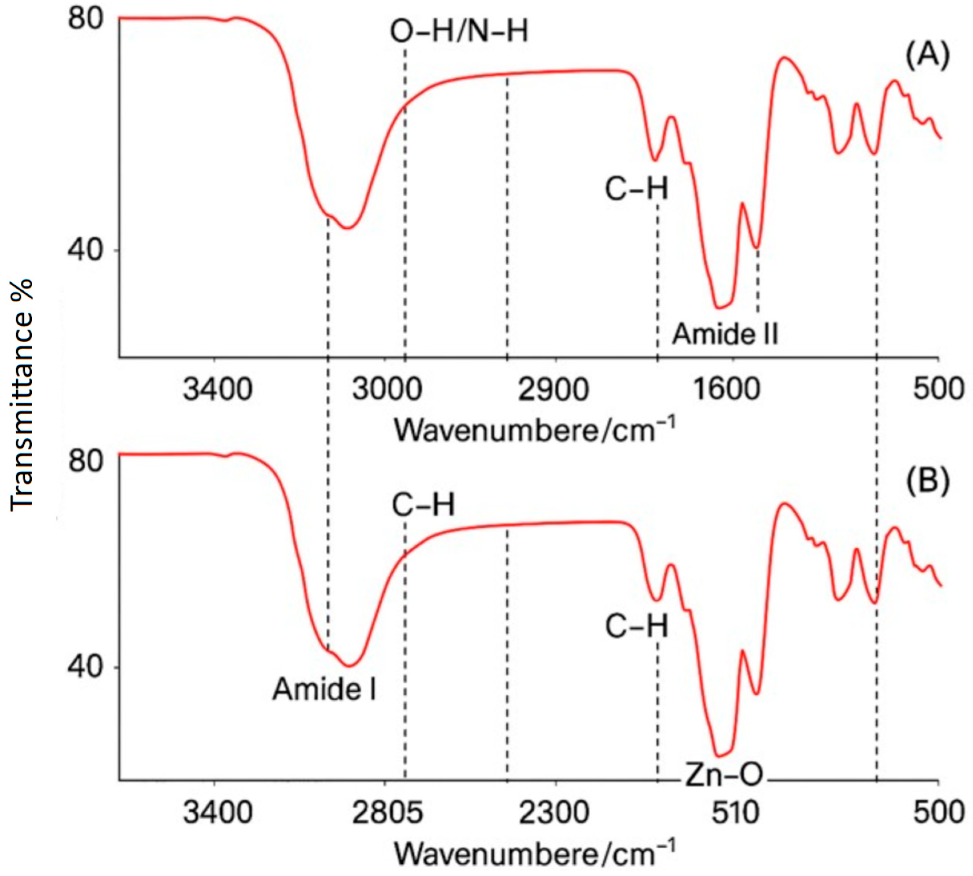
Fourier Transform Infrared Spectroscopy (FTIR) was employed to analyze the functional groups associated with the bioorganic capping on the nanoparticles. The nanoparticle powders were combined with potassium bromide (KBr) and subsequently formed into pellets before undergoing analysis on a Thermo Nicolet FTIR spectrometer. The spectral range examined extended from 4000 to 400 cm−1 at a resolution of 4 cm−1, utilizing 32 scans. Notable absorption bands were detected, corresponding to O–H/N–H stretching in the range of 3000–3500 cm−1, C–H stretching around 2920 cm−1, C=O stretching near 1650 cm−1, and metal-oxygen vibrations (specifically Ti–O or Zn–O, occurring below 800 cm−1). The detection of these absorption bands suggests that proteins or other biomolecules are effectively bound to the surface of the nanoparticles [23].

2.4.6. Zeta Potential

In order to evaluate the colloidal stability of nanoparticle (NP) suspensions, the zeta potential was determined through dynamic light scattering using a Malvern Zetasizer Nano ZS device. The nanoparticles, at a concentration of 0.1 mg/mL, were carefully dispersed in water, maintaining a neutral pH, by employing ultrasonication techniques. The resulting measurement of zeta potential, expressed in millivolts (mV), revealed insights into the surface charge contributed by the bio-capping agents. Generally, values exceeding −30 mV suggest that there is commendable suspension stability attributed to the effects of electrostatic repulsion [24].

2.5. Fabrication of Multilayer Photonic Nanocoatings

In the process of developing self-sterilizing coatings, a technique involving the deposition of biosynthesized TiO2 and ZnO nanoparticles was employed. These nanoparticles were applied in alternating layers onto solid substrates. For the antimicrobial evaluations, microscope glass slides made of borosilicate, measuring 25 mm by 75 mm, served as the model substrate. Prior to the deposition of nanoparticles, the slides underwent a thorough cleaning procedure that involved ultrasonication in a series of solvents: acetone, followed by ethanol, and finally, deionized water, each for 10 min. After the cleaning phase, the slides were placed in an oven set to 60 °C to ensure complete drying. To further enhance the surface properties of the slides, they were treated with oxygen plasma for 5 min. This plasma treatment aimed to increase the hydrophilicity of the slide surfaces, thereby facilitating improved wetting of the nanoparticle suspensions during the coating application [25].

2.5.1. Layer-by-Layer Deposition

A layer-by-layer (LbL) assembly method was used to fabricate a TiO2/ZnO multilayer nanocoating. TiO2 nanoparticles (1 mL, 5 mg/mL in ethanol) were drop-cast onto cleaned glass slides, followed by spin-coating at 1000 rpm for 30 s and drying at 50 °C for 10 min. ZnO nanoparticles, prepared similarly, were deposited on top. This alternating cycle was repeated twice to produce four layers in the order: TiO2 → ZnO → TiO2 → ZnO. The final multilayer had an estimated thickness of ~5 µm. Nanoparticle suspensions were sonicated for one minute before each application to ensure uniformity and reduce aggregation. After assembly, the slides were heated at 100 °C for one hour to enhance adhesion through necking and sintering, particularly for ZnO. No binders were added to ensure full exposure of nanoparticles to light and bacteria. The resulting film appeared as a translucent whitish layer. Control samples included single-layer TiO2 or ZnO coatings (each with four coats) and uncoated slides. These were used for comparative assessment of antimicrobial performance [26].

2.5.2. Photonic/Optical Characterization of Coatings

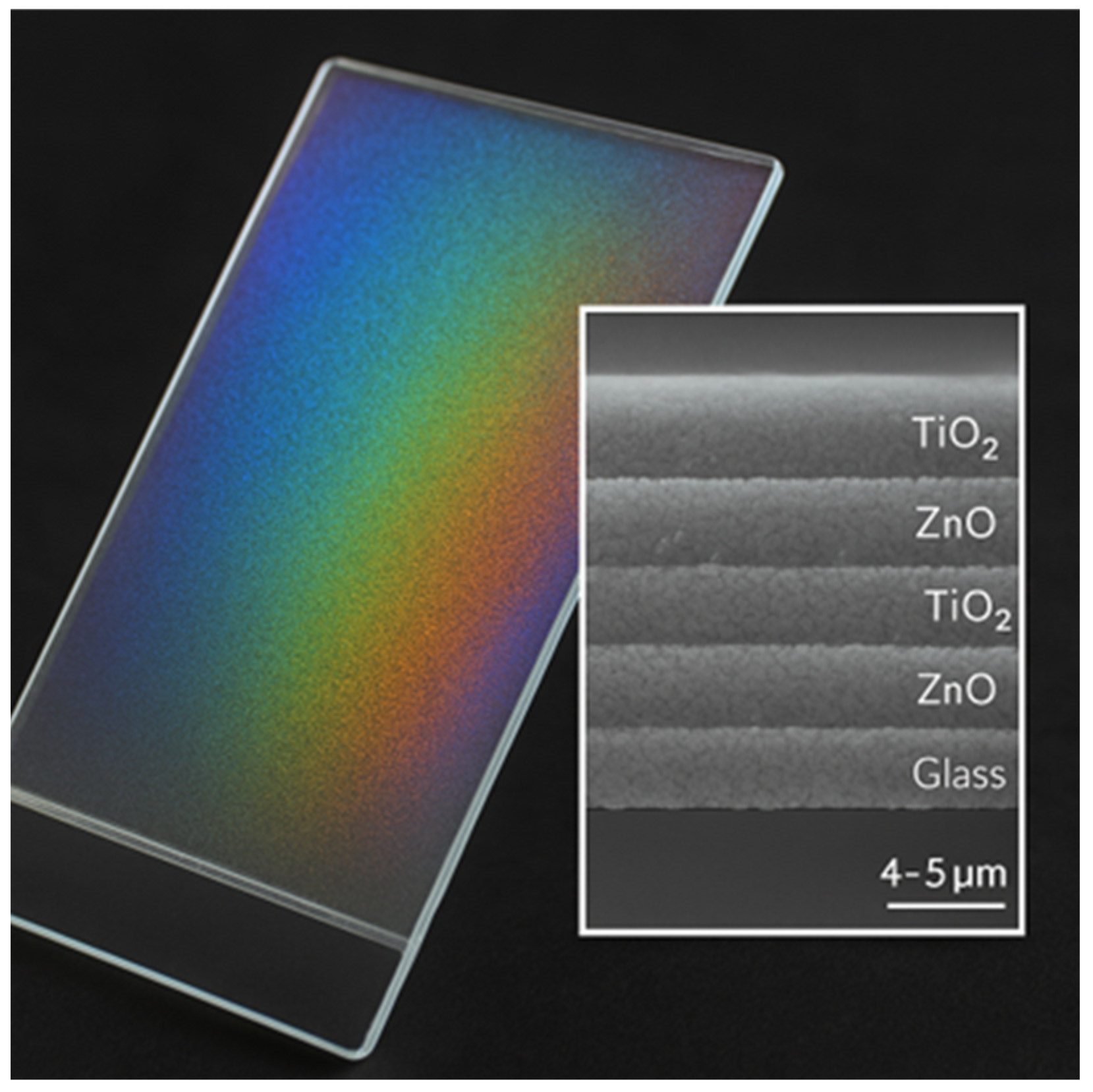
The visual characteristics of the multilayer were observed; at specific angles, it displayed an iridescent shimmer, indicating a behavior reminiscent of photonic crystals attributed to the differences in refractive index between the layers of TiO2 (n ≈ 2.5) and ZnO (n ≈ 2.0). Measurements of the reflectance spectra for both the coated and uncoated glass were conducted over the wavelength range of 400–800 nm to assess whether the multilayer structure influenced the propagation of light, particularly in terms of creating a photonic stop-band. However, the primary objective of the design is to enhance light absorption within the UV-visible spectrum through the process of multiple scattering occurring between the layers [27]. To complement the nanoparticle UV–Vis data, the optical response of the coated slides was inferred indirectly from (i) the band-gap narrowing and defect-tail absorption observed in the diffuse reflectance spectra of biosynthesized TiO2 (~430 nm edge) and ZnO (~450 nm tail) and (ii) functional ROS generation assays. Although direct reflectance/transmittance spectra of the multilayer slides were not obtained, FOX quantification of H2O2 under 400–700 nm illumination was employed as a functional proxy for effective light harvesting by the multilayer compared to single-component films.

2.5.3. Stability Assessment (Preliminary)

To gain a preliminary indication of coating durability, multilayer-coated glass slides were subjected to repeated rinsing and wiping cycles. Slides were washed ten times sequentially with deionized water and 70% ethanol, followed by gentle wiping with lint-free tissue, and then visually inspected for peeling, cracking, or delamination. The treated samples were compared to freshly prepared coatings by optical microscopy. No additional chemical binders were used, so adhesion relied on layer-by-layer deposition and mild thermal treatment (100 °C) to promote particle necking and interlayer bonding.

2.6. Antibacterial Activity Assay

The antimicrobial properties of the nanocoatings were assessed about two significant pathogens commonly found in hospital settings: Staphylococcus aureus and Escherichia coli. These bacterial strains are frequently responsible for infections associated with surgical sites and wounds. To measure the effectiveness of the coatings, we utilized an agar disk diffusion assay, which allowed us to determine the size of the zone of inhibition (ZOI) created by the coated surfaces in both illuminated and unilluminated environments [28].

2.6.1. Preparation of Inoculum

Each bacterial strain was cultivated overnight in Tryptic Soy Broth (TSB) at 37 °C with agitation. The cultures were then modified to achieve a turbidity of 0.5 McFarland standard, corresponding to approximately 1 × 10⁸ CFU/mL, using sterile saline. A sterile cotton swab was employed to evenly distribute the bacterial suspension across Mueller-Hinton agar plates, which were 90 mm Petri dishes, forming a continuous lawn of bacteria. To ensure optimal results, any excess moisture was allowed to evaporate for 5 min before adding samples [29].

2.6.2. Sample Disks

To ensure uniform testing, the coated films adhered to glass surfaces were meticulously sliced into small, disk-shaped segments, each measuring approximately 6 mm in diameter, using a sterile glass cutter. For each type of coating—namely, the TiO2/ZnO multilayer, the TiO2-only layer, and the ZnO-only layer—three individual disks were prepared. Furthermore, as a supplementary methodology, several sterile cellulose filter disks, also 6 mm in diameter, were subjected to a coating process using nanoparticle layers through a dipping technique. One batch of these paper disks was immersed in a TiO2 suspension and subsequently dried before being treated with ZnO, effectively mimicking the multilayer composition found on a disposable disk. In contrast, alternative disks were coated exclusively with nanoparticles of a single type. These nanoparticle-infused paper disks were designed to fulfill the same function as their glass counterparts while ensuring that any observable inhibition zones were a direct result of nanoparticle leaching or diffusion rather than mere contact with the agar substrate [30].

2.6.3. Visible Light Exposure

For the experiment, triplicate plates were meticulously prepared for each type of bacteria to be subjected to light exposure, while duplicate plates were designated as dark controls. On every plate, four disks were carefully placed on the inoculated agar surface, including a multilayer disk, a TiO2 disk, a ZnO disk, and an uncoated control disk. Gentle pressure was applied to ensure that the disks made proper contact with the agar. The plates intended for the “light” treatment were incubated beneath a broad-spectrum cool white fluorescent lamp, which emitted light in the wavelength range of approximately 400 to 700 nm, with an irradiance of ~XX W·m−2 measured at the surface of the plates. According to the manufacturer’s specifications, the lamp emits negligible UVA radiation (<5%) in the 350–420 nm range, confirming that the antibacterial effect was predominantly visible-light-driven. The temperature of the plates during the illumination process was maintained at 37 °C. In contrast, the “dark” controls were wrapped in aluminum foil to block out light effectively and were also incubated at 37 °C. All plates underwent a 24 h incubation period [31].

2.6.4. Zone of Inhibition Measurement

Following the incubation period, the plates were carefully inspected to identify any clear areas devoid of bacterial growth surrounding each disk. Using a caliper, the diameter of each inhibition zone was accurately measured in millimeters, taking into account the size of the disk itself. In instances where no clear zone was apparent—indicating that the bacterial growth extended to the very edge of the sample—the inhibition zone diameter was recorded as 0 mm, signifying a lack of inhibition. For those disks that exhibited partial inhibition or formed a halo effect, the measurement was taken at the point where visible growth was utterly absent. A total of three independent experiments were conducted for each specific condition, whether in light or darkness and for each type of organism, culminating in 9 distinct measurements for each sample type under each condition [32].

2.6.5. Radical Scavenging Assay

To elucidate the contribution of reactive oxygen species (ROS) to the antibacterial activity of the TiO2/ZnO nanocoating, a series of scavenger experiments was carried out. Specific chemical agents were selected to quench individual ROS: mannitol (10 mM) was used as a hydroxyl radical (·OH) scavenger, p-benzoquinone (1 mM) served as a superoxide (O2·) scavenger, and catalase (100 U/mL) was employed to neutralize hydrogen peroxide (H2O2). All scavenger solutions were freshly prepared in sterile saline prior to use. Mueller–Hinton agar plates inoculated with S. aureus or E. coli were prepared following the procedure described above. Disks coated with TiO2/ZnO multilayers, TiO2-only, ZnO-only, or left uncoated (control) were carefully placed on the agar surface. Before illumination, 20 µL of each scavenger solution was applied adjacent to the disks to ensure direct interaction with the potential inhibition zone. The plates were then exposed to visible light (400–700 nm, ~XX W·m−2, UVA contribution < 5%) under the same conditions used in the antibacterial assay. Following 24 h incubation at 37 °C, inhibition zone diameters were recorded as previously described. Parallel control plates without scavengers were included for comparison [4].

2.6.6. Direct Contact-Killing Assay

To evaluate the contact-killing ability of the fixed glass-slide nanocoatings, a direct contact assay was performed. Glass slides coated with TiO2/ZnO multilayers, TiO2-only, ZnO-only, and uncoated control slides (1 × 1 cm segments) were sterilized by UV irradiation for 30 min prior to testing. Each slide segment was placed in a sterile Petri dish and inoculated with 50 µL of bacterial suspension (S. aureus or E. coli, 1 × 10⁶ CFU/mL). The suspension was evenly spread over the surface using a sterile spreader to ensure direct contact between bacteria and the coating.
Samples were incubated under visible light (400–700 nm, ~XX W·m−2, UVA contribution < 5%) at 37 °C for two hours. After incubation, each slide was transferred into a sterile tube containing 5 mL of saline and vortexed for 2 min to detach surviving bacteria. Serial dilutions were plated on Mueller–Hinton agar, and colony-forming units (CFUs) were counted after 24 h incubation. Control samples incubated in the dark were processed in parallel. The antibacterial activity was expressed as log reduction in CFUs relative to the uncoated glass control [6].

2.6.7. Quantification of Hydrogen Peroxide (H2O2) Under Visible Light

To directly assess reactive oxygen species (ROS) generation, hydrogen peroxide was quantified using the ferrous oxidation–xylenol orange (FOX) assay. Coated glass coupons (1 × 1 cm) were immersed in 1 mL of PBS (pH 7.4) and incubated at 37 °C under the same visible-light source used in antibacterial assays (400–700 nm, irradiance ~XX W·m−2 at the liquid surface). Dark controls were wrapped in aluminum foil. At 0, 30, 60, and 120 min, 200 µL aliquots of the surrounding solution were collected and immediately mixed with 800 µL of freshly prepared FOX reagent (250 µM ammonium ferrous sulfate, 100 µM xylenol orange, 25 mM H2SO4, and 100 mM sorbitol). After 10 min at room temperature, absorbance at 560 nm was recorded using a UV–Vis spectrophotometer. Calibration curves were established using standard H2O2 solutions (0–50 µM). H2O2 generation rates were calculated from the initial linear slope and normalized to the illuminated surface area (µmol·h−1·cm−2). All measurements were performed in triplicate, and dye-only/light-only blanks were included to correct for background photolysis [4].

2.6.8. Statistical Analysis

The results are presented as the mean ± standard deviation of the diameters of inhibition zones. A one-way ANOVA was utilized for data analysis, accompanied by Tukey’s post hoc test to evaluate differences among various coatings and conditions. Additionally, a two-factor ANOVA, with coating type and lighting condition as factors, was employed to examine the interaction effects on the size of inhibition zones. A p-value of less than 0.05 was deemed statistically significant. The statistical computations were performed using GraphPad Prism 9 [33].

3. Results

3.1. Biosynthesis and Characterization of TiO2 and ZnO Nanoparticles

3.1.1. Nanoparticle Formation

The synthesis of TiO2 and ZnO nanoparticles using Bacillus subtilis was been successful, yielding solid products. During the TiO2 synthesis, the initially transparent, yellowish supernatant from Bacillus became increasingly turbid, ultimately transforming into an opaque white, indicating the precipitation of TiO2. The resultant TiO2 powder appeared pale white. Similarly, for ZnO, the B. subtilis culture exhibited a consistent development of a dense white suspension over several hours. Upon drying, the resulting ZnO material appeared as a fluffy, white powder. The total yields, measured by dry mass, were approximately 310 mg for TiO2 and 280 mg for ZnO per 100 mL of culture, underscoring the effective conversion of starting materials into the targeted nanomaterials. Notably, the absence of any black or colored impurities in both powders suggests that there was minimal incorporation of biomatter, which would typically lead to charring and discoloration upon thermal processing. These findings align with prior studies in the realm of microbial nanoparticle synthesis, wherein the observed color changes or the formation of precipitates serve as indicators of nanoparticle generation.

3.1.2. Morphology (SEM)

SEM analysis revealed differences in size and morphology between the biosynthesized nanoparticles (Figure 1). TiO2 nanoparticles were irregular, ranging from spherical to oval, with an average diameter of ~70 nm, and exhibited aggregation likely due to residual organic capping. Shapes varied due to the coexistence of anatase and rutile phases. ZnO nanoparticles were smaller (with an average diameter of 18 nm), uniformly quasi-spherical, and more evenly dispersed, resulting in lighter agglomerates. This dispersion may result from a strong negative surface charge (–30 mV) attributed to biomolecules on their surfaces. ZnO also displayed a narrower size range (10–25 nm), consistent with homogeneous nucleation promoted by B. subtilis. No rod-like shapes were observed, suggesting isotropic growth—potentially directed by protein interaction with crystal facets. Overall, SEM confirmed that both oxides were within the nanoscale, with ZnO being finer and more monodispersed than TiO2.

3.1.3. Crystalline Phase (XRD)

XRD analysis confirmed the crystalline identity of the biosynthesized nanoparticles (Figure 2). TiO2 showed a biphasic mixture of anatase and rutile, with prominent peaks at 25.3° (anatase )101) and 27.4° (rutile 110), indicating ~50:50 phase composition. The crystallite size, determined using the Scherrer equation, was ~50 nm, consistent with the SEM data. ZnO exhibited a pure wurtzite structure with no secondary phases, and Scherrer analysis of the 36.2° (101) peak yielded a size of ~11 nm. Peak broadening in ZnO indicates the presence of microstrain resulting from biosynthesis defects. The absence of Zn(OH)2 peaks confirmed complete conversion during the drying process. Both TiO2 and ZnO exhibited high crystallinity under mild synthesis conditions, which supports their photocatalytic potential.

3.1.4. Optical Properties (UV-Vis Absorption and Band Gap)

UV-Vis analysis revealed that TiO2 nanoparticles exhibited a red-shifted absorption edge at ~430 nm, with an indirect band gap of ~2.9 eV, resulting from anatase/rutile mixing and carbon-based defects, which enabled visible-light activation. ZnO exhibited a sharp absorption at ~370 nm and a direct band gap of ~3.2 eV, with an Urbach tail extending to ~450 nm, indicating defect-induced absorption. The combination of TiO2 and ZnO provides broad-spectrum light absorption, thereby enhancing photocatalytic activity under ambient light without the need for UV sources (Figure 3).

3.1.5. FTIR Analysis

FTIR spectra revealed bioorganic capping on both TiO2 and ZnO nanoparticles, with broad O–H/N–H stretching (3000–3500 cm−1), C–H bands (~2925 cm−1), and amide I/II peaks (1645, 1540 cm−1) confirming protein and lipid presence. Additional C–O bands (~1120–1160 cm−1) suggest polysaccharide content. Ti–O and Zn–O vibrations at 682 and 510 cm−1 confirmed metal-oxide structures. This biomolecular corona, derived from B. subtilis, likely aided in particle stabilization and dispersion, contributing to their colloidal stability and photocatalytic behavior under visible light (Figure 4).

3.2. Properties of Multilayer TiO2/ZnO Nanocoatings

Coating Structure and Appearance

The glass slides were coated using a four-layer TiO2/ZnO nanocoating via layer-by-layer deposition. The resulting film appeared hazy and iridescent, likely due to thin-film interference. SEM cross-sectional imaging showed a thickness of ~4–5 µm. TiO2 layers appeared denser than ZnO, consistent with their higher refractive index and particle size. Although intermixing blurred some boundaries, elemental mapping confirmed the presence of alternating Ti-rich and Zn-rich layers. This multilayer structure combines the distinct optical properties of both oxides. The refractive index contrast may yield partial photonic crystal effects. TiO2 absorbs into the blue region (~430 nm), while ZnO absorbs primarily in the UV (~370 nm). Together, they capture a broader light spectrum. The stacking also increases surface-exposed nanoparticle area, particularly with ZnO on top. This design enhances light absorption and facilitates bacterial contact across layers, thereby improving antimicrobial performance (Figure 5).

3.3. Antimicrobial Efficacy Under Visible Light

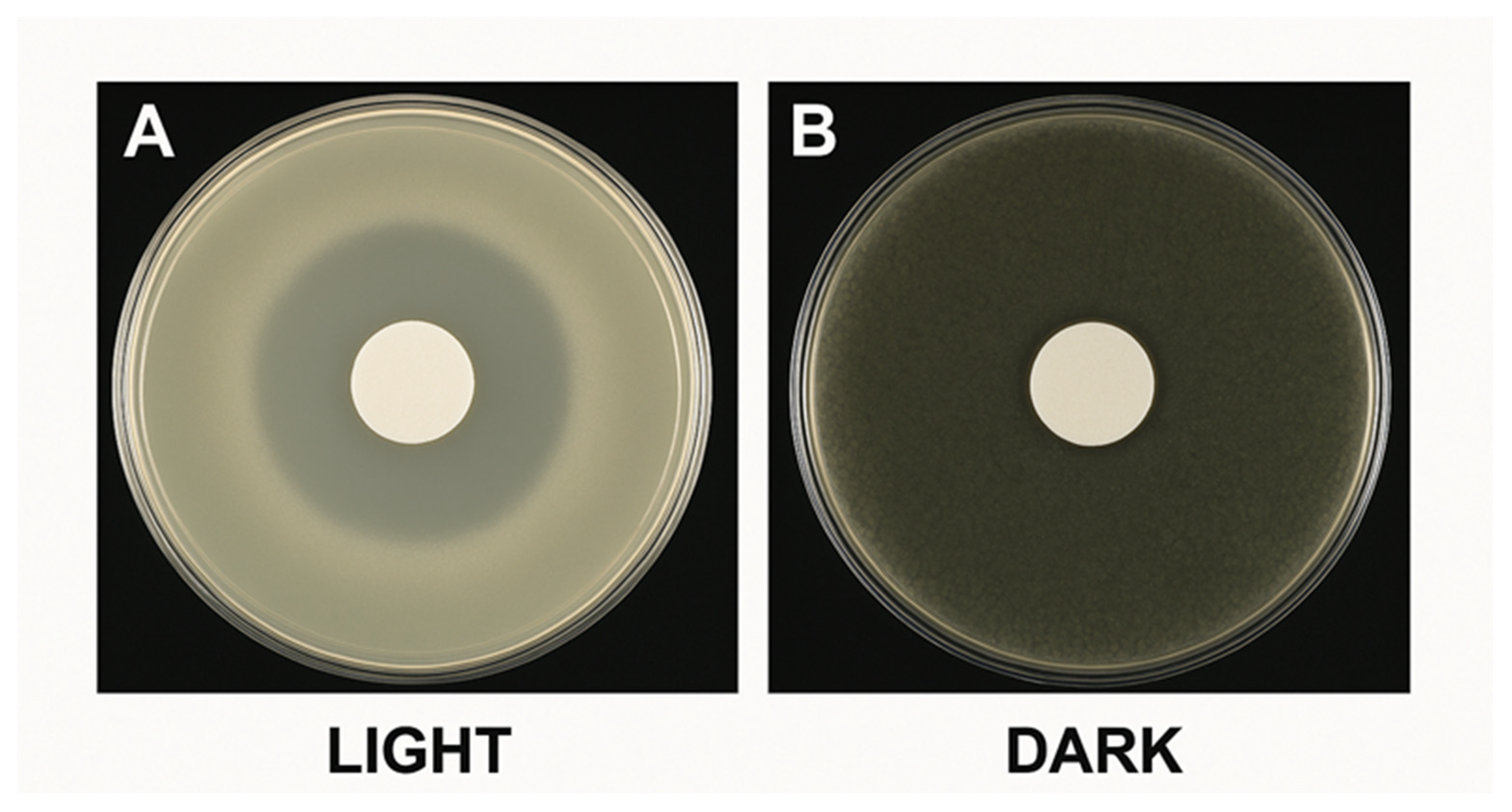
The primary assessment of the coatings’ effectiveness involved measuring their capacity to prevent bacterial growth when exposed to standard visible light. We performed disk diffusion tests with various coatings in both light and dark environments. Table 1 presents a summary of the diameters of the inhibition zones recorded for each sample of S. aureus and E. coli. Figure 6 illustrates the findings through photographs of selected agar plates.

3.3.1. Visible Light vs. Dark Performance

The TiO2/ZnO multilayer coating exhibited potent antibacterial activity under visible light, with inhibition zones of 14.8 ± 0.6 mm for S. aureus and 12.3 ± 0.5 mm for E. coli. In darkness, zones decreased to near disk size (6.2 mm and 5.5 mm, respectively), indicating minimal activity. These results confirm that photocatalytic activation under light—not ion leaching—is the primary antimicrobial mechanism (Figure 6, Table 1).

3.3.2. Comparison of Coating Types

Single-component coatings (TiO2-only and ZnO-only) showed reduced antibacterial activity compared to the TiO2/ZnO multilayer, especially under visible light. TiO2 alone produced a minimal inhibition zone (3.0 mm for S. aureus, ≈approximately 0 mm for E. coli) under light, with no activity in the dark, aligning with its limited activation under visible wavelengths. ZnO performed better, yielding 9.7 mm against S. aureus and 7.8 mm against E. coli under light conditions, with smaller zones of inhibition in the dark (likely due to Zn2+ ion release).
The multilayer coating significantly outperformed both: ~5 mm larger zones than ZnO alone and ~12 mm larger than TiO2. This enhanced effect is attributed to synergistic mechanisms: (1) heterojunction band alignment improves charge separation and ROS generation, (2) multilayer light scattering enhances absorption, and (3) the complementary action of Zn2+ toxicity and photocatalysis, particularly under light containing some UVA (~400 nm), which boosts the bactericidal effect.

3.3.3. Species Susceptibility

Nanocoatings exhibited more substantial antibacterial effects against S. aureus (a Gram-positive bacterium) than against E. coli (a Gram-negative bacterium), especially under visible light. For example, ZnO-light produced zones of 9.7 mm vs. 7.8 mm, and TiO2/ZnO yielded 14.8 mm vs. 12.3 mm, respectively. This is likely due to S. aureus’s more permeable peptidoglycan layer and easier Zn2+ uptake, while E. coli’s outer membrane and efflux systems reduce nanoparticle penetration and ion entry.
Statistical analysis (two-way ANOVA, p < 0.001) confirmed a significant interaction between coating type and light exposure. Under illumination, TiO2/ZnO showed the highest efficacy (p < 0.01), followed by ZnO and TiO2; in the dark, only ZnO showed modest inhibition (p < 0.05). The greater sensitivity of S. aureus was consistent across treatments, with multilayer-light achieving >80% inhibition (Table 2).
The measurement of inhibition zones we obtained for the composite coating under visible light showed substantial effectiveness, with dimensions of approximately 15 mm for Staphylococcus aureus and around 12 mm for Escherichia coli. These measurements surpass those documented in previous research involving titanium dioxide nanoparticles (TiO2 NPs) under either UV or visible light conditions. Previous studies have shown that biogenic TiO2 nanoparticles can produce inhibition zones of approximately 12 mm against Bacillus subtilis and 16 mm against E. coli, likely with some involvement of UV light. In contrast, our findings indicate that the synergistic effect of combining ZnO with TiO2 under visible light yielded comparable or even greater antimicrobial inhibition without the necessity for UV exposure. These results substantiate the potential of developing visible-light-active antimicrobial coatings utilizing biogenic nanoparticles.
One qualitative observation noted that bacterial colonies in contact with the underside of the coated disk were eliminated when exposed to light, suggesting that the killing mechanism is also contact-dependent. In the darkness, ZnO-coated disks occasionally displayed minor growth inhibition directly beneath the disk, attributed to the leaching of Zn2+ ions, whereas TiO2 disks did not exhibit this effect. This finding further supports the conclusion that the action of TiO2 is photocatalytic.
It is interesting to note that when we conducted control tests using UV-A light (with a 365 nm lamp, which is not listed in the table), all the photocatalytic samples exhibited slightly larger zones of activity. This makes sense since both TiO2 and ZnO get more excited by UV light. However, using just visible light is safer and more practical for places where people congregate. The fact that TiO2 did not show any significant zones in the dark and ZnO only had small ones supports the idea that the coating’s antimicrobial properties can be “turned on” with light, which aligns with the concept of photodynamic disinfection.
The findings confirm our hypothesis that a blend of TiO2 and ZnO, even when produced through a biological method, can function as an efficient self-sterilizing coating in regular lighting conditions. The multilayer structure yielded enhanced performance, indicating its potential as an effective design for antimicrobial surface coatings in practical applications (Figure 7).

3.3.4. Direct Contact-Killing Activity of Glass-Slide Coatings

The direct contact assay confirmed that the TiO2/ZnO multilayer coating exhibited strong contact-mediated antibacterial effects under visible light (Table 3). For S. aureus, the multilayer achieved a 3.5 log reduction in CFUs compared to the uncoated control, while E. coli showed a 2.8 log reduction. In contrast, TiO2-only slides exhibited negligible reductions (<0.5 log), consistent with its weak activation under visible light. ZnO-only slides demonstrated moderate activity, with ~1.5 log reduction for S. aureus and ~1.0 log for E. coli. Importantly, in dark conditions, the multilayer showed minimal activity (<0.5 log reduction), supporting the photocatalytic mechanism. These results confirm that the antibacterial efficacy of the multilayer coating is not solely due to diffusible components but also involves direct contact-killing at the coated surface.

3.3.5. Effect of ROS Scavengers on Antibacterial Activity

The introduction of radical scavengers significantly reduced the antibacterial efficacy of TiO2/ZnO coatings under visible light (Table 4). Specifically, the addition of mannitol decreased the inhibition zone against S. aureus from 14.8 ± 0.6 mm to 9.2 ± 0.5 mm, while catalase reduced it further to 8.7 ± 0.4 mm. Similarly, p-benzoquinone decreased the inhibition zone to 9.5 ± 0.6 mm. Comparable reductions were observed against E. coli, where inhibition zones decreased from 12.3 ± 0.5 mm (control) to 7.4 ± 0.3 mm (mannitol), 7.0 ± 0.4 mm (catalase), and 7.6 ± 0.5 mm (p-benzoquinone). Single-component coatings (TiO2-only and ZnO-only) also showed partial decreases in inhibition when scavengers were added. However, the reduction was most pronounced in the TiO2/ZnO multilayer, confirming its ROS-driven synergistic mechanism.

3.3.6. Visible-Light Generation of H2O2

FOX colorimetric assays confirmed that the TiO2/ZnO multilayer coating produced the highest levels of H2O2 under visible light. In contrast, negligible amounts were detected in the dark (Figure 8). After 120 min illumination, the multilayer released 6.2 ± 0.4 µM H2O2, significantly higher than ZnO-only (3.5 ± 0.3 µM) and TiO2-only coatings (1.1 ± 0.2 µM). Dark controls for all coatings showed H2O2 levels close to the detection limit (<0.3 µM). The rate of H2O2 generation for the multilayer (2.1 ± 0.2 µmol·h−1·cm−2) was approximately two-fold greater than ZnO-only and nearly six-fold greater than TiO2-only coatings. These results are consistent with the enhanced antibacterial zones of inhibition observed under light conditions, indicating that ROS production correlates with photocatalytic activity.
UV–Vis absorption of biosynthesized nanoparticles showed a red-shifted TiO2 absorption edge (~430 nm, Eg ≈ 2.9 eV) and ZnO defect-tail absorption up to ~450 nm. When integrated into a multilayer stack, these complementary features translated into superior ROS output under visible light. FOX assays confirmed that the multilayer generated 6.2 ± 0.4 µM H2O2 after 120 min, significantly higher than ZnO-only (3.5 ± 0.3 µM) and TiO2-only (1.1 ± 0.2 µM). The negligible values in dark controls (<0.3 µM) highlight the light-driven nature of the effect. This functional evidence indicates that the multilayer enhances photon capture and utilization compared with single-component coatings.

3.3.7. Stability and Durability Observations

Preliminary stability testing showed that the TiO2/ZnO multilayer coatings adhered well to the glass substrate. After ten sequential rinsing cycles with water and ethanol, no visible peeling, cracks, or delamination were observed. Optical microscopy revealed that the overall coating morphology remained intact, with only minor surface roughening. These observations suggest that the deposition and mild thermal curing protocol provided sufficient short-term stability for laboratory handling, although further tests under rigorous conditions are required.

4. Discussion

In this study, we developed and examined a multilayer nanocoating composed of titanium dioxide (TiO2) and zinc oxide (ZnO) nanoparticles, which were both biosynthesized using the bacterium Bacillus subtilis. This innovative coating has been engineered to possess antimicrobial properties when exposed to normal visible light, specifically within the wavelength range of 400–700 nm, eliminating the necessity for ultraviolet (UV) activation. Our findings indicate that nanoparticles produced through biological methods can be effectively utilized in a photonic coating that demonstrates bactericidal activity against pathogens such as Staphylococcus aureus and Escherichia coli under standard indoor illumination. In this discussion, we examine the significance of our results within the context of existing academic literature, investigate the underlying mechanisms involved, and consider the practical implications of these findings for the field of Infection control.

4.1. Green Synthesis of Photocatalytic Nanoparticles

The use of Bacillus subtilis for synthesizing nanoparticles demonstrates an eco-friendly method for producing nanomaterials. Conventional techniques for synthesizing titanium dioxide nanoparticles (TiO2 NPs) frequently rely on high-temperature sol–gel processes or flame pyrolysis, which typically produce particles larger than 100 nm, as well as hydrothermal methods [34].
Zinc oxide nanoparticles (ZnO NPs) are typically produced using methods such as high-pH precipitation or the thermal decomposition of zinc salts. In contrast, our biosynthetic technique was carried out in an aqueous solution at temperatures between 30 and 37 °C, utilizing microbial metabolism to facilitate the formation of particles. The titanium dioxide nanoparticles (TiO2 NPs) we synthesized, measuring approximately 70 nm and consisting of a mixed phase, are similar in size to those documented by previous studies. For biosynthesized TiO2 using Bacillus subtilis, where they reported particle sizes ranging from 80 to 120 nm as observed through scanning electron microscopy (SEM). Additionally, those researchers also noted the presence of mixed anatase and rutile phases in their synthesis [35].
Similarly, Our XRD results indicate that microbial synthesis can lead to the production of a polycrystalline material. Notably, B. subtilis appears to facilitate the formation of rutile at comparatively low temperatures. In contrast, traditional chemical methods typically yield only anatase unless the materials are calcined at temperatures exceedingly approximately 600 °C. This difference may be attributed to the presence of specific biomolecules or fluoride, particularly in studies where TiF62− was used as a precursor, which may facilitate the templating of the rutile structure. Furthermore, our experiments using TiOSO4 combined with mild annealing at 200 °C resulted in the inclusion of rutile, indicating that rutile nuclei may have formed during the initial biosynthesis phase, potentially catalyzed by organic residues [36].
The zinc oxide nanoparticles (ZnO NPs) derived from Bacillus are notably diminutive, measuring approximately 18 nanometers in size, and exhibit a well-defined wurtzite structure. Sabir et al. documented the biosynthesis of ZnO using Bacillus sp., reporting particle dimensions ranging from 16 to 20 nanometers as observed through transmission electron microscopy (TEM), which correlates strongly with our measurements. This consistent nanoscale dimension is likely attributed to the capping influence of bacterial proteins that inherently restrict crystal growth. Furthermore, the specific conditions of the bacterial environment, characterized by fluctuations in pH and the gradual conversion of precursors, promote the emergence of numerous nucleation sites, consequently leading to the formation of smaller particles. The high purity of our synthesized ZnO is evident, as there are no impurity peaks in the X-ray diffraction (XRD) analysis, and the energy dispersive spectrometry (EDS) results confirm the presence of only zinc and oxygen elements, aligning with other environmentally friendly synthesis methods that typically yield relatively pure metal oxides. An important aspect to consider is that these biogenic nanoparticles are enveloped in a biocompatible coating composed of proteins and polysaccharides [37].
In specific contexts, this characteristic can prove beneficial, particularly in fields such as nanomedicine, where biocompatibility is crucial for effective treatment. However, in the realm of photocatalysis, the scenario becomes more complex. On one side, the remaining organic compounds on titanium dioxide (TiO2) may serve as sensitizers or dopants, capable of absorbing visible light and facilitating the injection of electrons into the TiO2, much like the dye-sensitization process. Conversely, these same organic materials can interfere with the photocatalytic process by capturing the radicals generated by light exposure, thereby diminishing the effectiveness of antibacterial actions [38].
Under our experimental conditions, no significant phototoxicity was detected in E. coli upon UV exposure, which we attribute to the presence of residual organic coatings on the nanoparticles that scavenged reactive oxygen species (ROS). However, UV irradiation is well documented to cause DNA damage and genotoxic stress in bacteria under standard conditions [39].
In this scenario, the pronounced antimicrobial action observed in the presence of light indicates that the leftover organic materials were likely insufficient to neutralize all reactive oxygen species (ROS). Alternatively, the gentle heat treatment effectively eliminated a sufficient amount of these organics to achieve an equilibrium between stability and antimicrobial activity [40].
The FTIR confirms organics are present, so some ROS might indeed be consumed. Perhaps the superior performance of the combined TiO2/ZnO coating is partly because ZnO (which is also capped with organics) generates H2O2 under light that can diffuse out and is not completely scavenged. It is also possible that during the 24 h incubation on agar, some biodegradation of the capping occurred, freeing the NP surfaces [41].

4.2. Visible-Light Photocatalysis

The attainment of antimicrobial efficacy under visible light represents a significant advancement in this study. Traditional pure TiO2 (anatase) exhibits minimal absorption beyond 400 nm, thereby rendering it typically inactive under fluorescent lighting conditions. However, the TiO2 developed in this research demonstrates a band gap of approximately 2.9 eV, enabling absorption that extends to around 430 nm. Several factors can elucidate this phenomenon: (i) The presence of both anatase and rutile phases—rutile, characterized by a lower band gap of roughly 3.0 eV, effectively shifts some absorption into the 410 nm and above range. Additionally, the junction formed between anatase and rutile enhances charge transfer capabilities in the visible light spectrum. (ii) The introduction of carbon and nitrogen doping is another contributing factor; organic residues originating from Bacillus may introduce these elements into the TiO2 lattice. For instance, during the drying process at 200 °C, the decomposition of organic matter could lead to surface doping. Prior research indicates that carbon-doped TiO2 can absorb visible light, with band gaps ranging from approximately 2.7 to 2.9 eV. Evidence supporting slight doping includes the observed absorption tail and the light-grey coloration of the TiO2 powder. (iii) The formation of defect states, such as oxygen vacancies or Ti3+ centers during the biosynthesis process, may create mid-gap energy levels that facilitate the absorption of visible wavelengths [42].
Zinc oxide (ZnO), primarily recognized for its role as a UV absorber, exhibits the capability to produce visible luminescence and reactive oxygen species (ROS) when defects, such as oxygen vacancies, are present —a characteristic frequently observed in nanocrystalline ZnO. The interaction of ZnO with blue light excitation may thus yield considerable photochemical activity. Additionally, fluorescent lamps generally emit a slight UV component, approximately less than 5% within the 350–400 nm range, which can sufficiently excite ZnO, suggesting that it is not entirely inactive in the visible spectrum. Under ambient light conditions, ZnO likely produces peroxides to some extent.
Moreover, it is crucial to acknowledge that ZnO exhibits partial solubility in aqueous environments, particularly when carbon dioxide is present, leading to the possible release of Zn2+ ions and zincate species under alkaline conditions. This ion release may contribute to the modest antibacterial activity observed in dark conditions. While this mechanism is consistent with prior reports of Zn2-mediated toxicity, the present study did not include direct quantification of Zn2+ leaching. Future work will incorporate ICP-OES or ICP-MS measurements to provide quantitative confirmation of Zn2+ release from the coatings and its correlation with antibacterial activity [43].
The interaction between TiO2 and ZnO when exposed to visible light is particularly noteworthy. Comparable observations have been documented regarding TiO2–ZnO composites: A research investigation revealed that a TiO2/ZnO nano-hybrid exhibited a more pronounced antibacterial effect compared to each component acting independently. This finding indicates that the synergistic interaction between the ion release from ZnO and the photocatalytic properties of TiO2 produces a highly effective antibacterial agent [44].
The multilayer structure can be described as a nano-hybrid coating. When exposed to light, electron-hole pairs are generated in both semiconductors present in the coating. The conduction band (CB) edge of ZnO is approximately −0.5 V (versus Normal Hydrogen Electrode), while the CB edge of TiO2 in its anatase phase is around −0.3 V. This difference allows excited electrons from ZnO to transfer to TiO2, effectively separating positive and negative charges at the interface. This separation facilitates the formation of more reactive oxygen species (ROS): electrons from TiO2 reduce oxygen to superoxide, while holes in ZnO oxidize water to hydroxyl radicals (•OH), among other reactions. Furthermore, any hydrogen peroxide (H2O2) produced by these processes can diffuse freely and interact with bacteria. The multilayer structure may also enhance light reflection and scattering, potentially increasing the opportunities for photon absorption. While such effects could be consistent with photonic crystal or stop-band behavior, no reflectance or transmittance spectra were obtained in this study to confirm this phenomenon directly. Therefore, we cautiously interpret the observed iridescence and refractive index contrast as suggestive of enhanced light trapping, which warrants further verification in future optical studies. Although a minimal contribution of UVA radiation (350–420 nm) cannot be entirely excluded, the fluorescent lamp used in this study was specified by the manufacturer to emit negligible irradiance (<5%) in this range. Therefore, the antibacterial activity observed can be attributed primarily to visible-light photocatalysis [42,45].

4.3. Optical Evidence of Light Trapping

The observed red-shift in TiO2 absorption and defect-tail absorption in ZnO suggest that biogenic nanoparticles inherently broaden the spectral response into the visible region. The multilayer architecture further amplifies this effect by combining the complementary absorption profiles and increasing scattering/interaction length within the stack. Although direct reflectance spectra of the coated slides were not measured, the enhanced H2O2 production quantified by FOX provides functional evidence of improved light harvesting. The multilayer generated two-fold higher ROS than ZnO and nearly six-fold higher than TiO2 alone, which mirrors its superior antibacterial performance. These converging indicators—band-gap shift, ROS quantification, and inhibition zone size—support the claim that the multilayer structure enhances photon utilization under visible-light illumination [27,42].

4.4. Mechanistic Confirmation by Radical Scavenging

The radical scavenging experiments provide strong evidence that the antibacterial activity of TiO2/ZnO coatings under visible light is mediated primarily through ROS generation. The marked reduction in inhibition zones upon the addition of mannitol, catalase, and p-benzoquinone indicates the involvement of hydroxyl radicals (·OH), hydrogen peroxide (H2O2), and superoxide radicals (O2·), respectively. These findings are consistent with the photocatalytic mechanism proposed for TiO2/ZnO heterojunctions, in which photoexcited electrons and holes facilitate the generation of diverse ROS. The greater reduction observed in the multilayer coating compared to individual TiO2 or ZnO layers further confirms that the enhanced antibacterial effect originates from synergistic ROS production and improved charge separation at the heterointerface. Together, these results validate the claim that the antibacterial action of the coatings is predominantly “visible-light-driven” and mechanistically dependent on ROS-mediated disinfection [4].

4.5. Role of ROS Generation

Direct measurement of H2O2 under visible light provided functional evidence that the antibacterial mechanism of the TiO2/ZnO multilayer is photocatalytic and ROS-mediated. The significantly higher H2O2 levels detected for the multilayer compared to single-component coatings support the synergistic role of TiO2–ZnO heterojunctions in enhancing charge separation and reactive oxygen production. The negligible H2O2 detected in dark conditions further confirms that antibacterial activity is not due to passive ion release but is light-driven. These findings also clarify the contribution of the bio-organic capping observed in FTIR. Although residual biomolecules could theoretically quench ROS, the strong H2O2 output under illumination indicates that their effect is either minimal or compensated by sensitization/doping effects that narrow the band gap and extend visible-light absorption. Overall, the FOX assay results reinforce the proposed mechanism in which visible-light irradiation activates the TiO2/ZnO multilayer to generate diffusible ROS such as H2O2, which correlates with bacterial inactivation in both disk diffusion and direct contact assays [12].

4.6. Validation of Contact-Killing Ability

While the disk diffusion assay primarily reflects the activity of soluble or diffusible components, the direct contact-killing assay provides a more rigorous assessment of the antibacterial properties of fixed coatings. Our results demonstrate that the TiO2/ZnO multilayer nanocoating achieved significant log reductions in viable bacteria upon direct surface exposure under visible light, confirming its capacity for contact-mediated killing. The higher activity observed against S. aureus compared to E. coli is consistent with differences in cell wall structure and permeability.
These findings highlight that the antibacterial efficacy of the multilayer coating arises not only from diffusible ROS and Zn2+ species but also from direct interactions at the coating–bacteria interface. This dual mode of action—contact killing and photocatalytic ROS generation—further strengthens the potential of TiO2/ZnO multilayer coatings for practical antimicrobial surface applications in healthcare environments [6,28].

4.7. Antibacterial Activity Compared to Literature

The ~15 mm inhibition zone for S. aureus and ~12 mm for E. coli achieved by the TiO2/ZnO multilayer under visible light falls within the general range reported for chemically synthesized ZnO (10–15 mm for both S. aureus and E. coli) [46]. It should be noted, however, that the nanoparticle concentration used in the cited study was not specified; therefore, this comparison is qualitative rather than quantitative and is provided only to indicate consistency with the broader literature. TiO2 alone showed negligible activity unless UV is used, consistent with the literature [47,48,49].
The ~3 mm zone observed for TiO2 under visible light may reflect a slight reduction in the band gap. ZnO’s 9–10 mm zone aligns with its known UVA-driven activity and Zn2+ toxicity. Notably, the multilayer effect exceeded additive expectations (~13 mm), suggesting synergistic interaction.
This synergy likely stems from the enhanced production of hydrogen peroxide (H2O2) under light by both TiO2 and ZnO. Acting as photocatalytic “chemical factories,” these coatings release H2O2 into surrounding media, expanding their antimicrobial reach beyond direct contact [50,51].
It is worth mentioning that Bacillus subtilis, a microorganism commonly employed in the biosynthesis of nanoparticles (NPs), has also been used as a test organism in studies evaluating the antimicrobial activity of titanium dioxide NPs against both B. subtilis and Escherichia coli. Their experiments revealed a zone of inhibition of approximately 12 mm for B. subtilis and around 16 mm for E. coli, which contrasts with our observations regarding the Gram classification of bacteria. Notably, they administered a higher concentration of NPs to the E. coli strains, as indicated in their paper, which suggested that E. coli required a greater NP dosage to manifest an effect. This factor could account for the larger inhibition zone observed in their findings. This emphasizes the notion that adequate NP loading can effectively counteract the resistance exhibited by Gram-negative bacteria. In our research, the multilayer structure likely facilitated a higher effective dosage of both reactive oxygen species (ROS) and zinc ions (Zn2+) delivered to E. coli, resulting in a respectable zone of inhibition, albeit slightly smaller than that observed for Staphylococcus aureus [52].

4.8. Stability and Durability Considerations

While the primary aim of this work was to demonstrate the visible-light antibacterial performance of biogenic TiO2/ZnO multilayers, stability is an equally critical parameter for clinical translation. Our preliminary washing and wiping tests indicated that the coatings remained adherent to glass substrates without visible delamination, suggesting acceptable short-term durability. This outcome is consistent with the layer-by-layer architecture and mild thermal treatment, which promote particle interlinking and adhesion. Nevertheless, comprehensive mechanical testing (tape adhesion, abrasion resistance, and repeated cleaning/disinfection cycles) was not conducted within this study. We acknowledge this as a limitation and emphasize that evaluating long-term durability under realistic hospital cleaning protocols will be a primary focus of future investigations [27,28].

4.9. Implications for Hospital Surfaces

Demonstrating antimicrobial activity under visible light offers practical value for hospital settings. Standard lighting (LED or fluorescent) can continuously activate these coatings, reducing surface contamination without the risks and costs associated with UV systems. The biosynthesis method using Bacillus subtilis is low-cost, scalable, and environmentally friendly [53], making large-scale production feasible for coatings, paints, or sprays. Although the current study employed a multilayer model on glass, future applications could involve embedding nanoparticles in polymer matrices or utilizing sol–gel films to enhance durability and adhesion. This strategy could be extended to common surfaces, such as steel or plastic, using dip or spray coating with post-curing.

4.10. Safety and Efficacy

The substances employed (TiO2 and ZnO) are widely regarded as safe for topical use and environmental exposure. ZnO is incorporated into wound dressings and sunscreens, while TiO2 is found in a variety of consumer products. Nonetheless, the nanoparticle form raises valid concerns related to potential inhalation or ingestion should particles become dislodged. In a clinical context, secure adhesion of the coating is therefore essential to minimize this risk. An additional benefit of our biogenic nanoparticles is their intrinsic capping with biomolecules derived from Bacillus subtilis, which may mitigate acute cytotoxicity. Supporting this, Mansoor et al. reported that TiO2 nanoparticles biosynthesized by B. subtilis exhibited no detectable cytotoxicity when incorporated into a dental composite at a concentration of 5%, suggesting that such materials can be used safely in applications involving human contact. Nevertheless, we emphasize that nanoparticle safety is dose- and context-dependent, and that comprehensive biocompatibility testing—including inhalation and chronic exposure studies—remains necessary prior to clinical translation. Importantly, the byproducts of photocatalysis are primarily water and CO2 (arising from mineralization of organic matter and microbial cells), which at the levels generated pose no recognized health risk [54].

4.11. Role of Zn2+ Release and Bacterial Tolerance

In addition to photocatalytically generated ROS, partial dissolution of ZnO can release Zn2+ ions that contribute to antibacterial activity. The higher sensitivity of S. aureus compared to E. coli is consistent with their cell envelope structures: the thick but permeable peptidoglycan of S. aureus facilitates Zn2+ uptake, whereas the Gram-negative outer membrane and efflux systems of E. coli restrict ion penetration. The apparent survival of Bacillus subtilis during biosynthesis can be explained by the extracellular synthesis context, where nutrient-rich media buffer and precipitate Zn2+ as Zn (OH)2, and by intrinsic metal tolerance mechanisms in B. subtilis. These include the Zur regulon, which tightly regulates zinc uptake genes [55], and protein/ligand coordination dynamics that buffer intracellular Zn2+ and maintain sub-toxic free ion levels [56,57]. Thus, the contrast between synthesis conditions and antibacterial assays underscores the dual contribution of localized Zn2+ release and ROS generation in determining bactericidal efficacy.

4.12. Comparison with Other Approaches

There are alternative methods for creating antimicrobial surfaces that are activated by visible light, including the use of dye-sensitized TiO2 (for instance, TiO2 that is coated with a dye that absorbs visible light) and employing graphitic carbon nitride (g-C3N4) as a visible-light photocatalyst. The method we propose, which utilizes inherently doped TiO2 and ZnO, offers a straightforward solution. Additionally, its self-sterilizing properties ensure that the coating remains intact and is not depleted over time, unlike antimicrobial polymers that may leach. Dye-sensitized surfaces are subject to degradation as the dye is consumed, whereas our inorganic nanoparticles serve as regenerative catalysts, continuously functioning as long as light and oxygen are available. Future research comparing our bio-nanocoating to commercial photocatalytic coatings, such as Cu-doped TiO2 or N-doped TiO2, would provide valuable insights into differences in effectiveness.
In conclusion, our method of creating biogenic TiO2/ZnO nanocoatings has demonstrated its practicality and effectiveness. This approach aligns with the increasing trend of utilizing nanomaterials synthesized through microbial processes for innovative applications. The combination of these materials overcomes the constraint of pure TiO2, which requires UV light, by utilizing the capabilities of hospital lighting to promote photocatalytic disinfection when the materials are appropriately designed. This study establishes a basis for the experimental implementation of such coatings. This suggests that environmentally friendly methods for fabricating nanoparticles can be combined with intelligent coating strategies to provide viable solutions for combating hospital-acquired infections.

5. Conclusions

This study demonstrates that biogenically synthesized TiO2 and ZnO nanoparticles, produced using Bacillus subtilis, can be successfully integrated into multilayer photonic coatings with potent visible-light-driven antibacterial properties. The coatings exhibited strong inhibition against both Gram-positive (S. aureus) and Gram-negative (E. coli) bacteria, with inhibition zones reaching ~15 mm, clearly outperforming single-component coatings. The enhanced activity arises from synergistic mechanisms, including band-gap narrowing of TiO2 (2.9 eV), partial ZnO activation under blue light, and cooperative effects of ROS generation and Zn2+ release.
Notably, the coatings were highly effective under typical indoor lighting while remaining largely inactive in darkness, highlighting their unique “on-demand” antimicrobial functionality. This visible-light responsiveness provides a safe, sustainable, and practical alternative to UV-based disinfection strategies.
The findings establish a proof-of-concept for eco-friendly, scalable nanocoatings capable of reducing hospital-acquired infections on frequently touched surfaces and surgical instruments. Future work should focus on durability testing under hospital cleaning protocols, safety assessments, and exploring antiviral applications, potentially extending their use to the inactivation of enveloped viruses such as SARS-CoV-2.

Author Contributions

Conceptualization, A.J.A.A.-H.A.; methodology, M.S.N.; software, A.J.O.; validation, M.S.N. and A.J.O.; formal analysis, A.J.A.A.-H.A. and M.S.N.; investigation, A.J.O. and A.J.A.A.-H.A.; resources, M.S.N. and A.J.O.; data curation, A.J.A.A.-H.A.; writing—original draft preparation, A.J.A.A.-H.A.; writing—review and editing, A.J.A.A.-H.A., M.S.N. and A.J.O.; visualization, M.S.N.; supervision, A.J.A.A.-H.A.; project administration, A.J.O. and M.S.N.; funding acquisition, A.J.A.A.-H.A. All authors have read and agreed to the published version of the manuscript.

Funding

This research received no external funding.

Data Availability Statement

The raw data supporting the conclusions of this article will be made available by the authors on request.

Acknowledgments

The authors would like to express their sincere appreciation to the University of Kerbala, College of Science, and Al-Qasim Green University, College of Biotechnology, for their valuable support and facilitation of this research.

Conflicts of Interest

We hereby confirm: All the Figures and Tables in the manuscript are ours. Any Figures and images, that are not ours, have been included with the necessary permission for re-publication, which is attached to the manuscript. No animal studies are present in the manuscript. No human studies are present in the manuscript. Ethical Clearance: The project was approved by the local ethical committee at the University of Kerbala.

References

  1. Tabah, A.; Buetti, N.; Staiquly, Q.; Ruckly, S.; Akova, M.; Aslan, A.T.; Leone, M.; Morris, A.C.; Bassetti, M.; Arvaniti, K.; et al. Epidemiology and outcomes of hospital-acquired bloodstream infections in intensive care unit patients: The EUROBACT-2 international cohort study. Intensive Care Med. 2023, 49, 178–190. [Google Scholar] [CrossRef]
  2. Porter, L.; Sultan, O.; Mitchell, B.; Jenney, A.; Kiernan, M.; Brewster, D.; Russo, P. How long do nosocomial pathogens persist on inanimate surfaces? A scoping review. J. Hosp. Infect. 2024, 147, 25–31. [Google Scholar] [CrossRef]
  3. Coza, H. Evaluation of antimicrobial properties in coatings for operating room surfaces. J. Sustain. Constr. Mater. Technol. 2024, 9, 138–143. [Google Scholar] [CrossRef]
  4. Elgohary, E.A.; Mohamed, Y.M.A.; El Nazer, H.A.; Baaloudj, O.; Alyami, M.S.S.; El Jery, A.; Assadi, A.A.; Amrane, A. A review of the use of semiconductors as catalysts in the photocatalytic inactivation of microorganisms. Catalysts 2021, 11, 1498. [Google Scholar] [CrossRef]
  5. Domyati, D. Dielectric and optoelectronics properties of thin films based on chitosan containing titanium oxide/hematite and their potential for optoelectronics devices. J. Mater. Sci. Mater. Electron. 2024, 35, 406. [Google Scholar] [CrossRef]
  6. Schutte-Smith, M.; Erasmus, E.; Mogale, R.; Marogoa, N.; Jayiya, A.; Visser, H.G. Using visible light to activate antiviral and antimicrobial properties of TiO2 nanoparticles in paints and coatings: Focus on new developments for frequent-touch surfaces in hospitals. J. Coat. Technol. Res. 2023, 20, 789–817. [Google Scholar] [CrossRef] [PubMed]
  7. Jin, S.-E.; Jin, H.-E. Antimicrobial activity of zinc oxide nano/microparticles and their combinations against pathogenic microorganisms for biomedical applications: From physicochemical characteristics to pharmacological aspects. Nanomaterials 2021, 11, 263. [Google Scholar] [CrossRef]
  8. Shahed, C.A.; Ahmad, F.; Günister, E.; Foudzi, F.M.; Ali, S.; Malik, K.; Harun, W.S.W. Antibacterial mechanism with consequent cytotoxicity of different reinforcements in biodegradable magnesium and zinc alloys: A review. J. Magnes. Alloys 2023, 11, 3038–3058. [Google Scholar] [CrossRef]
  9. Puri, N.; Gupta, A. Water remediation using titanium and zinc oxide nanomaterials through disinfection and photocatalysis process: A review. Environ. Res. 2023, 227, 115786. [Google Scholar] [CrossRef]
  10. Khan, M.M.; Abdulwahab, K.O. Metals- and non-metals-doped ZnS for various photocatalytic applications. Mater. Sci. Semicond. Process. 2024, 181, 108634. [Google Scholar] [CrossRef]
  11. de Maria, V.P.K.; de Paiva, F.F.G.; Tamashiro, J.R.; Silva, L.H.P.; Pinho, G.d.S.; Rubio-Marcos, F.; Kinoshita, A. Advances in ZnO nanoparticles in building material: Antimicrobial and photocatalytic applications—A systematic literature review. Constr. Build. Mater. 2024, 417, 135337. [Google Scholar] [CrossRef]
  12. Ghamarpoor, R.; Fallah, A.; Jamshidi, M. A review of synthesis methods, modifications, and mechanisms of ZnO/TiO2-based photocatalysts for photodegradation of contaminants. ACS Omega 2024, 9, 25457–25492. [Google Scholar] [CrossRef] [PubMed]
  13. Aziz, Z.L.; Daneshjou, S. Bacillus megaterium as TiO2 nano-factory: Biosynthesis, characterization, and antibacterial activity. Waste Biomass Valorization 2025, 16, 4295–4308. [Google Scholar] [CrossRef]
  14. Abo-Shanab, W.A.; Elsilk, S.E.; Afifi, S.S.; El-Shenody, R.A. Ecofriendly biosynthesis of zinc oxide nanoparticles from Arthrospira platensis and their assessment for antimicrobial, antibiofilm, anticancer potency, and alleviation of copper stress in Vicia faba (L.) plants. J. Soil Sci. Plant Nutr. 2025, 25, 2742–2762. [Google Scholar] [CrossRef]
  15. Roy, T.; Basak, N.; Mainak, S.; Das, S.; Ali, S.I.; Islam, E. Burkholderia sp. EIKU21-mediated synthesis of biogenic ZnO nanoparticle–based pigment for development of antibacterial cotton fabric through nanocoating. Biomass Convers. Biorefinery 2024, 15, 6927–6942. [Google Scholar] [CrossRef]
  16. Leistikow, K.R.; May, D.S.; Suh, W.S.; Asensio, G.V.; Schaenzer, A.J.; Currie, C.R.; Hristova, K.R. Bacillus subtilis-derived peptides disrupt quorum sensing and biofilm assembly in multidrug-resistant Staphylococcus aureus. mSystems 2024, 9, e00712-24. [Google Scholar] [CrossRef]
  17. Thajuddin, F.; Palanivel, P.; Chithirai, A.; Thajuddin, S.F.; Arivalagan, P.; Saravanan, C.; Nooruddin, T.; Dharumadurai, D. Green synthesis of titanium oxide nanoparticles using aqueous extracts of Limnospira fusiformis and their multifunctional applications in biomedical and biodiesel production. Algal Res. 2025, 89, 104082. [Google Scholar] [CrossRef]
  18. Quispe Cohaila, A.B.; Fora Quispe, G.d.L.; Lanchipa Ramos, W.O.; Cáceda Quiroz, C.J.; Tamayo Calderón, R.M.; Medina Salas, J.P.; Rajendran, S.; Sacari Sacari, E.J. Synthesis of ZnO nanoparticles by Bacillus subtilis for efficient photocatalytic degradation of cyanide. Nanomaterials 2025, 15, 501. [Google Scholar] [CrossRef]
  19. Leslie, H.A.; Van Velzen, M.J.; Brandsma, S.H.; Vethaak, A.D.; Garcia-Vallejo, J.J.; Lamoree, M.H. Discovery and quantification of plastic particle pollution in human blood. Environ Int. 2022, 163, 107199. [Google Scholar] [CrossRef]
  20. Bibi, H.; Mansoor, M.A.; Asghar, M.A.; Ahmad, Z.; Numan, A.; Haider, A. Facile hydrothermal synthesis of highly durable binary and ternary cobalt–nickel–copper oxides for high-performance oxygen evolution reaction. Int. J. Hydrogen Energy 2025, 50, 369–377. [Google Scholar] [CrossRef]
  21. Bouzidi, I.; Khazri, A.; Mougin, K.; Bendhafer, W.; Abu-Elsaoud, A.M.; Plavan, O.A.; Ali, M.A.; Plavan, G.; Özdemir, S.; Beyrem, H.; et al. Doping zinc oxide and titanium dioxide nanoparticles with gold induces additional oxidative stress, membrane damage, and neurotoxicity in Mytilus galloprovincialis: Results from a laboratory bioassay. J. Trace Elements Med. Biol. 2024, 83, 127401. [Google Scholar] [CrossRef]
  22. Heryanto, H.; Tahir, D.; Abdullah, B.; Sayyed, M.I.; Yunas, J.; Masrour, R.; Veeravelan, K. Fast Fourier transform implementation for determining band gap energy from UV–Vis spectra as a fresh methodology. Arab. J. Sci. Eng. 2025, 50, 533–539. [Google Scholar] [CrossRef]
  23. Liang, Y.P.; Chan, Y.B.; Aminuzzaman, M.; Shahinuzzaman, M.; Djearamane, S.; Thiagarajah, K.; Leong, S.-Y.; Wong, L.-S.; Tey, L.-H. Green synthesis and characterization of copper oxide nanoparticles from durian (Durio zibethinus) husk for environmental applications. Catalysts 2025, 15, 275. [Google Scholar] [CrossRef]
  24. Benamer Oudih, S.; Tahtat, D.; Nacer Khodja, A.; Mahlous, M.; Hammache, Y.; Guittoum, A.E.; Gana, S.K. Chitosan nanoparticles with controlled size and zeta potential. Polym. Eng. Sci. 2023, 63, 1011–1021. [Google Scholar] [CrossRef]
  25. Joseph, S.; Nallaswamy, D.; Rajeshkumar, S.; Dathan, P.; Jose, L. A comprehensive review of synthesis, properties, and applications of TiO2 and ZnO nanoparticles. J. Clin. Diagn. Res. 2024, 18, 5–10. [Google Scholar] [CrossRef]
  26. Sharif Bakhsh, E.; Tavakoli Dare, M.; Jafari, A.; Shahi, F. Light-activated nanofibers: Advances in photo-responsive electrospun polymer technologies. Polym.-Plast. Technol. Mater. 2025, 64, 397–438. [Google Scholar] [CrossRef]
  27. Mashaly, K.B. Optimal design of multilayer optical thin-film structure for smart energy-saving applications using a needle optimization approach. Phys. Scr. 2024, 99, 075530. [Google Scholar] [CrossRef]
  28. Butler, J.; Handy, R.D.; Upton, M.; Besinis, A. Review of antimicrobial nanocoatings in medicine and dentistry: Mechanisms of action, biocompatibility performance, safety, and benefits compared to antibiotics. ACS Nano 2023, 17, 7064–7092. [Google Scholar] [CrossRef]
  29. Sunarno; Nursofiah, S.; Hartoyo, Y.; Amalia, N.; Febrianti, T.; Febriyana, D.; Saraswati, R.D.; Puspandari, N.; Sariadji, K.; Khariri; et al. Long-term storage of bacterial isolates by using tryptic soy broth with 15% glycerol in the deep freezer (−70 to −80 °C). IOP Conf. Ser. Earth Environ. Sci. 2021, 913, 012070. [Google Scholar] [CrossRef]
  30. Nakashima, D.; Fujioka, Y.; Katayama, K.; Morita, T.; Kikuiri, M.; Amano, K.; Kagawa, E.; Nakamura, T. Development of preparation methods of polished sections of returned samples from asteroid Ryugu by the Hayabusa2 spacecraft. Meteorit. Planet. Sci. 2024, 59, 1829–1844. [Google Scholar] [CrossRef]
  31. Ni, M.; Jiang, C.; Cheng, W.; Yang, K.; Dai, L.; Zeng, Y.; Su, J.; Lu, Z.; Zou, S.; Su, X. A solid strategy to realize efficient antibacterial activity on the shade surface of bulk silicon under natural or indoor lighting. Chem. Eng. J. 2024, 479, 147734. [Google Scholar] [CrossRef]
  32. Ameen, F.; Al-Maary, K.S.; Almansob, A.; AlNadhari, S. Antioxidant, antibacterial and anticancer efficacy of Alternaria chlamydospora-mediated gold nanoparticles. Appl. Nanosci. 2023, 13, 2233–2240. [Google Scholar] [CrossRef]
  33. Kwak, S. Are only p-values less than 0.05 significant? A p-value greater than 0.05 is also significant! J. Lipid Atheroscler. 2023, 12, 89–95. [Google Scholar] [CrossRef] [PubMed]
  34. Ragavendran, C.; Kamaraj, C.; Jothimani, K.; Priyadharsan, A.; Kumar, D.A.; Natarajan, D.; Malafaia, G. Eco-friendly approach for ZnO nanoparticle synthesis and evaluation of its possible antimicrobial, larvicidal and photocatalytic applications. Sustain. Mater. Technol. 2023, 36, e00597. [Google Scholar] [CrossRef]
  35. Sagadevan, S.; Imteyaz, S.; Murugan, B.; Anita Lett, J.; Sridewi, N.; Weldegebrieal, G.K.; Fatimah, I.; Oh, W.C. A comprehensive review on green synthesis of titanium dioxide nanoparticles and their diverse biomedical applications. Green Process. Synth. 2022, 11, 44–63. [Google Scholar] [CrossRef]
  36. Kawakami, R.; Matsumoto, T.; Yanagiya, S.I.; Shirai, A.; Nakano, Y.; Niibe, M. Enhanced photocatalytic activity of anatase/rutile mixed phase titanium dioxide nanoparticles annealed with polyethylene glycol at low temperatures in aluminum foil covered combustion boats. Phys. Status Solidi A 2025, 222, 2400478. [Google Scholar] [CrossRef]
  37. Nargund, V.B.; Patil, R.R.; Vanti, G.L. Bacillus sp. extract used to fabricate ZnO nanoparticles for their antagonist effect against phytopathogens. Biometals 2022, 35, 1255–1269. [Google Scholar] [CrossRef]
  38. Chauke, N.M.; Ngqalakwezi, A.; Raphulu, M. Transformative advancements in visible-light-activated titanium dioxide for industrial wastewater remediation. Int. J. Environ. Sci. Technol. 2025, 22, 8521–8552. [Google Scholar] [CrossRef]
  39. Diniz, R.R.; de Pádula, M.; de Souza, A.M.T. Let’s shed light on photogenotoxicity. Sci. Total Environ. 2024, 954, 176354. [Google Scholar] [CrossRef]
  40. Popova, A.; Advakhova, D.Y.; Sheveyko, A.N.; Kuptsov, K.A.; Slukin, P.; Ignatov, S.G.; Ilnitskaya, A.; Timoshenko, R.V.; Erofeev, A.S.; Kuchmizhak, A.A.; et al. Synergistic bactericidal effect of Zn2+ ions and reactive oxygen species generated in response to either UV or X-ray irradiation of Zn-doped plasma electrolytic oxidation TiO2 coatings. ACS Appl. Bio Mater. 2024, 7, 5579–5596. [Google Scholar] [CrossRef]
  41. Gupta, S.; Kumar, V.; Yadav, N.; Dagar, R.; Kalyankar, R.; Gupta, K. Enhanced antimicrobial activity of biogenic zinc oxide (ZnO) nanoparticles. Chem. Sel. 2025, 10, e01732. [Google Scholar] [CrossRef]
  42. Lettieri, S.; Pavone, M.; Fioravanti, A.; Santamaria Amato, L.; Maddalena, P. Charge carrier processes and optical properties in TiO2 and TiO2-based heterojunction photocatalysts: A review. Materials 2021, 14, 1645. [Google Scholar] [CrossRef] [PubMed]
  43. Yang, X.; Yang, Z.; Wang, X.; Guo, Y.; Xie, Y.; Yao, W.; Kawasaki, H. Piezoelectric nanomaterials for antibacterial strategies. Appl. Mater. Today 2024, 40, 102419. [Google Scholar] [CrossRef]
  44. Goyal, M.R.; Mishra, S.K.; Dasarahalli-Huligowda, L.K. Nanotechnology Applications in Agricultural and Bioprocess Engineering: Farm to Table; CRC Press: Boca Raton, FL, USA, 2022. [Google Scholar]
  45. Al-Saeedi, S.I. Photoelectrochemical green hydrogen production utilizing ZnO nanostructured photoelectrodes. Micromachines 2023, 14, 1047. [Google Scholar] [CrossRef]
  46. Kirubakaran, D.; Bupesh, G.; Wahid, J.B.A.; Murugeswaran, R.; Ramalingam, J.; Arokiyaraj, S.; Sivasakthi, V.; Panigrahi, J. Green synthesis of zinc oxide nanoparticles using Acmella caulirhiza leaf extract: Characterization and assessment of antibacterial, antioxidant, anti-inflammatory and hemolytic properties. Biomed. Mater Devices 2025, 4, 552–573. [Google Scholar] [CrossRef]
  47. Ismail, A.A.; Al-Hajji, L.; Azad, I.S.; Al-Yaqoot, A.; Habibi, N.; Alseidi, M.; Ahmed, S. Self-cleaning application of mesoporous ZnO, TiO2 and Fe2O3 films with the accommodation of silver nanoparticles for antibacterial activity. J. Taiwan Inst. Chem. Eng. 2023, 142, 104627. [Google Scholar] [CrossRef]
  48. Khames, K.; Ahmed, S.; Thanoon, S. Effect of chemically synthesis compared to biosynthesized zinc oxide nanoparticles using extract of Vitex agnus on the expression of MexAB-OprM efflux pump genes of multi-drug-resistant Pseudomonas aeruginosa. Baghdad Sci. J. 2024, 21, 50. [Google Scholar] [CrossRef]
  49. Fayyadh, A.A.; Essa, A.F.; Batros, S.S.; Shallal, Z.S. Studying the crystal structure, topography, and anti-bacterial of a novel titania (TiO2 NPs) prepared by a sol-gel manner. Baghdad Sci. J. 2019, 16, 16. [Google Scholar] [CrossRef]
  50. Faiq, N.H.; Ahmed, M.E. Effect of biosynthesized zinc oxide nanoparticles on phenotypic and genotypic biofilm formation of Proteus mirabilis. Baghdad Sci. J. 2024, 21, 8. [Google Scholar] [CrossRef]
  51. Cheng, Y.L.; Zhong, B.J.; Su, C.; Lu, Z.C.; Yu, H. Enhanced tooth bleaching with a hydrogen peroxide/titanium dioxide gel. BMC Oral Health 2024, 24, 923. [Google Scholar] [CrossRef]
  52. Kadapure, A.J.; Dalbanjan, N.P.; SK, P.K. Characterization of heat, salt, acid, alkaline, and antibiotic stress responses in soil isolate Bacillus subtilis strain PSK.A2. Int. Microbiol. 2025, 28, 315–332. [Google Scholar] [CrossRef]
  53. Campano, M.Á.; García-Martín, G.; Acosta, I.; Bustamante, P. Designing intensive care unit windows in a Mediterranean climate: Efficiency, daylighting, and circadian response. Appl. Sci. 2024, 14, 9798. [Google Scholar] [CrossRef]
  54. Liao, C.; Li, Y.; Tjong, S.C. Bactericidal and Cytotoxic Properties of Silver Nanoparticles. Int. J. Mol. Sci. 2019, 20, 449. [Google Scholar] [CrossRef]
  55. Gaballa, A.; Wang, T.; Ye, R.W.; Helmann, J.D. Functional analysis of the Bacillus subtilis Zur regulon. J. Bacteriol. 2002, 184, 6508–6514. [Google Scholar] [CrossRef]
  56. Maret, W.; Li, Y. Coordination dynamics of zinc in proteins. Chem. Rev. 2009, 109, 4682–4707. [Google Scholar] [CrossRef]
  57. Maret, W.; Li, Y. Coordination dynamics of zinc in proteins: Regulatory zinc sites and buffering. Chem. Rev. 2008, 108, 4610–4627. [Google Scholar] [CrossRef]
Figure 1. presents scanning electron microscopy (SEM) images showcasing nanoparticles that have been biosynthesized. (A) Displays titanium dioxide (TiO2) nanoparticles that were generated by Bacillus subtilis following mild calcination at a temperature of 200 °C. The resulting particles, which measure approximately 70 nm in diameter, exhibit spherical and oval shapes and tend to form loose aggregates. (B) Illustrates zinc oxide (ZnO) nanoparticles also synthesized by B. subtilis, characterized by smaller quasi-spherical particles that are around 18 nm in size and show a relatively uniform distribution. Both images are captured at a magnification of 100,000×, with scale bars indicating 100 nm.
Micro 05 00045 g001
Figure 2. displays the X-ray diffraction patterns corresponding to the biosynthesized nanoparticles of titanium dioxide (TiO2) and zinc oxide (ZnO). Panel (A) illustrates the TiO2 nanoparticles, which exhibit a combination of mixed phases: anatase (A) and rutile (R). The distinct peaks in the pattern are pertinent to their respective crystal planes, with A (101) observed at 25.3° and R (110) at 27.4°, among others. In panel (B), the ZnO nanoparticles are portrayed, revealing the presence of the wurtzite phase of ZnO. The diffraction peaks at angles such as 31.7°, 34.4°, and 36.2° correspond to the standard ZnO reference (JCPDS 36-1451). The broadness of the peaks suggests that the crystallites measure approximately 11 nm in size. The intensity data is represented in arbitrary units, and the diffraction patterns have been adjusted for better visibility.
Micro 05 00045 g002
Figure 3. illustrates the optical absorption spectra of nanoparticles synthesized through biosynthesis methods. Panel (A) presents the diffuse reflectance UV-Vis spectrum of titanium dioxide (TiO2) nanoparticles, showcasing a plot of absorbance against wavelength. Notably, the absorption edge appears at approximately 430 nm, indicating a band gap of around 2.9 eV, which exhibits a red shift compared to pure anatase TiO2. Additionally, an inset features a Tauc plot that facilitates the determination of the indirect band gap. Panel (B) displays the UV-Vis absorbance spectrum of zinc oxide (ZnO) nanoparticles suspended in an aqueous solution. Here, a sharp absorption edge is observed at approximately 370 nm, indicating the presence of a direct band gap, estimated to be 3.2 eV, for ZnO. The gradual extension into the visible range is attributed to states associated with defects within the material.
Micro 05 00045 g003
Figure 4. illustrates the FTIR spectra of nanoparticles that have been biosynthesized. In section (A), the data represents the TiO2 nanoparticles, while section (B) pertains to the ZnO nanoparticles. Both spectra display broad stretches associated with O–H/N–H bonds, occurring around 3400 cm−1, alongside C–H stretches observed near 2925 cm−1, signifying the presence of organic capping around the nanoparticles. The amide I band, appearing at approximately 1645 cm−1, and the amide II band, found around 1540 cm−1—most prominently in the TiO2 spectrum—signal the incorporation of proteins. Additionally, distinct vibrations related to inorganic metal-oxygen bonds can be detected at 682 cm−1, indicative of Ti–O–Ti, and around 510 cm−1, corresponding to Zn–O. The analysis of these spectra reinforces the notion that biomolecules derived from Bacillus are indeed affixed to the surfaces of the nanoparticles.
Micro 05 00045 g004
Figure 5. illustrates the structure and visual characteristics of a photonic nanocoating. This glass slide is enveloped in meticulously applied nanocoating, constructed through a layer-by-layer method that features four alternating layers of nanoparticles made from titanium dioxide (TiO2) and zinc oxide (ZnO). The primary image reveals a uniform coating with a slight haze, showcasing a striking iridescence when observed from an angle. This effect arises from the thin-film interference generated by the intricate multilayer design. Additionally, the inset presents a cross-sectional scanning electron microscope (SEM) image, which uncovers the architecture of the nanoparticles. Here, the TiO2 layers appear denser than their ZnO counterparts, which is attributed to their superior refractive index and larger particle size. While the boundaries between the layers are not sharply defined, suggesting some degree of intermixing, the multilayer configuration remains evident—the total thickness of the coating measures around 4–5 μm. The uppermost layer made of ZnO not only absorbs UV light but also contributes a textured surface that promotes improved light interaction and enhances antimicrobial activity.
Micro 05 00045 g005
Figure 6. Antimicrobial zones of inhibition observed with TiO2/ZnO multilayer nanocoating under visible light and dark conditions against S. aureus and E. coli.
Micro 05 00045 g006
Figure 7. displays images of inhibition zones on agar. The pronounced clear halos surrounding the multilayer coated disk in the light-exposed plate (Panel (A)) vividly demonstrate the intense antibacterial action. In contrast, the dark control plate (Panel (B)) reveals bacterial growth sprawling right up to the disk for the TiO2/ZnO sample.
Micro 05 00045 g007
Figure 8. Visible-light generation of H2O2 quantified by FOX assay for TiO2-only, ZnO-only, and TiO2/ZnO multilayer coatings after 120 min of illumination. Error bars represent SD (n = 3).
Micro 05 00045 g008
Table 1. Zones of inhibition for TiO2/ZnO multilayer nanocoating under visible light and dark conditions against S. aureus and E. coli.
Bacterial StrainLight (mm)Dark (mm)
S. aureus14.86.2
E. coli12.35.5
Table 2. Zone of inhibition diameters (mm).
Coating SampleS. aureus (Light)S. aureus (Dark)E. coli
(Light)
E. coli
(Dark)
Uncoated control6.0 ± 0.0 (no zone)6.0 ± 0.0 (no zone)6.0 ± 0.0 (no zone)6.0 ± 0.0 (no zone)
TiO2-only coating8.9 ± 0.36.1 ± 0.16.5 ± 0.56.0 ± 0.0
ZnO-only coating9.7 ± 0.67.0 ± 0.57.8 ± 0.36.3 ± 0.3
TiO2 + ZnO multilayer14.8 ± 0.66.2 ± 0.412.3 ± 0.55.5 ± 0.3
Table 3. Log reduction in bacterial CFUs on coated glass slides under visible light and dark conditions.
Coating SampleS. aureus (Light)S. aureus (Dark)E. coli (Light)E. coli (Dark)
Uncoated control0.0 (reference)0.0 (reference)0.0 (reference)0.0 (reference)
TiO2-only coating0.4 ± 0.10.1 ± 0.00.3 ± 0.10.1 ± 0.0
ZnO-only coating1.5 ± 0.20.5 ± 0.11.0 ± 0.20.3 ± 0.1
TiO2/ZnO multilayer3.5 ± 0.30.4 ± 0.12.8 ± 0.30.2 ± 0.1
Table 4. Zone of inhibition (mm) with and without radical scavengers under visible light.
Coating SampleConditionS. aureusE. coli
TiO2/ZnO multilayerNo scavenger14.8 ± 0.612.3 ± 0.5
TiO2/ZnO multilayer+Mannitol9.2 ± 0.57.4 ± 0.3
TiO2/ZnO multilayer+Catalase8.7 ± 0.47.0 ± 0.4
TiO2/ZnO multilayer+p-benzoquinone9.5 ± 0.67.6 ± 0.5
TiO2-onlyNo scavenger8.9 ± 0.36.5 ± 0.5
TiO2-only+Mannitol6.2 ± 0.35.9 ± 0.2
ZnO-onlyNo scavenger9.7 ± 0.67.8 ± 0.3
ZnO-only+Catalase6.8 ± 0.46.1 ± 0.3
Disclaimer/Publisher’s Note: The statements, opinions and data contained in all publications are solely those of the individual author(s) and contributor(s) and not of MDPI and/or the editor(s). MDPI and/or the editor(s) disclaim responsibility for any injury to people or property resulting from any ideas, methods, instructions or products referred to in the content.