Review

Quantifying c-di-GMP: A Call for Integration of Biological and Chemical Approaches

1
Laboratoire de Biotechnologie et Chimie Marines, Université Bretagne Sud, EMR CNRS 6076, IUEM, 56100 Lorient, France
2
Biodimar, LEMAR, UMR 6539, Institut Universitaire Européen de la Mer, Université Bretagne Occidentale, 29238 Brest, France
*
Author to whom correspondence should be addressed.
These authors contributed equally to this work.
Appl. Microbiol. 2026, 6(9), 105; https://doi.org/10.3390/applmicrobiol6090105 (registering DOI)
Submission received: 2 July 2026 / Revised: 27 August 2026 / Accepted: 28 August 2026 / Published: 3 September 2026

Abstract

Bacteria sense, respond, and adapt to rapidly changing environments through highly sensitive intracellular networks. Among them, cyclic-di-GMP (c-di-GMP) is a well-characterized second messenger controlling the transition from planktonic to sessile lifestyles, making it a key determinant of bacterial adaptation and survival. Two main approaches have been developed to investigate c-di-GMP dynamics. The first uses biological biosensors for live-cell monitoring of intracellular c-di-GMP through transcriptional reporters, RNA-based sensors, or protein-based sensors. Their main advantage is real-time, spatiotemporal analysis in living cells, although they generally do not provide absolute quantification. The second approach relies on analytical chemistry, particularly liquid chromatography coupled to tandem mass spectrometry (LC-MS/MS), which enables sensitive and accurate quantification of intracellular c-di-GMP across diverse bacterial species. However, the lack of standardized extraction protocols and the need for cell lysis prevent real-time measurements, providing only a snapshot of the total c-di-GMP pool. These approaches are complementary: biosensors reveal dynamic, single-cell responses, whereas LC-MS/MS provides precise global quantification. Nevertheless, most studies rely on only one method, limiting a comprehensive understanding of c-di-GMP biology. Combining both approaches would provide a more integrated view of c-di-GMP signaling and its role in bacterial physiology.

1. Introduction

Bacteria are remarkable for their ability to sense, respond and adapt to rapidly changing environments. This adaptability is largely mediated by sophisticated intracellular signaling networks that translate external cues into coordinated physiological responses. This enables them to colonize and persist across diverse habitats. Among the most pivotal signaling molecules in bacteria is the second messenger bis-(3′-5′)-cyclic diguanylic acid (cyclic-di-GMP or c-di-GMP) [1,2,3]. Since its initial discovery as an activator of cellulose synthase in Gluconacetobacter xylinus [4], c-di-GMP has emerged as a ubiquitous second messenger in the bacterial domain, orchestrating a wide array of fundamental processes essential for survival, colonization and pathogenicity [5]. This molecule is now recognized as a master regulator of the bacterial lifestyle switch between motile and sessile lifestyles. High intracellular concentrations of c-di-GMP typically promote the transition from planktonic state to a community-based lifestyle which is characterized by the formation of biofilms—structured, three-dimensional communities of microorganisms [6,7]. Biofilms represent the dominant form of bacterial life [8], providing protection against environmental stressors, antibiotics and host immune responses by producing an extracellular matrix [9,10,11]. Conversely, low levels of c-di-GMP favor motility, enabling bacteria to disperse and colonize new niches.
The centrality of c-di-GMP in bacterial physiology is underscored by the diversity and abundance of its metabolic enzymes and effectors. Structurally, c-di-GMP consists of two GMP moieties linked by a 5′-3′ macrocyclic ring. Its synthesis is catalyzed by diguanylate cyclases (DGCs) containing a conserved GGDEF domain, using two GTP molecules as substrates. In contrast, c-di-GMP degradation into pGpG is catalyzed by phosphodiesterases (PDEs) containing either EAL or HD-GYP domains [2,12].
c-di-GMP exerts its regulatory functions through a variety of molecular targets, including transcriptional regulators, PilZ domain-containing proteins, degenerate GGDEF/EAL domain proteins, riboswitches, histidine-kinase sensors, and I-site receptors [5,12]. These effectors convert fluctuations in intracellular c-di-GMP levels into changes in gene expression, protein activity, and cellular behavior. In pathogenic bacteria such as Pseudomonas aeruginosa, Vibrio cholerae and Clostridium difficile, c-di-GMP modulates the expression of virulence factors, adhesions and secretion systems, directly influencing infection outcomes [11].
In the context of biofilm formation, c-di-GMP plays a central role in both the motile-to-sessile transition and the regulation of key steps in the biofilm lifecycle, including the development of three-dimensional structures and biofilm dispersal. This regulation involves the temporal coordinated expression of specific enzymes that interact with other regulatory networks [12]. Several biofilm determinants are influenced by c-di-GMP signaling, such as flagellar rotation, type IV pilus retraction, exopolysaccharide production, surface adhesin expression, antimicrobial resistance and secondary metabolite production [3].
Studies in P. aeruginosa have shown that surface contact triggers a rapid increase in c-di-GMP levels [13], followed by the sequential activation of multiple DGCs, each specialized in distinct stages of biofilm formation, from initial surface attachment to microcolony formation and eventual dispersal [3]. In this opportunistic pathogen, approximatively 40 enzymes are involved in c-di-GMP synthesis or degradation and at least 13 distinct c-di-GMP receptor proteins have been identified [3,14], allowing the bacterium to integrate diverse environmental and cellular signals and regulate specific cellular processes.
Given the pivotal role of c-di-GMP in controlling diverse bacterial behaviors, the development of robust analytical tools is essential to dissect the impact of this signaling molecule in detail. Until recently, much of our knowledge regarding c-di-GMP regulation and its influence on bacterial physiology and behavior was derived from genetic studies and invasive biochemical assays that provided only static, population-level snapshots. These approaches lacked the resolution to capture single-cell dynamics, thereby overlooking heterogeneity within bacterial populations, failing to monitor real-time fluctuations in asynchronous cultures and missing distinctions in c-di-GMP-mediated cell fates within bacterial communities [15].
To address these limitations, the scientific community has developed innovative fluorescent biosensors enabling visualization of c-di-GMP dynamics at the single-cell level. These cutting-edge tools have evolved rapidly, now offering an unprecedented temporal and spatial resolution to reveal the dynamic choreography of c-di-GMP signaling within living cells. Current methodologies range from classical transcriptional fusions and riboswitch-based sensors to next-generation ratiometric and bimodular biosensors, providing powerful means to dissect the spatial organization and real-time fluctuations of c-di-GMP.
However, despite these advances, challenges remain, particularly in achieving absolute quantification of intracellular c-di-GMP levels. This is where the synergy with state-of-the-art analytical chemistry becomes indispensable, enabling the precise, quantitative profiling of this elusive second messenger. This review focuses on the development of various fluorescent biosensors and the application of advanced analytical chemical methods to study c-di-GMP, highlighting how their combined use can transform our understanding of c-di-GMP signaling. By bridging the gap between qualitative visualization and quantitative measurement, these approaches are set to unlock new frontiers in the study of bacterial physiology.

2. Live Quantification Through Biological Biosensors Tools

The “Makers and Breakers” mechanism induces rapid synthesis and degradation of c-di-GMP, enabling bacteria to respond swiftly to environmental stimuli [2]. The versatility of this second messenger highlights the need for techniques allowing real-time monitoring of intracellular c-di-GMP level in living cells and precise determination of its subcellular localization. To this end, several biosensors, based on distinct detection mechanisms, have been developed. Detection of c-di-GMP through those biosensors induce production or release of fluorescent molecules which can then be quantified and analysed through quantitative technics involving module with fluorescence signal detection (microscopy, plate reader…).

2.1. Transcriptional Biosensor

Due to the significant impact of its biofilms in the medical field and regarding the central role of c-di-GMP in P. aeruginosa biofilm formation, this bacterium has rapidly emerged as a model organism for studying of c-di-GMP signaling. Moreover, because P. aeruginosa biofilms are among the best characterized in terms of regulatory mechanisms, c-di-GMP dynamics are frequently studied in this species to better understand this second-messenger [16,17,18,19,20]. To this end, Ribtke et al. [16] developed a biosensor based on a transcriptional fusion approach to monitor c-di-GMP. This biosensor utilizes the promoter of the cdrA gene, which encode CdrA, an adhesin secreted into the extracellular environment that binds to polysaccharides to reinforce the biofilm matrix [17,18]. The cdrA gene is positively but indirectly regulated by c-di-GMP. The master regulator FleQ, represses transcription by binding to specific promoters, including cdrA gene and psl operon [19]. Via the FleN protein, c-di-GMP promotes dissociation of FleQ from these promoters, thereby enabling transcription of the associated gene [16,20]. Exploring this regulatory interaction, a plasmid-based biosensor was created in which the cdrA promoter drives expression of Green Fluorescent Protein (GFP). In this system, GFP is produced proportionally to intracellular c-di-GMP levels, enabling fluorescence monitoring without bacterial lysis (Figure 1a) [16].
This transcriptional biosensor has facilitated the elucidation of the mode of action of certain anti-biofilm molecules. For example, Andersen et al. [21] identified two molecules, H6-335 and H6-335-P1, with anti-biofilm activity. Fluorescence monitoring of P. aeruginosa carrying the pcdrA:gfp plasmid after treatment with these compounds revealed a dose-dependent decrease in GFP over 24 h. Further experiments demonstrated that this reduction in fluorescence was due to the stimulatory effect of H6-335 and H6-335-P1 on the BifA phosphodiesterase enzyme, which catalyses the degradation of c-di-GMP into pGpG, thereby promoting flagella-mediated motility while inhibiting biofilm formation [22]. This biosensor has also been used to investigate other c-di-GMP dependent processes. For instance, it revealed that a Psl-rich biofilm induced elevated c-di-GMP production in planktonic cells contacting the biofilm, demonstrating the direct influence of the Psl polysaccharide on c-di-GMP synthesis [23]. The impact of c-di-GMP on bacterial adhesion can also be observed with the transcriptional biosensor. Although the critical role of c-di-GMP in adhesion process is well-established, the mechanisms linking initial bacterial adhesion to the intracellular c-di-GMP elevation remain only partially understood. c-di-GMP signaling is also known to be a way to react to environmental stimuli like mechanical stress. This capacity to detect mechanical stimuli is called mechanosensing and it is based mainly on c-di-GMP production.
Moreover, this type of biosensor has demonstrated the capacity to monitor c-di-GMP production over time, revealing that stress induced by flow triggers c-di-GMP synthesis, highlighting the dynamic “Makers and Breakers” system. However, while this biosensor enables kinetic studies, the relatively long, ~26-h, half-life of GFP limits its ability to capture rapid fluctuations [24]. To overcome this limitation, Nair et al. [25] developed a plasmid in which the same cdrA gene promoter drives expression of a GFP variant, GFP-ASV, which is unstable with a shorter half-time life than the classic GFP. This improved biosensor, better suited for kinetic analysis, revealed that within a P. aeruginosa biofilm, c-di-GMP production is heterogenous [25]. After 96 h of growth, corresponding to P. aeruginosa biofilm dispersion stage, fluorescence “hotspots” appeared within the biofilm, indicated that c-di-GMP synthesis was no longer uniform across the biofilm.
The transcriptional biosensor offers several advantages. It preserves cell integrity, allowing real-time monitoring of live cells. This approach not only enables the identification of c-di-GMP producing cells within a biofilm but also facilitates the investigation of how various stimuli influence the production of this secondary messenger. Nevertheless, the transcriptional biosensor has inherent limitations for precise, single-cell resolution. Subcellular localization of c-di-GMP is known to influence cellular outcomes [26], yet this biosensor cannot provide instantaneous measurements. Because c-di-GMP triggers GFP production through a transcription-translation cascade, there is an inherent delay between the transcriptional activation and detectable fluorescent signal. This latency makes it challenging to visualize rapid c-di-GMP production, highlighting the need for biosensor systems capable of direct, real-time and spatially resolved detection of this critical second messenger.

2.2. Regulatory RNA Based Biosensor

To detect c-di-GMP without relying on the translation of a fluorescent molecule, regulatory RNA-based biosensors have been developed. Regulatory RNAs are small molecules or structural elements capable of controlling mRNA translation. A wide variety of regulatory RNAs exist, each responsive to specific stimuli. Among them are small RNAs (sRNAs) that regulate gene expression by promoting RNA degradation or altering RNA stability [27]. However, these mechanisms are not optimal for biosensor development, as the molecule of interest acts indirectly, often through complex regulatory pathways. In contrast, riboswitches offer a more suitable alternative. Riboswitches are cis-acting elements located at the 5′ untranslated region (UTR) of mRNAs. They typically consist of two main components: an aptamer region that specifically binds the ligand and an expression platform that modulates gene expression or protein synthesis [28]. Ligand binding to the aptamer region induces a conformational change in the riboswitch, leading to either repression or activation of transcription or translation. Ligand binding may promote the formation of a premature terminator stem, thereby repressing/halting transcription or it may disrupt an existing terminator stem, allowing transcription to proceed. Similarly, ligand-induced conformational changes can expose or sequester the Shine-Dalgarno sequence (necessary for translation initiation) thus modulating the translation process [27,28].
This complex mechanism of RNA regulation has been adapted for c-di-GMP quantification. Paige et al. [29] first described the interaction between 3,5-difluoro-4-hydroxybenzylidene imidazolinone (DFHBI) and the Spinach aptamer, in which fluorescence occurs only upon complex formation. Based on this concept, Nakayama et al. [30], developed a biosensor for c-di-GMP detection using the Vc2 aptamer, originally identified in V. cholerae, which specifically binds c-di-GMP [31]. In this biosensor, the Vc2 aptamer is fused to the Spinach aptamer via a short DNA sequence known as a transducer. Upon c-di-GMP binding, the Vc2 aptamer undergoes structural rearrangement, stabilizing the Spinach module. This stabilization allows DFHBI to interact with the RNA structure, triggering fluorescence (Figure 1b). The biosensor exhibits high sensitivity to c-di-GMP, with minimal fluorescence leakage (at least 40 times less fluorescence than in presence of c-di-GMP) [30]. Importantly, it displays exceptional specificity, as structurally similar nucleotides, like cGMP or even c-di-AMP-GMP, failed to induce fluorescence [30,32]. Moreover, the product of catalysis of the c-di-GMP, the 5′-phosphoguanylyl-(3′,5′)-guanosine (pGpG) doesn’t trigger fluorescence either.
Another approach involves the positioning of a fluorescent protein coding sequence, such as GFP, under the control of a c-di-GMP responsive aptamer. For example, researchers used the lchAA aptamer derived from Bacillus licheniformis to study c-di-GMP production in Bacillus subtilis. In this system, transcription of a Yellow Fluorescent Protein (YFP)-coding sequence was regulated by the aptamer: c-di-GMP binding promoted formation of a transcriptional terminator, halting yfp transcription (Figure 1b). This biosensor revealed that c-di-GMP levels in B. subtilis fluctuate according to physiological states: lower levels correlated with motility and competence, while higher levels were associated with matrix production and sporulation [33]. Similarly, a riboswitch fused to a Red Fluorescent Protein (RFP) gene was employed to monitor c-di-GMP in Vibrio fischeri. In this system, c-di-GMP binding induced fluorescent protein expression. Using this biosensor, Shrestha et al. [34] demonstrated the influence of Mg2+ and Ca2+ ions on c-di-GMP production and motility in V. fischeri.
As discussed, RNA-based biosensors provide a robust method for monitoring intracellular c-di-GMP concentrations. Their specificity is remarkable, with aptamers binding c-di-GMP exclusively, without cross-reactivity to other dinucleotides. Additionally, these biosensors are highly sensitive, capable of detecting nanomolar concentrations of c-di-GMP. For example, the first biosensor developed by Nakayama et al. [30] responded to c-di-GMP at concentrations as low as 320 nM, making it suitable for bacteria such as Escherichia coli, which naturally maintain low c-di-GMP levels [34]. Similarly, the biosensor described by Weiss et al. [33] reaches maximal fluorescence intensity at 100 nM. However, this high sensitivity can also be a limitation, as biosensors may saturate quickly, making them unsuitable for bacteria with naturally high c-di-GMP levels, such as P. aeruginosa [3]. To address this, Zhou et al. [35] developed a biosensor based on a natural triple riboswitch controlling GFP expression. This system uses three tandemly organized aptamers (Bc3-Bc4-Bc5), which, while slightly less sensitive, provide an extended dynamic range, compatible with a wider variety of bacterial species. Additionally, it exhibits a nearly linear correlation between fluorescence and c-di-GMP concentration, enabling quantitative estimation. This biosensor has been successfully used in E. coli [36] and V. fischeri [34]. Two fluorescence-reporting strategies have been explored for these riboswitch-based biosensors. The first one employs the Spinach aptamer and DFHBI interaction. This system enables immediate fluorescence upon c-di-GMP binding, without requiring fluorescent protein synthesis. It is particularly suitable to study anaerobic bacteria, as GFP maturation depends on molecular oxygen [37,38]. However, the potential effect of DFHBI accumulation in cells remains unclear, and the degradation kinetics of the aptamer-DFHBI complex have not been fully characterized, which may limit temporal observations. To overcome these limitation, alternative biosensors employ riboswitches to control GFP-type fluorescent protein expression. While this approach eliminates DFHBI-related issues, it requires additional time for fluorescence production and is less suitable under anaerobic conditions.
In conclusion, RNA-based biosensors are highly effective tools to detect c-di-GMP, offering both high sensitivity and specificity. They enable visualization of c-di-GMP dynamic across various bacterial species, in contrast to transcriptional biosensors discussed earlier. Nevertheless, careful selection of the appropriate biosensor is essential, as each approach presents trade-offs: potential DFHBI-related artifacts in the first type and slower fluorescence kinetics in the second.

2.3. Protein-Based Biosensors

The most recently developed class of biosensors is protein-based. This technology is based on Resonance Energy Transfer (RET), which involves the non-radiative transfer of energy from a donor molecule to an acceptor, resulting in light emission [39]. RET systems can be divided into three main types according to the source of energy used by the donor: Fluorescence Resonance Energy Transfer (FRET), Bioluminescence Resonance Energy Transfer (BRET), and Chemiluminescence Resonance Energy Transfer (CRET). Among these, FRET and BRET have been the most widely used approach for biosensor development. First proposed in 1948, FRET relies on the excitation of a fluorophore at a specific wavelength. When donor and acceptor molecules are in close proximity, energy is transferred from the donor to the acceptor, resulting in fluorescence emission from the acceptor. Over the years, several modifications have been developed to optimize this system, with a particular focus on optimizing the fluorescent donor, thus enhancing energy transfer efficiency. Early studies used an aromatic hydrocarbon, such as naphthalene, as an energy donor. While this system exhibited low resolution [40], it demonstrated the potential of RET for biological applications. As FRET-based approaches developed, a wide variety of fluorescent proteins have been used, with GFP and its derivatives being among the most commonly used. Due to the spectral overlap between the Yellow Fluorescent Protein (YFP) emission and the Cyan Fluorescent Protein (CFP) emission, the CFP-YFP pair has been described as the most effective for FRET applications [41]. BRET, in contrast, uses enzymatic activity, specifically luciferase, as an energy donor. Luciferase catalyzes the oxidation of luciferin, producing bioluminescence [42]. This energy is then transferred to an acceptor fluorophore, resulting in fluorescence emission. Improvements in BRET biosensors have largely focused on the identification of new luciferase substrates. However, concerns arose with the introduction of furimazine due to its cytotoxicity. Additionally, a key issue with BRET is its dependence to ATP. Indeed, some luciferases require ATP as a cofactor depending on the substrate used [41].
Both FRET and BRET are suitable for biosensor development, but each technique has specific advantages and limitations that must be considered. FRET is particularly well-suited for single-cell-level experiments due to its higher sensitivity, allowing the study of protein-protein interactions (PPI) at the subcellular level. However, FRET has significant limitations that BRET can overcome. Two major issues arise from the nature of FRET: donor excitation of the donor fluorophore with monochromatic light can lead to photobleaching, resulting in signal loss over time; second, donor excitation can induce cellular autofluorescence, which may interfere with accurate measurements. While autofluorescence can be partially corrected in single-cell experiments by measuring signal in an unlabeled region, it becomes a significant limitation in population-level studies. In contrast, BRET avoids the need for external light excitation, thereby reducing photobleaching and autofluorescence issues. Nevertheless, while BRET offers advantages over FRET, FRET remains more suitable for imaging PPI. Ultimately, the choice of the appropriate technique depends on the experimental context and the nature of the biological questions being addressed [39].
The principles discussed above have been used to develop c-di-GMP biosensors. The first FRET-based biosensor was developed by Christen et al. [43] in 2010. This biosensor is derived from Salmonella enterica serovar Typhimurium YcgR, a protein that inhibits motility by directly interacting with flagellar components, thereby reducing flagellar torque generation. YcgR contains a PilZ domain, capable of binding c-di-GMP [44]. To develop a FRET-based biosensor, a PilZ domain protein was flanked by CFP and YFP. In the absence of c-di-GMP, the two fluorescent molecules remain in close proximity, allowing energy transfer from CFP (donor) to YFP (acceptor), resulting in visible fluorescence from the acceptor. Upon binding of c-di-GMP to the PilZ domain, the protein undergoes a conformational change that separates the two fluorophores, reducing FRET efficiency and letting only the donor’s fluorescence visible. This biosensor exhibits high sensitivity, enabling single-cell-level observations and spatial visualization of c-di-GMP in bacteria during cell division [43]. In Caulobacter crescentus, c-di-GMP is heterogeneously distributed among the cell during the division process. C. crescentus undergoes asymmetrical division: prior to division, the bacterium is attached to a surface via a structure called “stalk”. This results in two distinct daughter cells: a stalked cell that remains attached to the surface and a motile swarmer cell with a polar flagellum. FRET-based biosensors have demonstrated that c-di-GMP accumulates in the stalked cell, leaving low concentrations of c-di-GMP in the swarmer cell [43]. Although P. aeruginosa does not undergo asymmetrical division, it also displays polarized c-di-GMP distribution during the cell cycle. Initially, c-di-GMP is evenly distributed, but as the cell cycle progresses, it rapidly accumulates at the non-flagellated pole after septum formation [43]. A subsequent study by the same team, using the same biosensor, demonstrated that this polarization of c-di-GMP is mediated by the phosphodiesterase Pch [45]. Pch is localized at the non-flagellated pole via CheA, a component of the chemotaxis machinery in P. aeruginosa. Two similar biosensors were later developed to quantify c-di-GMP in E. coli following exposure to biofilm-dispersing agents and macrophage engulfment [46]. One biosensor used the MrkH protein from Klebsiella pneumoniae [47], while the second used the VCA0042 protein from V. cholerae [48]. Both proteins contain a PilZ domain at their C-terminal region and were fused with mCerulean and mVenus, derivatives of CFP and YFP, respectively. These fluorescent molecules are more stable and brighter than the original CFP and YFP [41]. These biosensors were employed to elucidate the modes of action of five anti-biofilm compounds: N-acylhomoserine lactone autoinducers, the plant auxin 3-indoleacetonitrile (3-IAN), D-tyrosine, resveratrol, and nitric oxide. Results showed that all compounds, except D-tyrosine, increase FRET efficiency, reflecting a decrease in c-di-GMP concentration. Moreover, the biosensor revealed a decrease in c-di-GMP concentration when E. coli was engulfed by macrophages [46].
Compared to FRET-based biosensors, few BRET-based biosensors have been developed [49,50]. This biosensor uses the YcgR protein from E. coli, which contains a PilZ domain that allows c-di-GMP binding. Advancements in BRET technology led to the development of Nanoluc (Nluc), an ATP-independent luciferase, that is brighter than previously used luciferase [49]. Its high brightness enhances the sensitivity of the biosensor. The fluorescent protein mVenus, a YFP derivate, serves as the acceptor in the BRET system. The increase of c-di-GMP concentration induces a conformational change of YcgR, bringing the donor and acceptor molecules into close proximity, thus enabling energy transfer. This system is highly sensitive, allowing the detection of c-di-GMP concentration as low as 30 fmol [49]. This sensitivity is comparable to that achieved with LC-MS/MS-based methods.
c-di-GMP biosensors have been successfully applied across a wide range of bacterial species: E. coli [46], C. crescentus, P. aeruginosa [43], S. enterica [51], V. cholerae [49], and even Dictyostelium discoideum [52], the only known eukaryotic cell able to produce c-di-GMP. However, to date, BRET-based biosensors have not been used to quantify c-di-GMP in living cells [49,50]. FRET-based biosensors enable precise single-cell observations, providing valuable insights into bacterial physiology. The spatial distribution of c-di-GMP appears to play a key role in bacterial processes, paving the way for new research opportunities. However, RET-based biosensors also have several limitations. One key issue is the lack of knowledge regarding the dissociation constant (Kd) between PilZ domains and c-di-GMP. This limitation makes it difficult to determine optimal measurement intervals, thereby complicating kinetic analyses. Moreover, another key limitation is the signal loss due to donor photobleaching. Although technological advances have progressively reduced photobleaching [53], it can still interfere with the accurate interpretation of fluorescence signals and impair kinetic measurements.
Bimolecular Fluorescence Complementation (BiFC) was developed to overcome some of the limitations of RET-based biosensors. First described by Hu et al. [54], BiFC relies on the reconstitution of fluorescence from two non-fluorescent fragments of a fluorescent protein. When these fragments are brought into close proximity, they reassemble to form a functional fluorophore, emitting fluorescence. For c-di-GMP biosensor applications, each fragment is fused to a c-di-GMP-binding protein. The fluorescence activation mechanism differs from that of RET-based systems. When c-di-GMP is synthetized, both fragments are brought together, forming a stable complex, leading to fluorescence complementation (Figure 1c). The first BiFC biosensor was developed in 2011 by Halte et al. [55], using BldD, a c-di-GMP-binding transcriptional regulator from Streptomyces venezuelae that represses sporulation genes. YPet, a yellow fluorescent protein, was split into two fragments, each fused to BldD. To validate the efficiency of the biosensor, authors overexpressed the phosphodiesterase PdeH and the diguanylate cyclase DgcC in E. coli and S. Typhimurium, demonstrating that the fluorescence signal was dependent on both enzymes, which modulate c-di-GMP levels. Mutations introduced in the BldD binding site abolished the fluorescence response, confirming that fluorescence activation was specifically due to complex formation between BldD and c-di-GMP. BiFC biosensors represent a promising alternative to RET-based biosensors. Unlike FRET, they do not require excitation by monochromatic light, rendering them less susceptible to photobleaching and signal loss. Furthermore, they are compatible with single-cell level studies, overcoming the lack of sensitivity of BRET-biosensors for such applications. However, BiFC biosensors also present limitations. The formation of stable protein complexes by fusion of two sub-units upon c-di-GMP binding hinders dynamic measurements. Additionally, the high dissociation constant of BldD for c-di-GMP makes this biosensor unsuitable for kinetic studies.
Finally, Kaczmarczyk et al. [15] have recently developed two novel c-di-GMP biosensors, which combine features of previously described biosensors. In these constructs, two BldD protomers are fused to a circularly permutated EGFP (cpEGFP), repositioning the N- and C-termini closer to the chromophore [56]. This specific organization enhances the biosensor’s sensitivity to changes in c-di-GMP levels, enabling near-instantaneous detection of changes in c-di-GMP fluctuation. The biosensor exhibited high sensitivity, detecting c-di-GMP concentrations as low as 6 nM. To normalize changes in fluorescence intensity, the mScarlet-1 protein is inserted directly into the loop containing EGFP and serves as an internal fluorescent reference. Mutations in the c-di-GMP recognition site of BldD confirmed the specificity of the biosensor for c-di-GMP [56]. However, like other BiFC biosensors, CdGreen is not ideally suited to monitor dynamic fluctuations due to its low Kd, which results in slow dissociation kinetics. To address this limitation, CdGreen2 was engineered with point mutations in the c-di-GMP binding site, increasing the Kd nearly fivefold, from 386 nM for CdGreen to 1.83 µM for CdGreen2, and thereby accelerating dissociation kinetics [15]. To assess its performance for real-time monitoring, the authors monitored c-di-GMP dynamics during the C. crescentus cell cycle. They were able to visualize fluctuation at the single cell level, including changes in concentration and spatial gradient within the cell, at 5 min intervals over several hours [15]. This demonstrated that CdGreen2 can maintain stable performance over extended periods. This powerful tool allowed the identification of key regulatory factors involved in C. crescentus cell division and provided evidence supporting the “touch-seed-and-go” mechanism in P. aeruginosa. In this model, following adhesion and cell division, one daughter cell exhibits a lower c-di-GMP concentration, resulting in a more motile phenotype [26].
When comparing c-di-GMP biosensors, reported limits of detection (LOD) and quantification (LOQ) should be interpreted with caution. These parameters are generally determined under specific experimental conditions and cannot necessarily be directly extrapolated from one bacterial species to another. Differences in intracellular c-di-GMP concentrations, biosensor expression, cellular background fluorescence, and growth conditions can modify the signal-to-noise ratio and consequently the effective detection and quantification limits. Therefore, a LOD reported for a given biosensor in one bacterial species does not necessarily reflect its sensitivity when applied to another organism.
In summary, protein-based biosensors represent powerful tool to visualize c-di-GMP dynamics in bacteria (Figure 1). Their key advantage lies in the direct observation of c-di-GMP through fluorescence activation or attenuation, eliminating the need for transcription or translation of a fluorescent molecule. In theory, this enables more precise monitoring of intracellular c-di-GMP fluctuations. However, as discussed above, these experiments remain technically challenging, requiring careful evaluation of the dissociation kinetics between c-di-GMP and its binding partners. Among the biosensors discussed, only the cpEGFP-based CdGreen 2 incorporates this aspect, making it, to our knowledge, the only biosensor capable of real-time tracking with minimal time intervals. Furthermore, three out of the four protein-based biosensor classes discussed allow single-cell level studies, a major advantage over transcriptional or RNA-regulator-based biosensors. Such single-cell analyses have demonstrated the crucial role of c-di-GMP spatial distribution in shaping the lifestyle of C. crescentus and P. aeruginosa, underscoring the value of real-time, high-resolution biosensors in bacterial signaling research.

3. c-di-GMP Determination by Analytical Chemistry Approach

The analytical chemistry approach to quantify c-di-GMP in bacterial cultures enables precise, reproducible measurements of the molecule’s concentrations, thus facilitating the study of temporal variations within bacterial populations. The use of advanced techniques, such as liquid chromatography coupled to tandem mass spectrometry (LC-MS/MS), offers high sensitivity to detect low concentrations of c-di-GMP, contributing to a better understanding of signaling pathways. This section reviews the literature on the chemical quantification of c-di-GMP and is divided into two parts. First, procedures to extract c-di-GMP from bacterial cells are described. Second, analytical protocols for LC-MS/MS detection are examined.

3.1. Extraction Methods

In order to be quantified, c-di-GMP molecules must first be extracted from the bacterial cells. This molecule is subject to rapid, dynamic regulation in response to environmental signals. Without immediate cessation of enzymatic activity, measured levels can be artificially modified, no longer reflecting the true state of metabolism at the time of sampling. This is why metabolism quenching is crucial for accurate c-di-GMP determination, and for optimizing the extraction step.
Numerous challenges may arise during this extraction step. The cell lysate contains numerous components, along with extracellular products and constituents of the culture medium, thus considered as contaminants. These elements can interfere at various stages of the quantification analysis, particularly during ionization for mass spectrometry detection. Attention must therefore be paid to the nature and quantity of these interferents, which can distort the quantification of the c-di-GMP analyte.
Furthermore, the presence of numerous enzymes in the extraction medium (such as phosphodiesterases and nucleases) renders c-di-GMP highly unstable, necessitating, according to most protocols, that the initial extraction steps to be performed at 4 °C. Despite these challenges, the extraction remains critical, as successful quantification of c-di-GMP depends on this essential step. Moreover, the protocols emphasize the need for specific adaptation to the microorganism used. Most extraction protocols are described for Gram-negative bacteria, but there are also references for Gram-positive bacteria [57]. For this type of bacteria, protocols slightly differ, but mechanical disruption, such as bead beating, is necessary to break/disrupt the thicker, more rigid cell wall made up of multiple layers of peptidoglycan. Few references also focus on microorganisms other than bacteria: cyanobacteria [58,59] or bacteria/algae consortium [57]. Several factors, including culture conditions, are important in extraction procedures and can affect both yield or quality. These factors are detailed in the following points.
(a)
Culture medium and bacterial growth stage for extraction
In vivo quantification of c-di-GMP requires bacterial culture. Although protocols differ according to bacterial species, culture media mainly reported are Lysogeny Broth (LB) and Vogel-Bonner Minimal Media (VBMM). It should be noted, however, that LB medium has been reported to generate a compound that co-elutes with c-di-GMP, potentially obscuring the c-di-GMP signal during LC-MS analysis [20]. To avoid this interference, authors opted to use VBMM culture medium for c-di-GMP quantification. To detect and eliminate any interference caused by the culture medium, it is recommended that extraction and MS measurements also be performed on uninoculated culture medium. The resulting analytical signals can then be subtracted from those obtained from the cell [60].
Once the culture has been initiated, it is important to determine the appropriate sampling time. As c-di-GMP is a regulator of biofilm formation, levels vary according to incubation time. The timing of sampling and metabolic quenching therefore differs in the related studies. Kameda et al. [59] tested different sampling times during the exponential phase (OD730 0.01–0.2). A positive correlation was observed between bacterial culture density at the time of sampling and c-di-GMP levels, suggesting that c-di-GMP production is stimulated during the exponential phase. These observations are confirmed by the study of Spangler et al. [61], which examined variations in c-di-GMP levels throughout the growth curve in E. coli. To this end, c-di-GMP was extracted at an OD600 of 0.5 and then further extractions were performed on the culture every hour for 6 h. They observed an increase in c-di-GMP concentrations during the exponential growth phase, reaching a peak at an OD600 of 1.34 upon entry into the stationary phase, followed by a steady decrease throughout the stationary phase. Therefore, when culturing bacteria for c-di-GMP extraction, the growth duration should be determined based on the specific research question.
(b)
Extraction Methods
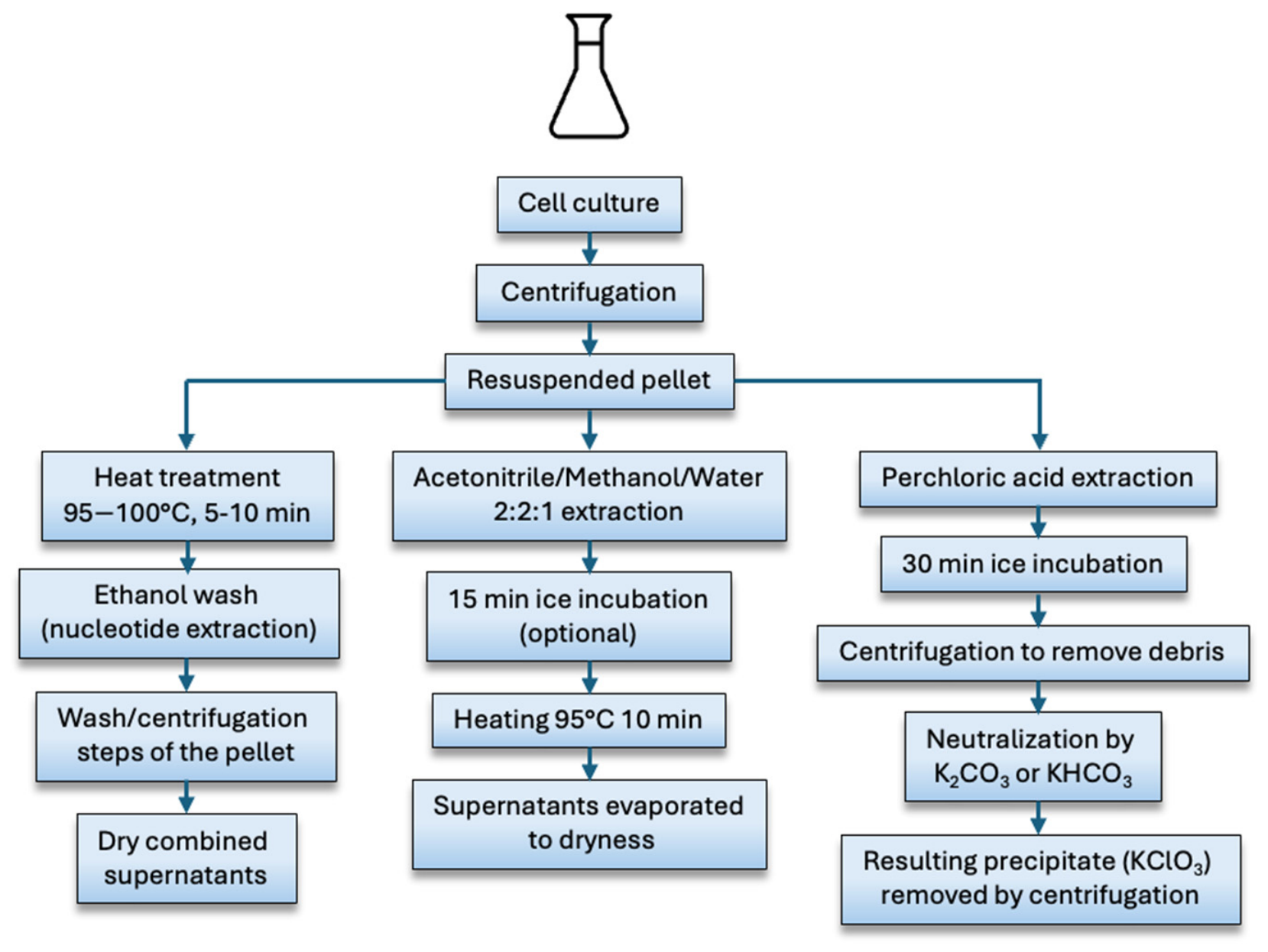
Among the chemical methods used for c-di-GMP quantification, extraction protocols often follow similar procedures. While these protocols are continually optimized and refined, three main categories can be distinguished based on their physico-chemical properties: heating treatment extraction, perchloric acid extraction and organic solvent extraction combined with heat. The diagrams of these protocols are shown in Figure 2.
i.
Heat treatment extraction
The heat extraction method was originally described by Amikam et al. to extract c-di-GMP from Molt4 cells incubated in the presence of 32P c-di-GMP [62]. Strong, short heating (100 °C, 3 min) induced cell lysis, followed by two nucleotide extractions with 65% ice-cold ethanol.
This method was subsequently used in other studies, like on Salmonella Typhimurium bacteria [63], or Shewanella oneidensis [64]. Heat treatment procedure is also used to extract c-di-AMP [65]. c-di-AMP is another signaling molecule of interest, present at approximately ten times the cellular concentration of c-di-GMP [59]. Heating to 100 °C over a short period of time causes rapid lysis of bacterial cells, releasing cell contents including c-di-GMP. This rapid extraction method minimizes changes in c-di-GMP levels that might occur if cells were left on ice or incubated for long periods. After heating, addition of cold ethanol solubilizes c-di-GMP and separates it from cell debris. An advantage of this extraction method is that it avoids the use of organic solvents such as acetonitrile. Nucleotides are extracted from the cell lysate with ethanol. An additional step can be incorporated after heat treatment, involving a subsequent extraction with acidic phenol. Typically employed in RNA extraction because the phenolic phase removes proteins, lipids and DNA from the aqueous phase, this step is applied here to minimize interfering substances in LC-MS [59].
ii.
Perchloric acid extraction
Initially presented by Weinhouse et al. [66] in 1997, perchloric acid extraction was used to isolate and quantify c-di-GMP at the cellular level in Acetobacter xylinum, as well as to analyse its membrane-associated protein-bound c-di-GMP. This extraction method, consisting of cell lysis with cold acid, followed by neutralization of the supernatant, has been used many times since, and adapted to a variety of bacterial types.
In their investigation of c-di-GMP’s role in P. aeruginosa, Hickman & Harwood also employed a perchloric acid extraction protocol. However, they introduced an initial centrifugation step to Weinhouse’s original protocol [20].
The extraction protocol of Hickman et al. was later revised to simplify and shorten the procedure. Specifically, omitting the centrifugation step prior to cell lysis shortened the analysis time and helped mitigate issues related to the chemical instability of c-di-GMP [60]. Moreover, centrifugation of cells can compromise the accurate determination of physiological c-di-GMP concentration [67]. Because this extraction occurs in an acid environment, metabolites stability is limited, making it necessary to neutralize the acid extract immediately after extraction using K2CO3 or KHCO3. The resulting KClO4 precipitate is then removed by centrifugation. Irie & Parsek [60] also caution that the addition of KHCO3 generates CO2, creating a potential explosion. Therefore, transferring the mixture to a large-volume conical tube is recommended.
iii.
Organic solvent extraction combined with heat
The solvent mixture used in all references is acetonitrile/methanol/water 2:2:1. This mixture allows rapid quenching of bacterial metabolism and counteracts the rapid loss of metabolites that occurs when exposed to cold solvents. The acetonitrile-containing extraction mixture significantly improves the measurement of key physiological parameters, such as nucleotide quantification [68]. Furthermore, the acetonitrile/methanol/water mixture has a low freezing point, which is advantageous for rapidly cooling cells to quench metabolism. A subsequent heating step (below 100 °C) is applied to inactivate residual phosphodiesterase and phosphatase activities.
This technique was first proposed by Spangler et al. [61] based on a nucleotide extraction protocol, originally reported by Rabinowitz and Kimball [68] for c-di-GMP extraction. This protocol was widely used in other studies [69] before being further refined by Burhenne et al. in 2013 for c-di-GMP extraction in Gram-negative bacteria [70]. Subsequently, the same team published a version of this protocol adapted for Gram-positive bacteria [71]. Although this article focuses on c-di-AMP extraction, the conditions described are also applicable to c-di-GMP extraction.
In a study of c-di-GMP signaling pathways in V. cholerae, a rapid sonication step was applied to cultures prior to extraction. Three short pulses were sufficient to disrupt bacterial aggregates without compromising cell viability, thereby optimizing the extraction process [72]. An alternative approach was proposed by Bobrov et al. which avoids heat treatment that could compromise cyclodinucleotide stability [73]. Yersinia pestis cell cultures were centrifuged, and the resulting pellet was resuspended in extraction buffer (40% methanol 40% acetonitrile in 0.1 N formic acid) and incubated for 30 min at −20 °C. A subsequent centrifugation removed insoluble material, after which a second extraction was performed under the same conditions. The supernatants were then neutralized with 15% ammonium bicarbonate and directly analyzed by liquid chromatography. More recently, another heat-free extraction protocol was reported for E. coli, in which bacterial pellets were subjected to 3 freeze/thaw cycles in liquid nitrogen, followed by homogenization via bead beating before extraction by chloroform/methanol 2:1 v/v [74].
Despite the diversity of these extraction protocols and their ongoing adaptation to different bacterial models, it is noteworthy that few studies report a systematic metrological characterization of the extraction step itself. The parameters typically required to validate an analytical method (extraction recovery, intra-day repeatability, inter-day reproducibility) are rarely comprehensively documented in published protocols, which tend to focus more on the operational conditions (time, temperature, solvents) than on quantitative performance. Only a few studies include an assessment of extraction recovery through spiking/recovery tests, allowing for a rough comparison between different categories of protocols. For the heat-treatment method, Simm et al. report a recovery rate of 85 ± 11% following the addition of 100 fmol of c-di-GMP per mg of cells, a quantity close to the limit of detection [75]. Kameda et al., also using heat treatment combined with sequential ethanol/phenol/chloroform extraction, reported recoveries ranging from 62 to 82% over a range of 10 to 100 nmol per injection [59]. For a method combining organic solvents and heating, Spangler et al. reported recoveries of 76.4%, 74.2% and 108.4% for standards at 10, 50 and 500 ng/mL respectively. The lowest values were obtained at the lowest concentrations (10 and 50 ng/mL), suggesting proportionally greater losses during the extraction of small quantities of analyte [61]. Although obtained under heterogeneous experimental conditions (including different matrices, concentration ranges, internal standards), these isolated data suggest that recoveries are broadly comparable between the two categories of methods, ranging from 60–110% depending on the analyte concentration. This lack of metrological standardization constitutes a significant limitation to the inter-study comparability of c-di-GMP levels reported in the literature, and highlights the need for future developments to systematically incorporate these validation parameters from the extraction stage onwards, prior to analytical characterization by LC-MS/MS.
(c)
Internal standards
The use of an internal standard is essential in c-di-GMP quantification method to correct variations arising from extraction, injection and instrumental response, thereby ensuring accurate and reproducible quantification measurements. Ideally, the standard should be structurally similar to c-di-GMP, such as an isotopically labeled analog, and absent from the sample matrix. Additionally, it should co-elute with the target molecule without interfering with its detection. Most protocols in the literature recommend adding the internal standard at the time of extraction. Earliest studies employed 13C2015N10 c-di-GMP, enzymatically synthesized from 13C1015N5 GTP [58,70,71,76]. This isotopically labeled internal standard, which is fully labelled on both guanine bases (20 labelled carbon atoms and 10 labelled nitrogen atoms), exhibits a mass shift relative to native c-di-GMP. It can therefore be detected by LC-MS/MS using the quantifier SRM transition m/z 721.198 → 162.056, compared with m/z 691.135 → 152.118 for native c-di-GMP. Its co-elution with native c-di-GMP enables compensation for matrix effects [69].
Other works use xanthosine 3,5-cyclic monophosphate (cXMP) to quantify c-di-GMP in Gram-positive bacteria [61,62]. The use of cXMP eliminates the need for stable isotopes. Furthermore, as cXMP is absent from bacterial cells, it minimizes analytical interferences, thereby enhancing reliability and accuracy. Moreover, cXMP is compatible with endogenous bacterial cell nucleotide extraction protocols. While Simm et al. employed c-di-AMP as an internal standard for LC-MS/MS-based c-di-GMP quantification, Irie & Parsek favored 2-chloro-adenosine-5′-O-monophosphate, as c-di-AMP had been identified as biologically active and was therefore unsuitable as internal standard [60,75].
Supplementary Table S1 summarizes the main materials and parameters used across different c-di-GMP extraction protocols. Several techniques have been developed, each with specific advantages and limitations. Perchloric acid extraction effectively precipitates proteins and rapidly releases cyclic nucleotides, but it can partially degrade c-di-GMP and requires careful neutralization, complicating the procedure. Heat treatment offers a simple and rapid extraction method, yet it can denature or transform sensitive metabolites, limiting its reliability for precise quantification. Organic solvents-based extraction (methanol, acetonitrile, and water) is the most widely employed method due to its compatibility with LC-MS/MS, but it requires strict compliance with quenching conditions to avoid metabolic bias [20,58,59,60,61,63,64,66,70,71,72,73,74,75,76,77,78,79,80,81,82,83,84,85,86,87,88].
In this context, standardizing extraction protocols is crucial to ensure comparability across studies and improve the reliability of cellular c-di-GMP measurements. However, few recent advancements have been made, highlighting the need for novel, more sensitive, selective, and standardizable extraction approaches suitable for analyzing c-di-GMP in complex or low-abundance matrices.

3.2. c-di-GMP Purification and Quantification by Liquid Chromatography with Mass Spectrometry

Liquid chromatography is used to separate the constituents of the cell extract obtained after the first extraction stage, regardless of the protocol employed. Molecules are retained on the column by affinity with the stationary phase. This is generally a silica gel functionalized with a C18 chain, known as the reverse phase. The retained molecules are then eluted by a solvent (mobile phase) according to their affinity with its polarity. For c-di-GMP separation, studies consistently report the use of polar solvents systems. The retention times measured during elution will be specific for each molecule and will enable rapid identification of the presence of c-di-GMP compared to standards. Molecules are then identified through a mass spectrometer coupled to the liquid chromatography chain. Owing to its high sensitivity, mass spectrometry enables precise detection of the molecules present in the extract. This combination of analytical techniques is the ideal tool for detection and quantification of c-di-GMP.
Historically, c-di-GMP was first identified using two-dimensional Thin Layer Chromatography (2D-TLC), during a comprehensive analysis of cellular nucleotides pool of S. Typhimurium [77]. Subsequent studies investigating the regulatory networks of biofilm formation in V. cholerae and P. aeruginosa also relied on 2D-TLC to assess intracellular c-di-GMP levels [78,79]. Although relative changes in c-di-GMP concentration can be determined using this technique, its resolution and quantitative accuracy is limited, thus requiring more precise techniques for fine quantification. Consequently, the remainder of this review will focus exclusively on LC-MS-based approaches.
(a)
High Performance Liquid Chromatography separation
The first studies identifying c-di-GMP as a bacterial second messenger relied on high-performance liquid chromatography (HPLC). This technic was used to purify this compound either from bacterial extracts or by chemical synthesis [4,80]. This approach subsequently served as reference for c-di-GMP quantification, including studies, such as that of Weinhouse et al. [66]. In this study, c-di-GMP was quantified by ultra-violet (UV) detection at 254 nm following HPLC separation. Nevertheless, this HPLC-UV detection was not optimal because cell extracts contain numerous nucleotides absorbing within the same wavelength range, leading to co-elution or signal interference. In 2006, Thormann and his team investigated the regulatory mechanisms underlying the role of c-di-GMP in biofilm regulation. To this end, they improved the initial quantification method by using LC-MS to measure c-di-GMP levels under different conditions [64]. This protocol was subsequently adapted in other studies investigating c-di-GMP [20]. In 2009, Simm et al. proposed a new protocol to determine and compare intracellular c-di-GMP concentrations across various bacterial species. To this end, they first used a preparative ion pair chromatography (AKTA column) to separate c-di-GMP. This was followed by matrix-assisted laser desorption/ionization-time-of-flight (MALDI-TOF) mass spectrometry for c-di-GMP detection and quantification [75]. The method enabled c-di-GMP quantification, with a reported limit of quantification of 5 nM. However, this approach has several limitations, including the preparative chromatography purification step (off-line) and the use of MALDI as a detector, leading to a limited linear dynamic range.
Today, almost all published protocols employ separation by liquid chromatography directly coupled to the mass spectrometer. The main advantage of HPLC lies in its ability to separate c-di-GMP from other compounds extracted from bacterial cells at the same time as the analyte. This technique also enables the identification of other compounds associated with c-di-GMP turnover, such as GTP, GMP and pGpG. HPLC monitoring thus makes it possible to assess c-di-GMP synthesis or degradation through monitoring its co-products [81]. As mentioned above, solvents used to separate compounds from the extract are polar solvents, such as ammonium acetate (in water), and methanol or acetonitrile. Ammonium acetate can be used in a dual role: as a chromatographic ion-pairing agent and as a volatile electrolyte for thermospray LC/MS detection [82]. This dual functionality provides high degree of selectivity for the analysis of complex biological mixtures.
Indeed, the resulting extracts may contain matrix- or culture medium-derived compounds that co-elute with c-di-GMP. In addition to ammonium acetate, which has been used as an ion-pairing reagent in several studies, more hydrophobic alkylamines represent a potential methodological option for improving chromatographic resolution. Donegan et al. demonstrated that the increasing hydrophobicity of these reagents enhances retention and resolution in ion-pair reverse-phase chromatography while maintaining MS compatibility at low concentrations (5 to 10 mM). Although this study focused on oligonucleotides rather than directly on c-di-GMP, these chromatographic principles may be transferable and could facilitate improved separation of c-di-GMP from co-eluting matrix compounds [83].
A solvent gradient is generally performed to elute molecules from the column, with a gradual increasing in the proportion of methanol or acetonitrile. In the solvent gradient, additional steps can be used, moving from a high to a low percentage of aqueous phase, then high again to eliminate carry-over between injections [20,64]. The use of an elution gradient enables the separation of c-di-GMP from other compounds present in cell extracts that may interfere with its quantification. For example, Petrova and Sauer separated c-di-GMP on a reverse-phase C18 column using methanol as solvent B and 10 mM ammonium acetate as solvent A. The following stepwise gradient was applied: 0–9 min, 1% B; 9–14 min, 15% B; 14–19 min, 25% B; 19–26 min, 90% B; 26–40 min, 1% B. Under these conditions, c-di-GMP eluted at approximately 14–15 min, and the final step allowed re-equilibration under highly aqueous conditions before the next run [81].
(b)
Mass spectrometry
The remarkable sensitivity of mass spectrometry enables accurate detection of intracellular c-di-GMP, even at very low concentration. This analytical power has enabled the scientific community to gain deeper insights into c-di-GMP regulatory networks through assessment and comparison of intracellular c-di-GMP levels from various bacterial strains and culture conditions, with various phenotypes, from high to low c-di-GMP producer. Beyond sensitivity, its specificity and quantitative capabilities make it a powerful tool to assess and compare intracellular c-di-GMP levels across different culture conditions or strains.
In the literature, detection relies on tandem mass spectrometry (MS/MS). This technique employs two sequential stages of mass analysis to selectively examine the fragmentation of specific ions within an ion mixture. c-di-GMP is detected in the multiple-reaction monitoring mode (MRM), where one ion of a particular mass is selected in the first stage of a tandem mass spectrometer and one ion product of a fragmentation reaction of the precursor ions is selected in the second mass spectrometer stage for detection. MRM mode is essential to discriminate compounds with the same mass (e.g., nucleotides). Only the different fragmentation of each molecule can unequivocally identify two molecules of the same mass based on specific transitions. In MRM mode, during fragmentation, a transition corresponds to the passage from a precursor ion (parent ion) to a fragment ion (product ion). Each transition is defined by an m/z pair corresponding to the parent ion and its fragment ion. Quantifier ion is the first fragment used to measure analyte concentration, as it provides the most intense or stable signal. Secondary, the qualifier ion is used to confirm analyte identity by checking that ratio of the quantifier remains constant. The combined use of quantifier and qualifier ensures both sensitivity and specificity in quantitative analyses.
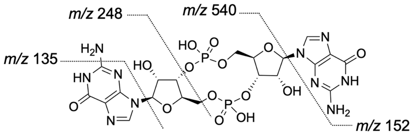
c-di-GMP is detected in MRM mode with the transitions m/z 691 → 152 (single bond fragmentation), m/z 691 → 248 (double bond fragmentation), and m/z 691 → 540. Some authors also mention/report the transition m/z 691 → 135 as specific. However, transitions such as m/z 691 → 152, 691 → 135 resulting from guanine fragmentation, may arise from other unidentified endogenous metabolites. On the other hand, the m/z 691 → 248 transition is highly specific to c-di-GMP and selected as qualifier transition while the m/z 691 → 152 transition is employed as quantifier [61,71]. The m/z 691 → 540 may also be monitored as confirmatory signal [20]. Most intensive mass transitions are used as quantifiers, whereas additional fragments serve as qualifiers. To minimize the risk of false-positive due to the presence of interferents, it is nevertheless necessary to use two qualifiers [76]. Fragmentation patterns are shown in Figure 3. Tandem mass spectrometers offer high selectivity by detecting specific molecular fragments. However, depending on the type of instrument and ion source, fragmentation can also occur within the ion source itself, a phenomenon known as “in-source fragmentation”. The analyte may decompose into small fragments directly in the ion source before reaching the collision cell. c-di-GMP can then be broken down into guanosine 3′,5′-cyclic monophosphate (cGMP) [61]. Despite some uncertainties related to c-di-GMP fragmentation at the source, ESI-MS/MS analysis coupled with HPLC remains more specific compared to single-MS or no-MS methods.
In most studies, characterization and quantification by mass spectrometry are performed using electrospray ionization (ESI) on triple quadrupole analyzers and detectors, operating in multiple reaction monitoring (MRM) mode on tandem mass spectrometry. Detection limits range from 0.002 nmol/mL to 2.5 nmol/mL, with quantification limits between 0.006 nmol/mL and 5 nmol/mL. Recovery percentages, meanwhile, remain unspecified, ranging from 62 to 108%. All protocols are performed using positive ionization, except for MALDI analyses [65,72,75], and one study using a triple quadrupole with ion trap [84] and negative ionization.
Optimization of collision energies used to obtain the fragments is critical to maintain the signal of the quantifier ion, as excessively high voltage can suppress it. These parameters were first optimized based on standards, before being evaluated on cell lysate. Under positive ionization, c-di-GMP produced an ion at m/z 691.2. The product ions were then identified, first with a collision energy of 30 V. Tandem mass spectra obtained by collision-induced dissociation (CID) generates fragment ions at m/z 540.1, 346.1, 248.1 and 152.1 (assigned to guanine). Different collision energies were also evaluated to optimize detection (energy resolved curves). Fragment ions intensities were plotted according to collision energy ranging from 0 to 50 V. Energy resolved curves profile of c-di-GMP obtained for fragment ions at m/z 540.1 and 248.1 present a maximum intensity at collision energy of 20 V, then rapidly decline at higher voltages. The ion at m/z 152.1 begins to increase at collision energy of 20 V. Hence, 20 V was used as optimal collision energy [74]. The various parameters used for detection and quantification by mass spectrometry are listed in Supplementary Table S2 [4,20,59,60,61,64,70,72,74,75,76,81,84,85].
Using tandem mass spectrometry detection offers the advantage of identifying additional metabolites present in the extract, providing a broader picture of compounds potentially involved in biofilm regulation. The protocol initially described by Spangler was later upgraded to enable the simultaneous quantification of other metabolites of interest. This method enables detection and distinction of c-di-GMP and c-di-AMP. Other dinucleotide metabolites such as 5′-phosphoadenylyl-3′,5′-adenosine (pApA) and 5′-phosphoguanylyl-3′-5′-guanosine (pGpG) can also be detected and distinguished. In addition, this approach enables the quantification of mononucleotides (e.g., AMP, GMP) as well as cyclic mononucleotides such as cAMP and cGMP [70]. This protocol was subsequently reused under identical conditions in a study highlighting the effect of light on intracellular c-di-GMP level during bacterial culture [58]. In 2021, a simple and robust method was developed to quantify four naturally occurring cyclic dinucleotides (including c-di-GMP) in both bacterial and vertebrate tissue cells [74].
However, c-di-GMP quantification through conventional methods previously presented has inherent limitations. Chemical methods based on LC/MS extraction and quantification provide only average value of c-di-GMP quantification without resolving metabolic gradients. In fact, biofilm environment is highly/intrinsically heterogeneous (oxygen, nutrients, signal molecules, etc.), leading to spatial variability for all the elements involved in the biofilm structure, including c-di-GMP. More recently, mass spectrometry imaging (MSI) techniques have been developed to address this gap, capable to generate spatially resolved chemical information with unprecedented detail. MSI enables a better understanding of environmental variables affecting biofilm formation and bacterial secretome as a function of culture conditions. It allows identification of numerous molecules, including secondary and specialized metabolites (chemical signals for bacterial communication, motility, virulence). Their spatial distribution profiles can be mapped, revealing dynamic production over time and gradient patterns both within and around the biofilm environment [89].
A promising label-free method approach for the direct detection of c-di-GMP in microbial colony biofilms using matrix-assisted laser desorption/ionization mass spectrometry imaging (MALDI-MSI) has recently been developed [90]. Unlike label-based methods, this strategy doesn’t require labeling or dyeing, avoiding chemical modification of metabolites. Furthermore, it allows direct detection and visualization of c-di-GMP spatial distribution within the biofilm. Applied to biofilms of V. cholerae, V. fischeri and P. aeruginosa PA14, MALDI-MSI captured spatial and temporal variations in the c-di-GMP levels in response to genetic perturbation of its metabolic enzymes. This versatile approach offers the possibility to explore and corelate spatial relationships between c-di-GMP and various specific metabolites, in both genetically modified bacterial strains and in clinical isolates. Notably, this method can be adapted for direct analysis on host tissues, extending its scope to more complex biological contexts. This versatility provides a deeper understanding of in situ molecular interactions, paving the way for new perspectives to investigate in situ molecular interactions and host-pathogen signaling dynamics.
Although not strictly an analytical chemistry method for c-di-GMP quantification, an ELISA kit to quantify c-di-GMP in bacterial lysates is commercially available. This kit is marketed by Cayman Chemical, Ann Arbor, MI, USA (https://www.caymanchem.com/product/501780/cyclic-di-gmp-elisa-kit, accessed on 31 August 2026). The manufacturer reports a lower detection limit of 5.3 pg/mL, which is below mass spectrometry detection limits, with results completed within 4 h. It is therefore worth considering this alternative for c-di-GMP quantification. Mass spectrometry-based methods provide precise quantification of c-di-GMP, enabling measurement at specific time points during biofilm establishment/development maturation. However, since this method relies on quenching of bacterial metabolism during extraction, analysis only reflects the total intracellular pool of c-di-GMP and does not capture spatial distribution within the biofilm. Nevertheless, tandem mass spectrometry enables highly sensitive detection, reaching quantification levels as low as the nanomolar range. This makes it a powerful tool to study c-di-GMP signaling dynamics in bacterial communities.

4. Discussion

In this review, we have outlined the current approaches used to monitor and quantify c-di-GMP, a versatile and important bacterial second messenger. These approaches can broadly be divided into two categories: biosensor-based strategies, which primarily provide relative measurements of c-di-GMP levels in living cells, and analytical approaches, mainly based on mass spectrometry, which enable absolute quantification. Although both strategies have contributed significantly to our understanding of c-di-GMP signaling, each presents inherent advantages and limitations. Importantly, no currently available approach can simultaneously provide highly sensitive and absolute quantification together with real-time and spatially resolved information. The choice of method therefore depends largely on the biological question being addressed.
One of the major challenges in c-di-GMP detection is sensitivity. The concentration at which c-di-GMP exerts physiological effects remains difficult to define and may vary depending on the organism and cellular context. Biosensors sensitivity remains limited compared to analytical approaches. Among the most sensitive biosensors developed to date are the protein-based sensor CdGreen2 and riboswitch-based biosensors derived from lchAA, both of which have been reported to detect c-di-GMP concentrations in the low nanomolar range [15,33]. However, the analytical sensitivity of a biosensor does not necessarily translate directly into an equivalent sensitivity in living cells. Biological variability and interference from other cellular processes can complicate signal interpretation and make the detection of small changes in intracellular c-di-GMP challenging. Consequently, biosensors are generally best suited to monitoring relative changes in c-di-GMP rather than providing absolute intracellular concentrations. By contrast, analytical chemistry approaches, particularly LC-MS/MS, provide sensitive and quantitative measurements of intracellular c-di-GMP. Tandem mass spectrometry enables low limits of quantification, including measurements in the nanomolar range, making this approach particularly valuable for bacteria with naturally low intracellular concentrations of c-di-GMP. More importantly, LC-MS/MS provides an absolute measurement of the amount of c-di-GMP recovered from a biological sample, allowing quantitative comparisons between experimental conditions. This represents a major advantage when the objective is to establish a relationship between intracellular c-di-GMP concentration and a specific bacterial phenotype.
Despite their relative simplicity, biosensor-based approaches can become technically demanding depending on the bacterial species studied. To monitor c-di-GMP, the organism must first be genetically equipped with the appropriate biosensor, and its performance may depend on species-specific characteristics such as promoter activity, gene expression, protein stability, intracellular background fluorescence, and the availability of suitable genetic tools. Most currently available biosensors have been developed and extensively characterized in a limited number of model organisms, including P. aeruginosa, C. crescentus, and E. coli [15,21,34]. Their application to less well-characterized species therefore often requires adaptation and optimization, which can be time-consuming and challenging, particularly when genetic manipulation systems are poorly established. Once a suitable biosensor has been established, however, fluorescence-based measurements can be experimentally straightforward. Global c-di-GMP dynamics can be monitored using a plate reader or epifluorescence microscope, requiring only bacteria carrying the biosensor and appropriate controls. Spatially resolved measurements, particularly within biofilms or at the single-cell level, require more advanced and sensitive microscopy and are consequently more technically demanding. Analytical approaches offer an important complement in this regard. LC-MS/MS can, in principle, be applied to a broad range of bacterial species without requiring the development of a species-specific biosensor. Once c-di-GMP has been efficiently extracted from the cells, chromatographic separation and mass spectrometric detection are independent of the biological origin of the sample. The main species-dependent step is therefore sample preparation. In Gram-positive bacteria, for example, additional cell-disruption steps such as bead beating may be required to efficiently release intracellular metabolites. Nevertheless, LC-MS/MS provides a comparatively robust and transferable strategy for absolute quantification across bacterial species.
One of the major advantages shared by biosensors is their ability to monitor c-di-GMP in living cells and, in some cases, in real time. This capability is particularly important because c-di-GMP signaling is highly dynamic. The rapid and reversible action of diguanylate cyclases (DGCs) and phosphodiesterases (PDEs), often referred to as the “makers and breakers” of c-di-GMP, can generate substantial changes in intracellular c-di-GMP over short timescales. Biosensors therefore provide information that is difficult to obtain using conventional endpoint analytical approaches. Both pcdrA-based transcriptional biosensors and protein-based biosensors have been used to monitor fluctuations in c-di-GMP levels, whereas riboswitch-based biosensors, although theoretically capable of dynamic measurements, have not yet been extensively applied to real-time monitoring. However, the temporal resolution of biosensors is determined by several factors. For transcriptional and riboswitch-based systems, the stability and maturation of the fluorescent reporter can introduce a delay between the change in c-di-GMP concentration and the observed fluorescence signal. The use of destabilized fluorescent proteins, such as GFP-ASV, can reduce this delay and improve the responsiveness of transcriptional biosensors [25]. Nevertheless, transcription, translation, protein maturation, and reporter degradation remain intrinsic limitations when very short timescale dynamics need to be resolved.
Another important parameter is the kinetics of c-di-GMP binding and dissociation from the biosensor. Rapid changes in c-di-GMP concentration can only be faithfully detected if the sensor responds sufficiently quickly to these changes. The dissociation kinetics of c-di-GMP have not been systematically characterized for many transcriptional, riboswitch-based, and protein-based biosensors. An exception is the cpEGFP-based CdGreen2 sensor, for which the response kinetics have been characterized, and which currently offers particularly high temporal resolution [15]. Consequently, while biosensors provide a unique opportunity to monitor c-di-GMP dynamics in vivo, the fluorescence signal should not necessarily be interpreted as an instantaneous representation of the intracellular c-di-GMP concentration. LC-MS/MS can also be used to investigate temporal changes in c-di-GMP by analyzing samples collected at different time points. However, this approach provides measurements from discrete samples rather than continuous monitoring in living cells. Capturing rapid fluctuations would therefore require a high sampling frequency and sufficient biological material at each time point. This becomes particularly challenging when studying processes occurring over extended periods, such as biofilm development, where changes in c-di-GMP may occur over minutes while the biological process itself develops over many hours.
Beyond temporal dynamics, an important feature of c-di-GMP signaling is its spatial organization within bacterial cells and communities. Increasing evidence indicates that c-di-GMP does not necessarily act as a uniformly distributed intracellular signal [15]. Instead, local production, degradation, and sequestration can generate heterogeneous c-di-GMP pools that influence specific cellular processes. Protein-based biosensors are particularly valuable in this context because they can provide spatial information at the single-cell level. This capability makes them powerful tools for investigating cell-to-cell heterogeneity and the relationship between local c-di-GMP levels and bacterial behaviors such as motility, adhesion, and biofilm formation. This spatial dimension represents a major limitation of analytical approaches. LC-MS/MS measures the total amount of c-di-GMP recovered from a population of cells and therefore provides an average intracellular concentration. Although fractionation or other specialized approaches may provide additional spatial information, conventional LC-MS/MS cannot directly reveal the distribution of c-di-GMP within individual cells or across a biofilm. Consequently, an increase in the global c-di-GMP concentration detected by LC-MS/MS may potentially conceal substantial cell-to-cell heterogeneity.
Although LC-MS/MS offers sensitive and absolute quantification, the accuracy of the measurement depends strongly on sample preparation. Quantification first requires efficient extraction of intracellular c-di-GMP while minimizing degradation and loss of the analyte. Three main extraction strategies are commonly described in the literature: heat treatment, perchloric acid extraction, and organic solvent-based extraction. Direct comparison of these approaches remains difficult because extraction yields and recovery efficiencies are not consistently reported. A standardized evaluation of extraction procedures would therefore represent an important methodological improvement. In particular, c-di-GMP measurements should ideally be normalized using clearly defined parameters, including culture cell density and the volume of culture processed prior to extraction. Reporting recovery efficiencies would also facilitate comparison between extraction protocols and between laboratories. Such standardization would help determine whether differences in reported intracellular c-di-GMP concentrations reflect biological variation or methodological differences. Although existing extraction protocols have been modified over time, major innovations in this step remain relatively limited. Future methodological developments could focus on improving metabolite recovery while minimizing degradation, as well as developing extraction procedures that use less hazardous or more environmentally sustainable solvents. Preservation of c-di-GMP integrity is particularly important because degradation during sample preparation can lead to an underestimation of the intracellular concentration. Improving extraction efficiency and reproducibility will therefore be essential for obtaining measurements that more accurately reflect the intracellular state.
Overall, the limitations of each strategy are partly compensated for by the strengths of the other. Biosensors provide information that is difficult to obtain using analytical chemistry, including real-time monitoring in living cells and, particularly for protein-based sensors, spatial information at the single-cell level. However, their fluorescence output is generally a relative proxy for c-di-GMP concentration, their sensitivity may be limited by biological and technical noise, and their application to less well-characterized organisms often requires substantial optimization. LC-MS/MS overcomes several of these limitations by providing sensitive and absolute quantification across a broad range of bacterial species. However, it requires cell disruption and metabolite extraction, provides measurements at defined sampling points rather than continuous monitoring, and does not directly resolve the spatial organization of c-di-GMP within individual cells or biofilms.
The choice between these approaches should therefore be driven by the biological question rather than by the assumption that one method is universally superior. LC-MS/MS is particularly well suited to determine absolute intracellular concentrations and to establish quantitative relationships between c-di-GMP levels and bacterial phenotypes. Biosensors, in contrast, are indispensable when the objective is to investigate temporal dynamics, cellular heterogeneity, or spatial organization. Combining both approaches can therefore provide a more comprehensive characterization of c-di-GMP signaling by linking absolute intracellular concentrations measured by analytical chemistry with their temporal and spatial dynamics observed using biosensors. Future methodological advances will likely depend not on the development of a single universal technique, but on the integration of complementary analytical and biosensor-based approaches. Such a combination, together with improved and standardized extraction procedures and the development of more sensitive and kinetically responsive biosensors, should enable a more accurate understanding of how c-di-GMP concentration, dynamics, and spatial distribution collectively shape bacterial physiology and behavior.

5. Conclusions

In conclusion, both approaches offer distinct advantages and limitations. Biosensors are uniquely suited to study spatial and temporal dynamics of c-di-GMP in living cell but suffer from limited sensitivity and lack of universality. In contrast, LC-MS/MS approach provides absolute quantification with high sensitivity and broad applicability but cannot resolve subcellular localization or real-time dynamics. We strongly believe that combining these complementary approaches or developing hybrid tools that integrate biosensor-based visualization with analytical quantification will be key to fully elucidate the role of c-di-GMP in bacterial physiology and signaling.

Supplementary Materials

The following supporting information can be downloaded at: https://www.mdpi.com/article/10.3390/applmicrobiol6090105/s1, Table S1: Comparison table of c-di-GMP extraction protocols; Table S2: Comparison table of c-di-GMP separation techniques, and detection parameters.

Author Contributions

Writing—original draft: A.A. and C.N. Writing—review and editing: A.A., C.N., K.V., S.R. and Y.B. Project administration: K.V. and S.R. Funding acquisition, K.V. All authors have read and agreed to the published version of the manuscript.

Funding

This work is part of the AA’s PhD thesis, funded by the region Bretagne, the Université de Bretagne Sud (UBS) and the Université de Bretagne Occidentale (UBO) (PhD AMPHIPOX project).

Institutional Review Board Statement

Not applicable.

Informed Consent Statement

Not applicable.

Data Availability Statement

No new data were created or analyzed in this study. Data sharing is not applicable to this article.

Conflicts of Interest

The authors declare no conflicts of interest.

Abbreviations

The following abbreviations are used in this manuscript:
c-di-GMP-(3′-5′)-cyclic diguanylic acid
DGCsdiguanylate cyclases
PDEsphosphodiesterases
GFPGreen Fluorescent Protein
sRNAssmall RNAs
UTRuntranslated region
DFHBI3,5-difluoro-4-hydroxybenzylidene imidazolinone
pGpG5′-phosphoguanylyl-(3′,5′)-guanosine
YFPYellow Fluorescent Protein
RFPRed Fluorescent Protein
RETResonance Energy Transfer
FRETFluorescence Energy Transfer
BRETBioluminescence Energy Transfer
CRETChemiluminescence Energy Transfer
KdConstant Dissociation Equilibrium
CFPCyan Fluorescent Protein
PPIprotein-protein interactions
3-IANauxin 3-indoleacetonitrile
BiFCBimolecular Fluorescence Complementation
cpEGFPcircularly permutated EGFP
LC-MS/MSliquid chromatography coupled to tandem mass spectrometry
LBLysogenic Broth
VBMMVogel-Bonner Minimal Media
cXMPxanthosine 3,5-cyclic monophosphate
2D-TLCtwo-dimensional Thin Layer Chromatography
UVultra-violet
MALDI-TOFmatrix-assisted laser desorption/ionization-time-of-flight
MRMmultiple-reaction monitoring mode
cGMPguanosine 3′,5′-cyclic monophosphate
ESIelectrospray ionization
CIDcollision-induced dissociation
pApA5′-phosphoadenylyl-3′,5′-adenosine
pGpG5′-phosphoguanylyl-3′-5′-guanosine
MSImass spectrometry imaging

References

  1. Cotter, P.A.; Stibitz, S. c-di-GMP-mediated regulation of virulence and biofilm formation. Curr. Opin. Microbiol. 2007, 10, 17–23. [Google Scholar] [CrossRef] [Scilit] [PubMed]
  2. Jenal, U.; Reinders, A.; Lori, C. Cyclic di-GMP: Second messenger extraordinaire. Nat. Rev. Microbiol. 2017, 15, 271–284. [Google Scholar] [CrossRef] [Scilit] [PubMed]
  3. Valentini, M.; Filloux, A. Biofilms and Cyclic di-GMP (c-di-GMP) Signaling: Lessons from Pseudomonas aeruginosa and other bacteria. J. Biol. Chem. 2016, 291, 12547–12555. [Google Scholar] [CrossRef] [Scilit] [PubMed]
  4. Ross, P.; Weinhouse, H.; Aloni, Y.; Michaeli, D.; Weinberger-Ohana, P.; Mayer, R.; Braun, S.; de Vroom, E.; van der Marel, G.A.; van Boom, J.H.; et al. Regulation of cellulose synthesis in Acetobacter xylinum by cyclic diguanylic acid. Nature 1987, 325, 279–281. [Google Scholar] [CrossRef] [Scilit] [PubMed]
  5. Boyd, C.D.; O’Toole, G.A. Second messenger regulation of biofilm formation: Breakthroughs in understanding c-di-GMP effector systems. Annu. Rev. Cell Dev. Biol. 2012, 28, 439–462. [Google Scholar] [CrossRef] [Scilit] [PubMed]
  6. Donlan, R.M.; Costerton, J.W. Biofilms: Survival mechanisms of clinically relevant microorganisms. Clin. Microbiol. Rev. 2002, 15, 167–193. [Google Scholar] [CrossRef] [Scilit] [PubMed]
  7. Donlan, R.M. Biofilms: Microbial Life on Surfaces. Emerg. Infect. Dis. 2002, 8, 881–890. [Google Scholar] [CrossRef] [Scilit] [PubMed]
  8. Flemming, H.C.; Wuertz, S. Bacteria and archaea on Earth and their abundance in biofilms. Nat. Rev. Microbiol. 2019, 17, 247–260. [Google Scholar] [CrossRef] [Scilit] [PubMed]
  9. Liu, H.Y.; Prentice, E.L.; Webber, M.A. Mechanisms of antimicrobial resistance in biofilms. npj Antimicrob. Resist. 2024, 2, 27. [Google Scholar] [CrossRef] [Scilit] [PubMed]
  10. Sauer, K.; Stoodley, P.; Goeres, D.M.; Hall-Stoodley, L.; Burmølle, M.; Stewart, P.S.; Bjarnsholt, T. The biofilm life cycle: Expanding the conceptual model of biofilm formation. Nat. Rev. Microbiol. 2022, 20, 608–620. [Google Scholar] [CrossRef] [Scilit] [PubMed]
  11. Snega Priya, P.; Meenatchi, R.; Pasupuleti, M.; Namasivayam, S.K.R.; Arockiaraj, J. Harnessing cyclic di-GMP signaling: A strategic approach to combat bacterial biofilm-associated chronic infections. Curr. Microbiol. 2025, 82, 118. [Google Scholar] [CrossRef] [Scilit] [PubMed]
  12. Römling, U.; Galperin, M.Y.; Gomelsky, M. Cyclic di-GMP: The first 25 years of a universal bacterial second messenger. Microbiol. Mol. Biol. Rev. 2013, 77, 1–52. [Google Scholar] [CrossRef] [Scilit] [PubMed]
  13. Manner, C.; Dias Teixeira, R.; Saha, D.; Kaczmarczyk, A.; Zemp, R.; Wyss, F.; Jaeger, T.; Laventie, B.-J.; Boyer, S.; Malone, J.G.; et al. A genetic switch controls Pseudomonas aeruginosa surface colonization. Nat. Microbiol. 2023, 8, 1520–1533. [Google Scholar] [CrossRef] [Scilit] [PubMed]
  14. Wei, Q.; Leclercq, S.; Bhasme, P.; Xu, A.; Zhu, B.; Zhang, Y.; Zhang, M.; Wang, S.; Ma, L.Z. Diguanylate Cyclases and Phosphodiesterases Required for Basal-Level c-di-GMP in Pseudomonas aeruginosa as Revealed by Systematic Phylogenetic and Transcriptomic Analyses. Appl. Environ. Microbiol. 2019, 85, e01194-19. [Google Scholar] [CrossRef] [Scilit] [PubMed]
  15. Kaczmarczyk, A.; van Vliet, S.; Jakob, R.P.; Teixeira, R.D.; Scheidat, I.; Reinders, A.; Klotz, A.; Maier, T.; Jenal, U. A genetically encoded biosensor to monitor dynamic changes of c-di-GMP with high temporal resolution. Nat. Commun. 2024, 15, 3920. [Google Scholar] [CrossRef] [Scilit] [PubMed]
  16. Rybtke, M.T.; Borlee, B.R.; Murakami, K.; Irie, Y.; Hentzer, M.; Nielsen, T.E.; Givskov, M.; Parsek, M.R.; Tolker-Nielsen, T. Fluorescence-based reporter for gauging cyclic di-GMP levels in Pseudomonas aeruginosa. Appl. Environ. Microbiol. 2012, 78, 5060–5069. [Google Scholar] [CrossRef] [Scilit] [PubMed]
  17. Borlee, B.R.; Goldman, A.D.; Murakami, K.; Samudrala, R.; Wozniak, D.J.; Parsek, M.R. Pseudomonas aeruginosa uses a cyclic-di-GMP-regulated adhesin to reinforce the biofilm extracellular matrix. Mol. Microbiol. 2010, 75, 827–842. [Google Scholar] [CrossRef] [Scilit] [PubMed]
  18. Flemming, H.C.; Wingender, J. The biofilm matrix. Nat. Rev. Microbiol. 2010, 8, 623–633. [Google Scholar] [CrossRef] [Scilit] [PubMed]
  19. Oladosu, V.I.; Park, S.; Sauer, K. Flip the switch: The role of FleQ in modulating the transition between the free-living and sessile mode of growth in Pseudomonas aeruginosa. J. Bacteriol. 2024, 206, e00365-23. [Google Scholar] [CrossRef] [Scilit] [PubMed]
  20. Hickman, J.W.; Harwood, C.S. Identification of FleQ from Pseudomonas aeruginosa as a c-di-GMP-responsive transcription factor. Mol. Microbiol. 2008, 69, 376–389. [Google Scholar] [CrossRef] [Scilit] [PubMed]
  21. Andersen, J.B.; Hultqvist, L.D.; Jansen, C.U.; Jakobsen, T.H.; Nilsson, M.; Rybtke, M.; Uhd, J.; Fritz, B.G.; Seifert, R.; Berthelsen, J.; et al. Identification of small molecules that interfere with c-di-GMP signaling and induce dispersal of Pseudomonas aeruginosa biofilms. npj Biofilms Microbiomes 2021, 7, 59. [Google Scholar] [CrossRef] [Scilit] [PubMed]
  22. Kuchma, S.L.; Brothers, K.M.; Merritt, J.H.; Liberati, N.T.; Ausubel, F.M.; O’Toole, G.A. BifA, a cyclic-di-GMP phosphodiesterase, inversely regulates biofilm formation and swarming motility by Pseudomonas aeruginosa PA14. J. Bacteriol. 2007, 189, 8165–8178. [Google Scholar] [CrossRef] [Scilit] [PubMed]
  23. Irie, Y.; Borlee, B.R.; O’Connor, J.R.; Hill, P.J.; Harwood, C.S.; Wozniak, D.J.; Parsek, M.R. Self-produced exopolysaccharide is a signal that stimulates biofilm formation in Pseudomonas aeruginosa. Proc. Natl. Acad. Sci. USA 2012, 109, 20632–20636. [Google Scholar] [CrossRef] [Scilit] [PubMed]
  24. Corish, P.; Tyler-Smith, C. Attenuation of green fluorescent protein half-life in mammalian cells. Protein Eng. 1999, 12, 1035–1040. [Google Scholar] [CrossRef] [Scilit] [PubMed]
  25. Nair, H.A.S.; Periasamy, S.; Yang, L.; Kjelleberg, S.; Rice, S.A. Real time, spatial and temporal mapping of the distribution of c-di-GMP during biofilm development. J. Biol. Chem. 2017, 292, 477–487. [Google Scholar] [CrossRef] [Scilit] [PubMed]
  26. Laventie, B.J.; Sangermani, M.; Estermann, F.; Manfredi, P.; Planes, R.; Hug, I.; Jaeger, T.; Meunier, E.; Broz, P.; Jenal, U. A surface-induced asymmetric program promotes tissue colonization by Pseudomonas aeruginosa. Cell Host Microbe 2019, 25, 140–152.e6. [Google Scholar] [CrossRef] [Scilit] [PubMed]
  27. Waters, L.S.; Storz, G. Regulatory RNAs in Bacteria. Cell 2009, 136, 615–628. [Google Scholar] [CrossRef] [Scilit] [PubMed]
  28. Abduljalil, J.M. Bacterial riboswitches and RNA thermometers: Nature and contributions to pathogenesis. Noncoding RNA Res. 2018, 3, 54–63. [Google Scholar] [CrossRef] [Scilit] [PubMed]
  29. Paige, J.S.; Wu, K.; Jaffrey, S.R. RNA mimics of green fluorescent protein. Science 2011, 333, 642–646. [Google Scholar] [CrossRef] [Scilit] [PubMed]
  30. Nakayama, S.; Luo, Y.; Zhou, J.; Dayie, T.K.; Sintim, H.O. Nanomolar fluorescent detection of c-di-GMP using a modular aptamer strategy. Chem. Commun. 2012, 48, 9059–9061. [Google Scholar] [CrossRef] [Scilit] [PubMed]
  31. Pursley, B.R.; Fernandez, N.L.; Severin, G.B.; Waters, C.M. The Vc2 cyclic di-GMP-dependent riboswitch of Vibrio cholerae regulates expression of an upstream putative small RNA by controlling RNA stability. J. Bacteriol. 2019, 201, e00293-19. [Google Scholar] [CrossRef] [Scilit] [PubMed]
  32. Kellenberger, C.A.; Wilson, S.C.; Sales-Lee, J.; Hammond, M.C. RNA-based fluorescent biosensors for live cell imaging of second messengers cyclic di-GMP and cyclic AMP-GMP. J. Am. Chem. Soc. 2013, 135, 4906–4909. [Google Scholar] [CrossRef] [Scilit] [PubMed]
  33. Weiss, C.A.; Hoberg, J.A.; Liu, K.; Tu, B.P.; Winkler, W.C. Single-cell microscopy reveals that levels of cyclic di-GMP vary among Bacillus subtilis dubpopulations. J. Bacteriol. 2019, 201, e00247-19. [Google Scholar] [CrossRef] [Scilit] [PubMed]
  34. Shrestha, P.; Razvi, A.; Fung, B.L.; Eichinger, S.J.; Visick, K.L. Mutational analysis of Vibrio fischeri c-di-GMP-modulating genes reveals complex regulation of motility. J. Bacteriol. 2022, 204, e00109-22. [Google Scholar] [CrossRef] [Scilit] [PubMed]
  35. Zhou, H.; Zheng, C.; Su, J.; Chen, B.; Fu, Y.; Xie, Y.; Tang, Q.; Chou, S.-H.; He, J. Characterization of a natural triple-tandem c-di-GMP riboswitch and application of the riboswitch-based dual-fluorescence reporter. Sci. Rep. 2016, 6, 20871. [Google Scholar] [CrossRef] [Scilit] [PubMed]
  36. Reinders, A.; Hee, C.S.; Ozaki, S.; Mazur, A.; Boehm, A.; Schirmer, T.; Jenal, U. Expression and genetic activation of cyclic di-GMP-specific phosphodiesterases in Escherichia coli. J. Bacteriol. 2016, 198, 448–462. [Google Scholar] [CrossRef] [Scilit] [PubMed]
  37. Craggs, T.D. Green fluorescent protein: Structure, folding and chromophore maturation. Chem. Soc. Rev. 2009, 38, 2865–2875. [Google Scholar] [CrossRef] [Scilit] [PubMed]
  38. Wang, X.C.; Wilson, S.C.; Hammond, M.C. Next-generation RNA-based fluorescent biosensors enable anaerobic detection of cyclic di-GMP. Nucleic Acids Res. 2016, 44, e139. [Google Scholar] [CrossRef] [Scilit] [PubMed]
  39. Boute, N.; Jockers, R.; Issad, T. The use of resonance energy transfer in high-throughput screening: BRET versus FRET. Trends Pharmacol. Sci. 2002, 23, 351–354. [Google Scholar] [CrossRef] [Scilit] [PubMed]
  40. Stryer, L. Fluorescence Energy Transfer as a Spectroscopic Ruler. Annu. Rev. Biochem. 1978, 47, 819–846. [Google Scholar] [CrossRef] [Scilit] [PubMed]
  41. Wu, Y.; Jiang, T. Developments in FRET- and BRET-based biosensors. Micromachines 2022, 13, 1789. [Google Scholar] [CrossRef] [Scilit] [PubMed]
  42. Sapsford, K.E.; Berti, L.; Medintz, I.L. Materials for fluorescence resonance energy transfer analysis: Beyond traditional donor-acceptor combinations. Angew. Chem. Int. Ed. Engl. 2006, 45, 4562–4589. [Google Scholar] [CrossRef] [Scilit] [PubMed]
  43. Christen, M.; Kulasekara, H.D.; Christen, B.; Kulasekara, B.R.; Hoffman, L.R.; Miller, S.I. Asymmetrical distribution of the second messenger c-di-GMP upon bacterial cell division. Science 2010, 328, 1295–1297. [Google Scholar] [CrossRef] [Scilit] [PubMed]
  44. Paul, K.; Nieto, V.; Carlquist, W.C.; Blair, D.F.; Harshey, R.M. The c-di-GMP binding protein YcgR controls flagellar motor direction and speed to affect chemotaxis by a “backstop brake” mechanism. Mol. Cell 2010, 38, 128–139. [Google Scholar] [CrossRef] [Scilit] [PubMed]
  45. Kulasekara, B.R.; Kamischke, C.; Kulasekara, H.D.; Christen, M.; Wiggins, P.A.; Miller, S.I. c-di-GMP heterogeneity is generated by the chemotaxis machinery to regulate flagellar motility. eLife 2013, 2, e01402. [Google Scholar] [CrossRef] [Scilit] [PubMed]
  46. Ho, C.L.; Chong, K.S.J.; Oppong, J.A.; Chuah, M.L.C.; Tan, S.M.; Liang, Z.X. Visualizing the perturbation of cellular cyclic di-GMP levels in bacterial cells. J. Am. Chem. Soc. 2013, 135, 566–569. [Google Scholar] [CrossRef] [Scilit] [PubMed]
  47. Jati, A.P.; Sola-Campoy, P.J.; Bosch, T.; Schouls, L.M.; Hendrickx, A.P.A.; Bautista, V.; Lara, N.; Raangs, E.; Aracil, B.; Rossen, J.W.A.; et al. Widespread detection of Yersiniabactin gene cluster and its encoding integrative conjugative elements (ICEKp) among nonoutbreak OXA-48-producing Klebsiella pneumoniae clinical isolates from Spain and the Netherlands. Microbiol. Spectr. 2023, 11, e0471622. [Google Scholar] [CrossRef] [Scilit] [PubMed]
  48. Benach, J.; Swaminathan, S.S.; Tamayo, R.; Handelman, S.K.; Folta-Stogniew, E.; Ramos, J.E.; Forouhar, F.; Neely, H.; Seetharaman, J.; Camilli, A.; et al. The structural basis of cyclic diguanylate signal transduction by PilZ domains. EMBO J. 2007, 26, 5153–5166. [Google Scholar] [CrossRef] [Scilit] [PubMed]
  49. Dippel, A.B.; Anderson, W.A.; Park, J.H.; Yildiz, F.H.; Hammond, M.C. Development of ratiometric bioluminescent sensors for in vivo detection of bacterial signaling. ACS Chem. Biol. 2020, 15, 904–914. [Google Scholar] [CrossRef] [Scilit] [PubMed]
  50. Dippel, A.B.; Anderson, W.A.; Evans, R.S.; Deutsch, S.; Hammond, M.C. Chemiluminescent biosensors for detection of second messenger cyclic di-GMP. ACS Chem. Biol. 2018, 13, 1872–1879. [Google Scholar] [CrossRef] [Scilit] [PubMed]
  51. Pultz, I.S.; Christen, M.; Kulasekara, H.D.; Kennard, A.; Kulasekara, B.; Miller, S.I. The response threshold of Salmonella domain proteins is determined by their binding affinities for c-di-GMP. Mol. Microbiol. 2012, 86, 1424–1440. [Google Scholar] [CrossRef] [Scilit] [PubMed]
  52. Ide, H.; Hayashida, Y.; Morimoto, Y.V. Visualization of c-di-GMP in multicellular Dictyostelium stages. Front. Cell Dev. Biol. 2023, 11, 1237778. [Google Scholar] [CrossRef] [Scilit] [PubMed]
  53. Wang, L.; Malengo, G.; Sanches-Medeiros, A.; Chen, X.; Teteneva, N.; Sierra, S.G.; Hammond, M.C.; Sourjik, V. A toolbox of FRET-based c-di-GMP biosensors and its FRET-To-Sort application for genome-wide mapping of the second messenger regulatory network. bioRxiv 2024. preprint. [Google Scholar] [CrossRef] [Scilit]
  54. Hu, C.D.; Chinenov, Y.; Kerppola, T.K. Visualization of interactions among bZIP and Rel family proteins in living cells using bimolecular fluorescence complementation. Mol. Cell 2002, 9, 789–798. [Google Scholar] [CrossRef] [Scilit] [PubMed]
  55. Halte, M.; Wörmann, M.E.; Bogisch, M.; Erhardt, M.; Tschowri, N. BldD-based bimolecular fluorescence complementation for in vivo detection of the second messenger cyclic di-GMP. Mol. Microbiol. 2022, 117, 705–713. [Google Scholar] [CrossRef] [Scilit] [PubMed]
  56. Kostyuk, A.I.; Demidovich, A.D.; Kotova, D.A.; Belousov, V.V.; Bilan, D.S. Circularly permuted fluorescent protein-based indicators: History, principles, and classification. Int. J. Mol. Sci. 2019, 20, 4200. [Google Scholar] [CrossRef] [Scilit] [PubMed]
  57. Ji, X.; Jiang, M.; Zhang, J.; Jiang, X.; Zheng, Z. The interactions of algae-bacteria symbiotic system and its effects on nutrients removal from synthetic wastewater. Bioresour. Technol. 2018, 247, 44–50. [Google Scholar] [CrossRef] [Scilit] [PubMed]
  58. Angerer, V.; Schwenk, P.; Wallner, T.; Kaever, V.; Hiltbrunner, A.; Wilde, A. The protein Slr1143 is an active diguanylate cyclase in Synechocystis sp. PCC 6803 and interacts with the photoreceptor Cph2. Microbiology 2017, 163, 920–930. [Google Scholar] [CrossRef] [Scilit] [PubMed]
  59. Kameda, M.; Kanaly, R.A.; Harada, M.; Aoki, S.; Tukada, H.; Kutsuna, S. Quantification of cyanobacterial cyclic di-guanosine monophosphate (c-di-GMP) by liquid chromatography electrospray ionization tandem mass spectrometry. J. Microbiol. Methods 2022, 196, 106468. [Google Scholar] [CrossRef] [Scilit] [PubMed]
  60. Irie, Y.; Parsek, M.R. LC/MS/MS-based quantitative assay for the secondary messenger molecule, c-di-GMP. Methods Mol. Biol. 2014, 1149, 271–279. [Google Scholar] [CrossRef] [Scilit] [PubMed]
  61. Spangler, C.; Böhm, A.; Jenal, U.; Seifert, R.; Kaever, V. A liquid chromatography-coupled tandem mass spectrometry method for quantitation of cyclic di-guanosine monophosphate. J. Microbiol. Methods 2010, 81, 226–231. [Google Scholar] [CrossRef] [Scilit] [PubMed]
  62. Amikam, D.; Steinberger, O.; Shkolnik, T.; Ben-Ishai, Z. The novel cyclic dinucleotide 3′-5′ cyclic diguanylic acid binds to p21ras and enhances DNA synthesis but not cell replication in the Molt 4 cell line. Biochem. J. 1995, 311, 921–927. [Google Scholar] [CrossRef] [Scilit] [PubMed]
  63. Simm, R.; Morr, M.; Kader, A.; Nimtz, M.; Römling, U. GGDEF and EAL domains inversely regulate cyclic di-GMP levels and transition from sessility to motility. Mol. Microbiol. 2004, 53, 1123–1134. [Google Scholar] [CrossRef] [Scilit] [PubMed]
  64. Thormann, K.M.; Duttler, S.; Saville, R.M.; Hyodo, M.; Shukla, S.; Hayakawa, Y.; Spormann, A.M. Control of formation and cellular detachment from Shewanella oneidensis MR-1 biofilms by cyclic di-GMP. J. Bacteriol. 2006, 188, 2681–2691. [Google Scholar] [CrossRef] [Scilit] [PubMed]
  65. Kamegaya, T.; Kuroda, K.; Hayakawa, Y. Identification of a Streptococcus pyogenes SF370 gene involved in production of c-di-AMP. Nagoya J. Med. Sci. 2011, 73, 49–57. [Google Scholar] [PubMed]
  66. Weinhouse, H.; Sapir, S.; Amikam, D.; Shilo, Y.; Volman, G.; Ohana, P.; Benziman, M. c-di-GMP-binding protein, a new factor regulating cellulose synthesis in Acetobacter xylinum. FEBS Lett. 1997, 416, 207–211. [Google Scholar] [CrossRef] [Scilit] [PubMed]
  67. Buckstein, M.H.; He, J.; Rubin, H. Characterization of nucleotide pools as a function of physiological state in Escherichia coli. J. Bacteriol. 2008, 190, 718–726. [Google Scholar] [CrossRef] [Scilit] [PubMed]
  68. Rabinowitz, J.D.; Kimball, E. Acidic acetonitrile for cellular metabolome extraction from Escherichia coli. Anal. Chem. 2007, 79, 6167–6173. [Google Scholar] [CrossRef] [Scilit] [PubMed]
  69. Gao, X.; Mukherjee, S.; Matthews, P.M.; Hammad, L.A.; Kearns, D.B.; Dann, C.E. Functional characterization of core components of the Bacillus subtilis cyclic-di-GMP signaling pathway. J. Bacteriol. 2013, 195, 4782–4792. [Google Scholar] [CrossRef] [Scilit] [PubMed]
  70. Burhenne, H.; Kaever, V. Quantification of cyclic dinucleotides by reversed-phase LC-MS/MS. Methods Mol. Biol. 2013, 1016, 27–37. [Google Scholar] [CrossRef] [Scilit] [PubMed]
  71. Corrigan, R.M.; Abbott, J.C.; Burhenne, H.; Kaever, V.; Gründling, A. c-di-AMP is a new second messenger in Staphylococcus aureus with a role in controlling cell size and envelope stress. PLoS Pathog. 2011, 7, e1002217. [Google Scholar] [CrossRef] [Scilit] [PubMed]
  72. Massie, J.P.; Reynolds, E.L.; Koestler, B.J.; Cong, J.P.; Agostoni, M.; Waters, C.M. Quantification of high-specificity cyclic diguanylate signaling. Proc. Natl. Acad. Sci. USA 2012, 109, 12746–12751. [Google Scholar] [CrossRef] [Scilit] [PubMed]
  73. Bobrov, A.G.; Kirillina, O.; Ryjenkov, D.A.; Waters, C.M.; Price, P.A.; Fetherston, J.D.; Mack, D.; Goldman, W.E.; Gomelsky, M.; Perry, R.D. Systematic analysis of cyclic di-GMP signalling enzymes and their role in biofilm formation and virulence in Yersinia pestis. Mol. Microbiol. 2011, 79, 533–551. [Google Scholar] [CrossRef] [Scilit] [PubMed]
  74. Annibal, A.; Ripa, R.; Ballhysa, E.; Latza, C.; Hochhard, N.; Antebi, A. Mass spectrometric characterization of cyclic dinucleotides (CDNs) in vivo. Anal. Bioanal. Chem. 2021, 413, 6457–6468. [Google Scholar] [CrossRef] [Scilit] [PubMed]
  75. Simm, R.; Morr, M.; Remminghorst, U.; Andersson, M.; Römling, U. Quantitative determination of cyclic diguanosine monophosphate concentrations in nucleotide extracts of bacteria by matrix-assisted laser desorption/ionization-time-of-flight mass spectrometry. Anal. Biochem. 2009, 386, 53–58. [Google Scholar] [CrossRef] [Scilit] [PubMed]
  76. Bähre, H.; Kaever, V. Identification and Quantification of Cyclic Di-Guanosine Monophosphate and Its Linear Metabolites by Reversed-Phase LC-MS/MS. Methods Mol. Biol. 2017, 1657, 45–58. [Google Scholar] [CrossRef] [Scilit] [PubMed]
  77. Bochner, B.R.; Ames, B.N. Complete analysis of cellular nucleotides by two-dimensional thin layer chromatography. J. Biol. Chem. 1982, 257, 9759–9769. [Google Scholar] [CrossRef] [Scilit]
  78. Hickman, J.W.; Tifrea, D.F.; Harwood, C.S. A chemosensory system that regulates biofilm formation through modulation of cyclic diguanylate levels. Proc. Natl. Acad. Sci. USA 2005, 102, 14422–14427. [Google Scholar] [CrossRef] [Scilit] [PubMed]
  79. Tischler, A.D.; Camilli, A. Cyclic diguanylate (c-di-GMP) regulates Vibrio cholerae biofilm formation. Mol. Microbiol. 2004, 53, 857–869. [Google Scholar] [CrossRef] [Scilit] [PubMed]
  80. Ross, P.; Mayer, R.; Weinhouse, H.; Amikam, D.; Huggirat, Y.; Benziman, M.; de Vroom, E.; Fidder, A.; de Paus, P.; Sliedregt, L. The cyclic diguanylic acid regulatory system of cellulose synthesis in Acetobacter xylinum. Chemical synthesis and biological activity of cyclic nucleotide dimer, trimer, and phosphothioate derivatives. J. Biol. Chem. 1990, 265, 18933–18943. [Google Scholar] [CrossRef] [Scilit]
  81. Petrova, O.E.; Sauer, K. High-Performance Liquid chromatography (HPLC)-based detection and quantitation of cellular c-di-GMP. Methods Mol. Biol. 2017, 1657, 33–43. [Google Scholar] [CrossRef] [Scilit] [PubMed]
  82. Escott, R.E.A.; Chandle, D.W. The use of ammonium acetate as an ion-pairing electrolyte for ethoxylated surfactant analysis by thermospray LC/MS. J. Chromatogr. Sci. 1989, 27, 134–138. [Google Scholar] [CrossRef] [Scilit]
  83. Donegan, M.; Nguyen, J.M.; Gilar, M. Effect of ion-pairing reagent hydrophobicity on liquid chromatography and mass spectrometry analysis of oligonucleotides. J. Chromatogr. A 2022, 1666, 462860. [Google Scholar] [CrossRef] [Scilit] [PubMed]
  84. Nicastro, G.G.; Kaihami, G.H.; Pulschen, A.A.; Hernandez-Montelongo, J.; Boechat, A.L.; de Oliveira Pereira, T.; Rosa, C.G.T.; Stefanello, E.; Colepicolo, P.; Bordi, C.; et al. c-di-GMP-related phenotypes are modulated by the interaction between a diguanylate cyclase and a polar hub protein. Sci. Rep. 2020, 10, 3077. [Google Scholar] [CrossRef] [Scilit] [PubMed]
  85. Roy, A.B.; Petrova, O.E.; Sauer, K. Extraction and quantification of cyclic di-GMP from P. aeruginosa. Bio Protoc. 2013, 3, e828. [Google Scholar] [CrossRef] [Scilit] [PubMed]
  86. Schumacher, M.A.; Wörmann, M.E.; Henderson, M.; Salinas, R.; Latoscha, A.; Al-Bassam, M.M.; Singh, K.S.; Barclay, E.; Gunka, K.; Tschowri, N. Allosteric regulation of glycogen breakdown by the second messenger cyclic di-GMP. Nat. Commun. 2022, 13, 5834. [Google Scholar] [CrossRef] [Scilit] [PubMed]
  87. Zhu, B.; Liu, C.; Liu, S.; Cong, H.; Chen, Y.; Gu, L.; Ma, L.Z. Membrane association of SadC enhances its diguanylate cyclase activity to control exopolysaccharides synthesis and biofilm formation in Pseudomonas aeruginosa. Environ. Microbiol. 2016, 18, 3440–3452. [Google Scholar] [CrossRef] [Scilit] [PubMed]
  88. Xu, G.; Han, S.; Huo, C.; Chin, K.H.; Chou, S.H.; Gomelsky, M.; Qian, G.; Liu, F. Signaling specificity in the c-di-GMP-dependent network regulating antibiotic synthesis in Lysobacter. Nucleic Acids Res. 2018, 46, 9276–9288. [Google Scholar] [CrossRef] [Scilit] [PubMed]
  89. Parmar, D.; Rosado-Rosa, J.M.; Shrout, J.D.; Sweedler, J.V. Metabolic insights from mass spectrometry imaging of biofilms: A perspective from model microorganisms. Methods 2024, 224, 21–34. [Google Scholar] [CrossRef] [Scilit] [PubMed]
  90. McCaughey, C.S.; Trebino, M.A.; McAtamney, A.; Isenberg, R.Y.; Mandel, M.J.; Yildiz, F.H.; Sanchez, L.M. A Label-Free Approach for Relative Spatial Quantitation of c-di-GMP in Microbial Biofilms. Anal. Chem. 2024, 96, 8308–8316. [Google Scholar] [CrossRef] [Scilit] [PubMed]
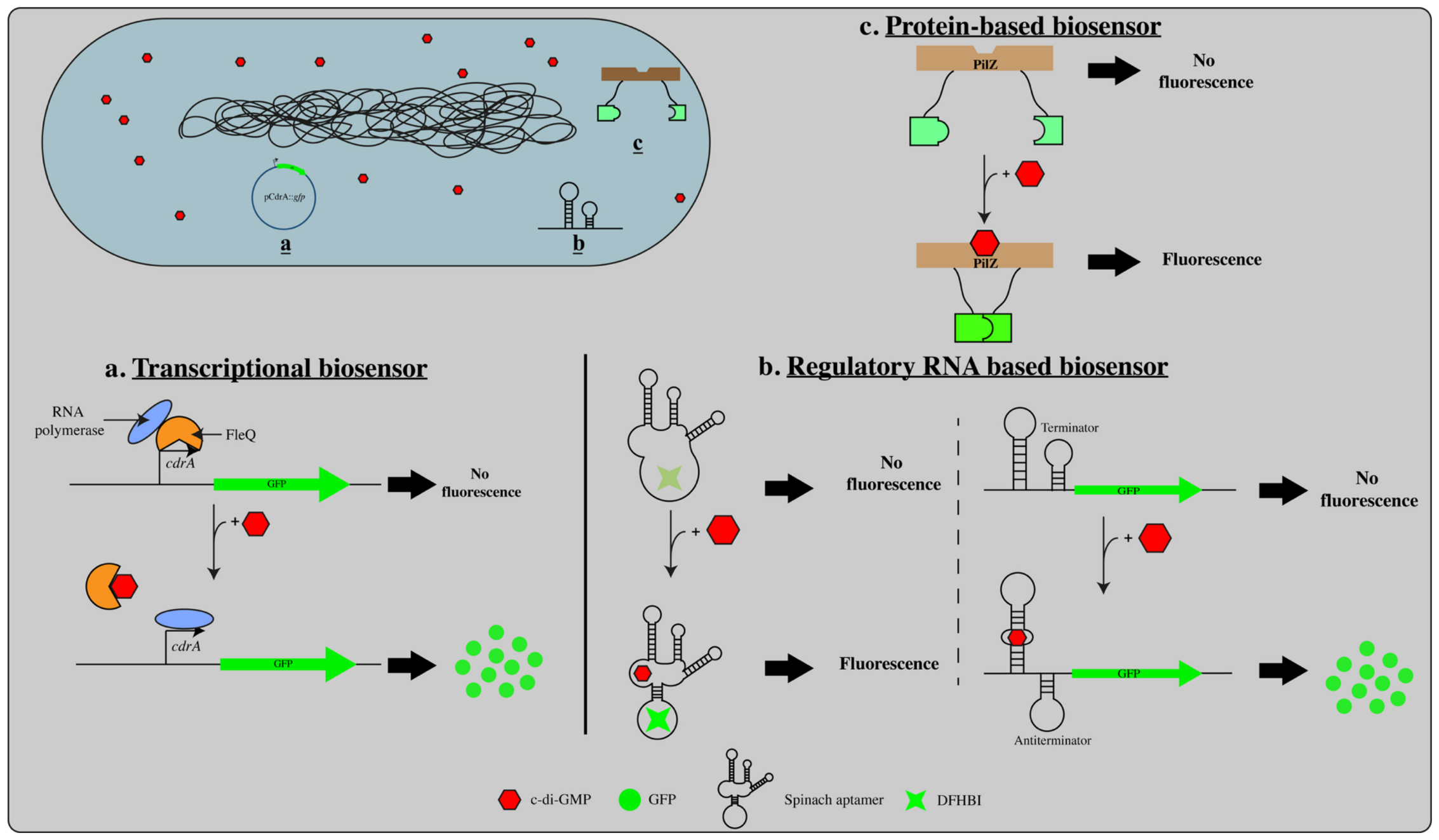
Figure 1. Schematic overview of the different biosensor-based techniques used to monitor c-di-GMP in bacterial cells. (a) The gene encoding the GFP fluorescent protein is placed after the c-di-GMP-dependent promoter of the cdrA gene. When c-di-GMP is produced, the affinity between the cdrA promoter and the FleQ repressor is lifted, allowing the production of the GFP fluorescent molecule. (b) This method involves an interaction between c-di-GMP and an aptamer structure. When c-di-GMP binds to the aptamer, it induces a conformational change in the RNA structure resulting in activation or repression of fluorescence through transcriptional or translational control, or via fluorogenic RNA aptamers. (c) c-di-GMP binding to PilZ protein promotes protein–protein interactions or conformational changes that enable fluorescence signal generation. Red dots represent c-di-GMP molecules, while green signals indicate fluorescence output, illustrating signal-off and signal-on states for each biosensing strategy.
Applmicrobiol 06 00105 g001
Figure 2. Experimental workflow for c-di-GMP extraction methods from bacterial cultures cells. Various experimental pathways for c-di-GMP extraction are described in the literature. Following cell centrifugation, the bacterial cell pellet could be treated through different extraction protocols with specific instructions (various solvent and treatment) allowing obtention of metabolites for downstream analytical analyses.
Applmicrobiol 06 00105 g002
Figure 3. MS/MS fragmentation scheme of c-di-GMP molecule. Ion fragmentation ESI-MS [M + H]+ by single glucosidic and double phosphodiester bond cleavage from c-di-GMP molecular ion at m/z 691 (adapted from [75]).
Applmicrobiol 06 00105 g003
Disclaimer/Publisher’s Note: The statements, opinions and data contained in all publications are solely those of the individual author(s) and contributor(s) and not of MDPI and/or the editor(s). MDPI and/or the editor(s) disclaim responsibility for any injury to people or property resulting from any ideas, methods, instructions or products referred to in the content.