Article

Heat Survival of Klebsiella pneumoniae in Infant Formula: The Role of clpC Heat Shock Resistance Genes


by
Mohamed T. Saad
1,*,
Nadia E. Sifennasr
2,
Mahmoud B. Agena
3,
Khaled M. Ibrahim
1,
Ahmed A. Zaghdani
1,
Abdlrhman M. Alsonosi
4,
Aya M. Saad
5,
Bassam A. Elgamoudi
6,* and
Stephen J. Forsythe
7
1
Microbiology Department, Libyan Biotechnology Research Centre, Tripoli P.O. Box 30313, Libya
2
Department of Medical Microbiology and Immunology, Faculty of Medicine, University of Tripoli, Tripoli P.O. Box 12456, Libya
3
Microbiology Department, Libyan Medical Research Centre, Azzawia P.O. Box 20311, Libya
4
Department of Medical Microbiology, Faculty of Medicine, Sebha University, Sebha P.O. Box 1000, Libya
5
School of Science & Technology, Nottingham Trent University, Nottingham NG1 4FQ, UK
6
Institute for Biomedicine and Glycomics, Griffith University, Southport, QLD 4215, Australia
7
Stephen J. Forsythe FoodMicrobe.com Ltd., Nottingham NG12 5GY, UK
*
Authors to whom correspondence should be addressed.
Appl. Microbiol. 2026, 6(5), 63; https://doi.org/10.3390/applmicrobiol6050063

Submission received: 9 April 2026 / Revised: 8 May 2026 / Accepted: 12 May 2026 / Published: 15 May 2026

Abstract

Klebsiella pneumoniae is a member of the six highly virulent and antibiotic-resistant bacterial pathogens group (ESKAPE) and poses a significant threat to public health due to its ability to cause both hospital and community-acquired infections. Recent health concerns have emerged about heat-tolerant bacterial contamination in hospital settings, particularly those associated with infant formula preparation. This study aims to evaluate the heat survival of 10 clinical K. pneumoniae strains in infant formula and to investigate the correlation between heat tolerance and the presence of heat shock resistance genes, particularly the clp family of ATPases. Ten strains of K. pneumoniae were exposed to heat at 55 °C for 30 min in infant formula. We assessed their survival rates and determined their D-values. Additionally, we screened for the presence of clpC family genes across representative strains. A wide variation in heat tolerance was observed among the strains. Strain 1701 (ST247, capsular antigen profile O3:K1) exhibited the highest heat tolerance, with a D-value of 12.9 min at 55 °C. The other strains exhibited moderate-to-low heat tolerance. Notably, strain 1701 was the only one that contained the clpC2 gene, suggesting a potential association between the clp gene family and heat resistance. Our results indicate that specific heat shock resistance genes, such as clpC2, may be associated with enhanced heat tolerance observed in K. pneumoniae strains. These findings highlight the potential role of heat shock proteins in bacterial persistence within neonatal healthcare environments.

1. Introduction

The nasogastric enteral feeding tube (NGET) has been identified as a potential niche for bacterial colonization, serving as a reservoir for pathogenic microorganisms that may alter the composition of the neonatal intestinal microbiome [1]. According to reports from the Food and Agriculture Organization (FAO) and World Health Organization (WHO), non-sterile powdered infant formula (PIF) is a significant source of bacterial contamination that can cause severe illness in infants under 1 year of age [2]. Members of the Enterobacteriaceae family isolated from PIF include Klebsiella pneumoniae, Salmonella spp., Escherichia coli, Enterobacter hormaechei, Cronobacter spp., and Citrobacter freundii [3,4]. Numerous studies have shown that approximately 76% of NGETs from neonatal intensive care units harbour biofilms, and bacteria can develop biofilms on non-living surfaces such as glass and plastic. In addition, extensive evidence shows that biofilm formation also occurs on a range of medical devices, including catheters, ventilator tubing, and enteral feeding tubes. The presence of biofilms on these medical devices has been linked to hospital-acquired infections (HAIs), which can result in prolonged hospital stays and severe, debilitating infections [1,5,6,7,8]. These biofilms harbored complex microbial communities, predominantly composed of Enterobacteriaceae species, including the clinically significant opportunistic pathogen K. pneumoniae. This organism is implicated in approximately 75% of both nosocomial and community-acquired infections, including septicemia, urinary tract infections, gastrointestinal disorders, and pneumonia [9]. K. pneumoniae is currently regarded as the second most prevalent pathogenic species within the Enterobacteriaceae family, following E. coli, which remains the primary cause of bacteraemia, septicaemia, and urinary tract infections (UTIs) [10,11]. K. pneumoniae demonstrates a greater capacity for transmission within hospital settings compared to E. coli. It is frequently implicated in both nosocomial and community-acquired infections, with particularly severe impacts observed in neonates and elderly populations [12,13].
K. pneumoniae has been identified as one of the most frequent causes of outbreaks reported in neonatal intensive care units [14]. Consequently, it is a leading cause of neonatal sepsis death worldwide [15]. Premature infants, defined as those born before 37 weeks of gestation, frequently experience complications related to feeding, respiration, and heightened susceptibility to infections. Consequently, they require specialized care within neonatal intensive care units (NICUs) [16]. Due to developmental limitations such as underdeveloped neurological function and respiratory difficulties, oral feeding is often contraindicated; thus, enteral nutrition via nasogastric or other feeding tubes is commonly employed. To mitigate microbial contamination risks in PIF, the WHO recommends strict hygiene practices during preparation, including the use of water at temperatures exceeding 70 °C and minimising storage at 4 °C [17].
The public health implications of K. pneumoniae are significant due to its opportunistic pathogenicity across diverse environments. Its virulence is attributed to multiple factors, including siderophore-mediated iron acquisition systems, polysaccharide capsule production, and its ability to adhere to medical surfaces and devices [18,19]. The K. pneumoniae capsule contributes to phagocytosis and evasion of the complement cascade, and capsular serotypes vary in their serum resistance. For example, K1, K2, and K5 are highly serum-resistant and associated with hypervirulent strains [18]. The hypermucoviscous strains can cause invasive infections even in healthy individuals. K. pneumoniae has evolved multiple adaptive strategies that enable it to withstand adverse environmental conditions. Among the various adaptive mechanisms, the utilisation of caseinolytic proteases (Clp ATPases), which are highly conserved across bacterial species, plays a crucial role in mediating stress tolerance and contributing to virulence across a wide range of pathogenic bacteria. It is implicated in the regulation of autolysis and is essential for bacterial growth under adverse environmental conditions, including heat shock [20]. These molecular chaperones are essential for maintaining protein quality control and cellular homeostasis, enabling bacteria to respond effectively to both intracellular and extracellular stressors. Consequently, they contribute to enhanced pathogenic potential and represent a significant clinical concern for patients. The ATP-dependent caseinolytic protease ClpCP is composed of two main components: the ATPase specificity factor ClpC, a member of the HSP100/Clp family, and the proteolytic subunit ClpP, which belongs to a distinct family of serine proteases. The genes encoding ClpC and ClpP are classified as class III heat shock genes and are negatively regulated by the class III stress gene repressor CtsR. The presence of ClpC, ClpP, and other Clp proteases is fundamental for bacterial survival under stress conditions, as mutations in Clp genes often result in impaired growth or inability to survive in stressful environments [20,21,22].
In light of these concerns, the present study aims to systematically evaluate the thermal tolerance of K. pneumoniae strains in reconstituted powdered infant formula, a food matrix of particular relevance to neonatal health. Specifically, this work seeks to quantify the survival of clinically relevant K. pneumoniae isolates following exposure to elevated temperatures and to investigate the association between observed heat resistance phenotypes and the presence of heat shock response-related genes identified through whole-genome sequencing, with particular emphasis on members of the clp ATPase gene family. The strains examined in this study were previously isolated from hospital environments, including neonatal nasogastric feeding tubes, thereby providing clinically and epidemiologically relevant insight into the potential persistence of K. pneumoniae in neonatal care settings.

2. Materials and Methods

2.1. Bacterial Strains and Growth Conditions

A total of 10 K. pneumoniae strains were selected for this study [23,24], each representing different sequence types (STs) and isolated from various sources, including neonatal nasogastric feeding tubes (NGETs), from two Jordanian hospitals (1681, 1699, 1701, 1725 and 1734), and Nottingham NGETs isolates (453 and 49) and sepsis isolates (2291, 2298, and 2312) were selected (Table 1). The strains were cultured in Luria–Bertani (LB) broth (Oxoid ThermoFischer, Basingstoke, UK) at 37 °C with shaking. In addition, whole-genome sequencing was used to determine the strains’ ST, O antigen, and K type.

2.2. Heat Tolerance Assay

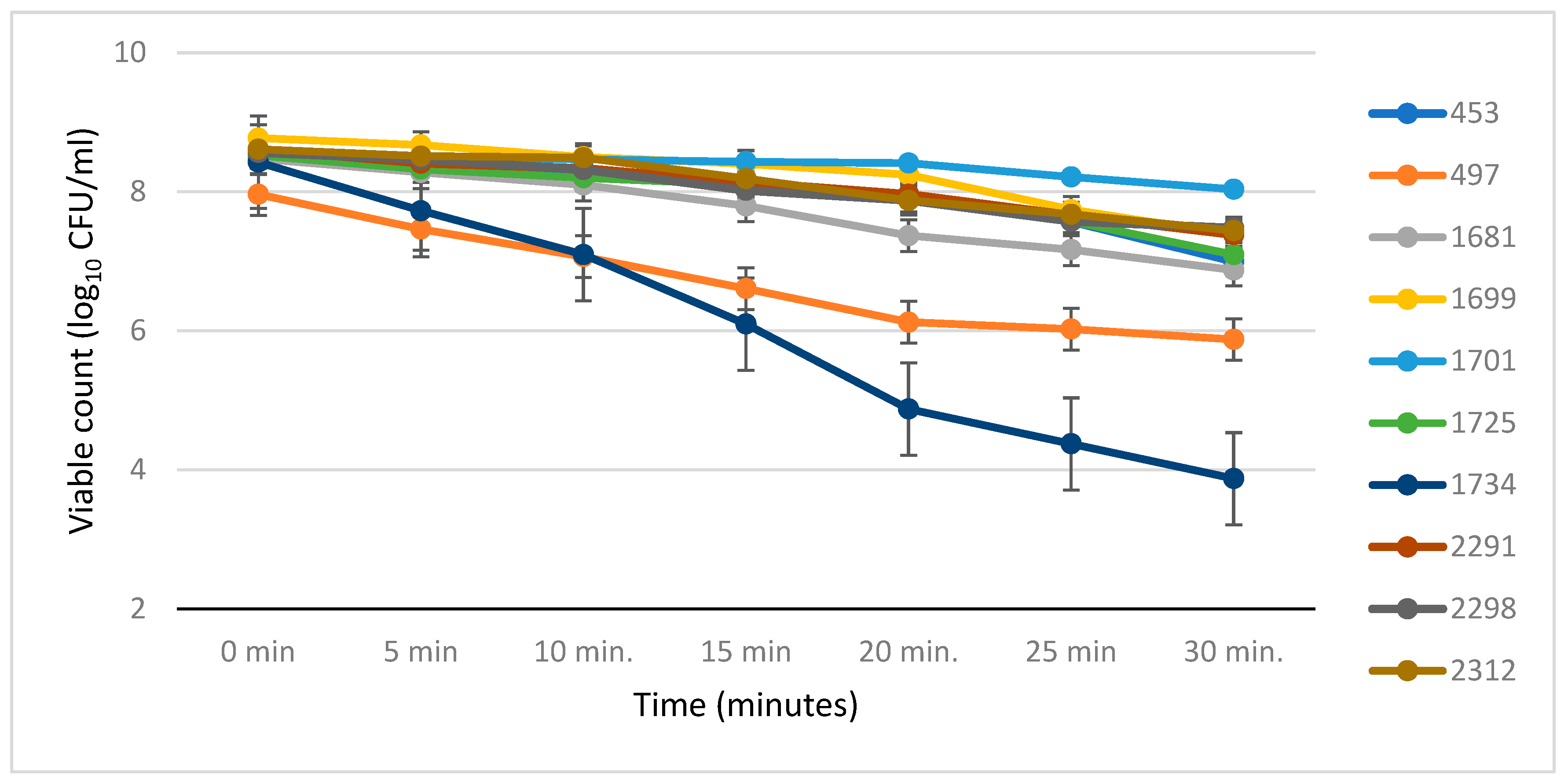
In the present study, 10 Klebsiella pneumoniae isolates were assessed for heat tolerance at 55 °C across seven time points (0, 5, 10, 15, 20, 25, and 30 min). The experiment aimed to identify strains capable of surviving and proliferating at elevated temperatures. Overnight cultures grown aerobically at 37 °C were adjusted to an optical density (OD600) of 0.1, after each 1 mL aliquot was inoculated into reconstituted powdered infant formula (Cow & Gate Premium 1) and incubated in a shaker water bath at 55 °C. At predetermined intervals, samples were withdrawn, serially diluted, and plated on LB agar to enumerate viable cells. The initial viable counts of the isolates ranged from 8.40 to 8.74 log10 CFU/mL (Figure 1) and (Table S1). Survivor counts at 55 °C were plotted as a function of time, and D-values were calculated by extrapolating the best-fit regression line (D = −1/slope), as described by Breeuwer et al. [25]. Each value represents the mean of three independent experiments, and the standard deviations of the D-values were calculated. Heat tolerance was evaluated following the methodology outlined by Breeuwer et al. [25].

2.3. Detection of Clp ATPase Genes

Genomic DNA was extracted from each strain using the GeneEluteTM kit (NA2110-1KT, Sigma, Hertfordshire, UK). The presence of Clp ATPase genes (clpC, clpB, clpA, and clpP) was determined by whole-genome sequencing, The identification of clp genes was based on a minimum sequence identity of 90% and coverage of at least 80% relative to reference sequences. Genomes were sequenced on an Illumina MiSeq (Illumina, San Diego, CA, USA). This comparative analysis was performed using WebACT comparative tool, Artemis Comparative Tool (ACT) for genome alignment [26]. In addition, BLAST (Version 2.6) searches were performed using NCBI BLAST research facility at https://blast.ncbi.nlm.nih.gov/Blast.cgi?PROGRAM=blastn&PAGE_TYPE=BlastSearch&LINK_LOC=blasthome (accessed on 15 June 2017).

3. Results

The thermal resistance of neonatal K. pneumoniae isolates at 55 °C was evaluated using a protocol adapted from Breeuwer et al. [25]. Bacterial survival at this temperature was measured over time, and decimal reduction times (D-values) were calculated accordingly. Based on the obtained D55 values, isolates were classified into three categories: heat sensitive (D55 ≤ 4 min), moderately tolerant (4.5 ≤ D55 ≤ 9 min), and highly heat tolerant (D55 ≥ 10 min). As illustrated in (Figure 1, Table 2 and Table S1), substantial variability in thermal tolerance was observed among the tested K. pneumoniae strains.
Table 1. K. pneumoniae isolates selected for the current study.
IsolateSTO-AntigenK-TypeHospitalSourceInfant Formula Feed
453105O4K5NCHNG tube biofilmNA
497147O12K5NCHNG tube biofilmNA
1681111O1K2PRHNG tube lumenBebelac
1699247O3K1KAHNG tube lumenNeosure
1701247O3K1KAHNG tube biofilmNeosure
1725111O12K2PRHNG tube lumenBebelac
1734526O2NDKAHNG tube biofilmNeosure
229135NDNDQMCSepsis caseNA
229835O8NDQMCSepsis caseNA
231234O2NDQMCSepsis caseNA
KAH = King Abdallah Hospital (Irbid—Jordan). PRH = Princes Rahma Hospital (Irbid—Jordan). QMC = Queen Medical Centre hospital (UK). NCH = Nottingham City hospital (UK). NG = Nasogastric. NA = Information not available. ST = Sequence type.
Figure 1. Survival curves of neonatal K. pneumoniae strains in infant formula at 55 °C. The thermal resistance of neonatal Klebsiella pneumoniae isolates was evaluated at 55 °C using a protocol adapted from Breeuwer et al. [25]. Bacterial survival was monitored over time, and decimal reduction times (D-values) were calculated. Data are presented as mean ± standard deviation of the recovered cell counts from three independent experiments, with error bars representing 95% confidence intervals.
Applmicrobiol 06 00063 g001
Table 2. Thermotolerance of neonatal K. pneumoniae in infant formula at 55 °C.
Isolate17012298229123121725169945316814971734
D-value (min)12.95.154.84.64.54.13.62.81.2
Heat toleranceHMMMMMSSSS
H = High, M = Moderate and S = Susceptible.
Strain 1701 (ST247-O3-K1), recovered from a neonatal nasogastric tube, exhibited the greatest heat resistance, with a D55 value of 12.9 min in liquid infant formula. In contrast, strains 2298 (ST35-O2-ND), 2291 (ST35-ND-ND), 2312 (ST34-O2-ND), 1725 (ST247-O12-K2), and 1699 (ST247-O3-K1) demonstrated intermediate thermal tolerance, maintaining viability for up to 20 min before a gradual decline was observed (D55 values of 5.1, 5.0, 4.8, 4.6, and 4.5 min, respectively). Conversely, strains 453 (ST105-O4-K5), 1681 (ST111-O1-K2), 497 (ST147-O12-K5), and 1734 (ST526-O2-ND) were highly susceptible to heat exposure, with D55 values below 4.5 min (4.1, 3.6, 2.8, and 1.2 min, respectively).
As shown in Table 2, the thermotolerance of neonatal K. pneumoniae in infant form was measured at 55 °C. Isolates were categorised into three groups based on thermal tolerance using decimal reduction times (D-values): heat sensitive (S), moderately heat-tolerant (M), and highly heat-tolerant (H).
Recent studies have identified a genetic locus (clpC ATPase) that could be responsible for heat tolerance, and it has been recognised in specific isolates such as E. coli, C. sakazakii and K. pneumoniae [27,28,29,30,31]. They suggested that the locus clpC ATPase plays an important role in the survival and persistence of K. pneumoniae in certain stressful hospital environments, such as hot water used to reconstitute powdered infant formula. As shown in Table 3, there was a clear variation among strains in the presence/absence of clpC ATPase family genes. Interestingly, strain 1701, the most heat-resistant strain, was the only strain with clpC2 gene, whereas strains 497 and 1734 each had 3 Clp genes missing and had the lowest D55 values.

4. Discussion

In recent decades, the emergence of heat-tolerant bacterial contaminants has become a growing concern for food safety, particularly in high-risk products such as powdered infant formula (PIF) [27]. Although many members of the Enterobacteriaceae family can acquire transient heat tolerance following sublethal stress, only a limited subset exhibits stable, intrinsic thermotolerance [30,31]. This distinction is critical, as current preparation guidelines assume that exposure to approximately 55 °C is sufficient to eliminate most pathogens [17]. However, evidence from Iversen and colleagues challenges this assumption, showing that certain Enterobacteriaceae can survive at this temperature [32]. This discrepancy reveals a potentially important limitation in existing safety recommendations. Moreover, the ability of Cronobacter sakazakii to grow across a wide temperature range (5–45 °C) and persist on feeding equipment further increases this risk, particularly in neonatal intensive care units (NICUs), where at-risk groups are exposed [32]. Reports from WHO and FAO reinforce this concern, indicating that variations from optimal preparation practices, such as reconstitution at lower temperatures or prolonged storage at room temperature, can facilitate bacterial survival and proliferation [17]. Taken together, these results indicate that current handling methods may not fully account for the adaptive capacity of certain pathogens, thereby calling for a more subtle understanding of bacterial heat resistance.
Within this wider context, the present study provides important knowledge of the thermotolerance of K. pneumoniae in rehydrated infant formula. The observed heterogeneity in heat resistance among isolates is especially remarkable, as it indicates that thermotolerance is not a uniform species-level trait, but rather a highly strain-dependent characteristic. The identification of three distinct tolerance groups, ranging from highly resistant (D55 = 12.9 min) to clearly heat-sensitive (D55 = 1.2–4.1 min), suggests that certain strains may be capable of surviving standard preparation conditions, while others are effectively inactivated. This variation has direct practical implications: even if guidelines are followed, the presence of highly tolerant strains could still pose a risk. Thus, the data not only confirm previous concerns about heat resistance but additionally extend them by demonstrating the extent of intra-species variability.
A key observation from this study is the absence of a clear relationship between thermotolerance and commonly used typing characteristics, including sequence type and capsule composition (O- and K-antigens). This lack of correlation is especially clear in the comparison between isolates 1701 and 1699, which are the same sequence type (ST247) yet exhibit markedly different heat resistance profiles. Such divergence strongly suggests that standard classification methods do not adequately capture the determinants of thermal resistance. This has important effects for both epidemiological tracking and risk assessment, as it indicates that strains cannot be reliably categorised as high- or low-risk based solely on genotype. Instead, thermotolerance appears to be governed by additional, possibly subtle, genetic or regulatory factors that remain uncharacterised.
In light of this, previous hypotheses linking capsular serotype and O-antigen composition to thermotolerance warrant reconsideration [33]. Although these surface structures may contribute to environmental resilience, their role appears insufficient to account for the patterns observed in this study. Rather, the data point towards the involvement of more complex stress response systems. One plausible mechanism is the contribution of chaperone proteins, particularly those associated with the Clp ATPase system. A thermotolerance-associated locus has been identified across multiple Enterobacteriaceae species, including E. coli, C. sakazakii, and K. pneumoniae [29,30,31,34], denoting a conserved functional role.
Among these, the clpC gene is of particular interest. Originally characterised in Danish clinical isolates of K. pneumoniae, this gene encodes a component of the ATP-dependent protease machinery, which is involved in protein quality control under stress conditions. Its prevalent distribution among Gram-negative bacteria and its presence in a large proportion of clinical isolates support the notion that it plays a central role in habitat adaptation. Importantly, the ability of multidrug-resistant K. pneumoniae to persist in contaminated endoscopes provides real-world evidence of this flexible capacity, linking heat tolerance to persistence in clinical environments. Furthermore, the apparent horizontal transfer of the clpC locus across Proteobacteria demonstrates its evolutionary advantage and raises the possibility that thermotolerance traits may be disseminated between strains and species [35,36,37].
Taken together, the findings of this study reinforce the concept that thermotolerance in K. pneumoniae is a multifactorial and strain-specific trait. More importantly, they emphasise a critical gap between current food safety practices and the biological realities of bacterial adaptation. While existing guidelines are based on general assumptions about pathogen susceptibility, the presence of highly tolerant strains challenges their universal effectiveness. This highlights the need for a more evidence-based approach that incorporates strain-level variability into risk assessments. Subsequent work should therefore focus on identifying the genetic and regulatory determinants of heat resistance and evaluating whether current preparation guidelines require revision to ensure adequate safeguarding of vulnerable populations. The present study is limited by a relatively small sample size (n = 10), which, while appropriate for a preliminary investigation aimed at identifying initial trends, may restrict statistical power and generalizability. Future studies incorporating larger, more diverse isolate collections and functional validation of stress response determinants are warranted to confirm and extend these findings and to better inform risk assessment and control strategies for neonatal infections associated with heat-resistant K. pneumoniae.

5. Conclusions

Heat resistance in K. pneumoniae strains, such as 1701, poses a health risk in neonatal intensive care units (NICUs), where powdered infant formula (PIF) is reconstituted at 55 °C since this temperature may not eliminate all pathogens. This study highlights the potential importance of Clp ATPase genes, particularly clpC2, in the heat tolerance of K. pneumoniae strains in rehydrated infant formula. The variability in heat tolerance observed among the strains emphasizes the complexity of bacterial adaptation mechanisms to thermal stress. Understanding these mechanisms is crucial for developing strategies to mitigate the risk of neonatal infections associated with K. pneumoniae.

Supplementary Materials

The following supporting information can be downloaded at: https://www.mdpi.com/article/10.3390/applmicrobiol6050063/s1, Table S1: Summary of the viable count (Log10 CFU/mL) of neonatal Klebsiella pneumoniae isolates in infant formula held at 55 °C at different time points.

Author Contributions

M.T.S. and S.J.F. conceived and designed the study. K.M.I., M.B.A., N.E.S., A.A.Z. and M.T.S. conducted the research. Experimental work was conducted by A.M.A., A.M.S. and M.T.S. Data analysis was performed by M.T.S., K.M.I., B.A.E. and N.E.S. All authors contributed to revising the manuscript, reviewed and approved the final version, and accept full responsibility for the integrity and accuracy of the work. All authors have read and agreed to the published version of the manuscript.

Funding

The authors declare that this research received no external funding.

Institutional Review Board Statement

Not applicable.

Informed Consent Statement

Not applicable.

Data Availability Statement

The datasets used and/or analyzed during the current study are available from the corresponding author upon request. We have also included some of the raw data in the Supplementary Materials for clarity.

Conflicts of Interest

Author Stephen J. Forsythe was employed by Foodmicrobe.com Ltd. The remaining authors declare that the research was conducted without any commercial or financial relationships that could be construed as a potential conflict of interest.

References

  1. Vongbhavit, K.; Salinero, L.K.; Kalanetra, K.M.; Masarweh, C.; Yu, A.; Taft, D.H.; Mills, D.A.; Underwood, M.A. A comparison of bacterial colonization between nasogastric and orogastric enteral feeding tubes in infants in the neonatal intensive care unit. J. Perinatol. 2022, 42, 1446–1452. [Google Scholar] [CrossRef] [PubMed]
  2. FAO/WHO. 10 Informe de reunión. Enterobacter sakazakii and Salmonella in Powdered Infant Formula 2006. Available online: http://www.fao.org/3/a-a0707e.pdf (accessed on 21 March 2026).
  3. Fusi, V.; Stella, S.; Bernardi, C.; Tirloni, E. Microbiological characteristics of powdered infant and follow-on formulae and safety concerns: A review. Heliyon 2025, 11, e42927. [Google Scholar] [CrossRef]
  4. Alsonosi, A.M.; Ibrahim, K.M.; Elgamoudi, B.A.; Agena, M.B.; Forsythe, S.J. The Potential role of rpoS and ompR in the acid resistance and desiccation tolerance of Cronobacter malonaticus strains. Microbiol. Res. 2025, 16, 53. [Google Scholar] [CrossRef]
  5. Dhiman, S.; Kumar, A.; Kaur, G.; Mukherjee, G.; Rustagi, S.; Shreaz, S.; Negi, R.; Yadav, A.N. Bacterial biofilms: Pathogenesis, monitoring, treatment approaches and associated challenges. Biologia 2024, 79, 3161–3181. [Google Scholar] [CrossRef]
  6. Jamal, M.; Ahmad, W.; Andleeb, S.; Jalil, F.; Imran, M.; Nawaz, M.A.; Hussain, T.; Ali, M.; Rafiq, M.; Kamil, M.A. Bacterial biofilm and associated infections. J. Chin. Med. Assoc. 2018, 81, 7–11. [Google Scholar] [CrossRef]
  7. Grari, O.; Ezrari, S.; El Yandouzi, I.; Benaissa, E.; Ben Lahlou, Y.; Lahmer, M.; Saddari, A.; Elouennass, M.; Maleb, A. A comprehensive review on biofilm-associated infections: Mechanisms, diagnostic challenges, and innovative therapeutic strategies. Microbe 2025, 8, 100436. [Google Scholar] [CrossRef]
  8. Ibrahim, K.M.; Alsonosi, A.M.; Agena, M.B.; Elgamoudi, B.A.; Forsythe, S.J. Multiplex determination of K-Antigen and colanic Acid capsule variants of Cronobacter sakazakii. Genes 2024, 15, 1282. [Google Scholar] [CrossRef]
  9. Kanevsky-Mullarky, I.; Nedrow, A.; Garst, S.; Wark, W.; Dickenson, M.; Petersson-Wolfe, C.; Zadoks, R.N. Short communication: Comparison of virulence factors in Klebsiella pneumoniae strains associated with multiple or single cases of mastitis. J. Dairy Sci. 2014, 97, 2213–2218. [Google Scholar] [CrossRef] [PubMed]
  10. Vuotto, C.; Longo, F.; Balice, M.P.; Donelli, G.; Varaldo, P.E. Antibiotic resistance related to biofilm formation in Klebsiella pneumoniae. Pathogens 2014, 3, 743–758. [Google Scholar] [CrossRef]
  11. Xu, M.; Fu, Y.; Kong, H.; Chen, X.; Chen, Y.; Li, L.; Yang, Q. Bloodstream infections caused by Klebsiella pneumoniae: Prevalence of blaKPC, virulence factors and their impacts on clinical outcome. BMC Infect. Dis. 2018, 18, 358. [Google Scholar] [CrossRef] [PubMed]
  12. Chung, D.; Lee, S.; Lee, H.; Kim, H.; Choi, H.; Eom, J.; Kim, J.; Choi, Y.; Lee, J.; Chung, M.; et al. Emerging invasive liver abscess caused by K1 serotype Klebsiella pneumoniae in Korea. J. Infect. 2007, 54, 578–583. [Google Scholar] [CrossRef]
  13. Hennequin, C.; Robin, F. Correlation between antimicrobial resistance and virulence in Klebsiella pneumoniae. Eur. J. Clin. Microbiol. Infect. Dis. 2015, 35, 333–341. [Google Scholar] [CrossRef]
  14. Priante, E.; Minotti, C.; Contessa, C.; Boschetto, M.; Stano, P.; Bello, F.D.; De Canale, E.; Lolli, E.; Baldo, V.; Baraldi, E.; et al. Successful control of an outbreak by Phenotypically Identified Extended-Spectrum Beta-Lactamase–Producing Klebsiella pneumoniae in a Neonatal Intensive Care Unit. Antibiotics 2022, 11, 1649. [Google Scholar] [CrossRef]
  15. Hetta, H.F.; Alanazi, F.E.; Ali, M.A.S.; Alatawi, A.D.; Aljohani, H.M.; Ahmed, R.; Alansari, N.A.; Alkhathami, F.M.; Albogmi, A.; Alharbi, B.M.; et al. Hypervirulent Klebsiella pneumoniae: Insights into Virulence, Antibiotic Resistance, and Fight Strategies Against a Superbug. Pharmaceuticals 2025, 18, 724. [Google Scholar] [CrossRef] [PubMed]
  16. American Academy of Pediatrics. Common Conditions, Concerns, and Equipment in the NICU. NICU J. A Parent’s Journey 2017, 99–114. [Google Scholar] [CrossRef]
  17. Commission Codex Alimentarius. Code of Hygienic Practice for Powdered Formulae for Infants and Young Children. CAC/RCP 66. Italy: Joint FAO/WHO Food Standards Programme. Available online: http://www.codexalimentarius.org/standards/list-ofstandards/en/?provide=standards&orderField=fullReference&sort=asc&num1=CAC/RCP (accessed on 13 April 2014).
  18. Brisse, S.; Passet, V.; Grimont, P.A.D. Description of Klebsiella quasipneumoniae sp. nov., isolated from human infections, with two subspecies, Klebsiella quasipneumoniae subsp. quasipneumoniae subsp. nov. and Klebsiella quasipneumoniae subsp. similipneumoniae subsp. nov., and demonstration that Klebsiella singaporensis is a junior heterotypic synonym of Klebsiella variicola. Int. J. Syst. Evol. Microbiol. 2014, 64, 3146–3152. [Google Scholar] [CrossRef]
  19. Cubero, M.; Marti, S.; Domínguez, M.Á.; González-Díaz, A.; Berbel, D.; Ardanuy, C. Hypervirulent Klebsiella pneumoniae serotype K1 clinical isolates form robust biofilms at the air-liquid interface. PLoS ONE 2019, 14, e0222628. [Google Scholar] [CrossRef]
  20. Ah Young, A.P.; Koehl, A.; Cascio, D.; Egea, P.F. Structural mapping of the ClpB ATPases of Plasmodium falciparum: Targeting protein folding and secretion for antimalarial drug design. Protein Sci. 2015, 24, 1508–1520. [Google Scholar] [CrossRef] [PubMed]
  21. Park, S.; Kwon, H.; Tran, T.D.; Choi, M.; Jung, S.; Lee, S.; Briles, D.E.; Rhee, D. ClpL is a chaperone without auxiliary factors. FEBS J. 2015, 282, 1352–1367. [Google Scholar] [CrossRef]
  22. Zolkiewski, M.; Zhang, T.; Nagy, M. Aggregate reactivation mediated by the Hsp100 chaperones. Arch. Biochem. Biophys. 2012, 520, 1–6. [Google Scholar] [CrossRef]
  23. Hurrell, E.; Kucerova, E.; Loughlin, M.; Caubilla-Barron, J.; Hilton, A.; Armstrong, R.; Smith, C.; Grant, J.; Shoo, S.; Forsythe, S. Neonatal enteral feeding tubes as loci for colonisation by members of the Enterobacteriaceae. BMC Infect. Dis. 2009, 9, 146. [Google Scholar] [CrossRef]
  24. Abudalla, H.; Lane, C. Neonatal Enteral Feeding Tube as Loci for Enterobacteriaceae Colonisation and Risk to Neonatal Health. 2014. Available online: https://irep.ntu.ac.uk/id/eprint/42/7/220453_Halema_Abudalla.pdf (accessed on 17 April 2016).
  25. Breeuwer, P.; Lardeau, A.; Peterz, M.; Joosten, H.M. Desiccation and heat tolerance of Enterobacter sakazakii. J. Appl. Microbiol. 2003, 95, 967–973. [Google Scholar] [CrossRef]
  26. Carver, T.J.; Rutherford, K.M.; Berriman, M.; Rajandream, M.-A.; Barrell, B.G.; Parkhill, J. ACT: The Artemis comparison tool. Bioinformatics 2005, 21, 3422–3423. [Google Scholar] [CrossRef]
  27. Guragain, M.; Brichta-Harhay, D.M.; Bono, J.L.; Bosilevac, J.M. Locus of Heat Resistance (LHR) in Meat-Borne Escherichia coli: Screening and Genetic Characterization. Appl. Environ. Microbiol. 2021, 87, e02343-20. [Google Scholar] [CrossRef]
  28. Ma, A.; Glassman, H.; Chui, L. Characterization of Escherichia coli possessing the locus of heat resistance isolated from human cases of acute gastroenteritis. Food Microbiol. 2020, 88, 103400. [Google Scholar] [CrossRef]
  29. Gajdosova, J.; Benedikovicova, K.; Kamodyova, N.; Tothova, L.; Kaclikova, E.; Stuchlik, S.; Turna, J.; Drahovska, H. Analysis of the DNA region mediating increased thermotolerance at 58 °C in Cronobacter sp. and other enterobacterial strains. Antonie Leeuwenhoek 2011, 100, 279–289. [Google Scholar] [CrossRef] [PubMed]
  30. Mercer, R.G.; Zheng, J.; Garcia-Hernandez, R.; Ruan, L.; Gänzle, M.G.; McMullen, L.M. Genetic determinants of heat resistance in Escherichia coli. Front. Microbiol. 2015, 6, 932. [Google Scholar] [CrossRef]
  31. Bojer, M.S.; Struve, C.; Ingmer, H.; Hansen, D.S.; Krogfelt, K.A. Heat Resistance Mediated by a New Plasmid Encoded Clp ATPase, ClpK, as a Possible Novel Mechanism for Nosocomial Persistence of Klebsiella pneumoniae. PLoS ONE 2010, 5, e15467. [Google Scholar] [CrossRef]
  32. Iversen, C.; Lane, M.; Forsythe, S.J. The growth profile, thermotolerance and biofilm formation of Enterobacter sakazakii grown in infant formula milk. Lett. Appl. Microbiol. 2004, 38, 378–382. [Google Scholar] [CrossRef] [PubMed]
  33. Pan, Y.-J.; Lin, T.-L.; Chen, Y.-H.; Hsu, C.-R.; Hsieh, P.-F.; Wu, M.-C.; Wang, J.-T. Capsular Types of Klebsiella pneumoniae Revisited by wzc Sequencing. PLoS ONE 2013, 8, e80670. [Google Scholar] [CrossRef]
  34. Bojer, M.S.; Krogfelt, K.A.; Struve, C. The newly discovered ClpK protein strongly promotes survival of Klebsiella pneumoniae biofilm subjected to heat shock. J. Med. Microbiol. 2011, 60, 1559–1561. [Google Scholar] [CrossRef] [PubMed]
  35. Jørgensen, S.; Bojer, M.; Boll, E.; Martin, Y.; Helmersen, K.; Skogstad, M.; Struve, C. Heat-resistant, extended-spectrum β-lactamase-producing Klebsiella pneumoniae in endoscope-mediated outbreak. J. Hosp. Infect. 2016, 93, 57–62. [Google Scholar] [CrossRef]
  36. Lee, C.; Wigren, E.; Heinrich Lünsdorf Römling, U. Protein homeostasis—More than resisting a hot bath. Curr. Opin. Microbiol. 2016, 30, 147–154. [Google Scholar] [CrossRef]
  37. Taylor, G.; Cui, H.; Leodolter, J.; Giese, C.; Weber-Ban, E. ClpC2 protects mycobacteria against a natural antibiotic targeting ClpC1-dependent protein degradation. Commun. Biol. 2023, 6, 301. [Google Scholar] [CrossRef]
Table 3. Presence/absence of the thermo-tolerant family of Clp ATPase genes in K. pneumoniae isolates.
GeneClp ATPase Family
clpAclpB1clpB2clpC1clpC2clpP1clpP2clpSclpX
FunctionProtease ATP-Binding SubunitChaperon ProteinChaperon ProteinProtease ATP-Binding SubunitProtease ATP-Binding SubunitProteolytic Subunit PrecursorProteolytic Subunit PrecursorProtease Adaptor ProteinProtease ATP-Binding Subunit
Strain
1701++++++-++
2298++++-+-++
2291++++-+-++
2312++++-+-++
1725++++-+-++
1699++++-+-++
453++++-+-++
1681++++-+-++
497++++---++
1734++---++++
A comparative analysis to assess the presence/absence of clpC ATPase family genes was performed using the WebACT comparative tool and the Artemis Comparative Tool (ACT) for whole-genome alignment. In addition, BLAST searches were conducted using the NCBI BLAST platform.
Disclaimer/Publisher’s Note: The statements, opinions and data contained in all publications are solely those of the individual author(s) and contributor(s) and not of MDPI and/or the editor(s). MDPI and/or the editor(s) disclaim responsibility for any injury to people or property resulting from any ideas, methods, instructions or products referred to in the content.