Article

Optimized Decolorization of Methylene Blue by Bacillus cereus: A Genomic and Analytical Approach


by
Fatima Hamadeh
1,*,
Thibaut Armel Chérif Gnimadi
2,3,4,
Mano Joseph Mathew
3,4,
Charbel Al-Bayssari
5,
Mounir Kassir
6,
Rana El Hajj
1 and
Dalia El Badan
1,7,*
1
Department of Biological Sciences, Faculty of Science, Beirut Arab University, Beirut P.O. Box 11-5020, Lebanon
2
Centre de Recherche et de Formation en Infectiologie de Guinée (CERFIG), Conakry BP 6629P, Guinea
3
EFREI Research Lab, Panthéon Assas Université, 30–32 Avenue de la République, 94800 Villejuif, France
4
Laboratoire Génomique, Bioinformatique et Chimie Moléculaire, EA7528, Conservatoire National des Arts et Métiers, HESAM Université, 2 Rue Conté, 75003 Paris, France
5
Department of Medical Laboratory Sciences, Faculty of Health Sciences, University of Balamand, Tripoli P.O. Box 100, Lebanon
6
PRASE Research Laboratory, Lebanese University, Beirut P.O. Box 6573/14, Lebanon
7
Botany and Microbiology Department, Faculty of Science, Alexandria University, Alexandria 21511, Egypt
*
Authors to whom correspondence should be addressed.
Appl. Microbiol. 2026, 6(4), 52; https://doi.org/10.3390/applmicrobiol6040052
Submission received: 12 February 2026 / Revised: 29 March 2026 / Accepted: 31 March 2026 / Published: 8 April 2026

Abstract

Synthetic dyes, such as methylene blue (MB), constitute a major category of environmental pollutants due to their toxicity, persistence, and resistance to standard treatment methods. In this study, Bacillus cereus BC WW Saida was isolated from the heavily polluted Saida dumpsite in Lebanon and evaluated for its MB degradation efficiency. The isolate was identified through whole-genome sequencing, which revealed the presence of key enzymatic systems involved in azo dye degradation. Under optimized conditions, the strain achieved 82% decolorization, as determined by optical density measurements using a microplate reader. The process was further examined using High-Performance Liquid Chromatography (HPLC), which revealed a significant reduction in the original dye peak and the emergence of new intermediate products. These findings suggest the strong biodegradation capability of B. cereus BC WW Saida isolated from contaminated environments and highlight its potential application in the eco-friendly treatment of azo dye-contaminated wastewater.

1. Introduction

Synthetic dyes represent one of the most critical classes of pollutants discharged into the environment from various industrial activities, especially textile, leather, paper, and cosmetic manufacturing. The extensive and continuous use of these dyes in industrial processes has resulted in the discharge of considerable amounts of colored wastewater into natural water bodies, leading to serious environmental concerns worldwide [1]. Among these dyes, methylene blue (MB), an azo dye with a heterocyclic aromatic cationic structure, is commonly used in the textile industry as well as in several other industrial applications. Despite its wide-ranging applications in industry, MB poses considerable environmental risks due to its toxicity, mutagenic effects, and persistence in aquatic environments [2]. The presence of dyes in wastewater interferes with aquatic photosynthesis by restricting sunlight penetration into the water column and by generating reactive oxygen species, which can cause oxidative damage to aquatic organisms and disturb the natural ecological balance. Additionally, the accumulation of dyes in aquatic environments can affect microbial communities and reduce water quality, making the water unsuitable for domestic and agricultural use [3]. Furthermore, the complex aromatic structure of these dyes poses challenges for degradation through conventional biological treatment methods. Consequently, the development of effective, sustainable, and eco-friendly strategies for the elimination of these persistent pollutants from wastewater systems has become essential [4].
A variety of physicochemical techniques, including membrane filtration, adsorption onto activated carbon, coagulation–flocculation, and advanced oxidation processes, have been employed to eliminate azo dyes from wastewater [5]. These techniques are commonly applied in many industrial treatment systems because they are capable of removing dyes quickly and effectively under controlled conditions. However, despite their potential for high dye removal efficiency, these approaches exhibit significant disadvantages, including high operating costs, incomplete dye mineralization, and the production of secondary pollution in the form of sludge or toxic by-products [6]. In addition, these treatments often require advanced equipment, continuous chemical additions, and strict operational control, which can limit their practical implementation in developing regions. Moreover, these techniques require large amounts of energy and chemicals, making them frequently unsuitable for large-scale applications and long-term environmental sustainability. For this reason, microbial degradation has emerged as an efficient and sustainable substitute for azo dye treatment [3,7]. Various microorganisms, including bacteria, algae, and fungi, display enzymatic systems, such as oxidoreductases and oxidative enzymes, capable of converting azo dyes into non-toxic compounds. These biological systems are able to convert complex dye molecules into simpler intermediates that can be further metabolized through microbial pathways [8]. In particular, bacterial degradation has demonstrated promising outcomes because some bacterial species can utilize azo dyes as sources of carbon and nitrogen for their metabolic activities. Through the activity of azoreductase enzymes, bacteria can break down azo bonds, causing decolorization of dye molecules and reducing their visible coloration in aqueous environments. Then, they can further metabolize degradation products using oxidative pathways that contribute to the detoxification of these compounds [9]. Among the bacterial genera investigated for dye degradation, Bacillus has shown considerable promise for azo dye degradation because of its metabolic adaptability and capacity to endure a range of environmental circumstances. This bacterium efficiently breaks down azo dyes by using enzymes such as oxidoreductases, which oxidize phenolic compounds, and azoreductases, which cleave the azo bonds [10]. Research has shown that it can decolorize a variety of dyes while producing non-toxic metabolites and maintaining activity under different environmental conditions; for example, Bacillus sp. AZ28, which was isolated from industrial effluent, demonstrated decolorization efficiencies between 84% and 95% for dyes, including methyl orange and novacron red FN 3GF, over a period of 14 to 72 h under optimal conditions [11]. Similarly, B. cereus has also shown significant activity, achieving decolorization rates of 72.66% and 85% for Eriochrome Black T and Red 3BN [12].
Therefore, the strong bioremediation potential of B. cereus supports the potential use of Bacillus species as efficient and eco-friendly candidates for the treatment of synthetic dyes in wastewater systems. Moreover, the environment from which bacterial isolates are obtained significantly affects their degradation capabilities. Microorganisms isolated from contaminated environments are often better adapted to endure toxic compounds and complex mixtures of contaminants [13]. In this Study, B. cereus was isolated from the Saida dump in Lebanon, which is a region that is highly affected by pollutants and industrial waste. This site holds considerable environmental significance characterized by years of unmanaged waste disposal, resulting in complex pollutant mixtures including dyes, heavy metals, and organic contaminants [14]. Such harsh and pollutant-rich environments impose selective pressure on native microbial communities, leading to the development of bacteria with improved adaptation strategies, survival mechanisms, and high biodegradation capacities [15]. Microorganisms isolated from these environments may consequently exhibit enhanced metabolic pathways, allowing them to degrade recalcitrant pollutants more efficiently than strains derived from less contaminated sites [16]. We used a combination of whole-genome sequencing and High-Performance Liquid Chromatography to detect the presence of enzymes involved in the process of degradation and optical density reduction measurement over time. This study aims to evaluate the degradation potential of isolated B. cereus on MB, providing insights into its effectiveness as a bioremediation agent for azo dye-contaminated environments, and supporting the development of future sustainable wastewater treatment technologies.

2. Materials and Methods

2.1. Sample Collection

Samples of soil and waste were collected from different parts of the Saida dump, with a focus on visibly contaminated areas, such as regions heavily stained with dye residues or organic waste. They were isolated from a depth of 5–10 cm. All samples were immediately transported to the laboratory for isolation procedures to be performed.

2.2. Bacterial Isolation and Enrichment

A total of 1 g of each soil sample was suspended in 9 mL of sterile saline solution (0.85% NaCl) and vortexed for 5 min. The homogenized suspension was serially diluted up to 10−6. A 100 µL aliquot from each dilution was spread onto nutrient agar plates supplemented with MB (50 mg/L) to isolate dye-degrading microorganisms. Dye-free control plates were used to track overall microbial growth. The plates were incubated at 37 °C for 24–48 h [17].

2.3. Biochemical Characterization

The most efficient bacterial isolate, exhibiting the highest MB decolorization efficiency, was selected for subsequent characterization and analysis. For the initial identification, colony morphology, Gram staining, endospore formation, and motility test using the hanging drop method were utilized [18]. Further biochemical characterization of the selected isolate was conducted using conventional biochemical assays, including the catalase activity test [19], Voges–Proskauer (VP) test [20], and nitrate reduction test [21].

2.4. Methylene Blue Degradation Assay

The MB degradation potential of the isolate was evaluated via a spectrophotometric measurement of optical density (OD). The bacterial isolate was cultured in nutrient broth containing 50 mg/L MB. A 24-h-old bacterial culture, adjusted to a turbidity equivalent of 0.6 McFarland standard, was used to inoculate 50 mL cultures. Cultures were incubated at 37 °C with shaking at 150 rpm. OD readings at 664 nm were taken at different time intervals: 0, 24, 48, 72, 96, 120, 144, and 168 h. The percentage of dye degradation was calculated using the following formula [22]:
D e g r a d a t i o n   ( % )   =   ( O D i n i t i a l   O D f i n a l ) O D   i n i t i a l     ×   100
where ODinitial represents the initial absorbance of the dye solution and ODfinal represents the absorbance after treatment with bacteria.

2.5. High-Performance Liquid Chromatography (HPLC) Analysis

MB degradation was evaluated using HPLC coupled with a diode array detection (DAD). Chromatographic separation was carried out on a reverse-phase C18 column (4.6 × 250 mm, 5 µm particle size) at room temperature. The mobile phase consisted of methanol and deionized water in a 35:65 (v/v) ratio, delivered under isocratic conditions at a flow rate of 1.0 mL/min. A injection volume of 20 µL was injected, and detection was performed at a wavelength of 254 nm. The analysis runtime was 10 min. Degradation efficiency was determined by comparing the retention time and peak area of MB in control and treated samples [23].

2.6. Whole-Genome Sequencing and Annotation

Bacterial genomic DNA was extracted from bacterial strains cultured on nutrient agar using the Quick-DNA™ Fungal/Bacterial Miniprep kit (Zymo Research, Irvine, CA, USA), followed by purification with the Genomic DNA Clean and Concentrator™ kit (Zymo Research, Irvine, CA, USA) according to the manufacturer’s protocols. Then, DNA from the selected bacterial isolate was sequenced on the Oxford Nanopore platform using a Flongle flow cell (R10.4.1 chemistry) and the Rapid Sequencing Kit V14 (SQK-RBK114) with barcoding, according to the manufacturer’s instructions. A high-accuracy model of basecalling and demultiplexing were performed using Dorado (7.2.13) [24]. De novo genome assembly was carried out with Bream (7.8.2) [25], optimized for Nanopore sequencing data, producing a draft genome. Genome annotation was then performed using Prokka (1.14.6), to predict coding sequences (CDSs), rRNAs, tRNAs, and functional gene categories [26]. The annotated genome was subsequently analyzed using the RASTk via the BV-BRC (3.54.6a) server to generate subsystem-based functional classifications [27].
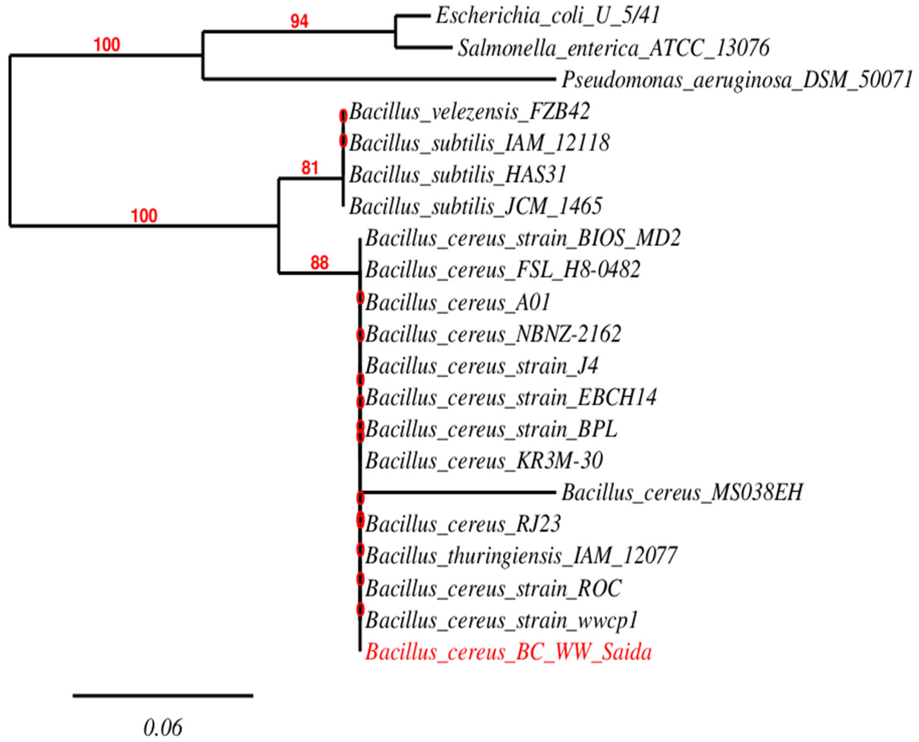
Candidate genes potentially involved in dye degradation were identified from Prokka outputs. Genome completeness and contamination were evaluated using CheckM2 (v1.0.2) on the Galaxy platform. Phylogenetic identity was further confirmed using 16S rRNA gene analysis and multilocus sequence typing (MLST) [28]. 16S rRNA genes were retrieved from NCBI GenBank and annotated using Prokka. These sequences were then aligned with reference 16S rRNA sequences using MAFFT (version 7) [29]. A maximum-likelihood phylogenetic tree was constructed with NGPhylogeny.fr, with bootstrap support values and a scale bar [30].

2.7. Statistical Analysis

To ensure statistical confidence, each experiment was conducted in triplicate. The experimental data values were documented as the mean ± standard deviation. A one-way ANOVA was used to evaluate the significance, with p < 0.05 considered statistically significant. The statistical analyses were conducted using Minitab® 21.2.

3. Results

3.1. Isolation of MB-Degrading Bacteria

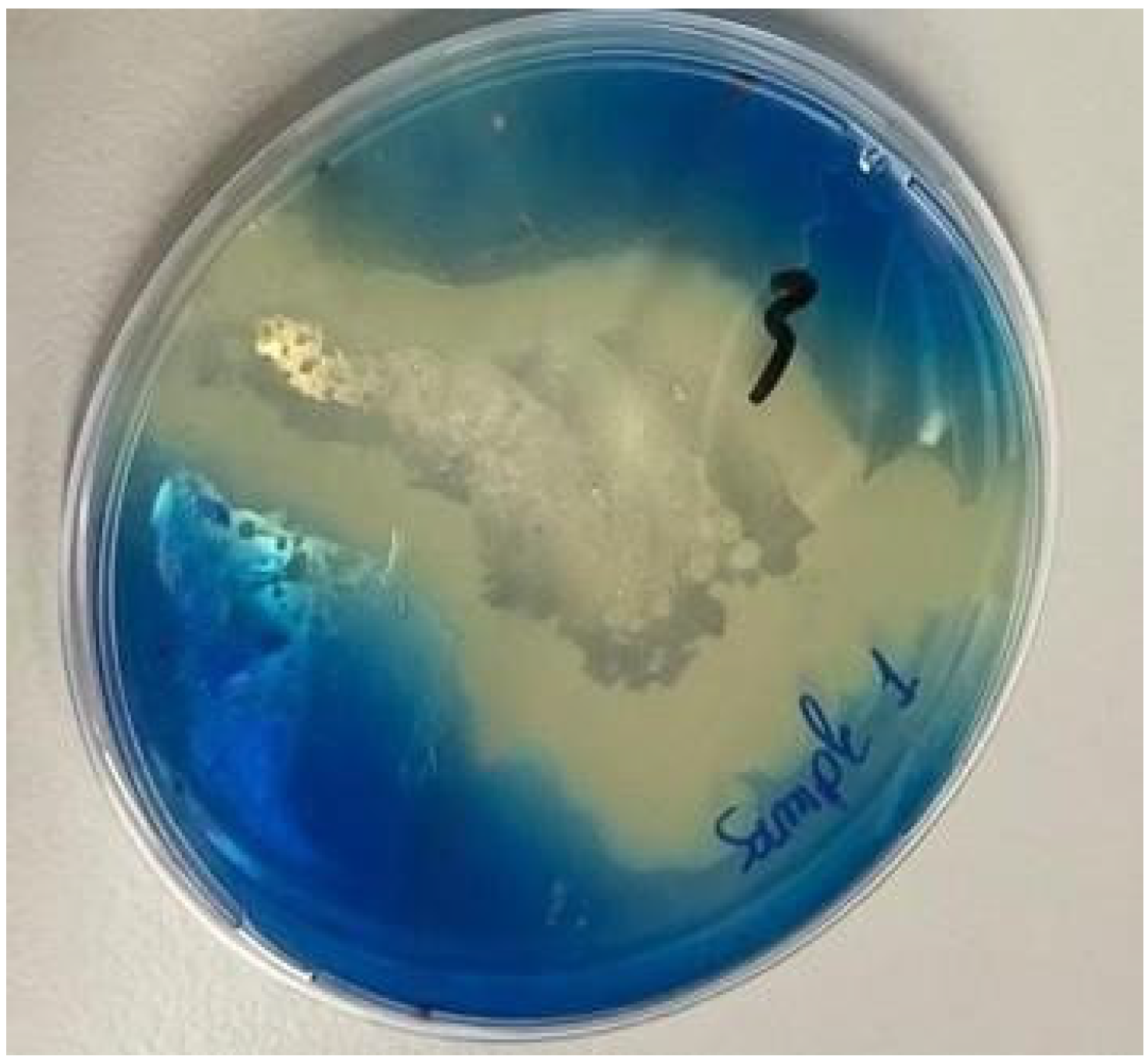
Samples collected from the Saida dumpsite were used for enrichment to isolate bacteria capable of degrading MB. After incubation at 37 °C for 48 h, on a nutrient agar plate supplemented with MB (50 mg/L), five bacterial isolates were obtained. Among these isolates, isolate 3 developed pronounced degrading activity, as indicated by the size and clarity of the clear zones surrounding the colonies compared to the other isolates. To confirm its degrading potential, strain 3 was subsequently re-cultivated on a fresh MB-supplemented plate, where it again demonstrated substantial decolorization (Figure 1). This distinct visual response set strain 3 apart from the other recovered isolates and highlighted it as the only one displaying a clear MB decolorizing phenotype on the plate. Accordingly, strain 3 was identified as the most promising isolate and selected for further investigation.

3.2. MB Degradation Assay

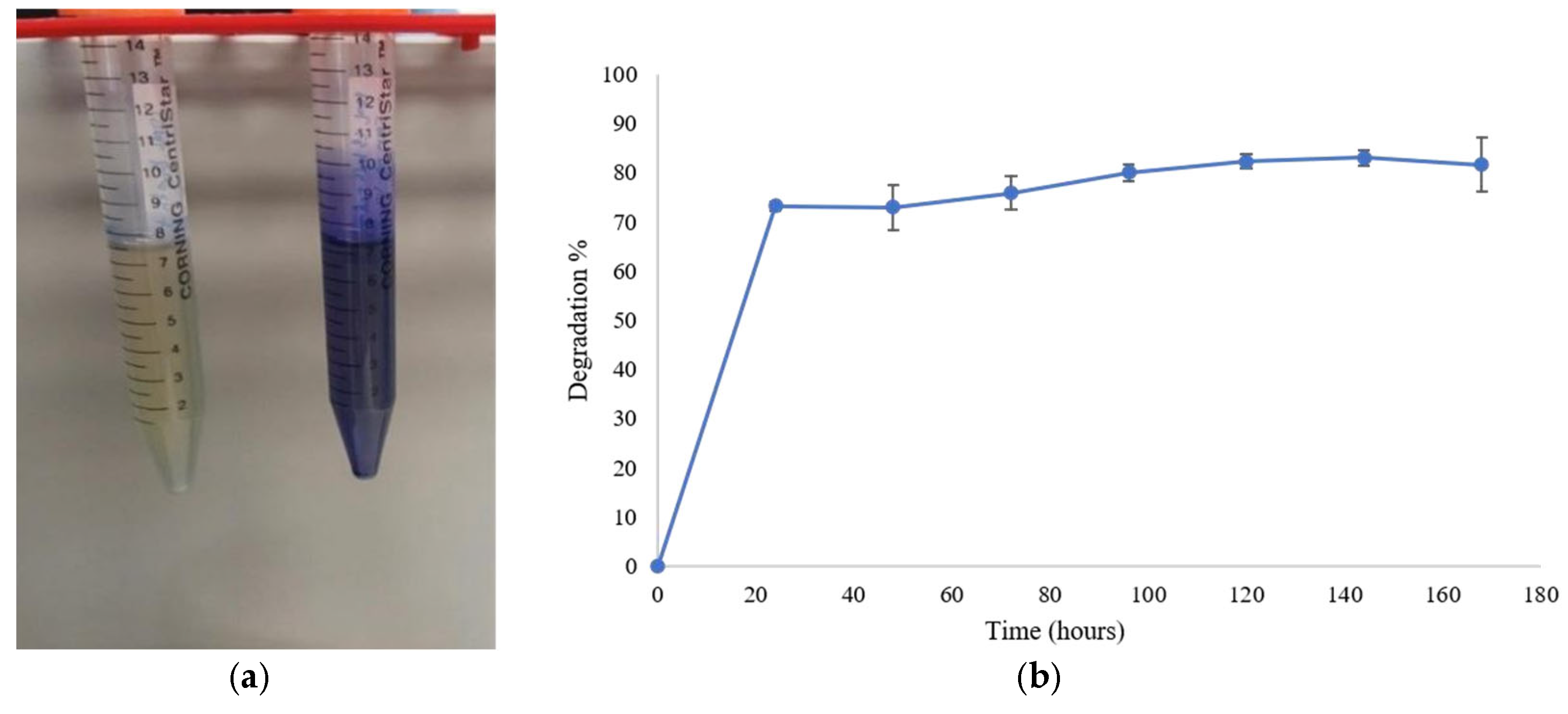
The decolorization efficiency of the selected bacterial strain was evaluated by measuring the optical density (OD) of MB (50 mg/L) at 664 nm at 24 h intervals over a total incubation duration of 168 h. As shown in Figure 2a, a progressive fading of the characteristic blue color of the medium was observed, indicating the occurrence of dye degradation. Spectrophotometric analysis revealed a progressive decrease in OD values over time, reflecting active biodegradation of the dye. The degradation percentage increased from 73.18% ± 0.64% after 24 h of incubation to 82.20% ± 1.54% after 140 h of incubation and remained relatively stable afterward, reaching 81.70% ± 5.06% at 168 h (Figure 2b).

3.3. HPLC Confirmation of Dye Degradation

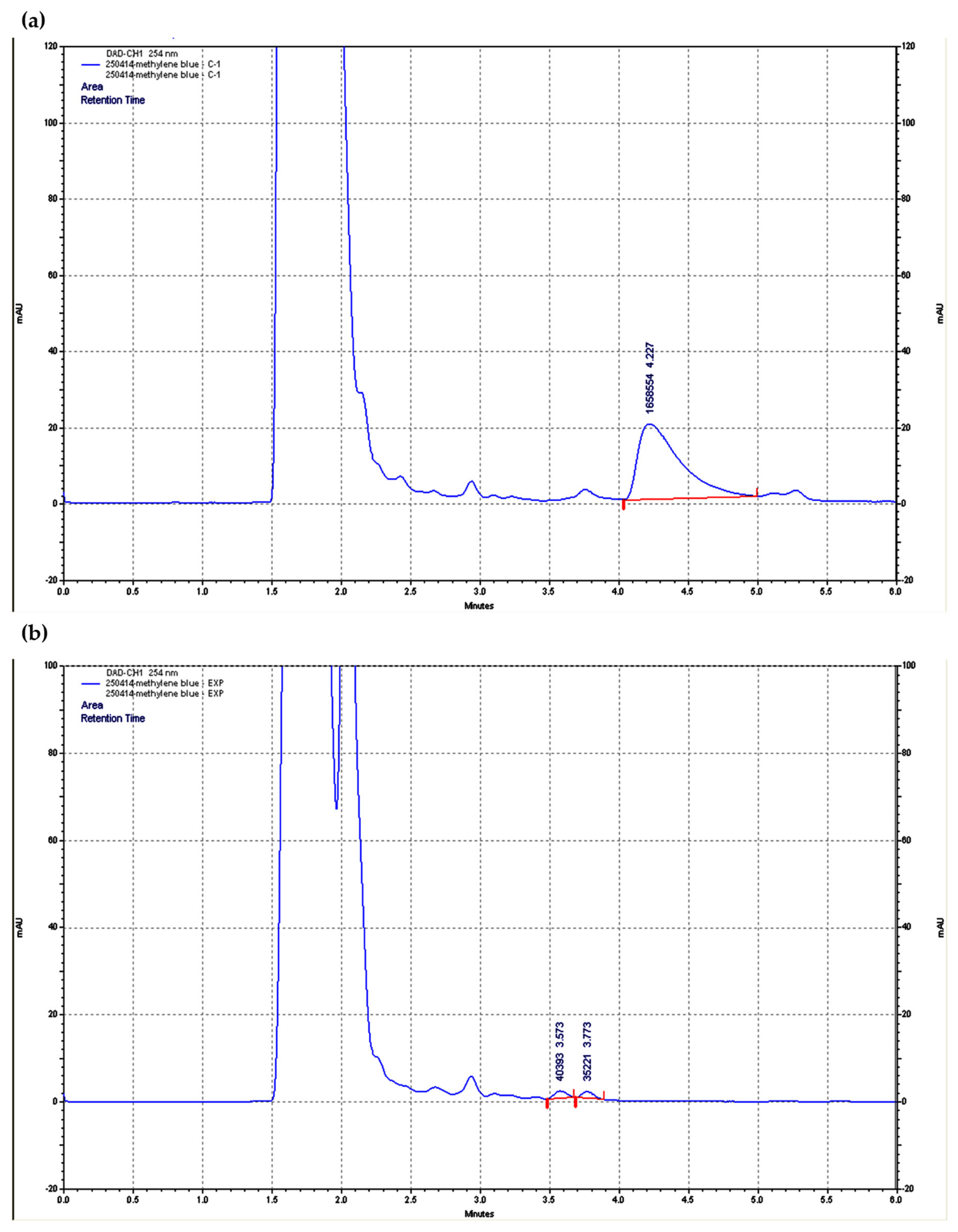
HPLC was conducted to monitor the degradation of MB following treatment with the bacterial isolate. The untreated control (Figure 3a) showed a dominant peak at a retention time (RT) of 4.227 min, representing the initial MB molecule.
Following bacterial treatment (Figure 3b), the original dye peak was markedly reduced in intensity, accompanied by the emergence of new auxiliary peaks. These chromatographic changes, characterized by the decrease in the initial dye concentration and the formation of new products, suggest the structural transformation of MB by the bacterial isolate.

3.4. Genomic Sequencing and Phylogenetic Analysis

The selected MB-degrading isolate was subjected to whole-genome sequencing, which confirmed the identity of the isolate as B. cereus. The genome of B. cereus BC WW Saida (accession no. SAMN46918858) comprised 5,137,921 base pairs with a GC content of 35.5%. Genome quality evaluation revealed 100% completeness and a low contamination level of 0.15%. Annotation performed using Prokka predicted 5519 genes, including 5384 protein-coding sequences (CDSs) and 135 RNA genes (Table 1), and revealed the presence of enzymes associated with dye degradation, including azoreductase, peroxidase, and oxidoreductase, along with antioxidant enzymes such as superoxide dismutase and catalase (Table 2). The identification result was further confirmed by 16S rRNA gene sequencing, as shown in Figure 4. Phylogenetic analysis revealed that the isolated strain was clustered within the B. cereus group. The isolate, marked in red, clustered closely with several B. cereus strains, including ROC, BIOS MD2, EBCH14, MS038EH, and J4, indicating a high level of similarity within this cluster. These members of the B. cereus group have been reported to degrade various synthetic dyes.

3.5. Genome Annotation and Biosynthetic Gene Cluster (BGC) Identification

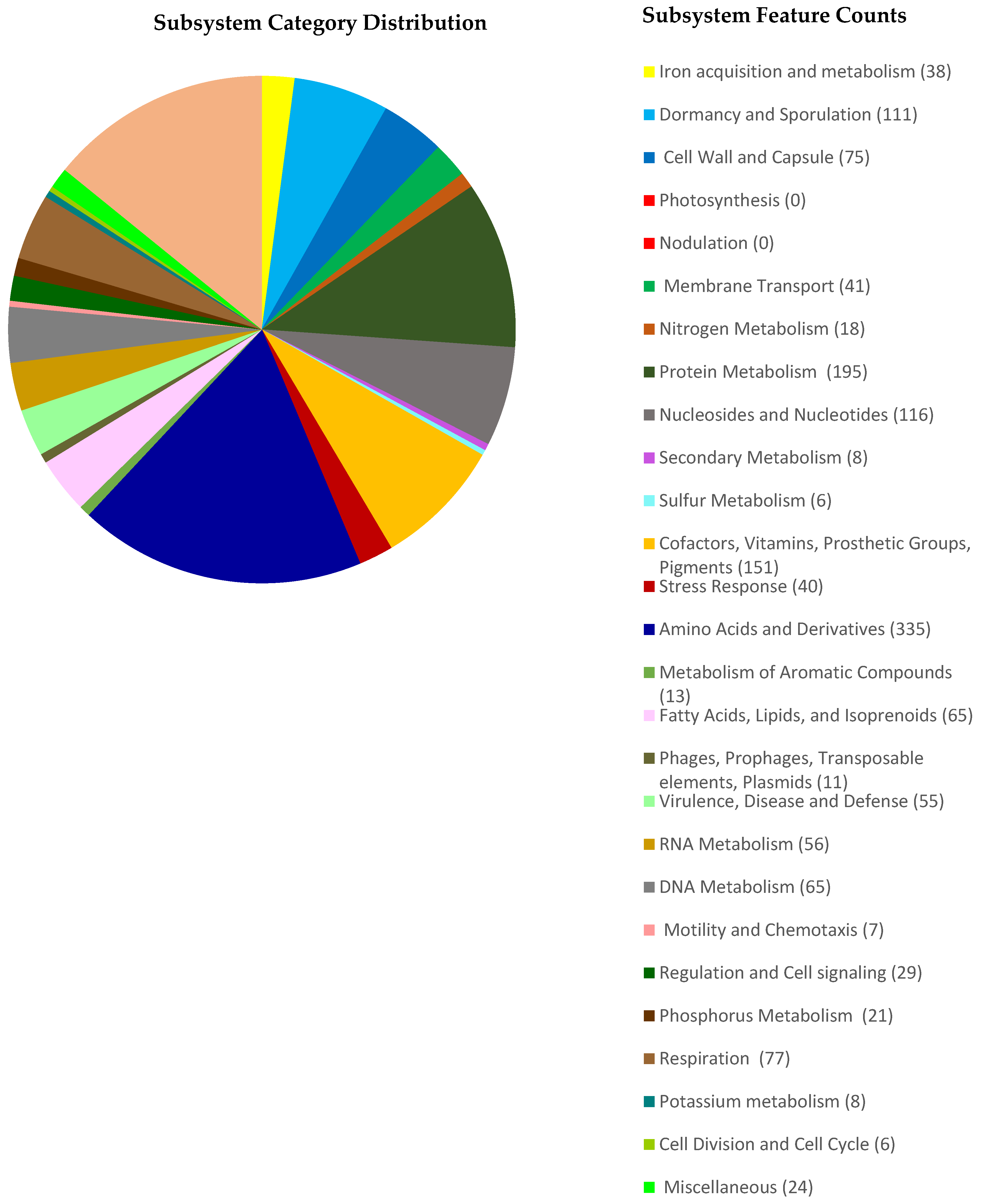
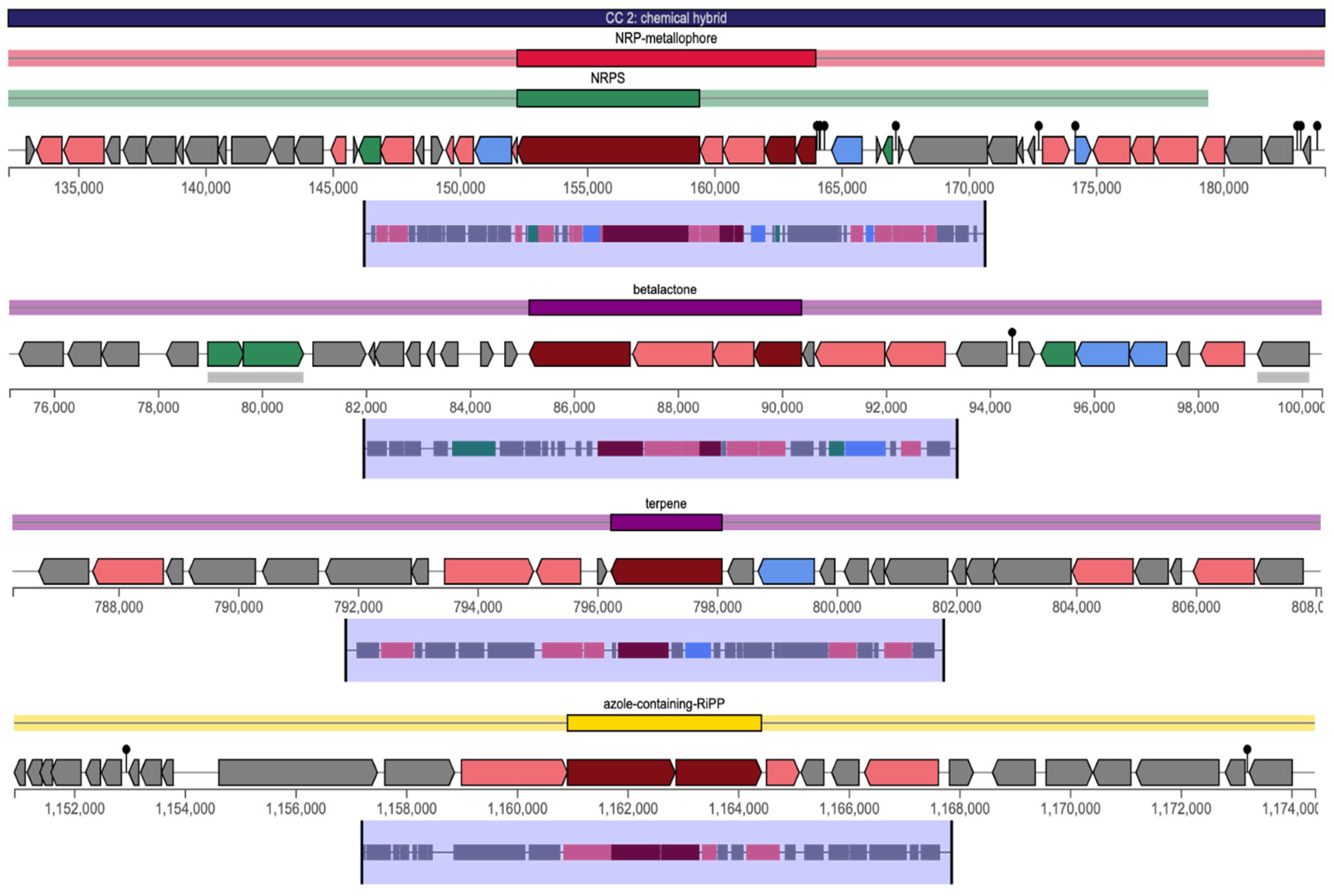
The pie diagram illustrates the distribution of genes assigned to different functional subsystems, as determined by annotation with RASTtk (Figure 5). The draft genome of B. cereus BC WW Saida consisted of 5,137,921 bp with a GC content of 35.5%, organized into 13 contigs (N50 = 2,668,635; L50 = 1). A total of 5384 coding sequences and 135 RNA genes were identified. Functional annotation identified 329 subsystems, with 24% of CDSs assigned to subsystems. The most abundant functional categories were amino acids and derivatives (335 genes), carbohydrates (259), cofactors, vitamins, prosthetic groups, and pigments (151), nucleosides and nucleotides (116), and protein metabolism (95). Additional categories included virulence and defense (55), stress response (40), respiration (77), fatty acids and lipids (65), RNA metabolism (56), DNA metabolism (65), membrane transport (41), iron acquisition (38), plasmids (11), and phages/transposable elements (13). A limited number of genes were associated with sulfur metabolism (6) and secondary metabolism (8), while no genes were detected for nodulation or photosynthesis. The B. cereus BC WW Saida genome harbors four biosynthetic gene clusters (BGCs), including a betalactone cluster related to fengycin (Region 9.1), an NRP-metallophore NRPS cluster corresponding to bacillibactin (Region 9.2), an azole-containing RiPP cluster (Region 9.3), and a terpene cluster (Region 1.1) (Figure 6). These BGCs consisted of core biosynthetic genes, as well as additional genes encoding regulatory proteins, transporters, and other accessory functions.

4. Discussion

Methylene blue (MB), a synthetic azo dye widely employed in textile, leather, and paper industries, poses significant environmental and ecological threats due to its chemical stability, aquatic toxicity, and resistance to traditional treatment processes [41]. Its persistence in wastewater can inhibit photosynthesis in aquatic systems, disrupt microbial ecosystems, and lead to mutagenic or carcinogenic effects on living organisms [42]. Furthermore, the accumulation of MB in aquatic environments may significantly decrease water clarity and impede oxygen transfer, further affecting aquatic biodiversity and disturbing ecological balance. These environmental and health-related issues have increased interest in sustainable alternatives such as microbial bioremediation, which provides practical simplicity, cost-effectiveness, and environmental compatibility [43]. Bacteria play a pivotal role in the breakdown of synthetic dyes using enzymatic systems that convert complex dye molecules into less harmful compounds. These bacterial processes typically rely on reduction and oxidation reactions mediated by enzymes such as azoreductases, peroxidases, and oxidoreductases. Among bacterial genera investigated for dye degradation, Bacillus species showed particular promise due to their robustness, adaptability to pollutant-rich environments, and ability to produce a wide spectrum of dye-degrading enzymes [44]. Their metabolic flexibility enabled survival in harsh environmental conditions where many other microorganisms could not persist.
In this study, a strain of B. cereus was isolated from the Saida dumpsite in Lebanon, a location heavily affected by industrial contamination. This isolate demonstrated significant potential for MB biodegradation. Spectrophotometric measurements indicated that the strain achieved 73% decolorization within 24 h and 82% after 168 h. This rapid initial response suggested prompt enzymatic reaction, followed by gradual breakdown of intermediate degradation products. The progressive increase in decolorization over time further indicated that the bacterial strain remained metabolically functional throughout the incubation period, confirming its ability to sustain activity under prolonged exposure to dye-contaminated environments. The HPLC data align with the spectrophotometric results, indicating that B. cereus BC WW Saida facilitates the chemical transformation of MB rather than mere physical adsorption. While the chromatographic profile demonstrated the degradative capacity of the strain, further detailed characterization of the resulting intermediates would provide a more comprehensive understanding of the metabolic pathways. Nevertheless, the integration of these results with the identified enzymatic repertoire from whole-genome sequencing supports the strain’s potential for efficient dye bioremediation.
To understand the enzymatic basis of this process, whole-genome sequencing was performed. Genome annotation revealed several key enzymes, including FMN-dependent NADH azoreductase (types 1, 2, and 4), NADPH-dependent azoreductase, Heme-dependent peroxidase, NAD(P)H-dependent FMN-containing oxidoreductase, nitrate reductase-like protein (NarX), vegetative catalase, and laccase (polyphenol oxidase). These enzymes are involved in catalyzing the reductive cleavage of azo bonds and oxidative breakdown of aromatic rings, both of which are key to effective dye degradation [45]. The coordinated activity of these enzymes likely facilitated sequential biochemical reactions resulting in the transformation of MB molecules. The genomic findings indicated that this isolate possesses a diverse set of enzymes typically associated with the degradation of synthetic dyes and aromatic compounds. This enzymatic profile suggests its potential to transform structurally complex dyes like methylene blue via both reductive and oxidative pathways [46]. Such metabolic versatility highlights the potential of this strain for application in environmental bioremediation processes.
The whole-genome analysis of B. cereus BC WW Saida highlights its broad metabolic capacity and adaptive strategies. The predominance of genes involved in amino acid, carbohydrate, and cofactor metabolism reflects the central role of these pathways in bacterial growth and energy generation. Genes being associated with virulence, disease, and defense is consistent with the strain’s pathogenic potential, which is known to cause foodborne illnesses and opportunistic infections [47]. Stress response and membrane transport genes further suggest its capacity to adapt to fluctuating environments and nutrient-limited conditions [48]. Such adaptive genetic features are commonly associated with bacteria inhabiting heavily contaminated environments. The relatively high number of genes related to respiration and lipid metabolism supports its metabolic flexibility under both aerobic and anaerobic conditions, while the absence of nodulation and photosynthetic genes confirms its heterotrophic, non-symbiotic lifestyle [49]. The genome also harbors four biosynthetic gene clusters related to fengycin, bacillibactin, RiPPs, and terpenes. These clusters encode secondary metabolites, regulatory proteins, and transporters that could enhance stress tolerance and redox activity, potentially supporting methylene blue degradation alongside azoreductase activity [50]. Together, these features highlight the strain’s adaptability and its potential for bioremediation in polluted environments.
Phylogenetically, BC WW Saida clusters within the B. cereus group, consistent with previous reports highlighting this species as a versatile environmental bacterium with strong dye-degradation potential. These closely related strains have been reported as effective azo dye degraders: strain ROC exhibits high decolorization of azo dyes [51], BIOS MD2 decolorizes Reactive Red dye [52], MS038EH removes Reactive Black-5 and Cr (VI) in 36 h, J4 removes methyl orange [53], and BPL was found to decolorize azo dye deep red glx [54]. The consistent detection of dye-degrading capabilities among closely related strains suggests that this metabolic trait may be relatively widespread within the B. cereus group.
The close phylogenetic relationship with these known dye-degrading strains, combined with the observed decolorization activity in this study, reinforces the functional role of BC WW Saida and confirms its capacity for synthetic dye degradation. The biodegradation efficiency of this isolated B. cereus strain demonstrates a notable efficiency in methylene blue degradation when compared to previously reported strains, with Acinetobacter johnsonii BP1 achieving approximately 60%, Bacillus weihenstephanensis BP2 reaching 67–70% [55], and Comamonas aquatica and Ralstonia mannitolilytica degrading around 60% under comparable experimental conditions [56]. The higher 82% decolorization achieved by B. cereus from the Saida dumpsite reflects a pronounced enzymatic specialization, possibly driven by prolonged exposure to a contaminated environment. An environment like the Saida dumpsite, contaminated with a variety of persistent pollutants, exerts selective evolutionary pressure that may favor the proliferation of bacterial populations possessing genes involved in xenobiotic degradation and stress adaptation [57]. This assumption is supported by the identification of oxidative and stress-related enzymes such as Heme-containing peroxidase and vegetative catalase. These enzymes likely enable the bacterium to process not only the primary dye compound but also the potentially toxic intermediates produced during the degradation process. Such functionality is particularly valuable for extensive bioremediation applications, where prolonged exposure to complex combinations of pollutants is expected [58]. Consequently, the metabolic capabilities exhibited by this strain support its potential application in future biological treatment processes designed for dye-contaminated wastewater.

5. Conclusions

In conclusion, the combined results from genomic, spectrophotometric, and HPLC analyses demonstrate that B. cereus BC WW Saida is a potent candidate for MB biodegradation. Although further analytical validation is required to definitively map the degradation products, these results establish a solid foundation for its application in industrial or municipal effluent treatment, particularly in scenarios where traditional methods are ineffective, expensive, or environmentally damaging. Future studies should focus on process optimization (pH, aeration, temperature), bioaugmentation approaches, and long-term surveillance of degradation by-products to ensure complete detoxification.

Author Contributions

F.H., writing—original draft, investigation, formal analysis; D.E.B. and R.E.H., supervision, conceptualization, methodology, formal analysis, writing—review and editing; T.A.C.G., M.J.M., C.A.-B. and M.K., review and editing. All authors have read and agreed to the published version of the manuscript.

Funding

This research received no external funding.

Institutional Review Board Statement

Not applicable.

Informed Consent Statement

Not applicable.

Data Availability Statement

The genome sequence of B. cereus BC WW Saida, used in this study for azo dye degradation analyses, has been deposited in the NCBI database under BioProject accession number PRJNA873891, and BioSample accession SAMN46918858. All other experimental data, including optical density measurements, HPLC results, and bacterial culture information, are held by the corresponding author and are fully available. This ensures complete transparency and reproducibility of the findings.

Acknowledgments

AI tools, GPT-5.3 architecture (SCI Mechanism Diagram Generator), were used solely to generate and refine the graphical abstract, as well as to assist with minor phrasing and grammatical improvements. No AI tools were used in the scientific writing, data analysis, interpretation, or conceptual development of the manuscript.

Conflicts of Interest

This work is original, has not been published, and is not under review elsewhere. All authors have approved the manuscript and its submission to AEM. The authors declare no competing interests and no external funding.

References

  1. Lellis, B.; Fávaro-Polonio, C.Z.; Pamphile, J.A.; Polonio, J.C. Effects of textile dyes on health and the environment and bioremediation potential of living organisms. Biotechnol. Res. Innov. 2019, 3, 275–290. [Google Scholar] [CrossRef]
  2. Yu, Y.; Liu, S.; Pei, Y.; Luo, X. Growing palladium nanoparticles on cellulose microspheres via in-situ reduction for catalytic decolorization of methylene blue. Int. J. Biol. Macromol. 2021, 166, 1419–1428. [Google Scholar] [CrossRef]
  3. Kumar, M.; Singh, V.P.; Bhat, S.B.; Kumar, R. Environmental risks of textile dyes and photocatalytic materials for sustainable treatment: Current status and future directions. Discov. Environ. 2025, 3, 132. [Google Scholar] [CrossRef]
  4. Alvarenga, G.; Lima, J.P.; Goszczynski, A.C.F.; Rosa, C.H.; Rosa, G.R.; Lopes, T.J. Methylene blue adsorption by timbaúva (Enterolobium contortisiliquum)-derived materials. Environ. Sci. Pollut. Res. 2020, 27, 27893–27903. [Google Scholar] [CrossRef] [PubMed]
  5. Cui, M.-H.; Liu, W.-Z.; Tang, Z.-E.; Cui, D. Recent advancements in azo dye decolorization in bio-electrochemical systems (BESs): Insights into decolorization mechanism and practical application. Water Res. 2021, 203, 117512. [Google Scholar] [CrossRef] [PubMed]
  6. Ledakowicz, S.; Paździor, K. Recent achievements in dyes removal focused on advanced oxidation processes integrated with biological methods. Molecules 2021, 26, 870. [Google Scholar] [CrossRef]
  7. Panwar, P.; Mahajan, P.; Kaushal, J. Microbial bioremediation of Azo dyes: An environment sustainable technology. Remediat. J. 2023, 33, 151–165. [Google Scholar] [CrossRef]
  8. Ngo, A.C.R.; Tischler, D. Microbial Degradation of Azo Dyes: Approaches and Prospects for a Hazard-Free Conversion by Microorganisms. Int. J. Environ. Res. Public Health 2022, 19, 4740. [Google Scholar] [CrossRef]
  9. Pinheiro, L.R.S.; Gradíssimo, D.G.; Xavier, L.P.; Santos, A.V. Degradation of Azo Dyes: Bacterial Potential for Bioremediation. Sustainability 2022, 14, 1510. [Google Scholar] [CrossRef]
  10. Hanis, K.K.A.; Nasri, A.R.M.; Farahiyah, W.K.W.; Rabani, M.Y.M. Bacterial Degradation of Azo Dye Congo Red by Bacillus sp. J. Phys. Conf. Ser. 2020, 1529, 022048. [Google Scholar] [CrossRef]
  11. Suhag, M.H.; Haque, K.A.U.; Hossen, Z.; Azad, A.K.; Younus, M. Biodegradation of azo dyes and dyes present in textile wastewaters using Bacillus sp. az28, obtained from industrial effluents. J. Bangladesh Acad. Sci. 2021, 45, 117–122. [Google Scholar] [CrossRef]
  12. Uppala, R.; Sundar, K.; Muthukumaran, A. Decolorization of Azo Dyes using Dried Biomass of Bacillus cereus RC1 and Kocuria kristinae RC3. J. Pure Appl. Microbiol. 2019, 13, 1969–1976. [Google Scholar] [CrossRef]
  13. Liu, J.; Bacosa, H.P.; Liu, Z. Potential Environmental Factors Affecting Oil-Degrading Bacterial Populations in Deep and Surface Waters of the Northern Gulf of Mexico. Front. Microbiol. 2017, 7, 2131. [Google Scholar] [CrossRef] [PubMed]
  14. Abbas, I.; Chaaban, J.K.; Shaar, A.A.; Al-Rabaa, A. Solid Waste Management in Lebanon: Challenges and Recommendations. J. Environ. Waste Manag. 2017, 4, 53–63. [Google Scholar]
  15. Ren, Y.; Manefield, M. Evolution of pollutant biodegradation. Appl. Microbiol. Biotechnol. 2025, 109, 36. [Google Scholar] [CrossRef]
  16. Putatunda, C.; Solanki, P.; Bhatia, R.; Kakkar, P.M.; Walia, A. Microbial metabolism of aquatic pollutants: Recent trends and future perspective. Blue Biotechnol. 2025, 2, 26. [Google Scholar] [CrossRef]
  17. Ariffin, F.; Anuar, N.E.M.C. Biodegradation of Methylene Blue by bacteria strains isolated from contaminated soil. Malays. Appl. Biol. 2022, 51, 25–35. [Google Scholar] [CrossRef]
  18. Shah, M.P.; Patel, K.A.; Nair, S.S.; Darji, A.M. Isolation, Identification and Screening of Dye Decolorizing Bacteria. Am. J. Microbiol. Res. 2013, 1, 62–70. [Google Scholar] [CrossRef][Green Version]
  19. Hadwan, M.H.; Hussein, M.J.; Mohammed, R.M.; Hadwan, A.M.; Al-Kawaz, H.S.; Al-Obaidy, S.S.M.; Al Talebi, Z.A. An improved method for measuring catalase activity in biological samples. Biol. Methods Protoc. 2024, 9, bpae015. [Google Scholar] [CrossRef]
  20. Barry, A.L.; Feeney, K.L. Two quick methods for Voges-Proskauer test. Appl. Microbiol. 1967, 15, 1138–1141. [Google Scholar] [CrossRef]
  21. Bhusal, A.; Muriana, P.M. Isolation and Characterization of Nitrate Reducing Bacteria for Conversion of Vegetable-Derived Nitrate to ‘Natural Nitrite’. Appl. Microbiol. 2021, 1, 11–23. [Google Scholar] [CrossRef]
  22. Ramakrishnan, G.; Reddy, G.K.; Allam, S.S.; Jagarlapudi, S.; Tummalacharla, P.; Kosuri, H.P.; Govind, G.; Shaik, B.; Ramaiah, J. Sustainable Biodegradation of Methylene Blue Dye by Enterobacter cloacae Strain BHPGT2024. Res. Sq. 2024. [Google Scholar] [CrossRef]
  23. Ullah, A.K.M.A.; Kibria, A.K.M.F.; Akter, M.; Khan, M.N.I.; Tareq, A.R.M.; Firoz, S.H. Oxidative Degradation of Methylene Blue Using Mn3O4 Nanoparticles. Water Conserv. Sci. Eng. 2017, 1, 249–256. [Google Scholar] [CrossRef]
  24. Wang, Y.; Zhao, Y.; Bollas, A.; Wang, Y.; Au, K.F. Nanopore sequencing technology, bioinformatics and applications. Nat. Biotechnol. 2021, 39, 1348–1365. [Google Scholar] [CrossRef] [PubMed]
  25. Freire, B.; Ladra, S.; Paramá, J.R. Memory-Efficient Assembly Using Flye. IEEE/ACM Trans. Comput. Biol. Bioinform. 2022, 19, 3564–3577. [Google Scholar] [CrossRef]
  26. Seemann, T. Prokka: Rapid prokaryotic genome annotation. Bioinformatics 2014, 30, 2068–2069. [Google Scholar] [CrossRef]
  27. Brettin, T.; Davis, J.J.; Disz, T.; Edwards, R.A.; Gerdes, S.; Olsen, G.J.; Olson, R.; Overbeek, R.; Parrello, B.; Pusch, G.D.; et al. RASTtk: A modular and extensible implementation of the RAST algorithm for building custom annotation pipelines and annotating batches of genomes. Sci. Rep. 2015, 5, 8365. [Google Scholar] [CrossRef]
  28. Larsen, M.V.; Cosentino, S.; Rasmussen, S.; Friis, C.; Hasman, H.; Marvig, R.L.; Jelsbak, L.; Sicheritz-Pontéen, T.; Ussery, D.W.; Aarestrup, F.M.; et al. Multilocus sequence typing of total-genome-sequenced bacteria. J. Clin. Microbiol. 2012, 50, 1355–1361. [Google Scholar] [CrossRef]
  29. Katoh, K.; Rozewicki, J.; Yamada, K.D. MAFFT online service: Multiple sequence alignment, interactive sequence choice and visualization. Brief. Bioinform. 2018, 20, 1160–1166. [Google Scholar] [CrossRef]
  30. Lemoine, F.; Correia, D.; Lefort, V.; Doppelt-Azeroual, O.; Mareuil, F.; Cohen-Boulakia, S.; Gascuel, O. NGPhylogeny.fr: New generation phylogenetic services for non-specialists. Nucleic Acids Res. 2019, 47, W260–W265. [Google Scholar] [CrossRef]
  31. Aragaw, T.A.; Bogale, F.M.; Gessesse, A. Adaptive Response of Thermophiles to Redox Stress and Their Role in the Process of dye Degradation From Textile Industry Wastewater. Front. Physiol. 2022, 13, 908370. [Google Scholar] [CrossRef] [PubMed]
  32. Jadhav, P.; Kishori Tarfe, D. Isolation of Methylene Blue dye degrading bacteria from effluent, screening and evaluation of their dye decolorization activity. Int. J. Sci. Dev. Res. 2019, 4, 224–230. [Google Scholar]
  33. Ferreira-Leitão, V.S.; da Silva, J.G.; Bon, E.P. Methylene blue and azure B oxidation by horseradish peroxidase: A comparative evaluation of class II and class III peroxidases. Appl. Catal. B Environ. 2003, 42, 213–221. [Google Scholar] [CrossRef]
  34. Buchholz, K.; Schirmer, R.H.; Eubel, J.K.; Akoachere, M.B.; Dandekar, T.; Becker, K.; Gromer, S. Interactions of methylene blue with human disulfide reductases and their orthologues from Plasmodium falciparum. Antimicrob. Agents Chemother. 2008, 52, 183–191. [Google Scholar] [CrossRef]
  35. Wu, K.; Shi, M.; Pan, X.; Zhang, J.; Zhang, X.; Shen, T.; Tian, Y. Decolourization and biodegradation of methylene blue dye by a ligninolytic enzyme-producing Bacillus thuringiensis: Degradation products and pathway. Enzym. Microb. Technol. 2022, 156, 109999. [Google Scholar] [CrossRef]
  36. Uddin, J.; Idrees, M.; Ahmed, H.; Batool, S.; Rahman, T.; Mehmood, S.; Tanoli, A.; bin Muhsinah, A.; Ullah, H.; Musharraf, S. Biodegradation and decolorization of methylene blue, reactive Black-5, and toluidine blue-O from an aqueous solution using the polyphenol oxidase enzyme. Front. Sustain. Food Syst. 2023, 7. [Google Scholar] [CrossRef]
  37. delCardayré, S.; Stock, K.; Newton, G.; Fahey, R.; Davies, J. Coenzyme A disulfide reductase, the primary low molecular weight disulfide reductase from Staphylococcus aureus: Purification and characterization of the native enzyme. J. Biol. Chem. 1998, 273, 5744–5751. [Google Scholar] [CrossRef]
  38. Haigler, B.E.; Nishino, S.F.; Spain, J.C. Biodegradation of 4-Methyl-5-Nitrocatechol by Pseudomonas sp. Strain DNT. J. Bacteriol. 1994, 176, 3433–3437. [Google Scholar] [CrossRef]
  39. Hu, S.; Yuan, D.; Liu, Y.; Zhao, L.; Guo, H.; Niu, Q.; Zong, W.; Liu, R. The toxic effects of alizarin red S on catalase at the molecular level. RSC Adv. 2019, 9, 33368–33377. [Google Scholar] [CrossRef]
  40. Zheng, M.; Liu, Y.; Zhang, G.; Yang, Z.; Xu, W.; Chen, Q. The Applications and Mechanisms of Superoxide Dismutase in Medicine, Food, and Cosmetics. Antioxidants 2023, 12, 1675. [Google Scholar] [CrossRef]
  41. Eltarahony, M.; El-Fakharany, E.; Abu-Serie, M.; ElKady, M.; Ibrahim, A. Statistical modeling of methylene blue degradation by yeast-bacteria consortium; optimization via agro-industrial waste, immobilization and application in real effluents. Microb. Cell Factories 2021, 20, 234. [Google Scholar] [CrossRef] [PubMed]
  42. Al-Tohamy, R.; Ali, S.S.; Li, F.; Okasha, K.M.; Mahmoud, Y.A.G.; Elsamahy, T.; Jiao, H.; Fu, Y.; Sun, J. A critical review on the treatment of dye-containing wastewater: Ecotoxicological and health concerns of textile dyes and possible remediation approaches for environmental safety. Ecotoxicol. Environ. Saf. 2022, 231, 113160. [Google Scholar] [CrossRef] [PubMed]
  43. Kuppan, N.; Padman, M.; Mahadeva, M.; Srinivasan, S.; Devarajan, R. A comprehensive review of sustainable bioremediation techniques: Eco friendly solutions for waste and pollution management. Waste Manag. Bull. 2024, 2, 100021. [Google Scholar] [CrossRef]
  44. Patel, J.; Vaishnav, D.; Joshi, P.; Tipre, D. Bacterial enzymes for azo dye degradation: An insight. Res. J. Chem. Environ. 2023, 27, 135–148. [Google Scholar] [CrossRef]
  45. Pham, V.H.T.; Kim, J.; Chang, S.; Bang, D. Investigating Bio-Inspired Degradation of Toxic Dyes Using Potential Multi-Enzyme Producing Extremophiles. Microorganisms 2023, 11, 1273. [Google Scholar] [CrossRef]
  46. Bedekar, P.A.; Saratale, R.G.; Saratale, G.D.; Govindwar, S.P. Oxidative stress response in dye degrading bacterium Lysinibacillus sp. RGS exposed to Reactive Orange 16, degradation of RO16 and evaluation of toxicity. Environ. Sci. Pollut. Res. 2014, 21, 11075–11085. [Google Scholar] [CrossRef]
  47. Barber, M.F.; Fitzgerald, J.R. Mechanisms of host adaptation by bacterial pathogens. FEMS Microbiol. Rev. 2024, 48, fuae019. [Google Scholar] [CrossRef]
  48. Fang, F.C.; Frawley, E.R.; Tapscott, T.; Vázquez-Torres, A. Bacterial Stress Responses during Host Infection. Cell Host Microbe 2016, 20, 133–143. [Google Scholar] [CrossRef]
  49. Chen, Y.-J.; Leung, P.M.; Wood, J.L.; Bay, S.K.; Hugenholtz, P.; Kessler, A.J.; Shelley, G.; Waite, D.W.; Franks, A.E.; Cook, P.L.M.; et al. Metabolic flexibility allows bacterial habitat generalists to become dominant in a frequently disturbed ecosystem. ISME J. 2021, 15, 2986–3004. [Google Scholar] [CrossRef]
  50. Kim, J.; Bashir, H.H.; Hwang, J.; Moon, G.-S. Mining Biosynthetic Gene Clusters of Bacillus subtilis MGE 2012 Using Whole Genome Sequencing. Processes 2025, 13, 1503. [Google Scholar] [CrossRef]
  51. Fareed, A.; Zaffar, H.; Bilal, M.; Hussain, J.; Jackson, C.; Alam Naqvi, T. Decolorization of azo dyes by a novel aerobic bacterial strain Bacillus cereus strain ROC. PLoS ONE 2022, 17, e0269559. [Google Scholar] [CrossRef]
  52. Dakshinamoorthi, B.M.; Jothiraman, S.; Rasappan, S.; Subramaniyam, S.; Sridharan, J. Biodecolorization of Reactive Red Using Bacillus cereus Isolated from Oil Contaminated Soil. Uttar Pradesh J. Zool. 2024, 45, 218–229. [Google Scholar] [CrossRef]
  53. Emadi, Z.; Sadeghi, R.; Forouzandeh, S.; Mohammadi-Moghadam, F.; Sadeghi, R.; Sadeghi, M. Simultaneous anaerobic decolorization/degradation of Reactive Black-5 azo dye and chromium(VI) removal by Bacillus cereus strain MS038EH followed by UV-C/H2O2 post-treatment for detoxification of biotransformed products. Arch. Microbiol. 2021, 203, 4993–5009. [Google Scholar] [CrossRef]
  54. Vasniwal, R.; Jadhav, I.; Shrivastava, D.; Jadhav, K. Biodegradation and Decolorization of Azo Dye (Deep Red Glx) by Alkaliphilic Bacillus Cereus Strain BPL Isolated from Textile Effluent Contaminant Soil. Int. J. Eng. Res. 2016, 5, 1–8. [Google Scholar] [CrossRef]
  55. Abai, N.; Babakhouya, N. The degradation of methylene blue by bacterial strains isolated from the peel of red beet. Alger. J. Environ. Sci. Technol. 2018, 4, 794–799. [Google Scholar]
  56. Michelle; Siregar, R.A.N.; Sanjaya, A.; Lucy, J.; Pinontoan, R. Methylene blue decolorizing bacteria isolated from water sewage in Yogyakarta, Indonesia. Biodiversitas J. Biol. Divers. 2020, 21, 1136–1141. [Google Scholar] [CrossRef]
  57. Tello, A.; Austin, B.; Telfer, T. Selective pressure of antibiotic pollution on bacteria of importance to public health. Environ. Health Perspect. 2012, 120, 1100–1106. [Google Scholar] [CrossRef] [PubMed]
  58. Bittencourt, G.A.; Vandenberghe, L.P.d.S.; Martínez-Burgos, W.J.; Valladares-Diestra, K.K.; de Mello, A.F.M.; Maske, B.L.; Brar, S.K.; Varjani, S.; Pereira, G.V.d.M.; Soccol, C.R. Emerging contaminants bioremediation by enzyme and nanozyme-based processes—A review. iScience 2023, 26, 106785. [Google Scholar] [CrossRef]
Figure 1. Bacterial isolate 3 grown on nutrient agar plate supplemented with MB showing clear zone around colonies.
Applmicrobiol 06 00052 g001
Figure 2. Methylene blue decolorization by the isolated bacterial strain over time: (a) MB (50 mg/L) in nutrient broth following incubation, with the inoculated tube on the left and the non-inoculated control on the right; (b) degradation profile of MB over time (24–168 h) based on OD (664 nm) readings. Data points represent the mean degradation percentage, and error bars indicate standard deviation (±SD).
Applmicrobiol 06 00052 g002
Figure 3. HPLC chromatograms of MB (a) before and (b) after bacterial treatment.
Applmicrobiol 06 00052 g003
Figure 4. Phylogenetic tree based on 16S rRNA gene sequences showing the position of B. cereus BC WW Saida (highlighted in red) among closely related Bacillus species. Numbers at the nodes represent branch support values indicating the confidence of the inferred phylogenetic relationships. Escherichia coli and Salmonella enterica were placed as outgroups. The scale bar represents 0.06 substitutions per nucleotide position.
Applmicrobiol 06 00052 g004
Figure 5. The RAST analysis-based subsystem distribution of whole-genome sequence of isolate. Each color in the pie chart represents a particular group of genes mentioned on the right side of the graph.
Applmicrobiol 06 00052 g005
Figure 6. Schematic diagram of secondary metabolite biosynthetic gene clusters in B. cereus BC WW Saida.
Applmicrobiol 06 00052 g006
Table 1. Genome annotation summary of B. cereus BC WW Saida.
CharacteristicsTerms
Taxonomy Firmicute > Bacilli > Bacilales > Bacilaceae > Bacillus > Bacillus cereus
Genomic statistics Complete size of genome 5,137,921 bp
Number of contigs 13
GC content (%) 35.5
Contig N50 value 2,668,635
Contig L50 value 1
Genomic feature CDS 5263
tRNA 93
rRNA 34
Genome quality Completeness 100%
Contamination 0.15
Overall remarks Good
Genome availability Bioproject PRJNA873891
Biosample SAMN46918858
SRA accession SRR32413154
Table 2. Decolorization and oxidative stress enzymes identified by Prokka annotation.
Enzyme GroupEnzyme NameGene/Locus Tag(s)Role in
Degradation
Reference
AzoreductasesFMN-dependent NADH azoreductase 1 HKCAGING_03932Reduces MB chromophore through
electron tranfer
[31]
FMN-dependent NADH
azoreductase 2
HKCAGING_00609;
HKCAGING_00695;
HKCAGING_03652
FMN-dependent NADH
azoreductase 4
HKCAGING_02709
NADPH
azoreductase
HKCAGING_03615Uses NADPH to reduce MB chromophores[32]
PeroxidasesHeme-dependent peroxidase HKCAGING_02688Decolorizes MB
(LiP, MnP, DyP-type)
[33]
NAD(P)H-dependent
FMN-containing oxidoreductase
HKCAGING_02465Disrupts aromatic structure of MB [34]
OxidoreductasesNitrate reductase-like protein NarXHKCAGING_03721;
HKCAGING_03722
Mediates dye degradation through NADH electron
transfer
[35]
Polyphenol oxidase HKCAGING_01201Oxidative dye degradation via electron transfer to O2 [36]
Coenzyme A disulfide reductase HKCAGING_04577;
HKCAGING_04579
Catalyzes reduction in MB to leucoMB by
NADPH
[37]
4-methyl-5nitrocatechol 5monooxygenaseHKCAGING_00816 Degradation of nitroaromatic azo compounds[38]
CatalaseHKCAGING_00291;
HKCAGING_00403;
HKCAGING_04665;
HKCAGING_04928;
HKCAGING_04981
Protects against oxidative damage from dyes[39]
Superoxide dismutaseHKCAGING_01574;
HKCAGING_02233;
HKCAGING_02747
Protects against oxidative stress during dye degradation [40]
Disclaimer/Publisher’s Note: The statements, opinions and data contained in all publications are solely those of the individual author(s) and contributor(s) and not of MDPI and/or the editor(s). MDPI and/or the editor(s) disclaim responsibility for any injury to people or property resulting from any ideas, methods, instructions or products referred to in the content.