Review

Functional Versatility of Vibrio cholerae Outer Membrane Proteins

by
Annabelle Mathieu-Denoncourt
1,2 and
Marylise Duperthuy
1,2,*
1
Département de Microbiologie, Infectiologie et Immunologie, Faculté de Médecine, Université de Montréal, Montréal, QC H3T 1J4, Canada
2
Institut Courtois d’Innovation Biomédicale, Faculté de Médecine, Université de Montréal, Montréal, QC H3T 1J4, Canada
*
Author to whom correspondence should be addressed.
Appl. Microbiol. 2025, 5(3), 64; https://doi.org/10.3390/applmicrobiol5030064
Submission received: 8 May 2025 / Revised: 27 June 2025 / Accepted: 28 June 2025 / Published: 3 July 2025

Abstract

A key feature that differentiates Gram-positive and Gram-negative bacteria is the outer membrane, an asymmetric membrane composed of lipopolysaccharides, phospholipids, lipoproteins and integral proteins, including the outer-membrane proteins (OMPs). By being in direct contact with the extracellular milieu, the outer membrane and OMPs participate in multiple functions in Gram-negative bacteria, including controlling nutrient and molecule access to the cytoplasm, membrane vesicle formation and resistance to environmental stresses. OMPs have a characteristic barrel shape formed by antiparallel β-strands, with or without channels that allow diffusion of substrates through the outer membrane. The marine bacterium Vibrio cholerae is responsible for non-invasive gastroenteritis and cholera disease by consumption of contaminated water or food. Its OMPs, besides having a porin function, contribute to resistance to osmotic pressure and antimicrobial agents, intracellular signaling, adhesion to host cells and biofilm formation, amongst other functions. In this review, in addition to quickly reviewing the general structure of the outer membrane, the OMPs and how they reach the outer membrane, the functions attributed to these proteins are compiled. The mechanisms used by each of the described OMP to accomplish these functions in the marine pathogenic bacterium V. cholerae are discussed. Potential clinical and bioengineering applications of OMPs, such as diagnostic tools, vaccine development, and targeted antimicrobial or anti-virulence strategies are presented. What is known about the OMPs of V. cholerae is presented below.

Graphical Abstract

1. Introduction

Resistance to antimicrobial agents (AMR) is a serious and growing public health threat. It represents a heavy economic and humanitarian burden, as the cost of heavy treatments, length of hospitalisation and mortality rates are increasing [1]. The misuse of antibiotics in human and veterinary medicines has led to the problem, which is exacerbated by the lack of new antibiotics discovery [1]. New resistance genes can emerge rapidly after the introduction of new drugs and can be transmitted and amplified quickly in bacteria [2]. Although AMR is globally growing, the emergence of antimicrobial resistance in Gram-negative bacteria is particularly worrying and is often associated with nosocomial infections [3]. Gram-negative bacteria such as Klebsiella pneumoniae, Acinetobacter baumannii and Pseudomonas aeruginosa, members of the highly virulent ESKAPE pathogens, are responsible for various diseases and are under the scope for AMR, as they can acquire multiple, extensive and pan-drug resistance [2,3].
The constitution of the Gram-negative bacterial envelope makes it a main barrier that contributes to antibiotic resistance by limiting the penetration of antibiotics, mostly targeting intracellular processes [4]. The distinction between Gram-negative and Gram-positive bacteria resides in the composition of their cellular envelope and the presence of the outer membrane (OM) [5]. While the Gram-negative bacterial envelope is composed of a thin peptidoglycan (PG) layer in a periplasmic space formed by the cytoplasmic and the outer membranes, the Gram-positive cell envelope is composed of a thick PG layer outside their sole cytoplasmic membrane. The OM is an asymmetrical lipid bilayer of glycerophospholipids and glycolipids, mainly lipopolysaccharides (LPSs) [6], in which the phospholipids are mostly confined to the inner leaflet. The OM also contains lipoproteins [7] and embedded outer membrane proteins (OMPs) [8]. In addition to maintaining the bacterial shape, the OM controls the passage of nutrients and molecules, while limiting the entry of antibiotics, and protects the cell from environmental stresses [5,6]. It can also participate in secretion, virulence and protection against phages and multiple antimicrobials [9,10,11,12,13,14,15].
Vibrio cholerae is a ubiquitous Gram-negative bacterium that resides in estuaries and brackish waters. It can be found as planktonic cells or in association with zooplankton, fishes and shellfishes [16,17]. There are as much as 200 serogroups of V. cholerae, of which the O1 and O139 are responsible for cholerae disease by production of cholera toxin and its coregulated pilus [16]. It is characterized by a severe diarrhea that, when left untreated, leads to dehydration and death. The O1 serogroup is further divided in Inaba and Ogawa based on the O antigen, and in El Tor and Classical biotypes, based on genetic markers [18]. Historically, the Classical biotype was responsible for six cholera pandemics from the early 1800s, but was replaced in the seventh ongoing pandemic by El Tor strains in the 1960s [18]. Cholera is endemic in developing countries, where sanitation and access to clean water is scarce, or in specific regions following environmental catastrophes [19]. As for the O139 strains, they appeared in the 1990s and are derived from O1 El Tor strains [20]. After a first rapid outburst, they are now less prevalent. The non-O1/non-O139 strains can be pathogenic but generally cause less severe diseases, or vibriosis. However, strains from non-O1/non-O139 serogroups that produce cholera toxin have been reported to cause outbreaks of cholera-like disease [21,22,23,24,25].
Several proteins are embedded in the OM. Some are part of big complexes that span both the inner and outer membranes, such as the flagellum, fimbriae and secretion systems, including pili. These will not be discussed in this review (see [26,27,28]). Rather, we will focus on V. cholerae integral OMPs. Integral OMPs make up to 2–3% of the genome and could cover up to 70% of the OM [29,30], thus representing a significant part of the total bacterial proteins. In addition to their essential role in the passage of molecules through the OM, they have diverse functions, which make them key components of bacterial cells. This review aims to provide an overview of current knowledge on OMPs in V. cholerae and to pinpoint areas requiring clarification regarding these proteins, in order to guide further research on OMPs and to identify OMPs whose characteristics—such as function and structure—remain poorly understood. OMPs, being abundantly present on the bacterial surface, could represent novel targets for the development of antimicrobials, anti-virulence agents, or vaccines. Therefore, a deeper understanding of these proteins is crucial, underscoring the rationale behind this review.

2. The Outer Membrane, a Quick Overview

The OM is a special feature of Gram-negative bacteria. Located at the surface of the bacterium, it is a complex assembly of LPS, phospholipids, proteins and lipoproteins, anchored to the PG (Figure 1). It creates an aqueous compartment between both the inner and the outer membranes called the periplasm, where a thin PG layer lies [31]. The periplasm is packed with enzymes, proteases, chaperones, and proteins implicated in nutrient transport, chemotaxis, and in membrane homeostasis. While the inner, or cytoplasmic, membrane (IM) is composed of two phospholipids leaflets, the OM is asymmetrical, with phospholipids in the inner leaflet and LPS on the outer leaflet [32]. LPS, also known as endotoxin, is composed of the hydrophobic lipid A forming the outer leaflet of the OM, the core oligosaccharide linked to lipid A, and the polysaccharidic O antigen [33]. LPSs are synthetized at the IM, then flipped by MsbA and translocated to the OM by the Lpt complex [33]. The asymmetry of the OM is maintained by the maintenance of lipid asymmetry (mla/vacJ) pathway, exporting phospholipids from the outer leaflet of the OM to the IM [34,35]. Because of the anchoring to the PG and LPS crosslinking, the OM is not as fluid as the cytoplasmic membrane [36]. The OM is selectively permeable through specific and nonspecific channels to amino acids and sugars, while it limits the penetration of large molecules, antibiotics and hydrophobic molecules [36,37]. The amphipathic nature of LPS, with the hydrophobic lipid A and a hydrophilic core and O antigen, confers resistance to bile, an important feature of enteric pathogens such as V. cholerae [38,39]. The composition and physical properties of the OM have been reviewed elsewhere [5,31,36,37,40].
It is important to note that bacteria produce membrane vesicles (MVs), small spherical structures derived from the bacterial cells. MV production is influenced by growth conditions [41]. In Gram-negative bacteria, MVs are formed by blebbing of the membrane due to unbalanced cell wall biosynthesis, or by explosive lysis. MV formation occurs during normal growth to release misfolded proteins, exchange cellular components, modify membrane composition, secrete factors, or communicate with other bacteria, for example [9]. It can also be induced in response to intercalant agents such as antimicrobial peptides (AMPs), phages, unbalanced biosynthesis of the cell wall, endolysin and cell lysis [41]. Their content varies depending on their type (outer, outer-inner, cytoplasmic, explosive) and synthesis pathway and can include nucleic acids, proteins, PG and phospholipids [40,41]. MVs have roles in pathogenesis and interactions with host cells [42], titration of phages and antimicrobials [43], horizontal gene transfer [44], adaptation to the environment, and in the secretion of virulence factors [9,26]. Since they are derived from bacterial membranes, outer membrane vesicles can be enriched in OMPs [40].
In V. cholerae, MVs and vesicular OMPs are important for antimicrobial resistance [13,15], secretion of virulence factors [11,45], and rapid modification of the OM after entry into the intestine [46,47], to name only a few. The outer leaflet of V. cholerae OM contains phospholipids, which can make it permeable to detergents such as bile because of a less compact LPS [48,49]. In response to bile acids or cationic AMPs, V. cholerae can release, through vacJ repression, MVs containing unmodified lipid A and OmpT. MV release produces decoys with an increased affinity to those molecules, while simultaneously allowing V. cholerae to modify rapidly its OM to decrease its affinity and permeability to them [47]. Vesicular OMPs also mediate the uptake of the MVs by host cells for the delivery of virulence factors [11].

3. The Outer Membrane Proteins: Structure and Transport to the Outer Membrane

While the proteins from the cytoplasmic membrane are α-helical, most integral transmembrane proteins from the OM are cylindrical transmembrane β-barrels [5,31,50]. OMPs contain an even number, from 8 to 36, of antiparallel amphipathic β-strands, which are generally connected by longer loops on the outside of the cell, and short turns in the periplasm [51,52]. Hydrogen bounds and interactions between paired residues stabilize the barrel structure [52]. Generally, the N- and C-terminal regions are located in the periplasm. OMPs can be monomeric or multimeric, such as OmpS, OmpT and OmpU, which form large homotrimers. The cylindrical shape of OMPs produces a lumen of variable size, which can form a channel between the periplasm and the extracellular milieu. The channel can be open, or completely or partially clogged [53]. Many OMPs are pore-forming proteins of specific size-exclusion properties. Some OMPs, such as OmpA, are anchored to the PG by a terminal domain, contributing to membrane stability and integrity [54]. The structure of OMPs has been reviewed in detail elsewhere [8,50,51,53].
The production and transportation of OMPs to the OM is a multistep complex task. First, unfolded precursors with signal peptide are synthesized in the cytoplasm. They are translocated to the periplasm in a (Sec)-dependent manner via the general secretory pathway, where the signal peptide is cleaved [55,56]. In the periplasm, the seventeen-kilodalton protein (Skp) (ompH, VC2251 in V. cholerae) and the survival protein A (SurA) chaperones protect and guide unfolded OMPs to the OM [57,58,59,60,61]. They are inserted in the OM by a β-barrel assembly machinery (BAM) complex that recognizes the C-terminal signal [62], where they are folded into their final form [63,64,65]. This process has been recently reviewed in [65,66].

4. A Wide Variety of Functions for Adaptation to Hostile Environments

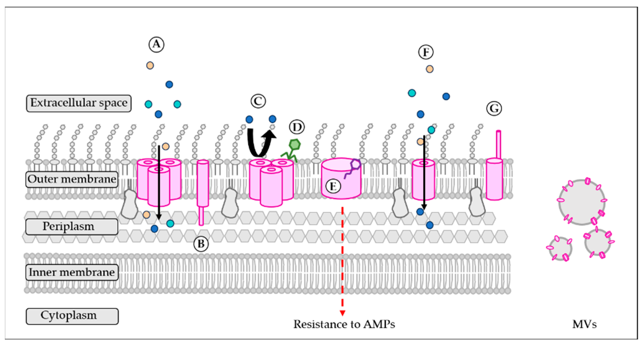
OMPs are often thought to as porins, but they are so much more than that. Although the mechanisms underlying their other functions have not all been clearly described yet, OMPs play major roles in bacteria that are not limited to molecules diffusion. By being displayed in great quantities at the surface of the bacteria, they are indeed implicated in a wide variety of bacterial functions (Figure 2). In response to the environmental changes while entering the host, bacteria can modulate the abundance of specific OMPs to increase permeability to essential nutrients, decrease the entry of toxic elements, modify its adhesion to surfaces and host cells and control the immune system. Porins provide the selective permeability of the OM. They can be divided in two families, including general porins and substrate-specific porins. General porins, which are stable trimers with independent pores, can transport a variety of small molecules across the OM [37,38], while sometimes showing a preference for anionic or cationic substrates. Substrate-specific porins allow transportation of specific substrates. In V. cholerae, OmpU and OmpT, which are homologous to OmpC and OmpF of E. coli, have been suggested to function as general porins [67], while OmpS, upregulated by maltose, specifically transports it to the periplasm [68]. After entering the host, V. cholerae modifies the abundance of OmpT and OmpU porins to resist bile. Although essential, available iron is scarce inside the hosts, as it is sequestered to heme, transferrin and ferritin [69]. Bacteria have evolved mechanisms to acquire iron efficiently from those iron-binding proteins and use OMPs to capture and transport iron inside the cell [70].
OMPs are also important for envelope integrity and MV formation. BamA and LptD are essential for OMP and LPS insertion in the OM, respectively. OmpA, by having a globular periplasmic domain non-covalently anchored to the PG layer, confers stability to the OM in E. coli [54,71,72]. V. cholerae can control MV formation, a crucial feature for survival, virulence and adaptation to environment, by modulating OmpA translation [73]. In addition, MV-associated OMPs contribute to the V. cholerae biofilm matrix, with OmpU contributing to adhesion and production of Vibrio polysaccharide (VPS) [74]. Cell-to-cell and surface adhesion not only leads to biofilm formation, but also to the colonization of the host. Their display at the bacterium’s surface and the presence of long extracellular loops make OMPs, including OmpU in V. cholerae and OmpV in Salmonella, potential bacterial adhesins [75,76]. These two proteins also act as sensors of AMPs and convey an intracellular signal for bacterial adaptation and resistance in V. cholerae [77,78,79]. OmpU leads to the activation of the σE membrane stress response, while OmpV leads to the expression of a specialized antimicrobial-resistance operon. V. cholerae can manipulate the inflammatory response using OmpU (see further sections) [79,80]. These are key features for survival and persistence inside the host.
It is also important to note that, by being exposed at the surface of the bacteria, OMPs are perfect targets for bacteriophages and bacteriocins, and were first described as bacteriophage receptors [8]. For example, OmpW is the target of VP5, while TolC is one of the VP3 receptors [81,82].

5. The Outer Membrane Proteins of V. cholerae

OMPs are characterized by their resistance to proteolytic agents such as trypsin and their apparent association with the peptidoglycan layer [83]. There were originally six major OMPs described in V. cholerae (i.e., OmpV, OmpS, OmpA, OmpT, OmpU and OmpX [84]); however, since the abundance of OMPs varies considerably with the growth conditions, the “major” OMPs vary accordingly [84]. Only two OMPs are considered essential: LptD, responsible for LPS transport to the OM, and BamA, facilitating the insertion of OMPs at the OM [66,85,86].

5.1. OmpV (VC1318)

Identified in the 80s, OmpV (23 kDa) is one of the major OMPs in V. cholerae and is common among the strains [84,87,88,89,90,91]. Antibodies against this protein were found in recovering patients [88,92], suggesting that OmpV is expressed during infection and might have a role in pathogenesis. First thought to be a porin based on its sequence [89], OmpV is a heat-shock protein in V. cholerae [92], with a role in osmoregulation in other marine bacteria, including other Vibrio species [93,94,95]. In Salmonella, OmpV is an adhesin that contributes to virulence [76,96] and is associated with virulence in Edwardsiella tarda [97]. Although abundant in the biofilm matrix through its high abundance in MVs [98], OmpV does not seem to be implicated in biofilm formation [74].
In V. cholerae, OmpV was upregulated by human α-defensin 5 and polymyxin B [79,99,100] and was more abundant in the MVs of bacteria grown with subinhibitory concentrations of the peptide polymyxin B [13]. Our group recently identified a new function in antimicrobial resistance for OmpV, as an ompV-deletion mutant was more sensible to polymyxin B [79]. OmpV was linked to the expression of the efflux pump VexAB, known for its implication in polymyxin B resistance [79,101]. OmpV would, in fact, act as a sensor for antimicrobials. A lateral opening in the β-barrel, formed by two short β-strands and accessible through the OM, leads to the electronegative lumen of OmpV, which could accommodate polymyxin B. A conformational change induced by this interaction could then lead to intracellular signaling and the expression of vexAB [79]. OmpV is part of a four-gene specialized operon encoding for (i) the two-component system (TCS) carRS (also known as vprAB), responsible for the activation of the lipid modification system alm in response to polymyxin B [102,103]; (ii) the sensor protein ompV controlling the expression of vexAB in response to polymyxin B; and (iii) the uncharacterized virK, having a role in antimicrobial resistance [79]. The operon organization might explain why in a carS (vprB) mutant, the expression of ompV is decreased [99]. OmpV is annotated as a protein of the MltA-interacting protein (MipA) family [104] and was shown to be an ortholog of Pseudomonas aeruginosa MipA, which also acts a sensor for polymyxin B using a lateral opening [79,105]. The main difference between those systems is that mipA is only present in strains lacking the arn operon, the lipid modification system in P. aeruginosa, while ompV is directly linked to alm in V. cholerae. The regulation cascade leading to vexAB expression and the proteins implicated in intracellular signaling remain to be confirmed experimentally.

5.2. OmpW (VCA0867)

OmpW is a protein of 22 kDa [106,107,108] conserved in Gram-negative bacteria and in V. cholerae. It is used for V. cholerae detection and identification by PCR [109,110,111]. It is also very immunogenic, making it an interesting vaccine candidate [107,112,113,114]. Similar to OmpV, our group showed that OmpW was more abundant in the MVs of V. cholerae grown with subinhibitory concentrations of AMPs, but no function in antimicrobial resistance has been attributed to this protein yet [13]. As a porin with a continuous apolar and hydrophobic channel, OmpW is most likely implicated in the transport of small or hydrophobic molecules through the OM, such as iron [115,116,117,118].
Gram-negative bacteria use transporters at their OM to accumulate molecules and maintain an osmotic pressure in the cytoplasm greater than in the environment, without interfering with bacterial processes [93,119,120,121]. OmpW has been shown to participate in osmoregulation in marine bacteria Photobacterium damselae, V. alginolyticus and V. parahaemolyticus [93,94,95]. Tolerance to osmotic pressure is an important feature of V. cholerae, allowing it to adapt to marine, estuarian and brackish waters, food and the hyperosmotic conditions of the intestine and host [122,123]. V. cholerae can grow at salt concentrations up to 5% by upregulating genes involved in sodium exclusion, potassium inclusion and glutamate biosynthesis, amongst others [122]. A study showed that the expression and abundance of OmpW increase with salt concentration, and that the growth of an OmpW-defective mutant is altered in hypersaline conditions [118]. While the presence of L-carnitine improved the growth of the wild-type strain, it had no impact on the growth of the ompW-defective mutant, for which the extracellular concentration of L-carnitine remained unchanged. The authors demonstrated that OmpW is, in fact, an L-carnitine transporter that enhances V. cholerae growth in hypersaline conditions. The accumulation of sugars, polyols, amino acids and amino acid derivatives, such as carnitine, is a known mechanism of tolerance to osmotic stress [118,124].

5.3. OmpU (VC0633)

OmpU is a 16-stranded β-barrel of 38 kDa and is the most abundant porin of the OM [84,125]. As such, OmpU is associated with multiple functions and is probably the most studied OMP. OmpU is highly conserved among V. cholerae strains. It is used for the discrimination of toxigenic and epidemic strains based on an OmpU-mass difference, since some alleles are associated with epidemic strains [126]. OmpU is osmoregulated, being more abundant in low osmolarity conditions [84]. At the OM, OmpU forms a trimeric structure, with large hydrophilic channels contributing to the permeability of the OM [67,84,125,127,128]. The N-terminal region of OmpU and its loop L3 constrict the pore, acting as gates [125,129]. The OmpU pore is highly selective for cations and shows oscillations between opened and closed states [125,130].
OmpU has diverse roles in virulence. Cell-to-cell and cell-to-surface adhesion is important for colonization processes and biofilm formation. It has been shown that purified OmpU binds to fibronectin, while the addition of OmpU-specific antibodies reduces the adhesion of V. cholerae to human cells, suggesting a role of OmpU in adhesion to host cells [75]. Further studies using atomic force microscopy revealed that OmpU contributes to adhesion to surfaces and to cell-to-cell adhesion [74]. OmpU is implicated in biofilm formation in V. cholerae not only by acting as an adhesin [75,131], but also by regulating the production of VPS, a major component of V. cholerae biofilm [74,132]. The colony morphology of an ompU-defective mutant was more corrugated than that of the wild-type strain, the mutant having more VPS in the biofilm matrix [74]. The abundance of VpsM and VpsT, a protein necessary for VPS production and a positive transcriptional regulator of vps operons, respectively, was increased in the ompU-mutant, which could explain the increased VPS production [74]. A proteomic analysis revealed that OMPs are present in high quantities in the biofilm matrix, with OmpU being the most abundant through the presence of MVs [74].
OmpU also plays a role in immunomodulation and control of the inflammatory response. An ompU-deleted mutant leads to a reduced production of the cytokines IL-1α, MCP-1, IL-8 and IL-6 by intestinal cells in comparison to the wild-type strain, suggesting a role for OmpU in the induction of inflammation [131,133,134]. OmpU can also activate dendritic cells and lead to the release of IL-6, TNF-α and IL-1β by these cells [135]. However, while inducing the release of pro-inflammatory cytokines by target cells, OmpU would also be responsible for a decrease in LPS-induced inflammation by inducing a negative regulator of TLR, thus leading to tolerance to LPS [80,136]. OmpU induces production of reactive oxygen species (ROS) by macrophages and dendritic cells, altering their functions and modulating inflammation [135,137], and a caspase-independent programmed cell death after translocation to the mitochondria [138]. In addition, an ompU-deleted mutant is more sensible to H2O2 compared to the wild-type strain [139]. By allowing, directly or indirectly, the influx of manganese into the cell, OmpU would indeed contribute to ROS resistance, manganese being a known protective agent against oxidative stress by scavenging superoxide and peroxide [139].
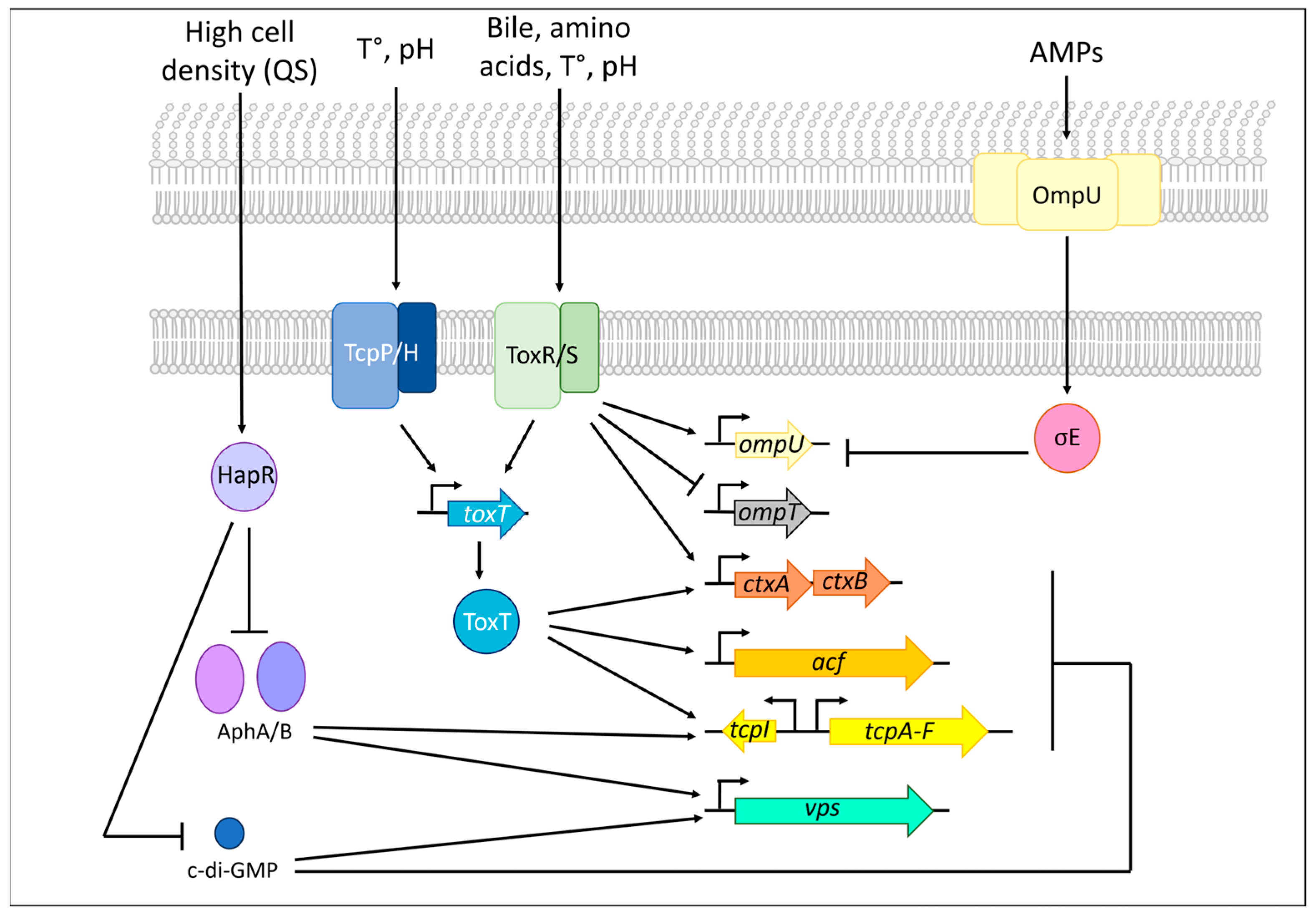
OmpU is a key effector of bile resistance, an essential feature for an enteric bacterium. Its expression is ToxRS-dependant and is inversely linked to that of ompT [140,141,142]. As such, the expression of ompU is increased by bile and deoxycholate, while the expression of ompT is reduced (Figure 3) [143]. Cells that express ompU, but not ompT, are resistant to bile and deoxycholate [144]. V. cholerae protects itself from bile by upregulating the expression of ompU in the gut to prevent the influx of the negatively charged bile salts through the cation-specific OmpU channel [67,130]. The fact that bile does not seem to interact with OmpU strengthens this hypothesis [67,125]. According to this model, early in the infection, bile enters the cell using OmpT, allowing signaling through ToxRS and increasing the transcription of ompU, which in turn blocks bile entry, while still allowing nutrient influx [67]. In addition to bile and deoxycholate resistance [143,145], OmpU is also implicated in resistance to organic acid and anionic detergents [144,146,147].
OmpU serves a sensor for antimicrobials and membrane damage and induces an intracellular signal leading to membrane repair through σE (Figure 3) [77,78,148,149]. OmpU is a major general regulator of σE, the absence of OmpU making σE non-essential in V. cholerae [78,149]. In absence of stress signals, RseA prevents the activity of σE by sequestration to the IM [150]. When cell-envelope perturbations occur, such as an excessive accumulation of OMPs at the OM or misfolded OMPs in the periplasm, the increasing concentration of a C-terminal YxF motif induces the cleaving of RseA by the protease DegS [149,151,152]. RseA is further cleaved by the protease RseP at the IM, releasing σE. σE further associates with polymerase and activates the transcription of envelope-stress response genes—including proteases, chaperones and foldases to reduce the burden of misfolded proteins—as well as small interfering RNA to regulate OMPs synthesis and its own transcription [149,153,154]. Further studies showed that OmpU is not only required for basal transcription and activation of σE, but also for the specific activation of σE in the presence of AMPs [78,149,155]. It has been proposed that the presence of AMPs leads to a conformational change in OmpU, unveiling its YDF motif and activating the DegS-RseP cascade. σE is then free to interact with RNA polymerase and induce its own expression, and that of chaperones, proteases, and membrane repair genes [78,156,157]. In the presence of AMPs, the transcription of rpoE was generally decreased in an OmpU-deficient mutant, and the transcription of σE-downstream genes was not activated, contrary to the wild-type strain [78]. The expression of ompU is regulated by toxR and σE, but it also has self-regulated negative feedback [155].
A polymorphism in ompU has an influence on the bacterium’s interactions with its environment and host, affecting its virulence and resistance to antimicrobial agents. A recent study analyzed more than 1600 V. cholerae genomes and identified 41 ompU alleles forming two distinct phylogenetic clusters [158]. In fact, this study revealed that OmpU from the pandemic cholera group strains differs from that of environmental strains, conferring varying levels of tolerance to bile depending on its origin, with some environmental alleles conferring no resistance to bile at all [158,159]. The ompU allele also has an influence on intestinal colonization and biofilm formation [159]. These variations are attributed to four conserved domains of OmpU, suggesting a strong environmental pressure for virulence on the protein and that some ompU variants confer greater virulence potential [158].
Another feature of OmpU is its implication in the formation of expelled food vacuoles (EFVs) by protozoa after feeding on bacteria [160]. These vacuoles are released in the environment and contain living bacteria, which can further evade from the vacuoles in favorable conditions [160]. Upon ompU mutation, fewer EFVs are produced [160]. The authors also showed that EFVs protect epidemic strains of V. cholerae from environmental and hosts stresses, such as pH, antibiotics, and starvation, increasing their survival and persistence in the environment [160]. Using an infant-mouse model of colonization, V. cholerae from EFVs can outcompete planktonic cells. The authors suggested that EFVs, by protecting V. cholerae from host stress, increase its survival in the host, or a potentiated/primed virulence in EFV [160]. The role of OmpU in EFVs could be linked to resistance to low pH and AMPs, but further studies will be necessary to determine the function and mechanism of OmpU in EFV formation [161].
Figure 3. Main regulation pathways in Vibrio cholerae. Virulence is under the control of many regulation pathways in V. cholerae. Environmental cues such as pH and temperature (T°) are sensed by TcpPH and ToxRS, thereby increasing toxT expression [162,163]. ToxT directly activates virulence by inducing the cholera toxin (ctxAB), the toxin-coregulated pilus (tcpAF) and the expression of accessory colonization factors (acf) [164]. ToxRS also directly induces ctxAB expression. In the presence of bile, V. cholerae represses the expression of ompT, an OMP that is permeable to bile, and induces the expression of ompU through ToxRS, leading to bile resistance [140,144]. OmpU can sense antimicrobial peptides (AMPs) and induce a periplasmic proteolytic cascade leading to the liberation and activation of σE [78]. σE inhibits ompU expression [155]. Virulence and colonization are also under the control of quorum sensing (QS). At low cell density, AphAB are present and induce the expression of the vps and tcp locus [165,166]. At high cell density, HapR inhibits AphAB and c-di-GMP signaling, thus reducing biofilm formation both directly and indirectly [167].
Applmicrobiol 05 00064 g003

5.4. OmpT (VC1854)

OmpT is another major OMP in V. cholerae [84]. OmpT is a 16-stranded β-barrel of 40 kDa forming a trimeric general porin, whose expression is regulated by ToxR (Figure 3) [84,125,141]. OmpT and OmpU are functional homologs of the E. coli general porins OmpC/F, even though they share less than 20% of sequence identity [125]. A crystal structure of OmpT revealed that its cationic-selective pore is constricted not only by the L3 loop but also by the L8 loop [125]. L8 would in fact bend from the extracellular space to interact with L3, greatly narrowing the channel [125]. L8 is also responsible for a small electric field inside OmpT’s pore, making it permeable to molecules such as deoxycholate, and regulating the open/close states of the channel [125]. OmpT’s channel can indeed exhibit frequent long closure periods [130,168].
OmpT is highly expressed in minimum media and would be the most abundant porin when V. cholerae is in the environment, while its expression is decreased inside the host. In addition to bile, rich media, mucin and the addition of amino acids to a minimal media decrease its expression [169]. As described in the previous section, OmpT is inversely regulated relative to OmpU (Figure 3). Since OmpT interacts with bile, ompT is downregulated in the presence of bile salts through ToxR in a ToxT-independent manner, making it the only downregulated gene of the ToxR regulon [125,141,170,171]. OmpT is also quickly removed from the cell surface as V. cholerae enters the host by vesiculation, a process induced by a downregulation of the VacJ/Yrb. OmpT withdrawal and LPS modification and replacement limit bile salt penetration and interaction with AMPs, protecting the bacteria from host defenses [11]. ompT expression is modulated by the carbon source signal. The promotor region of ompT has three CRP binding sites, CRP being a positive regulator when bound to the distal and proximal binding sides [172]. A CRP binding to the proximal sites only would, however, lead to a repression of ompT [172]. In addition to its direct repression, it has been shown that ToxR can prevent the CRP-dependent activation of ompT transcription [172]. Post-transcriptional regulation through base paring of the small non-coding RNA VrrA and Hfq also controls ompT expression [173]. Upon entry into the gut, acidic conditions activate the σE-mediated envelope stress response, leading to the expression of vrrA and the repression of ompT [173].
In addition to bile, OmpT is also more permeable to antibiotics [125]. OmpT is synthesized in larger amounts at high osmolarity, in contrast to OmpU [84,170]. Although the deletion of ompT does not alter CT and TCP production, the replacement of OmpU by OmpT reduced their production [144,146]. This could be due to the differential permeability of OmpU and OmpT, leading to a different influx of signaling molecules [146].
A study from our laboratory showed that OmpT is implicated in resistance to AMPs [13]. When V. cholerae is grown in the presence of PmB, the MVs produced provide protection against the human AMP LL-37 in a Bap1-OmpT dependent manner. Bap1 is a biofilm-associated extracellular matrix protein [174]. Our results showed that Bap1 binds to the LDV domain of OmpT, present in high levels in MVs produced by V. cholerae grown with PmB, and act as a scaffolding protein to capture LL-37. The segregation of LL-37 to the MVs leads to a better bacterial resistance to this AMP.
OmpT might have a role in adhesion, since an ompT-deletion mutant, although exhibiting similar biofilm production abilities, showed a significantly reduced adhesion force in comparison to the parental strain [74]. OmpT and OmpU have been reported to be necessary for the structural integrity of the flagellum [175]. V. cholerae’s flagellum is sheathed with the OM. In a study by Bari et al., OmpU was detected in the flagellum of V. cholerae, while an ompU-ompT mutant showed thinner flagella, fewer flagellated bacteria and lower motility. The authors suggested that the structure of the flagellum would be affected by the absence of OmpU and OmpT.
Studies revealed that MVs can transport bioactive CT and deliver it to human intestinal cells by caveolin-mediated endocytosis [11,176,177]. Using deletion mutants, OmpT and OmpU have been shown to mediate the uptake of CT-filled MVs by intestinal cells, with OmpU being more important for the uptake [11]. Aside from facilitating the transport of CT, MVs protect CT from degradation by intestinal proteases [11]. The mouse ileal loop model showed that while MVs increase CT stability, they also modify the toxin dynamic, inducing a moderate but sustained elevation of cAMP levels, thereby revealing its activity [11].

5.5. Maltoporin OmpS (LamB) (VCA1028)

OmpS is a 43 kDa protein, forming a trimeric porin in V. cholerae [84]. OmpS is common in V. cholerae strains and is one of the major OMPs depending on growth conditions [84,178,179]. OmpS is a maltoporin that is very similar to LamB from E. coli [68]. LamB allows the facilitated diffusion of maltose to the periplasm, where the ABC maltose transporter MalFGK transports it to the cytoplasm [180]. While repressed by glucose, the abundance of OmpS is indeed increased by up to 5 times by maltose or maltodextrins, the induction levels being strain-dependent [68,83,84,179]. The promotor region of ompS contains MalT and CRP binding sites, similarly to the malK promoter [68]. In addition to maltose and cAMP levels, the growth phase influences OmpS regulation, with a maximum abundance during the stationary phase of growth [68]. Indeed, under nutrient-deprived stress, maltoporins can play an important role in adaptation to the environment by allowing carbohydrate uptake [68]. They would also be important for colonization, as maltose and maltodextrins are the most abundant carbohydrates in the intestines, and for survival under starvation conditions [68]. Although the abundance of OmpS is reduced in vitro in a rabbit ileal loop model of infection, antibodies against this protein have been found [179], and OmpS has been identified multiple times in infected human stool samples [181].

5.6. OmpX

A study looking at the major OMPs has described OmpX as an osmoregulated 27 kDa protein [84]. Although very similar to OmpA, OmpX is more ellipsoid, with four long β-strands forming two loops on the extracellular side [8,182,183]. These protrusions could allow OmpX to interact with extracellular surfaces. OmpX is most likely not a porin, based on arabinose diffusion tests and structural analysis [84,183,184]. OmpX is more abundant at low osmolarity than at high osmolarity [84], and it is more abundant in strains lacking OmpU at the OM, independently of ToxR [146]. Little is known about OmpX in V. cholerae. It is annotated as an attachment invasion locus (Ail)/λ outer membrane (lom) protein in V. cholerae [8].

5.7. OmpA (VC2213)

V. cholerae possesses an OmpA-like protein of 35 kDa [185], sharing 47.8% similarity to that of E. coli [73]. It is composed of an N-terminal 8 β-strand barrel domain in the OM and a globular C-terminal periplasmic PG-binding domain [54,186]. The interaction of the PG-binding domain with the PG layer ensures cell envelope integrity, a main function of this protein [72].
In V. cholerae, OmpA was present in MVs and in the biofilm matrix, but an OmpA-deleted mutant had a similar biofilm formation capacity to the wild-type strain [74]. However, this might be a result of the culture conditions, and the role of OmpA in biofilm formation in V. cholerae is yet to be determined. An ompA deletion had no impact on adhesion to amoebas, a natural predator of V. cholerae, and intracellular survival, but improved bacterial survival when cultivated alone or in the presence of amoebas [187].
In V. cholerae, OmpA is part of the envelope stress response and controls MV formation [73,149]. VrrA (Vibrio regulatory RNA of ompA) is a small non-coding RNA implicated in virulence and under the direct control of the bacterial stress envelope regulator σE [73,188]. VrrA represses OmpA translation by base-pairing to the 5′ ribosome binding site and coding region of ompA mRNA [73,173]. Interestingly, a VrrA-induced repression or an ompA deletion promotes MV production, suggesting a role of OmpA in this process [73,187]. Under envelope stress conditions, σE induces the expression of vrrA, thus decreasing OmpA production [73]. The resulting increased vesiculation reduces envelope stress and could protect bacteria from environmental stresses, such as UV radiation damage [188]. VrrA also inhibits the expression of ompT. Its reduced expression under stress conditions could also decrease membrane permeability [173]. In low stress conditions, vrrA is low, promoting OmpA, biofilm (RbmC) and TCP production, thus enabling colonization [73,189].
The OmpA-deletion mutant’s capacity to colonize an infant mouse model is reduced by 10-fold, while the overexpression of OmpA through a vrrA-deletion mutant increases colonization ability, suggesting a role in virulence [73]. OmpA was identified in multiple infected human stool [181]. The hypervesiculation of an ompA-deletion mutant could also lead to the killing of amoebas and improved overall survival [187].
Another function of OmpA would be the export of SipA-bound AMPs [190]. SipA (VCA0732) is a periplasmic protein whose expression is induced by AMPs through the TCS VC1638-VC1639 [100]. SipA has an oligonucleotide/oligosaccharide-binding (OB) fold domain forming a pocket that could accommodate cationic molecules such as AMPs [190]. It has been suggested that SipA captures AMPs that reached the periplasm and chaperones them to OmpA, which could transport them out of the cell to detoxify the periplasm [190]. In classical strains where alm, the main resistance mechanism to AMP, is absent, SipA and OmpA highly contribute to antimicrobial resistance.

5.8. Chitoporin ChiP (VC0972)

V. cholerae is a marine bacterium often associated with shellfishes and crustaceans, covered by a chitinous exoskeleton [191]. V. cholerae can thrive on them by using chitin as a carbon source with a complex catabolic cascade. Chitin is first degraded by secreted chitinases to soluble chitin oligosaccharides [192]. Then, the chitoporin ChiP facilitates transportation of chitin oligosaccharides to the periplasm [191,193]. ChiP is a porin that is essential for growth on chitin, and its expression is induced by the presence of soluble chitin oligosaccharides [191,193]. Once in the periplasm, chitin is further degraded and translocated to the cytoplasm by inner membrane transporters for bacterial use [192]. The two-step chitin degradation cascade might bring an advantage to V. cholerae as many bacteria can transport chitin-derived mono- and disaccharides, while only a few can transport oligosaccharides [192]. Chitin also activates competence in V. cholerae [191].
Recently, a structural prediction, compared to the crystal structure of ChiP from other Vibrio species, revealed that ChiP from V. cholerae has a predicted molecular weight of 35.5 kDa, a structure composed of 16 β-strands and forms a homotrimeric structure [194,195]. The L3 loop constricts the ChiP channel and is predicted to control the size of molecules allowed through the channel, sugar binding and permeation [194]. ChiP has a high substrate specificity [196] and is permeable to monosaccharides and chitin oligosaccharides [194]. ChiP is under the control of the ChiS regulon, which activates its transcription in the presence of chitin, and the sRNA MicX (A10), downregulating its expression [191,197]. A high-throughput RNA sequencing of a V. cholerae OmpR-depleted mutant showed that OmpR, the regulator of the TCS EnvZ/OmpR, also plays a role in ChiP regulation [198]. The authors suggest that V. cholerae represses virulence factors before exiting the host, in preparation for its return to the aquatic ecosystem. A pH switch from acidic to alkaline in rice-water stool might activate OmpR, which further represses virulence and activates important genes for an aquatic lifestyle, such as ChiP [198].

5.9. LptD (VC0446)

As described in a previous section, LPS is a major component of the OM. LPSs are synthetized at the IM and translocated to the OM by the lipopolysaccharide transport (Lpt) machinery [199]. The Lpt complex is formed by LptA-G, requires ATP and is essential for Gram-negative bacteria. Lpt contains an ATP-binding cassette transporter (LptBFG), a bridge from the IM to the OM formed by LptC and multiple LptA subunits, as well as the translocation complex LptDE. LptD is the largest known β-barrel, formed by 26 β-strands, and is responsible for the unidirectional translocation of LPS to the OM [200]. The crystal structure of V. cholerae Lpt has not yet been characterized. It is highly conserved among Vibrio species [201]. Similar to BamA, LptD would have a lateral gate to allow the passage of mature LPSs [202].

5.10. Putative Phosphoporin PhoE (VCA1008)

PhoE has been identified in V. cholerae strain 569B as a 38 kDa OMP induced by inorganic phosphate starvation [203,204,205]. A structural prediction analysis showed that PhoE is in fact a putative phosphoporin composed of three 16 β-strands subunits organized in a homotrimer, sharing similarities with PhoE of E. coli [204,206]. It is the first anion-selective porin described in V. cholerae, a specificity conferred by positively charged lysine residues in the N-terminal [204,206]. Although it possesses a putative inorganic phosphate-binding site, PhoE is not a substrate-specific porin [206]. PhoE is under phoBR regulation, and it is a major OMP in inorganic phosphate starvation conditions [203,204,206].
PhoE transcription is induced during infection of the infant mouse small intestine, but not during growth in rich media, and a deletion mutant is highly attenuated [207,208]. In the El Tor strain N16961, PhoE also participates to bile resistance, with a mutant being more sensitive to sodium deoxycholate, and this is likely due to its high similarity to its paralogue OmpU [209]. In this strain, a deletion of PhoE impaired bacterial growth [209]. ToxR is implicated in the regulation of PhoE under low inorganic phosphate conditions, increasing its expression in the presence of sodium deoxycholate [209]. However, the authors suggest that these phenotypes are strain-dependent since, in the classical strain O395, growth and bile resistance are not affected by PhoE deletion, and phoE expression is ToxR-independent [209].

5.11. TonB-Dependent Siderophore Receptors

As an essential yet limited nutrient, iron acquisition is an important feature for survival inside a host, where it is mostly segregated to heme and other binding proteins [210,211]. Siderophores are organic iron chelators produced and secreted by bacteria, which scavenge the environment for ionic iron [212]. To acquire iron, Gram-negative bacteria can use their own siderophores, capture siderophores from other bacteria, capture exogenous iron carrier molecules or directly take up soluble ferrous iron [210]. The transport of captured iron to the periplasm depends on active transport through the ligand-specific TonB-dependent transporters (TBDTs) located at the OM. The TBDTs specifically serve as receptors for endogenous siderophores, exogenous siderophores and iron carrier molecules. Their expression is regulated by the presence of iron. An IM complex formed by TonB, ExbB and ExbD provides the required energy for transportation through the OM [213]. There are two TonB-ExbB-ExbD systems in V. cholerae [214]. The periplasmic chelated iron is then transported to an ABC transporter by a periplasmic binding protein [210]. Iron is enzymatically released from its binding protein into the cytoplasm.
V. cholerae can secrete and capture the catechol siderophore vibriobactin, and can also capture enterobactin fluvibactin from E. coli and V. fluvialis [215]. V. cholerae possesses many TBDTs, which act as siderophore receptors: the vibriobactin receptor ViuA (VC2211) [215], the enterobactin receptors VctA (VCA0232) and IrgA (VC0475) [216], the hemoglobin/transferrin/lactoferrin family receptors HutA (VCA0576), HasR (VCA0625) and HutR (VCA0064) [211] and the ferrichrome receptor FhuA (VC0200) [217]. Vitamin B12 is mainly transported by the TonB-dependent receptor BtuB (VC0156) [218]. Iron acquisition in Vibrio has been reviewed elsewhere [210].

5.12. BamA (VC2252)

The β-barrel assembly machinery (BAM) complex is located at the OM and allows the folding and insertion of OMPs in the OM [219]. The BAM complex is highly conserved in Gram-negative bacteria, and homologs are present in mitochondria and chloroplast. The BAM complex is composed of the β-barrel OMP BamA and the four lipoproteins VC0762 (BamB), VC2156 (BamC), VC0708 (BamD) and VC0851 (BamE), anchored to the OM [63]. Studies of BamA (Omp85) in Neisseria meningitidis paved the way for understanding the biogenesis of OM and OMPs [64,86]. BamA and BamD are essential for the activity of the BAM complex, as their deletion leads to an accumulation of unfolded OMPs in the periplasm, although the other components are required for efficient OMP biogenesis [219,220].
BamA has a 90 kDa molecular weight and is composed of a 16-stranded β-barrel C-terminal domain [221] and of five N-terminal polypeptide-transport-associated (POTRA) domains located in the periplasm [222,223]. The extracellular loops of the barrel enclose BamA’s channel, inserting into the lumen and preventing the passage of molecules through its pore [200,224]. The POTRA domains interact with the other components of the BAM complex to form a ring complex in the periplasm [219] and could thus serve as scaffolds for assembly and maintenance of the structure of the BAM complex [219,225]. They would also be implicated in recognizing, docking, chaperoning and contributing to the folding of new OMPs [226]. A study in E. coli using deletion mutants of each POTRA domain showed that while POTRA 1 and 2 are not essential, their deletion impaired bacterial growth [225]. Conversely, the deletion of POTRA 3, 4 and 5 led to cell death in E. coli [219,225]. POTRA domain 1 would interact with SurA, the chaperone transporting OMPs across the periplasm [200]. POTRA domains 3 and 5 would serve as a scaffold for BamB and BamCDE, respectively [225]. The conformation of the POTRA domains appears to be dynamic, as different structural analyses revealed four conformational states of BamA [227]. The dynamism of POTRA domains could thus lead to conformational changes in the BAM complex, facilitating the docking and insertion of OMPs to the OM [219].
Although the crystal structure of BamA in E. coli has been characterized [224,225,226,228,229,230], the mechanism of insertion and folding of OMPs is still unclear [230]. Two models are currently debated [200,230]. In the first model, a lateral opening in the barrel between the β-strands β1 and β16 caused by few hydrogen bonds could allow the insertion of OMPs in the OM by budding [221]. This opening would be dynamic, similar to a zipper of hydrogen bounds, and would open and close according to conformational changes in the POTRA domains and other components of the BAM complex [221,231]. In this model, the β-strands of the new OMP could integrate the OM in this lateral opening, forming a BamA/OMP hybrid barrel. The newly formed OMP and BamA could then close their barrels, with the new OMP budding from BamA [232]. In the second model, the new OMP could form in the periplasmic side of the OM and be incorporated into the OM near β1 and β16, where the membrane is distorted by BamA [221]. On this side of the barrel, the β-strands are shorter than on the opposite side, resulting in a thin hydrophobic belt and pinching of the membrane [200,221,227]. This feature was suggested to prime the membrane for the insertion of newly formed OMPs [221].
V. cholerae BamA shares 56% sequence identity with BamA from E. coli. It has five POTRA domains [233]. Although its function must include the insertion of newly formed OMPs into the OM, its structure has not been resolved yet.

5.13. ObfA (VC1154)

Recently, in an elegant study by Ebenberger et al., the 20 kDa outer membrane protein ObfA associated with the MVs was shown to participate in biofilm formation, in vivo colonization and a new intra-species quorum-sensing signalling mechanism in V. cholerae [234]. The authors demonstrated that ObfA was present in the MVs from V. cholerae biofilms, and that a deletion mutant produced less biofilm and was less virulent than the wildtype strain, due to a reduced vps expression and cholera toxin production, thereby suggesting a link between ObfA and HapR. In fact, their results showed that ObfA is linked to the transcription of the sRNAs CsrC and CsrD, which inhibit CsrA activity. CsrA is a post-transcriptional regulator stimulating HapR activity, HapR being a regulator of vps and virulence genes expression (Figure 3).
Furthermore, the addition of ObfA-containing MVs, but not the MVs from an obfA-deletion mutant, restored the biofilm formation capacity and the vps expression levels of the obfA mutant, suggesting that ObfA is sensed by bacteria and induces intracellular signalling. Their results showed a repressed HapR activity upon fusion of the ObfA-containing MVs with the recipient bacterium, thus leading to increased biofilm formation, colonization fitness and virulence. Their findings highlight a new intra-species quorum-sensing mechanism, which depends on MVs, and a new HapR regulatory effector.

5.14. Uncharacterized OMPs

Many ORF, annotated as OMPs, are uncharacterized in V. cholerae. VC2002 is a DUF2860 domain-containing hypothetical protein, but the prediction tool FoldSeek [235] marks it as an E. coli OmpG structural ortholog, sharing less than 10% sequence identity. The crystal structure of OmpG revealed that it is an unspecific, monomeric 14 β-strand, pore-forming OMP that facilitates sugar import in the absence of LamB [236,237,238].
VC2305 is annotated as OmpK, and it is a structural ortholog of Tsx [239,240]. In E. coli, Tsx is a monomeric 12-stranded barrel transporting nucleosides across the OM [56]. It could be implicated in nucleosides scavenging in V. cholerae, but its role has not been confirmed yet.
VC0935, annotated as the hypothetical protein VpsM, has been shown to be essential for vibrio polysaccharide (VPS) production and thus biofilm formation [132]. In V. cholerae, a mutant of this protein lost the corrugation phenotype, produced less biofilm and was not able to form a pellicle [132]. It was suggested to form, along with VpsN (a polysaccharide export protein, VC0936), the OM VPS transporter [241,242], but its role is yet to be confirmed.
VC1329 is a putative opacity protein-related protein regulated by histone-like nucleoid-structuring protein (H-NS) and VpsT control, suggesting a role in the biofilm matrix [243,244].
V. cholerae can incorporate long-chain fatty acids from bile and benthic sediments to its membrane phospholipids [245] and use exogenic fatty acids for energy production [246]. The uptake of fatty acids in E. coli is facilitated by the OMP FadL and is further transformed by FadD [247]. Alignment tools predicted VCA0862, VC1042 and VC1043 to be FadL homologs and would be implicated in the import of long-chain fatty acids [246,248]. A comparison of the predicted structure of these putative proteins with FadL and simulated docking analysis revealed that VC1042 and VC1043 would accommodate unsaturated rigid lipids, while VC0862 would require a conformational change to allow long-chain fatty acids passage through the barrel [248,249,250]. VC1043 of V. cholerae, which produces cholera toxin, is highly expressed in a neonatal rabbit intestine model, providing a fitness advantage to these strains [251]. VC1043 was also identified in a proteomic analysis in the stools of patients with clinical cholera, suggesting a role during infection [181,252]. These proteins remain to be characterized empirically.
VC2456, or vpvA, is the first gene of the vibrio phase variation operon, a target of VpsT responsible for the corrugated morphology of the rugose variant in the V. cholerae El Tor strain A1552 [243,253]. In contrast to a vpvC (diguanylate cyclase) deletion, the deletion of vpvA does not lead to a complete conversion to the smooth phenotype but increases motility in this strain [253]. The structure or exact function of VpvA has not been resolved yet.
VC0844 encodes for the accessory colonization factor subunit A, AcfA. VC0844 is located within the vibrio pathogenic island VPI-1, being therefore regulated by ToxR and ToxT, and is implicated in intestinal colonization by V. cholerae O395 classical strain and in the biogenesis of the toxin-coregulated pilus [254,255,256,257]. Its expression is upregulated in biofilms [258] and under hypoxic conditions and anaerobiosis [259]. AcfA has been considered for oral vaccine development [260].
VC1622 is a putative OMP with an OmpA domain that could bind peptidoglycan. VCA0195 (a porin-family protein), VCA0568 (vxrD or traD) [261], VCA0738 (traF, pilus) [262] and VCA0659 (an OmpA-family protein) [263] are putative porin-family proteins of unknown functions.

6. Discussion and Future Directions

We aimed to demonstrate throughout this review the diversity of V. cholerae’s OMPs and their association with a wide variety of functions (Table 1), making them important components of Gram-negative bacteria. Besides their role as general or substrate-specific porins, they are implicated in many regular cellular processes, including osmoregulation, structural integrity, and resistance to AMPs, the immune system and bile. The first and major observation that we made while writing this review is that although there are a lot of studies that characterized OMP-deficient mutants’ phenotypes and granted OMPs with general functions or roles, the mechanisms underlaying those functions, in fact, were most often not elucidated at all. OMPs are abundant at the cell surface, so it is expected that they would be implicated in multiple cell processes and have such diverse roles in interacting with the outside world. However, the molecular mechanisms behind those functions might help to understand their importance and their modulation during pathogenesis, guiding the development of vaccine, anti-virulence, anti-adhesion or antimicrobial molecules. In addition, roles have not been attributed to all OMPs, such as the major OMP OmpX in V. cholerae, and much work is still needed in this field. This OMP remains barely understood, as its structure is still unresolved, and its regulation and functions are still unknown.
With the rise of protein structure prediction tools such as AlphaFold3.0 [235,264,265,266], it has become more accessible to predict the structure of uncharacterized proteins, even for non-structural biologists [267]. Structural predictions can provide useful insights into the functions of proteins and reveal, using interaction prediction tools, potential mechanisms underlaying the functions of OMPs [268]. Such predictions provide precious leads, steering research toward fruitful avenues and elevating research to higher levels. For instance, these prediction tools revealed a potential mechanism of signalisation for OmpV (MipA) and the phosphoporin PhoE in V. cholerae [79,204], although the interaction partners relaying the intracellular signaling upon antimicrobial binding are still to be confirmed experimentally. The experimental validation of structural models is indeed important, particularly for proteins such as ChiP, LptD, PhoE and OmpV, for which only computational predictions are available. Techniques such as X-ray crystallography, solution and solid-state nuclear magnetic resonance, and cryo-electron microscopy are essential for confirming the accuracy of these models [269]. Additionally, experimental analyses can reveal functional insights, including multimerization and complex formation, which are critical for understanding the biological roles of these membrane proteins. Given the number of uncharacterized OMPs in V. cholerae and their potential applications, it would be pertinent to begin exploring information on that matter.
A key limitation of computational prediction tools is their reliance on resolved structures for comparison. Indeed, to efficiently predict a protein structure and function, a protein whose structure is resolved is needed. With OMPs, a high degree of structural homology with other known OMPs is often linked to the number of β-strands forming the barrel, while periplasmic and extracellular loops differ. These loops can inform on OMPs functions as it is the part interacting with periplasmic or extracellular components or can be used as a plug. Another limitation of computational prediction tools is the static nature of the predictions. Although they can predict interactions between multiple protein complexes, protein dynamics are not well integrated, which makes the prediction of the flexible protein regions or domains unreliable [270,271]. Some homologous proteins can have distinct folds, while other can change their secondary or tertiary structures in response to cellular stimuli, binding ligands or post-translational modifications [267,269]. Because the predictions are based on dominant folds, alternative conformations of fold-changing proteins or structural predictions of homologous proteins with different structures are thus still challenging [267,269,270], especially in complex or fluctuating physicochemical environments [270].
Other poorly resolved features of OMPs include their regulation, formation and insertion into the OM. Although their abundance has been quantified in different media, little is known about the regulators and pathways involved. This information would be invaluable for understanding their role in the virulence and physiology of Gram-negative bacteria. Substantial work has been conducted to untangle how the essential BAM complex governs the insertion of OMPs into the OM, but the mechanism is still unclear and debated [65,230].
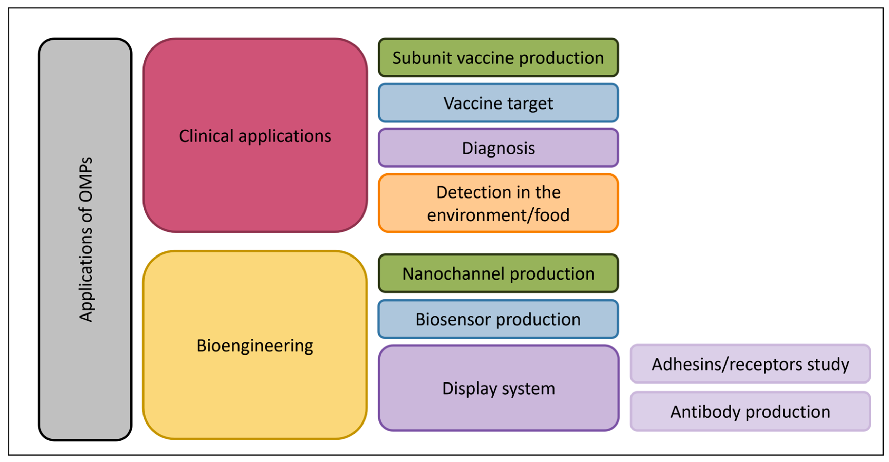
Their location at the cell surface makes OMPs a target for phages and bacteriocins [8], and they could be highly valuable for bioengineering and clinical purposes, as they could lead to vaccines [272,273], display systems [274] and the development of antibiotics/anti-virulence molecules [275], among others (Figure 4). From this perspective, it is crucial to pursue research on OMPs and gain a better understanding of what they are, what they do, and when they act. Several licensed MV-based vaccines (e.g., Bexsero™) targeting Neisseria meningitidis—built around outer-membrane proteins packaged in MVs—have demonstrated robust efficacy and are currently used in Canada [276]. Such a vaccine is not yet available for Vibrio, but OMPs such as OmpU, LptD, OmpK, OmpT and OmpW are promising candidates [272,273,277]. A recent review highlights the new findings on these OMPs and their immunoprotective potential as vaccines candidates [272].
In V. cholerae, OMPs can be used as a diagnostic tool [278,279] or as a biomarker for toxigenic V. cholerae detection in food or water [126]. The accessibility of OMPs to antibodies and T-cell receptors, their presence in high numbers at the surface of the bacteria, their conserved structure among a family and their expression throughout infection make them the perfect target for vaccine production [272,273,280].
Antibacterial molecules often target essential bacterial processes, while anti-virulence molecules target bacterial virulence factors such as adhesins, nutritional intake molecules and the proteins implicated in biofilm formation to reduce selective pressure and resistance [281]. Being exposed at the cell surface and implicated in iron (TBDTs) or B12 acquisition (BtuB) [282], cell wall integrity (OmpA, LptD) [201], or OMP export (BamA) [230], OMPs thus represent ideal candidates for antimicrobials or the development of anti-virulence molecules.
As for bioengineering, OMPs could be used as display systems [274,283]. The production of fusion proteins with an OMP could thus lead to the presentation of desired antigenic epitopes at the cell surface for antibody production or for studying adhesins/receptors [284]. The presentation of antigens on OMPs within MVs could even lead to the development of subunit vaccines. An ompA deletion could increase MV production, improving the yield of antigen-presenting particles. We can only imagine that OMPs could be coupled with drugs for specific MV-mediated specific delivery. The production of modified OMPs could also allow the production of nanopores used as biosensors, for protein–protein interaction and nutrient uptake studies, as well as sequencing, among other applications [285,286,287,288].

7. Conclusions

OMPs are highly versatile components that contribute to a broad spectrum of cellular processes beyond their traditional roles as porins. While phenotypic studies have highlighted their involvement in virulence, resistance, structural integrity and interaction with the environment, the underlying molecular mechanisms remain largely unexplored. Advances in structural prediction and interaction modeling tools now offer promising avenues to bridge this knowledge gap. Given their accessibility, abundance and functional diversity, OMPs hold immense potential for translational applications, including diagnostics, vaccine development and targeted antimicrobial or anti-virulence strategies. Continued investigation into their structure, regulation, and mechanistic roles will be critical to fully harness their biological and biotechnological value.

Author Contributions

A.M.-D.: project administration, conceptualization, formal analysis, writing—original draft, writing—review and editing and visualization. M.D.: writing—review and editing, supervision, funding acquisition and project administration. All authors have read and agreed to the published version of the manuscript.

Funding

This work was supported by the Natural Sciences and Engineering Research Council of Canada (NSERC http://www.nserc-crsng.gc.ca/index_eng.asp) Discovery Grant (grant number: RGPIN-2017-05322; MD). AM-D received financial support from the NSERC scholarship program (BESC D3-558624-2021) and a scholarship from Fonds de Recherche du Québec-Santé (FRQS).

Data Availability Statement

No new data were created or analyzed in this study. Data sharing is not applicable to this article.

Acknowledgments

The authors would like to thank Ressources Aquatiques Québec, an inter-institutional group supported financially by the Fonds de Recherche du Québec—Nature et Technologies (FRQNT) for their support.

Conflicts of Interest

The authors declare no conflicts of interest.

References

  1. World Health Organization. Antimicrobial Resistance: Global Report on Surveillance; World Health Organization: Geneva, Switzerland, 2014. [Google Scholar]
  2. Rossolini, G.M.; Arena, F.; Pecile, P.; Pollini, S. Update on the antibiotic resistance crisis. Curr. Opin. Pharmacol. 2014, 18, 56–60. [Google Scholar] [CrossRef] [PubMed]
  3. Santajit, S.; Indrawattana, N. Mechanisms of antimicrobial resistance in ESKAPE pathogens. Biomed Res. Int. 2016, 2016, 2475067. [Google Scholar] [CrossRef] [PubMed]
  4. Delcour, A.H. Outer membrane permeability and antibiotic resistance. Biochim. Biophys. Acta 2009, 1794, 808–816. [Google Scholar] [CrossRef]
  5. Guest, R.L.; Silhavy, T.J. Cracking outer membrane biogenesis. Biochim. Biophys. Acta-Mol. Cell Res. 2023, 1870, 119405. [Google Scholar] [CrossRef] [PubMed]
  6. Henderson, J.C.; Zimmerman, S.M.; Crofts, A.A.; Boll, J.M.; Kuhns, L.G.; Herrera, C.M.; Trent, M.S. The power of asymmetry: Architecture and assembly of the Gram-negative outer membrane lipid bilayer. Annu. Rev. Microbiol. 2016, 70, 255–278. [Google Scholar] [CrossRef]
  7. Narita, S.I.; Tokuda, H. Bacterial lipoproteins; biogenesis, sorting and quality control. Biochim. Biophys. Acta Mol. Cell Biol. Lipids 2017, 1862, 1414–1423. [Google Scholar] [CrossRef]
  8. Koebnik, R.; Locher, K.P.; Van Gelder, P. Structure and function of bacterial outer membrane proteins: Barrels in a nutshell. Mol. Microbiol. 2000, 37, 239–253. [Google Scholar] [CrossRef]
  9. Guerrero-Mandujano, A.; Hernandez-Cortez, C.; Ibarra, J.A.; Castro-Escarpulli, G. The outer membrane vesicles: Secretion system type zero. Traffic 2017, 18, 425–432. [Google Scholar] [CrossRef]
  10. Reyes-Robles, T.; Dillard, R.S.; Cairns, L.S.; Silva-Valenzuela, C.A.; Housman, M.; Ali, A.; Wright, E.R.; Camilli, A. Vibrio cholerae outer membrane vesicles inhibit bacteriophage infection. J. Bacteriol. 2018, 200. [Google Scholar] [CrossRef]
  11. Zingl, F.G.; Thapa, H.B.; Scharf, M.; Kohl, P.; Muller, A.M.; Schild, S. Outer membrane vesicles of Vibrio cholerae protect and deliver active cholera toxin to host cells via porin-dependent uptake. mBio 2021, 12, e0053421. [Google Scholar] [CrossRef]
  12. Bitar, A.; Aung, K.M.; Wai, S.N.; Hammarstrom, M.L. Vibrio cholerae derived outer membrane vesicles modulate the inflammatory response of human intestinal epithelial cells by inducing microRNA-146a. Sci. Rep. 2019, 9, 7212. [Google Scholar] [CrossRef]
  13. Duperthuy, M.; Sjostrom, A.E.; Sabharwal, D.; Damghani, F.; Uhlin, B.E.; Wai, S.N. Role of the Vibrio cholerae matrix protein Bap1 in cross-resistance to antimicrobial peptides. PLoS Pathog. 2013, 9, e1003620. [Google Scholar] [CrossRef] [PubMed]
  14. Vanhove, A.S.; Duperthuy, M.; Charriere, G.M.; Le Roux, F.; Goudenege, D.; Gourbal, B.; Kieffer-Jaquinod, S.; Coute, Y.; Wai, S.N.; Destoumieux-Garzon, D. Outer membrane vesicles are vehicles for the delivery of Vibrio tasmaniensis virulence factors to oyster immune cells. Environ. Microbiol. 2015, 17, 1152–1165. [Google Scholar] [CrossRef] [PubMed]
  15. Giacomucci, S.; Mathieu-Denoncourt, A.; Vincent, A.T.; Jannadi, H.; Duperthuy, M. Experimental evolution of Vibrio cholerae identifies hypervesiculation as a way to increase motility in the presence of polymyxin B. Front. Microbiol. 2022, 13, 932165. [Google Scholar] [CrossRef] [PubMed]
  16. Clemens, J.D.; Nair, G.B.; Ahmed, T.; Qadri, F.; Holmgren, J. Cholera. Lancet 2017, 390, 1539–1549. [Google Scholar] [CrossRef]
  17. Halpern, M.; Izhaki, I. Fish as hosts of Vibrio cholerae. Front. Microbiol. 2017, 8, 282. [Google Scholar] [CrossRef]
  18. Kaper, J.B.; Morris, J.G., Jr.; Levine, M.M. Cholera. Clin. Microbiol. Rev. 1995, 8, 48–86. [Google Scholar] [CrossRef]
  19. Deen, J.; Mengel, M.A.; Clemens, J.D. Epidemiology of cholera. Vaccine 2020, 38 (Suppl. 1), A31–A40. [Google Scholar] [CrossRef]
  20. Johnson, J.A.; Salles, C.A.; Panigrahi, P.; Albert, M.J.; Wright, A.C.; Johnson, R.J.; Morris, J.G., Jr. Vibrio cholerae O139 synonym bengal is closely related to Vibrio cholerae El Tor but has important differences. Infect. Immun. 1994, 62, 2108–2110. [Google Scholar] [CrossRef]
  21. Li, M.; Shimada, T.; Morris, J.G., Jr.; Sulakvelidze, A.; Sozhamannan, S. Evidence for the emergence of non-O1 and non-O139 Vibrio cholerae strains with pathogenic potential by exchange of O-antigen biosynthesis regions. Infect. Immun. 2002, 70, 2441–2453. [Google Scholar] [CrossRef]
  22. Tobin-D’Angelo, M.; Smith, A.R.; Bulens, S.N.; Thomas, S.; Hodel, M.; Izumiya, H.; Arakawa, E.; Morita, M.; Watanabe, H.; Marin, C.; et al. Severe diarrhea caused by cholera toxin-producing vibrio cholerae serogroup O75 infections acquired in the southeastern United States. Clin. Infect. Dis. 2008, 47, 1035–1040. [Google Scholar] [CrossRef] [PubMed]
  23. Haley, B.J.; Choi, S.Y.; Grim, C.J.; Onifade, T.J.; Cinar, H.N.; Tall, B.D.; Taviani, E.; Hasan, N.A.; Abdullah, A.H.; Carter, L.; et al. Genomic and phenotypic characterization of Vibrio cholerae non-O1 isolates from a US Gulf Coast cholera outbreak. PLoS ONE 2014, 9, e86264. [Google Scholar] [CrossRef] [PubMed]
  24. Crowe, S.J.; Newton, A.E.; Gould, L.H.; Parsons, M.B.; Stroika, S.; Bopp, C.A.; Freeman, M.; Greene, K.; Mahon, B.E. Vibriosis, not cholera: Toxigenic Vibrio cholerae non-O1, non-O139 infections in the United States, 1984–2014. Epidemiol. Infect. 2016, 144, 3335–3341. [Google Scholar] [CrossRef] [PubMed]
  25. Ke, B.; Pang, B.; He, D.; Xu, J.; Chen, Q.; Liang, J.; Chen, J.; Li, Z.; Zhou, H.; Deng, X.; et al. Phylogenetic analysis of serogroup O5 Vibrio cholerae that caused successive cholera outbreaks—Guangdong province, China, 2020–2021. China CDC Wkly. 2022, 4, 238–241. [Google Scholar] [CrossRef]
  26. Mathieu-Denoncourt, A.; Giacomucci, S.; Duperthuy, M. The secretome of Vibrio cholerae. In Infections and Sepsis Development; Huang, L., Li, J., Eds.; IntechOpen: London, UK, 2021. [Google Scholar]
  27. Echazarreta, M.A.; Klose, K.E. Vibrio Flagellar Synthesis. Front. Cell. Infect. Microbiol. 2019, 9, 131. [Google Scholar] [CrossRef]
  28. Oki, H.; Kawahara, K.; Iimori, M.; Imoto, Y.; Nishiumi, H.; Maruno, T.; Uchiyama, S.; Muroga, Y.; Yoshida, A.; Yoshida, T.; et al. Structural basis for the toxin-coregulated pilus–dependent secretion of Vibrio cholerae colonization factor. Sci. Adv. 2022, 8, eabo3013. [Google Scholar] [CrossRef]
  29. Jaroslawski, S.; Duquesne, K.; Sturgis, J.N.; Scheuring, S. High-resolution architecture of the outer membrane of the Gram-negative bacteria Roseobacter denitrificans. Mol. Microbiol. 2009, 74, 1211–1222. [Google Scholar] [CrossRef]
  30. Wimley, W.C. Toward genomic identification of beta-barrel membrane proteins: Composition and architecture of known structures. Protein Sci. 2002, 11, 301–312. [Google Scholar] [CrossRef]
  31. Silhavy, T.J.; Kahne, D.; Walker, S. The bacterial cell envelope. Cold Spring Harb. Perspect. Biol. 2010, 2, a000414. [Google Scholar] [CrossRef]
  32. Kamio, Y.; Nikaido, H. Outer membrane of Salmonella Typhimurium: Accessibility of phospholipid head groups to phospholipase c and cyanogen bromide activated dextran in the external medium. Biochemistry 1976, 15, 2561–2570. [Google Scholar] [CrossRef]
  33. Bertani, B.; Ruiz, N. Function and Biogenesis of Lipopolysaccharides. EcoSal Plus 2018, 8. [Google Scholar] [CrossRef] [PubMed]
  34. Malinverni, J.C.; Silhavy, T.J. An ABC transport system that maintains lipid asymmetry in the gram-negative outer membrane. Proc. Natl. Acad. Sci. USA 2009, 106, 8009–8014. [Google Scholar] [CrossRef]
  35. Hughes, G.W.; Hall, S.C.L.; Laxton, C.S.; Sridhar, P.; Mahadi, A.H.; Hatton, C.; Piggot, T.J.; Wotherspoon, P.J.; Leney, A.C.; Ward, D.G.; et al. Evidence for phospholipid export from the bacterial inner membrane by the Mla ABC transport system. Nat. Microbiol. 2019, 4, 1692–1705. [Google Scholar] [CrossRef]
  36. Sun, J.; Rutherford, S.T.; Silhavy, T.J.; Huang, K.C. Physical properties of the bacterial outer membrane. Nat. Rev. Microbiol. 2022, 20, 236–248. [Google Scholar] [CrossRef]
  37. Nikaido, H. Molecular basis of bacterial outer membrane permeability revisited. Microbiol. Mol. Biol. Rev. 2003, 67, 593–656. [Google Scholar] [CrossRef]
  38. Nikaido, H.; Vaara, M. Molecular basis of bacterial outer membrane permeability. Microbiol. Rev. 1985, 49, 1–32. [Google Scholar] [CrossRef] [PubMed]
  39. Carpenter, T.S.; Parkin, J.; Khalid, S. The free energy of small solute permeation through the Escherichia coli outer membrane has a distinctly asymmetric profile. J. Phys. Chem. Lett. 2016, 7, 3446–3451. [Google Scholar] [CrossRef]
  40. Beveridge, T.J. Structures of gram-negative cell walls and their derived membrane vesicles. J. Bacteriol. 1999, 181, 4725–4733. [Google Scholar] [CrossRef] [PubMed]
  41. Toyofuku, M.; Nomura, N.; Eberl, L. Types and origins of bacterial membrane vesicles. Nat. Rev. Microbiol. 2019, 17, 13–24. [Google Scholar] [CrossRef]
  42. Magana, G.; Harvey, C.; Taggart, C.C.; Rodgers, A.M. Bacterial outer membrane vesicles: Role in pathogenesis and host-cell interactions. Antibiotics 2023, 13, 32. [Google Scholar] [CrossRef]
  43. Toyofuku, M.; Schild, S.; Kaparakis-Liaskos, M.; Eberl, L. Composition and functions of bacterial membrane vesicles. Nat. Rev. Microbiol. 2023, 21, 415–430. [Google Scholar] [CrossRef]
  44. Marinacci, B.; Krzyzek, P.; Pellegrini, B.; Turacchio, G.; Grande, R. Latest update on outer membrane vesicles and their role in horizontal gene transfer: A mini-review. Membranes 2023, 13, 860. [Google Scholar] [CrossRef] [PubMed]
  45. Rompikuntal, P.K.; Vdovikova, S.; Duperthuy, M.; Johnson, T.L.; Ahlund, M.; Lundmark, R.; Oscarsson, J.; Sandkvist, M.; Uhlin, B.E.; Wai, S.N. Outer membrane vesicle-mediated export of processed PrtV protease from Vibrio cholerae. PLoS ONE 2015, 10, e0134098. [Google Scholar] [CrossRef]
  46. Zingl, F.G.; Kohl, P.; Cakar, F.; Leitner, D.R.; Mitterer, F.; Bonnington, K.E.; Rechberger, G.N.; Kuehn, M.J.; Guan, Z.; Reidl, J.; et al. Outer membrane vesiculation facilitates surface exchange and in vivo adaptation of Vibrio cholerae. Cell Host Microbe 2020, 27, 225–237.e228. [Google Scholar] [CrossRef] [PubMed]
  47. Jugder, B.-E.; Watnick, P.I. Vibrio cholerae sheds its coat to make itself comfortable in the gut. Cell Host Microbe 2020, 27, 161–163. [Google Scholar] [CrossRef] [PubMed]
  48. Paul, S.; Chaudhuri, K.; Chatterjee, A.N.; Das, J. Presence of exposed phospholipids in the outer membrane of Vibrio cholerae. J Gen. Microbiol. 1992, 138, 755–761. [Google Scholar] [CrossRef]
  49. Chatterjee, A.; Chaudhuri, S.; Saha, G.; Gupta, S.; Chowdhury, R. Effect of bile on the cell surface permeability barrier and efflux system of Vibrio cholerae. J. Bacteriol. 2004, 186, 6809–6814. [Google Scholar] [CrossRef]
  50. Schulz, G.E. The structure of bacterial outer membrane proteins. Biochim. Biophys. Acta 2002, 1565, 308–317. [Google Scholar] [CrossRef]
  51. Dhar, R.; Slusky, J.S. Outer membrane protein evolution. Curr. Opin. Struct. Biol. 2021, 68, 122–128. [Google Scholar] [CrossRef]
  52. Hartojo, A.; Doyle, M.T. β-barrel membrane proteins fold via hybrid-barrel intermediate states. Curr. Opin. Struct. Biol. 2024, 87, 102830. [Google Scholar] [CrossRef]
  53. Schulz, G.E. β-Barrel membrane proteins. Curr. Opin. Struct. Biol. 2000, 10, 443–447. [Google Scholar] [CrossRef] [PubMed]
  54. Samsudin, F.; Ortiz-Suarez, M.L.; Piggot, T.J.; Bond, P.J.; Khalid, S. OmpA: A flexible clamp for bacterial cell wall attachment. Structure 2016, 24, 2227–2235. [Google Scholar] [CrossRef] [PubMed]
  55. Tsirigotaki, A.; De Geyter, J.; Šoštaric’, N.; Economou, A.; Karamanou, S. Protein export through the bacterial Sec pathway. Nat. Rev. Microbiol. 2017, 15, 21–36. [Google Scholar] [CrossRef] [PubMed]
  56. Van den Berg, B.; Clemons, W.M., Jr.; Collinson, I.; Modis, Y.; Hartmann, E.; Harrison, S.C.; Rapoport, T.A. X-ray structure of a protein-conducting channel. Nature 2004, 427, 36–44. [Google Scholar] [CrossRef]
  57. Mas, G.; Thoma, J.; Hiller, S. The periplasmic chaperones Skp and SurA. Subcell Biochem. 2019, 92, 169–186. [Google Scholar] [CrossRef]
  58. Schafer, U.; Beck, K.; Muller, M. Skp, a molecular chaperone of Gram-negative bacteria, is required for the formation of soluble periplasmic intermediates of outer membrane proteins. J. Biol. Chem. 1999, 274, 24567–24574. [Google Scholar] [CrossRef]
  59. Chen, R.; Henning, U. A periplasmic protein (Skp) of Escherichia coli selectively binds a class of outer membrane proteins. Mol. Microbiol. 1996, 19, 1287–1294. [Google Scholar] [CrossRef]
  60. Rouviere, P.E.; Gross, C.A. SurA, a periplasmic protein with peptidyl-prolyl isomerase activity, participates in the assembly of outer membrane porins. Genes Dev. 1996, 10, 3170–3182. [Google Scholar] [CrossRef]
  61. Sklar, J.G.; Wu, T.; Kahne, D.; Silhavy, T.J. Defining the roles of the periplasmic chaperones SurA, Skp, and DegP in Escherichia coli. Genes Dev. 2007, 21, 2473–2484. [Google Scholar] [CrossRef]
  62. Struyvé, M.; Moons, M.; Tommassen, J. Carboxy-terminal phenylalanine is essential for the correct assembly of a bacterial outer membrane protein. J. Mol. Biol. 1991, 218, 141–148. [Google Scholar] [CrossRef]
  63. Wu, T.; Malinverni, J.; Ruiz, N.; Kim, S.; Silhavy, T.J.; Kahne, D. Identification of a multicomponent complex required for outer membrane biogenesis in Escherichia coli. Cell 2005, 121, 235–245. [Google Scholar] [CrossRef] [PubMed]
  64. Voulhoux, R.; Bos, M.P.; Geurtsen, J.; Mols, M.; Tommassen, J. Role of a highly conserved bacterial protein in outer membrane protein assembly. Science 2003, 299, 262–265. [Google Scholar] [CrossRef]
  65. Tomasek, D.; Kahne, D. The assembly of β-barrel outer membrane proteins. Curr. Opin. Microbiol. 2021, 60, 16–23. [Google Scholar] [CrossRef] [PubMed]
  66. Rollauer, S.E.; Sooreshjani, M.A.; Noinaj, N.; Buchanan, S.K. Outer membrane protein biogenesis in Gram-negative bacteria. Philos. Trans. R. Soc. B Biol. Sci. 2015, 370, 20150023. [Google Scholar] [CrossRef]
  67. Wibbenmeyer, J.A.; Provenzano, D.; Landry, C.F.; Klose, K.E.; Delcour, A.H. Vibrio cholerae OmpU and OmpT porins are differentially affected by bile. Infect. Immun. 2002, 70, 121–126. [Google Scholar] [CrossRef] [PubMed]
  68. Lång, H.; Palva, E.T. The ompS gene of Vibrio cholerae encodes a growth-phase-dependent maltoporin. Mol. Microbiol. 1993, 10, 891–901. [Google Scholar] [CrossRef]
  69. Anderson, G.J.; Frazer, D.M. Current understanding of iron homeostasis. Am. J. Clin. Nutr. 2017, 106, 1559S–1566S. [Google Scholar] [CrossRef]
  70. Silale, A.; van den Berg, B. TonB-dependent transport across the bacterial outer membrane. Annu. Rev. Microbiol. 2023, 77, 67–88. [Google Scholar] [CrossRef]
  71. Park, J.S.; Lee, W.C.; Yeo, K.J.; Ryu, K.-S.; Kumarasiri, M.; Hesek, D.; Lee, M.; Mobashery, S.; Song, J.H.; Kim, S.I. Mechanism of anchoring of OmpA protein to the cell wall peptidoglycan of the gram-negative bacterial outer membrane. FASEB J. 2012, 26, 219. [Google Scholar] [CrossRef]
  72. Sonntag, I.; Schwarz, H.; Hirota, Y.; Henning, U. Cell envelope and shape of Escherichia coli: Multiple mutants missing the outer membrane lipoprotein and other major outer membrane proteins. J. Bacteriol. 1978, 136, 280–285. [Google Scholar] [CrossRef]
  73. Song, T.; Mika, F.; Lindmark, B.; Liu, Z.; Schild, S.; Bishop, A.; Zhu, J.; Camilli, A.; Johansson, J.; Vogel, J.; et al. A new Vibrio cholerae sRNA modulates colonization and affects release of outer membrane vesicles. Mol. Microbiol. 2008, 70, 100–111. [Google Scholar] [CrossRef]
  74. Potapova, A.; Garvey, W.; Dahl, P.; Guo, S.; Chang, Y.; Schwechheimer, C.; Trebino, M.A.; Floyd, K.A.; Phinney, B.S.; Liu, J.; et al. Outer membrane vesicles and the outer membrane protein OmpU govern Vibrio cholerae biofilm matrix assembly. mBio 2024, 15, e0330423. [Google Scholar] [CrossRef] [PubMed]
  75. Sperandio, V.; Giron, J.A.; Silveira, W.D.; Kaper, J.B. The OmpU outer membrane protein, a potential adherence factor of Vibrio cholerae. Infect. Immun. 1995, 63, 4433–4438. [Google Scholar] [CrossRef]
  76. Kaur, D.; Mukhopadhaya, A. Outer membrane protein OmpV mediates Salmonella enterica serovar Typhimurium adhesion to intestinal epithelial cells via fibronectin and alpha1beta1 integrin. Cell. Microbiol. 2020, 22, e13172. [Google Scholar] [CrossRef] [PubMed]
  77. Mathur, J.; Waldor, M.K. The Vibrio cholerae ToxR-regulated porin OmpU confers resistance to antimicrobial peptides. Infect. Immun. 2004, 72, 3577–3583. [Google Scholar] [CrossRef] [PubMed]
  78. Mathur, J.; Davis, B.M.; Waldor, M.K. Antimicrobial peptides activate the Vibrio cholerae sigmaE regulon through an OmpU-dependent signalling pathway. Mol. Microbiol. 2007, 63, 848–858. [Google Scholar] [CrossRef]
  79. Mathieu-Denoncourt, A.; Whitfield, G.B.; Vincent, A.T.; Berne, C.; Pauzé-Foixet, J.; Mahieddine, F.C.; Brun, Y.V.; Duperthuy, M. The carRS-ompV-virK operon of Vibrio cholerae senses antimicrobial peptides and activates the expression of multiple resistance systems. Sci. Rep. 2025, 15, 13686. [Google Scholar] [CrossRef]
  80. Sakharwade, S.C.; Sharma, P.K.; Mukhopadhaya, A. Vibrio cholerae porin OmpU induces pro-inflammatory responses, but down-regulates LPS-mediated effects in RAW 264.7, THP-1 and human PBMCs. PLoS ONE 2013, 8, e76583. [Google Scholar] [CrossRef]
  81. Xu, D.; Zhang, J.; Liu, J.; Xu, J.; Zhou, H.; Zhang, L.; Zhu, J.; Kan, B. Outer membrane protein OmpW is the receptor for typing phage VP5 in the Vibrio cholerae O1 El Tor biotype. J. Virol. 2014, 88, 7109–7111. [Google Scholar] [CrossRef]
  82. Fan, F.; Li, X.; Pang, B.; Zhang, C.; Li, Z.; Zhang, L.; Li, J.; Zhang, J.; Yan, M.; Liang, W.; et al. The outer-membrane protein TolC of Vibrio cholerae serves as a second cell-surface receptor for the VP3 phage. J. Biol. Chem. 2018, 293, 4000–4013. [Google Scholar] [CrossRef]
  83. Lång, H.A.; Palva, E.T. A major outer membrane protein in Vibrio cholerae is maltose-inducible. Microb. Pathog. 1987, 3, 143–147. [Google Scholar] [CrossRef] [PubMed]
  84. Chakrabarti, S.R.; Chaudhuri, K.; Sen, K.; Das, J. Porins of Vibrio cholerae: Purification and characterization of OmpU. J. Bacteriol. 1996, 178, 524–530. [Google Scholar] [CrossRef] [PubMed]
  85. Wu, T.; McCandlish, A.C.; Gronenberg, L.S.; Chng, S.S.; Silhavy, T.J.; Kahne, D. Identification of a protein complex that assembles lipopolysaccharide in the outer membrane of Escherichia coli. Proc. Natl. Acad. Sci. USA 2006, 103, 11754–11759. [Google Scholar] [CrossRef]
  86. Genevrois, S.; Steeghs, L.; Roholl, P.; Letesson, J.J.; van der Ley, P. The Omp85 protein of Neisseria meningitidis is required for lipid export to the outer membrane. EMBO J. 2003, 22, 1780–1789. [Google Scholar] [CrossRef]
  87. Pohlner, J.; Meyer, T.F.; Jalajakumari, M.B.; Manning, P.A. Nucleotide sequence of ompV, the gene for a major Vibrio cholerae outer membrane protein. Mol. Gen. Genet. 1986, 205, 494–500. [Google Scholar] [CrossRef]
  88. Pohlner, J.; Meyer, T.F.; Manning, P.A. Serological properties and processing in Escherichia coli K12 of OmpV fusion proteins of Vibrio cholerae. Mol. Gen. Genet. 1986, 205, 501–506. [Google Scholar] [CrossRef] [PubMed]
  89. Stevenson, G.; Leavesley, D.I.; Lagnado, C.A.; Heuzenroeder, M.W.; Manning, P.A. Purification of the 25-kDa Vibrio cholerae major outer-membrane protein and the molecular cloning of its gene: ompV. Eur. J. Biochem. 1985, 148, 385–390. [Google Scholar] [CrossRef]
  90. Kabir, S. Composition and immunochemical properties of outer membrane proteins of Vibrio cholerae. J. Bacteriol. 1980, 144, 382–389. [Google Scholar] [CrossRef]
  91. Kelley, J.T.; Parker, C.D. Identification and preliminary characterization of Vibrio cholerae outer membrane proteins. J. Bacteriol. 1981, 145, 1018–1024. [Google Scholar] [CrossRef]
  92. Sahu, G.K.; Chowdhury, R.; Das, J. Heat shock response and heat shock protein antigens of Vibrio cholerae. Infect. Immun. 1994, 62, 5624–5631. [Google Scholar] [CrossRef]
  93. Xu, C.; Wang, S.; Ren, H.; Lin, X.; Wu, L.; Peng, X. Proteomic analysis on the expression of outer membrane proteins of Vibrio alginolyticus at different sodium concentrations. Proteomics 2005, 5, 3142–3152. [Google Scholar] [CrossRef] [PubMed]
  94. Xu, C.; Ren, H.; Wang, S.; Peng, X. Proteomic analysis of salt-sensitive outer membrane proteins of Vibrio parahaemolyticus. Res. Microbiol. 2004, 155, 835–842. [Google Scholar] [CrossRef] [PubMed]
  95. Wu, L.; Lin, X.; Wang, F.; Ye, D.; Xiao, X.; Wang, S.; Peng, X. OmpW and OmpV are required for NaCl regulation in Photobacterium damsela. J. Proteome Res. 2006, 5, 2250–2257. [Google Scholar] [CrossRef] [PubMed]
  96. Kaur, D.; Gandhi, S.; Mukhopadhaya, A. Salmonella Typhimurium adhesin OmpV activates host immunity to confer protection against systemic and gastrointestinal infection in mice. Infect. Immun. 2021, 89, e0012121. [Google Scholar] [CrossRef]
  97. Liu, F.; Tang, X.; Sheng, X.; Xing, J.; Zhan, W. Comparative study of the vaccine potential of six outer membrane proteins of Edwardsiella tarda and the immune responses of flounder (Paralichthys olivaceus) after vaccination. Vet. Immunol. Immunopathol. 2017, 185, 38–47. [Google Scholar] [CrossRef]
  98. Langlete, P.; Krabberod, A.K.; Winther-Larsen, H.C. Vesicles from Vibrio cholerae contain AT-rich DNA and shorter mRNAs that do not correlate with their protein products. Front. Microbiol. 2019, 10, 2708. [Google Scholar] [CrossRef]
  99. Liu, Y.; Xu, T.; Wang, Q.; Huang, J.; Zhu, Y.; Liu, X.; Liu, R.; Yang, B.; Zhou, K. Vibrio cholerae senses human enteric alpha-defensin 5 through a CarSR two-component system to promote bacterial pathogenicity. Commun. Biol. 2022, 5, 559. [Google Scholar] [CrossRef]
  100. Matson, J.S.; Livny, J.; DiRita, V.J. A putative Vibrio cholerae two-component system controls a conserved periplasmic protein in response to the antimicrobial peptide polymyxin B. PLoS ONE 2017, 12, e0186199. [Google Scholar] [CrossRef]
  101. Bina, J.E.; Provenzano, D.; Wang, C.; Bina, X.R.; Mekalanos, J.J. Characterization of the Vibrio cholerae vexAB and vexCD efflux systems. Arch. Microbiol. 2006, 186, 171–181. [Google Scholar] [CrossRef]
  102. Bilecen, K.; Fong, J.C.; Cheng, A.; Jones, C.J.; Zamorano-Sanchez, D.; Yildiz, F.H. Polymyxin B resistance and biofilm formation in Vibrio cholerae are controlled by the response regulator CarR. Infect. Immun. 2015, 83, 1199–1209. [Google Scholar] [CrossRef]
  103. Ko, D.; Sung, D.; Kim, T.Y.; Choi, G.; Bang, Y.J.; Choi, S.H. CarRS two-component system essential for Polymyxin B resistance of Vibrio vulnificus responds to multiple host environmental signals. Microbiol. Spectr. 2023, 11, e0030523. [Google Scholar] [CrossRef] [PubMed]
  104. Vollmer, W.; von Rechenberg, M.; Holtje, J.V. Demonstration of molecular interactions between the murein polymerase PBP1B, the lytic transglycosylase MltA, and the scaffolding protein MipA of Escherichia coli. J. Biol. Chem. 1999, 274, 6726–6734. [Google Scholar] [CrossRef] [PubMed]
  105. Janet-Maitre, M.; Job, V.; Bour, M.; Robert-Genthon, M.; Brugiere, S.; Triponney, P.; Cobessi, D.; Coute, Y.; Jeannot, K.; Attree, I. Pseudomonas aeruginosa MipA-MipB envelope proteins act as new sensors of polymyxins. mBio 2024, 15, e0221123. [Google Scholar] [CrossRef]
  106. Meza-Villezcas, A.; Gallego-Hernández, A.L.; Yildiz, F.H.; Jaime-Acuña, O.E.; Raymond-Herrera, O.; Huerta-Saquero, A. Effect of antimicrobial nanocomposites on Vibrio cholerae lifestyles: Pellicle biofilm, planktonic and surface-attached biofilm. PLoS ONE 2019, 14, e0217869. [Google Scholar] [CrossRef]
  107. Jalajakumari, M.B.; Manning, P.A. Nucleotide sequence of the gene, ompW, encoding a 22kDa immunogenic outer membrane protein of Vibrio cholerae. Nucleic Acids Res. 1990, 18, 2180. [Google Scholar] [CrossRef]
  108. Manning, P.A.; Bartowsky, E.J.; Leavesly, D.I.; Hackett, J.A.; Heuzenroeder, M.W. Molecular cloning using immune sera of a 22-kDal minor outer membrane protein of Vibrio cholerae. Gene 1985, 34, 95–103. [Google Scholar] [CrossRef]
  109. Guan, H.; Xue, P.; Zhou, H.; Sha, D.; Wang, D.; Gao, H.; Li, J.; Diao, B.; Zhao, H.; Kan, B.; et al. A multiplex PCR assay for the detection of five human pathogenic Vibrio species and Plesiomonas. Mol. Cell. Probes 2021, 55, 101689. [Google Scholar] [CrossRef]
  110. Li, Z.; Guan, H.; Wang, W.; Gao, H.; Feng, W.; Li, J.; Diao, B.; Zhao, H.; Kan, B.; Zhang, J. Development of a Rapid and Fully Automated Multiplex Real-Time PCR Assay for Identification and Differentiation of Vibrio cholerae and Vibrio parahaemolyticus on the BD MAX Platform. Front. Cell. Infect. Microbiol. 2021, 11, 639473. [Google Scholar] [CrossRef] [PubMed]
  111. Soleimani, F.; Taherkhani, R.; Dobaradaran, S.; Spitz, J.; Saeedi, R. Molecular detection of E. coli and Vibrio cholerae in ballast water of commercial ships: A primary study along the Persian Gulf. J. Environ. Health Sci. Eng. 2021, 19, 457–463. [Google Scholar] [CrossRef]
  112. Das, M.; Chopra, A.K.; Cantu, J.M.; Peterson, J.W. Antisera to selected outer membrane proteins of Vibrio cholerae protect against challenge with homologous and heterologous strains of V. cholerae. FEMS Immunol. Med. Microbiol. 1998, 22, 303–308. [Google Scholar] [CrossRef]
  113. Zareitaher, T.; Sadat Ahmadi, T.; Latif Mousavi Gargari, S. Immunogenic efficacy of DNA and protein-based vaccine from a chimeric gene consisting OmpW, TcpA and CtxB, of Vibrio cholerae. Immunobiology 2022, 227, 152190. [Google Scholar] [CrossRef] [PubMed]
  114. Tehrani, S.S.; Jahangiri, A.; Taheri-Anganeh, M.; Maghsoudi, H.; Khalili, S.; Fana, S.E.; Maniati, M.; Amani, J. Designing an Outer Membrane Protein (Omp-W) Based Vaccine for Immunization against Vibrio and Salmonella: An in silico Approach. Recent Pat. Biotechnol. 2020, 14, 312–324. [Google Scholar] [CrossRef]
  115. Hong, H.; Patel, D.R.; Tamm, L.K.; van den Berg, B. The outer membrane protein OmpW forms an eight-stranded beta-barrel with a hydrophobic channel. J. Biol. Chem. 2006, 281, 7568–7577. [Google Scholar] [CrossRef]
  116. Thompson Dorothea, K.; Beliaev Alexander, S.; Giometti Carol, S.; Tollaksen Sandra, L.; Khare, T.; Lies Douglas, P.; Nealson Kenneth, H.; Lim, H.; Yates, J.; Brandt Craig, C.; et al. Transcriptional and Proteomic Analysis of a Ferric Uptake Regulator (Fur) Mutant of Shewanella oneidensis: Possible Involvement of Fur in Energy Metabolism, Transcriptional Regulation, and Oxidative Stress. Appl. Environ. Microbiol. 2002, 68, 881–892. [Google Scholar] [CrossRef]
  117. Gil, F.; Ipinza, F.; Fuentes, J.; Fumeron, R.; Villarreal, J.M.; Aspée, A.; Mora, G.C.; Vásquez, C.C.; Saavedra, C. The ompW (porin) gene mediates methyl viologen (paraquat) efflux in Salmonella enterica serovar Typhimurium. Res. Microbiol. 2007, 158, 529–536. [Google Scholar] [CrossRef]
  118. Fu, X.; Zhang, J.; Li, T.; Zhang, M.; Li, J.; Kan, B. The outer membrane protein OmpW enhanced V. cholerae growth in hypersaline conditions by transporting carnitine. Front. Microbiol. 2017, 8, 2703. [Google Scholar] [CrossRef] [PubMed]
  119. Martinac, B.; Buechner, M.; Delcour, A.H.; Adler, J.; Kung, C. Pressure-sensitive ion channel in Escherichia coli. Proc. Natl. Acad. Sci. USA 1987, 84, 2297–2301. [Google Scholar] [CrossRef] [PubMed]
  120. Shahjee, H.M.; Banerjee, K.; Ahmad, F. Comparative analysis of naturally occurring L-amino acid osmolytes and their D-isomers on protection of Escherichia coli against environmental stresses. J. Biosci. 2002, 27, 515–520. [Google Scholar] [CrossRef]
  121. Brown, A.D. Microbial water stress. Bacteriol. Rev. 1976, 40, 803–846. [Google Scholar] [CrossRef]
  122. Fu, X.; Liang, W.; Du, P.; Yan, M.; Kan, B. Transcript changes in Vibrio cholerae in response to salt stress. Gut Pathog. 2014, 6, 47. [Google Scholar] [CrossRef]
  123. Singleton, F.L.; Attwell, R.; Jangi, S.; Colwell, R.R. Effects of temperature and salinity on Vibrio cholerae growth. Appl. Environ. Microbiol. 1982, 44, 1047–1058. [Google Scholar] [CrossRef] [PubMed]
  124. Roberts, M.F. Organic compatible solutes of halotolerant and halophilic microorganisms. Saline Syst. 2005, 1, 5. [Google Scholar] [CrossRef] [PubMed]
  125. Pathania, M.; Acosta-Gutierrez, S.; Bhamidimarri, S.P.; Baslé, A.; Winterhalter, M.; Ceccarelli, M.; van den Berg, B. Unusual constriction zones in the major porins OmpU and OmpT from Vibrio cholerae. Structure 2018, 26, 708–721.e4. [Google Scholar] [CrossRef]
  126. Paauw, A.; Trip, H.; Niemcewicz, M.; Sellek, R.; Heng, J.M.E.; Mars-Groenendijk, R.H.; de Jong, A.L.; Majchrzykiewicz-Koehorst, J.A.; Olsen, J.S.; Tsivtsivadze, E. OmpU as a biomarker for rapid discrimination between toxigenic and epidemic Vibrio cholerae O1/O139 and non-epidemic Vibrio cholerae in a modified MALDI-TOF MS assay. BMC Microbiol. 2014, 14, 158. [Google Scholar] [CrossRef] [PubMed]
  127. Duret, G.; Delcour, A.H. Size and dynamics of the Vibrio cholerae porins OmpU and OmpT probed by polymer exclusion. Biophys. J. 2010, 98, 1820–1829. [Google Scholar] [CrossRef]
  128. Benz, R.; Maier, E.; Chakraborty, T. Purification of OmpU from Vibrio cholerae classical strain 569B: Evidence for the formation of large cation-selective ion-permeable channels by OmpU. Microbiologia 1997, 13, 321–330. [Google Scholar]
  129. Li, H.; Zhang, W.; Dong, C. Crystal structure of the outer membrane protein OmpU from Vibrio cholerae at 2.2 Å resolution. Acta Crystallogr. D Struct. Biol. 2018, 74, 21–29. [Google Scholar] [CrossRef]
  130. Simonet, V.C.; Basle, A.; Klose, K.E.; Delcour, A.H. The Vibrio cholerae porins OmpU and OmpT have distinct channel properties. J. Biol. Chem. 2003, 278, 17539–17545. [Google Scholar] [CrossRef]
  131. Yang, J.S.; Jeon, J.H.; Jang, M.S.; Kang, S.-S.; Ahn, K.B.; Song, M.; Yun, C.-H.; Han, S.H. Vibrio cholerae OmpU induces IL-8 expression in human intestinal epithelial cells. Mol. Immunol. 2018, 93, 47–54. [Google Scholar] [CrossRef]
  132. Fong, J.C.N.; Syed, K.A.; Klose, K.E.; Yildiz, F.H. Role of Vibrio polysaccharide (vps) genes in VPS production, biofilm formation and Vibrio cholerae pathogenesis. Microbiology 2010, 156, 2757–2769. [Google Scholar] [CrossRef]
  133. Bandyopadhaya, A.; Bhowmick, S.; Chaudhuri, K. Activation of proinflammatory response in human intestinal epithelial cells following Vibrio cholerae infection through PI3K/Akt pathway. Can. J. Microbiol. 2009, 55, 1310–1318. [Google Scholar] [CrossRef] [PubMed]
  134. Bandyopadhaya, A.; Sarkar, M.; Chaudhuri, K. Human intestinal epithelial cell cytokine mRNA responses mediated by NF-κB are modulated by the motility and adhesion process of Vibrio cholerae. Int. J. Biochem. Cell Biol. 2007, 39, 1863–1876. [Google Scholar] [CrossRef]
  135. Dhar, V.; Gandhi, S.; Sakharwade, S.C.; Chawla, A.; Mukhopadhaya, A. Vibrio cholerae porin OmpU activates dendritic cells via TLR2 and the NLRP3 inflammasome. Infect. Immun. 2023, 91, e0033222. [Google Scholar] [CrossRef] [PubMed]
  136. Sakharwade, S.C.; Mukhopadhaya, A. Vibrio cholerae porin OmpU induces LPS tolerance by attenuating TLR-mediated signaling. Mol. Immunol. 2015, 68, 312–324. [Google Scholar] [CrossRef]
  137. Prasad, G.; Dhar, V.; Mukhopadhaya, A. Vibrio cholerae OmpU mediates CD36-dependent reactive oxygen species generation triggering an additional pathway of MAPK activation in macrophages. J. Immunol. 2019, 202, 2431–2450. [Google Scholar] [CrossRef] [PubMed]
  138. Gupta, S.; Prasad, G.V.; Mukhopadhaya, A. Vibrio cholerae porin OmpU induces Caspase-independent programmed cell death upon translocation to the host cell mitochondria. J. Biol. Chem. 2015, 290, 31051–31068. [Google Scholar] [CrossRef]
  139. Jiang, H.H.; Zhou, Y.; Liu, M.; Larios-Valencia, J.; Lee, Z.; Wang, H.; Gao, X.H.; Zhu, J. Vibrio cholerae Virulence Activator ToxR Regulates Manganese Transport and Resistance to Reactive Oxygen Species. Infect. Immun. 2020, 88. [Google Scholar] [CrossRef]
  140. Crawford, J.A.; Kaper, J.B.; DiRita, V.J. Analysis of ToxR-dependent transcription activation of ompU, the gene encoding a major envelope protein in Vibrio cholerae. Mol. Microbiol. 1998, 29, 235–246. [Google Scholar] [CrossRef]
  141. Li, C.C.; Crawford, J.A.; DiRita, V.J.; Kaper, J.B. Molecular cloning and transcriptional regulation of ompT, a ToxR-repressed gene in Vibrio cholerae. Mol. Microbiol. 2000, 35, 189–203. [Google Scholar] [CrossRef]
  142. Medrano, A.I.; DiRita, V.J.; Castillo, G.; Sanchez, J. Transient transcriptional activation of the Vibrio cholerae El Tor virulence regulator toxT in response to culture conditions. Infect. Immun. 1999, 67, 2178–2183. [Google Scholar] [CrossRef]
  143. Provenzano, D.; Schuhmacher, D.A.; Barker, J.L.; Klose, K.E. The virulence regulatory protein ToxR mediates enhanced bile resistance in Vibrio cholerae and other pathogenic Vibrio species. Infect. Immun. 2000, 68, 1491–1497. [Google Scholar] [CrossRef] [PubMed]
  144. Provenzano, D.; Klose, K.E. Altered expression of the ToxR-regulated porins OmpU and OmpT diminishes Vibrio cholerae bile resistance, virulence factor expression, and intestinal colonization. Proc. Natl. Acad. Sci. USA 2000, 97, 10220–10224. [Google Scholar] [CrossRef]
  145. Chomvarin, C.; Jumroenjit, W.; Chaicumpar, K.; Namwat, W. Association of ompU gene in Vibrio cholerae from patients and environment with bile resistance. Southeast Asian J. Trop. Med. Public Health 2008, 39, 876–881. [Google Scholar]
  146. Provenzano, D.; Lauriano, C.M.; Klose, K.E. Characterization of the role of the ToxR-modulated outer membrane porins OmpU and OmpT in Vibrio cholerae virulence. J. Bacteriol. 2001, 183, 3652–3662. [Google Scholar] [CrossRef] [PubMed]
  147. Merrell, D.S.; Bailey, C.; Kaper James, B.; Camilli, A. The ToxR-mediated organic acid tolerance response of Vibrio cholerae requires OmpU. J. Bacteriol. 2001, 183, 2746–2754. [Google Scholar] [CrossRef]
  148. Duperthuy, M.; Binesse, J.; Le Roux, F.; Romestand, B.; Caro, A.; Got, P.; Givaudan, A.; Mazel, D.; Bachere, E.; Destoumieux-Garzon, D. The major outer membrane protein OmpU of Vibrio splendidus contributes to host antimicrobial peptide resistance and is required for virulence in the oyster Crassostrea gigas. Environ. Microbiol. 2010, 12, 951–963. [Google Scholar] [CrossRef] [PubMed]
  149. Davis, B.M.; Waldor, M.K. High-throughput sequencing reveals suppressors of Vibrio cholerae rpoE mutations: One fewer porin is enough. Nucleic Acids Res. 2009, 37, 5757–5767. [Google Scholar] [CrossRef]
  150. Missiakas, D.; Mayer, M.P.; Lemaire, M.; Georgopoulos, C.; Raina, S. Modulation of the Escherichia coli sigmaE (RpoE) heat-shock transcription-factor activity by the RseA, RseB and RseC proteins. Mol. Microbiol. 1997, 24, 355–371. [Google Scholar] [CrossRef]
  151. Walsh, N.P.; Alba, B.M.; Bose, B.; Gross, C.A.; Sauer, R.T. OMP peptide signals initiate the envelope-stress response by activating DegS protease via relief of inhibition mediated by its PDZ domain. Cell 2003, 113, 61–71. [Google Scholar] [CrossRef]
  152. Wilken, C.; Kitzing, K.; Kurzbauer, R.; Ehrmann, M.; Clausen, T. Crystal structure of the DegS stress sensor: How a PDZ domain recognizes misfolded protein and activates a protease. Cell 2004, 117, 483–494. [Google Scholar] [CrossRef]
  153. Ades, S.E.; Connolly, L.E.; Alba, B.M.; Gross, C.A. The Escherichia coli sigma(E)-dependent extracytoplasmic stress response is controlled by the regulated proteolysis of an anti-sigma factor. Genes Dev. 1999, 13, 2449–2461. [Google Scholar] [CrossRef] [PubMed]
  154. Kanehara, K.; Ito, K.; Akiyama, Y. YaeL (EcfE) activates the sigma(E) pathway of stress response through a site-2 cleavage of anti-sigma(E), RseA. Genes Dev. 2002, 16, 2147–2155. [Google Scholar] [CrossRef]
  155. Pennetzdorfer, N.; Höfler, T.; Wölflingseder, M.; Tutz, S.; Schild, S.; Reidl, J. σ(E) controlled regulation of porin OmpU in Vibrio cholerae. Mol. Microbiol. 2021, 115, 1244–1261. [Google Scholar] [CrossRef]
  156. Dartigalongue, C.; Missiakas, D.; Raina, S. Characterization of the Escherichia coli sigma E regulon. J. Biol. Chem. 2001, 276, 20866–20875. [Google Scholar] [CrossRef]
  157. Kovacikova, G.; Skorupski, K. The alternative sigma factor sigma(E) plays an important role in intestinal survival and virulence in Vibrio cholerae. Infect. Immun. 2002, 70, 5355–5362. [Google Scholar] [CrossRef] [PubMed]
  158. Grant, T.A.; López-Pérez, M.; Haro-Moreno, J.M.; Almagro-Moreno, S. Allelic diversity uncovers protein domains contributing to the emergence of antimicrobial resistance. PLoS Genet. 2023, 19, e1010490. [Google Scholar] [CrossRef] [PubMed]
  159. Shapiro, B.J.; Levade, I.; Kovacikova, G.; Taylor, R.K.; Almagro-Moreno, S. Origins of pandemic Vibrio cholerae from environmental gene pools. Nat. Microbiol. 2016, 2, 16240. [Google Scholar] [CrossRef]
  160. Espinoza-Vergara, G.; Noorian, P.; Silva-Valenzuela, C.A.; Raymond, B.B.A.; Allen, C.; Hoque, M.M.; Sun, S.; Johnson, M.S.; Pernice, M.; Kjelleberg, S.; et al. Vibrio cholerae residing in food vacuoles expelled by protozoa are more infectious in vivo. Nat. Microbiol. 2019, 4, 2466–2474. [Google Scholar] [CrossRef]
  161. Mitterer, F.; Pombo, J.P.; Schild, S. Vibrio cholerae released by protozoa are hyperinfectious. Trends Microbiol. 2020, 28, 4–6. [Google Scholar] [CrossRef]
  162. Pennetzdorfer, N.; Lembke, M.; Pressler, K.; Matson, J.S.; Reidl, J.; Schild, S. Regulated proteolysis in Vibrio cholerae allowing rapid adaptation to stress conditions. Front. Cell. Infect. Microbiol. 2019, 9, 214. [Google Scholar] [CrossRef]
  163. Häse, C.C.; Mekalanos, J.J. TcpP protein is a positive regulator of virulence gene expression in Vibrio cholerae. Proc. Natl. Acad. Sci. USA 1998, 95, 730–734. [Google Scholar] [CrossRef] [PubMed]
  164. Weber, G.G.; Klose, K.E. The complexity of ToxT-dependent transcription in Vibrio cholerae. Indian J. Med. Res. 2011, 133, 201–206. [Google Scholar]
  165. Ramamurthy, T.; Nandy, R.K.; Mukhopadhyay, A.K.; Dutta, S.; Mutreja, A.; Okamoto, K.; Miyoshi, S.I.; Nair, G.B.; Ghosh, A. Virulence regulation and innate host response in the pathogenicity of Vibrio cholerae. Front. Cell. Infect. Microbiol. 2020, 10, 572096. [Google Scholar] [CrossRef]
  166. Herzog, R.; Peschek, N.; Fröhlich, K.S.; Schumacher, K.; Papenfort, K. Three autoinducer molecules act in concert to control virulence gene expression in Vibrio cholerae. Nucleic Acids Res. 2019, 47, 3171–3183. [Google Scholar] [CrossRef] [PubMed]
  167. Waters, C.M.; Lu, W.; Rabinowitz, J.D.; Bassler, B.L. Quorum sensing controls biofilm formation in Vibrio cholerae through modulation of cyclic di-GMP levels and repression of vpsT. J. Bacteriol. 2008, 190, 2527–2536. [Google Scholar] [CrossRef]
  168. Duret, G.; Simonet, V.; Delcour, A.H. Modulation of Vibrio cholerae porin function by acidic pH. Channels 2007, 1, 70–79. [Google Scholar] [CrossRef] [PubMed]
  169. Mey Alexandra, R.; Craig Stephanie, A.; Payne Shelley, M. Effects of amino acid supplementation on porin expression and ToxR levels in Vibrio cholerae. Infect. Immun. 2012, 80, 518–528. [Google Scholar] [CrossRef]
  170. Miller, V.L.; Mekalanos, J.J. A novel suicide vector and its use in construction of insertion mutations: Osmoregulation of outer membrane proteins and virulence determinants in Vibrio cholerae requires toxR. J. Bacteriol. 1988, 170, 2575–2583. [Google Scholar] [CrossRef]
  171. Duret, G.; Delcour, A.H. Deoxycholic acid blocks Vibrio cholerae OmpT but not OmpU porin. J. Biol. Chem. 2006, 281, 19899–19905. [Google Scholar] [CrossRef]
  172. Li, C.C.; Merrell, D.S.; Camilli, A.; Kaper, J.B. ToxR interferes with CRP-dependent transcriptional activation of ompT in Vibrio cholerae. Mol. Microbiol. 2002, 43, 1577–1589. [Google Scholar] [CrossRef]
  173. Song, T.; Sabharwal, D.; Wai, S.N. VrrA mediates Hfq-dependent regulation of OmpT synthesis in Vibrio cholerae. J. Mol. Biol. 2010, 400, 682–688. [Google Scholar] [CrossRef] [PubMed]
  174. Teschler, J.K.; Zamorano-Sanchez, D.; Utada, A.S.; Warner, C.J.; Wong, G.C.; Linington, R.G.; Yildiz, F.H. Living in the matrix: Assembly and control of Vibrio cholerae biofilms. Nat. Rev. Microbiol. 2015, 13, 255–268. [Google Scholar] [CrossRef] [PubMed]
  175. Bari, W.; Lee, K.-M.; Yoon, S.S. Structural and functional importance of outer membrane proteins in Vibrio cholerae flagellum. J. Microbiol. 2012, 50, 631–637. [Google Scholar] [CrossRef]
  176. Chatterjee, D.; Chaudhuri, K. Association of cholera toxin with Vibrio cholerae outer membrane vesicles which are internalized by human intestinal epithelial cells. FEBS Lett. 2011, 585, 1357–1362. [Google Scholar] [CrossRef] [PubMed]
  177. Rasti, E.S.; Brown, A.C. Cholera toxin encapsulated within several Vibrio cholerae O1 Serotype Inaba outer membrane vesicles lacks a functional B-subunit. Toxins 2019, 11, 207. [Google Scholar] [CrossRef]
  178. Lang, H.; Maki, M.; Rantakari, A.; Korhonen, T.K. Characterization of adhesive epitopes with the OmpS display system. Eur. J. Biochem. 2000, 267, 163–170. [Google Scholar] [CrossRef]
  179. Lång, H.A.; Jonson, G.; Svennerholm, A.M.; Palva, E.T. The maltose-inducible 43 kDa major outer membrane protein in Vibrio cholerae is immunogenic and common to different isolates. Microb. Pathog. 1988, 5, 169–175. [Google Scholar] [CrossRef]
  180. Norris, N.; Alcolombri, U.; Keegstra, J.M.; Yawata, Y.; Menolascina, F.; Frazzoli, E.; Levine, N.M.; Fernandez, V.I.; Stocker, R. Bacterial chemotaxis to saccharides is governed by a trade-off between sensing and uptake. Biophys. J. 2022, 121, 2046–2059. [Google Scholar] [CrossRef]
  181. LaRocque Regina, C.; Krastins, B.; Harris Jason, B.; Lebrun Lauren, M.; Parker Kenneth, C.; Chase, M.; Ryan Edward, T.; Qadri, F.; Sarracino, D.; Calderwood Stephen, B. Proteomic analysis of Vibrio cholerae in human stool. Infect. Immun. 2008, 76, 4145–4151. [Google Scholar] [CrossRef]
  182. Wimley, W.C. The versatile β-barrel membrane protein. Curr. Opin. Struct. Biol. 2003, 13, 404–411. [Google Scholar] [CrossRef]
  183. Vogt, J.; Schulz, G.E. The structure of the outer membrane protein OmpX from Escherichia coli reveals possible mechanisms of virulence. Structure 1999, 7, 1301–1309. [Google Scholar] [CrossRef] [PubMed]
  184. Rath, P.; Sharpe, T.; Hiller, S. The electrostatic core of the outer membrane protein X from E. coli. Biochim. Biophys. Acta (BBA)-Biomembr. 2020, 1862, 183031. [Google Scholar] [CrossRef]
  185. Alm, R.A.; Braun, G.; Morona, R.; Manning, P.A. Detection of an OmpA-like protein in Vibrio cholerae. FEMS Microbiol. Lett. 1986, 37, 99–104. [Google Scholar] [CrossRef]
  186. Smith, S.G.J.; Mahon, V.; Lambert, M.A.; Fagan, R.P. A molecular Swiss army knife: OmpA structure, function and expression. FEMS Microbiol. Lett. 2007, 273, 1–11. [Google Scholar] [CrossRef] [PubMed]
  187. Valeru, S.P.; Shanan, S.; Alossimi, H.; Saeed, A.; Sandstrom, G.; Abd, H. Lack of outer membrane protein A enhances the release of outer membrane vesicles and survival of Vibrio cholerae and suppresses viability of Acanthamoeba castellanii. Int. J. Microbiol. 2014, 2014, 610190. [Google Scholar] [CrossRef]
  188. Song, T.; Wai, S.N. A novel sRNA that modulates virulence and environmental fitness of Vibrio cholerae. RNA Biol. 2009, 6, 254–258. [Google Scholar] [CrossRef]
  189. Song, T.; Sabharwal, D.; Gurung, J.M.; Cheng, A.T.; Sjöström, A.E.; Yildiz, F.H.; Uhlin, B.E.; Wai, S.N. Vibrio cholerae utilizes direct sRNA regulation in expression of a biofilm matrix protein. PLoS ONE 2014, 9, e101280. [Google Scholar] [CrossRef]
  190. Saul-McBeth, J.; Matson, J.S. A periplasmic antimicrobial peptide-binding protein is required for stress survival in Vibrio cholerae. Front. Microbiol. 2019, 10, 161. [Google Scholar] [CrossRef]
  191. Meibom, K.L.; Li, X.B.; Nielsen, A.T.; Wu, C.Y.; Roseman, S.; Schoolnik, G.K. The Vibrio cholerae chitin utilization program. Proc. Natl. Acad. Sci. USA 2004, 101, 2524–2529. [Google Scholar] [CrossRef]
  192. Hayes, C.A.; Dalia, T.N.; Dalia, A.B. Systematic genetic dissection of chitin degradation and uptake in Vibrio cholerae. Environ. Microbiol. 2017, 19, 4154–4163. [Google Scholar] [CrossRef]
  193. Keyhani, N.O.; Li, X.B.; Roseman, S. Chitin catabolism in the marine bacterium Vibrio furnissii. Identification and molecular cloning of a chitoporin. J. Biol. Chem. 2000, 275, 33068–33076. [Google Scholar] [CrossRef] [PubMed]
  194. Soysa, H.S.M.; Aunkham, A.; Schulte, A.; Suginta, W. Single-channel properties, sugar specificity, and role of chitoporin in adaptive survival of Vibrio cholerae type strain O1. J. Biol. Chem. 2020, 295, 9421–9432. [Google Scholar] [CrossRef]
  195. Aunkham, A.; Zahn, M.; Kesireddy, A.; Pothula, K.R.; Schulte, A.; Baslé, A.; Kleinekathöfer, U.; Suginta, W.; van den Berg, B. Structural basis for chitin acquisition by marine Vibrio species. Nat. Commun. 2018, 9, 220. [Google Scholar] [CrossRef] [PubMed]
  196. Suginta, W.; Chumjan, W.; Mahendran, K.R.; Schulte, A.; Winterhalter, M. Chitoporin from Vibrio harveyi, a channel with exceptional sugar specificity. J. Biol. Chem. 2013, 288, 11038–11046. [Google Scholar] [CrossRef]
  197. Davis, B.M.; Waldor, M.K. RNase E-dependent processing stabilizes MicX, a Vibrio cholerae sRNA. Mol. Microbiol. 2007, 65, 373–385. [Google Scholar] [CrossRef] [PubMed]
  198. Kunkle, D.E.; Bina, X.R.; Bina, J.E. Vibrio cholerae OmpR contributes to virulence repression and fitness at alkaline pH. Infect. Immun. 2020, 88. [Google Scholar] [CrossRef]
  199. Yoon, Y.; Song, S. Structural insights into the lipopolysaccharide transport (Lpt) system as a novel antibiotic target. J. Microbiol. 2024, 62, 261–275. [Google Scholar] [CrossRef]
  200. Konovalova, A.; Kahne, D.E.; Silhavy, T.J. Outer membrane biogenesis. Annu. Rev. Microbiol. 2017, 71, 539–556. [Google Scholar] [CrossRef]
  201. Zha, Z.; Li, C.; Li, W.; Ye, Z.; Pan, J. LptD is a promising vaccine antigen and potential immunotherapeutic target for protection against Vibrio species infection. Sci. Rep. 2016, 6, 38577. [Google Scholar] [CrossRef]
  202. Qiao, S.; Luo, Q.; Zhao, Y.; Zhang, X.C.; Huang, Y. Structural basis for lipopolysaccharide insertion in the bacterial outer membrane. Nature 2014, 511, 108–111. [Google Scholar] [CrossRef]
  203. von Krüger, W.M.A.; Humphreys, S.; Ketley, J.M. A role for the PhoBR regulatory system homologue in the Vibrio cholerae phosphate-limitation response and intestinal colonization. Microbiology 1999, 145 Pt 9, 2463–2475. [Google Scholar] [CrossRef] [PubMed]
  204. Goulart, C.L.; Lery, L.M.S.; Diniz, M.M.P.; Vianez-Junior, J.L.; Neves-Ferreira, A.G.C.; Perales, J.; Bisch, P.M.; von Krüger, W.M.A. Molecular analysis of VCA1008: A putative phosphoporin of Vibrio cholerae. FEMS Microbiol. Lett. 2009, 298, 241–248. [Google Scholar] [CrossRef]
  205. von Kruger, W.M.; Lery, L.M.; Soares, M.R.; de Neves-Manta, F.S.; Batista e Silva, C.M.; Neves-Ferreira, A.G.; Perales, J.; Bisch, P.M. The phosphate-starvation response in Vibrio cholerae O1 and phoB mutant under proteomic analysis: Disclosing functions involved in adaptation, survival and virulence. Proteomics 2006, 6, 1495–1511. [Google Scholar] [CrossRef]
  206. Goulart, C.L.; Bisch, P.M.; von Krüger, W.M.A.; Homblé, F. VCA1008: An anion-selective porin of Vibrio cholerae. Biochim. Biophys. Acta (BBA)-Biomembr. 2015, 1848, 680–687. [Google Scholar] [CrossRef] [PubMed]
  207. Osorio, C.G.; Martinez-Wilson, H.; Camilli, A. The ompU Paralogue vca1008 is required for virulence of Vibrio cholerae. J. Bacteriol. 2004, 186, 5167–5171. [Google Scholar] [CrossRef]
  208. Osorio, C.G.; Crawford, J.A.; Michalski, J.; Martinez-Wilson, H.; Kaper, J.B.; Camilli, A. Second-generation recombination-based in vivo expression technology for large-scale screening for Vibrio cholerae genes induced during infection of the mouse small intestine. Infect. Immun. 2005, 73, 972–980. [Google Scholar] [CrossRef]
  209. Goulart, C.L.; Dos Santos, G.G.; Barbosa, L.C.; Lery, L.M.S.; Bisch, P.M.; von Krüger, W.M.A. A ToxR-dependent role for the putative phosphoporin VCA1008 in bile salt resistance in Vibrio cholerae El Tor N16961. Microbiology 2010, 156, 3011–3020. [Google Scholar] [CrossRef] [PubMed]
  210. Pramanik, A.; Vibhuti, R.K. Molecular mechanism of iron transport systems in Vibrio. J. Pure Appl. Microbiol. 2022, 16, 116–129. [Google Scholar] [CrossRef]
  211. Mey, A.R.; Payne, S.M. Haem utilization in Vibrio cholerae involves multiple TonB-dependent haem receptors. Mol. Microbiol. 2001, 42, 835–849. [Google Scholar] [CrossRef]
  212. Ferguson, A.D.; Deisenhofer, J. TonB-dependent receptors—Structural perspectives. Biochim. Biophys. Acta (BBA)-Biomembr. 2002, 1565, 318–332. [Google Scholar] [CrossRef]
  213. Noinaj, N.; Guillier, M.; Barnard, T.J.; Buchanan, S.K. TonB-dependent transporters: Regulation, structure, and function. Annu. Rev. Microbiol. 2010, 64, 43–60. [Google Scholar] [CrossRef]
  214. Occhino, D.A.; Wyckoff, E.E.; Henderson, D.P.; Wrona, T.J.; Payne, S.M. Vibrio cholerae iron transport: Haem transport genes are linked to one of two sets of tonB, exbB, exbD genes. Mol. Microbiol. 1998, 29, 1493–1507. [Google Scholar] [CrossRef]
  215. Wyckoff, E.E.; Allred, B.E.; Raymond, K.N.; Payne, S.M. Catechol siderophore transport by Vibrio cholerae. J. Bacteriol. 2015, 197, 2840–2849. [Google Scholar] [CrossRef]
  216. Mey, A.R.; Wyckoff, E.E.; Oglesby, A.G.; Rab, E.; Taylor, R.K.; Payne, S.M. Identification of the Vibrio cholerae enterobactin receptors VctA and IrgA: IrgA is not required for virulence. Infect. Immun. 2002, 70, 3419–3426. [Google Scholar] [CrossRef] [PubMed]
  217. Rogers Marc, B.; Sexton Jessica, A.; DeCastro, G.J.; Calderwood Stephen, B. Identification of an operon required for ferrichrome iron utilization in Vibrio cholerae. J. Bacteriol. 2000, 182, 2350–2353. [Google Scholar] [CrossRef] [PubMed]
  218. Ma, A.T.; Tyrell, B.; Beld, J. Specificity of cobamide remodeling, uptake and utilization in Vibrio cholerae. Mol. Microbiol. 2020, 113, 89–102. [Google Scholar] [CrossRef]
  219. Han, L.; Zheng, J.; Wang, Y.; Yang, X.; Liu, Y.; Sun, C.; Cao, B.; Zhou, H.; Ni, D.; Lou, J.; et al. Structure of the BAM complex and its implications for biogenesis of outer-membrane proteins. Nat. Struct. Mol. Biol. 2016, 23, 192–196. [Google Scholar] [CrossRef]
  220. Malinverni, J.C.; Werner, J.; Kim, S.; Sklar, J.G.; Kahne, D.; Misra, R.; Silhavy, T.J. YfiO stabilizes the YaeT complex and is essential for outer membrane protein assembly in Escherichia coli. Mol. Microbiol. 2006, 61, 151–164. [Google Scholar] [CrossRef] [PubMed]
  221. Noinaj, N.; Kuszak, A.J.; Gumbart, J.C.; Lukacik, P.; Chang, H.; Easley, N.C.; Lithgow, T.; Buchanan, S.K. Structural insight into the biogenesis of β-barrel membrane proteins. Nature 2013, 501, 385–390. [Google Scholar] [CrossRef]
  222. Sánchez-Pulido, L.; Devos, D.; Genevrois, S.; Vicente, M.; Valencia, A. POTRA: A conserved domain in the FtsQ family and a class of β-barrel outer membrane proteins. Trends Biochem. Sci. 2003, 28, 523–526. [Google Scholar] [CrossRef]
  223. Gentle, I.E.; Burri, L.; Lithgow, T. Molecular architecture and function of the Omp85 family of proteins. Mol. Microbiol. 2005, 58, 1216–1225. [Google Scholar] [CrossRef] [PubMed]
  224. Albrecht, R.; Schütz, M.; Oberhettinger, P.; Faulstich, M.; Bermejo, I.; Rudel, T.; Diederichs, K.; Zeth, K. Structure of BamA, an essential factor in outer membrane protein biogenesis. Acta Crystallogr. Sect. D Struct. Biol. 2014, 70, 1779–1789. [Google Scholar] [CrossRef]
  225. Kim, S.; Malinverni, J.C.; Sliz, P.; Silhavy, T.J.; Harrison, S.C.; Kahne, D. Structure and function of an essential component of the outer membrane protein assembly machine. Science 2007, 317, 961–964. [Google Scholar] [CrossRef] [PubMed]
  226. Gatzeva-Topalova, P.Z.; Warner, L.R.; Pardi, A.; Sousa, M.C. Structure and flexibility of the complete periplasmic domain of BamA: The protein insertion machine of the outer membrane. Structure 2010, 18, 1492–1501. [Google Scholar] [CrossRef]
  227. Noinaj, N.; Gumbart, J.C.; Buchanan, S.K. The β-barrel assembly machinery in motion. Nat. Rev. Microbiol. 2017, 15, 197–204. [Google Scholar] [CrossRef]
  228. Ni, D.; Wang, Y.; Yang, X.; Zhou, H.; Hou, X.; Cao, B.; Lu, Z.; Zhao, X.; Yang, K.; Huang, Y. Structural and functional analysis of the β-barrel domain of BamA from Escherichia coli. FASEB J. 2014, 28, 2677–2685. [Google Scholar] [CrossRef] [PubMed]
  229. Gatzeva-Topalova, P.Z.; Walton, T.A.; Sousa, M.C. Crystal structure of YaeT: Conformational flexibility and substrate recognition. Structure 2008, 16, 1873–1881. [Google Scholar] [CrossRef]
  230. Xu, Q.; Guo, M.; Yu, F. β-Barrel assembly machinery (BAM) complex as novel antibacterial drug target. Molecules 2023, 28, 3758. [Google Scholar] [CrossRef]
  231. Noinaj, N.; Kuszak, A.J.; Balusek, C.; Gumbart, J.C.; Buchanan, S.K. Lateral opening and exit pore formation are required for BamA function. Structure 2014, 22, 1055–1062. [Google Scholar] [CrossRef]
  232. Shen, C.; Chang, S.; Luo, Q.; Chan, K.C.; Zhang, Z.; Luo, B.; Xie, T.; Lu, G.; Zhu, X.; Wei, X.; et al. Structural basis of BAM-mediated outer membrane β-barrel protein assembly. Nature 2023, 617, 185–193. [Google Scholar] [CrossRef]
  233. Kim, K.H.; Aulakh, S.; Paetzel, M. The bacterial outer membrane β-barrel assembly machinery. Protein Sci. 2012, 21, 751–768. [Google Scholar] [CrossRef] [PubMed]
  234. Ebenberger, S.P.; Cakar, F.; Chen, Y.-C.; Pressler, K.; Eberl, L.; Schild, S. The activity of the quorum sensing regulator HapR is modulated by the bacterial extracellular vesicle (BEV)-associated protein ObfA of Vibrio cholerae. J. Extracell. Vesicles 2024, 13, e12507. [Google Scholar] [CrossRef]
  235. van Kempen, M.; Kim, S.S.; Tumescheit, C.; Mirdita, M.; Lee, J.; Gilchrist, C.L.M.; Soding, J.; Steinegger, M. Fast and accurate protein structure search with Foldseek. Nat. Biotechnol. 2024, 42, 243–246. [Google Scholar] [CrossRef]
  236. Yildiz, Ö.; Vinothkumar, K.R.; Goswami, P.; Kühlbrandt, W. Structure of the monomeric outer-membrane porin OmpG in the open and closed conformation. EMBO J. 2006, 25, 3702–3713. [Google Scholar] [CrossRef]
  237. Köster, S.; van Pee, K.; Yildiz, Ö. Chapter Eight—Purification, refolding, and crystallization of the outer membrane protein OmpG from Escherichia coli. In Methods in Enzymology; Shukla, A.K., Ed.; Academic Press: Cambridge, MA, USA, 2015; Volume 557, pp. 149–166. [Google Scholar]
  238. Misra, R.; Benson, S.A. A novel mutation, cog, which results in production of a new porin protein (OmpG) of Escherichia coli K-12. J. Bacteriol. 1989, 171, 4105–4111. [Google Scholar] [CrossRef] [PubMed]
  239. Antonova Elena, S.; Hammer Brian, K. Genetics of natural competence in Vibrio cholerae and other Vibrios. Microbiol. Spectr. 2015, 3. [Google Scholar] [CrossRef]
  240. Li, Y.-D.; Ren, H.-L.; Lu, S.-Y.; Zhou, Y.; Han, X.; Gong, B.-B.; Zhang, Y.-Y.; Liu, Z.-S. Cloning, expression, and genus-specificity analysis of 28-kDa OmpK from Vibrio alginolyticus. J. Food Sci. 2010, 75, M198–M203. [Google Scholar] [CrossRef] [PubMed]
  241. Schwechheimer, C.; Hebert, K.; Tripathi, S.; Singh, P.K.; Floyd, K.A.; Brown, E.R.; Porcella, M.E.; Osorio, J.; Kiblen, J.T.M.; Pagliai, F.A.; et al. A tyrosine phosphoregulatory system controls exopolysaccharide biosynthesis and biofilm formation in Vibrio cholerae. PLoS Pathog. 2020, 16, e1008745. [Google Scholar] [CrossRef]
  242. Schwabe, J.; Pérez-Burgos, M.; Herfurth, M.; Glatter, T.; Søgaard-Andersen, L. Evidence for a widespread third system for bacterial polysaccharide export across the outer membrane comprising a composite OPX/β-barrel translocon. mBio 2022, 13, e02032-22. [Google Scholar] [CrossRef]
  243. Guest, T.; Haycocks, J.R.J.; Warren, G.Z.L.; Grainger, D.C. Genome-wide mapping of Vibrio cholerae VpsT binding identifies a mechanism for c-di-GMP homeostasis. Nucleic Acids Res. 2022, 50, 149–159. [Google Scholar] [CrossRef]
  244. Ayala, J.C.; Wang, H.; Silva, A.J.; Benitez, J.A. Repression by H-NS of genes required for the biosynthesis of the Vibrio cholerae biofilm matrix is modulated by the second messenger cyclic diguanylic acid. Mol. Microbiol. 2015, 97, 630–645. [Google Scholar] [CrossRef] [PubMed]
  245. Giles, D.K.; Hankins, J.V.; Guan, Z.; Trent, M.S. Remodelling of the Vibrio cholerae membrane by incorporation of exogenous fatty acids from host and aquatic environments. Mol. Microbiol. 2011, 79, 716–728. [Google Scholar] [CrossRef] [PubMed]
  246. Pride Aaron, C.; Herrera Carmen, M.; Guan, Z.; Giles David, K.; Trent, M.S. The outer surface lipoprotein VolA mediates utilization of exogenous lipids by Vibrio cholerae. mBio 2013, 4. [Google Scholar] [CrossRef] [PubMed]
  247. Pavoncello, V.; Barras, F.; Bouveret, E. Degradation of exogenous fatty acids in Escherichia coli. Biomolecules 2022, 12, 1019. [Google Scholar] [CrossRef]
  248. Turgeson, A.; Morley, L.; Giles, D.; Harris, B. Simulated docking predicts putative channels for the transport of long-chain fatty acids in Vibrio cholerae. Biomolecules 2022, 12, 1269. [Google Scholar] [CrossRef]
  249. Heffernan, E.J.; Reed, S.; Hackett, J.; Fierer, J.; Roudier, C.; Guiney, D. Mechanism of resistance to complement-mediated killing of bacteria encoded by the Salmonella Typhimurium virulence plasmid gene rck. J. Clin. Investig. 1992, 90, 953–964. [Google Scholar] [CrossRef]
  250. Saksena, S.; Forbes, K.; Rajan, N.; Giles, D. Phylogenetic investigation of Gammaproteobacteria proteins involved in exogenous long-chain fatty acid acquisition and assimilation. Biochem. Biophys. Rep. 2023, 35, 101504. [Google Scholar] [CrossRef]
  251. Rivera-Chávez, F.; Mekalanos, J.J. Cholera toxin promotes pathogen acquisition of host-derived nutrients. Nature 2019, 572, 244–248. [Google Scholar] [CrossRef]
  252. Chapman Claire, M.L.; Kapinos, A.; Rivera-Chávez, F. Modulation of host-microbe metabolism by cholera toxin. Infect. Immun. 2023, 91, e00435-22. [Google Scholar] [CrossRef]
  253. Beyhan, S.; Yildiz, F.H. Smooth to rugose phase variation in Vibrio cholerae can be mediated by a single nucleotide change that targets c-di-GMP signalling pathway. Mol. Microbiol. 2007, 63, 995–1007. [Google Scholar] [CrossRef]
  254. Peterson, K.M.; Mekalanos, J.J. Characterization of the Vibrio cholerae ToxR regulon: Identification of novel genes involved in intestinal colonization. Infect. Immun. 1988, 56, 2822–2829. [Google Scholar] [CrossRef] [PubMed]
  255. Hughes, K.J.; Everiss, K.D.; Kovach, M.E.; Peterson, K.M. Isolation and characterization of the Vibrio cholerae acfA gene, required for efficient intestinal colonization. Gene 1995, 156, 59–61. [Google Scholar] [CrossRef] [PubMed]
  256. Parsot, C.; Mekalanos, J.J. Structural analysis of the acfA and acfD genes of Vibrio cholerae: Effects of DNA topology and transcriptional activators on expression. J. Bacteriol. 1992, 174, 5211–5218. [Google Scholar] [CrossRef]
  257. Withey, J.H.; DiRita, V.J. Activation of both acfA and acfD transcription by Vibrio cholerae ToxT requires binding to two centrally located DNA sites in an inverted repeat conformation. Mol. Microbiol. 2005, 56, 1062–1077. [Google Scholar] [CrossRef]
  258. Gallego-Hernandez, A.L.; DePas, W.H.; Park, J.H.; Teschler, J.K.; Hartmann, R.; Jeckel, H.; Drescher, K.; Beyhan, S.; Newman, D.K.; Yildiz, F.H. Upregulation of virulence genes promotes Vibrio cholerae biofilm hyperinfectivity. Proc. Natl. Acad. Sci. USA 2020, 117, 11010–11017. [Google Scholar] [CrossRef] [PubMed]
  259. Marrero, K.; Sánchez, A.; Rodríguez-Ulloa, A.; González, L.J.; Castellanos-Serra, L.; Paz-Lago, D.; Campos, J.; Rodríguez, B.L.; Suzarte, E.; Ledón, T.; et al. Anaerobic growth promotes synthesis of colonization factors encoded at the Vibrio pathogenicity island in Vibrio cholerae El Tor. Res. Microbiol. 2009, 160, 48–56. [Google Scholar] [CrossRef]
  260. Sharma, M.K.; Jani, D.; Thungapathra, M.; Gautam, J.K.; Meena, L.S.; Singh, Y.; Ghosh, A.; Tyagi, A.K.; Sharma, A.K. Expression of accessory colonization factor subunit A (ACFA) of Vibrio cholerae and ACFA fused to cholera toxin B subunit in transgenic tomato (Solanum lycopersicum). J. Biotechnol. 2008, 135, 22–27. [Google Scholar] [CrossRef]
  261. Cheng, A.T.; Ottemann, K.M.; Yildiz, F.H. Vibrio cholerae response regulator VxrB controls colonization and regulates the type VI secretion system. PLoS Pathog. 2015, 11, e1004933. [Google Scholar] [CrossRef]
  262. Beaber John, W.; Hochhut, B.; Waldor Matthew, K. Genomic and functional analyses of SXT, an integrating antibiotic resistance gene transfer element derived from Vibrio cholerae. J. Bacteriol. 2002, 184, 4259–4269. [Google Scholar] [CrossRef]
  263. Asakura, H.; Ishiwa, A.; Arakawa, E.; Makino, S.i.; Okada, Y.; Yamamoto, S.; Igimi, S. Gene expression profile of Vibrio cholerae in the cold stress-induced viable but non-culturable state. Environ. Microbiol. 2007, 9, 869–879. [Google Scholar] [CrossRef]
  264. Jumper, J.; Evans, R.; Pritzel, A.; Green, T.; Figurnov, M.; Ronneberger, O.; Tunyasuvunakool, K.; Bates, R.; Zidek, A.; Potapenko, A.; et al. Highly accurate protein structure prediction with AlphaFold. Nature 2021, 596, 583–589. [Google Scholar] [CrossRef] [PubMed]
  265. Mirdita, M.; Schutze, K.; Moriwaki, Y.; Heo, L.; Ovchinnikov, S.; Steinegger, M. ColabFold: Making protein folding accessible to all. Nat. Methods 2022, 19, 679–682. [Google Scholar] [CrossRef] [PubMed]
  266. Meng, E.C.; Goddard, T.D.; Pettersen, E.F.; Couch, G.S.; Pearson, Z.J.; Morris, J.H.; Ferrin, T.E. UCSF ChimeraX: Tools for structure building and analysis. Protein Sci. 2023, 32, e4792. [Google Scholar] [CrossRef] [PubMed]
  267. Varadi, M.; Tsenkov, M.; Velankar, S. Challenges in bridging the gap between protein structure prediction and functional interpretation. Proteins Struct. Funct. Bioinform. 2025, 93, 400–410. [Google Scholar] [CrossRef]
  268. Rosignoli, S.; Pacelli, M.; Manganiello, F.; Paiardini, A. An outlook on structural biology after AlphaFold: Tools, limits and perspectives. FEBS Open Bio 2025, 15, 202–222. [Google Scholar] [CrossRef]
  269. Chakravarty, D.; Lee, M.; Porter, L.L. Proteins with alternative folds reveal blind spots in AlphaFold-based protein structure prediction. Curr. Opin. Struct. Biol. 2025, 90, 102973. [Google Scholar] [CrossRef]
  270. Lin, P.-Y.; Huang, S.-C.; Chen, K.-L.; Huang, Y.-C.; Liao, C.-Y.; Lin, G.-J.; Lee, H.; Chen, P.-Y. Analysing protein complexes in plant science: Insights and limitation with AlphaFold 3. Bot. Stud. 2025, 66, 14. [Google Scholar] [CrossRef]
  271. Wee, J.; Wei, G.-W. Evaluation of AlphaFold 3’s protein–protein complexes for predicting binding free energy changes upon mutation. J. Chem. Inf. Model. 2024, 64, 6676–6683. [Google Scholar] [CrossRef]
  272. Singh, B.; Jaiswal, S.; Kodgire, P. Outer membrane proteins and vesicles as promising vaccine candidates against Vibrio spp. infections. Crit. Rev. Microbiol. 2024, 50, 417–433. [Google Scholar] [CrossRef]
  273. Rashid, M.I.; Rehman, S.; Ali, A.; Andleeb, S. Fishing for vaccines against Vibrio cholerae using in silico pan-proteomic reverse vaccinology approach. PeerJ 2019, 7, e6223. [Google Scholar] [CrossRef]
  274. Lång, H. Outer membrane proteins as surface display systems. Int. J. Med. Microbiol. 2000, 290, 579–585. [Google Scholar] [CrossRef] [PubMed]
  275. Zhou, G.; Wang, Q.; Wang, Y.; Wen, X.; Peng, H.; Peng, R.; Shi, Q.; Xie, X.; Li, L. Outer membrane porins contribute to antimicrobial resistance in Gram-negative bacteria. Microorganisms 2023, 11, 1690. [Google Scholar] [CrossRef]
  276. O’Ryan, M.; Stoddard, J.; Toneatto, D.; Wassil, J.; Dull, P.M. A multi-component meningococcal serogroup B vaccine (4CMenB): The clinical development program. Drugs 2014, 74, 15–30. [Google Scholar] [CrossRef]
  277. Lun, J.; Zheng, P.; Liang, X.; Hu, Y.; An, L.; Xiao, G.; Chen, X.; Chen, Y.; Gong, H.; Zhong, M.; et al. Identification of a conserved cryptic epitope with cross-immunoreactivity in outer membrane protein K (OmpK) from Vibrio species. Vaccine 2025, 53, 126964. [Google Scholar] [CrossRef]
  278. Kanoktippornchai, B.; Chomvarin, C.; Engchanil, C.; Wongboot, W. Triplex reverse transcription-PCR for detecting viable toxigenic Vibrio cholerae in water samples in Thailand. Southeast Asian J. Trop. Med. Public Health 2014, 45, 375. [Google Scholar]
  279. Martinez-Govea, A.; Ambrosio, J.; Gutierrez-Cogco, L.; Flisser, A. Identification and strain differentiation of Vibrio cholerae by using polyclonal antibodies against outer membrane proteins. Clin. Diagn. Lab. Immunol. 2001, 8, 768–771. [Google Scholar] [CrossRef] [PubMed]
  280. Maiti, B.; Dubey, S.; Munang’andu, H.M.; Karunasagar, I.; Karunasagar, I.; Evensen, Ø. Application of outer membrane protein-based vaccines against major bacterial fish pathogens in India. Front. Immunol. 2020, 11, 1362. [Google Scholar] [CrossRef]
  281. Dehbanipour, R.; Ghalavand, Z. Anti-virulence therapeutic strategies against bacterial infections: Recent advances. Germs 2022, 12, 262–275. [Google Scholar] [CrossRef] [PubMed]
  282. Wang, J.; Xiong, K.; Pan, Q.; He, W.; Cong, Y. Application of TonB-dependent transporters in vaccine development of Gram-Negative bacteria. Front. Cell. Infect. Microbiol. 2020, 10, 589115. [Google Scholar] [CrossRef]
  283. Rice, J.J.; Schohn, A.; Bessette, P.H.; Boulware, K.T.; Daugherty, P.S. Bacterial display using circularly permuted outer membrane protein OmpX yields high affinity peptide ligands. Protein Sci. 2006, 15, 825–836. [Google Scholar] [CrossRef]
  284. Chen, T.; Wang, K.; Chi, X.; Zhou, L.; Li, J.; Liu, L.; Zheng, Q.; Wang, Y.; Yu, H.; Gu, Y.; et al. Construction of a bacterial surface display system based on outer membrane protein F. Microb. Cell Factories 2019, 18, 70. [Google Scholar] [CrossRef] [PubMed]
  285. Chen, C.; Song, M.; Li, K.; Yan, S.; Chen, M.; Geng, J.E. coli outer membrane protein T (OmpT) nanopore for peptide sensing. Biochem. Biophys. Res. Commun. 2023, 677, 132–140. [Google Scholar] [CrossRef] [PubMed]
  286. Chen, M.; Khalid, S.; Sansom, M.S.P.; Bayley, H. Outer membrane protein G: Engineering a quiet pore for biosensing. Proc. Natl. Acad. Sci. USA 2008, 105, 6272–6277. [Google Scholar] [CrossRef] [PubMed]
  287. Samineni, L.; Acharya, B.; Behera, H.; Oh, H.; Kumar, M.; Chowdhury, R. Protein engineering of pores for separation, sensing, and sequencing. Cell Syst. 2023, 14, 676–691. [Google Scholar] [CrossRef]
  288. Sanganna Gari, R.R.; Seelheim, P.; Liang, B.; Tamm, L.K. Quiet outer membrane protein G (OmpG) nanopore for biosensing. ACS Sens. 2019, 4, 1230–1235. [Google Scholar] [CrossRef]
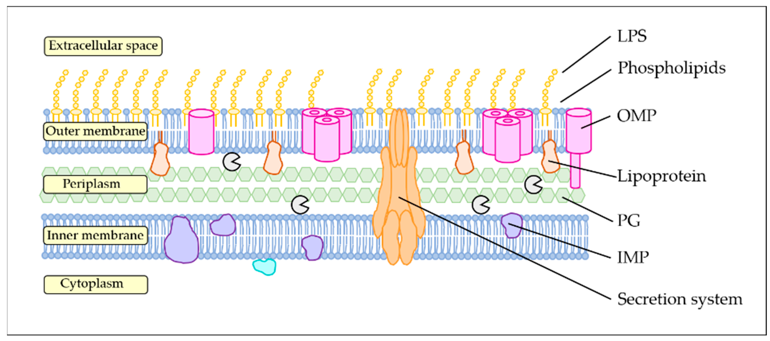
Figure 1. Gram-negative cell wall. The inner membrane, or cytoplasmic membrane, is fluid and composed of phospholipids. It contains inner membrane proteins (IMPs). The outer membrane of Gram-negative bacteria is composed of phospholipids and lipopolysaccharides (LPSs) constricted to the outer leaflet and contains integral proteins (OMPs) and lipoproteins. It is anchored to the peptidoglycan (PG) layer in the periplasm. Big protein structures such as secretion systems, pili and flagella can also be found at the cell surface.
Applmicrobiol 05 00064 g001
Figure 2. Functions of outer membrane proteins (OMPs). OMPs can allow (A) non-specific passive transport or (F) active specific transportation inside the cell. OMPs can also (B) participate in maintaining cell envelope integrity by binding the peptidoglycan layer in the periplasm. Bacteria can display a specific set of OMPs to decrease sensitivity or entry of (C) toxic molecules or (D) phages. (E) Some OMPs can act as a sensor for antimicrobial peptides (AMPs) and activate resistance gene transcription. (G) OMPs can play a role in adhesion to other cells or surfaces. Bacteria can modulate the OMPs at their surface by producing membrane vesicles (MVs). These organelles can specifically trap AMPs or phages, acting as decoys, by presenting the target at their surface.
Applmicrobiol 05 00064 g002
Figure 4. Potential applications of outer membrane proteins (OMPs).
Applmicrobiol 05 00064 g004
Table 1. Functions attributed to the outer membrane proteins of V. cholerae.
OmpVOmpWOmpUOmpTOmpSOmpXOmpALptDBamAChiPPhoETBDTs
General porin xx x
Substrate-specific porin x x x x x
Osmoregulation x
Antimicrobial resistancex xx x
Bile resistance x x
Membrane vesicle formation x
Biofilm matrixx x x
Biofilm formation x
Cell-to-cell adhesion x
Adhesion to surfaces x
Signal transductionx x
Immunomodulation x
Iron acquisition
Structure/cell envelope x (flagellum)x (flagellum) xxx
Virulence xx x xx
Legend: TBDTs, Ton-B-dependent siderophore receptors.
Disclaimer/Publisher’s Note: The statements, opinions and data contained in all publications are solely those of the individual author(s) and contributor(s) and not of MDPI and/or the editor(s). MDPI and/or the editor(s) disclaim responsibility for any injury to people or property resulting from any ideas, methods, instructions or products referred to in the content.