Article

Isolation, Optimization and Characterization of Rhodotorula alborubescens for Dietary Pigment β-Carotene Production

1
University Institute of Biotechnology, Chandigarh University, Gharuan, Mohali 140413, India
2
Department of Chemical Engineering, Chungbuk National University, Cheongju, Chungbuk 28644, Republic of Korea
*
Author to whom correspondence should be addressed.
Appl. Microbiol. 2025, 5(2), 54; https://doi.org/10.3390/applmicrobiol5020054
Submission received: 3 April 2025 / Revised: 18 May 2025 / Accepted: 29 May 2025 / Published: 4 June 2025

Abstract

:
In this study, pigment-producing yeast (Rhodotorula alborubescens) was isolated from the soil sample, demonstrating the potential for carotenoid biosynthesis. Physiological, morphological, biochemical, and molecular characterization confirmed the identity of the isolate. Optimization of the physical parameters for carotenoid production was achieved through batch shake flask experiments. The optimal conditions were determined to be 84 h of incubation at pH 6.0 and 28 °C under white light irradiation, utilizing the Yeast Peptone Dextrose (YPD) medium composed of 10 g/L yeast extract, 5 g/L of peptone, and 5 g/L dextrose, resulting in maximum carotenoid content. Further, the presence of β-carotene was confirmed using High-Performance Liquid Chromatography and Fourier Transform Infrared Spectroscopy. These findings highlight the potential of the isolated soil yeast (R. alborubescens) as a potential source of carotenoids, offering natural alternatives for various industrial applications.

1. Introduction

The color of a product significantly influences its representation and holds substantial sway over human life [1,2]. Numerous artificial pigments utilized in the fabrication of cosmetics, pharmaceuticals, healthcare and food industries elicit diverse deleterious ramifications such as hypersensitive reactions, neoplasms, carcinogenesis and severe detriment to vital body organs [3,4,5]. Furthermore, the discharge of synthetic colors and their by-products poses a significant threat to environmental preservation endeavors. Consequently, several synthetic pigments and by-products have been proscribed owing to their toxicological concerns and higher costs [6,7].
Heightened cognizance regarding the pernicious impacts of synthetic pigment on consumers’ well-being has spurred an escalating interest in harnessing pigments from natural habitats [8]. Further, pigments represent chemical entities capable of absorbing light within the visible spectrum. Their manifestation of color arises from the presence of a chromophore moiety, a molecular configuration that selectively captures solar energy, thereby instigating the excitation of electrons from lower to higher orbitals. Subsequent to this absorption, unabsorbed energy is either refracted or reflected, rendering it perceptible to the human eye. Demand for natural colors has increased recently across various industries, including healthcare, food, cosmetics, textiles, and pharmaceuticals [9,10,11]. Natural colorants come in a variety of forms, from red and purple to yellow and orange, and are sourced from plants, animals, and microbes. One of the most significant kinds of natural colorants is carotenoids [12,13]. Carotenoid pigments can be discovered throughout nature, ranging from cyanobacteria to the various kingdoms of fungi, plants and animals. In contrast with others, microorganisms produce carotenoids at better yields, with fewer batch-to-batch changes, more manipulation flexibility, and no seasonal or regional variations [14,15,16,17]. These pigments played an important and protective role in maintaining the normal physiological role under stress conditions. This inherent property of microbes was developed during evolution against exposure to different external environmental stresses, such as nutrient depletion, ultraviolet light, osmotic pressure, radiation and light exposure [18].
Yeasts are considered highly suitable candidates for carotenoid production due to their rapid growth rates and ease of cultivation [19]. They have demonstrated the capacity to produce effective and significant quantities of carotenoids, including lycopene, torulene, β-carotene, astaxanthin and torularhodin. The primary carotenoid-producing yeasts belong to genera such as Rhodotorula sp., Rhodosporidium sp., Sporobolomyces sp., and Xanthophyllomyces sp. [19,20,21]. The synthesis of carotenoids by yeasts such as Rhodotorula sp. has been delineated by researchers. Rhodotorula spp., found abundantly in various natural habitats, demonstrate the ability to biologically produce distinct carotenoids in varying proportions [22]. The carotenoid production among Rhodotorula spp. exhibits variability, influenced by the composition of growth media and prevailing environmental factors [23]. By this approach, we have aimed to isolate a novel species or species of efficient production from the local soil that has not yet been manipulated by Rhodotorula to streamline these processes and make them more efficient. Efforts were carried out to isolate the carotenoid-producing red yeast Rhodotorula alborubescens. There are no data in the literature that have examined its ability to produce carotenoids; thus, we can assert that our study constitutes novel research. The effect of different exogenous stress factors was determined based on the biosynthesis and production of carotenoids. Among various stress factors, different media, temperature, pH, and white light irradiation were chosen. Subsequently, proceed with the extraction, separation, and purification of pigment-producing yeast, specifically carotenoids.

2. Materials and Methods

2.1. Isolation and Screening

A soil sample was collected from the local gardens of Chandigarh University, Punjab, India, having coordinates 31°45′10.35″ N and 74°35′48.71″ E. The sample was collected at a depth of 10 cm in a sterile plastic bag and stored at 4 °C until use. Serial dilution was carried out to isolate desired colorful yeast from the soil samples on 20% potato dextrose agar (PDA) plates. The plates were incubated at 26 °C for 48 h to isolate yeast colonies. The single-colony isolation was achieved by streaking the obtained pigmented colonies and maintaining them on PDA plates.

2.2. Morphological, Molecular and Biochemical Characterization

The pure culture of the isolate was used to elucidate morphology, first on a PDA plate and under a microscope 100× using methyl blue staining, and then by performing biochemical characterization using [24]. Further, molecular identification of the isolated strain was carried out by performing PCR with the help of a DNA purification Kit and an ABI 3130 genetic analyzer to isolate genomic DNA. The PCR mixture was prepared by adding forward primer (TCCTGAGGGAAACTTCG), reverse primer (ACCCGCTGAACTTAAGC), 159 ng of extracted DNA, TAQ master mix (DNA polymerase, 0.5 mM of dNTPs, 3.2 mM of MgCl2, PCR enzyme buffer), 1 μL of template, 2 μL of primer, and 3 μL of Milli Q Water. PCR was set on 30 cycles for the initial denaturation (3 min; 94 °C), final denaturation at 94 °C for 1 min, primer annealing at 50 °C for 1 min, extension at 72 °C for 2 min., and final extension at 72 °C for 7 min. DNA separation was performed on 1% gel electrophoresis. The amplified and 18S purified DNA fragment was sequenced and compared with the existing data deposited in the gene bank (NCBI) utilizing BLASTn. Further, the phylogenetic tree was computed with the neighbor-joining method with a bootstrap value of 1000.

2.3. Blood Agar Testing

The selected isolate was streaked on a 5% (v/v) sheep blood agar plate and incubated at 26 °C for 24 h. Staphylococcus aureus and Bacillus cereus were taken as positive controls [25].

2.4. Selection of Media

The PDB (Potato Dextrose Broth) (10 mL) was inoculated with the yeast isolate and incubated at 26 °C for 48 h. After incubation, the culture was adjusted to 0.5 McFarland units, approximately a cell density of 1.5 × 10⁸ CFU/mL, and transferred into tubes with 5 mL of four different types of media, such as yeast malt extract (YME: 3 g/L yeast extract, 3 g/L malt extract), yeast peptone dextrose (YPD: 10 g/L yeast extract, 5 g/L peptone, 5 g/L dextrose), yeast peptone glycerol (YPG: 10 g/L yeast extract, 10 mL/L glycerol, 10 g/L peptone), and PDB with 500 μL of 0.5 McFarland standards [7]. Absorbance was taken at regular time intervals (0–96 h) at 630 nm of isolated strain. Media with maximum cellular growth was selected for further studies. All the experiments were performed in triplicate.

2.5. Carotenoid Production Optimization

Different abiotic factors such as temperature (26–34 °C), pH (4–8) and the effect of white light were assessed for maximal production of yeast biomass and carotenoid production after 84 h of interval at 150 rpm. Afterwards, cells were harvested. Culture media (100 mL) was centrifuged at 8000 rpm for 5 min. Further, the obtained pellets were washed three times with distilled water to remove all the unwanted residuals from the pellets. This pellet was dried at 60 °C and quantified for biomass estimation. Finally, the cells were collected for further extraction of desire pigment [26].

2.6. Extraction of Carotenoids

For pigment extraction, cells were lysed by using glass beads and 2 mL of preheated (at 55 °C) di-methyl sulphoxide (DMSO). Afterwards, the pellet was vortexed for 10 min., followed by centrifugation at 8000 rpm for 5 min. The whole process was repeated until the pink color disappeared completely from the pellet. After centrifugation, the colored supernatant was filtered through a 0.45 µm syringe membrane filter and collected in a separating funnel [22]. To the total volume of extracted pigment, an equal volume (v/v) of acetone: petroleum ether (1:2) was added. Then, 2 mL of saturated solution of NaCl (v/v) was added to the separating funnel and stirred continuously for 2 h. The separating funnel was then kept overnight in the dark to extract carotenoids. The assay was performed in triplicate under low light conditions [27].

2.7. Analysis of Carotenoids

The absorption coefficient (λmax) of carotenoids was analyzed using a spectrophotometer (UV-1800, Shimadzu, Japan) [28,29]. The given equation was used to quantify carotenoids in dried cell pellets ( µ g / g d . w ) [30].
M a s s   f r a c t i o n   o f   c a r o t e n o i d   c o n t e n t   ( µ g / g d . w ) = A × V ( m L ) × 10 6 A 1   c m 1 % × 100 × m ( g ) .
A = absorbance at 448 nm;
V = total extract volume;
m = dry cell biomass;
A 1   c m 1 % = 2592 (β-carotene extinction coefficient in petroleum ether).
The volumetric concentration of carotenoids (μg/L) was determined by multiplying the mass fraction of total carotenoids (μg/g) by the biomass concentration (g/L) [31,32].

2.8. Fourier Transform Infrared Spectroscopy (FTIR)

The analysis was determined using the FTIR spectrometer (Perkin Elmer) in the range of 400–4000 cm−1) to check the potential functional group present in the sample. FTIR analysis was carried out using two replicates of sample. For sample preparation, pigment was first condensed and then turned into a pellet by using potassium bromide. Standard graph of β-carotenoids was used to confirm the result [7].

2.9. High-Performance Liquid Chromatography (HPLC)

HPLC analysis was performed by using model D-14,163 equipped with a UV-visible photodiode detector, a C18 column (250 mm × 4.6 mm, 5 µm), and a mobile phase of acetonitrile (70): dichloromethane (20): methanol (10). FTIR analysis was carried out using two replicates of sample. The flow rate was set as 2.0 mL/min at 30 °C and 452 nm wavelength. Sample was entailed by the addition of 1 mL of pigment to 10 mL of diluent, followed by filtration through 0.45 Millipore membrane filters prior to its induction into the HPLC system. Subsequently the mobile phase was allowed to elute for a duration of 10 min. Sample analysis was carried out twice by comparing the retention time with the standards [7].

2.10. Statistical Analysis

The data were acquired through three separate experiments, and the ANOVA analysis (for p-value) was performed for determining their significance. Graphs were generated using ORIGIN (version 2018) data are presented as mean ± standard deviation from three independent replicates.

3. Results and Discussion

Numerous studies have been carried out on Rhodotorula mucilaginosa, Rhodotorula rubra and Rhodotorula glutinis and explored their potential role as antioxidants, production of β-carotene, torularhodin and torulene, and help in wastewater treatments [31,32,33,34]. In this study, we have isolated and explored a novel red yeast, R. alborubescens, for beta-carotene production.

3.1. Isolation, Identification and Biochemical Testing of the Isolate

Five distinct strains of yeast, specifically Rhodotorula mucilaginosa and Rhodotorula graminis, were isolated from a total of 242 samples collected from soil, leaves and fruit. All of these strains were classified as Rhodotorula glutinis. After determining the morphological and biochemical characteristics of the isolate, 18 s rRNA sequencing was performed. The isolates showed closest homology (>99%) to Rhodotorula alborubescens (Figure 1). It was found that the isolated yeast strains have the ability to produce carotenoid pigments as well [35]. ANPT (isolated R. alborubescens) gene sequencing was further submitted to the gene bank with accession number OR646828.
Libkind et al. [36] conducted a screening process to identify and select the yeast strain Rhodotorula mucilaginosa from the natural environment of Patagonia that has the ability to produce carotenoids. EL-Banna et al. [37] successfully extracted 46 yeast strains from various natural habitats. The transcribed spacers of ribosomal DNA were further tested to classify the genus to approve isolates classification. Among 32 isolated strains, a vibrant pink–red hue-producing isolate was selected as a pure culture and named ANPT. The strain ANPT showed excellent pigment-producing capacity with rapid growth; therefore, chosen for further study. The isolate ANPT developed ovoid, mucous, raised and pink-colored colonies on PDB agar plates. During microscopic analysis, the cells were seen as oval/round, indicating budding and the absence of ascospores, resulting in confirmation of the presence of yeast. This has assimilated that it utilizes glycerol, sucrose, glucose, maltose and galactose for fermentation. A similar observation was also reported by Hu et al. [38], R. mucilaginosa TZR2014 utilizes a range of sugars, including sucrose, raffinose, galactose, melezitose, sorbitol, trehalose, glycerol, and arabinose. The isolate was not able to utilize mannitol, D-glucitol, lactose, methanol-soluble starch, ethanol and citrate. R. alborubescens ANPT can withstand temperatures of 25 °C and 37 °C and is unable to grow at 42 °C and beyond. Different species of Rhodotorula were reported to grow between the range of 18–36 °C [38,39]. The isolate ANPT undergoes different biochemical testing as shown in Table 1.

3.2. Blood Agar Test

Blood agar is a rich, nutrient-dense microbiological growth medium used to cultivate a wide range of bacteria and to differentiate them based on their hemolytic properties. The test was performed to check the pathogenicity of R. alborubescens. There are three types of hemolysis that occur on blood agar: (i) alpha (α) appears in a greenish color and generally describes partial hemolysis of RBC; (ii) beta (β) appears in a clear zone around colonies and describes complete RBC lysis; and (iii) gamma (γ) shows no change in agar and describes no hemolysis [25,40,41]. Staphylococcus aureus and Bacillus cereus show β-hemolysis, which is considered pathogenic. It has been observed that R. alborubescens showed γ-hemolysis (non-hemolytic activity). It is evident that R. alborubescens passed biosafety measures, as it does not lyse RBCs, and it can proceed for further experimentation.

3.3. Optimization of Abiotic Parameters for Enhanced Total Carotenoid and Biomass Yield from R. alborubescens

3.3.1. Growth Medium and Incubation Time

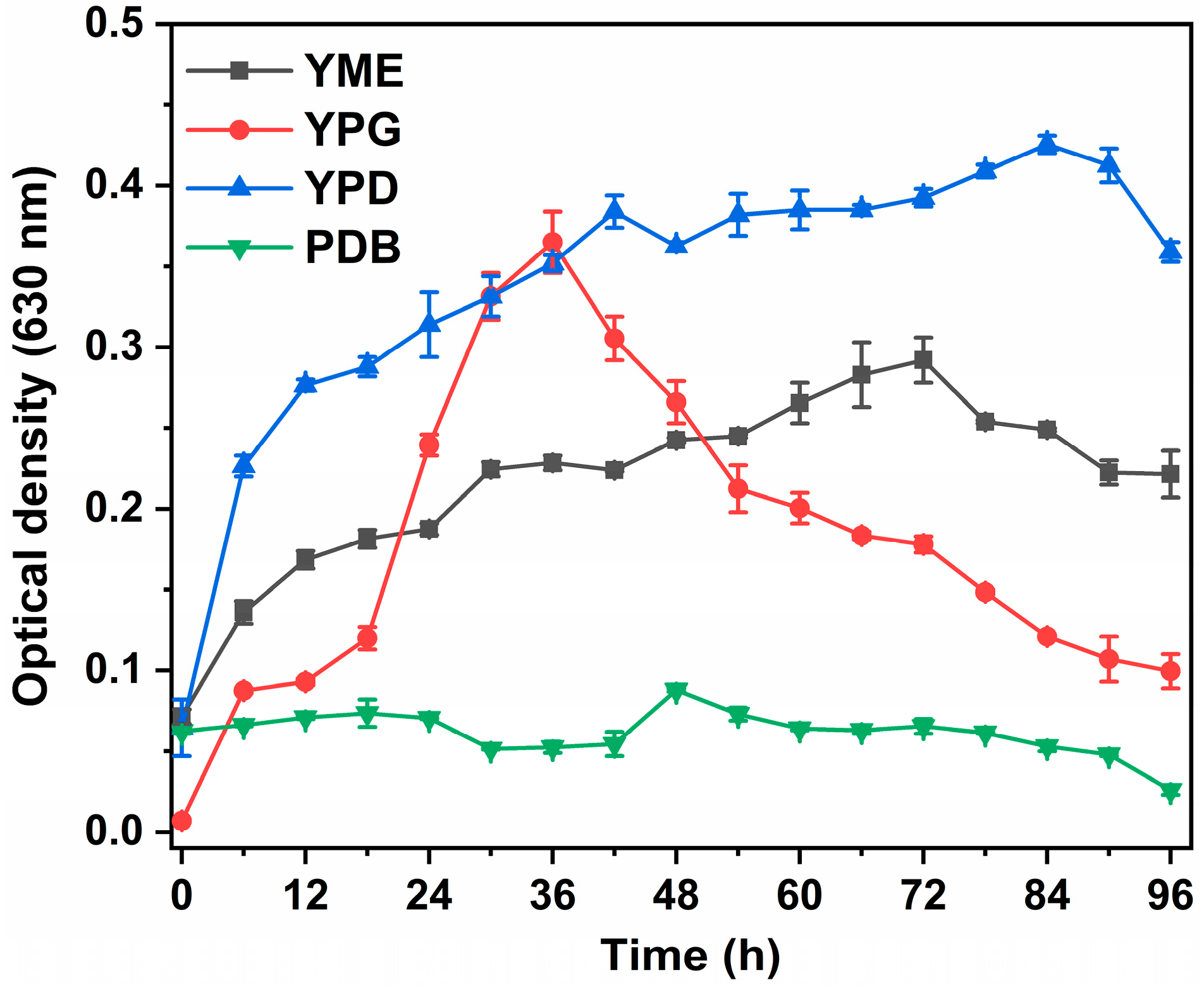
Growth kinetics were determined on the basis of the time taken by yeast to show full growth in all four different media. OD at 630 nm was taken at regular intervals (0–96 h) at 26 °C. It suggests that the stationary phase of our yeast was achieved at 42 h, which was observed by the pigment activity at the late stationary phase. This analysis aimed to determine the most favorable growth components by examining the impact of various culture media. The study evaluated the relative effectiveness of each culture medium in promoting cell growth. As shown in Figure 2, it has been clearly shown that YPD gives the most efficient biomass production.
ANOVA analysis was performed for the selection adequacy for all four different growth media and incubation times. Here, the p-value was very low (≤0.05), depicting the significance of the four-growth medium with respect to the incubation time (Table 2). The maximum growth time for R. alborubescens was seen at 84 h, but to enhance its pigment production, the incubation time was increased until the beginning of the death rate, as pigments are secondary metabolites. Hewedy et al. [42] reported that the ideal time for incubation for yeast Rhodotorula is 96 h. On the other hand, the optimal time for most bacteria and yeast was reported to be between 48 and 72 h [43]. To enhance the production of carotenoids in media, it is necessary to extend the cultivation time, as carotenoid biosynthesis in yeasts begins at the late logarithmic phase and continues in the late stationary phase [44].

3.3.2. Incubation Temperature

Synthesis of carotenoids was highly susceptible to different temperatures and their environment. We have observed carotenoid production at different temperatures ranging from 26 to 34 °C. As biomass increases, the production of carotenoids also increases. It is shown in Figure 3 that maximum carotenoid production was seen at 28 °C. The production of carotenoids kept on increasing up to 28 °C but sharply decreased after 32 °C. It has been observed in the study that higher temperatures do not support fungal growth, which could decline its biomass production and ultimately carotenoid production. The same study has been performed on R. mucilaginosa, in which the maximum carotenoid production was seen between 28 and 30 °C [45]. The current results were similar when compared with studies carried out on R. glutinis in which the optimal temperature for maximum biomass and carotenoid production is between 29 and 30 °C when observed in monoculture [46]. Monitoring carotenoid levels in cells was possible because, the production of carotenoids was influenced by temperature, which in turn affects the concentration of enzymes involved in the process. By observing the levels of carotenoids, we can gain insights into the impact of temperature on enzyme concentration [47].

3.3.3. Growth Media pH

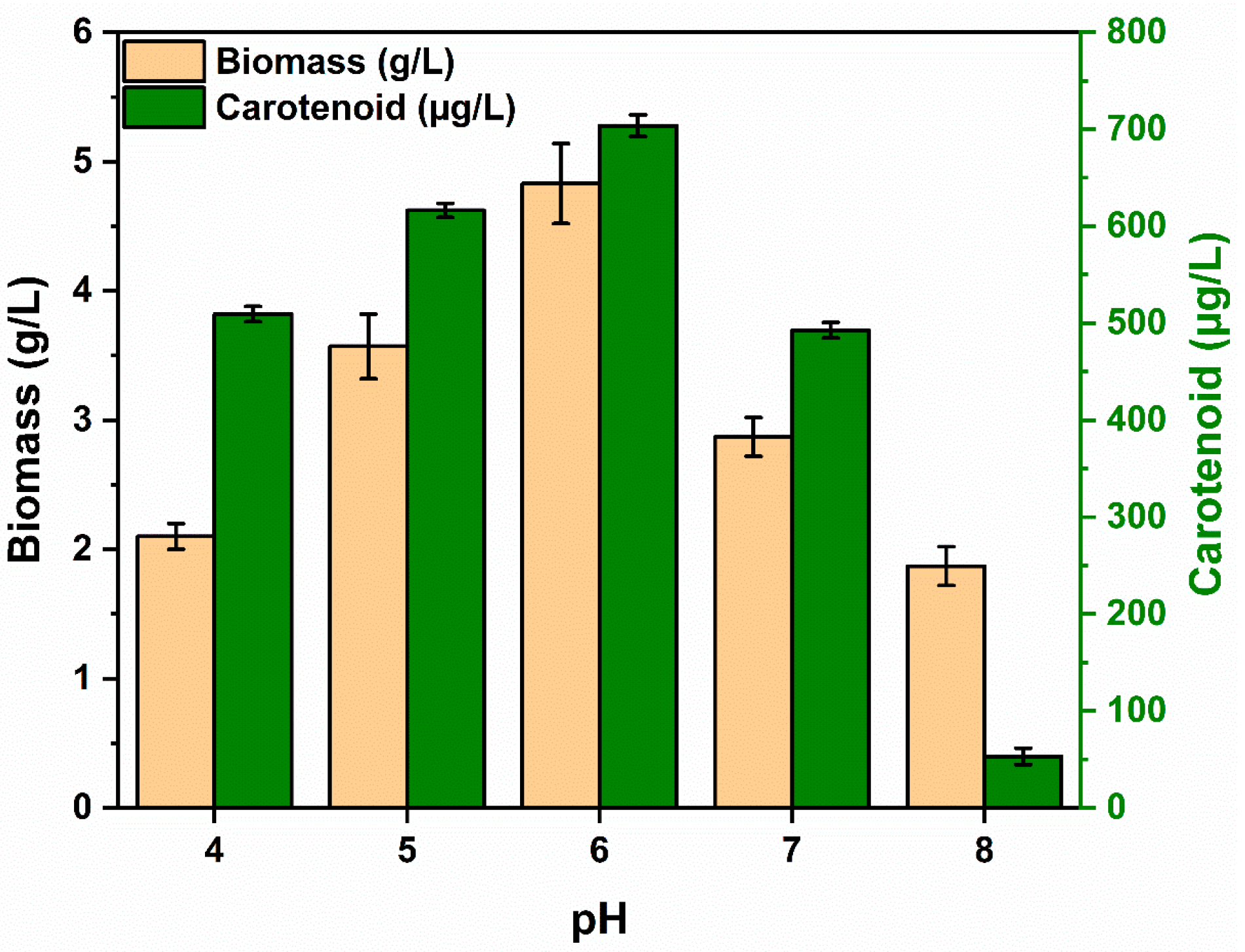
Another major factor that influences the growth of biomass in a selected culture media is pH. After the temperature was optimized and the media was finalized, the carotenoid production was determined by observing biomass production at different pH ranges from 4 to 8. YPD media was cultured at 28 °C as selected from previous results. The graph showed the maximal production of carotenoids was at pH 6. Therefore, the optimal pH condition for R. alborubescens was standardized at pH 6 (Figure 4). The same production rate was observed in R. mucilaginosa and R. graminis [48]. Some researchers also suggested that 6.1 is the most optimal pH for the maximum growth of carotenoids in R. mucilaginosa [35,49]. Latha et al. [50] also worked with R. glutinis, where he showed that biomass production increases as pH increases from 5.5 to 7.5. The optimal condition for carotenoid production was at pH 5.5, observed in R. acheniorum for β-carotene [51]. For most species of Rhodotorula, the ideal pH range for optimal growth and functioning falls between 5 and 6 [52,53]. The manipulation of culture medium pH can have a significant impact on the metabolic processes and nutrient uptake of yeast cells. When the culture medium becomes more alkaline, it acts as a stressor, leading to changes in cellular glucose metabolism and the activation of specific genes. As a result, the cells prioritize the synthesis of polysaccharides, particularly trehalose, instead of producing lipids and carotenoids [53].

3.3.4. White/Dark Period Analysis

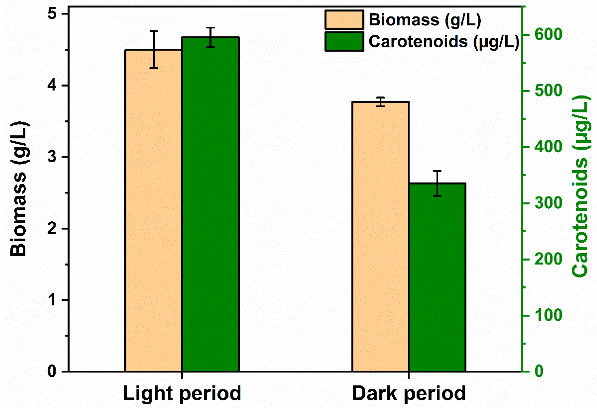
Light has been recognized as an essential factor in pigmentation, playing a crucial role in enhancing carotenogenesis. This process acts as a protective mechanism, preventing the cells from sustaining damage caused by exposure to light [30,54,55]. It has been determined from Figure 5 that biomass is enhanced when cells are incubated under white light with a wavelength of 395–520 nm. This showed a higher number of carotenoids as compared to when cultivated with no light at 28 °C with 6 pH on YPD broth. In the light and dark periods, the isolate produced 132.29 µg/g and 88.94 µg/g, respectively, of β-carotene. The intensity of the color of Rhodotorula also enhances under exposure to white light. This finding can be comparable to findings where artificial lights like LEDs advance pigment production and increase carotenoid production. In addition, Yen and Zhang [56] conducted an evaluation of β-carotene synthesis in R. glutinis using a batch reactor that included two LED lamps. This resulted in an increase of β-carotene from 14.69 μg/g to 24.6 μg/g. According to Moliné et al. [57], the increased pigmentation in Rhodotorula mucilaginosa strain was found to be associated with improved survival (250%) due to the connection between carotenoids, ergosterol and resistance to UV light at the cellular level.

3.4. Pigment Quantification and Characterization

The HPLC quantification was performed via using the standard β-carotene at different concentrations. Table 3 represents carotenoid production using different substrates by Rhodotorula species. Further, Balraj et al. [58] observed the best HPLC peak at 450 nm, which might correspond to the carotenoid family. Similar peaks were also observed in another research report given by Goswami and his co-workers, which might be due to the presence of carotenoids [59].
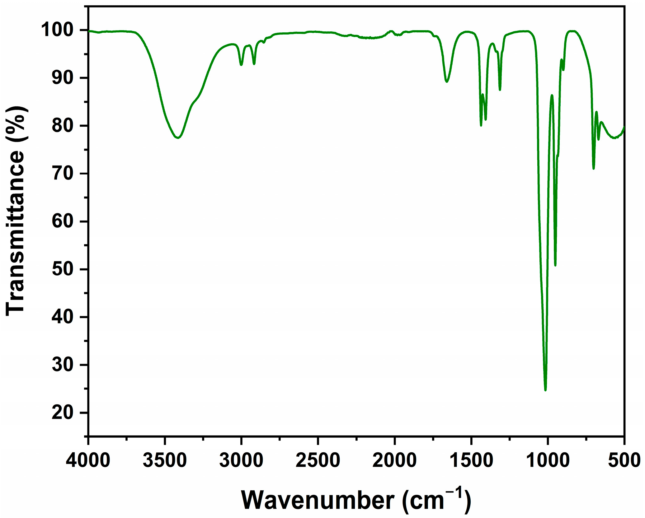
As illustrated in Figure 6, the FTIR spectrum analysis of the pigment extracted from R. alborubescens exhibited the prominent absorbance peaks at 3415, 3000, 2916, 1665, 1436, 1407, 1313, 963, and 701 cm−1. Unfortunately, there is limited research data available for the identification of the bacterial carotenoids characterized by the FTIR technique [66]. Figure 6 illustrates that the prominent spectral bands at 3415 cm−1 correspond to the presence of the stretching vibrations of the hydrogen-bonded –OH moiety in the yellow pigmented extract [66], which might arise owing to its interaction with the oxygen present in the air [67]. The weak-sharp bands at 3000 cm−1 and 2916 cm−1 indicated the existence of the trans –CH=CH group in β-carotene [68,69] and extending asymmetry stretching vibrations of the –CH2 group [66,67,68,69,70,71]. Furthermore, the distinctive polyene functional band present at 1665 cm−1 either represents the –C=C–C stretching vibrations [72] or the –C=O ketone’s stretching vibration [66,72]. Also, the bands present at 1436 cm−1, 1407 cm−1, and 1313 cm−1 correspond to the presence of asymmetric deformation/scissoring vibration of the methylene –CH2 group [66,69], –OH stretch [71], and –CH3 bending vibrations, respectively [71]. The prominent peak present at 963 cm−1 indicates the presence of out-of-plane/trans-conjugate-alkenes =CH wagging/deformation mode [67,72]. Moreover, the spectra peak at 701 cm−1 corresponds to the presence of the C=C bond and the rocking mode of the –CH2 group [69,71].
Given the prospective implementation for its application in the food industry, continuous efforts have been made for the commercial production of carotenoids with numerous degrees of accomplishment. The key driving force is the rising concern about substrate cost, as it consists of >40% of the overall economics involved [73,74]. Up-scaling of the production along with its recovery after the completion further needs to be optimized. More research is still to be performed that includes both upstream and downstream processing of the entire production process [75]. In addition, altering the batch or fed-batch approach to biorefinery systems can allow production in a marketable and viable manner, and industrial automation can sustain product stability founded on artificial intelligence controls [76,77]. And, at last, the life cycle assessment and techno-economic analysis should be performed and compared for the entire process [78]. Further, it is also reinforced by administrative guidelines that relevant Food and Drug Administration (FDA) approval should be taken prior to its application in the food industry [79,80,81].

4. Conclusions

Henceforth, the locally isolated Rhodotorula alborubescens bacterial species has clearly depicted its potential for pigment production. This study demonstrated that the enhancement of carotenoid biosynthesis can be achieved when yeast isolate was cultivated in a YPD medium under ideal carotenogenesis conditions. These conditions include an incubation period of 84 h, a temperature of 28 °C, pH 6 and exposure to white light. Apparently, R. alborubescens has garnered substantial interest from both academia and industry due to its unique physiological features and a wide range of valuable potentials, which have been applied in various industrial sectors. Local bacterial isolate Rhodotorula alborubescens is capable of conveniently producing carotenoids to meet the growing demand of consumers who are aware of the detrimental impact of synthetic colorants on health and the environment. Natural carotenoids demonstrate a plethora of advantages for human health, proven clinically safe owing to their strong antioxidant, anti-cancer and anti-inflammatory effects. In the present study, identifying the presence of commercially important carotenoid compounds, viz., β-carotene, was confirmed by purification of pigment by FTIR and HPLC. Further experiments are required for up-scaling the production of β-carotene. A holistic approach including genetic modification(s), process intensification, etc., is most needed for further research.

Author Contributions

Conceptualization, A. and V.S.; data acquisition and analyses, A., H.K. and S.K.; investigation, A., H.K. and S.K.; project administration, V.S.; supervision, V.S.; validation, A., H.K. and V.S.; writing—original draft, A.; writing—review and editing, L.G. and V.S. All authors have read and agreed to the published version of the manuscript.

Funding

This research does not receive any funding.

Data Availability Statement

The original contributions presented in the study are included in the article, further inquiries can be directed to the corresponding author/s.

Acknowledgments

Authors thank laboratory facilities provided by UIBT, Chandigarh University, India, to carry out all the research prospects.

Conflicts of Interest

The authors declare no conflicts of interest.

References

  1. Bashir, I.; Pandey, V.K.; Dar, A.H.; Dash, K.K.; Shams, R.; Mir, S.A.; Fayaz, U.; Khan, S.A.; Singh, R.; Zahoor, I. Exploring sources, extraction techniques and food applications: A review on biocolors as next-generation colorants. Phytochem. Rev. 2024, 23, 1–26. [Google Scholar] [CrossRef]
  2. Kushwaha, A.; Talukdar, S.; Mohanan, V.P.; Lata, S.; Gupta, M.; Goswami, L.; Kim, B.S. Bio-treatment of the swine wastewater and resource recovery: A sustainable approach towards circular bioeconomy. In Bio-Based Materials and Waste for Energy Generation and Resource Management; Elsevier: Amsterdam, The Netherlands, 2023; pp. 299–329. [Google Scholar]
  3. Dufossé, L. Current and potential natural pigments from microorganisms (bacteria, yeasts, fungi, and microalgae). In Handbook on Natural Pigments in Food and Beverages; Woodhead Publishing: Cambridge, UK, 2024; pp. 419–436. [Google Scholar]
  4. Anshi Kapil, S.; Goswami, L.; Sharma, V. Unveiling the intricacies of microbial pigments as sustainable alternatives to synthetic colorants: Recent trends and advancements. Micro 2024, 4, 621–640. [Google Scholar] [CrossRef]
  5. Singh, A.; Kushwaha, A.; Goswami, S.; Tripathi, A.; Bhasney, S.M.; Goswami, L.; Hussain, C.M. Roadmap from microalgae to biorefinery: A circular bioeconomy approach. In Emerging Trends to Approaching Zero Waste; Elsevier: Amsterdam, The Netherlands, 2022; pp. 339–360. [Google Scholar]
  6. Mussagy, C.U.; Khan, S.; Kot, A.M. Current developments on the application of microbial carotenoids as an alternative to synthetic pigments. Crit. Rev. Food Sci. Nutr. 2021, 62, 6932–6946. [Google Scholar] [CrossRef] [PubMed]
  7. Allahkarami, S.; Sepahi, A.A.; Hosseini, H.; Razavi, M.R. Isolation and identification of carotenoid-producing Rhodotorula sp. from Pinaceae forest ecosystems and optimization of in vitro carotenoid production. Biotechnol. Rep. 2021, 32, e00687. [Google Scholar] [CrossRef] [PubMed]
  8. El-Naggar, N.E.A.; El-Ewasy, S.M. Bioproduction, characterization, anticancer and antioxidant activities of extracellular melanin pigment produced by newly isolated microbial cell factories Streptomyces glaucescens NEAE-H. Sci. Rep. 2017, 7, 42129. [Google Scholar] [CrossRef]
  9. Hassan, M.G.; El-Sayyad, G.S.; Abdel-Monem, M.O.; Malash, M.N.; Kishk, M.A.; El Awady, M.E.; El-Khonezy, M.I. Unravelling the outcome of L-glutaminase produced by Streptomyces sp. strain 5 M as an anti-neoplasm activity. Microb. Cell Factories 2025, 24, 4. [Google Scholar] [CrossRef]
  10. Bozhuyuk, F.M.; Ozdal, M. Characterization of melanin from the fungus Scolecobasidium Musae and its antioxidant and photoprotective properties. Arch. Microbiol. 2025, 207, 77. [Google Scholar] [CrossRef]
  11. Mussagy, C.U.; Caicedo-Paz, A.V.; Farias, F.O.; de Souza Mesquita, L.M.; Giuffrida, D.; Dufossé, L. Microbial bacterioruberin: The new C50 carotenoid player in food industries. Food Microbiol. 2024, 124, 104623. [Google Scholar] [CrossRef]
  12. Zhao, D.; Li, C.; Zeng, N.; Wang, D.; Zhang, N.; Li, B. Current advances in the biosynthesis and sustainable production strategies of carotenoids and their multifaceted applications in the food industry: A comprehensive review. Food Biosci. 2025, 64, 105864. [Google Scholar] [CrossRef]
  13. Lin, Y.; Chen, M.; Jiang, W.; Zhang, L.; Shen, L.; Bai, Y. Functional Role and Mutational Analysis of the Phytoene Synthase from the Halophilic Euryarchaeon Haloferax volcanii in Bacterioruberin Biosynthesis. J. Agric. Food Chem. 2025, 73, 2393–2403. [Google Scholar] [CrossRef]
  14. Yadav AP, S.; Dwivedi, V.; Kumar, S.; Kushwaha, A.; Goswami, L.; Reddy, B.S. Cyanobacterial extracellular polymeric substances for heavy metal removal: A mini review. J. Compos. Sci. 2020, 5, 1. [Google Scholar] [CrossRef]
  15. Fang, Y.; Feng, P.; Wang, L.; Qin, L.; Wang, Z.; Zhu, S.; Fan, Y. Evaluation of culture conditions of Trichosporon oleaginosus DSM11815 for enhancement of growth and lipid production in sugarcane bagasse hydrolysate as a substrate. Process Biochemistry. 2025. [Google Scholar] [CrossRef]
  16. Bisht, A.; Sahu, S.C.; Kumar, A.; Maqsood, S.; Barwant, M.M.; Jaiswal, S.G. Recent Advances in Conventional and Innovative Extraction Techniques for Recovery of High-Added Value Compounds for Food Additives and Nutraceuticals. Food Phys. 2025, 2, 100047. [Google Scholar] [CrossRef]
  17. Siddiqui, S.A.; Kamran, F.; Stankov, S.; Rathod, N.B.; Teixeira-Costa, B.E.; Fidan, H.; Bhat, M.I.; Sofi, S.A. Unveiling the Diversity of Non Conventional Proteins-From Sources, Extraction, Technofunctionality, Nutraceutical Potential to Advancement in Food Applications-A Systematic Review. Waste Biomass Valoriz. 2025, 16, 29–51. [Google Scholar] [CrossRef]
  18. Chatragadda, R.; Dufossé, L. Ecological and biotechnological aspects of pigmented microbes: A way forward in development of food and pharmaceutical grade pigments. Microorganisms 2021, 9, 637. [Google Scholar] [CrossRef]
  19. Mannazzu, I.; Landolfo, S.; Da Silva, T.L.; Buzzini, P. Red yeasts and carotenoid production: Outlining a future for non-conventional yeasts of biotechnological interest. World J. Microbiol. Biotechnol. 2015, 31, 1665–1673. [Google Scholar] [CrossRef]
  20. Dufossé, L. Biotechnological approaches in the production of fungal pigments. In Fungal Biotechnology; Academic Press: Cambridge, MA, USA, 2025; pp. 449–466. [Google Scholar]
  21. Thilakan, M.L.J.; Amsaveni, S.; Dharani, G. Microwave pretreatment: A promising strategy to improve the clean extraction yield of microalgae-based products. In Algal Bioreactors; Elsevier Science Ltd.: Amsterdam, The Netherlands, 2025; pp. 233–247. [Google Scholar]
  22. Tinoi, J.; Rakariyatham, N.; Deming, R.L. Simplex optimization of carotenoid production by Rhodotorula glutinis using hydrolyzed mung bean waste flour as substrate. Process Biochem. 2005, 40, 2551–2557. [Google Scholar] [CrossRef]
  23. de Lima, J.G.O.; Veríssimo, N.V.P.; de Azevedo Lima, C.; Picheli, F.P.; de Paula, A.V.; Santos-Ebinuma, V.D.C. Improvement of torularhodin production by Rhodotorula glutinis through the stimulation of physicochemical stress and application of the bioproduct as an additive in the food industry. Bioprocess Biosyst. Eng. 2025, 48, 543–563. [Google Scholar] [CrossRef]
  24. Kurtzman, C.; Fell, J.W.; Boekhout, T. (Eds.) The Yeasts: A Taxonomic Study; Elsevier: Amsterdam, The Netherlands, 2011. [Google Scholar]
  25. Kapil, S.; Bhattu, M.; Sharma, V.; Kumar, T. Racemization rate and biomolecular characterization of D-serine synthesizing bacteria Bacillus tequilensis A1C1. Lett. Appl. Microbiol. 2023, 76, ovac017. [Google Scholar] [CrossRef]
  26. Gedela, R.; Prabhu, A.; Veeranki, V.D.; Kannan, P. High yield production of lipid and carotenoids in a newly isolated Rhodotorula mucilaginosa by adapting process optimization approach. Biofuels 2023, 14, 509–520. [Google Scholar] [CrossRef]
  27. Lopes, N.A.; Remedi, R.D.; dos Santos Sá, C.; Burkert, C.A.V.; de Medeiros Burkert, J.F. Different cell disruption methods for obtaining carotenoids by Sporodiobolus pararoseus and Rhodothorula mucilaginosa. Food Sci. Biotechnol. 2017, 26, 759–766. [Google Scholar] [CrossRef] [PubMed]
  28. Ravi, G.; Venkata Dasu, V.; Pakshirajan, K. Exploring the impact of sodium acetate on lipid and carotenoid production in Rhodotorula mucilaginosa. Prep. Biochem. Biotechnol. 2025, 55, 590–605. [Google Scholar] [CrossRef]
  29. Cutzu, R.; Coi, A.; Rosso, F.; Bardi, L.; Ciani, M.; Budroni, M.; Zara, G.; Zara, S.; Mannazzu, I. From crude glycerol to carotenoids by using a Rhodotorula glutinis mutant. World J. Microbiol. Biotechnol. 2013, 29, 1009–1017. [Google Scholar] [CrossRef]
  30. Goodwin, T.W. Biosynthesis of carotenoids: An overview. Methods Enzymol. 1993, 214, 330–340. [Google Scholar]
  31. Moise, A.R.; Al-Babili, S.; Wurtzel, E.T. Mechanistic aspects of carotenoid biosynthesis. Chem. Rev. 2014, 114, 164–193. [Google Scholar] [CrossRef] [PubMed]
  32. Rodrigues, T.V.D.; Amore, T.D.; Teixeira, E.C.; de Medeiros Burkert, J.F. Carotenoid production by Rhodotorula mucilaginosa in batch and fed-batch fermentation using agroindustrial byproducts. Food Technol. Biotechnol. 2019, 57, 388. [Google Scholar] [CrossRef]
  33. Mosqueda-Martínez, E.; Chiquete-Félix, N.; Castañeda-Tamez, P.; Ricardez-García, C.; Gutiérrez-Aguilar, M.; Uribe-Carvajal, S.; Mendez-Romero, O. In Rhodotorula mucilaginosa, active oxidative metabolism increases carotenoids to inactivate excess reactive oxygen species. Front. Fungal Biol. 2024, 5, 1378590. [Google Scholar] [CrossRef]
  34. Ghilardi, C.; Sanmartin Negrete, P.; Carelli, A.A.; Borroni, V. Evaluation of olive mill waste as substrate for carotenoid production by Rhodotorula mucilaginosa. Bioresour. Bioprocess. 2020, 7, 52. [Google Scholar] [CrossRef]
  35. Maldonade, I.R.; Rodriguez-Amaya, D.B.; Scamparini, A.R. Carotenoids of yeasts isolated from the Brazilian ecosystem. Food Chem. 2008, 107, 145–150. [Google Scholar] [CrossRef]
  36. Libkind, D.; Sommaruga, R.; Zagarese, H.; van Broock, M. Mycosporines in carotenogenic yeasts. Syst. Appl. Microbiol. 2005, 28, 749–754. [Google Scholar] [CrossRef]
  37. El-Banna, A.A.E.R.; Abd El-Razek, A.M.; El-Mahdy, A.R. Isolation, identification and screening of carotenoid-producing strains of Rhodotorula glutinis. Food Nutr. Sci. 2012, 3, 627–633. [Google Scholar]
  38. Hu, P.; Mao, J.; Zeng, Y.; Sun, Z.; Deng, H.; Chen, C.; Sun, W.; Tang, Z. Isolation, identification, and function of Rhodotorula mucilaginosa TZR2014 and its effects on the growth and health of weaned piglets. Front. Microbiol. 2022, 13, 922136. [Google Scholar] [CrossRef] [PubMed]
  39. Hof, H. Rhodotorula spp. in the gut–foe or friend? GMS Infect. Dis. 2019, 7, Doc02. [Google Scholar] [PubMed]
  40. Akinjogunla, O.J.; Adefiranye, O.O.; Edem, E.N.; Adenugba, I.T.; Ogboona, F.C.; Oshosanya, G.O. Antibiotic resistance profile, multidrug-, extensively drug-, and pandrug-resistant bacterial isolates: Hemagglutination and hemolytic activities against human erythrocytes. Proc. Natl. Acad. Sci. India Sect. B Biol. Sci. 2025, 95, 207–222. [Google Scholar] [CrossRef]
  41. Russell, F.M.; Biribo, S.S.N.; Selvaraj, G.; Oppedisano, F.; Warren, S.; Seduadua, A.; Mulholland, E.K.; Carapetis, J.R. As a bacterial culture medium, citrated sheep blood agar is a practical alternative to citrated human blood agar in laboratories of developing countries. J. Clin. Microbiol. 2006, 44, 3346–3351. [Google Scholar] [CrossRef]
  42. Hewedy, M.A.; Ashour, S.M. Production of a melanin like pigment by Kluyveromyces marxianus and Streptomyces chibaensis. Aust. J. Basic Appl. Sci. 2009, 3, 920–927. [Google Scholar]
  43. Keneni, A.; Gupta, V.K. Characterization of a Red bacterium strain isolated from root nodule of a Faba Bean (Vicia faba L.) for Growth Pigment Production. J. Adv. Lab. Res. Biol. 2011, 2, 138–146. [Google Scholar]
  44. Ramírez, J.; Obledo, N.; Arellano, M.; Herrera, E. Astaxanthin production by Phaffia rhodozyma in a fedbatch culture using a low cost medium feeding. e-Gnosis 2006, 4, 5. [Google Scholar]
  45. Sharma, R.; Ghoshal, G. Optimization of carotenoids production by Rhodotorula mucilaginosa (MTCC-1403) using agro-industrial waste in bioreactor: A statistical approach. Biotechnol. Rep. 2020, 25, e00407. [Google Scholar] [CrossRef]
  46. Malisorn, C.; Suntornsuk, W. Optimization of β-carotene production by Rhodotorula glutinis DM28 in fermented radish brine. Bioresour. Technol. 2008, 99, 2281–2287. [Google Scholar] [CrossRef]
  47. Hayman, E.P.; Yokoyama, H.; Chichester, C.O.; Simpson, K.L. Carotenoid biosynthesis in Rhodotorula glutinis. J. Bacteriol. 1974, 120, 1339–1343. [Google Scholar] [CrossRef] [PubMed]
  48. Maldonade, I.R.; Scamparini, A.R.; Rodriguez-Amaya, D.B. Selection and characterization of carotenoid-producing yeasts from Campinas region, Brazil. Braz. J. Microbiol. 2007, 38, 65–70. [Google Scholar] [CrossRef]
  49. Ochoa-Viñals, N.; Alonso-Estrada, D.; Pacios-Michelena, S.; García-Cruz, A.; Ramos-González, R.; Faife-Pérez, E.; Iliná, A. Current Advances in Carotenoid Production by Rhodotorula sp. Fermentation 2024, 10, 190. [Google Scholar] [CrossRef]
  50. Latha, B.V.; Jeevaratnam, K.; Murali, H.S.; Manja, K.S. Influence of growth factors on carotenoid pigmentation of Rhodotorula glutinis DFR-PDY from natural source. IJBT 2005, 4, 353–357. [Google Scholar]
  51. Nasrabadi, M.R.N.; Razavi, S.H. Optimization of β-carotene production by a mutant of the lactose-positive yeast Rhodotorula acheniorum from whey ultrafiltrate. Food Sci. Biotechnol. 2011, 20, 445–454. [Google Scholar] [CrossRef]
  52. Serra-Cardona, A.; Canadell, D.; Ariño, J. Coordinate responses to alkaline pH stress in budding yeast. Microb. Cell 2015, 2, 182. [Google Scholar] [CrossRef]
  53. Casado, C.; González, A.; Platara, M.; Ruiz, A.; Ariño, J. The role of the protein kinase A pathway in the response to alkaline pH stress in yeast. Biochem. J. 2011, 438, 523–533. [Google Scholar] [CrossRef]
  54. Raja, R.; Hemaiswarya, S.; Rengasamy, R. Exploitation of Dunaliella for β-carotene production. Appl. Microbiol. Biotechnol. 2007, 74, 517–523. [Google Scholar] [CrossRef]
  55. Weeks, O.B.; Saleh, F.K.; Wirahadiku-Sumah, M.; Berry, R.A. Photoregulated carotenoid biosynthesis in non-photosynthetic microorganisms. Pure Appl. Chem. 1973, 35, 63–80. [Google Scholar] [CrossRef]
  56. Yen, H.W.; Zhang, Z. Enhancement of cell growth rate by light irradiation in the cultivation of Rhodotorula glutinis. Bioresour. Technol. 2011, 102, 9279–9281. [Google Scholar] [CrossRef]
  57. Moliné, M.; Flores, M.R.; Libkind, D.; del Carmen Dieguez, M.; Farías, M.E.; van Broock, M. Photoprotection by carotenoid pigments in the yeast Rhodotorula mucilaginosa: The role of torularhodin. Photochem. Photobiol. Sci. 2010, 9, 1145–1151. [Google Scholar] [CrossRef] [PubMed]
  58. Balraj, J.; Pannerselvam, K.; Jayaraman, A. Isolation of pigmented marine bacteria Exiguobacterium sp. from peninsular region of India and a study on biological activity of purified pigment. Int. J. Sci. Technol. Res. 2014, 3, 375–384. [Google Scholar]
  59. Goswami, G.; Chaudhuri, S.; Dutta, D. Effect of pH and temperature on pigment production from an isolated bacterium. Chem. Eng. Trans. 2010, 20, 127–132. [Google Scholar]
  60. Szotkowski, M.; Plhalová, Ž.; Sniegoňová, P.; Holub, J.; Chujanov, O.; Špačková, D.; Blažková, J.; Márová, I. Conversion of mixed waste food substrates by carotenogenic yeasts of Rhodotorula sp. genus. Microorganisms 2023, 11, 1013. [Google Scholar] [CrossRef]
  61. Salih, A.I. Effect of some types of oils and Surfactant factors on the production of carotenoids from the yeast of Rhodotorula glutinis. Al-Kitab J. Pure Sci. 2022, 2, 292–304. [Google Scholar] [CrossRef]
  62. Moosavi Nasab Marzieh Abedi, E.; Moosavi-Nasab, S. Utilization of date syrup as a substrate for carotenoid production by Rhodotorula glutinis. Iran Agric. Res. 2015, 34, 8–13. [Google Scholar]
  63. Kot, A.M.; Błażejak, S.; Kieliszek, M.; Gientka, I.; Piwowarek, K.; Brzezińska, R. Production of lipids and carotenoids by Rhodotorula gracilis ATCC 10788 yeast in a bioreactor using low-cost wastes. Biocatal. Agric. Biotechnol. 2020, 26, 101634. [Google Scholar] [CrossRef]
  64. Banzatto, D.; Freita, L.A.D.; Mutton, M.J.R. Carotenoid production by Rhodotorula rubra cultivated in sugarcane juice, molasses, and syrup. Food Sci. Technol. 2013, 33, 14–18. [Google Scholar] [CrossRef]
  65. Keskin, A.; Fırat, M.; Büyüktopcu, A.E.Ü. Valorization of shalgam juice plant waste for the production of carotenoids by Rhodotorula glutinis. Int. J. Agric. Environ. Food Sci. 2023, 7, 79–87. [Google Scholar] [CrossRef]
  66. Ibrahim, G.S.; El-Shall, F.N.; Arafa, A.A.; Shalabi, A.; El Awady, M.E. Bio-production and characterization of carotenoid yellow pigment from Kocuria sp. GMA and exploring its sustainable antioxidant, antimicrobial and antibiofilm properties. Egypt. J. Chem. 2024, 67, 57–68. [Google Scholar] [CrossRef]
  67. Reksamunandar, R.P.; Edikresnha, D.; Munir, M.M.; Damayanti, S. Encapsulation of β-carotene in poly (vinylpyrrolidone)(PVP) by electrospinning Technique. Procedia Eng. 2017, 170, 19–23. [Google Scholar] [CrossRef]
  68. Hagos, M.; Redi-Abshiro, M.; Chandravanshi, B.S.; Yaya, E.E. Development of Analytical Methods for Determination of β-Carotene in Pumpkin (Cucurbita maxima) Flesh, Peel, and Seed Powder Samples. Int. J. Anal. Chem. 2022, 2022, 9363692. [Google Scholar] [CrossRef] [PubMed]
  69. Moh, M.H.; Man, Y.C.; Badlishah, B.S.; Jinap, S.; Saad, M.S.; Abdullah, W.J.W. Quantitative analysis of palm carotene using Fourier transform infrared and near infrared spectroscopy. J. Am. Oil Chem. Soc. 1999, 76, 249–254. [Google Scholar] [CrossRef]
  70. Martin, D.; Amado, A.M.; Gonzalvez, A.G.; Marques, M.P.M.; Batista de Carvalho, L.A.; Ureña, Á.G. FTIR Spectroscopy and DFT calculations to probe the kinetics of β-carotene thermal degradation. J. Phys. Chem. A 2019, 123, 5266–5273. [Google Scholar] [CrossRef]
  71. Iqbal, I.; Sarwar, W.; Ali, Q.; Ahmed, S. Characterization of Pigment Production by Endophytic Rhodotorula mucilaginosa MGI from Tagetes erecta. Microbiol. Biotechnol. Lett. 2024, 52, 314–324. [Google Scholar] [CrossRef]
  72. Noguchi, T.; Mitsuka, T.; Inoue, Y. Fourier transform infrared spectrum of the radical cation of β-carotene photoinduced in photosystem II. FEBS Lett. 1994, 356, 179–182. [Google Scholar] [CrossRef]
  73. Goswami, L.; Kayalvizhi, R.; Dikshit, P.K.; Sherpa, K.C.; Roy, S.; Kushwaha, A.; Kim, B.S.; Banerjee, R.; Jacob, S.; Rajak, R.C. A critical review on prospects of bio-refinery products from second and third generation biomasses. Chem. Eng. J. 2022, 448, 137677. [Google Scholar] [CrossRef]
  74. Saha, P.; Goswami, L.; Kim, B.S. Novel Biobased Non-isocyanate polyurethanes from Microbially Produced 7,10-Dihydroxy-8(E)-Octadecenoic acid for potential packaging and coating applications. ACS Sustain. Chem. Eng. 2022, 10, 4623–4633. [Google Scholar] [CrossRef]
  75. Kushwaha, A.; Yadav, A.N.; Singh, B.; Dwivedi, V.; Kumar, S.; Goswami, L.; Hussain, C.M. Life cycle assessment and techno-economic analysis of algae-derived biodiesel: Current challenges and future prospects. In Waste-to-Energy Approaches Towards Zero Waste; Elsevier: Amsterdam, The Netherlands, 2022; pp. 343–372. [Google Scholar]
  76. Goswami, L.; Kushwaha, A.; Napathorn, S.C.; Kim, B.S. Valorization of organic wastes using bioreactors for polyhydroxyalkanoate production: Recent advancement, sustainable approaches, challenges, and future perspectives. Int. J. Biol. Macromol. 2023, 247, 125743. [Google Scholar] [CrossRef]
  77. Kafle, S.R.; Kushwaha, A.; Goswami, L.; Maharjan, A.; Kim, B.S. A holistic approach for process intensification of nicotinamide mononucleotide production via high cell density cultivation under exponential feeding strategy. Bioresour. Technol. 2023, 390, 129911. [Google Scholar] [CrossRef]
  78. Kushwaha, A.; Goswami, L.; Kim, B.S. Advancement in innovative strategies for poly (ethylene terephthalate) biodegradation. Curr. Opin. Chem. Eng. 2025, 48, 101121. [Google Scholar] [CrossRef]
  79. Padigala, C.T.; Satpati, G.G.; Singhvi, M.; Goswami, L.; Kushwaha, A.; Oraon, S.; Aleksanyan, K.; Smykovskaya, R.S.; Rawindran, H.; Wei, L.J.; et al. Nanotechnological advancement in green hydrogen production from organic waste: Recent developments, techno–economic, and life cycle analyses. Int. J. Hydrogen Energy 2024, 92, 672–693. [Google Scholar] [CrossRef]
  80. Kushwaha, A.; Goswami, L.; Kim, B.S. An Integrated Chemical and Biological Approach for Poly (ethylene terephthalate) Depolymerization and Biopolyol Production. ACS Sustain. Chem. Eng. 2024, 12, 11866–11878. [Google Scholar] [CrossRef]
  81. Goswami, L.; Kushwaha, A.; Saha, P.; Kim, B.S. A novel integrated membrane bioreactor-stirred tank bioreactor system for simultaneous biodegradation of bisphenol A contaminated wastewater and biopolyol production: An approach towards circular bioeconomy. Chem. Eng. J. 2024, 490, 151740. [Google Scholar] [CrossRef]
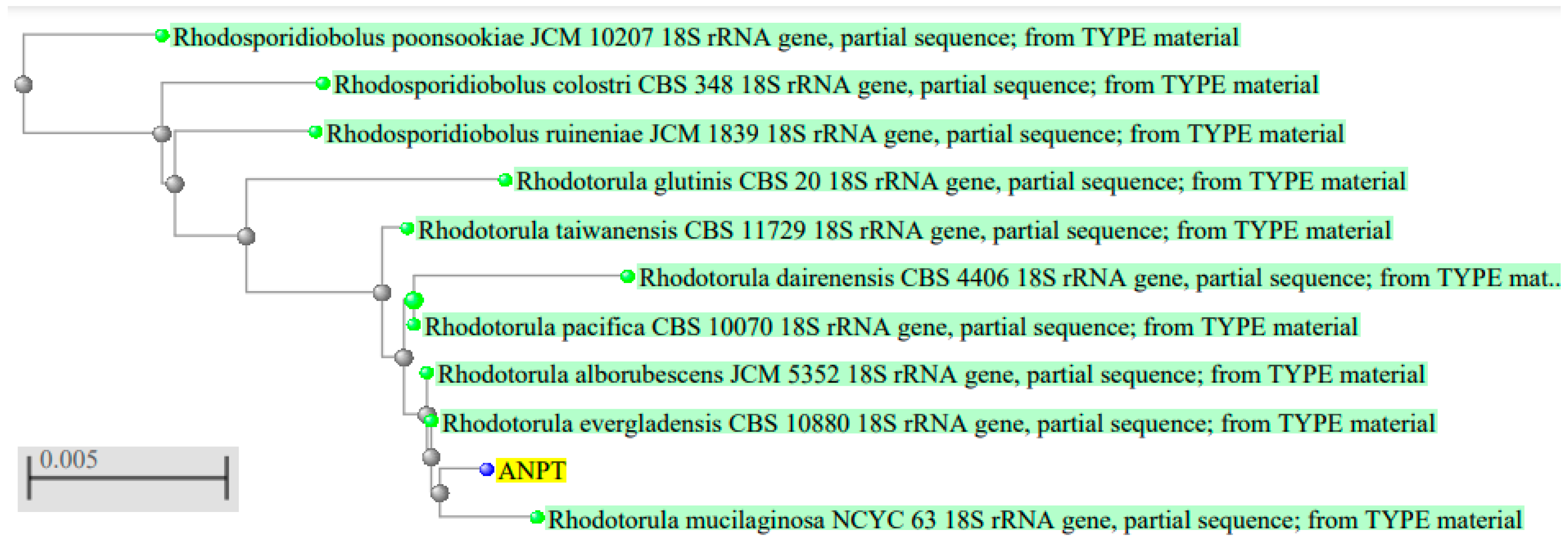
Figure 1. Phylogenetic tree showcasing closest homology of ANPT to R. alborubescens.
Applmicrobiol 05 00054 g001
Figure 2. Growth analysis of R. alborubescens by measuring OD630 nm for up to 96 h in different culture media of YME, YPG, YPD and PDB.
Applmicrobiol 05 00054 g002
Figure 3. Biomass extracted at different temperatures and the carotenoid content after incubation.
Applmicrobiol 05 00054 g003
Figure 4. Biomass extracted at different pH and the carotenoid content after incubation.
Applmicrobiol 05 00054 g004
Figure 5. Biomass extracted under light-free and white light conditions.
Applmicrobiol 05 00054 g005
Figure 6. FTIR spectra of extracted carotenoids from R. alborubescens.
Applmicrobiol 05 00054 g006
Table 1. The biochemical characteristics of R. alborubescens and their morphological and physiological properties.
CharacteristicsResultsCharacteristicsResults
colorPinkGlycerol+
AppearanceGlossy and SmoothD-Mannitol-
Colony shapeOvalD-Glucitol (10%)-
Cell ShapeOvoidCitrate-
TextureMucoidSuccinate (10%)+
ElevationRaisedVitamin-free-
Mycelium-Nitrate-
Conjugation-Starch formation-
Ascospore-Insulin-
Sucrose (10%)+Urease+
Galactose (10%)+Gelatin Liquification-
Maltose (10%)+Growth—25 °C+
Glucose (10%)+Growth—37 °C+
Lactose-Growth—42 °C-
Methanol-Sedimentation+
Ethanol-Soluble starch-
(- and + represent negative and positive tests, respectively).
Table 2. ANOVA analysis for all four different growth media and incubation times.
Source of VariationSSdfMSFp-ValueF Crit
Between Groups33,021.6948255.42350.58391.43 × 10−202.493696
Within Groups12,240.1975163.2026
Total45,261.8979
Table 3. Few studies have reviewed representing carotenoid production using different substrates by Rhodotorula species.
S. No.StrainSubstrate UtilizedCarotenoid YieldReference
1.Rhodotorula mucilaginosaAlpeerujo water0.78 mg/g[34]
2.Rhodotorula mucilaginosaSugarcane molasses and corn steep liquor1.248 mg/L[32]
3.Rhodotorula kratochvilovaeWaste animal fat4.930 mg/g[60]
4.Rhodotorula toruloidesWaste glycerol and coffee oil10.302 mg/g[60]
5.Rhodotorula glutinisBlack seed oil1.057 mg/L[61]
6.Rhodotorula glutinisDate syrup7.94 mg/L[62]
7.Rhodotorula gracilisPotato wastewater and glycerol6.24 mg/L[63]
8.Rhodotorula rubraMolasses2.74 mg/L[64]
9.Rhodotorula glutinisShalgam juice1.221 mg/L[65]
Disclaimer/Publisher’s Note: The statements, opinions and data contained in all publications are solely those of the individual author(s) and contributor(s) and not of MDPI and/or the editor(s). MDPI and/or the editor(s) disclaim responsibility for any injury to people or property resulting from any ideas, methods, instructions or products referred to in the content.