Article

Microwave-Assisted Dried Cells of the Fungus Arthrinium malaysianum as a Potential Biomaterial with Sustainable Bioremediation of Toxic Heavy Metals

1
Department of Biotechnology, School of Life Science & Biotechnology, Adamas University, Kolkata 700126, India
2
Department of Biological Sciences, School of Life Science & Biotechnology, Adamas University, Kolkata 700126, India
*
Authors to whom correspondence should be addressed.
Appl. Microbiol. 2025, 5(2), 55; https://doi.org/10.3390/applmicrobiol5020055
Submission received: 21 November 2024 / Revised: 2 December 2024 / Accepted: 6 December 2024 / Published: 11 June 2025

Abstract

:
Significant heavy metals contamination is often caused by rapid industrialization, which is devastating to both public health and the environment. Conventional processes of metal removal also result in the accumulation of secondary waste. This work proposes the use of a novel fungal biomass (microwave heat dried) from Arthrinium malaysianum for the biosorption of toxic chromium. We have meticulously explored and investigated the interactions of hexavalent chromium with dried biomass using several cutting-edge techniques like FTIR for studying the involvement of functional groups on the biomass surface, XRD for the surface architecture changes after metal binding, XPS to unravel the reduction of hexavalent chromium into its non-toxic form, and FESEM-EDX for the visualization of the ultra-structure of fungal cell surface. The Langmuir isotherm demonstrates that the maximum removal capacity Qmax of Cr(VI) is 102.310 mgg−1, at a pH of 3.5 with 100% removal of Cr(VI). There were substantial changes in the surface architecture during adsorption, confirmed by FESEM and AFM studies. FTIR and XPS data analysis indicated that carbonyl, hydroxyl, phosphate, and amine groups were responsible for the conversion of Cr(VI) (toxic) to Cr(III) (non-toxic). The IR spectra of biomass treated with Cr showed a decreased C-O stretching intensity and slight shriveling of the -OH band, and the bands in the FTIR spectra at 1642 cm−1 to 1635 cm−1 and at 1549 cm−1 to 1547 cm−1 shifted and appeared quite distinct. XRD revealed that the chromium-treated biomass had greater crystalline features and also the appearance of a wide peak where 2θ = 20°, approximately, indicating an amorphous nature at 576.0 eV and in highly loaded chromium (500 mg/L) biomass, with the Cr2p level displaying a slight shift, eventually terminating in a (576.0 eV) Cr2O3 to Cr(III) peak. Since the FTIR and XPS data obtained revealed that Cr(VI) reduces to Cr(III), this fungal biomass can also be used for generating metallic nanoparticles during biosorption. Thus, we suggest that the above-mentioned fungal biomass could be a very useful biomaterial for future translational research. We are in the process of fabricating beads with powdered biomass for further studies.

1. Introduction

In recent years, due to advancement in industrialization, one of the biggest environmental issues has been the release of various heavy metal wastes (arsenic, chromium, and lead) and dyeing colorants in the surface and ground water bodies without initially treating them [1,2,3,4,5]. This has made water unsuitable for household activities and drinking [6]. Since industrialization has a negative impact on soil fertility and productivity, it poses a severe danger to crop output, too. Toxic heavy metals released by a variety of sectors find their way into the soil and have a negative impact on human health. These heavy metals can remain in the soil for quite some time and are extremely harmful. Various heavy metals are released into agricultural soils and surface water bodies by the discharge of industrial effluents. Cadmium (Cd), lead (Pb), and chromium (Cr), on the other hand, are absorbed through organic and inorganic fertilizers [7].
A reduction in the toxicity concentrations of the approved heavy metals levels in the industrial effluents released in the water bodies can, to a certain extent, minimize the environmental issues and health risks [8,9,10,11]. Even though there are numerous other heavy metals that exist in the effluents of industrially progressive countries, the most dangerous and concerning of them all is chromium, present in more than acceptable concentrations and posing serious threats to the Indian ecosystem [12]. Tannery industries, situated in different belts of West Bengal and other states like Uttar Pradesh (Kanpur), India add tremendously to growing chromium pollution [13,14]. Neurotoxicity, genotoxicity, carcinogenicity, and immunotoxicity are all associated to Cr(VI) (hexavalent chromium) [12,15,16,17]. The well-documented detrimental consequences of Cr(VI) on human health has prompted researchers to focus their efforts on eradicating Cr(VI) from contaminated areas. Since traditional metal removal technologies are costly to use and, also, might release a few secondary contaminants, they are mostly ineffective [18,19,20,21,22]. Table 1 describes the Cr(VI) removal efficacy by different fungal species.
While comparing various hexavalent chromium Cr(VI) elimination techniques, it was found that a significant reduction in the consumption of chemicals and toxicity can be achieved by using a cutting-edge technology that employs a biosorbent for the biosorption of soluble chemicals [3,23]. The efficiencies and abilities of diverse algae, bacteria, and fungi to bind various metals have been studied in a number of ways [4,24,25,26,27,28,29,30,31,32]. The efficiency of metal bindings in bacteria, algae, and fungi has been examined and reported in a variety of studies [6,12,22,30]. Arthrinium malaysianum, a new indigenous strain of fungus having significant chromium removal efficiency, has been isolated by us. However, regarding the biomineralization and, also, biosorption of metal, so far, only one report on Arthrinium sp. biomass capacities have been published. Previously, only very few Arthrinium sp. had been identified as novel producers of a variety of industrially significant enzymes [33,34]. Non-pathogenic endophytes, Arthrinium sp., are used in various drug and medicinal arena [35], showing their antioxidant and antifungal and effects. We not so long ago demonstrated that the fungus Arthrinium malaysianum produces a number of essential enzymes [27]. In this regard, employing fungal biomass for toxic metal biosorption would give this organism a new dimension of utility for human wellbeing. We provide, here, an in-depth investigation into the behavior and processes of chromium adsorption as a battery of heavy metals. Fungal cell walls (living) are reported to be capable in the conversion of Cr(VI) soluble to Cr(III) insoluble, hence decreasing its toxic effects, confirmed with XPS (X-ray photoelectron spectra) studies. Additionally, in order to identify the interactions between the fungal cell wall and chromium ions, FTIR (Fourier transform infrared spectroscopy) and XRD (X-ray diffraction) analytical techniques were used, whereas, to evaluate the changes in morphology of the biomass exposed to Cr(VI), FESEM-EDX (field emission scanning electron spectroscopy with energy-dispersive X-ray analysis) was used [6,12,36]. Hence, hazardous heavy metals are removed from aqueous solution using Arthrinium malaysianum biomass as a potential and promising adsorbent. We would be fabricating the sustainable biomaterial out of it in the near future.
Table 1. Percentage of Cr(VI) elimination by different fungal species.
Sl. No.Name of the Fungi% Cr(VI) RemovalReferences
1A. niger (live biomass)48.7[37]
2P. Lilacinus (live biomass)100
3P. spp. (live biomass)100
4A. awamori (dead biomass)29
5A. spp. (live biomass)68
6A. flavus (live biomass)99.2
7A. niger (dead biomass)100
8P. chrysosporium (live biomass)98.5
9R. oryzae (live biomass)91.15
10T. clypeatus (dead biomass)84.5[38]
11A. malaysianum (dead and microwave dried biomass)100Present study
Present study has been done with an aim to check the hexavalent chromium biosorption by microwave dried biomass of Arthrinium malaysianum (Apiospora malaysiana).

2. Materials and Methods

2.1. Materials

Sigma-Aldrich (India) provided analytical-quality potassium dichromate (K2Cr2O7). Exact amounts of K2Cr2O7 (Potassium dichromate) were dissolved in deionized water to make hexavalent chromium stock solution of 1000 mgL−1. Usually, 1,5-diphenyl carbazide (DPC) technique was used to determine Cr(VI) levels. DPC solution was prepared by mixing 0.01 g of 1,5-diphenylcarbazide(DPC) with 5 mL ethanol and 20 mL of 1.8 M sulfuric acid. In order to measure, 0.6 mL of DPC solution and 0.05 mL of concentrated nitric acid were added to 10 mL of the sample. An intensely colored purple complex originating from the redox reaction between DPC and Cr(VI) was obtained. Cr(VI) is reduced to Cr(III), thereby forming a Cr(III)–diphenylcarbazone complex that gives the color [39]. However, this complex has a limited stability of about 15 min. Absorption is measured at 540 nm wavelength [40,41,42,43,44]. Chromium concentration (total) was determined with the help of atomic adsorption spectrometry [45]. The rest of the compounds were of analytical quality.

2.2. Functionalized Biomass Preparation

With only minor modifications, we followed the method outlined in [46,47], to prepare the dried biomass using microwave irradiation. To put it briefly, a simple Samsung commercial microwave oven (MW73AD-B/XTL) with a maximum power output of 1150 W was used for this purpose. After washing the mycelia with deionized water and filtering the culture medium, an appropriate amount of live biomass was recovered. Microwave irradiation (operated at 2.45 GHz) of Arthrinium malaysianum biomass (AMB) was performed for 240 s at an intensity of just around 20% (230 W) for 60 s intervals until the biomass became crunchy and no further weight loss was detected. Then, the obtained dried biomass was sealed in air-tight containers to avoid moisture contact for future usage.
This fungal microorganism was recently discovered as a contaminant from the Termitomyces clypeatus MTCC-5091 mushroom growth mat. The fungal genomic DNA was extracted for molecular identification purposes, and the 18s rRNITS region was amplified using gradient PCR with the use of specially designed primers [ITS1-‘TCCGTAGGTGAACCTGCGG’ and ITS4-‘TCCTCCGCTTATTGATATG’]. Later, we performed sequencing and the results were submitted to the GenBank under the accession number KY007521.1.

2.3. Efficacy of Cr(VI) Biosorption by Microwave-Assisted Heat-Dried Fungal Biosorbent

Erlenmeyer flasks were used to incubate at 30 °C at pH 4.0 (2N Phosphoric acid 2.5 mL was used to maintain a pH of 4 by deter mining the pH both at the beginning and end of the experiment using a pH meter) with a test solution of 25 mL of Cr(VI)having a 100 mgL−1. The pH 4.0 was used to make fungal cell wall protonated to facilitate the binding of chromate ions on the protonated cell wall effectively. The biomass dose that was required was added to the flasks, which were then shaken at 150 rpm. After that, with Millipore membrane (Burlington, MA, USA), vacuum filtrations were carried out so that the solutions from the biomass could be separated. Metal removal Efficiency (R), that is, the percentage of hexavalent chromium adsorbed by the biomass, can be estimated with the given Equation (1):
E f f i c i e n c y ( R ) = C i C e C i × 100
where Ci is the primary metal concentration and Ce is the equilibrium concentration. We calculated the amount of Cr(VI) adsorbed by (per gram) biomass using the following Equation (2):
Q e = V ( C i C e ) / M
where Ci = primary metal ion concentration (mg L−1); Ce = metal ion concentration post biosorption (mgL−1); M =biomass weight (g); V = metal solution volume (mL); and Qe = uptake of metal [mg Cr(VI) g−1 of biomass].

2.4. Different Interactions of Chromium with Oven-Dried Biomass of A. malaysianum: Various Investigations with Biophysical and Microscopic Studies

Powder X-ray diffraction (XRD) was carried out to determine the crystallization of the AMB loaded with metal. To achieve this, a Cu-Kα (λ = 1.54056 Å) radiation-equipped X-ray powder diffractometer with a scanning speed of 50 min−1 and a 2 with a range of 5–60 was employed [48,49].

2.5. (FTIR) Fourier Transform Infrared Spectroscopy

Chromium laden biomass and, also, the dried biomass were subjected to FTIR spectroscopy to obtain their FTIR spectra using the JASCO FTIR instrument-410 (Hachioji City, Tokyo, Japan). Samples having a KBr ratio of around 1/100 were pressed into KBr pellets having spectroscopic quality.

2.6. X-Ray Photoelectron Spectra Analysis

In order to collect the spectra of the XPS core-level of Arthrinium malaysianum mycelia before sorption and after sorption, a multiprobe spectrometer equipped with an EA125 hemispherical analyzer was used. To neutralize the material, a large-spot-size, low-energy electron gun was utilized. The electron gun’s ground potential was kept constant at −2 eV. A monochromatic source x-ray (Al-Kα) having 150 W was used to carry out the experiments. The analyzer’s pass energy was kept constant throughout the scans at 20 eV. 280.0 eV, which is the C1s peak that was used to calibrate the binding energies (BE). A combined Lorentzian–Gaussian function was used to fit the curves [50,51,52].

2.7. FESEM-EDX Analysis

For this analysis, we employed field emission scanning electron microscopy to examine the morphology of the surface and also the untreated and metal-laden fungal biomass microstructure, and, additionally, compositions of the elements were determined using EDX—energy-dispersive X-ray analysis. We conducted our experiments by incubation of dried biomass (0.2 gm) for 8 h put in a 25 mL K2Cr2O7 solution [53,54]. The acceleration tension of the electron beam was set at 15.0 KV, the angle of take-off was set at 35.0°, and the spectrum was acquired in 20 s. Each EDX analysis position area was set to roughly 0.25 µm × 0.25 µm so that different element concentrations could be compared between different positions [55].

2.8. Performance of Statistical Analysis

All investigational outcomes were reported from triplicate sets as mean data with a p value less than 0.05 using the program Origin 8.0.

3. Results

Endophytes such as Arthrinium malaysianum are a potential group of fungi, widely distributed in nature but not been studied completely for various biological uses [34]. Our aim behind the creation of the heat-inactivated/microwave-dried biomass of Arthrinium malaysianum (AMB) was to study, analyze, and determine the presence of bioremediation capabilities of AMB in an evident, visible, and ecofriendly way [34].

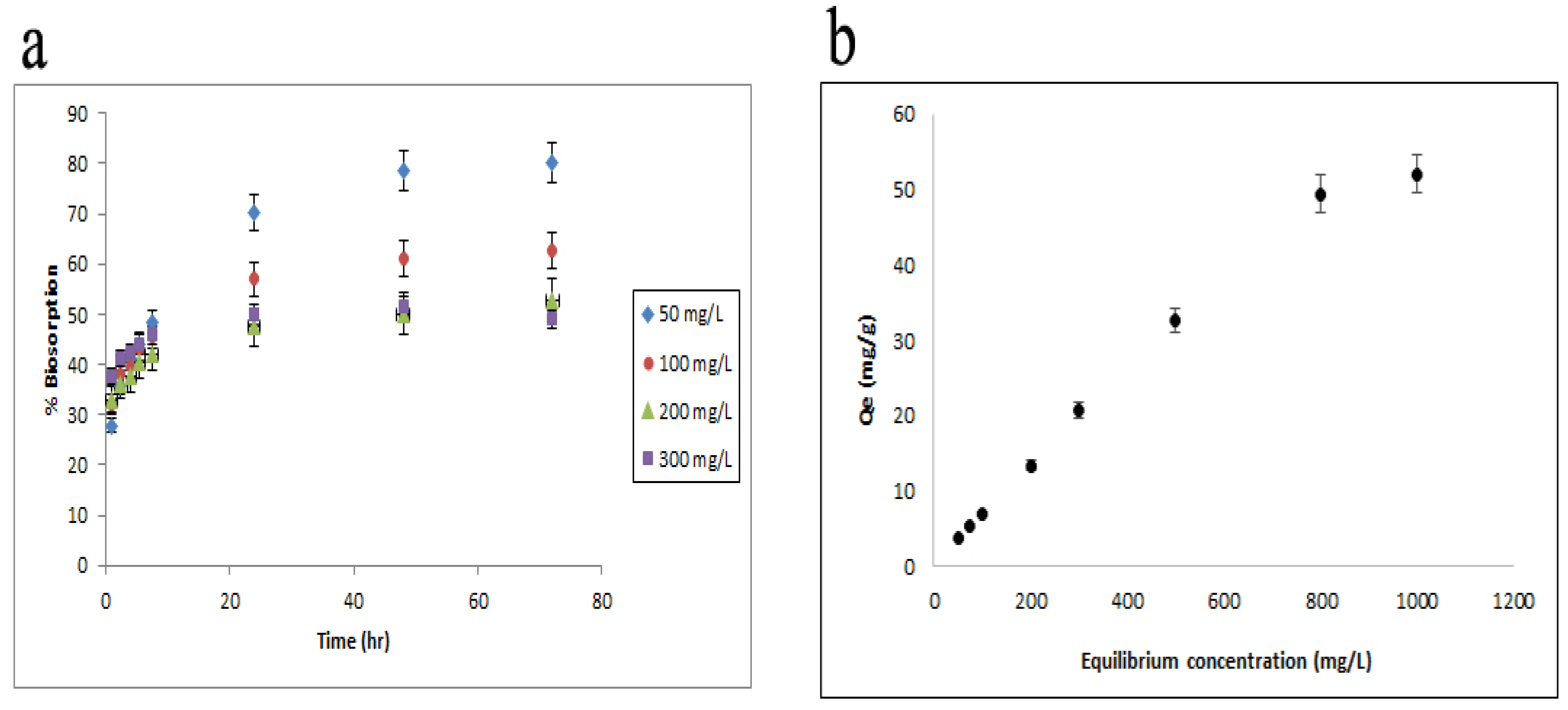
3.1. Mode of Adsorption Shown by Equilibrium Isotherm Studies

We aimed to assess the biosorption capacity of fungal biomass by varying the concentrations in a range from 50 to 300 mg L−1 of synthetic K2Cr2O7 solution. The removal capabilities of Cr(VI) were plotted as a function of the Cr(VI) concentration. The investigations were carried out with an 8 g/L dosage of biomass at a constant pH of 3 (to successfully and efficiently attach the protonated fungal cell wall to chromate ions) having an incubation period of 8 h at 38–40 °C.
A biosorption process that is required for the migration of metal ion mass from the aqueous to solid phases is aided by the preliminary concentrations of metal ions. With a chromium concentration of 50 mg/L, the percentage of biosorption increased to >80% after 72 h, which is much higher than expected (Figure 1a). The highest capacity of loading chromium (~52 mg g−1 of biomass) was achieved by elevating the Cr(VI) ion concentration to (1000 mg L−1), which resulted in improved metal absorption (Qe). When the solubility and adsorbent surface concentrations are dynamically adjusted, the values of the adsorption isotherm at equilibrium represent the equilibrium of adsorbent-sorption affinity and surface characteristics.
Due to the Chi square (χ2 = 6.24) value being the lowest and the regression coefficient value being the highest (R2 = 0.9807), the Langmuir provided the best fit (Table 2) [56]. The value in this investigation revealed that the adsorption process preferentially follows the Langmuir isotherm (R2 = 0.9807 and χ2 = 6.24) monolayer adsorption coverage features (ideally) with a predicted value of Qmax roughly 102.310 mg/g of dried fungal biomass. This figure for chromium is significantly higher than that reported in the literature for other fungal biosorbents [12,48,56,57,58,59,60,61,62,63,64,65,66,67,68,69,70,71,72].

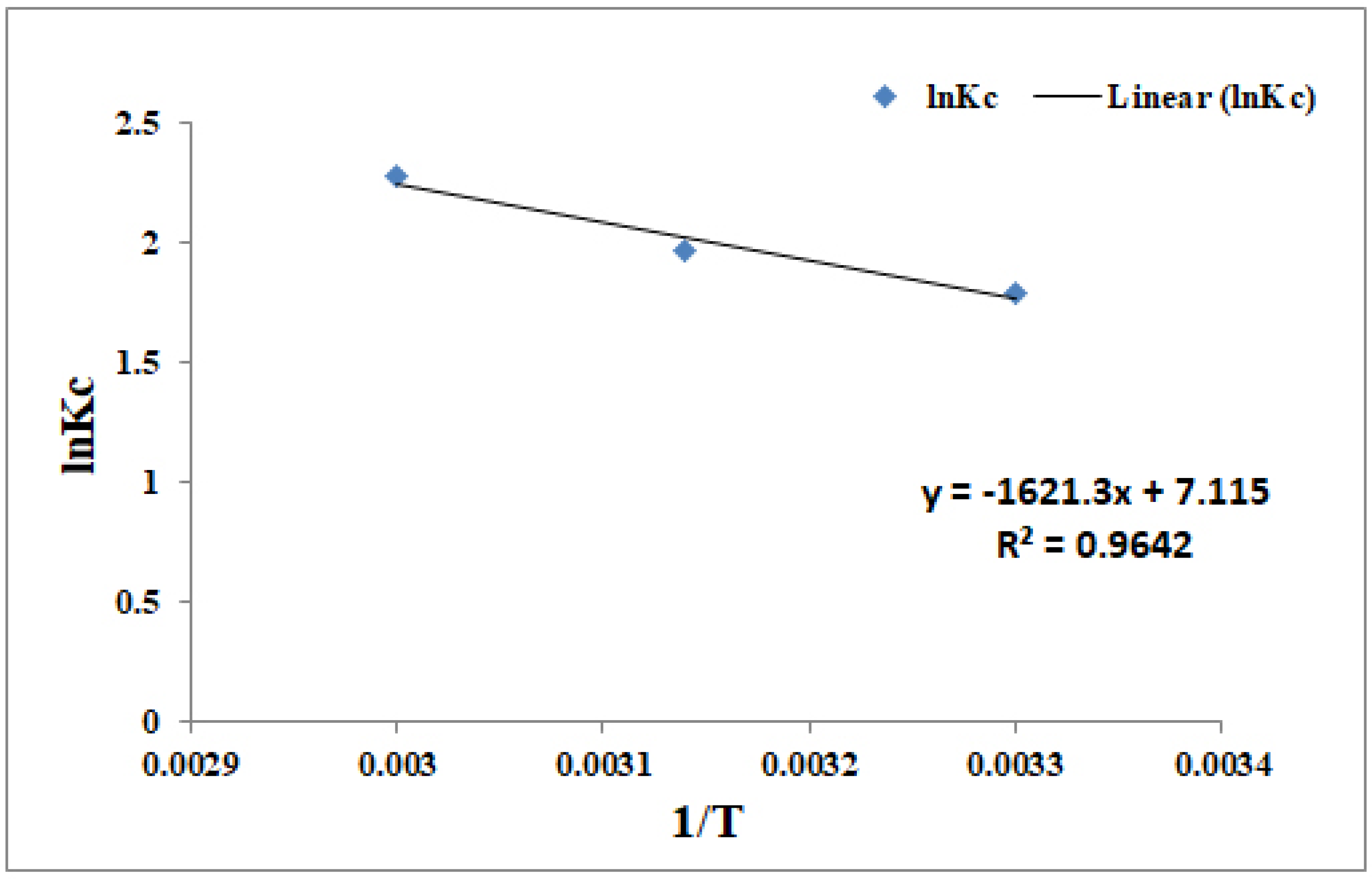
3.2. Endothermic and Entropy Driven Is the Biosorption of Cr(IV)

To understand and determine the efficiency of biomass (dried) in chromium biosorption, different thermodynamic parameters were developed. Figure 2 depicts that the temperature has an effect on adsorption.

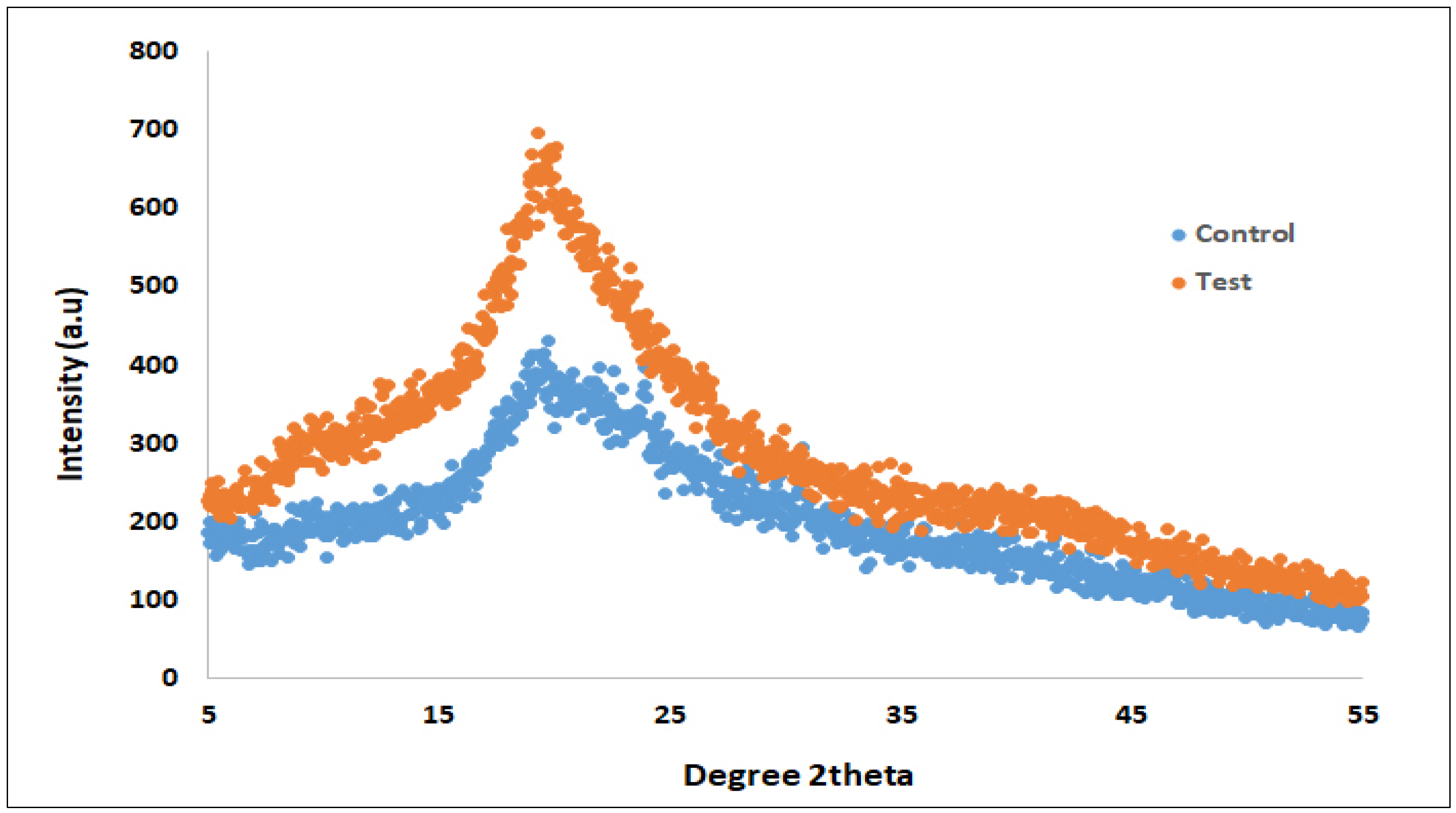
3.3. A Change in Structural Integrity Revealed by X-Ray Diffraction Studies

XRD data (control) exhibits signs of a crystalline (semi) structure of “chitosan” on the fungus cell wall [73], determined by the occurrence of a tiny halo at 2theta angle of 10.5. Moreover, the appearance of a wide peak near 19.5 indicated an amorphous nature (Figure 3).

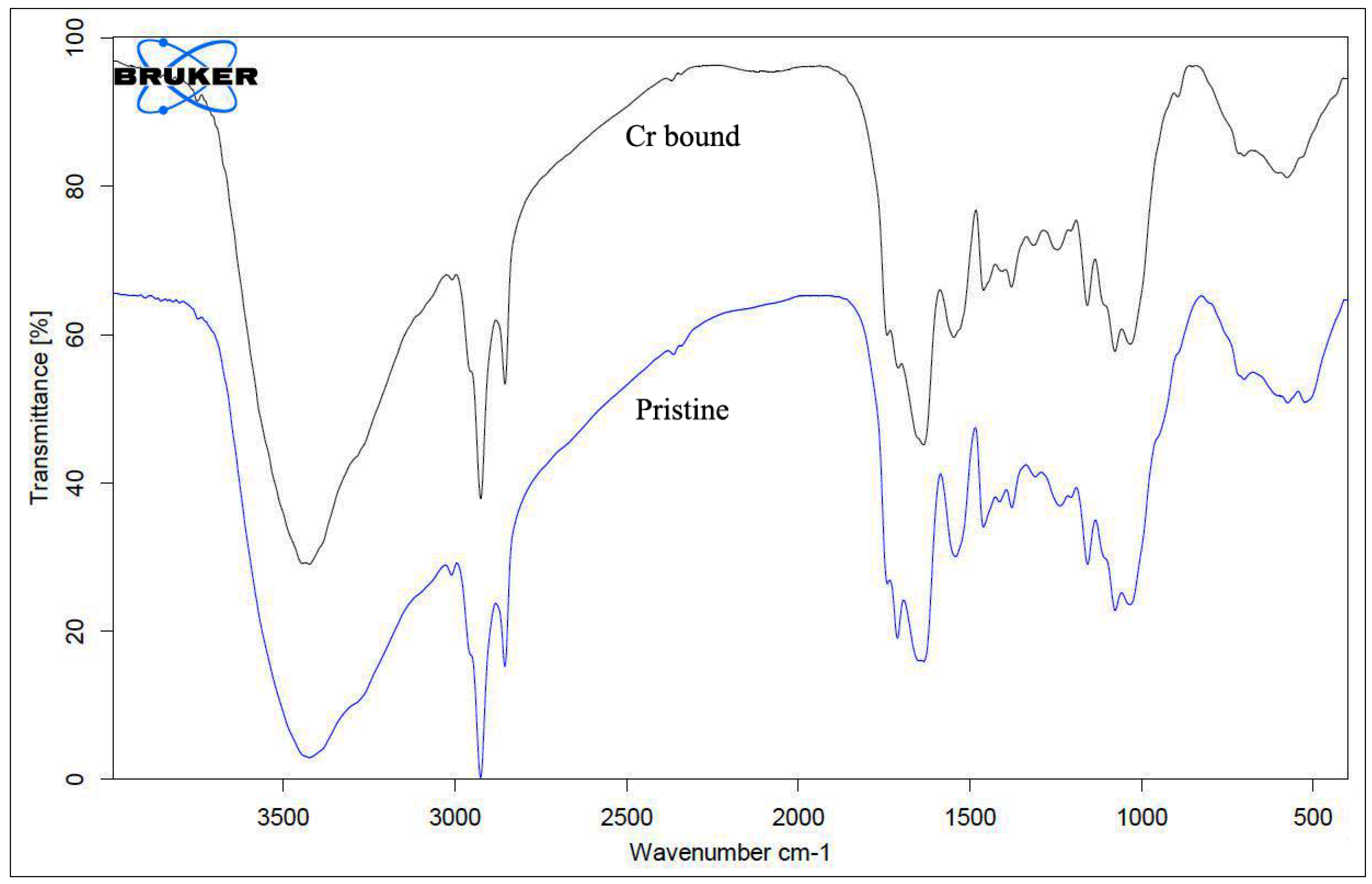
3.4. Occurrence of Functional Groups on Fungal Biomass Surface as Revealed by FTIR Analysis

The evident results helped us to identify the functional groups present on the surface that play important roles in the bioprocess. The presence of amine, hydroxyl, phosphate, and carbonyl (Figure 4) groups was confirmed by the FTIR results on the heat-pretreated and microwave-assisted Arthrinium malaysianum surface.

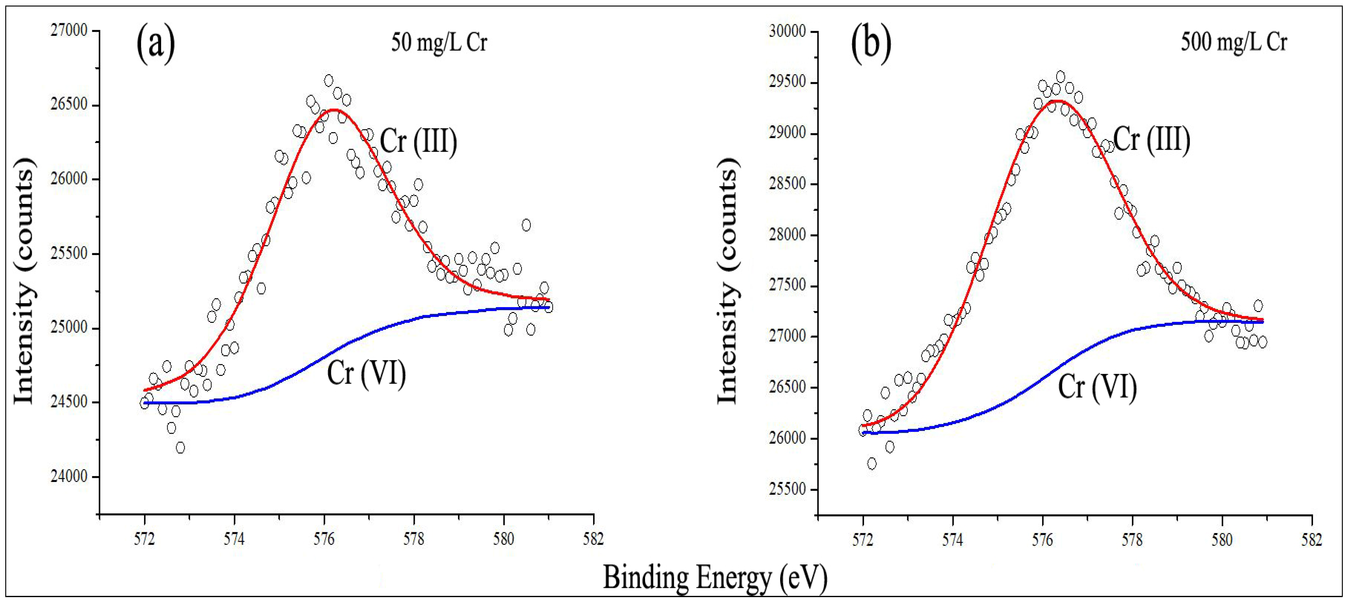
3.5. Existence of Two Types of Species of Chromium [Cr(III) and Cr(VI)] and Formation of Reduced Cr(III) from Cr(VI) as Confirmed with XPS Studies

The surface characterization using photoelectron spectroscopy by X-ray (XPS) is extremely strong, thereby providing data on a sample’s chemical bonding, chemical composition, chemical state, and atomic ratio. The chromium-laden fungal biomass survey scans were performed initially and quickly, which, thus, provides immediate data on the presence of different types of sample items. The occurrence of Cr, O, N, and C elements due to their binding energy (eV) peaks was unambiguously demonstrated by our data by survey scan [22,51,74].
We looked more deeply into the chemical properties of chromium adsorbed on the surface of biomass because of the appearance of a chromium peak. Figure 5a,b show peaks at ~576.0 and ~577.9 eV in mycelia-loaded chromium, indicating Cr2p core levels for Cr(III) and Cr(VI) species, respectively.
As the FTIR and XPS data were obtained, it was revealed that Cr(VI) reduces to Cr (III) after binding to protonated groups, then, subsequently, binding to the fungal mycelia having carboxyl groups. At higher energy (577.9 eV), the strength of the Cr2p signal elevates as the chromium concentration [mycelia highly loaded with chromium (500 mg/L)] increases. The reduction to Cr(III) from Cr(VI) using biosorbent was less at significantly greater chromium concentrations. These findings can be compared to studies carried out by other researchers [51,74], where the fungus Aspergillus versicolor has been studied and its mechanism of interaction with chromium analyzed. Figure 5b represents that at 576.0 eV, and in highly loaded chromium (500 mg/L) biomass, the Cr2p level displayed a slight shift, eventually terminating in a (576.0 eV) Cr2O3 to Cr(III) peak.

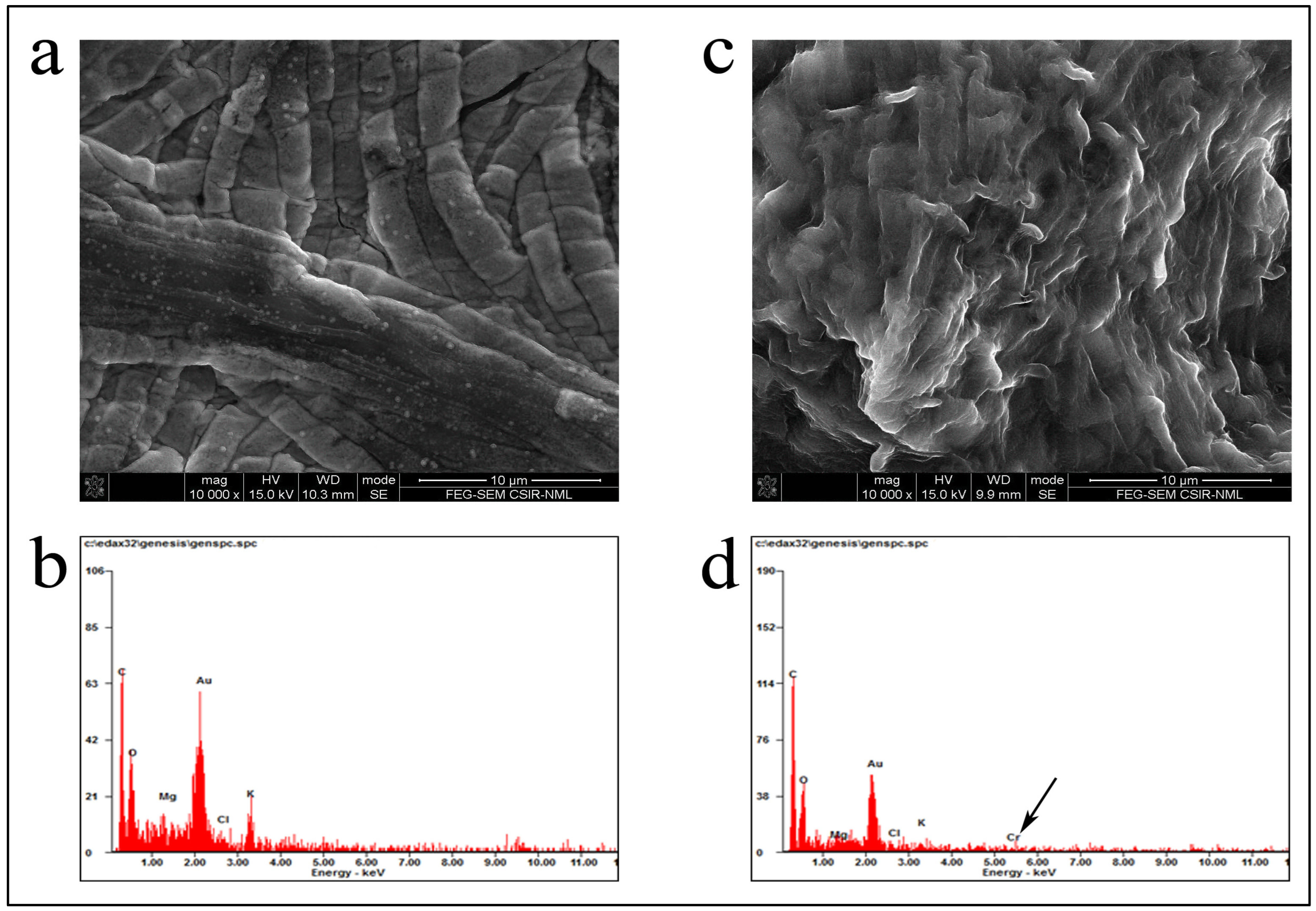
3.6. Cellular Ultrastructure Changes Were Apparent from EDX and FESEM Analyses

Pre (Figure 6a) and post biosorption of Cr(VI) onto heat-dried and microwave-assisted biomass (Figure 6c) was revealed by SEM micrographs (20,000 magnification). FESEM-EDX (field emission scanning electron spectroscopy with energy-dispersive X-ray spectroscopy) analysis was carried out to more effectively visualize the adsorption of Cr(VI) on biosorbent. EDX analysis revealed peaks of Cr in the spectra (Figure 6d), despite the absence of such a peak over the biomass surface (Figure 6b).

4. Discussion

In this present study, our equilibrium isotherm data could demonstrate the chromium adsorption abilities by the heat-inactivated (microwave-assisted) biomass of Arthrinium malaysianum biomass. Here, the Langmuir and Freundlich isotherms—two significant isotherm models—were used to investigate the AMB’s affinity for Cr(VI) ions [48,57,58]. In equilibrium isotherm curves, a lower χ2 (chi-square) and a larger R2 value usually indicate a better fitting. A smaller χ2 value indicates that the model closely matches the experimental data since there are less variations (residuals) between the experimental and expressed values while the higher R2 values indicate that our predicted data strongly correlated with the experimental data [4,56,75]. Both the Langmuir and Freundlich isotherm models described the involvement of physisorption as well as chemisorption in the Cr(VI) removal processes. According to the Langmuir model, adsorption occurred at certain homogeneous sites across the fungal cell wall, because the distinct functional groups on fungal cell walls interacted with heavy metals to provide a more consistent and predictable adsorption behavior. This feature is particularly significant for the bioremediation process. The Langmuir model also considers a limited number of binding sites on the fungal surface, which is equivalent to a finite number of adsorption sites. Fungi’s cell walls include unique functional groups such proteins, glucans, and chitin that provide particular sites for heavy metal binding. A surface adsorption model known as the Langmuir isotherm makes use of a limited number of identical sites, each of which has the capacity to bind a single molecule. This is particularly important for understanding the interactions between molecules and solid surfaces in processes like sensor design and catalysis. Once all of the adsorption sites are occupied, no further molecules can be adsorbed since it assumes a monolayer of adsorbate on the adsorbent. The Langmuir isotherm equation helps in determining key parameters such as the maximum adsorption capacity (Q_max) and the affinity between the adsorbate and the adsorbent. These parameters are crucial for designing and optimizing adsorption systems. It is widely used in environmental science for understanding pollutant removal, in material science for developing better adsorbents, and in chemistry for reaction kinetics. The Freundlich isotherm is more empirical and applicable to adsorption on heterogeneous surfaces where sites have different affinities for the adsorbate. It does not assume a monolayer, making it more flexible for various types of adsorbent surfaces. It accounts for the non-uniformity of the adsorbent surface, which is useful for describing adsorption in real-world scenarios where surfaces are rarely ideal. The Freundlich isotherm helps in understanding adsorption behavior. The Freundlich isotherm gives information about the relative binding strength of the adsorbate and aids in understanding adsorption behavior at low concentrations. It may be used with a variety of materials and systems, such as those where adsorption capacity increases with concentration and which the Langmuir model would not adequately represent. Comparative importance indicates that systems with a homogenous surface and similar adsorption sites are better suited for the Langmuir model. On heterogeneous surfaces with different adsorption energies, Freundlich functions well. Although adsorption data may be analyzed using both models, the choice between them depends on the application and the characteristics of the adsorbent and adsorbate [76].
The increase in the solubility of Cr(VI) ions is responsible for the endothermic- and entropy-driven biosorption of Cr(IV). The heat of physiosorption equalizes with the heat produced during the condensation, according to [77]. Hence, both physisorption and chemisorption processes might play an important role in chromium biosorption with fungal biomass.
X-ray diffraction studies reveal a change in structural integrity by the amalgamation of metal ions to the dried AMB surface due to the increase in relative intensity upon treatment with Cr(VI). Furthermore, comparisons between the crystalline structure of treated biomass and the semi-crystalline structure of crude biomass show that treated biomass has greater crystalline features [78]. It can thus be concluded that electrostatic interactions and/or chelation distorted the lattice organization of chitosan. This indicates that at higher temperatures, Cr(VI) biosorption is more feasible, which could be because at higher temperatures, the diffusion rates of Cr(VI) molecules become faster compared to the biosorbents from solution [45,79].
FTIR Analysis reveals the occurrence of functional groups on the fungal biomass surface. The blue line (Figure 4) of the FTIR spectra of pristine biomass revealed several distinct peaks at ~1077 cm−1 for the presence of phosphate (PO4−3) groups, 1240 cm−1 (due to stretching of C–H in amide III and stretching of C–O), 1404 cm−1 (due to stretching of C–N), 1455 cm−1 (due to bending of C–H), 1548 cm−1 (due to bending of N–H in amide II groups), 1642 cm−1 of protein bonds, 2925 cm−1 and 2955 cm−1 (stretching vibrations of –CH3 and >CH2 involving C–H), and 3420 cm−1 (the range of 3500–3000 cm−1 is attributable to -NH extending in amides and O-H vibratory stretching in alcohol and/or phenol [74,80,81]. When the biomass was subjected to Cr(VI) ions, then, the bands in the FTIR spectra at 1642 cm−1 to 1635 cm−1 and at 1549 cm−1 to 1547 cm−1 shifted and appeared quite distinct. The complexes formed between phosphate–chromate and sulfonyl–chromate groups led to a prominent peak appearance at 1404 cm−1 in the control sample, whereas this peak disappeared in the metal-loaded sample. The results suggested that carbonyl and amine groups were involved in metal ion biosorption, ion exchange, and/or complex formation [31]. In comparison to the control biomass, the metal treated biomass (blue line) IR spectra revealed a decrease in the intensity of C–O stretching, a minor shriveling of the –OH band and, also, an increase in the quantity along with the strength of C=O band. New peaks at 1742 and 1708 cm−1 provided additional evidence. It is feasible to conclude that few C-O groups were oxidized to C=O entities during the interaction of the biomass from fungi with Cr(VI) [81], resulting in the conversion of Cr(VI) to Cr(III). Others have also made similar findings [41,82,83]. N-substituted sulfonamides of solid materials and sulfonamides, and phosphate esters, have wave numbers in the range (at 1200–1400 cm−1). Thus, in samples that were Cr(VI) loaded, the peaks at ~1246 cm−1 reflected sulfonyl ester linkages of polysaccharide and those at 1379 cm−1 demonstrated phosphate. Furthermore, due to a noticeable peak shift from 1029 cm−1 to 1034 cm−1, the synthesis of the Cr(III)-phosphate complex can be concluded [84]. Disulfide and sulphide groups exist at frequency ranges of 400–550 cm−1 and 550–700 cm−1, respectively, whereas nitro (–NO2) compounds and sulphide groups exist at 450–650 cm−1 [38]. Due to the presence of nitro groups and disulfide groups on the surface of the biomass produced from the fungus, we noticed that upon metal exposure, there was a shift in the peak from 530 cm−1 for (pristine) biomass to ~576 cm−1. This area could be linked to the synthesis of Cr (OH)3, according to some earlier studies [25,51]. We hypothesized that Cr(VI), being anionic in nature, easily interacted well with the hydroxyl groups (CxOH), which acted as a reducing moiety throughout the present bioprocess [41,74].
XPS studies confirm the existence of two types of chromium species [Cr(III) and Cr(VI)], as well as the formation of reduced Cr(III) from Cr(VI).
At 576.0 eV, due to presence of similar peaks matching to the Cr2p core level, it can be predicted that chromic hydroxide was produced [51]. As the FTIR and XPS data were obtained, it was revealed that Cr(VI) reduces to Cr (III) after binding to protonated groups, then, subsequently binding to the fungal mycelia having carboxyl groups. At higher energy (577.9 eV), the strength of the Cr2p signal elevates as the chromium concentration [mycelia highly loaded with chromium (500 mg/L)] increases. The reduction to Cr(III) from Cr(VI) using biosorbent was less at significantly greater chromium concentrations. These findings can be compared to studies carried out by other researchers [51,74]. where the fungus Aspergillus versicolor has been studied and its mechanism of interaction with chromium analyzed.
In Figure 5b, at energy level (576.0 eV), the XPS spectrum shows a peak at 576.0 eV. This energy level is characteristic of the Cr2p binding energy. In XPS, the binding energy indicates the oxidation state of the element being analyzed. The presence of a peak at 576.0 eV suggests that chromium is primarily in the Cr(III) oxidation state. Specifically, this energy level is associated with Cr2p in Cr(III) oxide (Cr2O3). Highly loaded biomass (500 mg/L) indicates that the biomass sample has a high concentration of chromium, which could influence the XPS results. The “slight shift” refers to a minor change in the binding energy of the Cr2p peak. This shift could be due to various factors such as interactions between chromium species and the biomass or changes in the chemical state of chromium. The Cr2O3 to Cr(III) peak corresponds to Cr(III) in Cr2O3. Cr2O3 is a common form of chromium(III) oxide, and the XPS data suggest that the chromium in the biomass sample is present mainly in this oxidation state.
Hence, in summary, Figure 5b illustrates that in a biomass sample with high chromium content, the XPS spectrum shows a peak at 576.0 eV, corresponding to Cr(III) in the form of Cr2O3. The slight shift in this peak could indicate subtle changes in the oxidation state or chemical environment of chromium in the sample. Similar observations were made by [85], confirming Cr(III) species as the major signature. In fact, the goal of this research was to figure out what happens to toxic hexavalent chromium when it binds to a fungal surface, as well as the ability of the fungal surface to carry out the conversion of Cr(VI) (toxic) to Cr(III) (non-toxic).
EDX and FESEM analyses depict cellular ultrastructure changes. EDX, or energy-dispersive X-ray spectroscopy, is an effective analytical technique for determining the elemental makeup of materials. EDX is a commonly used technique in materials science to determine the elemental composition of metals, minerals, ceramics, and polymers. It aids in determining the material’s qualities and quality. In metallurgy, EDX helps to determine the composition of alloys and metal samples and guarantee that they fulfill needed criteria. EDX is capable of detecting trace elements and contaminants in soil, water, and air samples. This aids in monitoring environmental contamination and identifying pollution sources. In forensic science, EDX is used to assess trace evidence such as gunshot residues, paint, and fibers in order to gain insights into criminal investigations. In biological and medical research, EDX can be used to investigate biological tissues, cells, and biomaterials. It serves in understanding the elemental makeup of biological samples and investigating illness processes. EDX is used in the electronics industry to test the quality and performance of semiconductor materials, thin films, and microelectronic components. In art conservation and archaeology, EDX is used to analyze paints, ceramics, and metals to learn more about historical items and artworks. EDX detects the distinctive X-rays generated by a sample when activated by an electron beam. These X-rays have distinct energies that correspond to various elements, allowing for both qualitative and quantitative study.
Figure 6a–d help us to understand that a rough biomass surface was formed due to accumulation of circular/spherical particles that were microscopic as well. The chromium level in the entire examined area appeared to be high. Other researchers have come across similar observations [38,74].

5. Conclusions

In conclusion, the dried biomass of the Arthrinium malaysianum fungus was found to be a very effective biosorbent for eliminating harmful Cr(VI) from synthetic solutions. We found that this process is spontaneous and endothermic in nature, with chemisorption, chelation, physisorption, and oxidation reduction involved. The maximum calculated adsorption capacity (Qmax = 102.310 mg g−1) of the biomass employed an integrated Cr(VI) adsorption model with a preference for the Langmuir isotherm. The chromium has a higher Qmax value than other fungal biosorbents reported to date. We confirmed that fungal dried biomass converted Cr(VI) to Cr(III) regardless of the concentrations of chromium using XPS analysis. The reduction to Cr(III) ions from Cr(VI) ions by carbonyl and hydroxyl groups, and also the C–O oxidation to C=O, was confirmed by FTIR data. Future studies on the bioremediation of heavy metals by fungus should focus on understanding the molecular mechanisms that involve the absorption, transport, and detoxification of heavy metals in fungi, as well as the functions of certain genes and proteins. Recent advances in “omics”-based studies can determine important targets and pathways to increase the effectiveness of bioremediation. It is possible to produce fungus with improved metal-binding and detoxifying properties through genetic engineering. Furthermore, investigating the combined benefits of bacterial and fungal consortiums may enhance bioremediation procedures. Further investigation into the environmental parameters affecting fungal activity and heavy metal bioavailability, as well as the development of scalable and economical bioremediation techniques, are essential.

Author Contributions

S.R.C. and R.M. designed the experiments. S.R.C. performed the experiments. S.G., A.D., S.C. and R.M. analyzed the data, wrote the manuscript, and designed the figures and tables. All authors have read and agreed to the published version of the manuscript.

Funding

The research work was partially funded by the Adamas University Seed Money given to Dr. Rajib Majumder (SEED Grant No: AU/R&D/SEED/26/03-2020-21).

Data Availability Statement

The original contributions presented in this study are included in the article. Further inquiries can be directed to the corresponding authors.

Acknowledgments

We would like to express our heartfelt thanks to Samit Ray, Chancellor, Adamas University for providing us the infrastructure facilities.

Conflicts of Interest

The authors declare no competing financial interests.

Abbreviations

XRD: X-ray diffraction, FTIR: Fourier transform infrared spectroscopy, XPS: X-ray photoelectron spectroscopy, FESEM-EDX: field emission scanning electron microscopy and energy-dispersive X-ray spectroscopy.

References

  1. Vidu, R.; Matei, E.; Predescu, A.M.; Alhalaili, B.; Pantilimon, C.; Tarcea, C.; Predescu, C. Removal of heavy metals from wastewaters: A challenge from current treatment methods to nanotechnology applications. Toxics 2020, 8, 101. [Google Scholar] [CrossRef] [PubMed]
  2. Raji, Z.; Karim, A.; Karam, A.; Khalloufi, S. Adsorption of heavy metals: Mechanisms, kinetics, and applications of various adsorbents in wastewater remediation—A review. Waste 2023, 1, 775–805. [Google Scholar] [CrossRef]
  3. Netzahuatl-Muñoz, A.R.; Cristiani-Urbina, M.D.C.; Cristiani-Urbina, E. Chromium biosorption from Cr(VI) aqueous solutions by Cupressus lusitanica bark: Kinetics, equilibrium and thermodynamic studies. PLoS ONE 2015, 10, e0137086. [Google Scholar] [CrossRef]
  4. Zhang, J.; Chen, S.; Zhang, H.; Wang, X. Removal behaviors and mechanisms of hexavalent chromium from aqueous solution by cephalosporin residue and derived chars. Bioresour. Technol. 2017, 238, 484–491. [Google Scholar] [CrossRef] [PubMed]
  5. Chowdhury, S.R.; Roy, S.R.; Ganguly, A.; Ghosh, R.; Majumder, S.; Dasgupta, A.; Das, R.; Kumar, A.; Naskar, A.; Majumder, R. Biosorption of Acid dye by Jackfruit Leaf Powder: Isotherm, kinetics and Response surface methodology studies. Jebas 2022, 10, 254–265. [Google Scholar] [CrossRef]
  6. Abigail, M.; Samuel, M.S.; Chidambaram, R. Isotherm modelling, kinetic study and optimization of batch parameters using response surface methodology for effective removal of Cr(VI) using fungal biomass. PLoS ONE 2015, 10, e0116884. [Google Scholar]
  7. Tang, H.; Xiang, G.; Xiao, W.; Yang, Z.; Zhao, B. Microbial mediated remediation of heavy metals toxicity: Mechanisms and future prospects. Front. Plant Sci. 2024, 15, 1420408. [Google Scholar] [CrossRef] [PubMed]
  8. Abd Elnabi, M.K.; Elkaliny, N.E.; Elyazied, M.M.; Azab, S.H.; Elkhalifa, S.A.; Elmasry, S.; Mouhamed, M.S.; Shalamesh, E.M.; Alhorieny, N.A.; Abd Elaty, A.E.; et al. Toxicity of heavy metals and recent advances in their removal: A review. Toxics 2023, 11, 580. [Google Scholar] [CrossRef]
  9. Acar, F.; Malkoc, E. The removal of chromium (VI) from aqueous solutions by Fagus orientalis L. Bioresour. Technol. 2004, 94, 13–15. [Google Scholar] [CrossRef]
  10. Wu, S.; Zhang, X.; Sun, Y.; Wu, Z.; Li, T.; Hu, Y.; Su, D.; Lv, J.; Li, G.; Zhang, Z.; et al. Transformation and immobilization of chromium by arbuscular mycorrhizal fungi as revealed by SEM–EDS, TEM–EDS, and XAFS. Environ. Sci. Technol. 2015, 49, 14036–14047. [Google Scholar] [CrossRef]
  11. Staszak, K.; Regel-Rosocka, M. Removing heavy metals: Cutting-edge strategies and advancements in biosorption technology. Materials 2024, 17, 1155. [Google Scholar] [CrossRef]
  12. Samuel, M.S.; Chidambaram, R. Hexavalent chromium biosorption studies using Penicillium griseofulvum MSR1 a novel isolate from tannery effluent site: Box-Behnken optimization, equilibrium, kinetics and thermodynamic studies. J. Taiwan Inst. Chem. Eng. 2015, 49, 156–164. [Google Scholar]
  13. Ramamurthy, G.; Ramalingam, B.; Katheem, M.F.; Sastry, T.P.; Inbasekaran, S.; Thanveer, V.; Jayaramachandran, S.; Das, S.K.; Mandal, A.B. Total elimination of polluting chrome shavings, chrome, and dye exhaust liquors of tannery by a method using keratin hydrolysate. ACS Sustain. Chem. Eng. 2015, 3, 1348–1358. [Google Scholar]
  14. Sharma, P.; Bihari, V.; Agarwal, S.K.; Verma, V.; Kesavachandran, C.N.; Pangtey, B.S.; Mathur, N.; Singh, K.P.; Srivastava, M.; Goel, S.K. Groundwater contaminated with hexavalent chromium [Cr(VI)]: A health survey and clinical examination of community inhabitants (Kanpur, India). PLoS ONE 2012, 7, e47877. [Google Scholar] [CrossRef]
  15. Aftab, K.; Iqbal, S.; Khan, M.R.; Busquets, R.; Noreen, R.; Ahmad, N.; Kazimi, S.G.T.; Karami, A.M.; Al Suliman, N.M.S.; Ouladsmane, M. Wastewater-irrigated vegetables are a significant source of heavy metal contaminants: Toxicity and health risks. Molecules 2023, 28, 1371. [Google Scholar] [CrossRef]
  16. Li, Z.H.; Li, P.; Randak, T. Evaluating the toxicity of environmental concentrations of waterborne chromium (VI) to a model teleost, Oncorhynchus mykiss: A comparative study of in vivo and in vitro. Comp. Biochem. Physiol. C Toxicol. Pharmacol. 2011, 153, 402–407. [Google Scholar] [CrossRef]
  17. Zhang, P.; Yang, M.; Lan, J.; Huang, Y.; Zhang, J.; Huang, S.; Yang, Y.; Ru, J. Water quality degradation due to heavy metal contamination: Health impacts and eco-friendly approaches for heavy metal remediation. Toxics 2023, 11, 828. [Google Scholar] [CrossRef]
  18. Hkiri, N.; Olicón-Hernández, D.R.; Pozo, C.; Chouchani, C.; Asses, N.; Aranda, E. Simultaneous Heavy Metal-Polycyclic Aromatic Hydrocarbon Removal by Native Tunisian Fungal Species. J. Fungi 2023, 9, 299. [Google Scholar] [CrossRef]
  19. Ronda, A.; Della Zassa, M.; Martín-Lara, M.; Calero, M.; Canu, P. Combustion of a Pb (II)-loaded olive tree pruning used as biosorbent. J. Hazard. Mater. 2016, 308, 285–293. [Google Scholar] [CrossRef]
  20. Sounthararajah, D.; Loganathan, P.; Kandasamy, J.; Vigneswaran, S. Adsorptive removal of heavy metals from water using sodium titanate nanofibres loaded onto GAC in fixed-bed columns. J. Hazard. Mater. 2015, 28, 306–316. [Google Scholar] [CrossRef]
  21. Kim, N.; Park, M.; Park, D. A new efficient forest biowaste as biosorbent for removal of cationic heavy metals. Bioresour. Technol. 2015, 175, 629–632. [Google Scholar] [CrossRef] [PubMed]
  22. Ramrakhiani, L.; Halder, A.; Majumder, A.; Mandal, A.K.; Majumdar, S.; Ghosh, S. Industrial waste derived biosorbent for toxic metal remediation: Mechanism studies and spent biosorbent management. Chem. Eng. J. 2017, 308, 1048–1064. [Google Scholar] [CrossRef]
  23. Zhou, B.; Zhang, T.; Wang, F. Microbial-based heavy metal bioremediation: Toxicity and eco-friendly approaches to heavy metal decontamination. Appl. Sci. 2023, 13, 8439. [Google Scholar] [CrossRef]
  24. Alam, M.Z.; Ahmad, S. Chromium removal through biosorption and bioaccumulation by bacteria from tannery effluents contaminated soil. Clean–Soil Air Water 2011, 39, 226–237. [Google Scholar] [CrossRef]
  25. Das, S.K.; Guha, A.K. Biosorption of hexavalent chromium by Termitomyces clypeatus biomass: Kinetics and transmission electron microscopic study. J. Hazard. Mater 2009, 167, 685–691. [Google Scholar] [CrossRef]
  26. Bishnoi, N.R.; Kumar, R.; Kumar, S.; Rani, S.J. Biosorption of Cr(III) from aqueous solution using algal biomass Spirogyra spp. J. Hazard. Mater. 2007, 145, 142–147. [Google Scholar] [CrossRef]
  27. Yang, L.; Chen, J.P. Biosorption of hexavalent chromium onto raw and chemically modified Sargassum sp. Bioresour. Technol. 2008, 99, 297–307. [Google Scholar] [CrossRef] [PubMed]
  28. Elangovan, R.; Philip, L.; Chandraraj, K.J. Biosorption of chromium species by aquatic weeds: Kinetics and mechanism studies. Hazard. Mater. 2008, 152, 100–112. [Google Scholar] [CrossRef]
  29. Kang, S.Y.; Lee, J.U.; Kim, K.W. Biosorption of Cr(III) and Cr(VI) onto the cell surface of Pseudomonas aeruginosa. Biochem. Eng. J. 2007, 36, 54–58. [Google Scholar] [CrossRef]
  30. Salvadori, M.R.; Nascimento, C.A.O.; Corrêa, B. Nickel oxide nanoparticles film produced by dead biomass of filamentous fungus. Sci. Rep. 2014, 4, 6404. [Google Scholar] [CrossRef]
  31. Shen, L.; Xia Jl He, H.; Nie, Z.Y.; Qiu, G.Z. Biosorption mechanism of Cr(VI) onto cells of Synechococcus sp. J. Cent. South Univ. Technol. 2007, 14, 157–162. [Google Scholar] [CrossRef]
  32. Wei, W.; Wang, Q.; Li, A.; Yang, J.; Ma, F.; Pi, S.; Wu, D. Biosorption of Pb (II) from aqueous solution by extracellular polymeric substances extracted from Klebsiella sp. J1: Adsorption behavior and mechanism assessment. Sci. Rep. 2016, 6, 31575. [Google Scholar] [CrossRef] [PubMed]
  33. Shrestha, P.; Ibáñez, A.B.; Bauer, S.; Glassman, S.I.; Szaro, T.M.; Bruns, T.D.; Taylor, J.W. Fungi isolated from Miscanthus and sugarcane: Biomass conversion, fungal enzymes, and hydrolysis of plant cell wall polymers. Biotechnol. Biofuels 2015, 8, 38. [Google Scholar] [CrossRef]
  34. Mukherjee, S.; Chandrababunaidu, M.M.; Panda, A.; Khowala, S.; Tripathy, S. Tricking Arthrinium malaysianum into producing industrially important enzymes under 2-deoxy D-glucose treatment. Front. Microbiol. 2016, 7, 596. [Google Scholar] [CrossRef] [PubMed]
  35. Crous, P.W.; Groenewald, J.Z. A phylogenetic re-evaluation of Arthrinium. IMA Fungus 2013, 4, 133–154. [Google Scholar] [CrossRef]
  36. Majumder, R.; Banik, S.P.; Ramrakhiani, L.; Khowala, S.J. Bioremediation by alkaline protease (AkP) from edible mushroom Termitomyces clypeatus: Optimization approach based on statistical design and characterization for diverse applications. Chem. Technol. Biotechnol. 2015, 90, 1886–1896. [Google Scholar] [CrossRef]
  37. Kuanar, A.; Kabi, S.K.; Rath, M.; Dhal, N.K.; Bhuyan, R.; Das, S.; Kar, D. A Comparative Review on Bioremediation of Chromium by Bacterial, Fungal, Algal and Microbial Consortia. Geomicrobiol. J. 2022, 39, 515–530. [Google Scholar] [CrossRef]
  38. Ramrakhiani, L.; Majumder, R.; Khowala, S. Removal of hexavalent chromium by heat inactivated fungal biomass of Termitomyces clypeatus: Surface characterization and mechanism of biosorption. Chem. Eng. J. 2011, 171, 1060–1068. [Google Scholar] [CrossRef]
  39. Sanchez-Hachair, A.; Hofmann, A. Hexavalent chromium quantification in solution: Comparing direct UV–visible spectrometry with 1, 5-diphenylcarbazide colorimetry. C. R. Chim. 2018, 21, 890–896. [Google Scholar] [CrossRef]
  40. Cárdenas González, J.F.; Acosta Rodríguez, I.; Terán Figueroa, Y.; Lappe Oliveras, P.; Martínez Flores, R.; Rodríguez Pérez, A.S. Biotransformation of Chromium (VI) via a reductant activity from the fungal strain Purpureocillium lilacinum. J. Fungi 2021, 7, 1022. [Google Scholar] [CrossRef]
  41. Chatterjee, S.; De, R.; Gupta, A. Activated charcoal mediated purification of yellow sodium sulphate: A green process to utilize a hazardous by-product of the leather chemical industry. RSC Adv. 2016, 6, 53651–53656. [Google Scholar] [CrossRef]
  42. Li, M.H.; Gao, X.Y.; Li, C.; Yang, C.L.; Fu, C.A.; Liu, J.; Wang, R.; Chen, L.X.; Lin, J.Q.; Liu, X.M.; et al. Isolation and identification of chromium reducing Bacillus Cereus species from chromium-contaminated soil for the biological detoxification of chromium. Int. J. Environ. Res. Public Health 2020, 17, 2118. [Google Scholar] [CrossRef]
  43. McCracken, K.; Angus, S.; Reynolds, K.; Yoon, J. Multimodal imaging and lighting bias correction for improved μPAD-based water quality monitoring via smartphones. Sci. Rep. 2016, 6, 27529. [Google Scholar] [CrossRef]
  44. Kholisa, B.; Matsena, M.; Chirwa, E.M. Evaluation of Cr(VI) reduction using indigenous bacterial consortium isolated from a municipal wastewater sludge: Batch and kinetic studies. Catalysts 2021, 11, 1100. [Google Scholar] [CrossRef]
  45. Xu, F.; Liu, X.; Chen, Y.; Zhang, K.; Xu, H. Self-assembly modified-mushroom nanocomposite for rapid removal of hexavalent chromium from aqueous solution with bubbling fluidized bed. Sci. Rep. 2016, 6, 26201. [Google Scholar] [CrossRef] [PubMed]
  46. Sathvika, T.; Rajesh, V.; Rajesh, N. Microwave assisted immobilization of yeast in cellulose biopolymer as a green adsorbent for the sequestration of chromium. Chem. Eng. J. 2015, 279, 38–46. [Google Scholar] [CrossRef]
  47. Sebastian, J.; Rouissi, T.; Brar, S.K.; Hegde, K.; Verma, M. Microwave-assisted extraction of chitosan from Rhizopus oryzae NRRL 1526 biomass. Carbohydr. Polym. 2019, 219, 431–440. [Google Scholar] [CrossRef]
  48. Bhatt, A.S.; Sakaria, P.L.; Vasudevan, M.; Pawar, R.R.; Sudheesh, N.; Bajaj, H.C.; Mody, H.M. Adsorption of an anionic dye from aqueous medium by organoclays: Equilibrium modeling, kinetic and thermodynamic exploration. RSC Adv. 2012, 2, 8663–8671. [Google Scholar] [CrossRef]
  49. Ma, J.; Yang, M.; Yu, F.; Zheng, J. Water-enhanced removal of ciprofloxacin from water by porous graphene hydrogel. Sci. Rep. 2015, 5, 13578. [Google Scholar] [CrossRef] [PubMed]
  50. Samanta, T.; Sinha, S.; Mukherjee, M. Effect of added salt on swelling dynamics of ultrathin films of strong polyelectrolytes. Polymer 2016, 97, 285–294. [Google Scholar] [CrossRef]
  51. Das, S.K.; Mukherjee, M.; Guha, A.K. Interaction of chromium with resistant strain Aspergillus versicolor: Investigation with atomic force microscopy and other physical studies. Langmuir 2008, 24, 8643–8650. [Google Scholar] [CrossRef] [PubMed]
  52. Parandhaman, T.; Pentela, N.; Ramalingam, B.; Samanta, D.; Das, S.K. Metal nanoparticle loaded magnetic-chitosan microsphere: Water dispersible and easily separable hybrid metal nano-biomaterial for catalytic applications. ACS Sustain. Chem. Eng. 2017, 5, 489–501. [Google Scholar] [CrossRef]
  53. Baker, P.W.; Ito, K.; Watanabe, K. Marine prosthecate bacteria involved in the ennoblement of stainless steel. Environ. Microbiol. 2003, 5, 925–932. [Google Scholar] [CrossRef] [PubMed]
  54. Sheikh, L.; Tripathy, S.; Nayar, S. Biomimetic matrix mediated room temperature synthesis and characterization of nano-hydroxyapatite towards targeted drug delivery. RSC Adv. 2016, 6, 62556–62571. [Google Scholar] [CrossRef]
  55. Wu, S.; Zhang, X.; Sun, Y.; Wu, Z.; Li, T.; Hu, Y.; Lv, J.; Li, G.; Zhang, Z.; Zhang, J.; et al. Chromium immobilization by extra-and intraradical fungal structures of arbuscular mycorrhizal symbioses. J. Hazard. Mater. 2016, 316, 34–42. [Google Scholar] [CrossRef]
  56. Naskar, A.; Guha, A.K.; Mukherjee, M.; Ray, L. Adsorption of nickel onto Bacillus cereus M116: A mechanistic approach. Sep. Sci. Technol. 2016, 51, 427–438. [Google Scholar] [CrossRef]
  57. Qi, X.; Li, L.; Tan, T.; Chen, W.; Smith, R.L., Jr. Adsorption of 1-butyl-3-methylimidazolium chloride ionic liquid by functional carbon microspheres from hydrothermal carbonization of cellulose. Environ. Sci. Technol. 2013, 47, 2792–2798. [Google Scholar] [CrossRef]
  58. Duan, F.; Chen, C.; Wang, G.; Yang, Y.; Liu, X.; Qin, Y. Efficient adsorptive removal of dibenzothiophene by graphene oxide-based surface molecularly imprinted polymer. RSC Adv. 2014, 4, 1469–1475. [Google Scholar] [CrossRef]
  59. Arıca, M.Y.; Bayramoğlu, G. Cr(VI) biosorption from aqueous solutions using free and immobilized biomass of Lentinussajor-caju: Preparation and kinetic characterization. Colloids Surf. A Physicochem. Eng. Asp. 2005, 253, 203–211. [Google Scholar] [CrossRef]
  60. Cárdenas-González, J.F.; Acosta-Rodríguez, I. Hexavalent chromium removal by a Paecilomyces sp. fungal strain isolated from environment. Bioinorg. Chem Appl. 2010, 2010, 676243. [Google Scholar] [CrossRef]
  61. Deepa KKSathishkumar, M.; Binupriya, A.R.; Murugesan, G.S.; Swaminathan, K.; Yun, S.E. Sorption of Cr(VI) from dilute solutions and wastewater by live and pretreated biomass of Aspergillus flavus. Chemosphere 2006, 62, 833–840. [Google Scholar] [CrossRef]
  62. Kavita, B.; Limbachia, J.; Keharia, H. Hexavalent chromium sorption by biomass of chromium tolerant Pythium sp. J. Basic Microbiol. 2011, 51, 173–182. [Google Scholar] [CrossRef]
  63. Khambhaty, Y.; Mody, K.; Basha, S.; Jha, B. Kinetics, equilibrium and thermodynamic studies on biosorption of hexavalent chromium by dead fungal biomass of marine Aspergillus niger. Chem. Eng. J 2009, 145, 489–495. [Google Scholar] [CrossRef]
  64. Kumar, R.; Bishnoi, N.R.; Bishnoi, K. Biosorption of chromium (VI) from aqueous solution and electroplating wastewater using fungal biomass. Chem. Eng. J 2008, 135, 202–208. [Google Scholar] [CrossRef]
  65. Liu, T.; Li, H.; Deng, L. The optimum conditions, thermodynamical isotherm and mechanism of hexavalent chromium removal by fungal biomass of Mucor racemosus. World J. Microbiol. Biotechnol. 2007, 23, 1685–1693. [Google Scholar] [CrossRef] [PubMed]
  66. Mungasavalli, D.P.; Viraraghavan, T.; Jin, Y.C. Biosorption of chromium from aqueous solutions by pretreated Aspergillus niger: Batch and column studies. Colloids Surf. A Physicochem. Eng. Asp. 2007, 301, 214–223. [Google Scholar] [CrossRef]
  67. Park, D.; Yun, Y.S.; Jo, J.H.; Park, J.M. Mechanism of hexavalent chromium removal by dead fungal biomass of Aspergillus niger. Water Res. 2005, 39, 533–540. [Google Scholar] [CrossRef]
  68. Sanghi, R.; Sankararamakrishnan, N.; Dave, B.C. Fungal bioremediation of chromates: Conformational changes of biomass during sequestration, binding, and reduction of hexavalent chromium ions. J. Hazard. Matter. 2009, 169, 1074–1080. [Google Scholar] [CrossRef]
  69. Say, R.; Yilmaz, N.; Denizli, A. Removal of chromium (VI) ions from synthetic solutions by the fungus Penicillium purpurogenum. Eng. Life Sci. 2004, 4, 276–280. [Google Scholar] [CrossRef]
  70. Shroff, K.A.; Vaidya, V.K. Effect of pre-treatments on the biosorption of chromium (VI) ions by the dead biomass of Rhizopus arrhizus. J. Chem. Technol. Biotechnol. 2012, 87, 294–304. [Google Scholar] [CrossRef]
  71. Tewari, N.; Vasudevan, P.; Guha, B. Study on biosorption of Cr(VI) by Mucor hiemalis. Biochem. Eng. J 2005, 23, 185–192. [Google Scholar] [CrossRef]
  72. Ahluwalia, S.S.; Goyal, D. Removal of Cr(VI) from aqueous solution by fungal biomass. Eng. Life Sci 2010, 10, 480–485. [Google Scholar] [CrossRef]
  73. Kumar, S.; Koh, J. Physiochemical, optical and biological activity of chitosan-chromone derivative for biomedical applications. Int. J. Mol. Sci 2012, 13, 6102–6116. [Google Scholar] [CrossRef] [PubMed]
  74. Majumder, R.; Sheikh, L.; Naskar, A.; Mukherjee, M.; Tripathy, S. Depletion of Cr(VI) from aqueous solution by heat dried biomass of a newly isolated fungus Arthrinium malaysianum: A mechanistic approach. Sci. Rep. 2017, 7, 11254. [Google Scholar] [CrossRef] [PubMed]
  75. Fagundez, J.L.; Netto, M.S.; Dotto, G.L.; Salau, N.P. A new method of developing ANN-isotherm hybrid models for the determination of thermodynamic parameters in the adsorption of ions Ag+, Co2+ and Cu2+ onto zeolites ZSM-5, HY, and 4A. J. Environ. Chem. Eng. 2021, 9, 106126. [Google Scholar] [CrossRef]
  76. Vigdorowitsch, M.; Pchelintsev, A.; Tsygankova, L.; Tanygina, E. Freundlich isotherm: An adsorption model complete framework. Appl. Sci. 2021, 11, 8078. [Google Scholar] [CrossRef]
  77. Liu, Y.; Liu, Y.J. Biosorption isotherms, kinetics and thermodynamics. Sep. Purif. Technol. 2008, 61, 229–242. [Google Scholar] [CrossRef]
  78. Chen, Q.; Xu, A.; Li, Z.; Wang, J.; Zhang, S. Influence of anionic structure on the dissolution of chitosan in 1-butyl-3-methylimidazolium-based ionic liquids. Green Chem. 2011, 13, 3446–3452. [Google Scholar] [CrossRef]
  79. Vijayaraghavan, K.; Yun, Y.S. Bacterial biosorbents and biosorption. Biotechnol. Adv. 2008, 26, 266–291. [Google Scholar] [CrossRef]
  80. Chakravarty, S.; Mohanty, A.; Sudha, T.N.; Upadhyay, A.K.; Konar, J.; Sircar, J.K.; Madhukar, A.; Gupta, K.K. Removal of Pb (II) ions from aqueous solution by adsorption using bael leaves Aegle marmelos. J. Hazard. Mater. 2010, 173, 502–509. [Google Scholar] [CrossRef]
  81. Das, S.K.; Guha, A.K. Biosorption of chromium by Termitomyces clypeatus. Colloids Surf. B Biointerfaces 2007, 60, 46–54. [Google Scholar] [CrossRef] [PubMed]
  82. Hsu, N.H.; Wang, S.L.; Liao, Y.H.; Huang, S.T.; Tzou, Y.M.; Huang, Y.M. Removal of hexavalent chromium from acidic aqueous solutions using rice straw-derived carbon. J. Hazard. Mater. 2009, 171, 1066–1070. [Google Scholar] [CrossRef]
  83. Hsu, N.H.; Wang, S.L.; Lin, Y.C.; Sheng, G.D.; Lee, J.F. Reduction of Cr(VI) by crop-residue-derived black carbon. Environ. Sci. Technol. 2009, 43, 8801–8806. [Google Scholar] [CrossRef] [PubMed]
  84. Fuks, H.; Kaczmarek, S.; Bosacka, M. EPR and IR investigations of some chromium (III) phosphate (V) compounds. Rev. Adv. Mater. Sci. 2010, 23, 57–63. [Google Scholar]
  85. Vieira, R.S.; Oliveira, M.L.M.; Guibal, E.; Rodríguez-Castellón, E.; Beppu, M.M. Copper, mercury and chromium adsorption on natural and crosslinked chitosan films: An XPS investigation of mechanism. Colloids Surf. A Physicochem. Eng. Asp 2011, 374, 108–114. [Google Scholar] [CrossRef]
Figure 1. Studies of percentage of biosorption and equilibrium isotherms. (a) depicts that, at 72 h, for 50 mg/L (♦), maximum biosorption is up to 80%, followed by 100 mg/L (●), then 200 mg/L (▲), and ultimately, 300 mg/L (■). (b) depicts that, with the rise in Cr (IV) concentration up to 1000 mg L−1, the metal uptake Qe (●) increases (up to >60%) in 72 h.
Applmicrobiol 05 00055 g001
Figure 2. This figure demonstrates how temperature affects adsorption. This suggests that at higher temperatures (T), linear relationships with concentration Kc (♦) exist. Cr(VI) biosorption is more practicable, possibly due to the faster diffusion rate of Cr(VI) molecules on the biosorbents from the solution.
Applmicrobiol 05 00055 g002
Figure 3. XRD spectra of control and test biomass (chromium loaded). XRD data of blue line (●) of control sample reveals partially hydrated semi-crystalline nature of dried biomass. Understanding the structural changes in chitosan following metal ion loading is aided by analyzing the 2θ = 20° peak (●). This is essential for maximizing the material’s characteristics for certain uses, including metal ion adsorption.
Applmicrobiol 05 00055 g003
Figure 4. FTIR data is used in the identification of the functional groups responsible for biosorption. The FTIR spectra of pristine/untreated biomass (blue line ) indicated numerous unique peaks at ~1077 cm−1, 1240 cm−1, 404 cm−1, 1455 cm−1, 1548 cm−1, 1642 cm−1, 2925 cm−1, and 2955 cm−1, 3420 cm−1. The IR spectra of biomass treated with metal (blue line ) showed decreased C-O stretching intensity, modest shriveling of the -OH band, and a considerable increase in the amount and intensity of the C=O band as compared to (black line ) Cr bound.
Applmicrobiol 05 00055 g004
Figure 5. XPS data represent the Cr(III) formed from reduction of Cr(VI). (a,b) show two symmetric peaks at ~576.0 and ~577.9 eV in mycelia-loaded chromium, indicating Cr2p core levels for Cr(III) (--------) and Cr(VI) (--------), respectively. Figure 5b depicts the Cr2p level exhibited a slight shift in high chromium-loaded biomass (500 mg/L) at 576.0 eV, ultimately resulting in culmination of (576.0 eV) Cr2O3 to Cr(III) peak (--------).
Applmicrobiol 05 00055 g005
Figure 6. FESEM-EDX data show the changes in cellular microstructure and appearance of chromium peak only in chromium-laden biomass. (a,c) show comparisons between before and after biosorption of Cr(VI) onto heat-dried, microwave-assisted biomass using SEM micrographs (20,000 magnification). (b,d) depict absence and presence of Cr peak, respectively, by EDX analysis.
Applmicrobiol 05 00055 g006
Table 2. The constants of Langmuir and Freundlich isotherm for chromium(VI) adsorption onto the surface of Arthrinium malaysianum biomass. Due to Chi square (χ2 = 6.24) value being the lowest and the regression coefficient value being the highest (R2 = 0.9807), the Langmuir provided the best fit.
Isotherm Models Test Parameters Values
LangmuirQm (mg g−1) 102.310
Kl (L mg−1) 1.08 × 10⁻³
χ2 6.24
R20.9807
FreundlichKF (L g−1)0.370
n1.014
χ2 12.13
R20.9535
Disclaimer/Publisher’s Note: The statements, opinions and data contained in all publications are solely those of the individual author(s) and contributor(s) and not of MDPI and/or the editor(s). MDPI and/or the editor(s) disclaim responsibility for any injury to people or property resulting from any ideas, methods, instructions or products referred to in the content.

