Review

Metabolically Active Microbial Communities in Oilfields: A Systematic Review and Synthesis of RNA Preservation, Extraction, and Sequencing Methods

by
Rosimeire Floripes Gomes
1,
Glen Jasper Yupanqui García
2,
Joyce da Cruz Ferraz Dutra
1,
Mariana Santos Cardoso
2,
Eduardo Almeida Costa
3,
Vinicius de Abreu Waldow
4,
Claudia Julia Groposo
5,
Rubens Nobumoto Akamine
4,
Maira Paula de Sousa
4,
Henrique Figueiredo
6,
Vasco Ariston de Carvalho Azevedo
7 and
Aristóteles Góes-Neto
7,*
1
Graduate Program in Microbiology, Federal University of Minas Gerais, Belo Horizonte 31270-901, Brazil
2
Graduate Program in Bioinformatics, Federal University of Minas Gerais, Belo Horizonte 31270-901, Brazil
3
Center for Advanced and Multidisciplinary Computing, State University of Santa Cruz, Salobrinho 45662-900, Brazil
4
Petrobras Research, Development and Innovation Center Leopoldo Américo Miguez de Mello, Petrobras, Rio de Janeiro 21941-915, Brazil
5
Petrobras Research, Criciúma 88801-505, Brazil
6
Veterinary School, Federal University of Minas Gerais, Belo Horizonte 31270-901, Brazil
7
Institute of Biological Sciences, Federal University of Minas Gerais, Belo Horizonte 31270-901, Brazil
*
Author to whom correspondence should be addressed.
Appl. Microbiol. 2023, 3(4), 1144-1163; https://doi.org/10.3390/applmicrobiol3040079
Submission received: 16 August 2023 / Revised: 14 September 2023 / Accepted: 20 September 2023 / Published: 6 October 2023

Abstract

:
Characterizing metabolically active microorganisms using RNA-based methods is a crucial tool for monitoring and mitigating operational issues, such as oil biodegradation and biocorrosion of pipelines in the oil and gas industry. Our review, a pioneering study, addresses the main methods used to preserve, isolate, and sequence RNA from oilfield samples and describes the most abundant metabolically active genera studied. Using the MEDLINE/PubMed, PubMed Central, Scopus, and Web of Science databases, 2.561 potentially eligible records were identified. After screening, 20 studies were included in our review, underscoring the scarcity of studies related to the subject. Data were extracted and reported according to the Preferred Reporting Items for Systematic Reviews and Meta-Analysis (PRISMA). These studies evaluated different samples, including produced water (PW), injection water (IW), solid deposits (SD), oil (OIL), and oily sludge (OS) collected from oilfields located in Australia, China, India, Mexico, and the United Arab Emirates. Environmental samples accounted for 55% of the studies, while enriched cultures and microbial consortia represented 35% and 15% of studies, respectively. PW was the most frequently studied sample, comprising 72% of all samples. Filtration and centrifugation were the only processes employed to concentrate the biomass present in samples. For RNA preservation, the most used method was a solution composed of 95:5 v/v ethanol/TRIzol, while for RNA isolation, the TRIzol reagent was the most cited. The Sanger sequencing method was used in all studies evaluating functional genes (alkB, dsrA, aprA, assA, and mcrA), and the Next-Generation Sequencing (NGS) method was employed in studies for sequencing transcripts of the 16S rRNA gene and metatranscriptomes. Pseudomonas (16S rRNA = PW: 2%; IW: 8%; metatranscriptome = PW: 20%) and Acinetobacter (16S rRNA = PW: 1%; IW: 4%; metatranscriptome = PW: 17%) were the most abundant genera. This study outlined the primary methods employed in researching metabolically active microorganisms. These data provide a foundation for future research. However, it is essential to note that we cannot yet determine the most effective method. We hope that this study will inspire further research related to the standardization of RNA preservation, extraction, and sequencing methods and significantly contribute to our understanding of active microbial communities in oilfields.

1. Introduction

In the oil production process, the fluid that reaches the surface from the subsurface is triphasic, containing oil, water, and gas phases [1]. During the stage of oil primary recovery, the water phase is composed exclusively of formation water that occurs naturally within the pores of reservoir rocks, while waterflood during secondary recovery occurs through the injection of water (or gas) into reservoirs to repressurize the environment and displace the oil to the producing wells [2,3,4,5]. As oil production continues, the amount of produced water increases in relation to the oil and gas phases, and, as a consequence, produced water represents the largest volume of waste stream in oil and gas production operations [2].
Microbes occur in all these types of water (formation water, seawater, injection water, and produced water) and they colonize the internal environment of industrial equipment that interacts with water-containing streams, establishing biofilms on their internal surfaces [1,2,3,4,6]. The presence and activities of microbes, especially those present in biofilms, may engender a series of problems for the oil industry, such as biofouling, biocorrosion, and biological souring [6,7,8,9,10,11,12,13].
The identification and characterization of microbial communities, including hydrocarbon-degrading microorganisms (HDM) [14,15,16] and corrosion-influencing microorganisms (CIM) of pipelines [1,10,17,18], are crucial for developing strategies that minimize the biological impacts on oil quality, and transport and storage facilities of fluids resulting from oil production processes [1,10]. Traditionally, culture-dependent methods were used to isolate and identify microbial groups from oil reservoirs [4,11,19,20]. However, the so-called “culturable” strains usually represent about 1−5% of the total species present in a given environmental sample, casting doubt on the representativeness of the results [21]. To overcome these limitations, microbiological molecular methods that are culture-independent have been increasingly used to characterize microbial communities and identify unculturable and rare species present in complex environmental samples from the oil industry [21,22].
The most applied molecular methods involve DNA extraction, polymerase chain reaction (PCR), amplicon sequencing of 16S rRNA, and shotgun metagenomics [10,22,23,24,25,26,27,28]. However, RNA-based sequencing approaches that are presented have become alternatives to examine metabolically active microbial communities, including those from oil reservoirs [1,16,17,22].
RNA transcript sequencing is limited by difficulties in preserving and isolating high-quality RNA from environmental samples as RNA is an extremely unstable molecule and susceptible to degradation by the action of RNase enzymes [22]. Moreover, an adequate amount of RNA is critical to successfully carry out all stages of the analytical process, which involves the synthesis of complementary DNA (cDNA), construction of cDNA libraries, sequencing, and bioinformatic processing of obtained sequences [29]. Consequently, there is a lack of studies reporting the microbial diversity and composition in oil industry samples based on RNA transcripts [22].
The existence of standardized and established protocols for nucleic acid preservation ensures that samples collected in remote areas can be successfully and consistently transported to laboratories with suitable infrastructure for RNA extraction without significantly compromising their integrity [30,31,32]. Therefore, it allows for the characterization of metabolically active microbial communities, as well as the identification of rare, less abundant species [32].
In this context, the present review aimed to describe the methods applied in studies that evaluated metabolically active microbial communities (based on RNA) from oil industry samples. Additionally, the dominant genera of active microorganisms (bacteria and archaea) in the analyzed samples were identified. It should be noted that no previous review studies were identified in the literature aimed at active microorganisms in oil reservoirs or oil industry facilities. Therefore, this review is innovative in analyzing methodological data of preservation, extraction, and sequencing of RNA from samples related to the oil industry.

2. Materials and Methods

This systematic review was designed and carried out according to the guidelines of the preferred reporting items for systematic reviews and meta-analyses (PRISMA) [33]. The study consisted of the following steps: (1) identification of records in databases; (2) automated and manual screening of records; (3) assessment of document eligibility and inclusion of selected studies; and (4) synthesis and analysis of data. The PRISMA-S checklist [34] helped to describe the items applicable to these four stages (Supplementary Table S1).

2.1. Identification of Records in Databases

The search for records was carried out in the MEDLINE/PubMed (via National Library of Medicine), PubMed Central, Scopus, and Web of Science (Core Collection) databases. Records were identified by searching for combined terms (keywords) using the Boolean operators “OR” and “AND” (Supplementary Table S2). These terms were carefully defined to characterize the sampling points, types of samples, and analyses. The search was carried out in the title or abstract (MEDLINE/PubMed and PubMed Central) and title or abstract or keywords (Scopus and Web of Science) of publications. There were no restrictions on document types, language, and publication date to avoid pre-excluding relevant records. The last access to databases was on 20 January 2023.

2.2. Automated and Manual Screening of Records

Data from identified records were exported from databases (MEDLINE/PubMed, PubMed Central, Scopus, and Web of Science) in .csv file format (Supplementary Table S3). These were converted to the .xlsx format using the format_input.py script (https://github.com/lbmcf/format-input) (Supplementary Table S4). The format_input.py script also identified and removed records without DOI and those with identical titles or DOI.
Using the remove_duplicates.py script (https://github.com/lbmcf/remove-duplicates), the .xlsx files from databases were unified and duplicate data were removed (Supplementary Table S4). Documents corresponding to records listed in the unified file were downloaded in PDF format using an automated program that downloads scientific articles based on their DOI. This program is restricted to the network of the Federal University of Minas Gerais (UFMG), and inaccessible documents (closed access) were categorized as not available (NA).

2.3. Assessment of Eligibility and Inclusion of Studies

The downloaded PDF documents were converted to TXT format, using the pdf2txt.py script (https://github.com/lbmcf/pdf2txt). Using the search_keywords.py script (https://github.com/lbmcf/search-keywords), an automated screening was conducted on the methodology section in the TXT documents (Supplementary Table S4). For selection, terms analogous to those used in the database record search were applied (Supplementary Table S2).
The selected documents were independently reviewed by two reviewers (R.F.G. and J.d.C.F.D.). In cases of disagreement, a third reviewer (M.S.C.) was consulted to obtain definitive consensual information. According to eligibility criteria (Table 1), the screening was conducted by checking the title and methodology of the documents. Records without DOI, previously removed by the format_input.py script, were recovered, and the corresponding documents were manually downloaded (in PDF format). These records were reviewed by title and methodology (Supplementary Table S4). According to the adopted eligibility criteria, studies that evaluated metabolically active microbial communities based on RNA sequencing from samples collected from reservoirs, pipelines, and tanks in the oil industry were included (Table 1).
Studies were excluded if: (1) they only presented genomic DNA sequencing data; (2) they analyzed samples of soil, fauna and flora (marine and terrestrial) contaminated with oil; (3) they evaluated samples of oil-refined products; (4) they performed experiments with commercial strains or isolated from oilfields in which the isolation conditions and molecular analysis were not specified; and (5) they consisted of a literature review of documents, book chapters, conferences, or similar.

2.4. Synthesis and Analysis of Data of Included Studies

The methods used in different stages of development of the studies included in this review were evaluated. Data were collected on (1) sampling, (2) preservation and isolation of RNA, (3) RNA sequencing, and (4) identification of metabolically active microorganisms by analysis of 16S rRNA gene transcripts and metatranscriptome.

Analysis of Data from 16S rRNA Gene Transcripts and Metatranscriptome

All published data were compiled from studies related to the relative abundance of metabolically active microorganisms (Bacteria and Archaea). These data were obtained from the amplicon analyses of 16S rRNA gene transcripts (Supplementary Table S6). To normalize the data, only relative abundances greater than zero (>0) and with valid scientific nomenclature were considered (Supplementary Table S6).
Raw metatranscriptome data were downloaded from the NCBI SRA database with the following codes: SRR5352268 (W15-5), SRR5352269 (W9-18), and SRR5352270 (W2-71) for study 5 [16] and with codes: SRR11866712 (PW), SRR11866713 (O3), SRR11866714 (E3), and SRR11866715 (A3) for study 8 [35]. For study 19 [14], metatranscriptomes with codes OES118212 (HX_G1), OES053914 (XY_B2), OES053913 (XY_B1), OES118211 (HX_G6a, HX_G6b), OES053915 (XY_P1, XY_P2, XY_P3), and OES118213 (HX_G2) were downloaded from the Bio-Med Big Data Center database.
The analysis of metatranscriptomic data was carried out using the SqueezeMeta pipeline, which performs a complete analysis from quality control to taxonomic and functional annotation [36], and default parameters were used for the co-assembly mode. The pipeline also uses Trimmomatic [37] for quality control (trimming and filtering). Assembly was done using Megahit [38], followed by Prodigal [39] for open reading frame (ORF) prediction. The Diamond tool was used for the taxonomic classification of ORFs against the Genbank NR database [40]. Using the R package SQMtools v1.6.0 [41], a tabulated file containing the results of absolute taxonomic abundances was exported.
The 16S rRNA transcripts data from studies were grouped and analyzed by sample type, while the metatranscriptomic data were evaluated by study. For the analysis of phyla and genera abundance, the ggplot2 package (v3.3.6) was used [42]. To compare the similarity of shared phyla and genera, a Venn diagram was elaborated with the Venn package (v1.11) (https://github.com/dusadrian/venn).

3. Results

3.1. Identification and Selection of Studies

Potentially eligible records were identified in the four selected databases: MEDLINE/PubMed (n = 154), PubMed Central (n = 60), Scopus (n = 1650), and Web of Science (n = 697), totaling 2561 records (Figure 1). Duplicates by title or DOI identified in the Scopus database (n = 7) and those without a DOI detected in the MEDLINE/PubMed (n = 10), Scopus (n = 156), and Web of Science (n = 30) databases were removed (Figure 1), using the format input.py script. After removal, 2358 records were tracked (Figure 1). These were unified into a single file, and duplicate records (n = 624) were removed (Figure 1) using the remove_duplicates.py script. After duplicate removal, 1734 reports were selected for recovery (Figure 1). Using software with permissions to access UFMG’s internal network, 1648 reports were retrieved in PDF format (Figure 1). The remaining 86 reports were not available due to access restrictions (Figure 1). In the methodology section of recovered reports, a keyword screening (Supplementary Table S2) was performed using the pdf2txt.py and search_keywords.py scripts. A total of 1457 reports (Figure 1) that did not contain at least one of the determined terms were excluded. Following the eligibility (Table 1) and exclusion criteria, the remaining 191 reports were manually reviewed by title and methodology (Figure 1). Of these, 20 original articles were included in this systematic review for data synthesis and analysis (Figure 1; Supplementary Table S5). It is important to note that the reports corresponding to records without DOI (n = 196) removed at the identification stage were downloaded and reviewed. However, none of these reports were considered eligible for inclusion (Figure 1).

3.2. Included Studies

The included studies were published between 2011 and 2023 (Figure 2). These studies evaluated metabolically active microorganisms capable of degrading hydrocarbons and organic compounds and/or influencing corrosion processes in oil industry facilities. In 2011, only one study was published on the subject (Figure 2). However, since 2016, there has been a considerable increase in the number of publications, ranging from one to three articles per year, except in 2020, when seven studies were identified (Figure 2).
The studies evaluated samples collected from oilfields located in Australia, China, India, Mexico, and the United Arab Emirates (UAE) (Table 2). Notably, most of these studies (n = 13; 65%) were carried out with samples from Chinese fields, followed by Australia (n = 4), Mexico (n = 1), India (n = 1), and UAE (n = 1) (Table 2). Different types of environmental samples were evaluated, including produced water (PW), injection water (IW), solid deposits (SD), oil (OIL), and oily sludge (OS) (Table 2). The PW, IW, and OIL samples were mostly collected from production and injection wells, and surface pipelines [1,3,4,15,17,18,19,43,44,45,46]. The OS samples were obtained from storage tanks and fluid passages originating from oil reservoirs [13,14], while the DS was acquired from the internal scraping of pipelines [1]. Salgar-Chaparro and Machuca (2019) [1] were the only ones who proposed to evaluate planktonic microbiota circulating in fluids (PW and IW) and sessile microbiota adhered (SD) to the inner walls of the pipelines (Table 2).

3.3. Methods of Preprocessing, Preservation, and Extraction of RNA

In total, 50 samples were collected and analyzed in the 20 included studies (Table 2). The PW was the most studied sample (n = 36; 72%), followed by IW (n = 6), SD (n = 4), OS (n = 2), and OIL (n = 2) (Table 2). Altogether, 11 studies focused on the analysis of environmental samples (PW, IW, and SD) to investigate microorganisms degrading organic compounds (OCDM) and hydrocarbons (HDM) and/or corrosion-influencing microorganisms (CIM) (Table 2). The other seven studies analyzed cultures enriched with PW (PW.HDM and PW.SRB), IW (IW.HDM), OIL (OIL.HDM) and OS (OS.OCDM and OS.MET-HDM) (Table 2). Liu et al. (2020) [35] were the only ones who evaluated the environmental sample (PW) and methanogenic cultures (MET-HDM) in the same study (Table 2). Finally, three studies focused on the analysis of CIM consortia (PW.CIM, ND.CIM) (Table 2; Supplementary Table S6). Albahri et al. (2021) [18] evaluated a microbial consortium recovered from an oil production facility, but the authors did not declare the type of sample used as inoculum so this sample was named ND (no data).
The filtration process of water samples (PW and IW) was described in only four studies, three of which chose field preprocessing with sample filtration immediately after collection (Table 3). For biomass concentration, these studies used filter membranes with pore sizes of 0.1 µm [1,17,45]. In two studies, the microbial cells retained on filters were preserved with a 95:5 v/v ethanol/TRIzol solution, and in one study with RNAprotect Bacteria Reagent (Table 3). In the study by Nazina et al. (2017) [3], the IW sample was preserved with ethanol reagent during collection and, unlike the other three articles, the filtration process with a 0.22 µm diameter pore membrane filter took place in the laboratory (Table 3).
In the laboratory, 15 studies used centrifugation as preprocessing to concentrate microbial cells, of which six were environmental samples (PW), six were microbial cultures (PW.HDM, IW.HDM, PW.SRB, OS.MET-HDM, and OS.OCDM), and three were consortia (PW.CIM and ND.CIM) (Table 3). To preserve the pellets generated from PW samples, a 95:5 v/v ethanol/ TRIzol solution was used (Table 3). On the other hand, studies of cultures PW.SRB, OS.OCDM, and OS.MET-HDM used different methods to preserve the pellets: RNAprotect bacteria reagent, 20% glycerol, and liquid nitrogen (N2), respectively (Table 3). It is noteworthy that eight studies do not specify the use of RNA preservatives (Table 3). Furthermore, two studies [19,46] did not specify preprocessing and nucleic acid preservative agents (Table 3).
It is highlighted that SD samples [1] and OS samples [13] may present characteristics that make filtration and/or centrifugation a difficult process. As reported by Salgar-Chaparro and Machuca (2019) [1], deposit samples were collected from pipelines covered by approximately 3 cm of schmoo material. Zhou et al. (2022) [14] mention that the OS sample evaluated in their study was a mixture of water (27–46% w/w), crude oil (35–59% w/w), and sand (13–19% w/w).
For RNA extraction, studies used different methods, including commercial kits, TRIzol reagent, and acid phenol chloroform/isoamyl alcohol reagent (Table 3). Most studies (n = 11; 55%) opted for the use of a kit, describing seven types (Table 3). According to information provided by the manufacturers, these kits are specific for extracting RNA from bacteria and samples of water, soil, biofilm/microbiomes, and plants (Table 3). TRIzol reagent was used in four environmental sample studies (PW and IW) and four culture studies (PW.HDM, IW.HDM, OIL.HDM, and OS.OCDM) (Table 3). Zhou et al. (2022) [14] was the only one that used acid phenol chloroform/isoamyl alcohol reagent for RNA isolation from OS.MET-HDM samples (Table 3).
After RNA extraction, five studies did not specify the method of removing residual DNA [13,16,19,35,47]. Depletion of ribosomal RNA (rRNA) for enrichment of messenger RNA (mRNA) was described in six studies [4,13,14,16,19,35]. The synthesis of complementary DNA (cDNA) from total RNA or from mRNA was carried out in all studies. Liu et al. (2020) [47] was the only study that did not specify the preprocessing of RNA samples before sequencing.

3.4. Methods for Amplification and Sequencing RNA

Among the eligibility criteria considered, studies were included that carried out the amplicon sequencing of 16S rRNA and functional gene transcripts, as well as transcriptome and metatranscriptome of studied samples (Table 4). It was observed that amplicon sequencing of 16S rRNA transcripts was the most applied (n = 8; 40%). This sequencing was used to identify metabolically active microorganisms (bacteria and archaea) from environmental samples (PW and IW), cultures (PW.HDM and IW.HDM), and CIM consortia (PW.CIM and ND.CIM) (Table 4).
Primers directed at the Bacteria domain (827F-519R, 515F-907R, and 341F-806R) and the Archaea domain (341F-806R, A109F-A1041R, 344F-915R, and 524F10extF-Arch958RmodR) were used for 16S rRNA gene amplification (Table 4). The pair of primers 341F (5′-CCTAYGGGRBGCASCAG-3′) and 806R (5′-GGACTACNNGGGTATCTAAT-3′) were used in four of the eight studies that sequenced 16S rRNA gene (Table 4). It should be highlighted that in one study [46], isolated RNA from PW and IW samples was used solely for quantitative PCR (qPCR) based on the 16S rRNA gene (EUB341F-EUB534R) and functional genes narG (narG1575F/1748R) and nirS (nirS1189F/1376R) (Table 4).
Amplicon sequencing of functional gene transcripts alkB (AlkBFB/AlkBRB), dsrA (DSRAVibF/DSRAVIbR and DSR-1Fdeg/PJdsr853Rdeg), aprA (aprA-1-FW/aprA-5-RV), assA (assA2F/assA2R) and mcrA (mlas-mod-F/mcrA-rev-R) were assessed in six studies, four of which were environmental samples (PW and IW) and two were culture studies (PW.HDM, IW.HDM, and PW.SRB) (Table 4). It is important to note that the clone library of functional gene transcripts was constructed from cDNA libraries obtained from evaluated samples [4,15,17,45,48,49].
In the study by Shestakova et al. (2011) [15], alkB gene transcripts were analyzed to investigate the microbial diversity and identify microorganisms responsible for hydrocarbon degradation in cultures enriched with PW (PW.HDM) and IW (IW.HDM) (Table 4). In contrast, two PW studies evaluated the sequencing of mcrA gene transcripts to identify methanogens (Table 4). In addition, the biomarker assA was assessed in three studies to detect the microorganisms responsible for the activation of alkane during anaerobic degradation processes.
Two studies were identified that investigated sulfate-reducing microorganisms based on dsrA and aprA transcripts (Table 4). In one of these studies, conducted by Zapata-Peñasco et al. (2016) [4], the dsrA gene was amplified using the pair of primers DSRAVibF (‘5-CGGCGTTATCGGCCGTTACTG-3′) and DSRAVibR (5′-GA[A/G]CCCGAACC GCCGAGGTCGG-3′), designed specifically to recover sequences from the Desulfovibrionales order, obtained from SRB cultures (PW.SRB) (Table 4). In another study conducted by Li et al. (2017) [17], the microbial diversity and composition of the sulfate-reducing community were analyzed, using transcripts from the aprA and dsrA genes of PW samples (Table 4).
In studies of cultures enriched with oil samples (OIL.HDM) [19], oily sludge (OS.OCDM) [13] and PW (PW.HDM) [20], RNA from the bacterial species Bacillus licheniformis, Bacillus cereus, and Exiguobacterium aurantiacum SW-20, respectively, were isolated for transcriptome analysis (Table 4). In four other studies, the metatranscriptomes of environmental samples (PW) and cultures enriched with PW samples (PW.HDM) and oily sludge (OS.MET-HDM) (Table 4) were analyzed. In the study by Liu et al. (2020) [47], the metatranscriptomic data were not disclosed.
The studies reported the use of two sequencing methods, first-generation (Sanger) and next-generation sequencing (NGS) (Table 4). Sanger sequencing was applied in all six studies that assessed the sequences of functional gene transcripts (alkB, drsA, aprA, assA, and mcrA) using ABI310, ABI377, and ABI3730 sequencers (Table 4). The studies by Shestakova et al. (2011) [15] and Nazina et al. (2017) [3] also used Sanger sequencing to assess 16S rRNA gene sequences (Table 4). On the other hand, 13 studies used the NGS method to sequence 16S rRNA gene transcripts, transcriptomes, and metatranscriptomes from environmental samples (PW), cultures (OIL.HDM, PW.HDM, OS.OCDM and OS.MET-HDM), and consortia (PW.CIM and ND.CIM), using MiSeq, HiSeq X ten, NextSeq, and NovaSeq 6000 sequencers (Table 4). The NGS MiSeq sequencer was the most frequently used, being mentioned in eight studies that sequenced 16S rRNA gene transcripts, as well as the metatranscriptome (Table 4). In contrast, the NextSeq [19], HiSeq X ten [20,35], and NovaSeq 6000 [14] sequencers were used solely for transcriptome and metatranscriptome sequencing (Table 4).

3.5. Composition of the Metabolically Active Microbiota

The composition of metabolically active microorganisms based on RNA was described in eight studies through the analysis of 16S rRNA gene transcript amplicons (Supplementary Table S6). These studies covered environmental samples (PW and IW) [1,3,17,22], cultures (PW.HDM and IW.HDM) [15], and a CIM consortium (PW.CIM and ND.CIM) [18,43,44].
After normalization, data from the environmental samples, cultures, and consortia were grouped as follows: (1) PW (PW, PW.HDM, and PW.CIM); (2) IW (IW and IW.HDM); and (3) ND.CIM (Figure 3). This approach was adopted because, in cultures, PW and IW samples were used as an inoculum to maximize the growth of specific microbial groups. Also, different cultures enriched with PW and ND were used to form CIM consortia (Supplementary Table S7). It is important to mention that the ND sample was not grouped with other samples, as it cannot be confirmed whether the fluid was PW, OIL or a PW/OIL mixture.
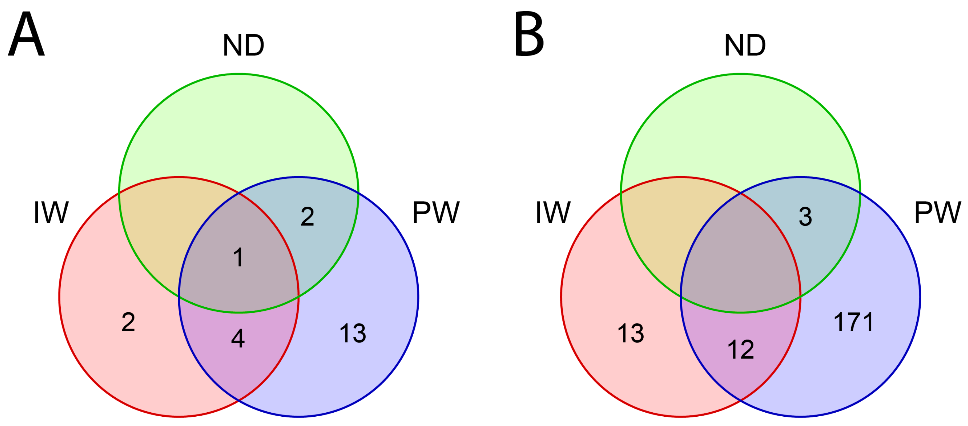
The aggregated data from PW, IW, and ND (Figure 3, Supplementary Table S6) reveals patterns of sharing and exclusivity, and a Venn diagram analysis was performed to highlight the number of shared and exclusive phyla and genera of metabolically active microorganisms.
The PW sample had a larger number of phyla (n = 20) compared to IW samples (n = 7) and ND (n = 3) (Figure 3). In the comparative analysis, it was observed that PW and IW samples had 13 and two exclusive phyla, respectively. On the other hand, PW and IW shared five bacterial phyla (Bacteroidetes, Deferribacteres, Firmicutes, Ignavibacteriae, and Proteobacteria) (Figure 3 and Figure 4, Supplementary Table S6). Also, the ND sample shares three phyla with PW, two bacterial (Synergistetes and Firmicutes), and one archaeal (Euryarchaeota) (Figure 3 and Figure 4, Supplementary Table S6). No shared phyla were observed between ND and IW (Figure 3).
As mentioned earlier, PW samples presented a larger quantity of phyla. Consequently, a larger number of genera was identified in PW (n = 186), followed by IW (n = 25) and ND (n = 3) (Figure 3). PW and IW share 12 bacterial genera (Figure 3 and Figure 4). On the other hand, PW and ND share three genera, two belonging to the Bacteria domain (Thermoanaerobacter and Thermovirga) and one belonging to the Archaea domain (Methanothermobacter) (Figure 3 and Figure 4, Supplementary Table S6). Unique genera were also identified, being 171 in PW and 13 in IW (Figure 3, Supplementary Table S6). On the other hand, no exclusive genera were observed in the ND sample (Figure 3).
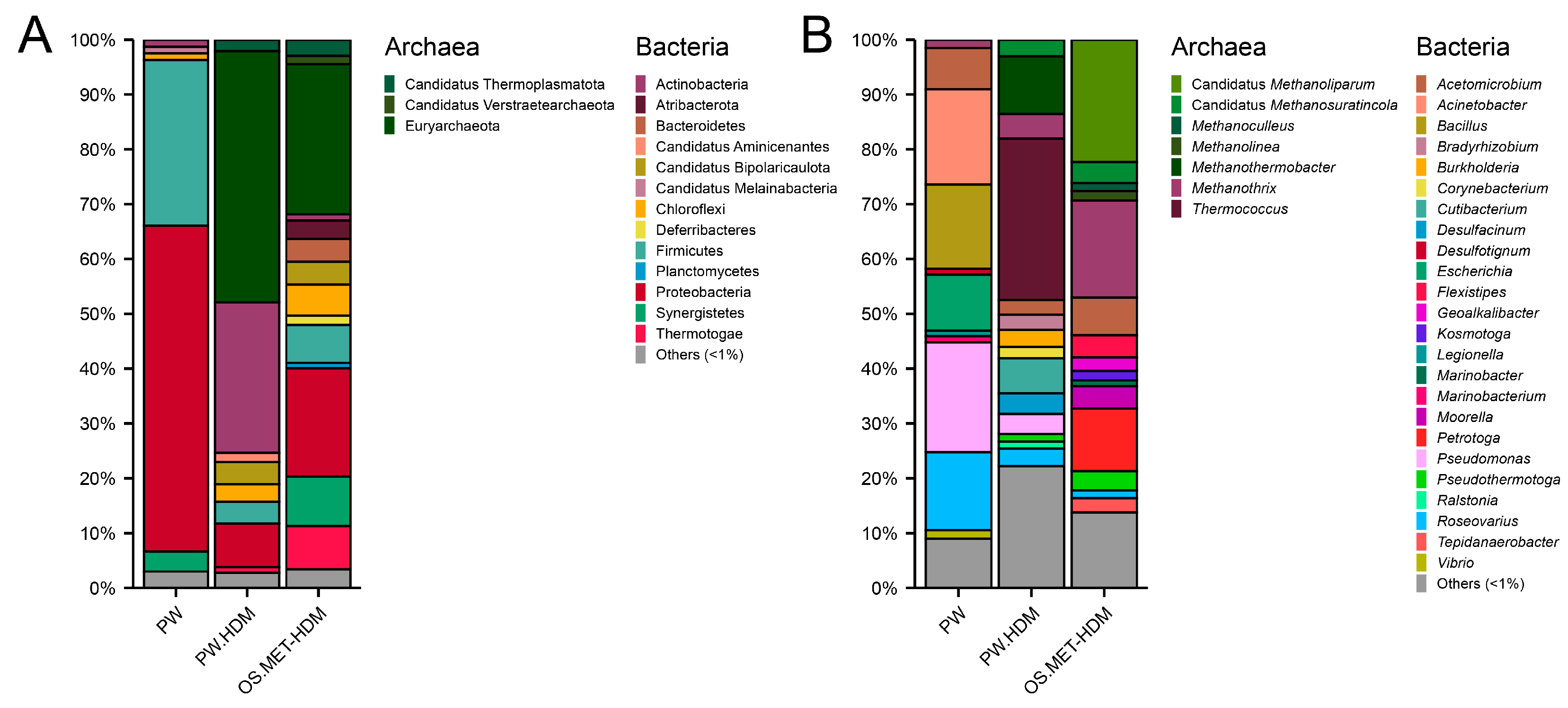
The phylum Proteobacteria was predominant in PW (57%) and IW (65%) samples, followed by Euryarchaeota (PW: 11%), Deferribacteres (IW: 8%), Firmicutes (PW: 12% and IW: 7%), and Bacteroidetes (PW: 6% and IW: 4%) (Figure 4, Supplementary Table S6). On the other hand, only the Euryarchaeota, Firmicutes, and Synergistetes phyla were identified in the ND sample, with similar abundances of 33% (Figure 4, Supplementary Table S6).
For the analysis at the genus level, the relative abundances above one (>1%) were considered. Therefore, genera with abundances lower than 1% were grouped and named as “others” (Figure 4). The Bacteria domain was predominant in PW (99%), IW (100%), and ND (67%) samples (Figure 4). The Acinetobacter, Pseudomonas, Smithella, and Tepidiphilus genera, all belonging to the Bacteria domain, were identified in PW and IW samples (Figure 4, Supplementary Table S6). The genus Pseudomonas showed a higher relative abundance (PW: 2%, IW: 8%) (Figure 4). The Methanothermobacter, Thermoanaerobacter, and Thermovirga genera were observed in the ND sample and demonstrated similar abundances of 33%. Among them, the genus Methanothermobacter is the only representative of the Archaea domain (Figure 4).
Metatranscriptomic data were analyzed in three studies. These evaluated environmental samples (PW) [16] and cultures (PW.HDM and OS.MET-HDM) [14,35]. Metatranscriptome data were downloaded from the NCBI [16,35] and NODE databases [14]. The composition of the metabolically active community was evaluated separately in each study since raw data were used (Figure 5, Supplementary Table S6).
Using metatranscriptomic data, a Venn diagram was made to show the number of shared and exclusive phyla and genera of metabolically active microorganisms detected in PW, PW.HDM, and OS.MET-HDM samples (Figure 5, Supplementary Table S6). A total of 65,119, and 164 phyla were identified in the analysis of PW, PW.HDM, and OS.MET-HDM samples, respectively. Of the 65 phyla of PW, 64 are shared with PW.HDM (Figure 5, Supplementary Table S6). The OS.MET-HDM sample presented 44 exclusive phyla. However, it shared 65 phyla with PW and 119 phyla with PW.HDM (Figure 5, Supplementary Table S6). At the genus level, a total of 1864 genera were observed (Figure 5, Supplementary Table S6). Of these, ion in PW, 327 in PW.HDM and 615 in OS.MET-HDM samples were exclusive.
The relative abundance of phyla identified in PW, PW.HDM, and OS.MET-HDM samples was also evaluated using metatranscriptome data. The Proteobacteria phylum, belonging to the Bacteria domain, had the highest abundance in the PW sample (59%), while the Euryarchaeota phylum, belonging to the Archaea domain, was more abundant in the PW.HDM (46%) and OS.MET-HDM (27%) samples (Figure 6, Supplementary Table S6).
Similar to what was done with the 16S rRNA gene data, relative abundances above one (>1%) were considered for the analysis at the genus level, while the rest were grouped and named as “others” (<1%) (Figure 6, Supplementary Table S6). In the PW sample, the most abundant genera were Pseudomonas (20%) and Acinetobacter (17%), whereas in the PW.HDM sample, the predominant genera were Thermococcus (29%) and Methanothermobacter (11%) (Figure 6, Supplementary Table S6). On the other hand, in the OS.MET-HDM sample, the most abundant genera were “Candidatus Methanoliparum” (22%) and Methanothrix (18%) (Figure 6, Supplementary Table S6).

4. Discussion

4.1. Identification and Selection of Studies

The present systematic review was based on the PRISMA guidelines and aimed to evaluate the methods applied in studies based on RNA that analyzed metabolically active microbial communities from oilfields. In the four selected databases (MEDLINE/PubMed, PubMed Central, Scopus and Web of Science), 2561 potentially eligible records were identified. After automated and manual screening, only 20 eligible original articles were included, highlighting the scarcity of studies directed at the analysis of 16S rRNA transcripts and functional genes, as well as transcriptome and metatranscriptome of samples of reservoirs and oil industry facilities. It is noteworthy that, in recent decades, several studies have investigated microbial communities from the oil industry [10,23,24,25,26,27,28,30,31]. However, most of these analyses are primarily focused on DNA sequences.
Probably, the limited number of studies directed at active microorganisms is attributed to the challenges associated with preserving and isolating high-quality RNA [22]. RNA is an inherently unstable molecule and highly vulnerable to degradation caused by RNases [30]. This instability represents a significant challenge in obtaining reliable results for the characterization of active microbial communities.

4.2. Studies Included

The studies analyzed environmental samples (PW, IW, OIL, and SD) collected from oilfields in Australia, China, India, Mexico, and the United Arab Emirates. The majority of evaluated samples were from PW, which is the most abundant waste stream of the oil industry and, consequently, one of the most available sources of samples in oilfield systems [2,31]. Salgar-Chaparro and Machuca (2019) [1] were the only authors who investigated both the planktonic microorganisms in circulating fluids and the sessile microbiota adhered to the inner walls of the pipelines. The comprehensive analysis of both fluids (PW, IW, and OIL) and solids (sediments, sludge, biofilm, pig residue, or similar) provides complementary insights into microbial activities primarily involved in hydrocarbon biodegradation and pipeline biocorrosion processes [21].
Some studies have examined microorganisms involved in the degradation of organic compounds (OCDM) [13] and hydrocarbons (HDM) [14,15,19,20,35], as well as corrosion-influencing microorganisms (CIM) [4,18,43,44], through the analysis of enriched cultures inoculated with environmental samples (PW, IW, OIL, and OS). The oil industry has a vested interest in monitoring HDM and CIM [21] since these groups can cause significant economic and environmental damage.
Therefore, the analysis of metabolically active microbial communities using RNA sequencing provides valuable insights into identifying microorganisms involved in hydrocarbon biodegradation and corrosion in oil industry facilities [16,35,43,44,47]. High-quality RNA sequencing allows for comprehensive coverage and characterization of active microorganisms [32], but the successful application of these methods depends primarily on effective protocols to preserve RNA integrity [30,32].

4.3. RNA Preprocessing, Preservation, and Extraction Method

Despite the implementation of effective protocols for sample collection, transportation, and temporary storage, the use of nucleic acid preservatives is increasingly employed, particularly for samples collected from remote areas such as offshore petroleum platforms [22]. In the studies included in the review, various preservation methods were identified. The 95:5 v/v ethanol/TRIzol solution was mentioned in eight out of 20 studies. However, information regarding the impact of preservatives on the RNA integrity of oilfield samples remains limited.
The evaluated studies employed various methods for RNA isolation, including the use of commercial kits, TRIzol reagent, and acid phenol chloroform/isoamyl alcohol reagent (the TRIzol reagent was utilized in eight studies). While nucleic acid extraction protocols should be tailored to the specific characteristics of each sample [32], the TRIzol method stands out as a rapid, accessible, and cost-effective protocol [50]. Its favorable attributes likely contribute to its widespread use for RNA isolation across diverse sample types, such as medicinal plants [51], human visceral adipose tissue [50], the SARS-CoV-2 virus [52], and fungi [53].
It is worth noting that filtration and centrifugation processes were mentioned in four studies and 14 studies, respectively. While aqueous fluids such as PW and IW are commonly sampled in petroleum systems, these samples typically exhibit low biomass and diversity [31,54]. To address this limitation, larger sample volumes are collected and subjected to preprocessing steps to concentrate biomass and facilitate the isolation of nucleic acids [1,10,17,31,45,54].

4.4. RNA Amplification and Sequencing Method

Among the molecular methods mentioned in the studies, sequencing of 16S rRNA gene transcripts was the most implemented approach for studying the active microbiota. However, there is a challenge in interpreting the data due to variations in the number of copies of the 16S rRNA gene among species, ranging from one to 15 in Bacteria and from one to four in Archaea [55,56]. Some studies suggest that the number of ribosomes increases in actively growing cells, and ribosomal RNA analysis can be used to identify metabolically active cell forms [55,56]. However, evidence indicates that the use of rRNA as an indicator of microbial activity has limitations.
Blazewicz et al. (2013) [57] reviewed several studies and concluded that (a) rRNA concentration and growth rate are not always correlated, (b) the relationship between rRNA concentration and growth rate can vary significantly among taxa, and (c) dormant cells can contain a large number of ribosomes. While there are inherent uncertainties in 16S rRNA gene sequencing, and DNA-based analysis cannot differentiate active species among all those present in the environment, the comparison of DNA and RNA libraries provides a more comprehensive characterization of microorganisms in petroleum environments [1,3,17,22,48,49].
One study in the review utilized the sequencing of the functional gene alkB, which codes for the enzyme alkane hydroxylase, to investigate microorganisms involved in the aerobic degradation of hydrocarbons [15]. In contrast, three studies sequenced the functional genes assA and mcrA to analyze microorganisms associated with the anaerobic degradation of hydrocarbons and methanogenesis, respectively. The assA gene has been employed as a biomarker for detecting microorganisms responsible for the initial activation of alkanes during the anaerobic degradation process [48,49]. Meanwhile, the mcrA functional gene is widely recognized as a biomarker for identifying methanogens [45,48,49].
The HDM can use hydrocarbons as a source of energy and carbon [58,59], preferably alkanes, cyclic and aromatic, and they metabolize organic and carbon compounds, drastically reducing oil quality [45,60]. With increasing levels of biodegradation, the content of asphaltenes and resins, acidity, and oil viscosity increase, while the content of saturated and aromatic hydrocarbons decreases [60,61]. These alterations have a negative effect on oil production by reducing the flow rates from reservoirs, as well as refining operations, and increasing process costs [61,62].
In turn, CIM represents another significant concern for the oil industry since biocorrosion processes involve the degradation of metallic materials caused by the presence and activity of various microorganisms that are in direct or close contact with the metal surface [43,63]. It is estimated that MIC contributes to nearly 40% of internal corrosion problems and 20−30% of external pipeline corrosion [63]. Corroborating the data obtained in this review, sulfate-reducing bacteria (SRB) have been widely cited in studies related to pipeline corrosion [4,10]. Functional genes such as aprA and drsA, encoding the enzymes adenosine-5-reductase and dissimilatory sulfate reductase, respectively, are used to track SRB [4,17].
Metatranscriptomic analysis has been evaluated with data from three studies. This analysis is considered a powerful technique that enables the examination of the gene expression profile of microorganisms present in a sample, providing valuable insights into their metabolic activity in a given environment [64]. Despite the advancements in microbiome studies of oilfield samples, there is still a notable scarcity of studies analyzing the metatranscriptome, and this can be attributed to several challenges, including the extraction of high-quality RNA, the presence of inhibitors, limited quantities of genetic material in the samples, and rapid RNA degradation. Additionally, the collection and proper preservation of samples for metatranscriptomic analysis can be complex due to the demanding conditions of the petroleum environment, such as high pressure and temperature, as well as the presence of toxic compounds.

4.5. Composition of the Metabolically Active Microbiota

In total, eight studies performed amplicon sequencing of 16S rRNA gene transcripts [1,3,15,17,18,22,43,44] and three performed metatranscriptome analysis [14,16,35]. The phylum Proteobacteria was predominant in the analysis of 16S rRNA gene data from PW (57%) and IW (65%) samples (Figure 4), as well as in the metatranscriptome analysis of PW samples (59%) (Figure 6). This phylum comprises one of the largest divisions within prokaryotes and represents the vast majority of known gram-negative bacteria, consisting of more than 200 genera [65].
When analyzing data at the genus level, higher relative abundances of the genera Pseudomonas (16S rRNA= PW: 2%; IW: 8%; and metatranscriptome = PW: 20%) and Acinetobacter (16S rRNA = PW: 1%; IW: 4%; and metatranscriptome = PW: 17%) were observed. These genera belong to the phylum Proteobacteria and are often found in oil environments and can degrade complex hydrocarbons into simpler compounds, reducing oil quality [3]. It is worth mentioning that some genera degrade n-alkanes, while others degrade aromatic compounds [66]. However, Pseudomonas can degrade both classes of hydrocarbons [3,67] due to the presence of specific genes, such as alkB, xylXYZ and benABCD [15,68].
Analyzing the metatranscriptome from culture samples, it was observed that the phylum Euryarchaeota showed greater abundance (PW.HDM: 46% and OS.MET-HDM: 27%), a phylum affiliated with the Archaea domain, which harbors hydrogenotrophic and acetotrophic methanogens microorganisms [69]. In oil reservoirs, under methanogenic conditions, hydrocarbon degradation occurs when communities of bacteria and archaea cooperate syntrophically to produce methane through thermodynamically favorable pathways [70].
Analyzing metatranscriptomic data at the genus level, it was observed that in the samples from the PW.HDM cultures, the most abundant genera were Thermococcus (29%) and Methanothermobacter (11%). While in the study that evaluated OS.MET-HDM cultures, the most abundant genera were “Candidatus Methanoliparum” (22%) and Methanothrix (18%). The genera Thermococcus, Methanothermobacter, and Methanothrix belong to the phylum Euryarchaeota and are often detected in oil reservoirs [45].

5. Conclusions

This systematic review reveals the limited research on RNA-based analysis of metabolically active microbial communities in oilfields. Despite substantial prior research on microbial communities in the oil industry, there is a significant gap in the analysis of 16S rRNA transcripts, functional genes, transcriptomes, and metatranscriptomes. It is believed that challenges related to RNA preservation and isolation have hindered progress in this area. The selected studies predominantly focus on produced water samples, highlighting the need for a broader exploration of various sample types within oilfield systems. Additionally, metatranscriptomic analysis remains underutilized due to its technical complexities. The predominance of Proteobacteria and specific genera like Pseudomonas and Acinetobacter underscores the importance of studying these microorganisms’ roles in hydrocarbon degradation. Furthermore, the presence of Euryarchaeota points to the significance of methanogenic processes in oil reservoirs. This review serves as a call to action for further research in this area, emphasizing the importance of standardizing RNA preservation, extraction, and sequencing methods. Advancements in RNA-based analysis have the potential to significantly enhance our understanding of active microbial communities in oilfields and their impacts on the industry.

Supplementary Materials

The following supporting information can be downloaded at: https://www.mdpi.com/article/10.3390/applmicrobiol3040079/s1. Table S1: PRISMA-S checklist. Table S2: Terms used in the search for records in databases and selection of documents by automation tools. Table S3: Records identified in the databases. Table S4: Automated and manual screening of records. Table S5. References, title, and DOI of the studies included in the present systematic review Table S5. Composition of the metabolically active microorganisms. Table S6. References, title, and DOI of the original articles are included in the present systematic review. Table S7: Description of the microbial groups corrosion-influencing microorganisms.

Author Contributions

Conceptualization, A.G.-N., R.F.G., G.J.Y.G., M.S.C. and V.d.A.W.; Methodology, A.G.-N., R.F.G., G.J.Y.G., J.d.C.F.D. and M.S.C.; Software, G.J.Y.G. and E.A.C.; Formal Analysis, R.F.G. and G.J.Y.G.; Investigation, R.F.G. and J.d.C.F.D.; Data Curation, R.F.G., G.J.Y.G., J.d.C.F.D. and M.S.C.; Writing—Original Draft Preparation, R.F.G.; Writing—Review & Editing, A.G.-N., R.F.G., G.J.Y.G., M.S.C., V.d.A.W., R.N.A., M.P.d.S. and C.J.G.; Visualization, R.F.G., G.J.Y.G., E.A.C., M.S.C., V.d.A.W., R.N.A., M.P.d.S., C.J.G., A.G.-N.; V.A.d.C.A. and H.F.; Supervision, A.G.-N. and M.S.C.; Project Administration, A.G.-N.; V.A.d.C.A. and H.F.; Funding, A.G.-N. All authors have read and agreed to the published version of the manuscript.

Funding

This work was supported by Petróleo Brasileiro S.A—Petrobras (ANP nº 21035-1) and the Graduate Programs of Microbiology (http://www.microbiologia.icb.ufmg.br/pos) and Bioinformatics (http://www.pgbioinfo.icb.ufmg.br) of the Institute of Biological Sciences of Federal University of Minas Gerais (UFMG). The funders had no role in study design, data collection and analysis, decision to publish, or preparation of the manuscript.

Data Availability Statement

Information available in the Supplementary Material.

Acknowledgments

We would like to thank all researchers who contributed directly or indirectly to this study.

Conflicts of Interest

The authors declare no conflict of interest.

References

  1. Salgar-Chaparro, S.J.; Machuca, L.L. Complementary DNA/RNA-based profiling: Characterization of corrosive microbial communities and their functional profiles in an oil production facility. Front. Microbiol. 2019, 10, 2587. [Google Scholar] [CrossRef] [PubMed]
  2. Neff, J.; Lee, K.; DeBlois, E.M. Produced water: Overview of composition, fates, and effects. In Produced Water: Environmental Risks and Advances in Mitigation Technologies; Springer Science Business Media: New York, NY, USA, 2011; pp. 3–54. [Google Scholar]
  3. Nazina, T.N.; Shestakova, N.M.; Semenova, E.M.; Korshunova, A.V.; Kostrukova, N.K.; Tourova, T.P.; Liu, M.; Feng, Q.; Poltaraus, A.B. Diversity of metabolically active bacteria in water-flooded high-temperature heavy oil reservoir. Front. Microbiol. 2017, 8, 707. [Google Scholar] [CrossRef] [PubMed]
  4. Zapata-Peñasco, I.; Salazar-Coria, L.; Saucedo-García, M.; Villa-Tanaca, L.; Hernández-Rodríguez, C. Bisulfite reductase gene expression of thermophilic sulphate-reducing bacteria from saline connate water of oil reservoirs with high temperature. Int. Biodeterior. Biodegrad. 2016, 108, 198–206. [Google Scholar] [CrossRef]
  5. Liang, Y.; Ning, Y.; Liao, L.; Yuan, B. Special focus on produced water in oil and gas fields: Origin, management, and reinjection practice. In Formation Damage during Improved Oil Recovery: Fundamentals and Applications; Yuan, B., Wood, D.A., Eds.; Elsevier: Amsterdam, The Netherlands, 2018; pp. 515–586. [Google Scholar]
  6. Sanders, P.F.; Sturman, P.J. Biofouling in the oil industry. In Petroleum Microbiology; Ollivier, B., Magot, M., Eds.; ASM Press: Washington, DC, USA, 2005; pp. 171–198. [Google Scholar]
  7. Gieg, L.M.; Jack, T.R.; Foght, J.M. Biological souring and mitigation in oil reservoirs. Appl. Microbiol. Biotechnol. 2011, 92, 263–282. [Google Scholar] [CrossRef]
  8. Johnson, R.J.; Folwell, B.D.; Wirekoh, A.; Frenzel, M.; Skovhus, T.L. Reservoir Souring–Latest developments for application and mitigation. J. Biotechnol. 2017, 256, 57–67. [Google Scholar] [CrossRef]
  9. Xu, D.; Gu, T.; Lovley, D.R. Microbially mediated metal corrosion. Nat. Rev. Microbiol. 2023, 1–14. [Google Scholar] [CrossRef]
  10. Dutra, J.; García, G.; Gomes, R.; Cardoso, M.; Côrtes, Á.; Silva, T.; de Jesus, L.; Rodrigues, L.; Freitas, A.; Góes-Neto, A.; et al. Effective biocorrosive control in oil industry facilities: 16S rRNA gene metabarcoding for monitoring microbial communities in produced water. Microorganisms 2023, 11, 846. [Google Scholar] [CrossRef]
  11. Eckert, R.B. Emphasis on biofilms can improve mitigation of microbiologically influenced corrosion in oil and gas industry. Corros. Eng. Sci. Technol. 2015, 50, 163–168. [Google Scholar] [CrossRef]
  12. Vigneron, A.; Head, I.M.; Tsesmetzis, N. Damage to offshore production facilities by corrosive microbial biofilms. Appl. Microbiol. Biotechnol. 2018, 102, 2525–2533. [Google Scholar] [CrossRef]
  13. Alhefeiti, M.A.; Athamneh, K.; Vijayan, R.; Ashraf, S.S. Bioremediation of various aromatic and emerging pollutants by Bacillus cereus sp. isolated from petroleum sludge. Water Sci. Technol. 2021, 83, 1535–1547. [Google Scholar] [CrossRef]
  14. Zhou, Z.; Zhang, C.J.; Liu, P.F.; Fu, L.; Laso-Pérez, R.; Yang, L.; Bai, L.-P.; Li, J.; Yang, M.; Cheng, L.; et al. Non-syntrophic methanogenic hydrocarbon degradation by an archaeal species. Nature 2022, 601, 257–262. [Google Scholar] [CrossRef] [PubMed]
  15. Shestakova, N.M.; Korshunova, A.V.; Mikhailova, E.M.; Sokolova, D.S.; Tourova, T.P.; Belyaev, S.S.; Poltaraus, A.B.; Nazina, T.N. Characterization of the aerobic hydrocarbon-oxidizing enrichments from a high-temperature petroleum reservoir by comparative analysis of DNA-and RNA-derived clone libraries. Microbiology 2011, 80, 60–69. [Google Scholar] [CrossRef]
  16. Liu, Y.F.; Galzerani, D.D.; Mbadinga, S.M.; Zaramela, L.S.; Gu, J.D.; Mu, B.Z.; Zengler, K. Metabolic capability and in situ activity of microorganisms in an oil reservoir. Microbiome 2018, 6, 5. [Google Scholar] [CrossRef] [PubMed]
  17. Li, X.X.; Liu, J.F.; Zhou, L.; Mbadinga, S.M.; Yang, S.Z.; Gu, J.D.; Mu, B.Z. Diversity and composition of sulfate-reducing microbial communities based on genomic DNA and RNA transcription in production water of high temperature and corrosive oil reservoir. Front. Microbiol. 2017, 8, 1011. [Google Scholar] [CrossRef] [PubMed]
  18. Albahri, M.B.; Barifcani, A.; Iglauer, S.; Lebedev, M.; O’Neil, C.; Salgar-Chaparro, S.J.; Machuca, L.L. Investigating the mechanism of microbiologically influenced corrosion of carbon steel using X-ray micro-computed tomography. J. Mater. Sci. 2021, 56, 13337–13371. [Google Scholar] [CrossRef]
  19. Zheng, X.; Lin, X.; Yan, Y.; Wen, Y.; Shi, Y.; Xie, X.; Hou, A.; Lai, N. Effect of reservoir salinity between bioacid and carbonate rock based on biometabolic analysis. Energy Fuels 2019, 33, 8135–8144. [Google Scholar] [CrossRef]
  20. Su, Z.; Wang, S.; Yang, S.; Yin, Y.; Cao, Y.; Li, G.; Ma, T. Genetic and comparative genome analysis of Exiguobacterium aurantiacum SW-20, a petroleum-degrading bacteria with salt tolerance and heavy metal-tolerance isolated from produced water of changqing oilfield, China. Microorganisms 2022, 10, 66. [Google Scholar] [CrossRef]
  21. Lomans, B.P.; de Paula, R.; Geissler, B.; Kuijvenhoven, C.A.; Tsesmetzis, N. Proposal of improved biomonitoring standard for purpose of microbiologically influenced corrosion risk assessment. In Proceedings of the SPE International Oilfield Corrosion Conference and Exhibition, Aberdeen, UK, 9–10 May 2016; OnePetro: Richardson, TX, USA, 2016. [Google Scholar]
  22. Zhou, L.; Lu, Y.W.; Wang, D.W.; Zhang, S.L.; Tang, E.G.; Qi, Z.Z.; Xie, S.-N.; Wu, J.; Liang, B.; Mu, B.Z.; et al. Microbial community composition and diversity in production water of a high-temperature offshore oil reservoir assessed by DNA-and RNA-based analyses. Int. Biodeterior. Biodegrad. 2020, 151, 104970. [Google Scholar] [CrossRef]
  23. Neria-González, I.; Wang, E.T.; Ramírez, F.; Romero, J.M.; Hernández-Rodríguez, C. Characterization of bacterial community associated to biofilms of corroded oil pipelines from the southeast of Mexico. Anaerobe 2006, 12, 122–133. [Google Scholar] [CrossRef]
  24. Gittel, A.; Sørensen, K.B.; Skovhus, T.L.; Ingvorsen, K.; Schramm, A. Prokaryotic community structure and sulfate reducer activity in water from high-temperature oil reservoirs with and without nitrate treatment. Appl. Environ. Microbiol. 2009, 75, 7086–7096. [Google Scholar] [CrossRef]
  25. Li, H.; Chen, S.; Mu, B.Z.; Gu, J.D. Molecular detection of anaerobic ammonium-oxidizing (anammox) bacteria in high-temperature petroleum reservoirs. Microb. Ecol. 2010, 60, 771–783. [Google Scholar] [CrossRef] [PubMed]
  26. Lenchi, N.; İnceoğlu, Ö.; Kebbouche-Gana, S.; Gana, M.L.; Llirós, M.; Servais, P.; García-Armisen, T. Diversity of microbial communities in production and injection waters of Algerian oilfields revealed by 16S rRNA gene amplicon 454 pyrosequencing. PLoS ONE 2013, 8, 66588. [Google Scholar] [CrossRef] [PubMed]
  27. Machuca Suarez, L.; Salgar-Chaparro, S. Effect of sample storage conditions on the molecular assessment of MIC. In Proceedings of the Corrosion & Prevention Conference, Adelaide, Australia, 11–14 November 2018. [Google Scholar]
  28. Nasser, B.; Saito, Y.; Alarawi, M.; Al-Humam, A.A.; Mineta, K.; Gojobori, T. Characterization of microbiologically influenced corrosion by comprehensive metagenomic analysis of an inland oil field. Gene 2021, 774, 145425. [Google Scholar] [CrossRef] [PubMed]
  29. Hampton-Marcell, J.T.; Frazier, A.; Moormann, S.M.; Owens, S.M.; Gilbert, J.A. Preparation and analysis of metatranscriptomic libraries in petroleum hydrocarbon microbe systems. In Hydrocarbon and Lipid Microbiology Protocols: Genetic, Genomic and System Analyses of Communities; McGenity, T., Timmis, K., Nogales, B., Eds.; Springer Protocols Handbooks; Springer: Berlin/Heidelberg, Germany, 2017; pp. 51–67. [Google Scholar]
  30. Simister, R.L.; Schmitt, S.; Taylor, M.W. Evaluating methods for the preservation and extraction of DNA and RNA for analysis of microbial communities in marine sponges. J. Exp. Mar. Biol. Ecol. 2011, 397, 38–43. [Google Scholar] [CrossRef]
  31. Rachel, N.M.; Gieg, L.M. Preserving microbial community integrity in oilfield produced water. Front. Microbiol. 2020, 11, 581387. [Google Scholar] [CrossRef] [PubMed]
  32. Silva, L.C.F.; Lopes, D.R.G.; Lima, H.S.; Quartaroli, L.; de Sousa, M.P.; de Abreu Waldow, V.; Akamine, R.N.; de Paula, S.O.; da Silva, C.C. Comparison of methods for preservation of activated sludge samples for high-throughput nucleic acid sequencing and bacterial diversity analysis. Int. Biodeterior. Biodegrad. 2021, 157, 105139. [Google Scholar] [CrossRef]
  33. Page, M.J.; McKenzie, J.E.; Bossuyt, P.M.; Boutron, I.; Hoffmann, T.C.; Mulrow, C.D.; Shamseer, L.; Tetzlaff, J.M.; Akl, E.A.; Moher, D.; et al. The PRISMA 2020 statement: An updated guideline for reporting systematic reviews. Int. J. Surg. 2021, 88, 105906. [Google Scholar] [CrossRef]
  34. Rethlefsen, M.L.; Kirtley, S.; Waffenschmidt, S.; Ayala, A.P.; Moher, D.; Page, M.J.; Koffel, J.B. PRISMA-S: An extension to the PRISMA statement for reporting literature searches in systematic reviews. Syst. Rev. 2021, 10, 39. [Google Scholar] [CrossRef]
  35. Liu, Y.F.; Chen, J.; Liu, Z.L.; Shou, L.B.; Lin, D.D.; Zhou, L.; Yang, S.Z.; Liu, J.F.; Li, W.; Mu, B.Z.; et al. Anaerobic degradation of paraffins by thermophilic Actinobacteria under methanogenic conditions. Environ. Sci. Technol. 2020, 54, 10610–10620. [Google Scholar] [CrossRef]
  36. Tamames, J.; Puente-Sánchez, F. SqueezeMeta, a highly portable, fully automatic metagenomic analysis pipeline. Front. Microbiol. 2019, 9, 3349. [Google Scholar] [CrossRef]
  37. Bolger, A.M.; Lohse, M.; Usadel, B. Trimmomatic: A flexible trimmer for Illumina sequence data. Bioinformatics 2014, 30, 2114–2120. [Google Scholar] [CrossRef] [PubMed]
  38. Li, D.; Liu, C.M.; Luo, R.; Sadakane, K.; Lam, T.W. MEGAHIT: An ultra-fast single-node solution for large and complex metagenomics assembly via succinct de Bruijn graph. Bioinformatics 2015, 31, 1674–1676. [Google Scholar] [CrossRef] [PubMed]
  39. Hyatt, D.; Chen, G.L.; LoCascio, P.F.; Land, M.L.; Larimer, F.W.; Hauser, L.J. Prodigal: Prokaryotic gene recognition and translation initiation site identification. BMC Bioinform. 2010, 11, 119. [Google Scholar] [CrossRef] [PubMed]
  40. Buchfink, B.; Xie, C.; Huson, D.H. Fast and sensitive protein alignment using DIAMOND. Nat. Methods 2015, 12, 59–60. [Google Scholar] [CrossRef] [PubMed]
  41. Puente-Sánchez, F.; García-García, N.; Tamames, J. SQMtools: Automated processing and visual analysis of’omics data with R and anvi’o. BMC Bioinform. 2020, 21, 358. [Google Scholar] [CrossRef] [PubMed]
  42. Wickham, H. ggplot2. Wiley interdisciplinary reviews. Comput. Stat. 2011, 3, 180–185. [Google Scholar] [CrossRef]
  43. Salgar-Chaparro, S.J.; Lepkova, K.; Pojtanabuntoeng, T.; Darwin, A.; Machuca, L.L. Microbiologically influenced corrosion as a function of environmental conditions: A laboratory study using oilfield multispecies biofilms. Corros. Sci. 2020, 169, 108595. [Google Scholar] [CrossRef]
  44. Salgar-Chaparro, S.J.; Lepkova, K.; Pojtanabuntoeng, T.; Darwin, A.; Machuca, L.L. Nutrient level determines biofilm characteristics and subsequent impact on microbial corrosion and biocide effectiveness. Appl. Environ. Microbiol. 2020, 86, e02885-19. [Google Scholar] [CrossRef]
  45. Zhou, L.; Hu, Q.Q.; Lu, Y.W.; Mbadinga, S.M.; Liu, Y.F.; Li, X.X.; Wang, B.; Lv, H.; Liu, J.F.; Mu, B.Z.; et al. Dominant and active methanogens in the production waters from a high-temperature petroleum reservoir by DNA-and RNA-based analysis. Geomicrobiol. J. 2021, 38, 191–198. [Google Scholar] [CrossRef]
  46. Prajapat, G.; Jain, S.; Lal, B.; Lavania, M.; Agrawal, A. Control of reservoir souring by incomplete nitrate reduction in Indian oil fields. Bioresour. Technol. Rep. 2023, 21, 101302. [Google Scholar] [CrossRef]
  47. Liu, Y.F.; Chen, J.; Zaramela, L.S.; Wang, L.Y.; Mbadinga, S.M.; Hou, Z.W.; Wu, X.L.; Gu, J.D.; Zengler, K.; Mu, B.Z. Genomic and transcriptomic evidence supports methane metabolism in Archaeoglobi. mSystems 2020, 5, e00651-19. [Google Scholar] [CrossRef] [PubMed]
  48. Liu, J.F.; Lu, Y.W.; Liu, X.B.; Li, B.G.; Sun, Y.F.; Zhou, L.; Liu, Y.F.; Yang, S.Z.; Gu, J.D.; Mu, B.Z. Dominance of Pseudomonas in bacterial community and inhibition of fumarate addition pathway by injection of nutrients in oil reservoir revealed by functional gene and their transcript analyses. Int. Biodeterior. Biodegrad. 2020, 153, 105039. [Google Scholar] [CrossRef]
  49. Liu, J.F.; Lu, Y.W.; Zhou, L.; Li, W.; Hou, Z.W.; Yang, S.Z.; Wu, X.L.; Gu, J.D.; Mu, B.Z. Simultaneous detection of transcribed functional assA gene and the corresponding metabolites of linear alkanes (C4, C5, and C7) in production water of a low-temperature oil reservoir. Sci. Total Environ. 2020, 746, 141290. [Google Scholar] [CrossRef] [PubMed]
  50. Roy, D.; Tomo, S.; Modi, A.; Purohit, P.; Sharma, P. Optimising total RNA quality and quantity by phenol-chloroform extraction method from human visceral adipose tissue: A standardisation study. MethodsX 2020, 7, 101113. [Google Scholar] [CrossRef] [PubMed]
  51. Chang, E.; Zhao, Y.; Wei, Q.; Shi, S.; Jiang, Z. Isolation of high-quality RNA from Platycladus orientalis and other Cupressaceae plants. Electron. J. Biotechnol. 2016, 23, 21–27. [Google Scholar] [CrossRef]
  52. Amirouche, A.; Ait-Ali, D.; Nouri, H.; Boudrahme-Hannou, L.; Tliba, S.; Ghidouche, A.; Bitam, I. TRIzol-based RNA extraction for detection protocol for SARS-CoV-2 of coronavirus disease 2019. New Microbes New Infect. 2021, 41, 100874. [Google Scholar] [CrossRef]
  53. Nóbrega, B.B.; Soares, D.M.; Zamuner, C.K.; Stevani, C.V. Optimized methodology for obtention of high-yield and-quality RNA from the mycelium of the bioluminescent fungus Neonothopanus gardneri. J. Microbiol. Methods 2021, 191, 106348. [Google Scholar] [CrossRef]
  54. Oldham, A.L.; Sandifer, V.; Duncan, K.E. Effects of sample preservation on marine microbial diversity analysis. J. Microbiol. Methods 2019, 158, 6–13. [Google Scholar] [CrossRef]
  55. Klappenbach, J.A.; Saxman, P.R.; Cole, J.R.; Schmidt, T.M. rrndb: The ribosomal RNA operon copy number database. Nucleic Acids Res. 2001, 29, 181–184. [Google Scholar] [CrossRef]
  56. Moeseneder, M.M.; Arrieta, J.M.; Herndl, G.J. A comparison of DNA-and RNA-based clone libraries from the same marine bacterioplankton community. FEMS Microbiol. Ecol. 2005, 51, 341–352. [Google Scholar] [CrossRef]
  57. Blazewicz, S.J.; Barnard, R.L.; Daly, R.A.; Firestone, M.K. Evaluating rRNA as an indicator of microbial activity in environmental communities: Limitations and uses. ISME J. 2013, 7, 2061–2068. [Google Scholar] [CrossRef] [PubMed]
  58. Atlas, R.M.; Atlas, M.C. Biodegradation of oil and bioremediation of oil spills. Curr. Opin. Biotechnol. 1991, 2, 440–443. [Google Scholar] [CrossRef]
  59. Salama, A.M.; Rashad, E.; Elgarahy, A.M.; Elwakeel, K. Effect of green synthesized iron oxide nanoparticles on bacterial microbiome for clean up the crude oil. Aswan Univ. J. Environ. Stud. 2023, 4, 49–81. [Google Scholar] [CrossRef]
  60. Yang, Z.; Li, M.; Cheng, D.; Xiao, H.; Lai, H.; Chen, Q. Geochemistry and possible origins of biodegraded oils in the Cretaceous reservoir of the Muglad Basin and their application in hydrocarbon exploration. J. Pet. Sci. Eng. 2019, 173, 889–898. [Google Scholar] [CrossRef]
  61. Cruz, G.F.D.; Marsaioli, A.J. Processos naturais de biodegradação do petróleo em reservatórios. Química Nova 2012, 35, 1628–1634. [Google Scholar] [CrossRef]
  62. Head, I.M.; Jones, D.M.; Larter, S.R. Biological activity in the deep subsurface and the origin of heavy oil. Nature 2003, 426, 344–352. [Google Scholar] [CrossRef]
  63. Wolodko, J.; Haile, T.; Khan, F.; Taylor, C.; Eckert, R.; Hashemi, S.J.; Ramirez, A.M.; Skovhus, T.L. Modeling of microbiologically influenced corrosion (MIC) in the oil and gas industry-past, present and future. In Proceedings of the NACE Corrosion, Phoenix, AZ, USA, 15–19 April 2018. [Google Scholar]
  64. Hampton-Marcell, J.T.; Frazier, A.; Moormann, S.M.; Owens, S.M.; Gilbert, J.A. Preparation and Analysis of Metatranscriptomic Libraries in Petroleum Hydrocarbon Microbe Systems. In Laboratory Methods in Food Microbiology; McGenity, T., Timmis, K., Nogales, B., Eds.; Gulf Professional Publishing: Houston, TX, USA, 2014. [Google Scholar]
  65. Gupta, R.S. The phylogeny of proteobacteria: Relationships to other eubacterial phyla and eukaryotes. FEMS Microbiol. Rev. 2000, 24, 367–402. [Google Scholar] [CrossRef]
  66. Goveas, L.C.; Selvaraj, R.; Vinayagam, R.; Alsaiari, A.A.; Alharthi, N.S.; Sajankila, S.P. Nitrogen dependence of rhamnolipid mediated degradation of petroleum crude oil by indigenous Pseudomonas sp. WD23 in seawater. Chemosphere 2022, 304, 135235. [Google Scholar] [CrossRef]
  67. Muthukumar, B.; Al Salhi, M.S.; Narenkumar, J.; Devanesan, S.; Rao, T.N.; Kim, W.; Rajasekar, A. Characterization of two novel strains of Pseudomonas aeruginosa on biodegradation of crude oil and its enzyme activities. Environ. Pollut. 2022, 304, 119223. [Google Scholar] [CrossRef]
  68. de Oliveira, H.L.; Dias, G.M.; Neves, B.C. Genome sequence of Pseudomonas aeruginosa PA1-Petro—A role model of environmental adaptation and a potential biotechnological tool. Heliyon 2022, 8, e11566. [Google Scholar] [CrossRef]
  69. Sengupta, K.; Pal, S. A review on microbial diversity and genetic markers involved in methanogenic degradation of hydrocarbons: Futuristic prospects of biofuel recovery from contaminated regions. Environ. Sci. Pollut. Res. 2021, 28, 40288–40307. [Google Scholar] [CrossRef] [PubMed]
  70. Dolfing, J.; Larter, S.R.; Head, I.M. Thermodynamic constraints on methanogenic crude oil biodegradation. ISME J. 2008, 2, 442–452. [Google Scholar] [CrossRef] [PubMed]
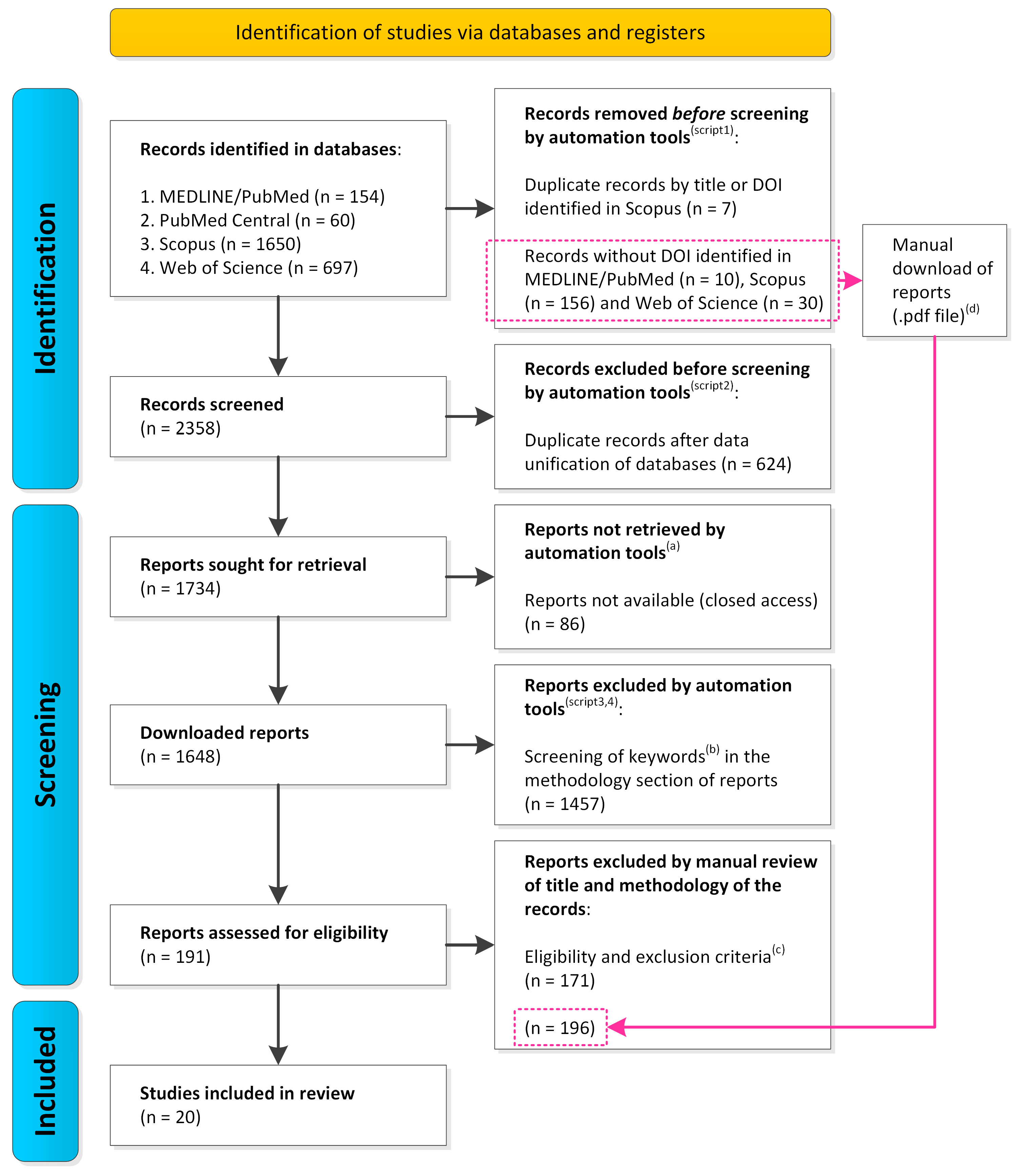
Figure 1. PRISMA flow diagram [33] of identification, screening, and inclusion of studies in the systematic review. Script1 format_input.py; Script2 remove_duplicates.py; Script3 pdf2txt.py; Script4 search_keywords.py (Supplementary Table S4). (a) Software based on permissions of the UFMG internal network. (b) Keywords (Supplementary Table S2). (c) Eligibility and exclusion criteria (Methodology Section 2.3). (d) Reports screened manually.
Applmicrobiol 03 00079 g001
Figure 2. Number of publications per year of studies that evaluated metabolically active microorganisms in oilfields.
Applmicrobiol 03 00079 g002
Figure 3. Venn diagram presents the number of shared and exclusive (A) phyla and (B) genera of metabolically active microorganisms based on RNA obtained from the analyses of 16S rRNA gene transcript amplicons. Sample type: produced water (PW) and injection water (IW), no data (ND). ND: not available in the study.
Applmicrobiol 03 00079 g003
Figure 4. Relative abundance of metabolically active microorganisms based on RNA at the (A) phylum and (B) genus levels obtained from 16S rRNA gene transcript amplicon data. Sample type: produced water (PW) and injection water (IW), no data (ND). ND: not available in the study.
Applmicrobiol 03 00079 g004
Figure 5. The Venn diagram shows the number of shared and exclusive (A) phyla and (B) genera of metabolically active microorganisms based on RNA obtained from metatranscriptomic data. Produced water (PW); PW hydrocarbon-degrading microorganisms (PW.HDM) and Oily sludge (OS) methanogenic hydrocarbon-degrading microorganisms (OS.MET-HDM).
Applmicrobiol 03 00079 g005
Figure 6. Relative abundance of metabolically active microorganisms based on RNA at the (A) phylum and (B) genus level obtained from metatranscriptomic data. Sample type: Produced water (PW); PW hydrocarbon-degrading microorganisms (PW.HDM) and Oily sludge (OS) methanogenic hydrocarbon-degrading microorganisms (OS.MET-HDM).
Applmicrobiol 03 00079 g006
Table 1. Eligibility criteria for the inclusion of articles in the systematic review.
CriteriaDescription
Sampling locationReservoir; pipeline; tank
Sample typeenvironmental (oil, injection water, produced water, biofilm, pig residue, and oil sludge); laboratory (cultures inoculated with environmental samples)
AnalysisRNA sequencing
StudyOriginal
Table 2. Reference, country, and samples studied in the articles included in this systematic review.
ReferenceSampling CountrySample TypeNSample StudiedID
Shestakova et al. (2011) [15]Hebei, ChinaPW1CulturePW.HDM (a)
IW1CultureIW.HDM (a)
Zapata-Peñasco et al. (2016) [4]MexicoPW2CulturePW.SRB (a)
Nazina et al. (2017) [3]Hebei, ChinaIW1EnvironmentalIW (a)
Li et al. (2017) [17]Jiangsu, ChinaPW6EnvironmentalPW (a)
Liu et al. (2018) [16]Jiangsu, ChinaPW3EnvironmentalPW (a)
Salgar-Chaparro e Machuca (2019) [1]AustraliaPW6EnvironmentalPW (a)
IW1EnvironmentalIW (a)
SD4EnvironmentalSD (a)
Zheng et al. (2019) [19]ChinaOIL1CultureOIL.HDM (a)
Liu et al. (2020) [35]Shandong, ChinaPW1EnvironmentalPW (a)
CulturePW.HDM (a)
Liu et al. (2020) [47]ChinaPW2EnvironmentalPW (a)
Liu et al. (2020) [48]Jiangsu, ChinaPW1EnvironmentalPW (a)
Zhou et al. (2020) [22]ChinaPW2EnvironmentalPW (a)
Liu et al. (2020) [49]ChinaPW1EnvironmentalPW (a)
Salgar-Chaparro et al. (2020) [43]AustraliaPW1ConsortiumPW.CIM (a),(b)
Salgar-Chaparro et al. (2020) [44]AustraliaPW2ConsortiumPW.CIM (a),(b)
Alhefeiti et al. (2021) [13]United Arab EmiratesOS1CultureOS.OCDM (a)
Zhou et al. (2021) [45]Jiangsu, ChinaPW6EnvironmentalPW (a)
Albahri et al. (2021) [18]AustraliaOIL1ConsortiumND.CIM (a),(b),(c)
Su et al. (2022) [20]ChinaPW1CulturePW.HDM (a)
Zhou et al. (2022) [14]Shengli, ChinaOS1CultureOS.MET-HDM (a)
Prajapat et al. (2023) [46]Rajasthan, IndiaPW1EnvironmentalPW (a)
IW3EnvironmentalIW (a)
Produced water (PW); injection water (IW); solid deposits (SD); oil (OIL); oily sludge (OS); corrosion-influencing microorganisms (CIM); hydrocarbon-degrading microorganisms (HDM); organic compounds degrading microorganisms (OCDM); sulfate-reducing bacteria (SRB); methanogenic hydrocarbon-degrading microorganisms (MET-HDM). Sample number (n). Identification code (ID). (a) Samples used for RNA sequencing: (1) environmental and/or microbial, (2) microbial culture and/or (3) consortium enriched with environmental samples. (b) Description of CIM groups (Supplementary Table S7). (c) Sample type (inoculum) not available in the study, no data (ND).
Table 3. Methods of preprocessing, preservation, and extraction of RNA from the studied samples in the articles included in this systematic review.
ReferenceSamplePreprocessingRNA Preserving AgentRNA Extraction Method
Shestakova et al. (2011) [15]PW.HDMCentrifugation (b)Not declaredTRIzol reagent (d)
IW.HDMCentrifugation (b)Not declaredTRIzol reagent (d)
Zapata-Peñasco et al. (2016) [4]PW.SRBCentrifugation (b)RNAProtect Bacteria Reagent (e)RNeasy Protect Bacteria kit (e)
Nazina et al. (2017) [3]IWFiltration (b)Ethanol reagent (h)TRIzol reagent (d)
Li et al. (2017) [17]PWFiltration (a)95:5 v/v ethanol/trizol (h)High Pure RNA Isolation Kit (f)
Liu et al. (2018) [16]PWCentrifugation (b)95:5 v/v ethanol/trizol (h)PowerMicrobiome RNA Isolation kit (g)
Salgar-Chaparro e Machuca (2019) [1]PWFiltration (a)RNAProtect Bacteria Reagent (e)RNeasy PowerWater kit (e)
IWFiltration (a)RNAProtect Bacteria Reagent (e)RNeasy PowerWater kit (e)
SDNot declaredRNAProtect Bacteria Reagent (e)RNeasy PowerSoil kit (e)
Zheng et al. (2019) [19]OIL.HDMNot declaredNot declaredTRIzol reagent (d)
Liu et al. (2020) [35]PWCentrifugation (b)95:5 v/v ethanol/trizol (h)PowerMicrobiome RNA Isolation kit (g)
PW.HDMCentrifugation (b)Not declaredPowerMicrobiome RNA Isolation kit (g)
Liu et al. (2020) [48]PWCentrifugation (b)95:5 v/v ethanol/trizol (h)TRIzol reagent (d)
Liu et al. (2020) [47]PWCentrifugation (b)95:5 v/v ethanol/trizol (h)PowerMicrobiome RNA Isolation kit (g)
Zhou et al. (2020) [22]PWCentrifugation (b)95:5 v/v ethanol/trizol (h)TRIzol reagent (d)
Liu et al. (2020) [49]PWCentrifugation (b)95:5 v/v ethanol/trizol (h)TRIzol reagent (d)
Salgar-Chaparro et al. (2020) [43]PW.CIM (c)Centrifugation (b)Not declaredRNeasy PowerBiofilm kit (e)
Salgar-Chaparro et al. (2020) [44]PW.CIM (c)Centrifugation (b)Not declaredRNeasy PowerBiofilm kit (e)
Alhefeiti et al. (2021) [13]OS.OCDMCentrifugation (b)20% glycerol reagent (h)TRIzol reagent (i)
Zhou et al. (2021) [45]PWFiltration (a)95:5 v/v ethanol/trizol (h)High Pure RNA Isolation kit (f)
Albahri et al. (2021) [18]ND.CIM (c),(i)Centrifugation (b)Not declaredRNeasy PowerBiofilm kit (e)
Su et al. (2022) [20]PW.HDMCentrifugation (b)Not declaredTRIzol reagent (d)
Zhou et al. (2022) [14]OS.MET-HDMCentrifugation (b)Liquid nitrogen (n2)acid phenol chloroform/
isoamyl alcohol reagent (h)
Prajapat et al. (2023) [46]PWNot declaredNot declaredRNeasy plant mini kit (e)
IWNot declaredNot declaredRNeasy plant mini kit (e)
Produced water (PW); injection water (IW); solid deposits (SD); oil (OIL); oily sludge (OS); corrosion-influencing microorganisms (CIM); hydrocarbon-degrading microorganisms (HDM); organic compounds degrading microorganisms (OCDM); sulfate-reducing bacteria (SRB); methanogenic hydrocarbon-degrading microorganisms (MET-HDM). Preprocessing: (a) before preserving the samples; (b) after preserving or not preserving the samples. (c) Description of CIM groups (Supplementary Table S7). Manufacturer of RNA extraction reagent and kits: (d) Thermo Fisher Scientific/Invitrogen; (e) QIAGEN; (f) Roche. (g) Mobi. (h) Not declared. (i) Sample type (inoculum) not available in the study, no data (ND).
Table 4. Methods for amplification and sequencing of RNA transcripts from the samples studied in the articles included in this systematic review.
ReferenceSampleGenePrimerPlatform (Sequencer Model)
Shestakova et al. (2011) [15]PW.HDM and IW.HDMalkBAlkBFB/AlkBRBABI (3730)
16S rRNABact-827F/519R
16S rRNAArch-A109F/A1041R
Zapata-Penãsco et al. (2016) [4]PW.SRBdsrADSRAVibF/DSRAVIbRABI (310)
Nazina et al. (2017) [3]IW16S rRNABact-827F/519RABI (3730)
Li et al. (2017) [17]PW16S rRNABact-515F/907RNGS (Miseq)
16S rRNAArch-344F/915RNGS (Miseq)
aprAaprA-1-FW/aprA-5-RV’ABI (377)
dsrADSR-1Fdeg/PJdsr853RdegABI (377)
Liu et al. (2018) [16]PWMetatranscriptomicNGS (Miseq)
Salgar-Chaparro and Machuca (2019) [1]PW, IW (a) and DS (b)16S rRNABact-341F/806RNGS (Miseq)
Zheng et al. (2019) [19]OIL.HDMTranscriptomic (Bacillus licheniformis)NGS (NextSeq)
Liu et al. (2020) [35]PW and PW.HDMMetatranscriptomicNGS (HiSeq X ten)
Liu et al. (2020) [48]PWassAassA2F/assA2RSanger (not specified)
mcrAmlas-mod-F/mcrA-rev-R
Liu et al. (2020) [47]PWMetatranscriptomic (c)NGS (MiSeq)
Zhou et al. (2020) [22]PW16S rRNABact-515F/907RNGS (MiSeq)
16S rRNAArch-524F10extF/Arch958RmodR
Liu et al. (2020) [49]PWassAassA2F/assA2RABI (377)
mcrAmlas-mod-F/mcrA-rev-R
Salgar-Chaparro et al. (2020) [43]PW.CIM (f)16S rRNABact-341F/806RNGS (MiSeq)
Salgar-Chaparro et al. (2020) [44]PW.CIM (f)16S rRNABact-341F/806RNGS (MiSeq)
Alhefeiti et al. (2021) [13]OS.OCDMTranscriptomic (Bacillus cereus)NGS (not specified)
Zhou et al. (2021) [45]PWmcrAmlas-mod-F/mcrA-rev-RABI (377)
Albahri et al. (2021) [18]ND.CIM (e),(f)16S rRNABact-341F/806RNGS (MiSeq)
Su et al. (2022) [20]PW.HDMTranscriptomic (Exiguobacterium aurantiacum SW-20)NGS (HiSeq X ten)
Zhou et al. (2022) [14]OS.MET-HDMMetatranscriptomicNGS (NovaSeq 6000)
Prajapat et al. (2023) [46]PW and IW16S rRNAEUB341F/EUB534RUnrealized (d)
narGnarG 1575F/narG 1748R
nirSnirS 1189F/nirS 1376R
Produced water (PW); injection water (IW); solid deposits (SD); oil (OIL); oily sludge (OS); corrosion-influencing microorganisms (CIM); hydrocarbon-degrading microorganisms (HDM); organic compounds degrading microorganisms (OCDM); sulfate-reducing bacteria (SRB); methanogenic hydrocarbon-degrading microorganisms (MET-HDM). Next Generation Sequencing (NGS), Illumina company. (a) Despite several attempts with modified conditions, the extraction of high-quality RNA from the IW sample or cDNA synthesis from SD samples failed [1]. (b) Description of CIM groups (Supplementary Table S7). (c) Metatranscriptomic data was not disclosed. (d) Isolated RNA was used for quantitative PCR (qPCR). (e) Sample type (inoculum) not available in the study, no data (ND). (f) Description of CIM groups (Supplementary Table S7).
Disclaimer/Publisher’s Note: The statements, opinions and data contained in all publications are solely those of the individual author(s) and contributor(s) and not of MDPI and/or the editor(s). MDPI and/or the editor(s) disclaim responsibility for any injury to people or property resulting from any ideas, methods, instructions or products referred to in the content.