Article

Usage of Cultured Human Fecal Microbiota for Colonization of Caenorhabditis elegans to Study Host–Microbe Interaction

by
Katrine V. Møller
1,
Jonas Bruhn Wesseltoft
1,
Richelle Malazarte
1,
Sabrina J. Kousgaard
2,3,
Hans L. Nielsen
3,4,
Erika Yashiro
1 and
Anders Olsen
1,*
1
Department of Chemistry and Biosciences, Medical Biotechnology, Aalborg University, 9220 Aalborg, Denmark
2
Department of Gastrointestinal Surgery, Aalborg University Hospital, 9000 Aalborg, Denmark
3
Department of Clinical Medicine, Aalborg University, 9000 Aalborg, Denmark
4
Department of Clinical Microbiology, Aalborg University Hospital, 9000 Aalborg, Denmark
*
Author to whom correspondence should be addressed.
Appl. Microbiol. 2023, 3(4), 1130-1143; https://doi.org/10.3390/applmicrobiol3040078
Submission received: 31 August 2023 / Revised: 22 September 2023 / Accepted: 25 September 2023 / Published: 28 September 2023

Abstract

:
The role of the microbiota in health and disease is a research area receiving much attention in academia and industry. A person’s microbiota refers to a community of microorganisms found mainly in the gut. It is estimated that around 39 trillion bacteria can be found on and inside the human body and there is increasing evidence that they influence human health. Advances in sequencing techniques are revolutionizing characterization of the human microbiome. However, causality and underlying molecular mechanisms are still largely unknown due to the complexity of the human microbiome and its interaction with the host. Turning towards simpler host organisms and using well-defined microbiomes are two ways to strengthen studies of causality and mechanism. Here, we show that the nematode Caenorhabditis elegans can be used as host to study sub-microbiomes derived from human feces samples prepared for fecal microbiota transplantation following a simple feeding protocol. Approximately 200 amplicon sequence variants were identified in the worm gut following transplantation with human fecal microbiota samples. We find that the gut microbiome does not simply reflect the bacterial community initially fed to the worms. Hence, our experimental setup can be used to identify and characterize host genetic factors shaping the microbiota and improving our understanding of host–human microbiome interactions.

1. Introduction

The human microbiota refers to the community of microorganisms found mainly in the gut but there are also abundant communities on the skin, in the oral cavity, and vagina. It is estimated to contain around 39 trillion bacteria belonging to ~2000 unique bacterial species [1,2]. Many of these bacteria are critical in digestion as they break down complex carbohydrates, protein, and fat [3]. It has become increasingly clear that metabolites produced by the microbial community in the human gut affects signaling pathways, both near and distant (after absorption into the bloodstream) to the intestinal lumen, which impact health and disease states. For example, the microbiota has been implicated in cancer [4], metabolic diseases [5], obesity [6], intestinal inflammation [7], and various neurological diseases [8].
Many of the microbiome studies report correlations between the microbiomes and a given condition/effect but causality and mechanism are extremely hard to uncover, especially in humans due to the complexity of the microbiome and the host interaction [9]. Model organisms, cell-culture-based systems, and more recently also organoids have been used as alternatives to human studies [10,11,12]. The (relative) simplicity of these compared to humans makes it easier to uncover the underlying molecular mechanisms. Another way of making it easier to study the human microbiome and address mechanisms and causality is by reducing the complexity of the microbiome. In an elegant example of the latter, Cheng et al. constructed and characterized a synthetic microbiome from humans containing 104 different bacterial species [13]. When germ-free mice were colonized by this defined microbiome, it conferred protection against pathogenic infections. Interestingly, in gnotobiotic mice, this defined microbiome and a complete human microbiome had similar phenotypic effects. Thus, using the defined microbiome greatly reduces the complexity but retains the beneficial effects of a complete microbiome.
The use of simple and extremely well-characterized model organisms is a way of reducing the complexity and variation of the host found in humans. Due to the development of strong genetic tools, such model organisms have been instrumental in understanding the molecular mechanisms underlying complex biological processes. For example, the small soil nematode Caenorhabditis elegans (C. elegans) is a popular model organism that has been used to genetically dissect many processes including development, apoptosis, and aging [14]. Traditionally, in a laboratory setting, C. elegans is fed a monoculture of Escherichia coli (E. coli) OP50. However, changing their diet to other bacteria can have profound phenotypic effects, affecting for example lifespan, development, brood size, and gene expression [15,16]. We have recently shown that introducing a probiotic Lactobacillus spp. diet to C. elegans increases lifespan and offers a remarkable protection against the pathogenic methicillin-resistant Staphylococcus aureus (MRSA) due to activation of the host immune response [17]. Furthermore, metabolic profiling revealed significantly different metabolomes of the probiotic Lactobacillus spp. and OP50 which may also contribute to the beneficial effects [18]. There is a strong correlation between stress resistance and lifespan in C. elegans [19] and there is increasing evidence suggesting that the microbiota may affect aging [20].
In the wild, C. elegans develop a natural, complex microbiota [21,22,23]. Interestingly, a study culturing C. elegans in different types of soil containing different microbial communities [23] demonstrated that the microbiota of C. elegans does not directly reflect the microbiome of the environment; rather, there appear to be genetic factors that shape the microbiota. TGF-β signaling [24] and insulin signaling [25] are some of the host signaling pathways found to influence the microbiota of C. elegans likely through their involvement in innate immunity.
To obtain a more controlled research platform, further facilitating studies of cause-effect relationships, a naturally derived synthetic microbiome (CeMbio) was recently developed [26]. CeMbio comprises 12 different bacterial strains found in natural C. elegans habitats, all capable of robustly colonizing the nematode intestine. The work on characterizing and understanding the effects of the complex native microbiome of C. elegans clearly shows that the strong genetic tools available allow uncovering of the underlying molecular mechanisms. Furthermore, it has been suggested that understanding the interaction between the host and the native microbiome could reveal functions of currently uncharacterized worm genes [27].
Because of the high genetic conservation between C. elegans and humans [28], C. elegans have also been used as model to study bacteria relevant for human health, not necessarily found in the natural habitat of C. elegans. Particularly, studies uncovering host responses to a range of pathogenic bacteria have been very successful [29,30]. Such studies typically involve monocultures, but more complex cultures and microbiomes can also be studied. Since C. elegans is a bacterivore, microbiome transplantations can be performed simply by feeding. A fresh fecal microbiome transplantation from humans to C. elegans has been shown to elevate the levels of antioxidant glutathione GSH and protect against nano-plastics induced toxicity [31]. Given the much simpler anatomy (only 20 intestinal cells) and physiology of C. elegans compared to humans, such transplantation studies offer a nice experimental platform to study causality and molecular mechanisms of the microbiome, provided that a stringent protocol is developed for maintaining a stable and diverse microbiome between experiments.
In this study, we demonstrate that C. elegans can develop normally and establish a microbiota when fed a cultivated human fecal microbiota. Introduction of an overnight (o/n) culture step ensures that the starting microbiome is relatively stable but still with high complexity of the microbial community. Consistent with previous observations from natural microbiomes [21,22,23] and fresh human FMT transfer [31], we find that the gut microbiome does not simply reflect the bacterial community initially fed to the worms. Hence, host genetic factors influencing the compositions of the bacterial communities within the gut microbiota can be uncovered using the described approach.

2. Materials and Methods

2.1. Strains and Maintenance

Bristol N2 and SS104, and glp-4(bn2) were obtained from the Caenorhabditis Genetics Center (CGC). N2 worms were maintained at 20 °C whereas glp-4 was maintained at 16 °C. For 16S rRNA analysis, glp-4 was cultured on large (9 cm) Nematode Growth Media (NGM) plates (17 g/L agar (Sigma-Aldrich), 3 g/L NaCl, 2.5 g/L soy peptone (VWR) with 25 mM KPO4 (pH 6.0), 1 mM CaCl2, 1 mM MgSO4, and 5 μg/mL cholesterol (Sigma-Aldrich) added after autoclaving) seeded with 40× concentrated E. coli OP50.

2.2. Human Fecal Microbiota (HFM)

Fecal samples were collected from healthy (no report of diseases or intake of medication) individuals recruited at the Blood Bank at Aalborg University Hospital for the intentional use as fecal microbiota transplantation (FMT) . These samples will be referred to as HFM onwards. The HFM sample was made from 20 g filtered feces diluted in 90 mL demineralized water with 10% glycerol and kept at −80 °C. Overnight (o/n) cultures were made by inoculating HFM (kept on ice) (1:100) in Luria Broth (LB) medium (Sigma-Aldrich) incubated at 37 °C for 19 h at 175 rpm under aerobe conditions. The following day, 9 cm NGM plates were spotted with 7 drops of 120 µL/spot HFM o/n culture and left to dry either at 37 °C or at room temperature (RT). No additional normalization was performed because the bacteria continue their growth on the plates.

2.3. Lawn Harvest for 16S Analysis

Five milliliters of S-basal was transferred to each plate and an inoculation loop was used to scratch off the bacteria. Resuspended lawn was then transferred to a 15 mL tube and centrifuged for 5 min at 4000 rpm. Supernatant was removed until 500 µL and the pellet was redissolved, transferred to Eppendorf tubes, flash-frozen in liquid nitrogen, and stored at −20 °C until DNA extraction.

2.4. C. elegans Growth and Harvest for 16S Analysis

Gravid glp-4 worms were treated with a hypochlorite solution (5 mL H2O, 940 µL hypochlorite (10–15%), 315 µL 5 M KOH) for 1 min to release germ-free eggs and washed three times with S-basal (5.8 g NaCl, 50 mL 1 M KPO4 buffer (pH 6.0), 950 mL H2O). Eggs in 50 mL S-basal were transferred to 250 mL conical flasks and left to hatch o/n with rotation to oxygenize the buffer. To remove worm debris from the hypo-treatment, L1 larvae were filtered through a 40µm cell strainer and centrifuged in 15 mL tubes for 1 min at 4000 rpm to pellet the L1s. Then, ~600 larvae were transferred to each of six HFM plates and grown at 25 °C to prevent proper development of the gonads and make them sterile. glp-4 mutants were used to prevent bagging and cross-contamination with offspring.
Six-day-old sterile worms were washed off the plate with 6 mL S-basal (~350 worms/plate) and transferred to individual 15 mL tubes and set to precipitate. The nematode pellet was then transferred to a 1.5 mL tube and washed five times in the following series of buffers to remove bacteria from the cuticle:
Twice with 150 µL 25 mM levamisole in S-basal. Levamisole anesthetizes the worms, inhibiting pumping and defecation among others, thereby preventing the following hypochlorite solution from entering the gut. Twice with 150 µL 25 mM levamisole and 1:100 sodium hypochlorite (10–15% active chlorine, Acros Organics) in S-basal to surface sterilize the cuticle of the worms, leaving only the gut bacteria for analysis. Finally, 150 µL 0.05% Triton-x-100 in S-basal was added to the worm pellet before flash-freezing in liquid nitrogen. Samples were stored at −20 °C until DNA extraction.

2.5. DNA Extraction

The FastDNA Spin Kit for Soil (MP biomedicals) was used for the extraction of DNA from both lawn and worm samples following manufacture’s instruction, including the optional 55 °C incubation step to increase DNA yields of worm samples. In total, 200 µL lawn solution was used per sample for extraction and 100 µL for each worm sample, making sure to include the entire worm pellet containing all ~350 worms.
Quantity of the DNA content was assessed using Qubit dsDNA BR assay kit (Thermo Fisher Scientific) and a Qubit 3 Fluorometer (Thermo Fisher Scientific).

2.6. Library Prep and 16S Sequencing

The V1–3 hypervariable region of the bacterial 16S rRNA gene was amplified in duplicate PCR reactions of 25 µL using a platinum Taq DNA polymerase high fidelity (Thermo Fisher Scientific) and the primer set used by the Human Microbiome Project: 27F (forward primer) 5′-AGAGTTTGATCCTGGCTCAG-3′ and 534R (revers primer) 5′-ATTACCGCGGCTGCTGG-3′ [32] both fused with Illumina adaptors. Quality control was performed using Qubit measurements and tape station gel electrophoresis (Agilent Technologies). Paired-end sequencing was performed on the Illumina Miseq platform (Illumina). All relevant data can be found at the European Nucleotide Archive under PRJEB65062.

2.7. Software and Analysis

The raw sequencing data were summarized into Zero-radius Operational Taxonomic Unit (ZOTU), commonly referred to as amplicon sequence variants (ASVs), using an in-house pipeline, AmpProc v5.1.0.beta2.11.0 [33], which operates primarily on the USEARCH11 [34] workflow. The ASVs were assigned taxonomy using the database, SILVA v138, and with 99% sequence identity. Data analysis was performed using R 4.2.1 software and the packages used were as follows: ampvis2, ggplot2, tidyverse, readxl, ggpubr, here, and phyloseq.. Beta diversity and the microbial community differences between samples were examined using correspondence analysis (CA). Alpha diversity of the microbial community richness was calculated using Chao1. The community diversity was assessed using Shannon index, Simpson diversity, and Pielou’s evenness. Pielou’s evenness was defined as the Shannon entropy divided by the log of species richness [35]. p-values were determined using Wilcoxon signed-rank test and adjusted with the Benjamin–Hochberg procedure with a significance at p < 0.05.

2.8. Egg Lay and Development

Gravid N2 worms were left on an OP50 plate for one hour to synchronize the offspring. For the egg lay assay, 10 eggs were distributed on individual plates for both OP50 plates and cultured HFM plates. In the OP50 o/n culture, 1:1000 100% glycerol was added to match the concentration in the cultured HFM. From 72 h, the worms were moved to new plates every 24 h and the old plate placed at 25 °C until the offspring were big enough to count with a manual cell counter. For development, at the indicated time point, 10 worms were mounted on a glass slide in a drop of 1 M sodium azide for immobilization on a 2% agarose pad, topped with a cover slip. Images were captured using a Leica DMI3000B microscope with an Olympus DP72 camera at 10× magnification. Fiji was used for measuring the length of the worms. Data analysis was performed using the R v4.2.1 software. A student’s t-test was used for statistical analysis.

2.9. GST-4 Expression

CL2166 worms were synchronized by picking eggs onto fresh NGM plates spotted with OP50.
Overnight (o/n) cultures were made by inoculating HFM (1:100) in Luria Broth (LB) medium (Sigma-Aldrich) incubated at 37 °C for 19 h at 175 rpm under aerobic conditions. The following day, 4 cm NGM plates were spotted with HFM o/n culture and left to dry at room temperature (RT).
Then, 3-day-old CL2166 worms were transferred to an NGM plate containing cHFM or OP50 and left for 24 h at 20 °C. In the OP50 o/n culture, 100% glycerol was added (1:1000) to match the concentration in the cultured HFM. The worms were then mounted on a glass slide in a drop of 1M azide for immobilization on a 2% agarose pad and topped with a cover slip before being sealed. Images were captured using an Olympus IX83 inverted microscope with a Yokogawa CSU-W1 spinning disk unit equipped with a Hamamatsu Orca-Flash 4.0 camera, using 10× magnification. Fiji was used for measuring the average intensity across the entire area of each worm. Data analysis was performed using R v4.2.1 software. A student’s t-test was used for statistical analysis.

3. Results

In the laboratory, C. elegans is typically cultured on NGM plates spotted with E. coli OP50 as food source. To allow comparisons with traditional studies, we wanted to establish an HFM feeding protocol as similar as possible to the traditional OP50 protocol.

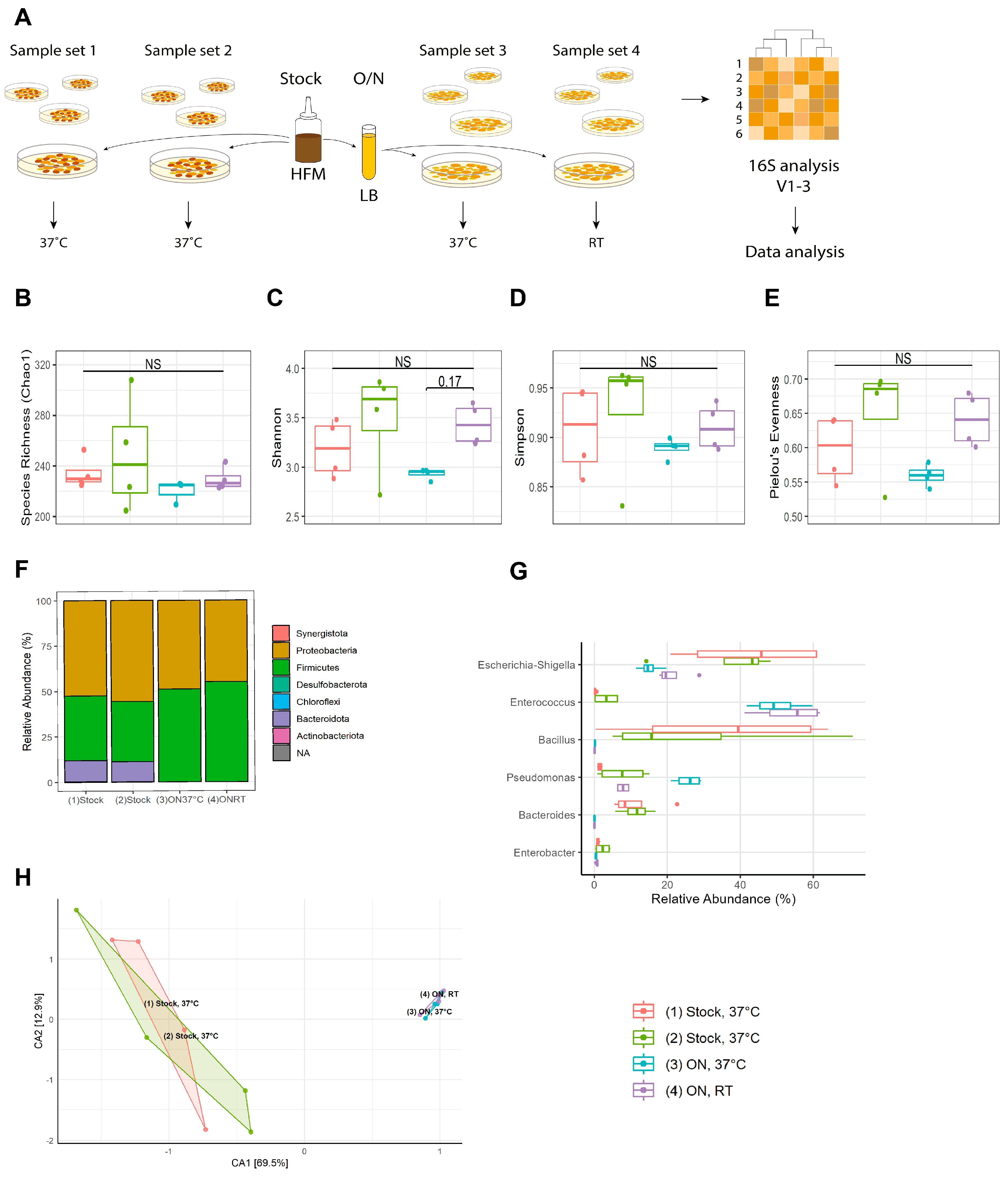
3.1. A Stable Starting Material Is Obtained through Overnight Cultivation

In order to replace OP50 with HFM, we established two different experimental protocols using an HFM culture directly from a frozen (−80 °C) stock and an HFM culture cultivated o/n in LB media, respectively (Figure 1A). For both protocols, we obtained NGM plates with bacterial lawns that clearly differed in bacterial content by visual inspection. The lawns on the plates spotted with cultured HFM appeared more uniform and transparent than those spotted with HFM directly from the frozen stock which were very thick and with dark colonies.
Therefore, we wanted to investigate in more detail the effects on bacterial composition by introduction of the o/n cultivation step and growth temperature after the HFM cultures were spotted on the NGM plates. Four individual sample sets, each consisting of four samples, were subjected to 16S rRNA gene amplicon sequencing of the variable region V1-V3 (Figure 1A). Sample set 1 and 2 consisted of non-cultivated HFM spotted on NGM plates directly from the frozen stock and incubated at 37 °C o/n. We included two replicates of the same sample type to assess reproducibility. Sample sets 3 and 4 were o/n cultures of HFM cultured in LB media and spotted on NGM plates left to dry and grow o/n at either 37 °C (sample set 3) or RT (sample set 4).
High-quality sequences were obtained for all 16 samples with a total number of 502,268 and an average of 31,391 ± 735 sequences per sample. Rarefaction curves with beginning horizontal asymptote were seen for every sample, meaning enough reads had been sequenced to capture the diversity and to continue with downstream analysis (Figure S1).
Next, we analyzed alpha diversity. Pairwise Wilcoxon rank sum test of the microbial community richness revealed no significant differences between the four samples sets with median species richness of 225–230 per sample (Figure 1B). Furthermore, no significant differences were observed between the four sample sets in terms of Shannon index (Figure 1C), Simpson index (Figure 1D), and Pielou’s evenness (Figure 1E). The index scores for Pielou’s evenness are relatively low, meaning that the members of the community are not evenly represented but dominated by few genera.
Although not significant, the microbial community of cultivated HFM was affected by growing on the plates at either 37 °C (sample set 3) or at RT (sample set 4). The bacterial communities on the plates incubated at lower temperature had higher diversity and evenness, suggesting that keeping the plates at RT after spotting would be preferable. It is possible that the dominating bacteria grow slower at lower temperatures, leaving resources for other species.
At the phylum level, we found that half of the stock samples were Proteobacteria and that Firmicutes were the second most abundant phylum. Bacteroidota make up 1:10 of the initial stock community but are lost during the LB cultivation step (Figure 1F). At the genus level, Escherichia-Shigella, Enterococcus, Bacillus, Pseudomonas, and Bacteroides were the most abundant across all the samples (Figures S2 and S3). Enterococcus was enriched in cultivated samples compared to non-cultivated whereas Escherichia-Shigella, Bacillus, and Bacteroides were less abundant (Figure 1G). An increased number of Pseudomonas was observed for cultivated HFM plates grown at 37 °C compared to plates grown at RT. Enterobacter were most abundant in the stock samples of the second set.
To visualize and compare the variability and diversity between the samples sets, beta diversity analysis was performed using correspondence analysis (CA) taking differences of less abundant ASVs into account (Figure 1H). The sample sets primarily segregate across the first dimension (69.5%), indicating that the cultivation step is the dominant factor in determining the microbial community composition and diversity. Sample sets 1 and 2 of non-cultivated HFM have a large spread but do overlap, which suggests that sampling date appears to have a minor effect. On the contrary, sample set 3 and 4 of o/n culture HFM have very low spread and overlap to a small degree, meaning that the cultivation step gives a more repeatable representation of communities across technical replicates compared to non-cultivated HFM.
Based on alpha and beta diversity analyses, we find that a cultivation step of an HFM in LB media provides a more stable starting material with a richness not significantly different from the non-cultivated samples. Furthermore, the cultivated HFM created more transparent bacterial lawns, which is advantageous when setting up experiments with C. elegans as this allow visual phenotyping and identification of animals. Since no significant difference was observed between the two sample sets cultivated in LB media, it can be argued that incubation at RT is preferable for both ease of use and compatibility with commonly utilized C. elegans protocols. The resulting microbial community originating from cultured human fecal microbiota in LB media overnight, spotted on NGM plates and grown at room temperature, will be referred to as cultured HFM (cHFM) onwards.

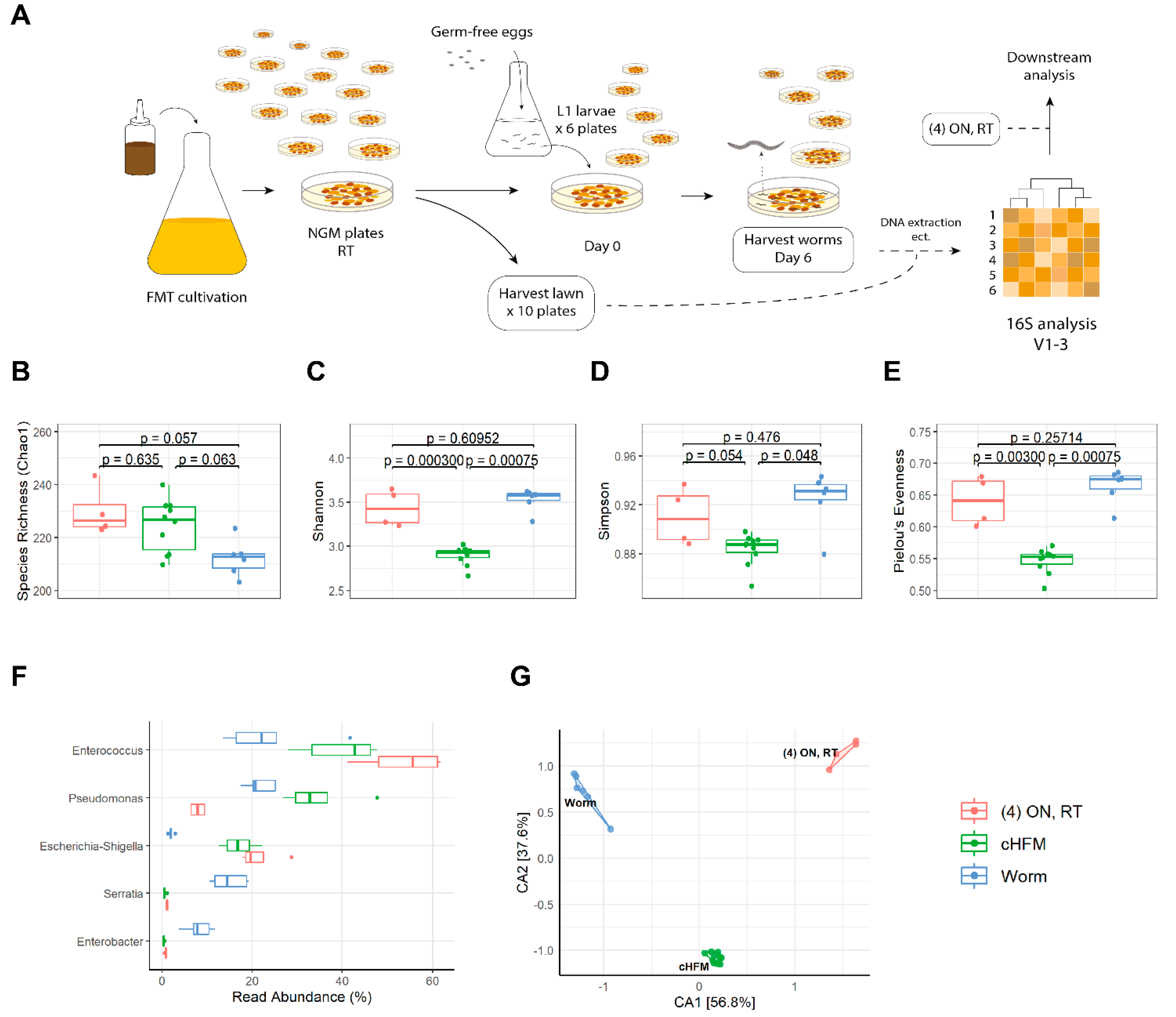
3.2. Transplantation of Cultured Human Fecal Microbiota to C. elegans

Next, we wanted to see if the cHFM could support normal growth and development of C. elegans. For this, synchronized wildtype N2 eggs were transferred to either OP50 or cHFM and both egg laying and size (body length) of the worms were recorded. We found that the cHFM accelerated the development of the worms as the size of 2-day-old worms were significantly increased compared to controls eating OP50 (Figure 2A). Furthermore, time from embryo to first egg lay was accelerated in worms fed cHFM compared to controls fed OP50 (Figure 2B). Thus, the cHFM provides a food source with sufficient nutritional value to support C. elegans growth and development. Consistent with this we did not observe any dauers.
To provide further evidence that the cHFM has a biological effect on C. elegans, we examined the levels of the phase II detoxification enzyme, glutathione S-transferase (GST-4) using a GST-4::GFP reporter strain [36]. Oxidative stress induces GST-4 expression and thus this reporter can estimate the degree of oxidative stress. Interestingly, worms fed the cHFM diet had significantly lower levels of GST-4 induction (Figure 2C), indicating that the cHFM diet causes lesser oxidative stress than a traditional OP50 diet.
Previous studies have shown that when worms are fed a fresh FMT community or various soil communities, only a subset of the bacteria in these communities can colonize the worm gut [23,31]. Next, we wondered which bacteria of the cHFM would be found in the gut microbiota of the worms. To test this, approximately 600 L1-synchronized glp-4 larvae were grown on six individual cHFM plates for six days before harvesting. Analyses of the 16S rRNA variable region V1-V3 were performed from both lawns and worms to determine if the starting material provided as feed was different from the nematode intestinal microbiota (Figure 3A).
Again, all 16 samples were of high quality with a total number of 602,373 and an average of 37,648 ± 802 sequences per sample. Beginning horizontal asymptotes were observed for the rarefaction curves and thus sequencing depth was sufficient to capture diversity (Figure S4).
The difference in species richness was close to being significant between the ten lawn samples and the six worm samples, with median species richness of 237 and 217, respectively (Figure 3B). However, we found that the diversity was significantly higher for both the Shannon and Simpson index in the worm samples compared to lawn samples (Figure 3C,D). Furthermore, Pielou’s evenness score was also significantly higher for the worms than for the starting lawns (Figure 3E). Thus, the microbiota of the worms is more even than the starting bacterial lawns where fewer genera dominate the microbial community. This is also visualized in the heatmap (Figures S5 and S6).
At the genus level, we found that the abundance of Enterococcus and Escherichia-Shigella in the worms was around half of that present in the lawn that they had been eating. The same was observed for Pseudomonas, although to a smaller extent (Figure 3F and Supplementary Figures S5 and S6). On the contrary, Serratia and Enterobacter were more abundant in the worms compared to the lawn subcommunity. Taken together, this shows that there is a clear distinction between the bacterial community available as food on the plates and the resulting gut microbiota in the worms.
The four lawn samples from the initial study were made in the same way as the ten lawn samples from this setup. A side-by-side comparison of the two sample sets showed that the difference in species richness and the Simpson index are not significantly different (Figure 3B,D), but in terms of the diversity we find that both Shannon and Pielou indices are significantly different (Figure 3C,E). Furthermore, the CA plot of the three sample sets shows that they cluster together in their respective groups (Figure 3G), and taken together, the first and second dimension explain 94.4% of the segregation. The two lawn groups do not overlap, and all groups have relatively low internal spread. However, looking at the heatmap (Figures S5 and S6), it is clear that the lawn samples from the two different setups look more similar to each other compared to the worm samples. The read abundances (Figures S5 and S6) also show that the two lawn sample sets lie closely, except from the lower abundance of Pseudomonas, which could explain why the two sample sets do not overlap in the CA.

4. Discussion

There is a tremendous therapeutic potential in manipulating and shaping our microbiota towards promoting health. Consequently, there is an increasing focus on finding new ways of uncovering causality between the microbiota and the host response. Studying monocultures of bacteria has long been the preferred way of reducing complexity and uncovering underlying molecular mechanisms. Unfortunately, monocultures are a poor representation of the much more complex intact microbiota found in humans. To overcome this problem, focus has shifted towards defined microbiotas containing bacterial communities rather than monocultures, but still with lesser complexity and diversity than intact microbiotas [13,31,37,38]. In this study we have demonstrated that the soil nematode C. elegans can be used as host to study a sub-set of the human microbiota.

4.1. The Typical E. coli Diet Can Be Replaced with Complex Sub-Set of the Human Microbiota

C. elegans is typically cultured on NGM plates spotted with an o/n culture of E. coli OP50 as food source. The OP50 culture is usually left to grow at RT for two days before C. elegans is moved to the plates. This protocol can be adapted to study complex bacterial communities simply by replacing OP50 with a complete microbiota. In this study, we used human fecal samples as the complex microbiota. Since our microbiota samples were stored at −80 °C, we speculated that there could potentially be a large plate-to-plate variation if separate aliquots of the frozen stock were added to each NGM plate. We therefore tested the effect of introducing an o/n culture step, allowing each plate to be spotted with a bacterial community originating from the same aliquot of the frozen stock in each experiment. One might expect that a culturing step would decrease the diversity, but interestingly, we find that species richness, diversity, and evenness were not significantly different across the sample sets. However, we did see differences in abundance: Enterococcus was enriched in cultivated samples compared to non-cultivated whereas Escherichia-Shigella, Bacillus, and Bacteroides were less abundant. Enterobacter were most abundant in the stock samples of the second set. Bacteroides are a group of obligate anaerobic bacteria which likely explains why this group of bacteria is lost during aerobic cultivation [39]. Bacillus are either obligate aerobe or facultative anaerobe [40] and reduced abundance of Bacillus in the cultivated community must be the result of some other selection, for example arising from nutrient utilization or production of inhibitory metabolites.
In humans, the microbiota is experiencing a temperature of around 37 °C, but C. elegans cannot be cultured at 37 °C. However, the bacterial growth on the NGM after spotting and before worms are moved to the plates can be performed at any temperature. To determine if the growth temperature influenced the microbiota composition, we compared growth at RT with growth at 37 °C. Interestingly, the culture temperature did not significantly change alpha diversity.
When we compare the starting microbiotas on plates generated from two independent experiments on different days, we find that they differ in beta diversity. This stresses the need for using a proper number of technical replicates and performing independent experimental replicates. It also highlights the need for determining the starting microbiotas in every independent experimental setup. Variation from experiment to experiment cannot be avoided and can in fact ease the analysis of the results. Provided that the host phenotype is consistently observed across the experiments, the number of possible causal bacteria can be reduced by focusing on bacteria found in all independent experiments.
In conclusion, introducing an o/n culture step in LB followed by growth on the NGM plates at RT provides an easy protocol for having a controlled starting microbiota with sufficient complexity on each plate to study the effect of bacterial communities rather than on monocultures.

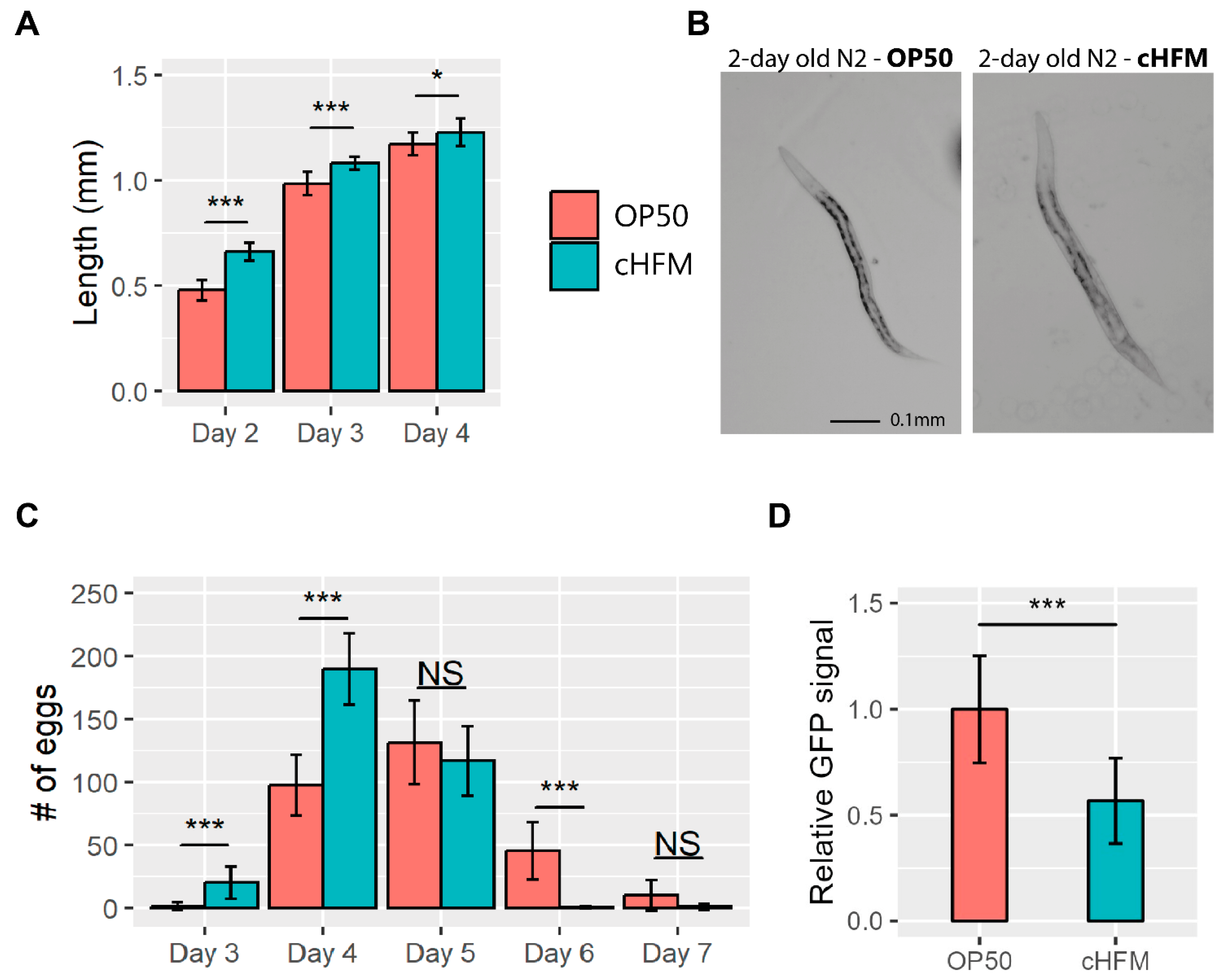
4.2. C. elegans Can Develop and Reproduce Normally When Being Fed a Sub-Microbiota of Cultivated Human Fecal Microbiota

C. elegans induces larval arrest or dauer formation if the bacterial food source does not have the right nutritional composition [41]. Complete starvation induces L1 arrest, and this can be utilized to make the animals germ-free after hypochlorite treatment. Exposure to pathogens can also induce larval arrest or avoidance behavior [42].
We find that C. elegans can develop normally when cultured on NGM plates spotted directly with a frozen HFM stock or with cHFM. This is consistent with a previous study in which C. elegans was cultured on a fresh human fecal microbiota [31]. In fact, in this study we find that the worms develop significantly faster on human microbiota compared to their traditional OP50 diet. Thus, it appears to be a general feature that human fecal microbiotas have sufficient nutritional value to support C. elegans development. Interestingly, we find that the cHFM diet reduces the levels of the phase II detoxification enzyme, glutathione S-transferase (GST-4) in the worms, providing proof of principle that sub-microbiomes can be used to manipulate host physiology. Consistent with this, increased levels of the antioxidant glutathione have also been observed following transplantation of fresh human microbiota to C. elegans, which protects the intestine against nano-plastic toxicity [31]. Since oxidative stress is implicated in many diseases, understanding how the cHFM diet reduces oxidative stress could lead to novel treatment strategies.

4.3. You Are Not What You Eat—Sub-Microbiomes Colonize the Worm Intestine

It is well known that different soil bacteria and fresh human FMT can colonize the intestine of C. elegans as complex communities [15,22,23,26,31]. We find that approximately 200 out of 230 ASVs from a cultured human microbiota can be found in the worm intestine when fed to the worms for 6 days from the L1 stage.
Consistent with previous observations for soil bacteria and fresh human FMT, we find that the resulting worm microbiota is not simply a direct reflection of the human microbiota initially fed to the worms. Rather, only a subset of the starting microbiota can colonize the worm intestine. In our study, the bacteria that colonize the intestine are dominated by the genera Enterococcus, Pseudomonas, Serratia, and Enterobacter.
The fact that C. elegans can develop normally and reproduce on human microbiota and that a subset of this can colonize the worm intestine opens a new line of exciting experiments dissecting the host microbiome interactions. When fed soil bacteria, the innate immune system has been shown to be an important factor for shaping the resulting worm microbiota [24]. The innate immune system is also involved with mediating a beneficial effect of probiotic bacteria [17,43] and it is most likely also determining which subset of the human microbiota can colonize the bacteria. However, many additional genetic factors determining microbiota colonization and effects on the host are likely to exist, which can be studied with a sub-microbiota of cHFM.
The choice of growth medium in the liquid culture step and in the nematode growth plates provides a tool to modify the resulting complex bacterial community’s composition. In our study, this is reflected by the relatively low Pielou’s evenness scores. Some bacteria favor growth in LB media, and these will be represented in higher number compared to bacteria that favors another growth medium. Thus, the protocol can easily be adapted to support the growth of specific bacteria of interest simply by changing the growth media accordingly.
If C. elegans are exposed to anerobic conditions, they quickly undergo suspended animation during which they stop feeding, cell cycling, and developmental progression [44]. Therefore, our current protocol is not suitable for studying the anaerobic bacteria found in the human gut. For this purpose, mouse models with custom-made synthetic and defined microbial communities would be better suited, including for example the Oligo-Mouse Microbiota (OMM) 12 and OMM19.1 [37,38]. However, it would be possible to culture the bacteria anaerobically in the steps prior to introducing C. elegans. This way, metabolites from the anaerobic bacteria and their effects on the host and microbiota could be studied.
The metabolites may hold an important key to understanding how the microbiota can influence host physiology and health. We have recently characterized the metabolome of probiotic Lactobacillus bacteria compared to the traditional OP50 diet and found significant differences [18]. Interestingly, when the metabolomes of the bacterial diets are compared to the metabolomes of C. elegans fed a batch of the same bacteria, we find that these are not identical. Thus, the metabolomic profiles support the notion that you are not simply what you eat, which is one of the main conclusions of this study. Increased levels of the antioxidant glutathione are among the 20 most important metabolites distinguishing worms fed a probiotic diet from worms fed OP50.

4.4. Future Perspectives

It is widely recognized that the human microbiota influences our health to a much higher degree than previously thought. However, the complexity of the human microbiome as well as the human host is a challenge for understanding the underlying mechanisms. Focusing on sub-sets of the human microbiota as well as well-defined model organisms is one way to overcome this challenge. Conservation across species has made C. elegans popular for uncovering the genetic basis for many complex biological processes. C. elegans is well established as a model system to study host–microbe interactions and has the benefit of strong genetic tools. Notably, the responses to probiotic and pathogenic bacteria have successfully been mapped using genetic and omics approaches in C. elegans [45]. Our study demonstrates that C. elegans can equally be used to study the effects of subsets of the human microbiota.

Supplementary Materials

The following supporting information can be downloaded at: https://www.mdpi.com/article/10.3390/applmicrobiol3040078/s1, Figure S1: Rarefaction curves of the individual samples.; Figure S2: Heatmap of the 25 most abundant ASVs identified across the four sample sets.; Figure S3: Heatmap of the 15 most abundant ASVs at family level identified across the four sample sets.; Figure S4: Rarefaction curves of the individual samples.; Figure S5: Heatmap of the 25 most abundant ASVs identified across the three sample sets.; Figure S6: Heatmap of the 15 most abundant ASVs identified at family level across the three sample sets.

Author Contributions

Conceptualization, K.V.M. and A.O.; Methodology, K.V.M., J.B.W., S.J.K., H.L.N., E.Y. and A.O.; Formal analysis, K.V.M., J.B.W., R.M. and E.Y.; Investigation, K.V.M., J.B.W. and R.M.; Resources, S.J.K. and H.L.N.; Data curation, J.B.W., R.M. and E.Y.; Writing—original draft, A.O.; Writing—review & editing, J.B.W. and A.O.; Supervision, K.V.M. and A.O.; Funding acquisition, A.O. All authors have read and agreed to the published version of the manuscript.

Funding

The work was funded by the Lundbeckfonden (324-2019-2035) and Carlsbergfondet (CF19-0668).

Institutional Review Board Statement

Samples from faecal donors were collected in accordance with the requirements of Good Clinical Practice and the Revised Declaration of Helsinki. All participants provided signed written informed consent. The Regional Research Ethics Committee of Northern Jutland, Denmark approved collection of the samples, and gave concent for the use of the samples in this study.

Informed Consent Statement

Not applicable.

Data Availability Statement

All relevant data can be found at the European Nucleotide Archive under PRJEB65062.

Acknowledgments

Some strains were provided by the CGC, which is funded by NIH Office of Research Infrastructure Programs (P40 OD010440). We thank Nadieh de Jonge for helpful discussions and suggestions regarding data analysis.

Conflicts of Interest

The authors declare no conflict of interest.

References

  1. Sender, R.; Fuchs, S.; Milo, R. Are We Really Vastly Outnumbered? Revisiting the Ratio of Bacterial to Host Cells in Humans. Cell 2016, 164, 337–340. [Google Scholar] [CrossRef] [PubMed]
  2. Almeida, A.; Mitchell, A.L.; Boland, M.; Forster, S.C.; Gloor, G.B.; Tarkowska, A.; Lawley, T.D.; Finn, R.D. A New Genomic Blueprint of the Human Gut Microbiota. Nature 2019, 568, 499–504. [Google Scholar] [CrossRef] [PubMed]
  3. Valdes, A.M.; Walter, J.; Segal, E.; Spector, T.D. Role of the Gut Microbiota in Nutrition and Health. BMJ 2018, 361, 36–44. [Google Scholar] [CrossRef] [PubMed]
  4. Yang, H.J.; Kim, J.H. Role of Microbiome and Its Metabolite, Short Chain Fatty Acid in Prostate Cancer. Investig. Clin. Urol. 2023, 64, 3–12. [Google Scholar] [CrossRef]
  5. Fujisaka, S.; Watanabe, Y.; Tobe, K. The Gut Microbiome: A Core Regulator of Metabolism. J. Endocrinol. 2023, 256, e220111. [Google Scholar] [CrossRef]
  6. Boekhorst, J.; Venlet, N.; Procházková, N.; Hansen, M.L.; Lieberoth, C.B.; Bahl, M.I.; Lauritzen, L.; Pedersen, O.; Licht, T.R.; Kleerebezem, M.; et al. Stool Energy Density Is Positively Correlated to Intestinal Transit Time and Related to Microbial Enterotypes. Microbiome 2022, 10, 1–10. [Google Scholar] [CrossRef]
  7. Clemente, J.C.; Manasson, J.; Scher, J.U. The Role of the Gut Microbiome in Systemic Inflammatory Disease. BMJ 2018, 360, j5145. [Google Scholar] [CrossRef]
  8. Morais, L.H.; Schreiber, H.L.; Mazmanian, S.K. The Gut Microbiota–Brain Axis in Behaviour and Brain Disorders. Nat. Rev. Microbiol. 2021, 19, 241–255. [Google Scholar] [CrossRef]
  9. Fischbach, M.A. Microbiome: Focus on Causation and Mechanism. Cell 2018, 174, 785–790. [Google Scholar] [CrossRef]
  10. de Jonge, N.; Michaelsen, T.Y.; Ejbye-Ernst, R.; Jensen, A.; Nielsen, M.E.; Bahrndorff, S.; Nielsen, J.L. Housefly (Musca Domestica L.) Associated Microbiota across Different Life Stages. Sci. Rep. 2020, 10, 7842. [Google Scholar] [CrossRef]
  11. Xia, H.; Chen, H.; Cheng, X.; Yin, M.; Yao, X.; Ma, J.; Huang, M.; Chen, G.; Liu, H. Zebrafish: An Efficient Vertebrate Model for Understanding Role of Gut Microbiota. Mol. Med. 2022, 28, 161. [Google Scholar] [CrossRef] [PubMed]
  12. Poletti, M.; Arnauts, K.; Ferrante, M.; Korcsmaros, T. Organoid-Based Models to Study the Role of Host-Microbiota Interactions in IBD. J. Crohn’s Colitis 2021, 15, 1222–1235. [Google Scholar] [CrossRef]
  13. Cheng, A.G.; Ho, P.Y.; Aranda-Díaz, A.; Jain, S.; Yu, F.B.; Meng, X.; Wang, M.; Iakiviak, M.; Nagashima, K.; Zhao, A.; et al. Design, Construction, and in Vivo Augmentation of a Complex Gut Microbiome. Cell 2022, 185, 3617–3636. [Google Scholar] [CrossRef] [PubMed]
  14. Olsen, A.; Gill, M.S. (Eds.) Ageing: Lessons from C. Elegans; Springer International Publishing: Berlin/Heidelberg, Germany, 2017. [Google Scholar]
  15. Christensen, K.; Mørch, M.; Morthorst, T.; Lykkemark, S.; Olsen, A. Microbiota, Probiotic Bacteria and Ageing. In Ageing: Lessons from C. elegans; Olsen, A., Gill, M., Eds.; Springer International Publishing: Berlin/Heidelberg, Germany, 2017; pp. 411–429. [Google Scholar]
  16. Cabreiro, F.; Gems, D. Worms Need Microbes Too: Microbiota, Health and Aging in Caenorhabditis elegans. EMBO Mol. Med. 2013, 5, 1300–1310. [Google Scholar] [CrossRef] [PubMed]
  17. Mørch, M.G.M.; Møller, K.V.; Hesselager, M.O.; Harders, R.H.; Kidmose, C.L.; Buhl, T.; Fuursted, K.; Bendixen, E.; Shen, C.; Christensen, L.G.; et al. The TGF-β Ligand DBL-1 Is a Key Player in a Multifaceted Probiotic Protection against MRSA in C. elegans. Sci. Rep. 2021, 11, 10717. [Google Scholar] [CrossRef] [PubMed]
  18. Møller, K.V.; Nguyen, H.T.T.; Mørch, M.G.M.; Hesselager, M.O.; Mulder, F.A.A.; Fuursted, K.; Olsen, A. A Lactobacilli Diet That Confers MRSA Resistance Causes Amino Acid Depletion and Increased Antioxidant Levels in the C. elegans Host. Front. Microbiol. 2022, 13, 886206. [Google Scholar] [CrossRef]
  19. Brejning, J.; Nørgaard, S.; Schøler, L.; Morthorst, T.H.; Jakobsen, H.; Lithgow, G.J.; Jensen, L.T.; Olsen, A. Loss of NDG-4 Extends Lifespan and Stress Resistance in Caenorhabditis elegans. Aging Cell 2014, 13, 156–164. [Google Scholar] [CrossRef]
  20. Miller, B.C.; Mathai, M.; Yadav, H.; Jain, S. Geroprotective Potential of Microbiome Modulators in the Caenorhabditis elegans Model. Geroscience 2023. [Google Scholar] [CrossRef]
  21. Samuel, B.S.; Rowedder, H.; Braendle, C.; Félix, M.-A.; Ruvkun, G. Caenorhabditis elegans Responses to Bacteria from Its Natural Habitats. Proc. Natl. Acad. Sci. USA 2016, 113, E3941–E3949. [Google Scholar] [CrossRef]
  22. Dirksen, P.; Marsh, S.A.; Braker, I.; Heitland, N.; Wagner, S.; Nakad, R.; Mader, S.; Petersen, C.; Kowallik, V.; Rosenstiel, P.; et al. The Native Microbiome of the Nematode Caenorhabditis elegans: Gateway to a New Host-Microbiome Model. BMC Biol. 2016, 14, 38. [Google Scholar] [CrossRef]
  23. Berg, M.; Stenuit, B.; Ho, J.; Wang, A.; Parke, C.; Knight, M.; Alvarez-Cohen, L.; Shapira, M. Assembly of the Caenorhabditis elegans Gut Microbiota from Diverse Soil Microbial Environments. ISME J. 2016, 10, 1998–2009. [Google Scholar] [CrossRef] [PubMed]
  24. Berg, M.; Monnin, D.; Cho, J.; Nelson, L.; Crits-Christoph, A.; Shapira, M. TGFβ/BMP Immune Signaling Affects Abundance and Function of C. elegans Gut Commensals. Nat. Commun. 2019, 10, 604. [Google Scholar] [CrossRef] [PubMed]
  25. Zhang, F.; Weckhorst, J.L.; Assié, A.; Hosea, C.; Ayoub, C.A.; Khodakova, A.S.; Cabrera, M.L.; Vidal Vilchis, D.; Félix, M.-A.; Samuel, B.S. Natural Genetic Variation Drives Microbiome Selection in the Caenorhabditis elegans Gut. Curr. Biol. 2021, 31, 2603–2618.e9. [Google Scholar] [CrossRef] [PubMed]
  26. Dirksen, P.; Assié, A.; Zimmermann, J.; Zhang, F.; Tietje, A.M.; Marsh, S.A.; Félix, M.A.; Shapira, M.; Kaleta, C.; Schulenburg, H.; et al. CeMbio—The Caenorhabditis elegans Microbiome Resource. G3 Genes. Genomes Genet. 2020, 10, 3025–3039. [Google Scholar] [CrossRef]
  27. Petersen, C.; Dirksen, P.; Schulenburg, H. Why We Need More Ecology for Genetic Models Such as C. elegans. Trends Genet. 2015, 31, 120–127. [Google Scholar] [CrossRef]
  28. Corsi, A.K.; Wightman, B.; Chalfie, M. A Transparent Window into Biology: A Primer on Caenorhabditis Elegans. Genetics 2015, 200, 387–407. [Google Scholar] [CrossRef]
  29. Kumar, A.; Baruah, A.; Tomioka, M.; Iino, Y.; Kalita, M.C.; Khan, M. Caenorhabditis elegans: A Model to Understand Host–Microbe Interactions. Cell. Mol. Life Sci. 2020, 77, 1229–1249. [Google Scholar] [CrossRef]
  30. Walker, A.C.; Bhargava, R.; Vaziriyan-Sani, A.S.; Pourciau, C.; Donahue, E.T.; Dove, A.S.; Gebhardt, M.J.; Ellward, G.L.; Romeo, T.; Czyż, D.M. Colonization of the Caenorhabditis elegans Gut with Human Enteric Bacterial Pathogens Leads to Proteostasis Disruption That Is Rescued by Butyrate. PLoS Pathog. 2021, 17, e1009510. [Google Scholar] [CrossRef]
  31. Chu, Q.; Zhang, S.; Yu, X.; Wang, Y.; Zhang, M.; Zheng, X. Fecal Microbiota Transplantation Attenuates Nano-Plastics Induced Toxicity in Caenorhabditis elegans. Sci. Total Environ. 2021, 779, 146454. [Google Scholar] [CrossRef]
  32. Jumpstart Consortium Human Microbiome Project Data Generation Working Group. Evaluation of 16S RDNA-Based Community Profiling for Human Microbiome Research. PLoS ONE 2012, 7, e39315. [Google Scholar]
  33. Yashiro, E. Illumina Sequenced Amplicon Reads Processing Workflow. Available online: https://github.com/eyashiro/AmpProc (accessed on 26 September 2023).
  34. Edgar, R.C. Search and Clustering Orders of Magnitude Faster than BLAST. Bioinformatics 2010, 26, 2460–2461. [Google Scholar] [CrossRef] [PubMed]
  35. Smith, B.; Wilson, J.B. A Consumer’s Guide to Evenness Indices. Oikos 1996, 76, 70–82. [Google Scholar] [CrossRef]
  36. Leiers, B.; Kampkötter, A.; Grevelding, C.G.; Link, C.D.; Johnson, T.E.; Henkle-Dührsen, K. A Stress-Responsive Glutathione S-Transferase Confers Resistance to Oxidative Stress in Caenorhabditis elegans. Free Radic. Biol. Med. 2003, 34, 1405–1415. [Google Scholar] [CrossRef]
  37. Afrizal, A.; Jennings, S.A.V.; Hitch, T.C.A.; Riedel, T.; Basic, M.; Panyot, A.; Treichel, N.; Hager, F.T.; Wong, E.O.-Y.; Wolter, B.; et al. Enhanced Cultured Diversity of the Mouse Gut Microbiota Enables Custom-Made Synthetic Communities. Cell Host Microbe 2022, 30, 1630–1645. [Google Scholar] [CrossRef] [PubMed]
  38. Brugiroux, S.; Beutler, M.; Pfann, C.; Garzetti, D.; Ruscheweyh, H.-J.; Ring, D.; Diehl, M.; Herp, S.; Lötscher, Y.; Hussain, S.; et al. Genome-Guided Design of a Defined Mouse Microbiota That Confers Colonization Resistance against Salmonella Enterica Serovar Typhimurium. Nat. Microbiol. 2016, 2, 16215. [Google Scholar] [CrossRef]
  39. Wexler, H.M. Bacteroides: The Good, the Bad, and the Nitty-Gritty. Clin. Microbiol. Rev. 2007, 20, 593–621. [Google Scholar] [CrossRef]
  40. Turnbull, P.C.B. Bacillus. In Medical Microbiology; Baron, S., Ed.; University of Texas Medical Branch at Galveston: Galveston, TX, USA, 1996; ISBN 0-9631172-1-1. [Google Scholar]
  41. Baugh, L.R.; Hu, P.J. Starvation Responses Throughout the Caenorhabditis elegans Life Cycle. Genetics 2020, 216, 837–878. [Google Scholar] [CrossRef]
  42. Cohen, L.B.; Troemel, E.R. Microbial Pathogenesis and Host Defense in the Nematode C. elegans. Curr. Opin. Microbiol. 2015, 23, 94–101. [Google Scholar] [CrossRef]
  43. Baillo, A.; Villena, J.; Albarracín, L.; Tomokiyo, M.; Elean, M.; Fukuyama, K.; Quilodrán-Vega, S.; Fadda, S.; Kitazawa, H. Lactiplantibacillus Plantarum Strains Modulate Intestinal Innate Immune Response and Increase Resistance to Enterotoxigenic Escherichia Coli Infection. Microorganisms 2023, 11, 63. [Google Scholar] [CrossRef]
  44. Padilla, P.A.; Nystul, T.G.; Zager, R.A.; Johnson, A.C.M.; Roth, M.B. Dephosphorylation of Cell Cycle–Regulated Proteins Correlates with Anoxia-Induced Suspended Animation in Caenorhabditis elegans. Mol. Biol. Cell 2002, 13, 1473–1483. [Google Scholar] [CrossRef]
  45. Harding, B.W.; Ewbank, J.J. An Integrated View of Innate Immune Mechanisms in C. elegans. Biochem. Soc. Trans. 2021, 49, 2307–2317. [Google Scholar] [CrossRef] [PubMed]
Figure 1. An overnight culture step reduces variability in bacterial communities between replicates. (A) Experimental setup. (BE) Alpha diversity measurements on the observed ASVs. (F) Microbial community structure at phylum level across the four sample sets. (G) Relative abundance of the first six genera from heatmap. (H) Beta diversity, Correspondence analysis (CA). All samples were rarefied to the lowest number of reads (25,391).
Applmicrobiol 03 00078 g001
Figure 2. A cultured human microbiome supports and accelerates nematode development. (A) Graph of the average size of worm on day 2, 3, and 4 when fed control bacteria (OP50) or cHFM, n = 10/day. (B) Representative pictures of 2-day-old N2 worms fed control bacteria (OP50) and cHFM. (C) Average number of eggs laid by worms fed control bacteria (OP50) or cHFM on days 3 to 7, n = 8–10 for each treatment in one experiment. (D) Relative intensity of GFP expressed under the gst-4 promotor. Bars represent means across three independent experiments standardized to OP50 levels, n > 80. A student’s t-test was used for statistical analysis. * p < 0.01, *** p < 0.0005.
Applmicrobiol 03 00078 g002
Figure 3. Not all bacteria from the cultivated human fecal microbiome colonize the nematode intestine. (A) Experimental setup. (BE) Alpha diversity measurements on the observed ASVs. (F) Relative abundance of the first six genera from heatmap. (G) Beta diversity—Correspondence analysis (CA). All samples were rarefied to the lowest number of reads (28,973).
Applmicrobiol 03 00078 g003
Disclaimer/Publisher’s Note: The statements, opinions and data contained in all publications are solely those of the individual author(s) and contributor(s) and not of MDPI and/or the editor(s). MDPI and/or the editor(s) disclaim responsibility for any injury to people or property resulting from any ideas, methods, instructions or products referred to in the content.

