Article

In Vitro Leaf-Based Method for Agrobacterium-Mediated Genetic Transformation of Sugar Beet

by
Dmitry N. Miroshnichenko
*,
Anna Klementyeva
,
Lilia Mourenets
,
Alexander S. Pushin
,
Aleksey P. Firsov
and
Sergey V. Dolgov
Branch of Shemyakin and Ovchinnikov Institute of Bioorganic Chemistry, Russian Academy of Sciences, 142290 Pushchino, Russia
*
Author to whom correspondence should be addressed.
Crops 2026, 6(1), 12; https://doi.org/10.3390/crops6010012
Submission received: 2 December 2025 / Revised: 24 December 2025 / Accepted: 8 January 2026 / Published: 13 January 2026
(This article belongs to the Topic Genetic Engineering in Agriculture, 2nd Edition)

Abstract

Sugar beet, one of the most important natural sources of sugars in the world, is well known as a recalcitrant crop for genetic transformation. In the present study, several key components of Agrobacterium-mediated transformation of sugar beet have been studied. The correct choice of explant and plant regeneration potential of domestic breeding lines was evaluated; however, most attention was paid to the search for the most efficient selectable marker gene and selection agents. To produce transgenic plants, we applied a method based on the agrobacterial inoculation of wounded morphogenic structures previously initiated on in vitro cultivated leaves. Four selective marker genes conferring antibiotic or herbicide resistance were evaluated. In the case of selection using kanamycin or G418 (nptII gene controlled by the nos promoter), no transgenic plants were obtained, while the addition of the aminoglycoside antibiotic hygromycin (hpt gene, driven by the nos promoter) to the medium ensured the successful production of transgenic plants from three breeding lines with a frequency ranging from 1.5 to 5.1%. The selection of transgenic tissues using herbicides such as phosphinothricin and glyphosate after transformation with the bar and cp4-epsps genes (both controlled by the CaMV 35S promoter) also ensured the obtaining of transgenic plants, but the transformation efficiency was significantly low, reaching only 1.0 and 0.4%, respectively. Primary transgenic sugar beet plants grown in the greenhouse demonstrated enhanced resistance to herbicides in dosages commonly used in the field. In addition, after self-pollination of the primary T0 transgenic lines, homozygous T2 offspring were successfully selected, which demonstrated stable resistance to glyphosate due to the constitutive expression of the introduced cp4-epsps gene.

1. Introduction

Sugar beet (Beta vulgaris L. var. saccharifera) is the second-largest sugar-producing crop after sugarcane, accounting for 21% of global sugar production, which amounted to 41.0 million tons in the 2023–2024 season [1]. Due to the high importance of sugar beet as an industrial crop, significant efforts have been directed towards developing genetic engineering methods for this crop since the early 1990s. Thus, in 1997, transgenic sugar beet plants resistant to the herbicide glyphosate were obtained [2], and since 2009, more than 95% of the sugar beet cultivation area in the United States has been planted with transgenic lines resistant to herbicides [3]. Currently, molecular breeding of sugar beet is aimed at increasing resistance to abiotic (salinity, high soil moisture, frost) and biotic stresses (resistance to rhizomania, cercospora, and nematodes). In the last decade, one of the rapidly developing techniques in molecular breeding is target gene editing, which requires reliable protocol for stable genetic transformation of sugar beet [4,5,6].
The first report of genetic transformation in sugar beets dates back to 1991 [7]. Despite the huge amount of research that has been conducted since then, sugar beet remains a recalcitrant for genetic transformation crops. The vast majority of studies on the genetic transformation of sugar beets have been conducted using the Agrobacterium-mediated method [8]. Two approaches have been developed to date: direct transformation, when transgenic shoots are regenerated directly from explants subjected to bacterial inoculation, and indirect transformation, when transgenic plants are produced by the transgenic morphogenic callus previously obtained after co-cultivation with Agrobacterium. Each approach has its own strengths and drawbacks. Transformation through an intermediate callus phase is relatively time-consuming and more labor-intensive; a prolonged callus phase increases the probability of somaclonal variants. Considering the undesirability of somaclonal variants, the direct transformation method appears to be more preferable. It is worth noting that both approaches are highly dependent on the genotype.
The main problem with direct transformation is its low efficiency, as in most studies the transformation frequency ranges from a few tenths to 5 percent and rarely exceeds 10% of the number of explants. Only in the studies by Konwar [9] and Hisano et al. [10], the transformation frequency of sugar beet was over 10 and 15%, respectively. As shown in these studies, the main factor determining the transformation potential of sugar beet cultivars is their ability to regenerate adventitious shoots from somatic tissues. Approximately a quarter of the studied genotypes were unable to regenerate adventitious shoots and, therefore, were hardly transformed [2,9,11,12,13]. The dependence of the regeneration capacity on the genetic background can be partially overcome by selecting the optimal explant type, the composition of the regeneration medium, and the in vitro culture conditions [10,14,15,16,17].
Another key factor determining the efficiency of sugar beet transformation is the choice of a selective antibiotic and its concentration in the medium, ensuring a minimum number of escapes. Sugar beet is characterized by the appearance of a large number of chimeric shoots consisting of both transformed and untransformed cells that are formed as a result of direct regeneration from transformed plant tissues. When kanamycin was used, only 5–50% of the adventitious shoots grown on selective media were found to be transgenic. In the case of selection using phosphinothricin and hygromycin, approximately 30% of resistant shoots were transgenic [14,18,19,20,21,22]. In most studies, the choice of selective agent has not been explained and seems to be determined by the design of the transformation vector used.
In almost all published studies, the problem of low regeneration was solved by fine-tuning the composition of the medium or searching for genotypes with high regeneration potential. Attempts to increase the explants’ regeneration frequency beyond varying the medium composition were very limited. Moreover, little attention has been paid to systematic studying the influence of the type of selective agents and their concentration on the transformation efficiency.
Thus, there are certain gaps in our knowledge regarding Agrobacterium-mediated transformation of sugar beet. In particular, the interactions between the genotype, the medium composition, the type, and concentration of selective agents remain insufficiently studied and require further investigation. The objective of this study was to comprehensively examine these factors in order to improve the transformation protocol for sugar beet and contribute to successful molecular breeding research on this crop. Additionally, using the developed protocol, transgenic herbicide-resistant sugar beet plants were obtained, their T1 and T2 generations were analyzed, and homozygous lines were selected.

2. Materials and Methods

2.1. Plant Material

In the present study, several domestic sugar beet (Beta vulgaris L. var. saccharifera) breeding lines named as №1, №2, №3, №4-28, and №4-40 (kindly provided by The A.L. Mazlumov All-Russian Research Institute of Sugar Beet, VNIISS, Russia), and OP5063, OP4977, OT11301 and OT7994 (kindly provided by the FSBSI Pervomayskaya breeding and experimental station of sugar beet, Gulkevichi, Russia), were used.

2.2. In Vitro Culture

All experiments were conducted using the basal MS media consisted of Murashige and Skoog mineral salts and vitamins [23], supplemented with 30 g L−1 sucrose and 100 mg L−1 myo-inositol, solidified with 7 g L−1 agar, and adjusted to a pH range of 5.6–5.8 with 0.1 N NaOH. For initiation of in vitro culture, seeds were washed overnight in running water to simplify the scarification. The mature embryos were mechanically isolated from slightly swollen seeds by removal of coat, and then intact embryos were surface-sterilized for 22 min using 15% commercial bleach containing a few droplets of Tween 20 followed by several rinses with sterile water. Sterilized embryos were cultured in test tubes containing 10 mL of MS medium containing 0.25 mg L−1 6-Benzylaminopurine (BAP). After 10–14 days of cultivation, aseptic shoots were transferred for further growth in glass jars containing 50 mL of the same medium. Every 30 days, plants were divided and transferred onto the fresh medium. In vitro plants were cultivated at an illumination of 3000–3500 lux, day/night length of 16/8 h, and day/night temperature of 25/22 °C. To assess the regeneration potential, whole leaves were separated from young two-week-old plants. Twelve to fifteen explants were placed in 90 mm Petri dishes with 25 mL of regeneration medium, representing the MS medium containing various combinations of 6-BAP (0.5 or 1.0 mg L−1) and indo1e-3-acetic acid (IAA) (0.1 mg L−1). No less than 45 explants were used in a single repetition. Regeneration was carried out in the dark for 21 days at 25 °C, and then the explants were transferred to fresh regeneration medium and cultivated under the same conditions as the aseptic in vitro cultures of sugar beets. When the plantlets reached a size of 10–12 mm, they were separated from the explant and rooted on hormone-free MS medium.

2.3. Effect of Selective Agents Type and Concentration

Leaf explants of two sugar beet lines №4-28 and OP5063 were cultured on a regeneration medium containing 1.0 mg L−1 6-BAP, 0.1 mg L−1 IAA for 15 days. The explants starting to regenerate were transferred 15 days after culture initiation onto the same medium supplemented with one of the selective agents. The following antibiotics were used: kanamycin (concentrations of 0, 150, 200, 250, 300 mg L−1), G418 (concentrations of 0, 10, 20, 30, 40 mg L−1), and hygromycin (concentrations of 0, 5, 7.5, 10, 12.5 mg L−1). Additionally, the sensitivity of the OP5063 line to herbicides such as phosphinothricin (0, 5, 7.5, 10, 12.5, 15 mg L−1) and glyphosate (concentration of 0, 16.9, 33.8, 42.5, 84.5 mg L−1) was studied. The explants were cultured on a medium containing the selective agent for 21 days in the dark; after that, the regeneration abilities of explants were analyzed. Three independent experiments were carried out, each consisting of two Petri dishes as replicates comprising 8 to 11 explants each.

2.4. Bacterial Strains and Plasmids

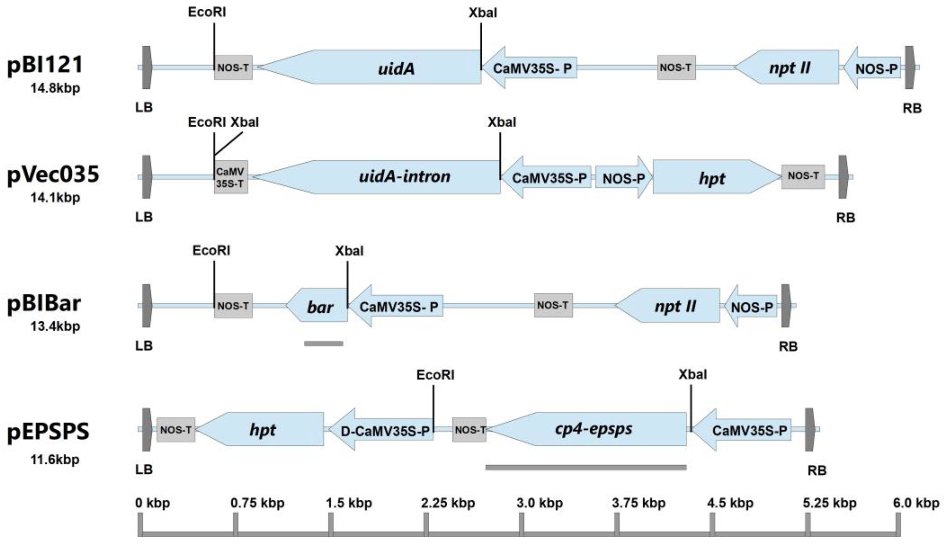
The disarmed super virulent strain Agrobacterium tumefaciens CBE21 (this strain was constructed by removing T-DNA oncogenes from the Ti plasmid pTiBo542 of strain A281 [24]) was used in genetic transformation experiments. Four binary vectors, pBI121 [25], pVec035 [26], pEPSPS, and pBIBar [27], were used for transformation (Figure 1). The pEPSPS vector was constructed on the basis of pCambia vector and contains in T-DNA the synthetic gene cp4-epsps, which encodes protein CP4-EPSPS from Agrobacterium tumefaciens strain CP4 with a N-terminal chloroplast signal peptide CTP2 of the epsps gene from Arabidopsis thaliana. The nucleotide sequence of gene cp4-epsps corresponds to that published in GenBank under accession number GV597339.1.
Prior to transformation, the single Agrobacterium colony was grown for 16 h on a shaker (300 rpm, 28 °C) in the presence of the appropriate antibiotic. Bacterial cells were pelleted by centrifugation at 4000 rpm for 5 min; the pellet was gently washed one time with liquid hormone-free MS medium and diluted to OD600 = 0.4 prior to inoculation.

2.5. Agrobacterium-Mediated Transformation

Two to three upper expanded leaves collected from two-week-old in vitro plants were used as explants. Leaves were placed in Petri dishes (12 to 15 pcs. per dish) which contained 25 mL of MS medium, supplemented with 1.0 mg L−1 6-BAP and 0.1 mg L−1 IAA, and cultivated in the dark at 25 °C. After 15–20 days of cultivation, the explants demonstrating early stages of morphogenesis on petioles were used for agrobacterial inoculation (Figure S1). Morphogenic areas and clusters of young adventitious shoots were cut off or wounded in such a way that the only bud/shoot bases of 0.2–0.4 mm in height were left. If needed, the regenerating leaf was divided perpendicularly to the central vein onto a 1.0–1.5 cm pieces (Figure S1). The leaf parts without sites of morphogenesis were cut off as much as possible. Wounded explants (10 to 20 pcs. per dish) were transferred to the same regeneration medium and additionally enriched with acetosyringone (200 μM). Wounded areas were covered with drops of Agrobacterium suspension (5–20 µL) by pipeting. The dishes with inoculated explants were placed in the dark and cultured for 5 days at 25 °C. After co-cultivation, explants were transferred to the selection medium, representing the regeneration medium additionally supplemented with 500 mg L−1 cefotaxime and the appropriate selective agent. Survived explants or parts of explants demonstrating new morphogenic sites were transferred to the fresh selection medium every 21 days until the buds or plantlets were visible (Figure S1). Such explants were transferred to the illumination of 3000–3500 lux, day/night length of 16/8 h, and day/night temperature of 25/22 °C. Developed plantlets of 10–15 mm long were cut from explants and transferred to a proliferation medium (MS medium containing 0.25 mg L−1 BAP, 250 mg L−1 cefotaxime, and the appropriate selective agent). For rooting, the single proliferated shoots were transferred to a rooting medium representing the hormone-free MS medium supplemented with the appropriate selective agent. The rooted plants were then adapted to ex vitro condition in a growth chamber using standard practices and transferred to a greenhouse for further analysis.

2.6. PCR and Southern Blot Analysis

Genomic DNA was isolated from leaves of in vitro-grown rooted sugar beet plants using the 2 × CTAB method. Target sequences were amplified using Taq DNA polymerase (Thermo Fisher Scientific, Waltham, MA, USA) in the manufacturer’s recommended buffer. The reaction mixture contained genomic DNA (200 ng), forward and reverse primers (0.5 μM each), and 0,1 U of polymerase; the reaction volume was 25 μL. The primer sequences used to detect target genes in transgenic plants and the size of PCR products are presented in Supplementary Table S1. To further confirm the integration of the herbicide resistance genes (bar and cp4-epsps), Southern blot analysis was carried out. Genomic DNA of transgenic sugar beet plants (100 μg) islolated from greenhouse-grown plants was treated for 16 h at 37 °C with 100 units of restriction enzyme EcoRI (plants transformed with the pBIBar vector) or Xba I (transformation with the pEPSPS vector). After electrophoresis in agarose gel (0.8%), the restriction products were transferred to a Hybond-N+ membrane and immobilized according to the manufacturer’s instructions (Amersham Bioscience, Amersham, UK). The DNA probe was obtained by PCR using the pBIBar and pEPSPS plasmids as a template and primer pairs bar-1 and bar-2, and clon for and clon rev (Supplementary Table S1), respectively. The probes (0.3 kbp for the bar gene and 1.6 kbp for the cp4-epsps gene) were labeled with alkaline phosphatase using Amersham Gene Images AlkPhos Direct Labelling and Detection System (GE Healthcare, Amersham Bioscience, Amersham, UK). Prehybridization, hybridization (overnight at 60 °C) with alkaline phosphatase-labeled probes, and subsequent washings of membranes were carried out according to the AlkPhos Direct Labeling System protocol. Detection was performed using the CDP-Star detection reagent following the manufacturer’s directions (Amersham CDP-Star Detection reagent, GE Healthcare). The signal from the blot was accumulated at room temperature for 24 h on X-ray film (Retina XBE blue sensitive, Carestream Health INC., NY, USA) placed in the film cassette. X-ray films were scanned on EPSON Perfection V750 Pro scanner (Seiko Epson Corporation, Suwa, Japan).

2.7. Analysis of Transgenic Lines for Herbicide Tolerance

To test for herbicide resistance, non-transgenic and transgenic plant lines were grown in the greenhouse in 3 L pots filled with a mixture of sterile sand, peat, and vermiculite in a ratio of 1:10:1. Environmental conditions were as follows: 16 h/8 h (day/night) photoperiod, temperature 25 °C ± 2 °C/20 °C ± 2 °C (day/night), and air humidity 50–70%. Month-old plants were treated once with a 1% solution of Roundup (482 g L−1 of glyphosate, Monsanto Company, Creve Coeur, MO, USA) or a 0.01% solution of Bialaphos (Sigma-Aldrich, St. Louis, MO, USA) using a hand sprayer. Plant health was monitored every 3 days after treatment. The experiment continued until the death of control non-transgenic plants.
Primary T0 plants were grown for 6 months until the formation of tap-root of 5–6 cm in diameter and then vernalized for 4 months at 5 °C (light/dark, 12 h/12 h). After vernalization, plants were grown under standard growth conditions and spatially isolated from each other. To prevent possible cross pollination, the inflorescence of each plant was covered with a hand-made spunbound bag. To analyze the inheritance of transgenic herbicide resistance trait, freshly harvested T1 seeds were sown in trays in the same soil mixture as the primary T0 plants. One-week-old seedlings were sprayed with a 1% aqueous solution of Roundup. After two weeks, the number of surviving seedlings was counted, and surviving T1 plants were transplanted into 3 L pots and cultivated as described above to produce T2 seeds. To select transgenic homozygous sub-lines, T2 seedlings of individual T1 plants were sprayed with herbicide and analyzed for survival and inheritance of transgenic traits.

2.8. Statistical Analysis

The data presented are the mean ± SD. Within experiments, data were analyzed by Statistica10 software (©StatSoft Inc., Tulsa, OK, USA) using analysis of variance (ANOVA), followed by Tukey’s HSD test. The inheritance of herbicide resistance was assessed using chi-square analysis (χ2) for goodness-of-fit corresponding to 3:1 and 15:1 categories of the segregation pattern.

3. Results

3.1. Evaluation of Adventitious Shoot Regeneration

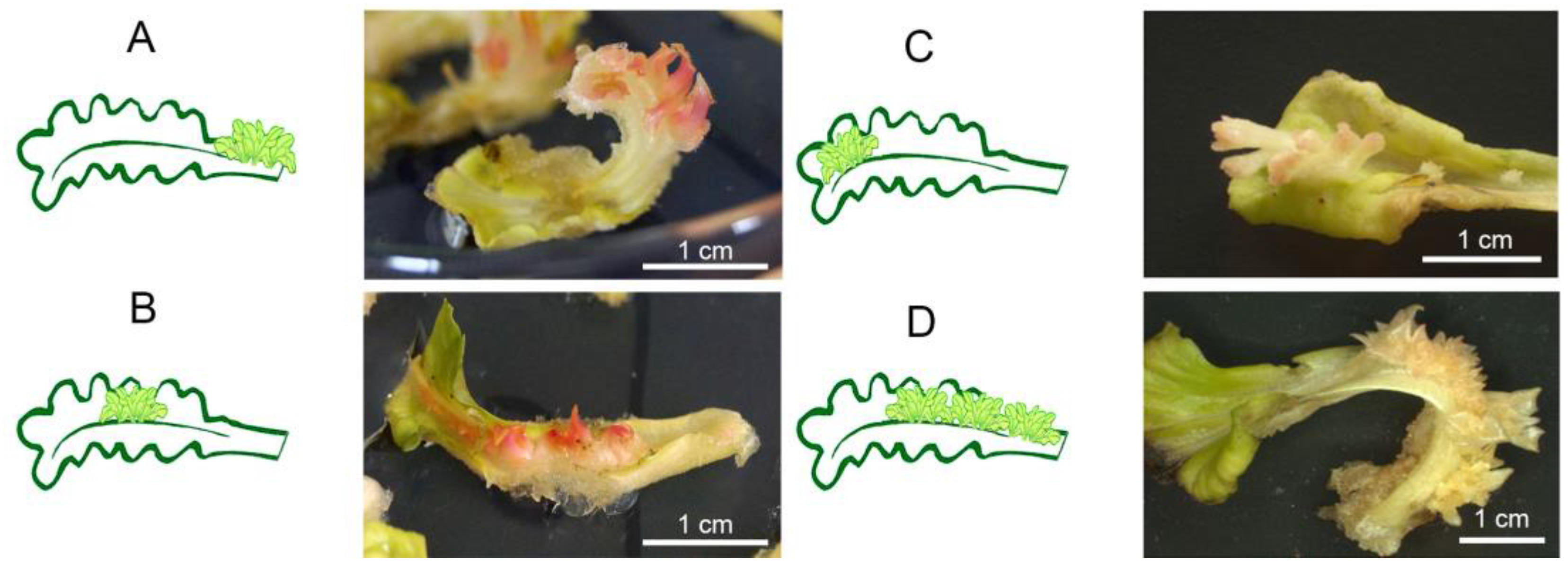
A preliminary assessment of the morphogenic potential of in vitro leaf explants of several sugar beet breeding lines revealed that the combination of 1.0 mg L−1 6-BAP with 0.1 mg L−1 IAA showed better induction of adventitious shoot regeneration (data in preparation). Using this combination, we analyzed regeneration abilities of various sugar beet breeding lines, including O-type lines (№4-28 and №4-40), MS lines (№1, №2, E11301 and C7994), and pollinator lines (№3, H4977 and O5063). Depending on the breeding lines, 250 (hybrid line C7994) to 1136 (hybrid line H4977) leaf explants overall were cultivated in vitro in independent experiments. The summarized results are presented in Table 1. Morphogenic foci arise at 12–15 days after the beginning of the culture. Adventitious shoots developed in various parts of cultivated leaves, including leaf blades, central or basal parts of petioles, clustering primarily near the midrib (Figure 2). At the end of the cultivation, we distributed the regenerating explants in the several categories according to the main area of regeneration (Figure 2, Table 1). Some leaves displayed ability to generate adventitious shoots simultaneously in different parts, mostly in the middle and basal petiole parts, which were covered with morphogenic areas. Such explants we regarded as the most desirable explants for transformation experiments.
Among nine breeding lines, the highest capacity for regeneration was demonstrated by O-type lines №4-28 and №4-40, as an average 85% and 62% of explants, correspondently, developed adventitious shoots. The leaves of O5063 also showed a good regeneration response; on average, more than half of the cultured explants produced morphogenic clusters. The breeding line №4-40 regenerated the highest number of adventitious shoots (12 plantlets per explant), while morphogenic clusters equally formed at different parts of leaves. Most of the explants of O5063 predominantly produced adventitious plantlets in the middle part of leaves (on average, eight plantlets per one explant). The line №4-28 was characterized by formation of adventitious shoots strictly in the middle and basal parts of cultivated explant (seven plantlets per one explant). Two MS breeding lines, C7994 and E11301, were almost unable to produce adventitious shoots, as only single explants developed 1–2 plantlets (Table 1). The other lines occupied an intermediate position in terms of percentage of regenerating explants, which varied from 16% (H4977) to 34% (№2), primarily producing 1 to 4 plantlets in the basal or middle part of cultivated leaves. As a result of regeneration experiments, three breeding lines, №4-28, №4-40, and O5063, which displayed the better ability for in vitro plantlets regeneration, were chosen for further studies.

3.2. Effect of Selective Substances on the Regeneration Abilities

To determine the sub-lethal concentrations of antibiotics and herbicides at which the ability of regenerating explants to produce new adventitious shoots would be significantly suppressed, explants of two sugar beet lines, №4-28 and OP5063, were cultured on a regeneration medium containing different concentrations of selective substances. The obtained data demonstrated resistance of sugar beet tissues to high kanamycin levels. Even at a concentration of 300 mg/L, the adventitious shoot regeneration continued without inhibition; the explant tissues remained green until the end of the experiment (Figures S2 and S3). When using the antibiotic G418, the inhibition of adventitious shoot formation due to explant browning was observed at a concentration of 30 mg L−1, while at the highest concentration of 40 mg L−1, plant tissue died within 2 weeks (Figures S2 and S3). The toxic effect of hygromycin was clearly noticeable even at a minimum applied concentration of 5 mg L−1; significant inhibition of shoot regeneration was observed at an antibiotic concentration of 10 mg L−1 and higher (Figures S2 and S3). The herbicides phosphinothricin and glyphosate were also effective in the suppression of adventitious regeneration. The supplementation of 10 mg L−1 phosphinothricin resulted in the browning of explants with significant termination of morphogenesis (Figure S3). A glyphosate level of lower than 33.8 mg L−1 was not sufficient to suppress the plantlets’ regeneration while, starting from 42.5 mg L−1, no development of adventitious shoot occurred, and most explants significantly browned or even died at the end of the experiment (Figure S3). Based on the experiments, the following concentrations were chosen for selection for further transformation study: 300 mg L−1 of kanamycin, 30 mg L−1 of G418, 10 mg L−1 of hygromycin, 10 mg L−1 of phosphinothricin, and 42.5 mg L−1 of glyphosate.

3.3. Agrobacterium-Mediated Genetic Transformation Using Genetic Vectors Encoded Various Selective Genes

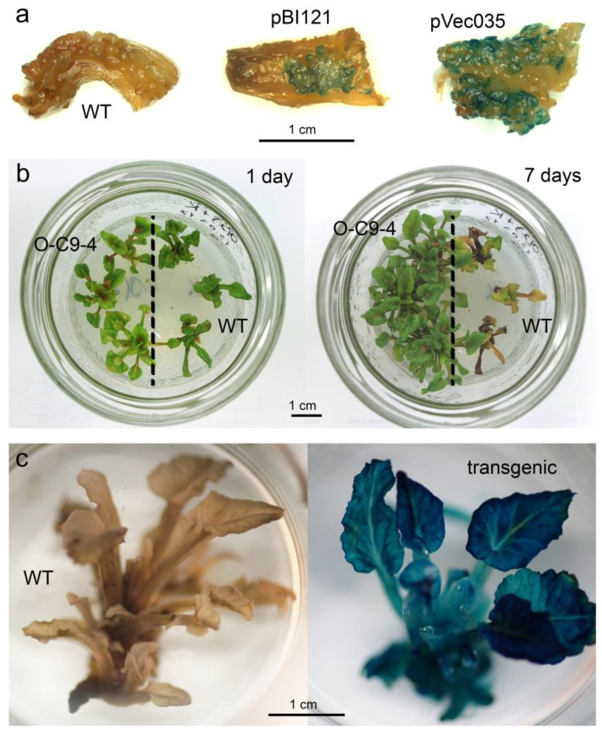
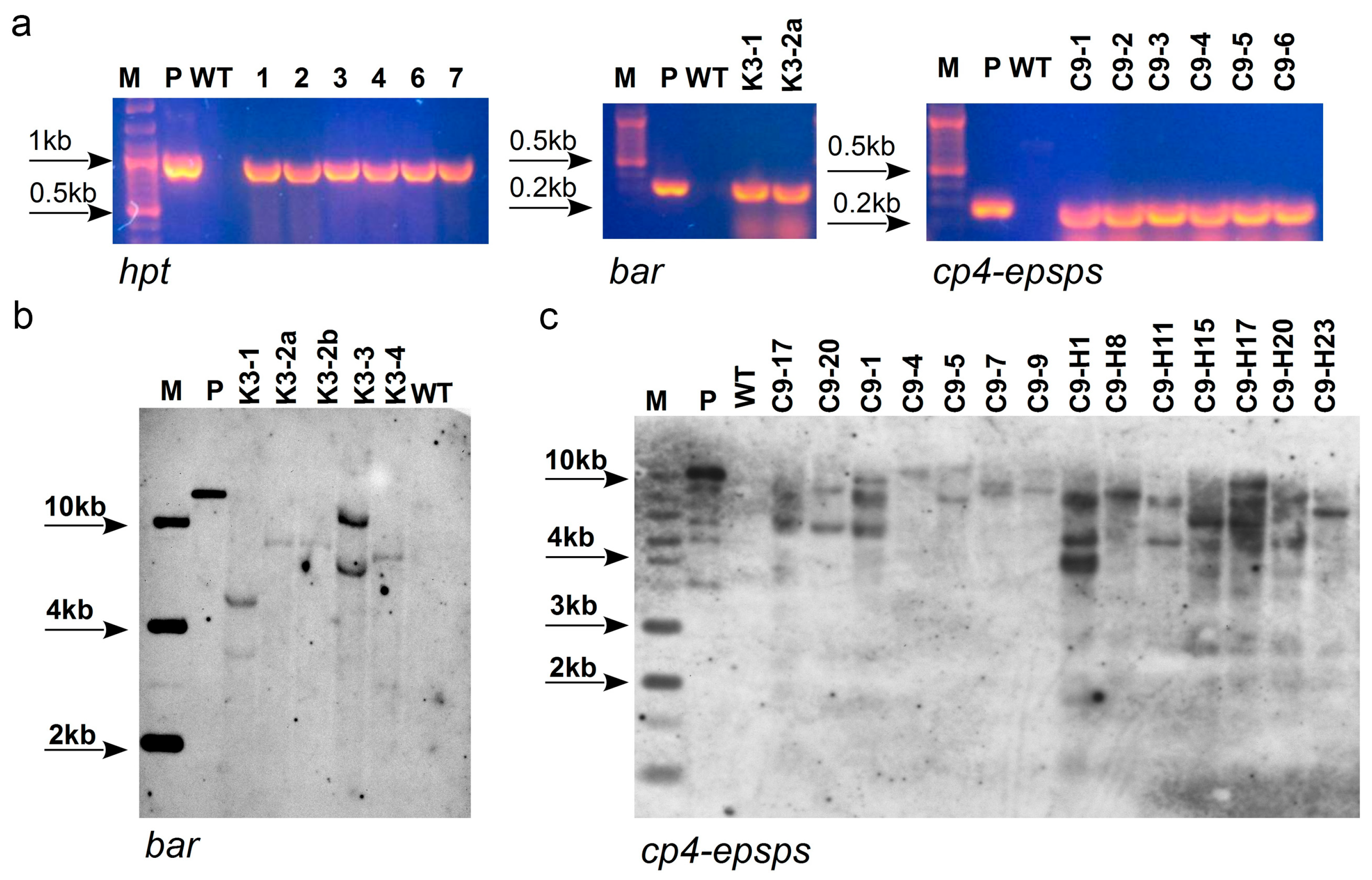
In the first experiments, leaf explants of three breeding lines, №4-28, №4-40, and O5063, were co-cultivated with Agrobacterium carrying pVec035 or pBI121 plasmids, encoded with the resistance to the most popular selective antibiotics, hygromycine and kanamycin, correspondently. Both vectors also encoded the reporter gene b-glucuronidase, which helped us to monitor the transformation progress. Analysis for transient expression showed the successful transferring of foreign sequences into the cells of wounded explants from both vectors (Figure 3a) after the agrobacterial inoculation. Between 5.0 and 10.0% of explants of all three studied breeding lines produced resistant plantlets during long-term selection on kanamycin-enriched medium. Subsequent PCR analysis of resulting regenerants did not confirm integration of the nptII and uidA genes into the plant genome. Thus, no transgenic plants were obtained after transformation with the pBI121 vector and kanamycin selection. In contrast, when explants were transformed with the pVec035 vector the number of hygromycin-resistant regenerants was significantly lower (2.3–2.8% of the number of transformed explants), while the most of the selected plants displayed integration of the hpt and uidA genes (Table 1, Figure 4a). A total of 24 independent transgenic lines carrying the hpt gene were generated, which were easily proliferated on the hygromycin-containing medium (Figure 3b). The frequency of genetic transformation varied from 1.48 (№4-40) to 2.45% (O5063) depending on the transforming breeding line (Table 1). Most of the transgenic plants (21 out of 24) also showed the presence of reporter uidA gene sequence in their genome and demonstrated specific blue staining in all plant tissues, confirming successful expression of the transferred genes (Figure 3c).
In the next experiments, we aimed to produce herbicide-resistant sugar beet plants; the OP5063 breeding line, which displayed a higher transformation rate, was used. The explants transformed with the pBIBar vector, whose T-DNA contain sequences of the bar and the nptII genes, were cultivated on the medium supplemented with phosphinothricin or G418. Despite the increased number of explants and more rigorous selection compared to previous transformation experiments, transgenic shoots were also not obtained using G418 as a selective substance. Selection on phosphinothricin-containing medium using the same number of explants yielded several herbicide-resistant shoots. They showed the integration of the bar gene according to PCR analysis (Figure 4a), resulting in a transformation frequency of 0.99%.
Genetic transformation of OP5063 explants with the pEPSPS vector using glyphosate as the selective agent also occurred at a low frequency, as only two glyphosate-resistant lines containing the cp4-epsps gene were produced. This corresponded to a transformation frequency of 0.4%. In parallel, inoculated leaf explants were also cultivated on the medium supplemented with hygromycin, as the T-DNA of the pEPSPS vector also contains the sequence of the hpt-selective gene. Selection of transgenic shoots using hygromycin resulted in recovering 42 antibiotic-resistant lines. Subsequent PCR analysis (Figure 4a) of genomic DNA proved that 38 independent lines contained the sequence of the hpt gene, while the insertion of the cp4-epsps gene was detected only in the genome of 27 lines. Counting the number of transgenic lines carrying the antibiotic resistance gene, the transformation rate reached 5.07%; the transformation efficiency with the gene of interest was 3.6% (Table 2).
In general, the selection process of sugar beet transgenic shoots was similar for all transformation vectors and types of selective substance used. By the end of the second month of cultivation, surviving adventitious shoots were developed on individual explants. During the third month of cultivation, some plantlets died while others continued to grow normally, showing no signs of toxic effects from selective agents. When the shoots reached a size of 10–15 mm, they were separated from the explants and transferred to proliferation medium containing the appropriate selective substances. Multiplicated herbicide-resistant transgenic plants were then normally rooted within 2–3 weeks of culture. A part of independent transgenic lines was transferred to the greenhouse to grow mature plants for estimation of transgene copy number and integrity of insertions by Southern blot analysis.
Southern blot analysis confirmed the independent nature of the discovered transformation events, with one exception (Figure 4b,c). Two lines generated after transformation of the pBIBar plasmid, named K3-2a and K3-2b, were originally generated from the same explants. As shown in Figure 4c, they expectedly represent the same transformation event because the hybridization with the specific bar probe resulted in the same size band in DNA extracts (Figure 4b). The rest of the sugar beet transgenic lines produced after Agrobacterium-mediated transformation with pBIBar plasmid contained two copies of the bar gene (Figure 4b). Southern blotting assay of 14 independent transgenic lines generated after transformation with pEPSPS plasmid confirmed the cp4-epsps gene integration in the genome of all analyzed transformants. In general, two to four insertions of foreign genes were observed, while a single copy of the cp4-epsps gene was clearly detectable only in two independent lines (Figure 4c).

3.4. Herbicide Resistance Analysis of Primary Transgenic Sugar Beet Lines

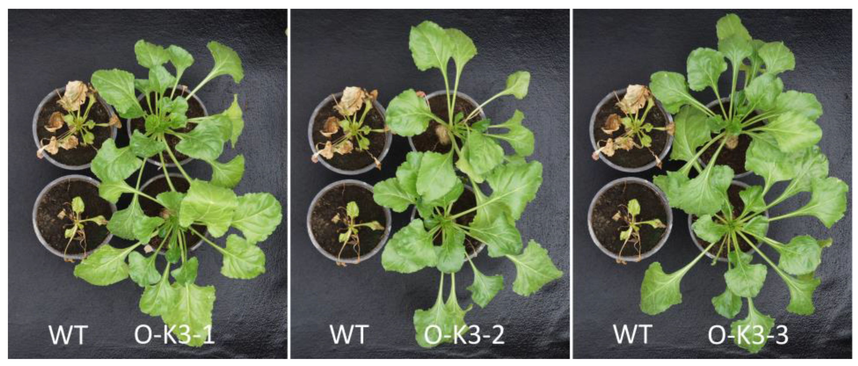
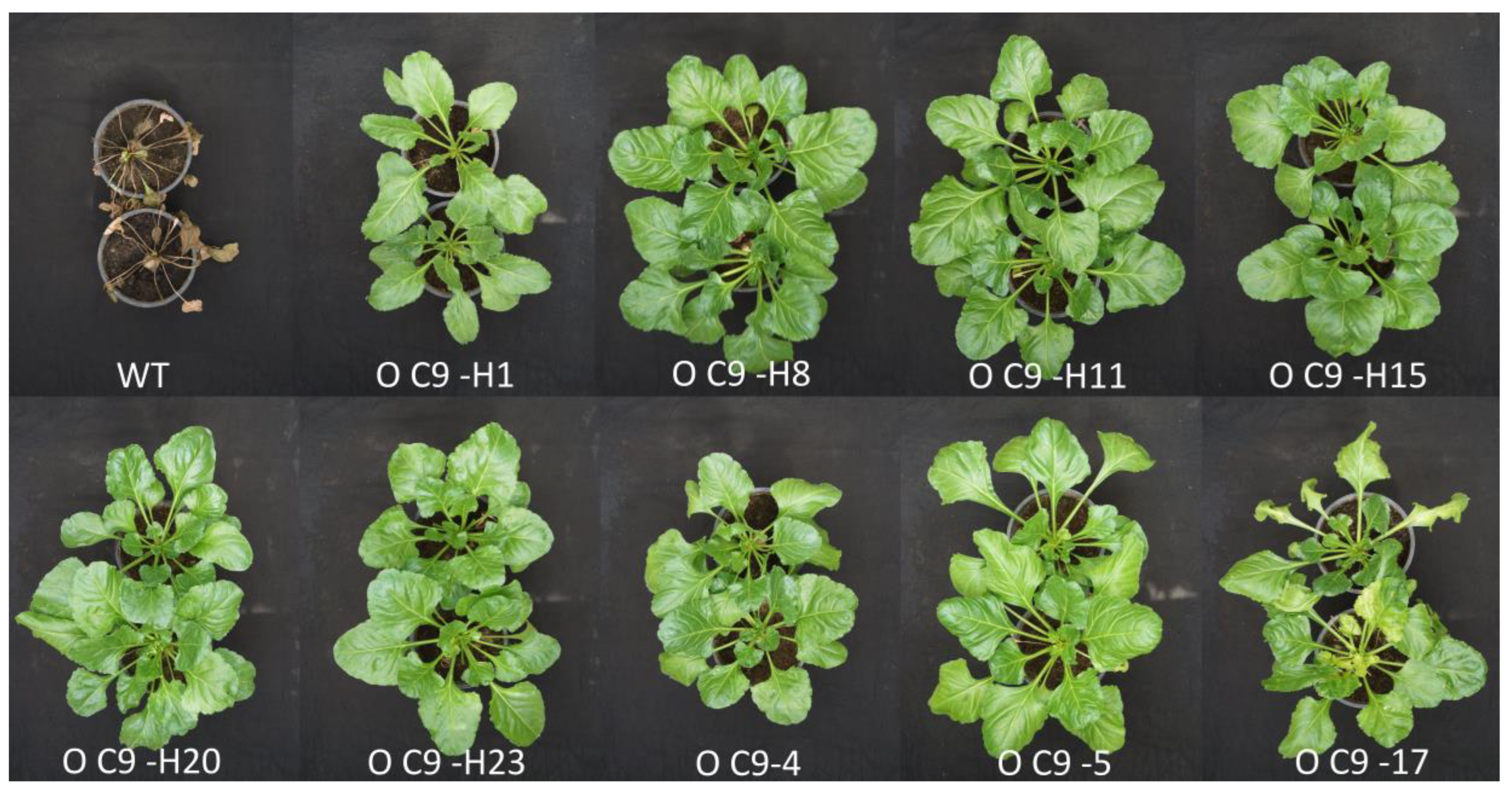
Transgenic lines with confirmed integration of herbicide resistance genes were cultivated in the greenhouse to check the identity of the original breeding line O5063 and test the herbicide resistance. The majority of lines showed no differences from the original breeding line, except line C9-H1, which displayed a delay in the growth and some differences in the shape of leaves. A total of 19 transgenic lines, including three lines transformed with the bar gene and 16 transgenic lines transformed with the cp4-epsps gene, were treated with the corresponding 0.01% Bialaphos solution or 1% Roundup solution to test the resistance. The first signs of herbicide toxicity (chlorotic spots) on non-transformed plants appeared 3–5 days after treatment. Chlorosis then intensified, and by 10–12 days after treatment, necrotic areas became visible on the leaf blades and plant growth completely stopped. By days 15–17, all non-transformed plants died (Figure 5 and Figure 6).
The plants of three independent lines, K3-1, K3-2, and K3-3, showed no signs of susceptibility to Bialaphos treatment (Figure 5). All the tested transgenic lines carrying the cp4-epsps gene showed resistance to Roundup, including the line OC-H1 which visually differed from the parent breeding line. Only one of the lines, OC-9, showed some yellowing of young leaves after the treatment. At the same time, compared to non-transgenic control plants, which completely died after treatment, this line remained alive and capable of satisfactory growth. (Figure 6).

3.5. Inheritance of Herbicide Resistance in the Next Progenies of Primary Transgenic Sugar Beet Lines

To investigate heritability of glyphosate resistance in the next progenies, seeds obtained after self-fertilization of 16 T0 primary transgenic lines were sown in the greenhouse and assessed for resistance. Depending on seed availability, 24 to 157 T1 seedlings were sprayed with Roundup solution to determine the pattern of inheritance. Some sprayed T1 seedlings died, indicating the segregation of the glyphosate resistance trait (Figure S4). Inheritance was found to follow a single-locus segregation model (3:1) in T1 progeny of four T0 transgenic lines: O-C9-4, O-C9-9, O-C9-H8, and O-C9-H15 (Table S2). Seeds obtained from self-pollination of the primary lines O-C9-H20, O-C9-H23, and O-C9-20 segregated at a ratio of 15:1, in accordance with a two-locus Mendelian inheritance model. The ratio of resistant and dead seedlings in the progeny of nine primary transgenic lines could not be reliably attributed to any classic inheritance models (Table S2).
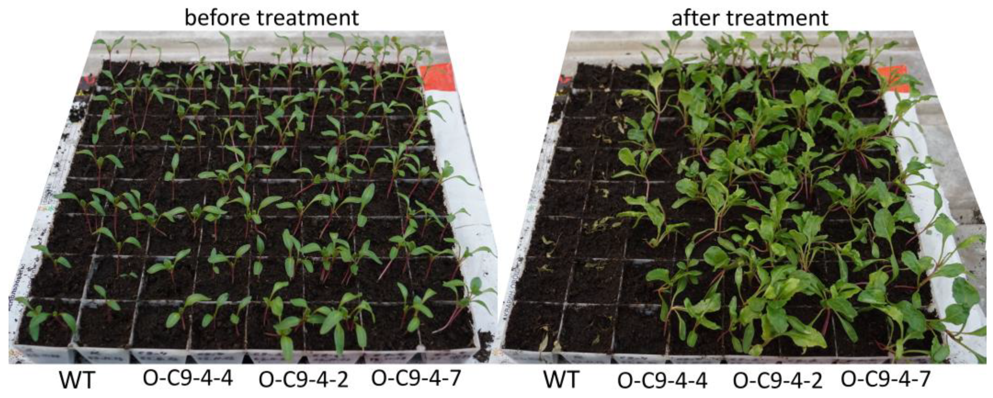
Transgenic lines, which segregated the herbicide resistance trait in accordance with a single-locus inheritance model, were further studied in more detail for identification of homozygous T2 populations. Eight to nine surviving T1 seedlings of three primary transgenic lines, O-C9-9, O-C9-4, and O-C9-H8, were randomly selected, grown to maturity, and self-pollinated to produce T2 seeds. After spraying with Roundup solution, all the T2 seedlings derived from five individual T1 plants of primary lines O-C9-H8 and O-C9-4 survived (Figure 7), while the other T1plants seemed to be heterozygous as they segregated according to a 3:1 ratio Mendelian pattern of inheritance (Table S3). No homozygous T2 seedlings were found among the T1 progenies of primary transgenic line O-C9-9; several T1 progenies completely died, indicating the lack of resistance-trait inheritance, while the others were segregated as heterozygous plants according to a single-locus inheritance model (Table S3).

4. Discussion

In recent decades, considerable efforts have been invested in the development of efficient protocols for the genetic transformation of sugar beet [2,8,9,10,13,14,16,21,28,29]. Various factors should be considered to successfully achieve the foreign gene transfer into the recipient genome of sugar beet. In the present study, the choice of explant, genotype, and selective agent (selective gene) was the primary focus of the Agrobacterium-mediated transformation protocol.
Some prerequisites for the successful production of transgenic sugar beet plants are the evaluation of the morphogenetic potential of a number of genotypes and lines and the selection of the appropriate explant type that exhibits good regenerative capacity and meets the objectives of the study. Although various sugar beet tissues, including leaves (in vitro and in vivo), basal parts of shoots, hypocotyls, cotyledons, and young inflorescences, have been reported to exhibit in vitro morphogenetic response [13,17,18,22,28,29,30,31,32], leaves taken from aseptically propagated plants are still the most convenient tissue available year-round. Our experiments showed that not all parts of the cultured leaf are capable of regenerating adventitious shoots (Figure 2). Plant regeneration was observed mainly along the leaf near the main vein. Shoots formed on the leaf blade and the central and basal parts of the petiole but almost never appeared on the cut leaf petiole (Figure 2). This feature complicates genetic transformation, as agrobacteria transfers T-DNA into wounded cells, which subsequently develop transgenic morphogenic structures. Preliminary experiments showed that the wounding of the leaf vain prior to regeneration initiation does not promote the adventitious shoot formation or stop it. To overcome this problem, we first induced the formation of adventitious structures in the cultured leaf, then wounded or cut off young morphogenic clusters, and only then began target inoculation of the wounded explants with Agrobacterium. This approach allows us to select for transformation only in parts with morphogenic clusters, discarding “empty” non-morphogenic parts of the cultured leaves.
Our results are consistent with previous studies demonstrating the predominant role of genotypes in the abilities of sugar beet breeding lines and cultivars to regenerate plants [8,14,16,17,29,30,31,33]. An analysis of the morphogenic potential of the studied sugar beet breeding lines showed that it varied widely not only by the average number of the developed adventitious shoots and overall efficiency but also by the abilities to form shoots at various parts of the cultured leaves (Figure 2, Table 1). Screening of the regenerative potential of nine sugar beet lines allowed us to identify genotypes with acceptable regenerative capacity, which showed multiple formations of morphogenic clusters in various parts of the cultured leaf.
Further experiments confirmed that the presence of abundant clusters significantly increases the probability of producing transgenic plants. Of the three breeding lines used in the experiments, line O5063, characterized by the predominant location of morphogenic clusters in the central part of the leaf petiole, proved more amenable to genetic transformation than lines №4-28 and №4-40 (Table 2), which had a comparable or even higher percentage of regenerating explants (Table 1), although the clusters were more unevenly distributed across different parts of the leaf.
As has been shown by numerous studies, the optimal choice of a selective agent is of great importance for the successful production of transgenic plants [8,10,18,19,20,21,28]. In the present study, the efficiency of application of selective marker genes and the corresponding selective agents was analyzed in same genotypes and explant type using the same transformation protocol. In most studies, kanamycin at concentrations of 50–200 mg/L is used for selection of sugar beet transformants. Most often, the working concentration of kanamycin is in the range of 100–150 mg/L [10,12,21,34,35], but in some studies it was increased to 200 mg/L [11,36]. Thus, sugar beet appears to be resistant to kanamycin, which is due to the specific features of its biochemistry and genetically determined traits. The possibility of an unusually high resistance, as observed in our experiments, should be considered when developing a protocol for the genetic transformation of this crop.
In experiments simulating the transformation, when morphogenic clusters were injured and cut, cultivation of explants at various levels of selective substances revealed that the studied sugar beet lines are characterized by high sensitivity to other selective agents. However, as in the case of kanamycin, we did not obtain any transgenic shoots when selecting transformants using aminoglycoside antibiotic G418 (resistance encoded by nptII gene). Supplementation of herbicides to medium, such as phosphinothricin (resistance encoded by bar gene) and glyphosate (resistance encoded by ESPS gene), provided the selection of transgenic shoots, but the transformation efficiency was low. The ineffectiveness of G418 and low effectiveness of phosphinothricin and glyphosate as selective agents appears to be due to the excessive sensitivity of adventitious shoot formation to these substances. Among all studied substances, antibiotic hygromycin (resistance encoded by hpt gene) proved to be the most suitable selective agent for transforming sugar beet in our experiments. Furthermore, to obtain glyphosate-resistant lines, it was more beneficial to select transformants using hygromycin rather than glyphosate, followed by PCR selection of lines with the cp4-epsps gene insertion.
Hygromycin has previously been used for the selection of sugar beet transgenic shoots, usually at a concentration of 50–100 mg L−1 [34,37,38,39], and less often at 10–15 mg L−1 [10,22], providing a transformation frequency ranging from 3.1 to 8.8%. In general, the process of regeneration and selection of transgenic shoots on hygromycin-containing media in our experiments was similar to that described by other researchers. The hybrid sugar beet lines that we studied were relatively sensitive to hygromycin, which resulted in a transformation frequency that varied between 1.5% and 5.1%, depending on the specific line. In addition to antibiotics, herbicides such as bialaphos (phosphinothricin) [40,41,42] or glyphosate [2] were previously used to produce transgenic sugar beet plants. In rare cases, the mannose-based method of positive selection was applied [13,43]. Previous studies have shown that these substances are less effective for the genetic transformation of sugar beet than kanamycin or hygromycin, since the frequency of transformation varied from 0.3% to 2.8% for mannose, from 0.2% to 1.7% for phosphinothricin, and up to 1.5% for glyphosate [2,13,40,41,42,43]. In our experiments, we observed transformation frequencies of 1.0% and 0.4% for the O5063 sugar beet line when selecting transgenic plants with phosphinothricin and glyphosate, respectively, which are consistent with the data from other researchers.
Southern blot analysis confirmed the independent nature of our transgenic lines. Given that the expected size of the inserted fragments after transformation with the pBIBar or pEPSPS vectors is at least 4.2 kb or 4.25 kb, respectively, we frequently observed inserts of two or four foreign genes in the studied transgenic lines. However, single-copy inserts of the cp4-epsps gene were only detected in two lines. It is important to note that there is no direct correlation between the number of copies of foreign genes and their expression. For example, experiments conducted by Mannerlof et al. [2] have shown that complete resistance to glyphosate is most often observed in transgenic plants containing one copy of the cp4-epsps gene and less often in plants with five copies. This may be due to complex interactions between copies of the transgene, including possible deficiency in transcription factors and the presence of cross-links between them. Additionally, there may be defective copies of the transgene resulting from incomplete T-DNA transfer, which are not detected by Southern blotting. These factors together explain the lack of a clear correlation between the number of cp4-epsps gene copies and the pattern of glyphosate resistance which we observed in the self-pollinated progenies of the primary transgenic plants.
In the present study, all analyzed transgenic lines transformed with the bar and ESPS genes demonstrated resistance to treatment with the corresponding herbicide at the dosage commonly used in the field. The vast majority of transgenic lines grown in the greenhouse were visually indistinguishable from the parental breeding line in both morphology and growth rate. However, the presence of dissimilarity in a single transgenic line confirms the need to screen mass-produced lines for varietal conformity, especially if further commercial use of the obtained material is anticipated. Analysis of T1 and T2 seed progeny obtained by self-pollination of 16 primary T0 plants transformed with the pEPSPS vector allowed us to isolate lines with a stable inheritance of herbicide resistance. Inheritance of the resistance trait did not always correspond to the results of the Southern blot analysis. Most primary transgenic lines containing three or more copies of the introduced cp4-epsps gene showed unpredictable segregation of the resistance trait, ranging from nearly homozygous inheritance (O-C9-H20) to lack of resistance (O-C9-H1) in the progeny. The presence of one or two copies estimated by Southern blot analysis generally did not correspond directly to the single- or two-locus Mendelian models of segregation. Of the transgenic lines exhibiting segregation of the resistance trait as a single dominant allele, only two lines (O-C9-4 and O-C9-H8) harbored a single cp4-epsps sequence insertion, while the banding pattern in others lines corresponded to two (O-C9-9) or even multiple bands (O-C9-H15). The same situation was observed in transgenic sugar beet lines in which the inheritance of resistance fitted to the 15:1 categories of segregation (=two-locus pattern); only the O-C9-20 line yielded two bands that hybridized with the probe (Figure 4). The observed effects may be associated with a change in the functional activity of the introduced copies, presumably caused by unpredictable truncation or rearrangements of T-DNA insertions resulting from the transformation process, as well as the positional effect of transgene integration. Despite the observed inheritance issues, careful analysis and screening of the seed generation resulted in the production of homozygous T2 progeny for the glyphosate resistance gene. Due to constitutive expression of the introduced cp4-epsps gene, the selected homozygous populations of transgenic sugar beet demonstrated stable herbicide resistance and can be used for further research or in breeding programs.
As a result of our study, we obtained transgenic lines of three hybrid sugar beet lines. Our proposed approach is applicable to a wide range of studies on the genetic transformation of sugar beet. It primarily involves evaluating the maximum number of available breeding lines and varieties that meet the research objectives for the ability to regenerate adventitious shoots, then determining their sensitivity to various selective agents and agrobacterial genetic transformation with a vector carrying the optimal selective gene. Comparative studies of the most accessible selective marker genes, conducted on the same genetic material, allow us to recommend a selection system using hygromycin (resistance is encoded by the hpt gene). However, the use of vectors conferring resistance to kanamycin or G418 (encoded by the nptII gene) for the genetic transformation of sugar beet should be avoided. The data presented here also suggest inoculation of in vitro leaf fragments with already-initiated morphogenesis for the successful production of transgenic plants. The approach described can ensure efficient Agrobacterium-mediated transformation of sugar beet and the production of various transgenic lines for further improvement of this important crop using molecular breeding methods, including genome editing.

Supplementary Materials

The following supporting information can be downloaded at: https://www.mdpi.com/article/10.3390/crops6010012/s1, Table S1: List of primers used in the study; Table S2: Segregation analysis of herbicide resistance in T1 progenies of primary transgenic plants of sugar beet transformed with pEPSPS plasmid; Table S3: Segregation analysis of herbicide resistance in T2 progenies resulted after self-pollination of T1 transgenic plants; Figure S1: The schematic overview of the in vitro leaf-based method for Agrobacterium-mediated genetic transformation of sugar beet; Figure S2: Effect of selective antibiotics on regeneration abilities of sugar beet leaf explants; Figure S3: Effect of kanamycin, G418, glyphosate, hygromycin, and phosphinothricin concentrations on the ability of sugar beet leaf explants to re-regenerate morphogenetic structures/plantlets. Figure S4: Example of herbicide resistance evaluation of T1 seedlings derived from primary sugar beet transgenic lines transformed with pEPSPS plasmid. References [44,45,46] are cited in the Supplementary Materials.

Author Contributions

D.N.M. and S.V.D. conceived and designed the study. S.V.D. supervised the research. A.S.P. designed plasmids. D.N.M., A.K. and L.M. produced and maintained transgenic plants. A.S.P. and A.K. carried out PCR and Southern blot analysis. D.N.M. and L.M. conducted herbicide resistance analysis. D.N.M. and A.P.F. performed data analysis and wrote the manuscript. A.S.P. and S.V.D. contributed to writing and revision. All authors have read and agreed to the published version of the manuscript.

Funding

This research was performed under the framework of state budget project FFEV-2024-0042.

Data Availability Statement

The presented data are available upon reasonable request from the corresponding author.

Acknowledgments

The authors would like to thank the staff of the Large-Scale Research Facility of the Branch of Shemyakin and Ovchinnikov Institute of Bioorganic Chemistry of Russian Academy of Science, code 73597, for the growing of the donor and transgenic plants.

Conflicts of Interest

The authors declare no conflicts of interest.

Abbreviations

The following abbreviations are used in this manuscript:
MSMurashige and Skoog
BAP6-benzylaminopurine
Kmkanamycin
Hyghygromycine
G418geneticin
PPTphosphinothricin
bargene of phosphinothricin acetyltransferase
epspsgene of 5-enolpyruvulshikimate-3-phosphate synthase
uidAgene of β-glucuronidase
nptIIgene of neomycin phosphotransferase
hptgene of hygromicin phosphotransferase

References

  1. Sugartalk Industrial Insight. Available online: https://www.ragus.co.uk/sugar-talk (accessed on 1 December 2025).
  2. Mannerlöf, M.; Tuvesson, S.; Steen, P.; Tenning, P. Transgenic sugar beet tolerant to glyphosate. Euphytica 1997, 94, 83–91. [Google Scholar] [CrossRef]
  3. Nehls, R.; Kraus, J.; Matzk, A.; Jansen, R. Transgenic Varieties: Sugarbeet. Sugar Tech 2010, 12, 194–200. [Google Scholar] [CrossRef]
  4. Eini, O.; Varrelmann, M. Challenges and potential applications of genome editing in sugar beet. Sugar Ind. 2022, 147, 274–283. [Google Scholar] [CrossRef]
  5. Misra, V.; Mall, A.K.; Pandey, H.; Srivastava, S.; Sharma, A. Advancements and prospects of CRISPR/Cas9 technologies for abiotic and biotic stresses in sugar beet. Front. Genet. 2023, 14, 1235855. [Google Scholar] [CrossRef]
  6. Amaresh, G.; Nunavath, A.; Appunu, C.; Viswanathan, C.; Kumar, R.; Gujjar, R.S.; Manimekalai, R. Advanced Genome Editing Technologies: Potentials and Prospects in Improvement of Sugar crops. Sugar Tech 2025, 27, 14–28. [Google Scholar] [CrossRef]
  7. Fry, J.E.; Barnason, A.R.; Hinchee, M. Genotype-independent transformation of sugarbeet using Agrobacterium tumefaciens. In Molecular Biology of Plant Growth and Development, Proceedings of the 3rd International Congress of the International Society for Plant Molecular Biology, Tuckon, AZ, USA, 6–11 October 1991; Hallick, R.B., Ed.; Plenum Press: New York, NY, USA, 1991; p. 384. [Google Scholar]
  8. Smigocki, A.; Campbell, L.; Larson, R.; Wozniak, C. Sugar Beet. In Compendium of Transgenic Crop Plants; Kole, C., Hall, T.C., Eds.; Wiley: Hoboken, NJ, USA, 2008; pp. 59–96. ISBN 9781405169240. [Google Scholar]
  9. Konwar, B.K. Agrobacterium tumefaciens-Mediated Genetic Transformation of Sugar Beet (Beta vulgaris L.). J. Plant Biochem. Biotechnol. 1994, 3, 37–41. [Google Scholar] [CrossRef]

  10. Hisano, H.; Kimoto, Y.; Hayakawa, H.; Takeichi, J.; Domae, T.; Hashimoto, R.; Abe, J.; Asano, S.; Kanazawa, A.; Shimamoto, Y. High frequency Agrobacterium-mediated transformation and plant regeneration via direct shoot formation from leaf explants in Beta vulgaris and Beta maritima. Plant Cell Rep. 2004, 22, 910–918. [Google Scholar] [CrossRef]
  11. Snyder, G.W.; Ingersoll, J.C.; Smigocki, A.C.; Owens, L.D. Introduction of pathogen defense genes and a cytokinin biosynthesis gene into sugarbeet (Beta vulgaris L.) by Agrobacterium or particle bombardment. Plant Cell Rep. 1999, 18, 829–834. [Google Scholar] [CrossRef]
  12. Zhang, C.L.; Chen, D.F.; McCormac, A.C.; Scott, N.W.; Elliott, M.C.; Slater, A. Use of the GFP reporter as a vital marker for Agrobacterium-mediated transformation of sugar beet (Beta vulgaris L.). Mol. Biotechnol. 2001, 17, 109–117. [Google Scholar] [CrossRef]
  13. Gurel, S. Sand-Wounding of Shoot and Petiole Explants Enhances Transformation Efficiency in Sugar Beet (Beta vulgaris L.) by Agrobacterium-mediated Transformation. Sugar Tech 2021, 23, 415–427. [Google Scholar] [CrossRef]
  14. Freytag, A.H.; Anand, S.C.; Rao-Arelli, A.P.; Owens, L.D. An improved medium for adventitious shoot formation and callus induction in Beta vulgaris L. in vitro. Plant Cell Rep. 1988, 7, 30–34. [Google Scholar] [CrossRef]
  15. Detrez, C.; Tetu, T.; Sangwan, R.S.; Sangwan-Norreel, B.S. Direct Organogenesis from Petiole and Thin Cell Layer Explants in Sugar Beet Cultured In Vitro. J. Exp. Bot. 1988, 39, 917–926. Available online: http://www.jstor.org/stable/23692095 (accessed on 1 January 2026). [CrossRef]
  16. Ivic-Haymes, S.D.; Smigocki, A.C. Identification of highly regenerative plants within sugar beet (Beta vulgaris L.) breeding lines for molecular breeding. Vitr. Cell. Dev. Biol.-Plant 2005, 41, 483–488. [Google Scholar] [CrossRef]
  17. Mishutkina, Y.V.; Gaponenko, A.K. Sugar beet (Beta vulgaris L.) morphogenesis in vitro: Effects of phytohormone type and concentration in the culture medium, type of explants, and plant genotype on shoot regeneration frequency. Russ. J. Genet. 2006, 42, 150–157. [Google Scholar] [CrossRef]
  18. Lindsey, K.; Gallois, P. Transformation of Sugarbeet (Beta vulgaris) by Agrobacterium tumefaciens. J. Exp. Bot. 1990, 41, 529–536. [Google Scholar] [CrossRef]
  19. D’Halluin, K.; Bossut, M.; Bonne, E.; Mazur, B.; Leemans, J.; Botterman, J. Transformation of Sugarbeet (Beta vulgaris L.) and Evaluation of Herbicide Resistance in Transgenic Plants. Nat. Biotechnol. 1992, 10, 309–314. [Google Scholar] [CrossRef]
  20. Krens, F.A.; Trifonova, A.; Paul Keizer, L.C.; Hall, R.D. The effect of exogenously-applied phytohormones on gene transfer efficiency in sugarbeet (Beta vulgaris L.). Plant Sci. 1996, 116, 97–106. [Google Scholar] [CrossRef]
  21. Norouzi, P.; Malboobi, M.A.; Zamani, K.; Yazdi-Samadi, H. Using a competent tissue for efficient transformation of sugarbeet (Beta vulgaris L.). Vitr. Cell. Dev. Biol.-Plant 2005, 41, 11–16. [Google Scholar] [CrossRef]
  22. Yang, A.F.; Duan, X.G.; Gu, X.F.; Gao, F.; Zhang, J.R. Efficient transformation of beet (Beta vulgaris) and production of plants with improved salt-tolerance. Plant Cell Tissue Organ Cult. 2005, 83, 259–270. [Google Scholar] [CrossRef]
  23. Murashige, T.; Skoog, F. A Revised Medium for Rapid Growth and Bio Assays with Tobacco Tissue Cultures. Physiol. Plant. 1962, 15, 473–497. [Google Scholar] [CrossRef]
  24. Revenkova, E.V.; Kraev, A.S.; Skryabin, K.G. Construction of a disarmed derivative of the super virulent Ti-plasmid pTiBo542. In Plant Biotechnology and Molecular Biology; Skryabin, K.G., Ed.; Pushchino Research Centre: Moscow, Russia, 1993; p. 281. [Google Scholar]
  25. Jefferson, R.A.; Kavanagh, T.A.; Bevan, M.W. GUS fusions: Beta-glucuronidase as a sensitive and versatile gene fusion marker in higher plants. EMBO J. 1987, 6, 3901–3907. [Google Scholar] [CrossRef]
  26. Santarém, E.R.; Trick, H.N.; Essig, J.S.; Finer, J.J. Sonication-assisted Agrobacterium-mediated transformation of soybean immature cotyledons: Optimization of transient expression. Plant Cell Rep. 1998, 17, 752–759. [Google Scholar] [CrossRef]
  27. Padegimas, L.; Shul’ga, O.A.; Skriabin, K.G. Creation of transgenic plants Nicotiana tabacum and Solanum tuberosum, resistant to the herbicide phosphinothricin. Mol. Biol. 1994, 28, 437–443. [Google Scholar] [PubMed]
  28. Zhang, Z.; Zhang, H.; Wang, L.; Zhang, B.; Fu, Z.; Zhao, S.; Yuanyuan, E.; Zheng, W.; Zhang, H.; Han, P.; et al. An Efficient Protocol for Agrobacterium-Mediated Transformation and Regeneration of Sugar Beet (Beta vulgaris L.) based on Blade–Petiole Transition Zone Explants. Sugar Tech 2023, 25, 154–159. [Google Scholar] [CrossRef]
  29. Khan, Z.; Hathaway, T.; Chu, C.; Wyatt, N.; Bolton, M.; Ramachandran, V. Development of an improved Agrobacterium-mediated transformation and callus-based regeneration system for sugar beet (Beta vulgaris L.). Plant Cell Tissue Organ Cult. 2025, 163, 30. [Google Scholar] [CrossRef]
  30. Roussy, I.; Dubois, F.; Sangwan, R.S.; Sangwan-Norreel, B.S. In planta 2,3,5 truodobenzoic acid treatment promotes high frequency and routine in vitro regeneration of sugar beet (Beta vulgaris L.) plants. Plant Cell Rep. 1996, 16, 142–146. [Google Scholar] [CrossRef]
  31. de Marchis, F.; Wang, Y.; Stevanato, P.; Arcioni, S.; Bellucci, M. Genetic transformation of the sugar beet plastome. Transgenic Res. 2009, 18, 17–30. [Google Scholar] [CrossRef]
  32. Jafari, M.; Norouzi, P.; Malboobi, M.A.; Ghareyazie, B.; Valizadeh, M.; Mohammadi, S.A.; Mousavi, M. Enhanced resistance to a lepidopteran pest in transgenic sugar beet plants expressing synthetic cry1Ab gene. Euphytica 2009, 165, 333–344. [Google Scholar] [CrossRef]
  33. Abe, J.; Nakashima, H.; Mitsui, K.; Mikami, T.; Shimamoto, Y. Tissue culture response of Beta germplasm: Callus induction and plant regeneration. Plant Cell Tissue Organ Cult. 1991, 27, 123–127. [Google Scholar] [CrossRef]
  34. Liu, H.; Wang, Q.; Yu, M.; Zhang, Y.; Wu, Y.; Zhang, H. Transgenic salt-tolerant sugar beet (Beta vulgaris L.) constitutively expressing an Arabidopsis thaliana vacuolar Na/H antiporter gene, AtNHX3, accumulates more soluble sugar but less salt in storage roots. Plant Cell Environ. 2008, 31, 1325–1334. [Google Scholar] [CrossRef]
  35. Zare, B.; Niazi, A.; Sattari, R.; Aghelpasand, H.; Zamani, K.; Sabet, M.S.; Moshiri, F.; Darabie, S.; Daneshvar, M.H.; Norouzi, P.; et al. Resistance against rhizomania disease via RNA silencing in sugar beet. Plant Pathol. 2015, 64, 35–42. [Google Scholar] [CrossRef]
  36. Ivic, S.; Smigocki, A. Evaluation of the biolistic transformation method for commercially important sugarbeet breeding lines. Am. Soc. Sugar Beet Tech. Proc. 2001, 31, 207–213. [Google Scholar]
  37. Zhan, C.L.; Chen, D.F.; Kubis, S.K.; McCormac, A.C.; Kublakova, M.; Zhang, J.; Bao, M.Z.; Scott, N.W.; Slater, A.; Heslop-Harrison, J.S.; et al. Improved procedure for transformation of sugar beet. In Proceedings of the 61st IIRB Congress, Brussels, Belgium, 11–12 February 1998; Volume 61, pp. 381–389. [Google Scholar]
  38. Lisson, R.; Hellert, J.; Ringleb, M.; Machens, F.; Kraus, J.; Hehl, R. Alternative splicing of the maize Ac transposase transcript in transgenic sugar beet (Beta vulgaris L.). Plant Mol. Biol. 2010, 74, 19–32. [Google Scholar] [CrossRef]
  39. Kagami, H.; Kurata, M.; Matsuhira, H.; Taguchi, K.; Mikami, T.; Tamagake, H.; Kubo, T. Sugar beet (Beta vulgaris L.). Methods Mol. Biol. 2015, 1223, 335–347. [Google Scholar] [CrossRef]
  40. Hall, R.D.; Riksen-Bruinsma, T.; Weyens, G.J.; Rosquin, I.J.; Denys, P.N.; Evans, I.J.; Lathouwers, J.E.; Lefèbvre, M.P.; Dunwell, J.M.; van Tunen, A.; et al. A high efficiency technique for the generation of transgenic sugar beets from stomatal guard cells. Nat. Biotechnol. 1996, 14, 1133–1138. [Google Scholar] [CrossRef]
  41. Yamada, N.; Takahashi, H.; Kitou, K.; Sahashi, K.; Tamagake, H.; Tanaka, Y.; Takabe, T. Suppressed expression of choline monooxygenase in sugar beet on the accumulation of glycine betaine. Plant Physiol. Biochem. 2015, 96, 217–221. [Google Scholar] [CrossRef]
  42. Mishutkina, Y.V.; Kamionskaya, A.M.; Skryabin, K.G. The creation of sugar beet transgenic plants expressing bar gene. Appl. Biochem. Microbiol. 2010, 46, 80–86. [Google Scholar] [CrossRef]
  43. Joersbo, M.; Mikkelsen, J.D.; Brunstedt, J. Relationship between promoter strength and transformation frequencies using mannose selection for the production of transgenic sugar beet. Mol. Breed. 2000, 6, 207–213. [Google Scholar] [CrossRef]
  44. Miroshnichenko, D.; Pushin, A.; Dolgov, S. Assessment of the pollen-mediated transgene flow from the plants of herbicide resistant wheat to conventional wheat (Triticum aestivum L.). Euphytica 2016, 209, 71–84. [Google Scholar] [CrossRef]
  45. Demeke, T.; Giroux, R.W.; Reitmeier, S.; Simon, S.L. Development of a polymerase chain reaction assay for detection of three canola transgenes. J. Am. Oil Chem. Soc. 2002, 79, 1015–1019. [Google Scholar] [CrossRef]
  46. Dolgov, S.V.; Firsov, A.P.; Pushin, A.S.; Tarasenko, I.V.; Golikov, A.G. Gene-engineered details of a Raudis commercial rapeseed variety naturally tolerant to glyphosate. Biotehnologiâ 2015, 59–64. [Google Scholar] [CrossRef]
Figure 1. Schematic representation of expression cassettes used for genetic transformation of sugar beet. Abbreviations: CaMV35S-p, 35S RNA promoter of Cauliflower Mosaic Virus; uidA, gene of β-glucuronidase (GUS); uidA-intron, intron-containing uidA gene; NOS-T, nopaline synthase terminator with polyadenylation signal; nptII, neomycin phosphotransferase (NPT) coding sequence; hpt, hygromycin phosphotransferase (HPT) coding sequence; NOS-P, nopaline synthase promoter; CaMV35S-T, CaMV35S terminator with polyadenylation signal; bar, phosphinothricin N-acetyltransferase (PAT) coding sequence; cp4-epsps, synthetic gene on basis of the EPSPS (5-enolpyruvulshikimate-3-phosphate synthase) sequence of Agrobacterium tumefaciens CP4 strain with a N-terminal chloroplast signal peptide CTP2 of the Arabidopsis thaliana epsps gene; RB, right border of transferring DNA (T-DNA); LB, left border of T-DNA; gray bars represent the elements of T-DNA, which were detected by Southern blot in transgenic plants.
Crops 06 00012 g001
Figure 2. Different types of adventitious shoot regeneration on leaf explants of sugar beet. (A) Adventitious shoots developed in the basal part of the leaf petiole; (B) adventitious shoots developed in the central part of leaf petiole; (C) adventitious shoots developed on the leaf blade; (D) multiple formations of adventitious shoots are observed in various parts of the leaf explant.
Crops 06 00012 g002
Figure 3. Genetic transformation of sugar beet. (a) Transient expression of the uidA reporter gene on the morphogenic leaf explants of №4-28 breeding line, 10 days after transformation with pBI121 or pVec035 vectors; (b) growth of transgenic and non-transgenic sugar beet plants on the medium supplemented with 10 mg L−1 of hygromycin; (c) expression of the uidA gene in the tissues of one of sugar beet line, demonstrated after transformation with pVec035 vector using hygromycin selection.
Crops 06 00012 g003
Figure 4. Molecular analysis of transgenic sugar beet lines. (a) An example of PCR analysis of a part of lines generated using pVec035 (left panel), pBIBar (central panel), and pEPSPS (right panel) for the insertion of hpt, bar or cp4-epsps genes, correspondently; lane M, DNA ladder as molecular weight marker; lane P, corresponding plasmid; lane WT, DNA of untransformed sugar beet breeding line. (b) Southern blot analyses of genomic DNAs isolated from leaves of PCR-positive transgenic plants generated after transformation using pBIBar plasmid; bands larger than 4.2 kb on the blot correspond to the complete insert. (c) Southern blot of genomic DNAs isolated from leaves of PCR-positive transgenic plants generated after transformation using pEPSPS plasmid; bands larger than 4.25 kb on the blot correspond to the complete insert. Primary transgenic plants generated after transformation with pVec035 labeled as 1, 2, 3, 4, 5, 6, 7; plants generated after transformation with pBIBar labeled as K3-1, K3-2, K3-3, K3-4; plants generated after transformation with pEPSPS labeled as C9-1, C9-2, C9-3, C9-4, C9-5, C9-6, C9-7, C9-9, C9-17, C9-20, C9-H1, C9-H8, C9-H11, C9-H15, C9-H17, C9-H20, C9-H23.
Crops 06 00012 g004
Figure 5. Evaluation of herbicide resistance of transgenic lines obtained after Agrobacterium–mediated transformation using the pBIBar plasmid. Transgenic plants and the breeding line O5063 (WT) were grown in greenhouse and sprayed with 0.01% Bialaphos.
Crops 06 00012 g005
Figure 6. Evaluation of herbicide resistance of transgenic lines obtained after Agrobacterium-mediated transformation with pEPSPS plasmid. Transgenic plants and the breeding line O5063 (WT) were grown in greenhouse and sprayed with 1% Roundup.
Crops 06 00012 g006
Figure 7. Evaluation of T2 seedlings derived from three individual T1 plants obtained after self-pollination of primary transgenic line O-C9-4. Transgenic seedlings and seedlings of breeding line O5063 (WT) were grown in greenhouse and sprayed with 1% Roundup solution.
Crops 06 00012 g007
Table 1. Efficiency of the adventitious shoot regeneration from in vitro leaf explants of nine breeding lines of sugar beet (Beta vulgaris L. var. saccharifera).
LineNumber of ExplantsThe Percentage of
Regenerating Explants (%)
Number of Plantlets per Regenerating ExplantThe Ratio of Explants with Different Types of Regeneration ** (%)
ABCD
№198334 ± 21 c2.8 ± 1.7 c771445
№253434 ± 21 c3.7 ± 1.7 c592877
№351329 ± 26 cd3.7 ± 1.9 c2852514
№4-2855885 ± 4 a6.9 ± 0.4 b3338022
№4-4053162 ± 7 ab12.41 ± 1.3 a28282730
C79942501 ± 1 e1.0 ± 0.4 d100000
E113012511 ± 1 e2.0 ± 0.4 d100000
H4977113616 ± 10 de1.1 ± 1.1 d642861
O506371052 ± 16 b8.2 ± 0.6 b13491919
** A, adventitious shoots develop in the basal part of the leaf petiole (see Figure 2). B, adventitious shoots develop in the central part of the leaf petiole (see Figure 2). C, adventitious shoots develop on the leaf blade (see Figure 2). D, multiple formations of adventitious shoots are observed in various parts of the leaf explant (see Figure 2). Means with the same letter in the column had no significant differences according to Tukey’s multiple range test (p < 0.05).
Table 2. Efficiency of Agrobacterium-mediated production of sugar beet transgenic plants using various selective substances and vectors.
Vector
Transferred Genes
Selective
Substance *
LineNo. of ExplantsNo. of Primary Selected PlantsNumber of PCR Positive
Transgenic Events
Transformation Efficiency, %
hptnptIIbaruidAcp4-epsps
pVec035
hpt, uidA
Hygromycine№4-28478129 8 1.9
№4-40481117 6 1.5
O506332798 7 2.5
pBI121
nptII, uidA
Kanamycin№4-2852052 0 0 0
№4-4047747 0 0 0
O506337618 0 0 0
G-418O50631356 0 0 0
pBIBar
nptII, bar
G-418O50634037 00 0
PPTO50634064 44 1.0
pEPSPS
hpt, epsps
GlyphosateO506350722 20.4
HygromycineO50637494238 275.1
* Selection of transgenic plants was performed using MS medium supplemented with 1 mg L−1 BA and 0.1 mg L−1 IAA and one of following selective substances: 10 mg L−1 hygromycine, 250 mg L−1 kanamycin, 50 mg L−1 G-418 (geneticin), 5 mg L−1 PPT (phosphinothricin), 2.5 mg L−1 glyphosate.
Disclaimer/Publisher’s Note: The statements, opinions and data contained in all publications are solely those of the individual author(s) and contributor(s) and not of MDPI and/or the editor(s). MDPI and/or the editor(s) disclaim responsibility for any injury to people or property resulting from any ideas, methods, instructions or products referred to in the content.