Review

Immunotherapy in Small Cell Lung Cancer: Advances, Barriers, and Emerging Strategies

Department of Pathology, Ohio State University, Columbus, OH 43210, USA
*
Author to whom correspondence should be addressed.
Submission received: 2 January 2026 / Revised: 26 January 2026 / Accepted: 3 February 2026 / Published: 5 February 2026

Simple Summary

Small cell lung cancer (SCLC) is an aggressive form of lung cancer associated with limited treatment options and poor survival outcomes. In recent years, immunotherapy, particularly immune checkpoint inhibitors, has been added to standard chemotherapy and has modestly improved survival for some patients. However, unlike other lung cancers, most SCLC tumors do not respond well to immunotherapy because they evade immune detection and create a highly suppressive tumor environment. This review highlights key translational insights into the SCLC immune landscape, including tumor molecular subtypes with distinct immune profiles and differential responses to immunotherapy. We discuss emerging biomarkers of response and resistance, such as antigen presentation defects and immune checkpoint pathway alterations, that may guide patient selection. In addition, we review novel therapeutic strategies—including next-generation immune checkpoints, antibody–drug conjugates, and cellular immunotherapies—that aim to overcome resistance in immunologically “cold” tumors. Understanding the immune biology of SCLC is critical for developing more personalized and effective therapies for this challenging disease.

Abstract

Small cell lung cancer (SCLC) is a highly aggressive neuroendocrine disease marked by rapid growth, early metastatic spread, and poor outcomes. The addition of immune checkpoint inhibitors (ICIs) targeting the PD-1/PD-L1 axis to first-line chemotherapy has recently reshaped the treatment landscape for extensive-stage SCLC (ES-SCLC); however, the resulting survival gains remain modest compared with non-small lung cancer (NSCLC). This review explores the molecular features of the SCLC immune landscape that contribute to its predominantly “cold” tumor phenotype, including low MHC class I expression, T-cell exhaustion, and a profoundly immunosuppressive tumor microenvironment (TME). We summarize key clinical findings from landmark trials and examine mechanisms of both primary and acquired resistance against ICIs in SCLC. In addition, we have reviewed the growing role of precision medicine in SCLC, including molecular subtyping (SCLC-A, -N, -P, and -I) and the development of next-generation immunotherapies such as bispecific T-cell engagers (BiTEs), B7-H3, targeted therapy, and antibody–drug conjugates. By combining existing clinical evidence with new molecular insights, this review article presents strategies to overcome the existing therapeutic plateau and enhance personalized immunotherapy approaches in SCLC.

1. Introduction

Small cell lung cancer (SCLC) is an exceptionally aggressive neuroendocrine malignancy characterized by a rapid doubling time, early systemic metastasis, and a dismal prognosis [1]. Representing approximately 13–15% of all lung cancer cases, SCLC is clinically categorized into two stages [2]. Patients with limited-stage (LS-SCLC), where the disease is confined to a single hemithorax, often achieve high initial response rates to concurrent chemoradiotherapy. In contrast, those with extensive-stage (ES-SCLC) face a recalcitrant disease course [3,4]. While initially sensitive to platinum-based doublets, most patients experience rapid relapse, with a median survival that has historically plateaued at less than one year [5]. Despite decades of clinical trials, survival outcomes have improved only marginally, highlighting a critical need for novel therapeutic paradigms.
From a molecular standpoint, SCLC is almost exclusively associated with heavy tobacco exposure and exhibits one of the highest tumor mutational burdens (TMBs) among human cancers. This high mutational load generates a wealth of neoantigens, which should, in principle, render SCLC highly immunogenic [6,7]. However, SCLC tumors are remarkably adept at immune evasion. This “cold” tumor phenotype is driven by multiple mechanisms, including the downregulation of major histocompatibility complex (MHC) molecules, a lack of tumor-infiltrating lymphocytes (TILs), and a suppressive tumor microenvironment (TME) enriched with myeloid-derived suppressor cells (MDSCs) and inhibitory checkpoint signaling [7,8].
The recent use of immune checkpoint inhibitors targeting programmed cell death protein-1 (PD-1), its ligand (PD-L1), and cytotoxic T-lymphocyte-associated protein 4 (CTLA-4) has significantly transformed the cancer treatment landscape. While the addition of ICIs to standard chemotherapy has achieved statistically significant improvements in overall survival, these gains remain modest compared to the transformative successes seen in non-small cell lung cancer (NSCLC) [9,10]. Emerging evidence suggests that this heterogeneity in response may be explained by the molecular diversity of SCLC [2]. The recent classification of the disease into four distinct subtypes based on master transcription factors, specifically SCLC-A (ASCL1), SCLC-N (NEUROD1), SCLC-P (POU2F3), and SCLC-I (Inflamed), provides a new framework for precision medicine.
Each subtype displays unique neuroendocrine features, immune profiles, and therapeutic vulnerabilities. These subtypes also show differing responses to immunotherapy. Notably, the SCLC-I subtype, characterized by an inflamed gene signature and greater immune infiltration, appears to derive the most substantial benefit from immunotherapy [7,11]. Although this patient stratification approach is still emerging and not yet implemented in standard practice, it represents the most promising framework for ‘precision immunotherapy’ in SCLC.
The neuroendocrine-high SCLC-A and SCLC-N subtypes tend to be “immune-cold” with low T-cell infiltration and limited response. SCLC-P tumors exhibit features of an intermediate immune microenvironment with moderate T cell infiltration and PD-L1 expression, and the immunotherapy responsiveness is less pronounced compared to the SCLC-I subtype [12,13]. Understanding these subtype-specific immune phenotypes not only helps explain the heterogeneous outcomes seen in clinical trials but also guides rational development of personalized immunotherapeutic approaches for SCLC.
Recent clinical trials have established immune checkpoint blockade, in combination with chemotherapy, as a standard component of first-line therapy for extensive-stage SCLC. Nevertheless, the overall survival benefit remains limited, and predictive biomarkers for patient selection are still poorly defined [14,15]. A deeper understanding of the molecular mechanisms governing immune checkpoint signaling, immune evasion, and therapeutic resistance in SCLC is therefore essential [16].
This review focuses on the molecular basis of immune checkpoint regulation in SCLC, summarizes current clinical applications of ICIs, and discusses key challenges and future directions aimed at improving immunotherapeutic outcomes in this aggressive disease.

2. Mechanisms of Action of Immune Checkpoint Inhibitors

ICIs can benefit patients with SCLC by reducing immune suppression in the TME. SCLC is known to have a high tumor mutational burden (TMB) that generates a diverse array of neoantigens that could help in activating T cell responses. A retrospective cohort of SCLC patients treated with checkpoint inhibitors found that high TMB by targeted next-generation sequencing was associated with longer progression-free survival (PFS) and overall survival (OS) compared with low TMB [17].
KEYNOTE-158 trial showed that high TMB correlated with clinical benefit (ORR and OS) with pembrolizumab, including in SCLC patients in the later-line setting [18]. Furthermore, SCLC patients treated with nivolumab alone or in combination with ipilimumab were stratified according to TMB. Higher TMB was associated with improved clinical outcomes, including higher objective response rates as well as longer progression-free and overall survival compared with those with low or intermediate TMB [18]. These findings support a potential predictive role for TMB in identifying SCLC patients who are more likely to derive benefit from immune checkpoint blockade, particularly with dual-agent immunotherapy. SCLC TME is notoriously “cold,” with a dearth of tumor-infiltrating lymphocytes and an abundance of immunosuppressive signaling [19,20]. Tumors can broadly be classified as “hot” or “cold” based on their immune microenvironment, which has significant implications for therapeutic responsiveness [8]. Hot tumors are characterized by high infiltration of cytotoxic T cells, elevated expression of immune checkpoint molecules, and a pro-inflammatory cytokine milieu [21]. These tumors generally exhibit pre-existing anti-tumor immunity and are more likely to respond to immunotherapies, including checkpoint blockade [8]. In contrast, cold tumors display limited immune cell infiltration, low antigen presentation, and immunosuppressive microenvironments, often dominated by regulatory T cells (Tregs) and MDSCs [22]. These tumors typically show resistance to current immunotherapies [22]. These differences are shaped not only by immune cell composition but also by tumor-intrinsic features such as antigen presentation capacity and oncogenic signaling [23,24]. Importantly, tumors frequently defy clear categorization, often exhibiting a blend of mixed or intermediate immune features [25]. Understanding the mechanisms underlying these distinct tumor immune landscapes is critical for improving the efficacy of immunotherapeutic approaches.
ICIs function by blocking these inhibitory pathways, primarily targeting the PD-1/PD-L1 and CTLA-4 pathways to restore T-cell fitness (Figure 1).
The mechanism of PD-1/PD-L1 blockade operates primarily within the TME, where chronic antigen exposure drives T cells into a state of exhaustion. PD-1 ligation on activated T cells triggers a biochemical cascade that dephosphorylates the T-cell receptor (TCR), effectively halting signal transduction. Simultaneously, CTLA-4 blockade acts more proximally within the secondary lymphoid organs during the priming phase [26,27]. By outcompeting the costimulatory receptor CD28 for B7 ligands on antigen-presenting cells, CTLA-4 inhibitors lower the threshold for T-cell activation, thereby expanding the diversity of the TCR repertoire and increasing the pool of tumor-specific clones [28].
Despite these robust mechanisms, predicting clinical response remains a major challenge. In contrast to non-small cell lung cancer (NSCLC), PD-L1 expression has proven to be an inconsistent predictor in SCLC, often failing to correlate with overall survival [29,30,31]. In NSCLC, PD-L1 expression frequently reflects an adaptive immune resistance mechanism driven by interferon-γ signaling in an inflamed tumor microenvironment [32,33]. As a result, higher PD-L1 expression correlates with increased tumor-infiltrating lymphocytes and improved response rates and survival outcomes following immune checkpoint inhibition, especially with monotherapy.
In contrast, SCLC is characterized by profound immune evasion despite its high tumor mutational burden. PD-L1 expression in SCLC is typically low, heterogeneous, and often confined to immune or stromal cells rather than tumor cells themselves [34,35]. Moreover, SCLC exhibits marked deficiencies in antigen presentation machinery, including downregulation of MHC class I, along with sparse effector T-cell infiltration and abundance of immunosuppressive cells, and inhibitory cytokine signaling [36]. These features suggest a more globally immunosuppressed or “immune-excluded” tumor microenvironment in which PD-L1 expression alone fails to capture immune responsiveness.
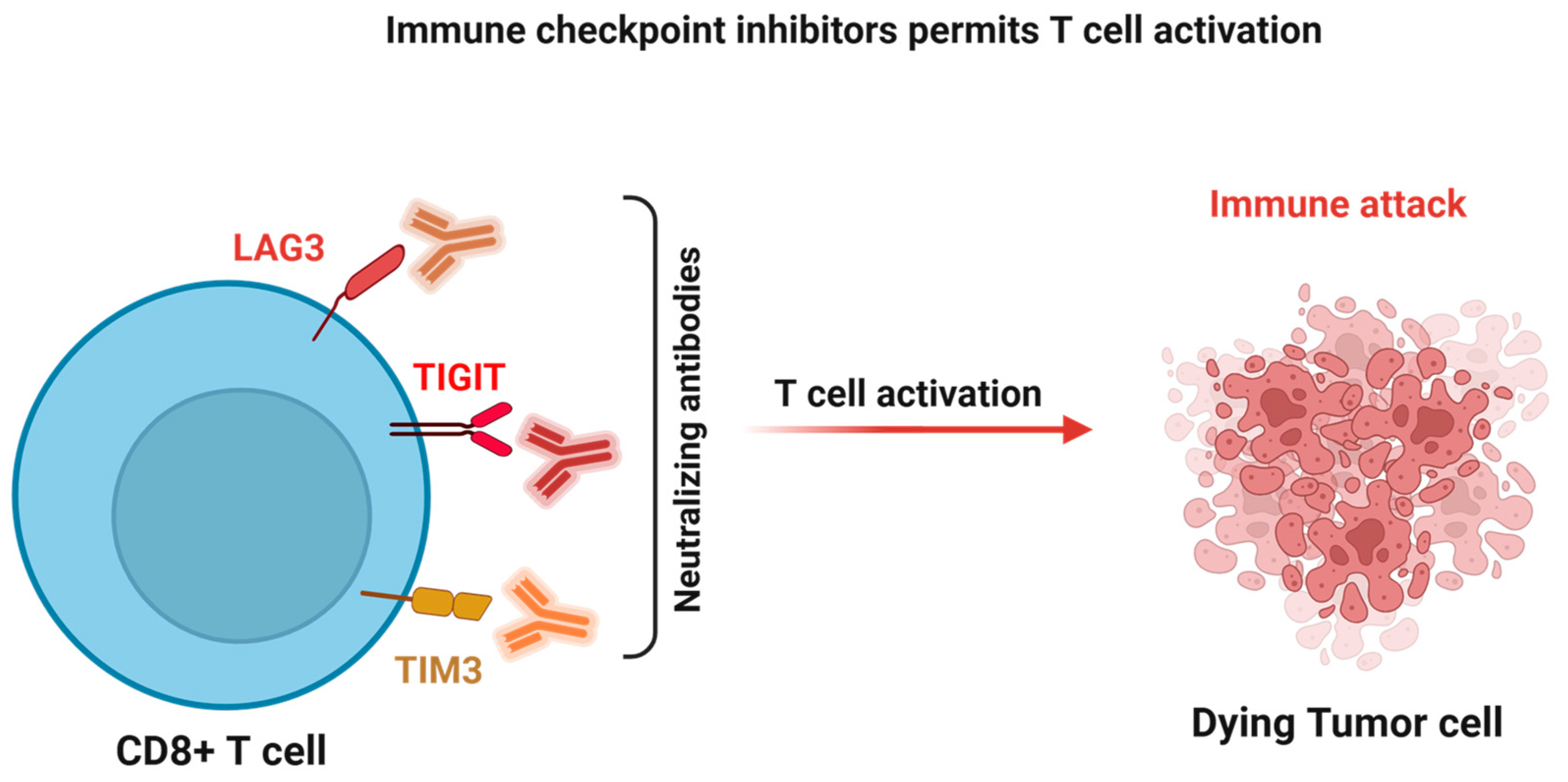
As research moves toward targeting emerging checkpoints like LAG-3 and TIGIT, the goal is to address the non-redundant pathways of immune escape that persist despite standard therapy. Importantly, the therapeutic efficacy of ICIs in this aggressive disease relies on a coordinated and multi-layered antitumor immune response to reinvigorate exhausted effector cells and alleviate immunosuppression. Understanding these intricate molecular mechanisms is quintessential for the rational design of next-generation combination therapies aimed at overcoming resistance and improving long-term clinical outcomes.

3. The Evolving Immunotherapy Landscape in SCLC

The immune system plays a central role in controlling tumor progression as well as distant metastasis; however, tumors such as SCLC often exploit multiple immune checkpoint pathways to evade immune surveillance and clearance. Immune checkpoints are regulatory molecules expressed on immune cells that maintain self-tolerance and prevent excessive immune activation under physiological conditions [37,38,39]. Tumors co-opt these pathways to suppress cytotoxic T-cell activity, limit antigen-specific immune responses, and establish an immunosuppressive TME [40]. The immune landscape of SCLC plays a crucial role in the effectiveness of immune checkpoint therapies [41]. Gaining deeper insights into this microenvironment could facilitate the identification of new therapeutic targets to address the shortcomings of current treatments.
For several decades, the combination of etoposide and platinum was the undisputed standard of care for extensive-stage SCLC. However, the integration of ICIs such as PD-L1 inhibitors has established a new therapeutic benchmark [42,43]. Two landmark phase III trials (IMpower133 and CASPIAN) have redefined the first-line setting, demonstrating that the addition of ICIs to standard chemotherapy significantly extends survival (Table 1). Immunotherapy with ICIs has become part of the therapeutic landscape for ES-SCLC, often in combination with chemotherapy [44]. Immune-related adverse events (irAEs) represent a distinct toxicity profile that differs from traditional therapies [45]. IrAEs develop as a result of immunotherapy with ICIs, and they can affect multiple organ systems, including dermatologic, pulmonary, endocrine, gastrointestinal, and hematologic systems, with sparse but severe events such as myocarditis and pneumonitis reported in SCLC patients treated with ICIs. These toxicities are unpredictable but can be life-threatening, accentuating the need for vigilant monitoring and management in clinical practice [46].
Meta-analyses of randomized controlled trials indicate that adding ICIs to chemotherapy in ES-SCLC can increase the risk of high-grade treatment-related adverse events and treatment discontinuation due to toxicity compared with chemotherapy alone [47]. Despite the growing use of ICIs in SCLC, health-related quality of life (HRQoL) and real-world tolerability data remain limited. Systematic reviews indicate that only a minority of trials include HRQoL measures, and while the addition of ICIs to chemotherapy did not seem to worsen overall symptoms in trial settings, domain-specific comparisons are limited [48]. Real-world evidence also suggests that SCLC histology may be associated with a higher incidence of irAEs compared with other lung cancers [49]. The impact of these events on long-term outcomes such as progression-free survival or overall survival warrants further validation.
Frail patients with SCLC, frequently characterized by older age, comorbidities, or poor performance status, are underrepresented in clinical trials but form a substantial proportion of the real-world treated population [50]. Comorbidity and frailty have been associated with trends toward increased toxicity and higher rates of treatment discontinuation in older patients receiving immunotherapy, although evidence specific to SCLC is sparse. Moreover, frailty is linked to lower quality of life and reduced physical functioning, which may worsen irAEs and complicate their management [51].Future studies that integrate patient-reported outcomes, real-world evidence, and frailty assessments will be essential to optimize immunotherapy strategies and improve supportive care in this vulnerable group.

3.1. The PD-1 and PD-L1 Axis: Biological Rationale

The PD-1/PD-L1 pathway represents the most extensively studied immune checkpoint axis in SCLC. PD-1 is primarily expressed on activated T cells, whereas PD-L1 is often upregulated on tumor cells and antigen-presenting cells within the TME. The engagement of PD-1 by PD-L1 triggers inhibitory signaling that curtails T-cell proliferation, reduces cytokine secretion, and diminishes cytotoxic effector function, thereby facilitating immune escape [52,53,54].
In SCLC, PD-L1 expression levels tend to be lower and exhibit a greater degree of heterogeneity compared to non-small cell lung cancer (NSCLC). This variance underscores the complexities of SCLC and highlights the challenges in developing targeted therapies. This disparity may partially account for the modest clinical responses to PD-1 or PD-L1 blockade in SCLC. Nonetheless, the high tumor mutational burden and the presence of smoking-induced neoantigens provide a compelling mechanistic rationale for targeting this pathway [41,55,56].

3.2. First-Line Immunotherapy in Extensive-Stage SCLC

For several decades, the combination of etoposide and platinum (EP) was the undisputed standard of care for extensive-stage SCLC (ES-SCLC). The integration of ICIs has recently established a new therapeutic benchmark [57,58,59]. Two landmark phase III trials have redefined the first-line setting by demonstrating that the addition of anti-PD-L1 agents to standard chemotherapy significantly improves survival outcomes [60].
IMpower133 (Atezolizumab): This landmark, randomized, double-blind phase III trial enrolled patients with previously untreated ES-SCLC. Participants received carboplatin and etoposide with either atezolizumab or placebo for four cycles, followed by maintenance atezolizumab or placebo. The addition of atezolizumab significantly improved median overall survival (OS) to 12.3 months vs. 10.3 months with chemotherapy alone (HR ≈ 0.70), marking the first meaningful OS benefit in this disease in more than 30 years. Progression-free survival (PFS) was also modestly improved. The survival benefit was maintained with longer follow-up, and toxicity was manageable, with immune-related adverse events consistent with prior atezolizumab experience and no major detriment to quality of life [61].
CASPIAN (Durvalumab): This open-label, randomized phase III trial further validated the chemo-immunotherapy strategy for first-line ES-SCLC patients. Participants received platinum-etoposide (carboplatin or cisplatin) with or without durvalumab, followed by maintenance durvalumab in the experimental arm. Durvalumab plus chemotherapy significantly improved median OS to 12.9 months compared with 10.5 months in the control group, with a durable survival benefit. Median PFS was similar between arms but favored durvalumab numerically. Safety outcomes were acceptable, with no new safety signals and comparable rates of adverse events [62].

3.3. Immunotherapy for Limited-Stage SCLC

The success of ICIs in the extensive-stage setting has prompted significant interest in their application for limited-stage SCLC (LS-SCLC). The clinical landscape for LS-SCLC shifted significantly in 2024 following the publication of the phase III ADRIATIC trial [41,63]. This study demonstrated that durvalumab consolidation therapy after concurrent chemoradiation significantly improved overall survival, leading to U.S. FDA approval for this indication.
However, the integration of immunotherapy into LS-SCLC remains complex. In contrast to the ADRIATIC results, the phase II ACHILES trial, presented at ASCO 2025, found that adding atezolizumab to chemoradiation did not provide a statistically significant improvement in overall survival or progression-free survival [41]. These divergent results suggest that the timing of ICI administration and the specific choice of agent may be critical factors in achieving clinical success in the limited-stage setting.

3.4. Beyond PD-1: CTLA-4 and Next-Generation Checkpoints

CTLA-4 is another critical immune checkpoint that regulates early T-cell activation [64]. While PD-L1 inhibitors have moved into first-line care, they only benefit a subset of patients. To overcome resistance, research has pivoted toward targeting the “spatial and temporal” variety of immune checkpoints. Unlike the PD-1/PD-L1 axis, which primarily functions in the TME during the effector phase, CTLA-4 acts earlier within the secondary lymphoid organs during the priming phase [64,65].
In Tregs, CTLA-4 is constitutively expressed and is essential for suppressive function, acting by limiting costimulatory ligand availability on APCs and maintaining peripheral immune tolerance [64,66]. Beyond its direct inhibitory signaling in T cells, CTLA-4 is unique among immune checkpoints because antibody targeting can physically remove Tregs from the TME. This Treg-depleting effect occurs mainly within the tumor itself and is associated with tumor rejection, because the TME is rich in myeloid cells that express Fcγ receptors [67,68]. Mechanistically, α-CTLA-4 antibodies engage FcγRIV expressed on CD11b+ myeloid cells, triggering antibody-dependent cellular cytotoxicity (ADCC) against CTLA-4 high Tregs [69]. Genetic deletion of FcγRIV restores intratumoral Treg populations and abolishes the antitumor efficacy of α-CTLA-4 therapy, establishing FcγRIV as a dominant mediator of Treg depletion in vivo [70].
Clinical Lessons (CASPIAN Trial): In the CASPIAN study, the addition of tremelimumab (a CTLA-4 inhibitor) to durvalumab and chemotherapy did not significantly improve overall survival compared to durvalumab plus chemo alone [71]. This suggests that in SCLC, simply adding a second “broad” checkpoint inhibitor may increase toxicity without a proportional increase in efficacy. Recent studies are exploring the development of CTLA-4 antibodies with modified Fc regions that are designed to more effectively eliminate regulatory T-cells (Tregs) in tumors. Because Tregs suppress anti-tumor immune responses, their targeted depletion could enhance the activity of immune cells that attack cancer. This approach differs from previous CTLA-4 inhibitors, such as ipilimumab, which block CTLA-4 more broadly and can cause widespread immune side effects. By engineering the Fc portion of the antibody to improve its ability to recruit immune cells that kill Tregs, these next-generation therapies aim to provide stronger anti-tumor effects with fewer systemic toxicities [72].
The limited success of CTLA-4 inhibition in the CASPIAN trial has shifted focus toward checkpoints that may more precisely address the “exhausted” phenotype of T cells specifically found in the SCLC TME.

3.5. TIGIT (T-Cell Immunoglobulin and ITIM Domain)

TIGIT is a prominent co-inhibitory receptor expressed on NK cells and activated T cells. It competes with the costimulatory receptor CD226 for the same ligands (CD155 and CD112) expressed on APCs and tumor cells.
SKYSCRAPER-02 trial: Unlike the promising results seen in non-small cell lung cancer (NSCLC), the Phase III SKYSCRAPER-02 trial showed that adding tiragolumab (α-TIGIT) to atezolizumab and chemotherapy did not improve progression-free survival PFS or OS in ES-SCLC [73].
This study points to potential biological differences between NSCLC and SCLC. This correlation prompted researchers to investigate whether TIGIT blockade is only effective in specific SCLC molecular subtypes, such as the SCLC-I phenotype, which exhibits enhanced immune infiltration.

3.6. LAG-3 (Lymphocyte Activation Gene-3)

LAG-3 is a cell-surface molecule expressed on effector T cells and Tregs. It binds to MHC class II with higher affinity than CD4, leading to the inhibition of T-cell proliferation and cytokine production [74]. SCLC often exhibits low MHC expression as an immune-evasion tactic; however, LAG-3 is frequently co-expressed with PD-1 on tumor-infiltrating lymphocytes in SCLC samples.
Trials are underway for evaluating dual PD-1/LAG-3 blockade (e.g., using relatlimab) based on the synergy seen in melanoma. In SCLC, this combination is being tested primarily in the refractory setting to see if it can “re-prime” the immune response in patients who failed initial PD-L1 therapy [75].

3.7. TIM-3 (T-Cell Immunoglobulin and Mucin-Domain Containing-3)

TIM-3 has emerged as a critical marker of terminal exhaustion within the Small Cell Lung Cancer (SCLC) tumor microenvironment. While early-stage exhausted T cells may still be reinvigorated by PD-1 blockade, cells that co-express both PD-1 and TIM-3 represent a deeply dysfunctional state characterized by a near-total loss of proliferative capacity and cytokine production. TIM-3 interacts with its ligand, Galectin-9, which is often upregulated in the SCLC TME. When Galectin-9 binds to TIM-3 on the surface of T cells, it triggers intracellular calcium flux that induces apoptosis in Th1 and CD8+ effector cells [76].
Clinical evaluation of TIM-3 blockade remains limited. In a phase I/II trial (NCT02608268), the anti-TIM-3 antibody sabatolimab (MBG453) was tested alone and in combination with the anti-PD-1 antibody spartalizumab. Both regimens were generally well tolerated, and a maximum tolerated dose was not reached. No objective responses were observed with sabatolimab monotherapy; however, the combination achieved partial responses in 6% of patients, including one individual with SCLC, with responses lasting up to 27 months [77].
Overall, clinical data supporting TIM-3 inhibition in SCLC, including agents such as cobolimab, are still emerging. Consequently, the therapeutic role of TIM-3–targeted approaches in SCLC remains preliminary and exploratory, with no definitive evidence of efficacy to date.

3.8. B7-H3 (CD276): A High-Priority SCLC Target

B7-H3, a member of the B7 family, is uniquely overexpressed in SCLC, where it correlates with poor prognosis and the promotion of epithelial–mesenchymal transition (EMT). Given its high tumor-to-normal tissue expression ratio, B7-H3 is increasingly targeted through direct cytotoxic modalities rather than simple checkpoint blockade. A prime example is ifinatamab talirine (DS-7300), a B7-H3-directed antibody–drug conjugate (ADC) that has demonstrated impressive early-phase activity in heavily pre-treated SCLC patients [78]. These early results suggest that for specific checkpoints like B7-H3, direct cytotoxicity via an ADC may be more effective than traditional methods of releasing immune “brakes.”
Early and later-phase trials have reported negative efficacy signals in some settings, as well as important safety concerns such as cytokine release syndrome, neurotoxicity, and other limiting toxicities that constrain the therapeutic window of these agents [79]. This trial may have seemingly valuable preclinical data, but these findings do not support the efficacy of this trial. Despite initially compelling data, later findings on the real-world relevance of the trials underscore that not all targeted strategies translate into reliable and durable clinical benefit or acceptable tolerability in heavily pretreated SCLC populations.
Building on this clinical momentum, the IDeate-Lung02 (NCT06203210) trial is a global Phase 3 study evaluating ifinatamab deruxtecan (I-DXd) against the physician’s choice of chemotherapy in 540 adults with relapsed SCLC. IDeate-Lung02 is designed to test whether I-DXd provides greater benefit than commonly used chemotherapies when given after the first relapse of SCLC. The failure of ICIs in many SCLC patients is attributed to the “cold” nature of the tumor, largely driven by myeloid and stromal cells that upregulate checkpoint ligands [80].
Overall, the complex interplay between tumor cells, immune checkpoints, and the surrounding microenvironment defines the immunosuppressive landscape of SCLC, and novel checkpoint inhibitors have the potential to restore T cell activity and enhance anti-tumor immune responses (Figure 2). A mechanistic understanding of these pathways not only provides the rationale for existing immune checkpoint inhibitors but also informs the development of next-generation immunotherapies aimed at overcoming resistance and improving clinical outcomes in this aggressive malignancy.

4. The Evolution Toward Adoptive Cellular Therapies in SCLC

Growing recognition of the limitations of immune checkpoint inhibitors has prompted a shift in the therapeutic approach to SCLC, with increasing attention focused on cellular immunotherapies such as Chimeric Antigen Receptor (CAR) T-cell therapy and bispecific T-cell engagers (BiTEs). Unlike checkpoint blockade, which seeks to revive an often profoundly impaired endogenous immune response, CAR T-cell therapy involves reprogramming a patient’s own T cells to directly recognize and attack malignant cells through engineered receptors that function independently of major histocompatibility complex (MHC) presentation [81]. A major barrier to translating this strategy to SCLC has been the lack of a sufficiently selective tumor antigen, as inadequate specificity raises the risk of damaging healthy tissues. Among the targets investigated, delta-like ligand 3 (DLL3) has emerged as the most compelling, given its high expression in nearly 80% of SCLC tumors and its limited presence in normal tissues [82].
As of 2025, DLL3-targeted CAR T-cell therapies such as LB2102 have shown encouraging dose-dependent antitumor activity in early-phase clinical trials. However, the first cellular immunotherapy to achieve standard-of-care status in SCLC is the bispecific T-cell engager tarlatamab [83]. Tarlatamab exerts its effect by binding CD3 on T cells and DLL3 on tumor cells simultaneously, thereby promoting direct T cell-mediated cytotoxicity. Despite this progress, translating the success of CAR T-cell therapy from hematologic malignancies to solid tumors like SCLC remains difficult due to the immunosuppressive TME and significant antigen heterogeneity [84]. Current research efforts are therefore focused on developing next-generation armored CAR T cells that can better withstand these barriers through mechanisms such as cytokine secretion, including IL-12, or disruption of inhibitory pathways like PD-1. Altogether, combining these enhanced cellular therapies with established ICIs may represent a promising strategy for achieving durable remissions in extensive-stage SCLC.

5. Resistance and Challenges

Despite the paradigm shift brought about by ICIs, the majority of patients with SCLC eventually succumb to disease due to primary or acquired resistance. The “recalcitrant” nature of SCLC is driven by a complex interplay of tumor-intrinsic and extrinsic factors that systematically disable the immune response.

5.1. Tumor-Intrinsic Mechanisms of Escape

A hallmark of SCLC resistance is the downregulation of MHC Class I molecules, which occurs in most cases. This defect renders tumor cells virtually “invisible” to CD8+ cytotoxic T cells, as the lack of MHC-I prevents the presentation of tumor antigens and neoantigens. Furthermore, genomic instability in SCLC leads to antigenic changes and clonal evolution [85]. As a result, even when ICIs eliminate some tumor cells, resistant subclones that no longer express the targeted antigens can survive and eventually cause relapses.

5.2. The Immunosuppressive Microenvironment

The SCLC TME acts as a hostile immunosuppressive barrier that limits the infiltration of tumor-reactive lymphocytes, leading to poor antitumor immune responses. Rapid tumor cell proliferation rate creates a hypoxic, glucose-poor, and nutrient-deprived environment, resulting in the accumulation of lactic acid, thereby acidifying the TME [86]. Lactate enrichment lowers the pH of TME and directly impairs T cell effector functions and cytokine production. In parallel, the microenvironment is abundant with myeloid immunosuppressive cell populations, including M2-polarized tumor-associated macrophages (TAMs) and MDSCs, which are well known to secrete inhibitory cytokines such as TGF-β and IL-10 [87]. The abundance of highly immunosuppressive myeloid cells causes T cell dysfunction and apoptosis and contributes to an immune-excluded or “cold” tumor phenotype. Adding to these challenges, the abnormal and disorganized tumor architecture and vasculature of SCLC restrict the physical penetration of anti-tumor lymphocytes into the tumor core, which limits the efficacy of immunotherapies [88].

5.3. Genetic Deletions and ICI Resistance in SCLC

In SCLC, mutations or deletions in B2M (Beta-2 Microglobulin) and components of the JAK1/2 signaling pathways have emerged as key mechanisms driving primary resistance to ICIs [89]. B2M is critical for the proper assembly of MHC class I molecules, which present tumor antigens to cytotoxic T cells [90]. Loss-of-function mutations or deletions in B2M lead to defective antigen presentation, preventing T cells from recognizing and killing SCLC cells, thereby rendering ICIs ineffective from the outset.
Similarly, alterations in JAK1 or JAK2 disrupt interferon-gamma (IFN-γ) signaling. This pathway is essential for upregulating MHC-I expression and other genes involved in T-cell-mediated cytotoxicity. Mutations in JAK1 or JAK2 disrupt interferon-gamma (IFN-γ) signaling. Tumor cells are unable to respond to IFN-γ, which reduces the expression of PD-L1 and other immune-related genes. This prevents T cells from recognizing and killing the tumor effectively. As a result, tumors with these mutations often show resistance to PD-1–targeted immunotherapy [91].
In SCLC, these defects impair the tumor’s ability to respond to IFN-γ, further limiting T-cell infiltration and antitumor activity. Clinical studies have reported that SCLC tumors harboring such genetic deletions are often intrinsically resistant to PD-1/PD-L1 inhibitors [92], highlighting the need for genomic profiling to guide immunotherapy decisions. These molecular alterations serve as negative biomarkers and serve as markers of resistance rather than response. Additionally, these mutations/deletions help identify patients unlikely to benefit from immunotherapies. These insights suggest that targeting alternative pathways to restore antigen presentation or bypass IFN-γ signaling could enhance immunotherapy efficacy in genetically resistant SCLC, representing a potential strategy to overcome primary ICI resistance in this aggressive disease.

6. Conclusions and Future Directions

While ICIs represent a significant advance in SCLC therapy, limited efficacy underscores the need for improved biomarkers and rational combination strategies. The therapeutic landscape of SCLC has undergone a fundamental shift with the integration of ICIs into the standard of care. After years of minimal therapeutic advancement, a series of pivotal trials has reformed the treatment modalities of SCLC. IMpower133 and CASPIAN clinical trials (Table 1) have transformed care in patients with extensive-stage disease, and the subsequent ADRIATIC trial (Table 1) extended these gains to those with limited-stage SCLC, solidifying immunotherapy as a foundational component of SCLC management. However, SCLC remains a formidable clinical challenge.
Despite its high mutational burden, the disease frequently exhibits a “cold” tumor phenotype, characterized by MHC-I downregulation, profound T cell exhaustion, and a myeloid-rich immunosuppressive microenvironment. The modest survival gains indicate a significant limit in the effectiveness of current PD-1/PD-L1 inhibitors, suggesting that a uniform approach is inadequate for this molecularly diverse malignancy. Overcoming these barriers requires a shift from monotherapy to rational combination strategies. Current research is exploring the use of epigenetic modifiers (like HDAC inhibitors) to restore MHC-I expression and the integration of anti-angiogenic agents to normalize the tumor vasculature.
Moving forward, transitioning from broad-spectrum immunotherapy to precision immuno-oncology is essential. Future research must prioritize the classification of SCLC into A, N, P, and I subtypes to guide personalized therapy, specifically investigating whether “cold” subtypes require priming strategies to enhance immunogenicity. The emergence of targeted modalities, such as the DLL3-targeted bispecific T-cell engager tarlatamab and B7-H3-directed antibody–drug conjugates, offers a promising frontier that bypasses the limitations of traditional checkpoint blockade. Addressing the hostile, highly immunosuppressive SCLC TME through the modulation of immunosuppressive myeloid cells and the engineering of “armored” CAR-T cells will be critical to achieving durable remissions. Integrating composite biomarkers, including HLA-I status and genomic signatures like B2M or JAK1/2 mutations, will be crucial in identifying responders and overcoming the primary and acquired resistance that currently limits the long-term success of immunotherapy in SCLC.
In summary, advances in multi-omics technologies, preclinical modeling, and translational research offer opportunities to develop personalized immunotherapeutic strategies tailored to the unique biology of SCLC. Continued exploration of novel immune checkpoints, neoantigen-targeted therapies, and approaches to enhance antigen presentation holds the potential to improve response rates and long-term outcomes. In conclusion, while ICIs have introduced a new era in SCLC treatment, a deeper molecular understanding and rational combination strategies are essential to fully realize their clinical potential.

Author Contributions

Conceptualization, M.C.; writing—original draft preparation, M.C., T.M. and K.P.; writing—review and editing, M.C., R.K.G., T.M. and K.P.; visualization, M.C., R.K.G., T.M. and K.P.; supervision, M.C.; project administration, M.C.; M.C. compiled and finalized the full manuscript. All authors have read and agreed to the published version of the manuscript.

Funding

This work is supported in part by the Department of Defense Lung Cancer Idea Award (HT942524LCRPIDA) and Lung Cancer Concept Award (LC220065) to R.K.G.

Institutional Review Board Statement

Not applicable. Ethical approval was not required as this study is a review article and does not involve human participants or patient data.

Informed Consent Statement

Not applicable. This study did not involve human subjects or individual patient data.

Data Availability Statement

No new data were created or analyzed in this study. Data sharing is not applicable to this article.

Conflicts of Interest

The authors declare no competing interests.

References

  1. Shalata, W.; Naamneh, R.; Najjar, W.; Asla, M.; Abu Gameh, A.; Abu Amna, M.; Saiegh, L.; Agbarya, A. Current and Emerging Therapeutic Strategies for Limited- and Extensive-Stage Small-Cell Lung Cancer. Med. Sci. 2025, 13, 142. [Google Scholar] [CrossRef] [Scilit]
  2. Schwendenwein, A.; Megyesfalvi, Z.; Barany, N.; Valko, Z.; Bugyik, E.; Lang, C.; Ferencz, B.; Paku, S.; Lantos, A.; Fillinger, J.; et al. Molecular profiles of small cell lung cancer subtypes: Therapeutic implications. Mol. Ther. Oncolytics 2021, 20, 470–483. [Google Scholar] [CrossRef] [Scilit]
  3. Zhang, J.; Zhong, X.; Wang, S.; Wang, L. Advances in Immunotherapy and Chemoradiotherapy Combination for Limited-Stage Small-Cell Lung Cancer: Current Landscape and Future Frontiers. Curr. Oncol. Rep. 2025, 27, 1416–1426. [Google Scholar] [CrossRef] [Scilit] [PubMed]
  4. Oronsky, B.; Abrouk, N.; Caroen, S.; Lybeck, M.; Guo, X.; Wang, X.; Yu, Z.; Reid, T. A 2022 Update on Extensive Stage Small-Cell Lung Cancer (SCLC). J. Cancer 2022, 13, 2945–2953. [Google Scholar] [CrossRef] [Scilit] [PubMed]
  5. Gong, J.; Salgia, R. Managing Patients with Relapsed Small-Cell Lung Cancer. J. Oncol. Pract. 2018, 14, 359–366. [Google Scholar] [CrossRef] [Scilit] [PubMed]
  6. Sun, S.; Liu, L.; Zhang, J.; Sun, L.; Shu, W.; Yang, Z.; Yao, H.; Zhang, Z. The role of neoantigens and tumor mutational burden in cancer immunotherapy: Advances, mechanisms, and perspectives. J. Hematol. Oncol. 2025, 18, 84. [Google Scholar] [CrossRef] [Scilit]
  7. Huang, D.; Wang, J.; Chen, L.; Jiang, W.; Inuzuka, H.; Simon, D.K.; Wei, W. Molecular Subtypes and Targeted Therapeutic Strategies in Small Cell Lung Cancer: Advances, Challenges, and Future Perspectives. Molecules 2025, 30, 1731. [Google Scholar] [CrossRef] [Scilit]
  8. Wu, B.; Zhang, B.; Li, B.; Wu, H.; Jiang, M. Cold and hot tumors: From molecular mechanisms to targeted therapy. Signal Transduct. Target. Ther. 2024, 9, 274. [Google Scholar] [CrossRef] [Scilit]
  9. Park, J.; Skalhegg, B.S. Combination of PD-1/PD-L1 and CTLA-4 inhibitors in the treatment of cancer—A brief update. Front. Immunol. 2025, 16, 1680838. [Google Scholar] [CrossRef] [Scilit]
  10. Huang, X.; Tian, B.; Ren, Z.; Zhang, J.; Yan, W.; Mo, Y.; Yuan, J.; Ma, Y.; Wang, R.; Liu, R.; et al. CD34 as a potential prognostic indicator for camrelizumab response in advanced non-small-cell lung cancer: Insights from digital spatial profiling. Ther. Adv. Med. Oncol. 2024, 16, 17588359241289671. [Google Scholar] [CrossRef] [Scilit]
  11. Caliman, E.; Fancelli, S.; Petroni, G.; Gatta Michelet, M.R.; Cosso, F.; Ottanelli, C.; Mazzoni, F.; Voltolini, L.; Pillozzi, S.; Antonuzzo, L. Challenges in the treatment of small cell lung cancer in the era of immunotherapy and molecular classification. Lung Cancer 2023, 175, 88–100. [Google Scholar] [CrossRef] [Scilit]
  12. Zhu, Y.; Wu, J.; Wang, H.; Chi, K.; Diao, X.; Zhuo, M.; Lin, D. Whole-section digital analysis of immune profiles in surgically resected small cell lung carcinoma and their associations with molecular subtypes. Transl. Lung Cancer Res. 2025, 14, 449–466. [Google Scholar] [CrossRef] [Scilit]
  13. Zhu, L.; Qin, J. Predictive biomarkers for immunotherapy response in extensive-stage SCLC. J. Cancer Res. Clin. Oncol. 2024, 150, 22. [Google Scholar] [CrossRef] [Scilit]
  14. Dong, X.; Yao, X.; Li, R.; Li, Y.; Li, Y. Comparison of efficacy and safety of first-line immunotherapy combined with chemotherapy in extensive-stage small cell lung cancer, a retrospective study. Sci. Rep. 2025, 15, 40167. [Google Scholar] [CrossRef] [Scilit]
  15. Han, Y.; Wang, J.; Sun, T.; Ouyang, Q.; Li, J.; Yuan, J.; Xu, B. Predictive biomarkers of response and survival following immunotherapy with a PD-L1 inhibitor benmelstobart (TQB2450) and antiangiogenic therapy with a VEGFR inhibitor anlotinib for pretreated advanced triple negative breast cancer. Signal Transduct. Target. Ther. 2023, 8, 429. [Google Scholar] [CrossRef] [Scilit]
  16. Meder, L.; Orschel, C.I.; Bouchez, C.L.; Gholamipoorfard, R.; Orschel, C.V.; Stahl, D.; Kreer, C.; Koker, M.; Nill, M.; Kocak, I.G.; et al. ERBB2 signaling drives immune cell evasion and resistance against immunotherapy in small cell lung cancer. Nat. Commun. 2025, 16, 10983. [Google Scholar] [CrossRef] [Scilit]
  17. Ricciuti, B.; Kravets, S.; Dahlberg, S.E.; Umeton, R.; Albayrak, A.; Subegdjo, S.J.; Johnson, B.E.; Nishino, M.; Sholl, L.M.; Awad, M.M. Use of targeted next generation sequencing to characterize tumor mutational burden and efficacy of immune checkpoint inhibition in small cell lung cancer. J. Immunother. Cancer 2019, 7, 87. [Google Scholar] [CrossRef] [Scilit]
  18. Chen, T.; Wang, M.; Chen, Y.; Cao, Y.; Liu, Y. Advances in predictive biomarkers associated with immunotherapy in extensive-stage small cell lung cancer. Cell Biosci. 2024, 14, 117. [Google Scholar] [CrossRef] [Scilit]
  19. Liu, Z.; Zhou, Z.; Dang, Q.; Xu, H.; Lv, J.; Li, H.; Han, X. Immunosuppression in tumor immune microenvironment and its optimization from CAR-T cell therapy. Theranostics 2022, 12, 6273–6290. [Google Scholar] [CrossRef] [Scilit]
  20. Li, X.; Li, Y.; Tuerxun, H.; Zhao, Y.; Liu, X.; Zhao, Y. Firing up “cold” tumors: Ferroptosis causes immune activation by improving T cell infiltration. Biomed. Pharmacother. 2024, 179, 117298. [Google Scholar] [CrossRef] [Scilit]
  21. Wang, L.; Geng, H.; Liu, Y.; Liu, L.; Chen, Y.; Wu, F.; Liu, Z.; Ling, S.; Wang, Y.; Zhou, L. Hot and cold tumors: Immunological features and the therapeutic strategies. MedComm 2023, 4, e343. [Google Scholar] [CrossRef] [Scilit]
  22. Khosravi, G.R.; Mostafavi, S.; Bastan, S.; Ebrahimi, N.; Gharibvand, R.S.; Eskandari, N. Immunologic tumor microenvironment modulators for turning cold tumors hot. Cancer Commun. 2024, 44, 521–553. [Google Scholar] [CrossRef] [Scilit]
  23. Aliazis, K.; Christofides, A.; Shah, R.; Yeo, Y.Y.; Jiang, S.; Charest, A.; Boussiotis, V.A. The tumor microenvironment’s role in the response to immune checkpoint blockade. Nat. Cancer 2025, 6, 924–937. [Google Scholar] [CrossRef] [Scilit]
  24. Wellenstein, M.D.; de Visser, K.E. Cancer-Cell-Intrinsic Mechanisms Shaping the Tumor Immune Landscape. Immunity 2018, 48, 399–416. [Google Scholar] [CrossRef] [Scilit]
  25. Ren, X.; Guo, S.; Guan, X.; Kang, Y.; Liu, J.; Yang, X. Immunological Classification of Tumor Types and Advances in Precision Combination Immunotherapy. Front. Immunol. 2022, 13, 790113. [Google Scholar] [CrossRef] [Scilit]
  26. Arasanz, H.; Gato-Canas, M.; Zuazo, M.; Ibanez-Vea, M.; Breckpot, K.; Kochan, G.; Escors, D. PD1 signal transduction pathways in T cells. Oncotarget 2017, 8, 51936–51945. [Google Scholar] [CrossRef] [Scilit]
  27. Xu-Monette, Z.Y.; Zhang, M.; Li, J.; Young, K.H. PD-1/PD-L1 Blockade: Have We Found the Key to Unleash the Antitumor Immune Response? Front. Immunol. 2017, 8, 1597. [Google Scholar] [CrossRef] [Scilit]
  28. Burke, K.P.; Chaudhri, A.; Freeman, G.J.; Sharpe, A.H. The B7:CD28 family and friends: Unraveling coinhibitory interactions. Immunity 2024, 57, 223–244. [Google Scholar] [CrossRef] [Scilit]
  29. Herbst, R.S.; Soria, J.C.; Kowanetz, M.; Fine, G.D.; Hamid, O.; Gordon, M.S.; Sosman, J.A.; McDermott, D.F.; Powderly, J.D.; Gettinger, S.N.; et al. Predictive correlates of response to the anti-PD-L1 antibody MPDL3280A in cancer patients. Nature 2014, 515, 563–567. [Google Scholar] [CrossRef] [Scilit]
  30. Carvajal-Hausdorf, D.; Altan, M.; Velcheti, V.; Gettinger, S.N.; Herbst, R.S.; Rimm, D.L.; Schalper, K.A. Expression and clinical significance of PD-L1, B7-H3, B7-H4 and TILs in human small cell lung Cancer (SCLC). J. Immunother. Cancer 2019, 7, 65. [Google Scholar] [CrossRef] [Scilit]
  31. Zhang, Q.; Wang, G.; Yan, W.; Wang, D.; Yin, J.; Song, Y.; Ye, M.; Lv, T. Molecular subtyping dictates therapeutic response to anti-PD-L1 immunotherapy in ES-SCLC. Cancer Immunol. Immunother. 2025, 74, 213. [Google Scholar] [CrossRef] [Scilit]
  32. Patel, S.P.; Kurzrock, R. PD-L1 Expression as a Predictive Biomarker in Cancer Immunotherapy. Mol. Cancer Ther. 2015, 14, 847–856. [Google Scholar] [CrossRef] [Scilit]
  33. Toki, M.I.; Mani, N.; Smithy, J.W.; Liu, Y.; Altan, M.; Wasserman, B.; Tuktamyshov, R.; Schalper, K.; Syrigos, K.N.; Rimm, D.L. Immune Marker Profiling and Programmed Death Ligand 1 Expression Across NSCLC Mutations. J. Thorac. Oncol. 2018, 13, 1884–1896. [Google Scholar] [CrossRef] [Scilit]
  34. Ullah, A.; Pulliam, S.; Karki, N.R.; Khan, J.; Jogezai, S.; Sultan, S.; Muhammad, L.; Khan, M.; Jamil, N.; Waheed, A.; et al. PD-L1 Over-Expression Varies in Different Subtypes of Lung Cancer: Will This Affect Future Therapies? Clin. Pract. 2022, 12, 653–671. [Google Scholar] [CrossRef] [Scilit]
  35. Acheampong, E.; Abed, A.; Morici, M.; Bowyer, S.; Amanuel, B.; Lin, W.; Millward, M.; Gray, E.S. Tumour PD-L1 Expression in Small-Cell Lung Cancer: A Systematic Review and Meta-Analysis. Cells 2020, 9, 2393. [Google Scholar] [CrossRef] [Scilit]
  36. Chen, Y.; Li, H.; Fan, Y. Shaping the tumor immune microenvironment of SCLC: Mechanisms, and opportunities for immunotherapy. Cancer Treat. Rev. 2023, 120, 102606. [Google Scholar] [CrossRef] [Scilit]
  37. Kang, L.P.; Huang, H.J.; Xu, C.; Chen, H.H.; Huang, D.H.; Jiang, Z.B. Breakthroughs in immune checkpoint therapy: Overcoming NSCLC immune checkpoint therapy resistance with novel techniques. Front. Immunol. 2025, 16, 1630940. [Google Scholar] [CrossRef] [Scilit]
  38. Wang, H.; Niu, X.; Jin, Z.; Zhang, S.; Fan, R.; Xiao, H.; Hu, S.S. Immunotherapy resistance in non-small cell lung cancer: From mechanisms to therapeutic opportunities. J. Exp. Clin. Cancer Res. 2025, 44, 250. [Google Scholar] [CrossRef] [Scilit]
  39. Meng, L.; Wu, H.; Wu, J.; Ding, P.; He, J.; Sang, M.; Liu, L. Mechanisms of immune checkpoint inhibitors: Insights into the regulation of circular RNAS involved in cancer hallmarks. Cell Death Dis. 2024, 15, 3. [Google Scholar] [CrossRef] [Scilit]
  40. Cao, P.; Sun, Z.; Zhang, F.; Zhang, J.; Zheng, X.; Yu, B.; Zhao, Y.; Wang, W.; Wang, W. TGF-beta Enhances Immunosuppression of Myeloid-Derived Suppressor Cells to Induce Transplant Immune Tolerance Through Affecting Arg-1 Expression. Front. Immunol. 2022, 13, 919674. [Google Scholar] [CrossRef] [Scilit]
  41. Zhong, J.; Jie, G.; Qin, H.; Li, H.; Chen, N.; Aerxiding, P.; Zou, X.; Niu, X. Immunotherapy for small cell lung cancer: Current challenges and prospects. Exp. Hematol. Oncol. 2025, 14, 130. [Google Scholar] [CrossRef] [Scilit]
  42. Chen, Y.; Shang, H.; Yang, Y.; Wang, Q.; Gao, X.; Huang, G. Efficacy and safety of immune checkpoint inhibitors plus platinum-etoposide vs. platinum-etoposide in the first-line treatment of extensive-stage small cell lung cancer: A systematic review and a meta-analysis. Transl. Cancer Res. 2024, 13, 4146–4158. [Google Scholar] [CrossRef] [Scilit]
  43. Sands, J.; Subramanian, J. Treating patients with platinum-sensitive extensive-stage small-cell lung cancer in a real-world setting. Front. Oncol. 2023, 13, 1161931. [Google Scholar] [CrossRef] [Scilit]
  44. Chi, X.; Dong, Y.; Zhu, L.; Su, D.; Wu, H. Expanding the immunotherapy universe in extensive-stage small cell lung cancer: From chemoimmunotherapy backbone to next-wave combinations. Front. Immunol. 2025, 16, 1693401. [Google Scholar] [CrossRef] [Scilit]
  45. Yin, Q.; Wu, L.; Han, L.; Zheng, X.; Tong, R.; Li, L.; Bai, L.; Bian, Y. Immune-related adverse events of immune checkpoint inhibitors: A review. Front. Immunol. 2023, 14, 1167975. [Google Scholar] [CrossRef] [Scilit]
  46. Fletcher, K.; Johnson, D.B. Chronic immune-related adverse events arising from immune checkpoint inhibitors: An update. J. Immunother. Cancer 2024, 12, e008591. [Google Scholar] [CrossRef] [Scilit]
  47. Longo, V.; Rizzo, A.; Catino, A.; Montrone, M.; Galetta, D. Safety evaluation of immune checkpoint inhibitors combined with chemotherapy for the treatment of small cell lung cancer: A meta-analysis of randomized controlled trials. Thorac. Cancer 2023, 14, 1029–1035. [Google Scholar] [CrossRef] [Scilit]
  48. Dunker, A.M.; Malik, N.; Krause, K.J.; Keung, E.Z.; Liu, J.B.; Nassif Haddad, E.F.; Somaiah, N.; Lyu, H.G.; Roland, C.L. Health-Related Quality of Life in the Era of Immune Checkpoint Blockade: What Do Patient-Reported Outcomes Reveal? Cancers 2025, 17, 3917. [Google Scholar] [CrossRef] [Scilit]
  49. Hu, X.; Rodday, A.M.; Gurinovich, A.; Pan, S.; Salei, Y.V.; Lin, J.H.; Byrne, M.M.; Cao, Y.; Pai, L.; Parsons, S.K. Real-world data of immune-related adverse events in lung cancer patients receiving immune-checkpoint inhibitors. Immunotherapy 2025, 17, 321–329. [Google Scholar] [CrossRef] [Scilit]
  50. Damiano, P.; Stefani, A.; Avancini, A.; Belluomini, L.; Bria, E.; Pilotto, S. Real-world evidence in extensive disease small cell lung cancer: The missing piece of the puzzle. Crit. Rev. Oncol. Hematol. 2025, 207, 104618. [Google Scholar] [CrossRef] [Scilit]
  51. Ozkan, A.; de Joode, K.; Kapiteijn, E.; Slingerland, M.; Zunder, S.; van den Bos, F.; Mooijaart, S.; Uit den Boogaard, A.; Trompet, S.; Westgeest, H.; et al. Impact of geriatric impairments on outcomes of single-agent immunotherapy in solid tumors. Int. J. Cancer 2026, 158, 1370–1382. [Google Scholar] [CrossRef] [Scilit]
  52. Javed, S.A.; Najmi, A.; Ahsan, W.; Zoghebi, K. Targeting PD-1/PD-L-1 immune checkpoint inhibition for cancer immunotherapy: Success and challenges. Front. Immunol. 2024, 15, 1383456. [Google Scholar] [CrossRef] [Scilit]
  53. Jiang, X.; Wang, J.; Deng, X.; Xiong, F.; Ge, J.; Xiang, B.; Wu, X.; Ma, J.; Zhou, M.; Li, X.; et al. Role of the tumor microenvironment in PD-L1/PD-1-mediated tumor immune escape. Mol. Cancer 2019, 18, 10. [Google Scholar] [CrossRef] [Scilit]
  54. Lin, X.; Kang, K.; Chen, P.; Zeng, Z.; Li, G.; Xiong, W.; Yi, M.; Xiang, B. Regulatory mechanisms of PD-1/PD-L1 in cancers. Mol. Cancer 2024, 23, 108. [Google Scholar] [CrossRef] [Scilit]
  55. Regzedmaa, O.; Zhang, H.; Liu, H.; Chen, J. Immune checkpoint inhibitors for small cell lung cancer: Opportunities and challenges. Onco Targets Ther. 2019, 12, 4605–4620. [Google Scholar] [CrossRef] [Scilit]
  56. Zheng, S.; Cao, Y.; Randall, J.; Yu, H.; Thomas, T.O. Integrating POLE/POLD1 mutated for immunotherapy treatment planning of advanced stage non-small cell lung cancer. Thorac. Cancer 2023, 14, 2269–2274. [Google Scholar] [CrossRef] [Scilit]
  57. Sathiyapalan, A.; Febbraro, M.; Pond, G.R.; Ellis, P.M. Chemo-Immunotherapy in First Line Extensive Stage Small Cell Lung Cancer (ES-SCLC): A Systematic Review and Meta-Analysis. Curr. Oncol. 2022, 29, 9046–9065. [Google Scholar] [CrossRef] [Scilit]
  58. Lazzari, C.; Mirabile, A.; Bulotta, A.; Vigano, M.G.; Ogliari, F.R.; Ippati, S.; DellOca, I.; Santarpia, M.; Lorusso, V.; Reck, M.; et al. History of Extensive Disease Small Cell Lung Cancer Treatment: Time to Raise the Bar? A Review of the Literature. Cancers 2021, 13, 998. [Google Scholar] [CrossRef] [Scilit] [PubMed]
  59. Li, J.; Fu, T.; Wen, Z.; Liang, J.; Qiu, Y.; Li, K.; Yang, J.; Tong, Y.; Cai, H. Advances in the Use of Immune Checkpoint Inhibitors for Colorectal Cancer Treatment. Onco Targets Ther. 2025, 18, 1159–1168. [Google Scholar] [CrossRef] [Scilit] [PubMed]
  60. Xu, Y.; Zhong, B.; Yu, C.; Hou, Q.; Chen, W.; Zheng, W.; Zhang, W.; Zhou, T. The benefits and risks of adding PD-1/PD-L1 inhibitors to chemotherapy for stage IIIb-IV non-small-cell lung cancer: An updated meta-analysis based on phase 3 randomized controlled trials. Front. Oncol. 2025, 15, 1590017. [Google Scholar] [CrossRef] [Scilit] [PubMed]
  61. Liu, S.V.; Reck, M.; Mansfield, A.S.; Mok, T.; Scherpereel, A.; Reinmuth, N.; Garassino, M.C.; De Castro Carpeno, J.; Califano, R.; Nishio, M.; et al. Updated Overall Survival and PD-L1 Subgroup Analysis of Patients With Extensive-Stage Small-Cell Lung Cancer Treated With Atezolizumab, Carboplatin, and Etoposide (IMpower133). J. Clin. Oncol. 2021, 39, 619–630. [Google Scholar] [CrossRef] [Scilit]
  62. Paz-Ares, L.; Chen, Y.; Reinmuth, N.; Hotta, K.; Trukhin, D.; Statsenko, G.; Hochmair, M.J.; Ozguroglu, M.; Ji, J.H.; Garassino, M.C.; et al. Durvalumab, with or without tremelimumab, plus platinum-etoposide in first-line treatment of extensive-stage small-cell lung cancer: 3-year overall survival update from CASPIAN. ESMO Open 2022, 7, 100408. [Google Scholar] [CrossRef] [Scilit]
  63. Cheng, Y.; Spigel, D.R.; Cho, B.C.; Laktionov, K.K.; Fang, J.; Chen, Y.; Zenke, Y.; Lee, K.H.; Wang, Q.; Navarro, A.; et al. Durvalumab after Chemoradiotherapy in Limited-Stage Small-Cell Lung Cancer. N. Engl. J. Med. 2024, 391, 1313–1327. [Google Scholar] [CrossRef] [Scilit]
  64. Hossen, M.M.; Ma, Y.; Yin, Z.; Xia, Y.; Du, J.; Huang, J.Y.; Huang, J.J.; Zou, L.; Ye, Z.; Huang, Z. Current understanding of CTLA-4: From mechanism to autoimmune diseases. Front. Immunol. 2023, 14, 1198365. [Google Scholar] [CrossRef] [Scilit] [PubMed]
  65. Lippman, S.M.; Abate-Shen, C.; Colbert Maresso, K.L.; Colditz, G.A.; Dannenberg, A.J.; Davidson, N.E.; Disis, M.L.; DuBois, R.N.; Szabo, E.; Giuliano, A.R.; et al. AACR White Paper: Shaping the Future of Cancer Prevention—A Roadmap for Advancing Science and Public Health. Cancer Prev. Res. 2018, 11, 735–778. [Google Scholar] [CrossRef] [Scilit]
  66. Wing, K.; Onishi, Y.; Prieto-Martin, P.; Yamaguchi, T.; Miyara, M.; Fehervari, Z.; Nomura, T.; Sakaguchi, S. CTLA-4 control over Foxp3+ regulatory T cell function. Science 2008, 322, 271–275. [Google Scholar] [CrossRef] [Scilit] [PubMed]
  67. Quezada, S.A.; Peggs, K.S.; Simpson, T.R.; Shen, Y.; Littman, D.R.; Allison, J.P. Limited tumor infiltration by activated T effector cells restricts the therapeutic activity of regulatory T cell depletion against established melanoma. J. Exp. Med. 2008, 205, 2125–2138. [Google Scholar] [CrossRef] [Scilit] [PubMed]
  68. Griffiths, R.W.; Elkord, E.; Gilham, D.E.; Ramani, V.; Clarke, N.; Stern, P.L.; Hawkins, R.E. Frequency of regulatory T cells in renal cell carcinoma patients and investigation of correlation with survival. Cancer Immunol. Immunother. 2007, 56, 1743–1753. [Google Scholar] [CrossRef] [Scilit]
  69. Nimmerjahn, F.; Ravetch, J.V. Fcgamma receptors as regulators of immune responses. Nat. Rev. Immunol. 2008, 8, 34–47. [Google Scholar] [CrossRef] [Scilit]
  70. Nimmerjahn, F.; Lux, A.; Albert, H.; Woigk, M.; Lehmann, C.; Dudziak, D.; Smith, P.; Ravetch, J.V. FcgammaRIV deletion reveals its central role for IgG2a and IgG2b activity in vivo. Proc. Natl. Acad. Sci. USA 2010, 107, 19396–19401. [Google Scholar] [CrossRef] [Scilit]
  71. Goldman, J.W.; Dvorkin, M.; Chen, Y.; Reinmuth, N.; Hotta, K.; Trukhin, D.; Statsenko, G.; Hochmair, M.J.; Ozguroglu, M.; Ji, J.H.; et al. Durvalumab, with or without tremelimumab, plus platinum-etoposide versus platinum-etoposide alone in first-line treatment of extensive-stage small-cell lung cancer (CASPIAN): Updated results from a randomised, controlled, open-label, phase 3 trial. Lancet Oncol. 2021, 22, 51–65. [Google Scholar] [CrossRef] [Scilit] [PubMed]
  72. Chand, D.; Savitsky, D.A.; Krishnan, S.; Mednick, G.; Delepine, C.; Garcia-Broncano, P.; Soh, K.T.; Wu, W.; Wilkens, M.K.; Udartseva, O.; et al. Botensilimab, an Fc-Enhanced Anti-CTLA-4 Antibody, Is Effective against Tumors Poorly Responsive to Conventional Immunotherapy. Cancer Discov. 2024, 14, 2407–2429. [Google Scholar] [CrossRef] [Scilit] [PubMed]
  73. Rudin, C.M.; Liu, S.V.; Soo, R.A.; Lu, S.; Hong, M.H.; Lee, J.S.; Bryl, M.; Dumoulin, D.W.; Rittmeyer, A.; Chiu, C.H.; et al. SKYSCRAPER-02: Tiragolumab in Combination with Atezolizumab Plus Chemotherapy in Untreated Extensive-Stage Small-Cell Lung Cancer. J. Clin. Oncol. 2024, 42, 324–335. [Google Scholar] [CrossRef] [Scilit]
  74. Mariuzza, R.A.; Shahid, S.; Karade, S.S. The immune checkpoint receptor LAG3: Structure, function, and target for cancer immunotherapy. J. Biol. Chem. 2024, 300, 107241. [Google Scholar] [CrossRef] [Scilit]
  75. Qiu, X.; Yu, Z.; Lu, X.; Jin, X.; Zhu, J.; Zhang, R. PD-1 and LAG-3 dual blockade: Emerging mechanisms and potential therapeutic prospects in cancer. Cancer Biol. Med. 2024, 21, 970–976. [Google Scholar] [CrossRef] [Scilit]
  76. Chen, C.; Zhao, F.; Peng, J.; Zhao, D.; Xu, L.; Li, H.; Ma, S.; Peng, X.; Sheng, X.; Sun, Y.; et al. Soluble Tim-3 serves as a tumor prognostic marker and therapeutic target for CD8+ T cell exhaustion and anti-PD-1 resistance. Cell Rep. Med. 2024, 5, 101686. [Google Scholar] [CrossRef] [Scilit]
  77. Curigliano, G.; Gelderblom, H.; Mach, N.; Doi, T.; Tai, D.; Forde, P.M.; Sarantopoulos, J.; Bedard, P.L.; Lin, C.C.; Hodi, F.S.; et al. Phase I/Ib Clinical Trial of Sabatolimab, an Anti-TIM-3 Antibody, Alone and in Combination with Spartalizumab, an Anti-PD-1 Antibody, in Advanced Solid Tumors. Clin. Cancer Res. 2021, 27, 3620–3629. [Google Scholar] [CrossRef] [Scilit] [PubMed]
  78. Fabrizio, F.P.; Muscarella, L.A.; Rossi, A. B7-H3/CD276 and small-cell lung cancer: What’s new? Transl. Oncol. 2024, 39, 101801. [Google Scholar] [CrossRef] [Scilit]
  79. Morris, E.C.; Neelapu, S.S.; Giavridis, T.; Sadelain, M. Cytokine release syndrome and associated neurotoxicity in cancer immunotherapy. Nat. Rev. Immunol. 2022, 22, 85–96. [Google Scholar] [CrossRef] [Scilit]
  80. Owonikoko, T.K.; Byers, L.; Cheng, Y.; Hayashi, H.; Paz-Ares, L.; Perol, M.; Hu, H.; Qian, M.; Garcia, C.R.; Godard, J.; et al. IDeate-Lung02: A Phase 3 study of second-line ifinatamab deruxtecan in patients with relapsed small cell lung cancer. Future Oncol. 2025, 21, 3275–3282. [Google Scholar] [CrossRef] [Scilit]
  81. Zhang, W.Y.; Yang, L.Y.; Fan, X.X. CAR-T therapy-based innovations in the enhancement of contemporary anti-tumor therapies. Front. Immunol. 2025, 16, 1622433. [Google Scholar] [CrossRef] [Scilit]
  82. Rudin, C.M.; Reck, M.; Johnson, M.L.; Blackhall, F.; Hann, C.L.; Yang, J.C.; Bailis, J.M.; Bebb, G.; Goldrick, A.; Umejiego, J.; et al. Emerging therapies targeting the delta-like ligand 3 (DLL3) in small cell lung cancer. J. Hematol. Oncol. 2023, 16, 66. [Google Scholar] [CrossRef] [Scilit]
  83. Mullard, A. FDA approves first DLL3 × CD3 bispecific T-cell engager for lung cancer. Nat. Rev. Drug Discov. 2024, 23, 487. [Google Scholar] [CrossRef] [Scilit] [PubMed]
  84. Tang, D.; Kang, R. Tarlatamab: The promising immunotherapy on its way from the lab to the clinic. Transl. Lung Cancer Res. 2023, 12, 1355–1357. [Google Scholar] [CrossRef] [Scilit]
  85. Zhao, K.; Huang, Y.; Chang, L.; Wang, B.; Ye, M.; Qi, J. Rewiring tumor visibility: The immunopeptidome as a dynamic interface between antigen processing, microenvironmental stress, and immune recognition. Front. Oncol. 2025, 15, 1691719. [Google Scholar] [CrossRef] [Scilit]
  86. Chen, Y.; Jin, Y.; Hu, X.; Chen, M. Infiltrating T lymphocytes in the tumor microenvironment of small cell lung cancer: A state of knowledge review. J. Cancer Res. Clin. Oncol. 2022, 148, 881–895. [Google Scholar] [CrossRef] [Scilit]
  87. Basak, U.; Sarkar, T.; Mukherjee, S.; Chakraborty, S.; Dutta, A.; Dutta, S.; Nayak, D.; Kaushik, S.; Das, T.; Sa, G. Tumor-associated macrophages: An effective player of the tumor microenvironment. Front. Immunol. 2023, 14, 1295257. [Google Scholar] [CrossRef] [Scilit]
  88. Liu, Y.T.; Wang, Y.L.; Wang, S.; Li, J.J.; He, W.; Fan, X.J.; Wan, X.B. Turning cold tumors into hot tumors to ignite immunotherapy. Mol. Cancer 2025, 24, 254. [Google Scholar] [CrossRef] [Scilit] [PubMed]
  89. Nie, Y.; Schalper, K.A.; Chiang, A. Mechanisms of immunotherapy resistance in small cell lung cancer. Cancer Drug Resist. 2024, 7, 55. [Google Scholar] [CrossRef] [Scilit] [PubMed]
  90. Zaretsky, J.M.; Garcia-Diaz, A.; Shin, D.S.; Escuin-Ordinas, H.; Hugo, W.; Hu-Lieskovan, S.; Torrejon, D.Y.; Abril-Rodriguez, G.; Sandoval, S.; Barthly, L.; et al. Mutations Associated with Acquired Resistance to PD-1 Blockade in Melanoma. N. Engl. J. Med. 2016, 375, 819–829. [Google Scholar] [CrossRef] [Scilit]
  91. Shin, D.S.; Zaretsky, J.M.; Escuin-Ordinas, H.; Garcia-Diaz, A.; Hu-Lieskovan, S.; Kalbasi, A.; Grasso, C.S.; Hugo, W.; Sandoval, S.; Torrejon, D.Y.; et al. Primary Resistance to PD-1 Blockade Mediated by JAK1/2 Mutations. Cancer Discov. 2017, 7, 188–201. [Google Scholar] [CrossRef] [Scilit] [PubMed]
  92. Lim, J.U.; Kang, H.S. A narrative review of current and potential prognostic biomarkers for immunotherapy in small-cell lung cancer. Ann. Transl. Med. 2021, 9, 809. [Google Scholar] [CrossRef] [Scilit] [PubMed]
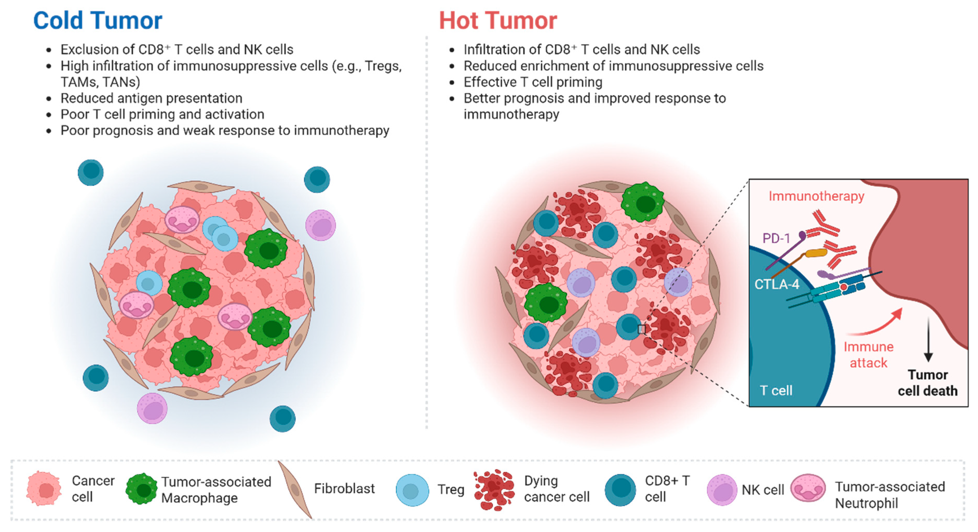
Figure 1. ICIs can reshape cold TMEs into hot TMEs. Hot TME is characterized by abundant cytotoxic T cells and pro-inflammatory signaling, whereas cold TME shows limited immune infiltration and high levels of immunosuppressive factors. By converting cold TME into hot, ICIs enhance T-cell activity and promote cancer cell killing. Created in BioRender. Charan, M. (2026) https://BioRender.com/lraiwjd (accessed on 26 January 2026).
Onco 06 00010 g001
Figure 2. Checkpoint receptor inhibition restores T cell anti-tumor activity. Monoclonal antibodies targeting inhibitory receptors such as LAG-3, TIM-3, and TIGIT block their suppressive signaling on T cells. This releases the inhibitory “brakes,” allowing T cells to become activated, proliferate, and directly kill tumor cells. Created in BioRender. Charan, M. (2026) https://BioRender.com/1thnwue (accessed on 26 January 2026).
Onco 06 00010 g002
Table 1. Clinical Trials: ICI Alone or in Combination for ES-SCLC.
Trial Name, NCT #Agent(s)PhaseStatusPrimary EndpointsHeterogeneitySafety/Toxicity
IMpower133, NCT 02763579Atezolizumab + ChemoIIICompletedAtezolizumab + EP
significantly improved:
PFS: HR 0.77; 95% CI
0.62–0.96; p = 0.02
OS: HR
0.76; 95% CI 0.60–0.95, p = 0.0154
Patients were enrolled from: U.S., Poland, Czech Republic, Hong Kong, Japan, Austria, GermanyNE tumors with high T-cells and low TAMs: greater survival benefit from atezolizumab + carboplatin/etoposide.
Non-NE tumors with high TAMs and high T-cell: less benefit from immunotherapy.
CASPIAN, NCT 03043872Durvalumab ± ChemoIIICompletedDurvalumab + EP
significantly
improved
OS: HR 0.71, 95% CI 0.60–
0.86, p = 0.0003
Patients were enrolled from various countries around Europe, Asia, and the Americas Durvalumab + EP offers the most favorable balance between:
survivability,
toxicity, and
low immune-related adverse event rates.
ASTRIUM-005,
NCT 04063163
Serplulimab + EPIIICompletedSerplulimab + EP
significantly
improved
OS: HR 0.63, 95% CI 0.49
Patients were enrolled from:
China, Georgia,
Poland, Russia,
Turkey, and Ukraine.
Randomized and Double-Blind study
A longer survival and better tumor response than chemotherapy alone, at the cost of increased treatment related toxicity.
CAPSTONE-1,
NCT 03711305
Adebrelimab + EPIIICompletedAdebrelimab + EP
significantly
improved
OS: HR 0.72, 95% CI 0.58–
0.90, p = 0.0017
Conducted in 47 hospitals around ChinaA survival benefit from adding adebrelimab, but additional immune-related toxicity risks layered on top of standard chemotherapy toxicities.
ETER701,
NCT 04234607
Benmelstobart + anolitinib + EPIIICompletedBenmelstobart +
anlotinib + EP
significantly improved:
PFS: 0.32, 95%
CI 0.26–0.41,
p < 0.0001
OS: HR 0.61,
95% CI 0.45–0.79, p = 0.0003
Exclusively in China throughout many provincesGreat survival benefit but higher rates of treatment-related toxicities: hematologic adverse events
KEYNOTE-604,
NCT 03066778
Pembrolizumab + Chemo
+ Etoposide Platinum
IIICompletedPFS: 12-month PFS estimates were 13.6%
OS: prolonged but did not meet statistical significance
Many countries across the Middle East, America, Europe, and AsiaProduced similarly high rates of grade 3–4 adverse events compared to standard chemotherapy
CheckMate
451,
NCT 02538666
Nivolumab +/−
ipilimumab
maintenance after EP
IIICompletedOS: Primary endpoint not reachedMultiple global
regions including Argentina, Australia, and more
Higher immune-related toxicity burden compared to placebo
CA184-156,
NCT 01450761
Ipilimumab + EPIIICompletedOS: Primary endpoint not reachedUnited States,
Argentina, and Australia
Increased immune related toxicity consistent with typical CTLA-4 blockage
CheckMate
331,
NCT 02481830
NivolumabIIICompletedOS: Primary endpoint not reachedUnited States, countries around Asia and EuropeManageable immune-related toxicity profile relative to known PD-1 inhibitor effects.
CheckMate
032,
NCT 01928394
Nivolumab +/−
Ipilumumab
IIICompletedORR: higher in Nivolumab
+ ipilumumab group (odds ratio 2.12,
95% CI 1.06–4.25, p = 0.03)
OS: was similar
United States and countries around EuropeResponses with a manageable but dose-dependent increase in immune-related toxicities
RATIONALE-312,
NCT 04017182
Tislelizumab + ChemoIIICompletedOS: 15.5 vs. 13.5 mo.
Significant 25% reduction
in risk of death.
Conducted exclusively in provinces in China Survival benefits with a manageable safety profile and no new safety signals
SKYSCRAPER-02,
NCT 04256421
Tiragolumab + Atezolizumab + ChemoIIICompletedPFS, OS:
Adding Tiragolumab did
not improve survival
over Atezo + Chemo.
United States, New Zealand, Japan, South Korea,
Spain, Italy,
Poland, Brazil, Germany, and Türkiye
No additional efficacy benefit but a well-tolerated safety profile with no
new safety signals
CheckMate 451,
NCT 02538666
Nivolumab, Nivolumab
+ Ipililimab or placebo
IIICompletedOS:
Nivolumab + Ipiliumab
did not improve
compared to placebo after
1st line chemotherapy
Argentina and AustraliaToxicities consistent with known PD-1/CTLA-4 inhibitor effects; no added clinical benefit
KEYNOTE
158,
NCT 02628067
Pembrolizumab
IICompletedORR: 18.7% overallCountries in North America, Europe, and AsiaGrade 3–5 treatment-related
adverse events
occurring in
approximately 11–23% of participants
KEYNOTE
028,
NCT 02054806
PembrolizumabICompletedORR: 33%, 95% CI
16–55%
North America, Europe, and AsiaSafety consistent
with known safety
profile of
pembrolizumab
Treatment-related adverse events occurred in 68–75% of patients
EXTENTORCH,
NCT 04012606
Toripalimab + ChemoIIICompletedSignificant improvements
in PFS and OS in
patients with ES-SCLC
Conducted
exclusively in
China
Adverse-event rates similar to placebo-chemo
STIMULI,
NCT 02046733
Ipilimumab/nivolumab
consolidation after
cCRT
IICompletedClosed early due to slow
accrual
Belgium, France, Germany, Netherlands, Spain, Switzerland, United Kingdom, AustraliaNo improvement in progression-free or overall survival; toxicities were higher with treatment than without
ADRIATIC, NCT 03703297Durvalumab +/−
tremilimumab
consolidation after
cCRT
IIICompletedDurvalumab consolidation
significantly improved:
PFS: HR 0.76,
95% CI 0.59–0.98, p = 0.02
OS: HR 0.73, 95% 0.54–0.98, p = 0.01
United States, Russia, and JapanDurvalumab treatment showed a favorable and consistent safety profile with no new safety signals
ACHIEVE,
NCT 06096844
Pembrolizumab [ICI] Monotherapy vs. Chemo-ICIIIIOngoingOS (Older Adults)Illinois and South CarolinaAim is to reduce toxicity for older patient populations
Compare toxicity between chemo-immunotherapy and pembrolizumab alone
Disclaimer/Publisher’s Note: The statements, opinions and data contained in all publications are solely those of the individual author(s) and contributor(s) and not of MDPI and/or the editor(s). MDPI and/or the editor(s) disclaim responsibility for any injury to people or property resulting from any ideas, methods, instructions or products referred to in the content.