Review

Advancing Immunotherapy in Cervical Cancer: Biological Rationale, Clinical Evidence, and Biomarker Standardization


by
Sofia Carralas Antunes
1,*,
Joana Nogueira
1,
Daniel Gomes Pinto
1,2 and
Leda Viegas de Carvalho
3
1
Pathology Department, Hospital Garcia de Orta, Unidade Local de Saúde de Almada-Seixal, Avenida Torrado da Silva, 2805-267 Almada, Portugal
2
NOVA Medical School, Faculdade de Ciências Médicas, Universidade NOVA de Lisboa, Campo dos Mártires da Pátria, 1169-056 Lisboa, Portugal
3
Pathology Department, Champalimaud Foundation, Avenida de Brasília, 1400-038 Lisboa, Portugal
*
Author to whom correspondence should be addressed.
Submission received: 19 December 2025 / Revised: 13 January 2026 / Accepted: 28 January 2026 / Published: 2 February 2026

Simple Summary

Cervical cancer continues to be a major cause of cancer-related mortality among women throughout the world, mainly in regions where access to screening and vaccination is limited. Most cervical cancer cases are caused by persistent infection with high-risk human papillomavirus (HPV) types. Research into cervical cancer immune biology has created new therapeutic options for patients, mainly with advanced disease, where immune checkpoint inhibitors are being used to enable the body’s immune system to identify and fight cancer cells. This article reviews the biological mechanisms underlying immunotherapy response, summarizes the main clinical evidence supporting its use, and discusses biomarker standardization for patient selection.

Abstract

Cervical cancer is strongly associated with persistent infection by high-risk human papillomavirus (HPV). Recent advances in immunotherapy have redefined the therapeutic landscape of this disease. We aim to review the biological rationale, clinical evidence, and biomarker standardization supporting the use of immune checkpoint inhibitors (ICIs) in cervical cancer. A comprehensive review of recent literature and pivotal phase II–III clinical trials was performed, focusing on the PD-1/PD-L1 and CTLA-4 pathways, mechanisms of immune evasion, and predictive biomarkers. Persistent HPV infection leads to immune dysregulation and PD-L1 upregulation through E6/E7-mediated activation of the PI3K/AKT/mTOR and JAK/STAT pathways. ICIs have demonstrated significant improvements in overall survival, progression-free survival, and objective response rates in advanced and recurrent disease. PD-L1 immunohistochemistry using standardized assays such as 22C3 pharmDx and SP263 remains the key biomarker for treatment selection, while emerging molecular markers (TMB, MSI, HLA-I expression) are under investigation. Immunotherapy represents a major step forward in cervical cancer management, integrating molecular diagnostics and immune modulation into clinical practice. Continued efforts to refine biomarkers, optimize combination strategies, and expand global access will be essential to achieve equitable outcomes and disease elimination goals.

1. Introduction

1.1. Epidemiology and Global Burden

Cervical cancer is the fourth most common malignancy in females worldwide, with 600,000 new cases and 340,000 deaths annually [1]. The majority (approximately 70%) cases are related to persistent high-risk human papillomavirus (HPV) infection, particularly with types 16 and 18 [2]. Notwithstanding advances in prevention through vaccination, the disease disproportionately affects women from low- and middle-income countries (LMIC), for whom access to HPV vaccines, organized screening programs and early treatment is limited [1,3]. In 2020, the World Health Organization established the 90-70-90 global strategy (90% of girls vaccinated by age 15; 70% of women screened at ages 35 and 45; 90% of diagnosed cases treated) to make cervical cancer a non-public health threat by 2030 [3].

1.2. Pathogenesis and Molecular Mechanisms

Cervical cancer development starts with persistent infection with high-risk HPV which causes E6 and E7 viral oncoproteins expression that inactivate p53 and Rb tumor suppressors, respectively. This leads to genomic instability which causes cells to grow uncontrollably and become resistant to apoptosis [2,4]. HPV infection leads to immune system changes in the tumor environment through direct oncogenic effects and immune system modifications. The viral oncoproteins trigger various signaling pathways that result in PD-L1 expression increase and subsequent immune system suppression of cytotoxic T-cell activity [5,6]. The cervical tumor microenvironment (TME) consists of chronic inflammation, numerous tumor-infiltrating lymphocytes (TILs) and frequent PD-L1 expression which creates an “immunologically hot” environment that makes tumors responsive to immune checkpoint blockade [7,8]. Cervical cancer serves as an excellent model to study viral oncogenesis because its biological features enable researchers to observe how viral infection causes immune system alterations which result in tumor formation.

1.3. Diagnosis and Standard Treatment

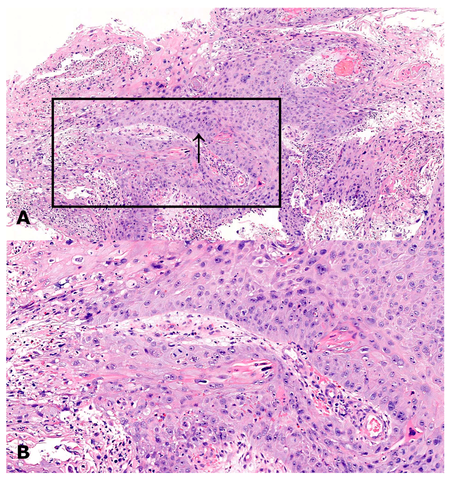
Diagnosis is based on cytologic screening (Papanicolaou test) and HPV DNA testing, followed by colposcopy and histopathologic confirmation (Figure 1). Radiologic staging follows, being a crucial step in assessment of disease extent to further guide therapy.
The FIGO 2018 classification system integrates multiple imaging modalities: magnetic resonance imaging (MRI) is the gold standard for assessment of local tumor extent and parametrial invasion due to its superior soft-tissue visualization; PET/CT scans are performed to inform lymph-node status and the presence of distant metastasis; CT scans are used to evaluate thoracic and abdominal status, particularly where access to MRI and PET/CT is limited [3].
The management of cervical cancer is stage-dependent and must balance oncologic safety with quality-of-life considerations, particularly fertility preservation. According to the 2018 FIGO staging system, treatment is dictated by tumor size, depth of invasion, and the involvement of surrounding tissues or lymph nodes. Depth of stromal invasion is a critical determinant of both staging and treatment intensity. Microinvasive disease is defined by limited stromal penetration (≤3 mm in IA1 and 3–5 mm in IA2), allowing for conservative surgical approaches. In contrast, deep stromal invasion—particularly extension into the outer third of the cervix—is associated with increased recurrence risk and often necessitates more radical surgery or adjuvant radiotherapy, even in otherwise early-stage disease [2,3]. Lymphovascular space invasion (LVSI), although not incorporated into FIGO staging, is a major prognostic factor. Its presence markedly increases the risk of lymph node metastasis and influences surgical planning. For example, in FIGO IA1 disease, LVSI may justify pelvic lymphadenectomy, whereas its absence allows for less extensive treatment [2,3]. Fertility preservation is a key consideration in young patients with early-stage cervical cancer. Fertility-sparing procedures are feasible in carefully selected cases, typically tumors < 2 cm, with limited stromal invasion and no radiologic evidence of nodal disease. Options include cervical conization for selected IA1–IA2 tumors and radical trachelectomy for small IB1 lesions. While oncologic outcomes are favorable, patients should be counseled regarding increased risks of obstetric complications, including preterm delivery and cervical stenosis [2,3]. For very early microinvasive disease (Stage IA), conservative options such as cervical conization are often adequate. As the disease progresses to Stage IB1 and IB2, surgical intervention—specifically radical hysterectomy with pelvic lymphadenectomy—remains the standard of care. However, for tumors larger than 4 cm (Stage IB3 and IIA2) or those involving the parametria (Stage IIB and above), concurrent platinum-based chemoradiation (CCRT) is the preferred modality [2,3].
The GOG-240 trial confirmed that platinum–paclitaxel plus bevacizumab should be used as the primary treatment for patients who have recurrent or metastatic disease [9]. The use of multiple treatment approaches has improved patient outcomes; however, patients with advanced or recurrent cancer continue to experience poor results. Consequently, the development of novel therapeutic strategies has become essential, as existing treatments fail to provide adequate patient benefit [10,11].
In the last decade, immune checkpoint inhibitors (ICIs) have redefined cancer treatment, as they enable the immune system to fight cancer cells by blocking CTLA-4 and PD-1/PD-L1 pathways [4,12,13]. The viral etiology related to the majority of cervical cancers, along with their high PD-L1 expression and immune-active tumor environment make immunotherapy a rational treatment choice [5,6,7,8,14]. Table 1 summarizes cervical cancer management.

2. Immunopathogenesis of Cervical Cancer and Mechanisms of Checkpoint Inhibition

Cervical cancer is a paradigmatic example of virus-driven oncogenesis, with persistent infection with high-risk HPV promoting malignant transformation through genetic and immune dysregulation. The viral oncoproteins E6 and E7 cause p53 and Rb tumor suppressor degradation, resulting in uncontrolled cell growth, genomic instability and resistance to apoptosis [2,4]. Beyond their oncogenic effects, HPV E6 and E7 generate highly immunogenic viral neoantigens and induce a chronic inflammatory state that profoundly reshapes the local immune response, facilitating tumor persistence and progression [5,6,13].

2.1. Innate and Adaptive Immune Interactions in HPV-Driven Tumorigenesis

HPV infection activates both innate and adaptive immune responses. The initial host defense relies on natural killer (NK) cell cytotoxicity, dendritic cell activation, and interferon (IFN) signaling. However, high-risk HPV subtypes evade immune surveillance by downregulating pattern recognition receptors (PRRs), including Toll-like receptors (TLRs), and impairing antigen processing and presentation [5,6]. Despite these evasion mechanisms, HPV-derived antigens promote infiltration of tumor-specific CD8 + T lymphocytes, leading to an inflamed tumor microenvironment. Persistent antigen exposure, however, drives immune adaptation rather than clearance. Over time, the tumor microenvironment becomes enriched with regulatory T cells (Tregs), myeloid-derived suppressor cells (MDSCs), and M2-polarized macrophages, which suppress cytotoxic T-cell activity and enable immune escape [5]. Chronic antigen stimulation ultimately results in T-cell exhaustion, characterized by upregulation of inhibitory receptors such as programmed cell death protein 1 (PD-1), T-cell immunoglobulin and mucin-domain containing-3 (TIM-3), and lymphocyte activation gene 3 (LAG-3) [14,15].

2.2. The PD-1/PD-L1 Axis in Cervical Cancer

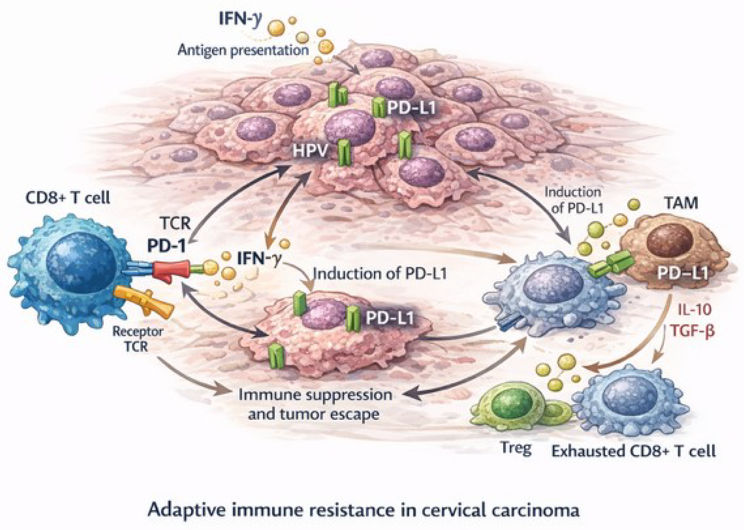
The PD-1/PD-L1 signaling pathway plays a central role in immune system evasion in cervical cancer. PD-L1, also known as B7-H1, is a 40-kDa type 1 transmembrane protein that contains extracellular immunoglobulin C and immunoglobulin V domains, being coded by CD274 gene, which is located on human chromosome 9 [16]. HPV-infected tumor cells present viral antigens that are recognized by infiltrating CD8 + T lymphocytes, leading to T-cell activation and secretion of interferon- γ (IFN- γ ). IFN- γ induces PD-L1 expression on both tumor cells and tumor-associated macrophages (TAMs) as part of a mechanism of adaptive immune resistance, as explained in Figure 2. Engagement of PD-L1 with PD-1 receptors on activated T cells inhibits T-cell receptor (TCR) signaling, reduces cytokine production, and culminates in functional T-cell exhaustion [4,12,14,15].
This adaptive immune resistance is further reinforced by the tumor microenvironment. TAMs and regulatory T cells secrete immunosuppressive cytokines such as interleukin-10 (IL-10) and transforming growth factor- β (TGF- β ), amplifying immune dysfunction and promoting tumor progression [4,12,14,15]. In this context, PD-L1 expression in cervical cancer represents a dynamic response to an active anti-tumor immune infiltrate driven by HPV antigens, rather than a purely constitutive tumor trait.
At the molecular level, activation of the PI3K/AKT/mTOR and JAK/STAT signaling pathways by HPV oncoproteins, as well as Hsp90 overexpression, contributes to PD-L1 upregulation in cervical cancer cells [5,6]. Immunohistochemical studies demonstrate PD-L1 expression in approximately 50–80% of cervical cancer cases, depending on tumor subtype, HPV status, and testing methodology [7,8,14]. PD-L1 is more strongly expressed in neoplastic cells of HPV-induced cervical intraepithelial neoplasia (CIN) and squamous cell carcinomas, with expression increasing alongside higher CIN grades [5,6,7,16]. Huang et al. showed that PD-L1 positivity correlates with PIK3CA and PTEN mutations and an inflamed immune phenotype, supporting its value as a biomarker reflecting immune activation rather than intrinsic tumor aggressiveness [3,6]. This interpretation is reinforced by studies demonstrating that PD-L1 expression, particularly when associated with high tumor-infiltrating lymphocytes (TILs) and an IFN- γ -enriched microenvironment, correlates with improved progression-free and overall survival in patients treated with immune checkpoint inhibitors [7,8]. These findings validate PD-L1 as a surrogate marker of an ongoing anti-tumor immune response. Additionally, PD-L1–positive cervical carcinomas exhibit distinct genomic and immune characteristics that may inform immunotherapeutic targeting [17]. PD-L1 expression has also been identified in cervical intraepithelial neoplasia and invasive squamous cell carcinoma in both HIV-infected and non-infected patients, extending the relevance of immune modulation across disease stages and patient populations [18].

3. Clinical Evidence and Ongoing Trials in Advanced Cervical Cancer

3.1. Clinical Validation of Immune Checkpoint Inhibitors

Multiple phase II and III clinical trials have established ICIs as standard treatment for advanced and locally advanced cervical cancer. The KEYNOTE-158 trial evaluated pembrolizumab monotherapy for patients with recurrent or metastatic cervical cancer who received previous treatments, and showed a 14.3% ORR and 17% response rate for patients with PD-L1-positive tumors and CPS scores of 1 or higher [19,20,21,22,23]. The study results led to the U. S. Food and Drug Administration (FDA) approval of pembrolizumab for treating PD-L1-positive recurrent or metastatic cervical cancer. Subsequently, the KEYNOTE-826 trial showed that pembrolizumab combined with standard chemotherapy (platinum–paclitaxel ± bevacizumab) for first-line treatment of recurrent or metastatic disease resulted in better OS and PFS outcomes [11,22,24,25]. Median OS improved to 24.4 months versus 16.5 months with chemotherapy alone, reducing death risk by 37%, with the benefit being consistent across all PD-L1-positive patient subgroups (CPS ≥ 1 and CPS ≥ 10). The study results helped establish pembrolizumab combined with chemotherapy as the new first-line standard treatment for recurrent, persistent or metastatic disease [11,22,24,25]. A meta-analysis of phase III clinical trials demonstrated that ICIs in combination with standard treatments achieved better OS, PFS and ORR results with acceptable safety profiles [19]. The EMPOWER-Cervical 1 trial demonstrated that cemiplimab significantly improved overall survival compared with chemotherapy (median 12.0 vs. 8.5 months), across histologic subtypes and irrespective of PD-L1 expression, with fewer severe adverse events, supporting its role as a superior second-line treatment after platinum failure [26].

3.2. ICIs Combined with Chemoradiation and Novel Agents

The KEYNOTE-A18/ENGOT-cx11/GOG-3047 trial evaluated pembrolizumab combined with concurrent chemoradiation (CRT) for patients with newly diagnosed FIGO stage III–IVA disease. The study results showed that pembrolizumab addition to CRT treatment improved PFS by 59% and demonstrated positive OS results which established a new treatment standard for locally advanced cervical cancer patients [10,24,27,28], which led to FDA approval for pembrolizumab combined with CRT for patients with FIGO III–IVA cervical cancer regardless of their PD-L1 status in 2024 [10,24,27,28]. The CALLA trial investigated durvalumab combined with chemoradiation for locally advanced cervical cancer patients. While the study did not meet its primary endpoint of progression-free survival (PFS) in the overall intention-to-treat population, exploratory analyses suggested improved PFS specifically among patients with high PD-L1 expression [22,24,29]. The BEATcc trial showed that atezolizumab together with platinum–paclitaxel–bevacizumab extended PFS and OS for patients with advanced disease beyond what standard chemotherapy achieves [24,30]. Clinical trials have proven that ICIs should be used in combination with other treatments for advanced and locally advanced cervical cancer patients, in a shift of the standard of care from cytotoxic approaches to immune-modulated treatment paradigms [12,19,24]. However, the discrepancy between studies highlights the importance of patient selection and biomarker stratification [19,24].
Table 2 provides a comparison of the key clinical trials exploring immunotherapy in cervical cancer, as detailed in the sources.

4. Pathological Assessment of PD-L1 Expression

4.1. Biomarkers and Predictors of Response

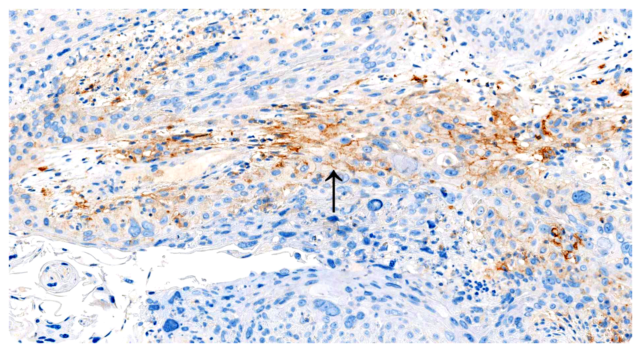
The primary biomarker for immunotherapy eligibility in cervical cancer is PD-L1 expression, assessed through immunohistochemistry (IHC) using the combined positive score (CPS) [14,31]. Studies show that patients with higher CPS values achieve better results from ICIs, although PD-L1-negative tumors also show responses to treatment, which suggests additional immune mechanisms at play [31]. The assessment of PD-L1 expression in cervical cancer uses two main IHC validated clones which include 22C3 pharmDx (Dako/Agilent) (Figure 3) and SP263 (Ventana/Roche), both with excellent interobserver agreement among gynecologic pathologists [17,31].
The 2023 research by Vainer et al. demonstrated that the laboratory-developed 22C3 test produces results identical to the commercial pharmDx platform which enables flexible implementation in different pathology laboratories [32]. The implementation of standardized pre-analytical procedures for tissue fixation duration and sample thickness and staining platform selection becomes essential to achieve consistent results between different laboratories [31,32].
Despite its clinical significance, PD-L1 evaluation has various analytical challenges across global diagnostic systems. The evaluation of test results depends on both the specific assay platform and antibody clone and the established clinical eligibility cutoff values for different cancer types. These aspects are particularly relevant in cervical cancer, where PD-L1 testing is required to guide pembrolizumab-based therapy.

4.2. Methodological Considerations and Scoring Systems

The evaluation of PD-L1 expression through immunohistochemistry (IHC) has become a cornerstone method for selecting patients for ICI therapy based on biomarkers. In cervical cancer, PD-L1 expression assessment is performed using the combined positive score (CPS), which is calculated as:
CPS = number of PD-L1 staining tumor cells , lymphocytes , and macrophages total number of viable tumor cells × 100
Both FDA and the European Medicines Agency (EMA) require PD-L1 testing for pembrolizumab treatment eligibility in patients with recurrent or metastatic cervical cancer, where CPS   1 qualifies for treatment either as monotherapy or in combination with platinum-based chemotherapy ± bevacizumab [13,24,32]. A CPS   1 defines PD-L1 positivity, while CPS   10 has been associated with better responses to PD-1/PD-L1 blockade treatments [14,23,24]. However, there are instances in which ICIs can be used without PD-L1 testing: in locally advanced disease (FIGO III-IVA), treatment with pembrolizumab combined with chemoradiation is not biomarker-restricted, reflecting evolving clinical integration of ICIs [23]. Additionally, in China, cadonilimab + chemotherapy (±bevacizumab) has been approved for first-line treatment of recurrent or metastatic cervical cancer without PD-L1 restriction [10].

4.3. Validated Clones and Analytical Platforms

The diversity of PD-L1 antibody clones and assay platforms currently available reflects both regulatory requirements and the ongoing technical evolution of immunohistochemistry-based companion diagnostics. As summarized in Table 3, the 22C3 pharmDx assay (Dako/Agilent) remains the only FDA-approved companion diagnostic for pembrolizumab treatment in cervical carcinoma, serving as the analytical reference standard for comparison with alternative assays [17,31,33]. Multiple PD-L1 antibody clones have received regulatory clearance or clinical validation for use across different tumor types, but the 22C3 pharmDx assay (Dako/Agilent) is the only officially approved companion diagnostic for pembrolizumab treatment in cervical carcinoma [17,31,33]. Other clones, including SP263 (Ventana/Roche), 28-8 (Agilent), and E1L3N (Cell Signaling Technology), are considered complementary diagnostics and show high analytical concordance in most studies, although subtle variations in staining intensity, cellular distribution, and immune background have been reported [17,31,33]. Comparative analyses demonstrate that even though 22C3 and SP263 achieve similar overall positivity rates, discrepancies tend to occur in tumors with low CPS or borderline PD-L1 expression, which can influence clinical eligibility decisions, while E1L3N demonstrates strong agreement with 22C3 results when standardized protocols and scoring systems are applied [17,31,33]. A critical technical issue involves the balance between commercial “closed-kit” systems (such as 22C3 or SP263 on proprietary automated platforms) and laboratory-developed tests (LDTs) implemented on open immunohistochemistry platforms. LDTs offer greater flexibility and cost-effectiveness but require rigorous validation and quality assurance to ensure reproducibility and diagnostic accuracy across laboratories [32,33]. Recent cross-platform studies have demonstrated that optimized LDT protocols can achieve analytical equivalence to 22C3 pharmDx assay, reinforcing their utility in laboratories lacking access to proprietary systems [32]. However, interlaboratory variability in pre-analytical handling, antigen retrieval conditions, and interpretation of CPS scoring continues to be a relevant source of inconsistent results [31,33]. The companion diagnostic model, originally developed under FDA regulatory frameworks, has spread across the world, albeit with regional adaptations. The FDA requires strict assay-drug combinations for ICI eligibility, whereas the EMA permits the use of appropriately validated LDTs under accredited national quality systems [33]. These two realities underscore why international validation frameworks are needed to ensure technical standardization, analytical comparability, and equitable access to biomarker-driven treatments in cervical cancer.
Table 4 summarizes the different regulatory and methodological frameworks.

4.4. Limitations of PD-L1 as a Predictive Biomarker in Cervical Cancer

Notwithstanding its widespread clinical adoption, PD-L1 is an imperfect biomarker due to several biological and technical limitations. Its expression is highly heterogeneous, showing substantial variability both within the same tumor and between primary lesions and metastatic sites. In addition, PD-L1 expression is dynamically regulated by cytokine signaling within the tumor microenvironment, further contributing to temporal and spatial variability. Together, these factors lead to inconsistent assay results and limit the ability of PD-L1 testing to reliably predict either response or non-response to ICI therapy [7,8,31]. Although PD-L1 expression measured by immunohistochemistry is commonly used to stratify patients with advanced cervical cancer for ICI therapy, its predictive performance remains imperfect. Clinical trials highlight these limitations: in KEYNOTE-158 [20], pembrolizumab responses occurred mainly in PD-L1 CPS   1 tumors, yet many PD-L1–positive patients did not derive durable benefit, and few PD-L1–negative tumors were included. In KEYNOTE-826 [11,25], higher CPS was associated with greater benefit, but outcomes overlapped across strata, limiting its discriminatory power. Similarly, EMPOWER-Cervical 1 [26] demonstrated that cemiplimab improved survival irrespective of PD-L1 subgroup, underscoring the limitations of PD-L1 as a stand-alone biomarker. Importantly, clinical evidence demonstrates that PD-L1 fails as a standalone predictive marker, as some patients with low or undetectable PD-L1 expression still derive meaningful clinical benefit from ICIs, while not all patients with high PD-L1 expression respond to treatment. This discordance indicates that PD-L1 functions primarily as an enrichment marker rather than a definitive determinant of therapeutic benefit. Consequently, although regulatory agencies such as the FDA have adopted PD-L1 as a companion or complementary diagnostic for selected immunotherapies, owing to its capacity to enrich for responders in clinical trials, its inherent limitations preclude its use as a reliable standalone biomarker.
Table 5 summarizes PD-L1 pitfalls as a predictive biomarker in cervical cancer.

4.5. Future Directions in PD-L1 Determination

From an analytical standpoint, clone selection and scoring reproducibility remain critical challenges. Although 22C3 and SP263 assays demonstrate high concordance, discrepancies in borderline CPS ranges and variations in staining intensity persist, particularly across laboratories using distinct automated systems or LDT protocols [17,31,33]. The increasing use of LDTs has reinforced the need for cross-platform harmonization, external proficiency testing, and standardized interpretation protocols [32,33]. Lessons learned from the PD-L1 rollout across tumor types underline that assay standardization must integrate both technical validation and pathologist training, ensuring consistent interpretation and reproducible CPS scoring [33]. In this context, digital pathology and artificial intelligence (AI) are emerging as transformative tools. AI-powered image analysis systems, as studied by Baxi et al. [34], have demonstrated strong concordance with manual scoring and predictive value for clinical outcomes in ICI-treated patients, and the evaluation pipeline proposed by Knudsen et al. [35] for AI/ML-assisted PD-L1 quantification introduced standardized algorithmic validation, explainability metrics, and quality controls that support integration into diagnostic practice. These tools can help overcome observer variability, optimize cut-off definitions, and enable consistent reporting across institutions and regions. Regulatory frameworks are also evolving to accommodate these innovations: while the FDA still mandates approved companion diagnostics for specific assay–drug pairs, the EMA and several Asian agencies are recognizing AI-assisted quantification and validated LDTs as acceptable diagnostic approaches under accredited systems [33,34,35]. Pathology is thus transitioning from a purely visual to a data-driven digital discipline, where algorithms augment human expertise and improve reproducibility in immune biomarker assessment. The integration of standardized IHC, multiplex imaging, RNA-based assays, and AI-assisted analysis represents the next frontier of precision immunodiagnostics in cervical cancer [17,32,33,34,35].

5. Beyong PD-L1: Tumor Immunogenicity and Antigen-Presentation Biomarkers

The observation of clinical benefit in PD-L1–negative tumors underscores the need for additional biomarkers to more accurately capture tumor immunogenicity and immune competence. These include tumor mutational burden (TMB), microsatellite instability (MSI), Human Leukocyte Antigen class I (HLA-I) expression, and tumor microenvironment, all of which provide complementary biological information. The integration of these parameters into future diagnostic algorithms is essential to refine patient selection, improve predictive accuracy, and guide rational combination therapy strategies in immuno-oncology [10,36].

5.1. Tumor Mutational Burden (TMB) and Microsatellite Instability (MSI)

TMB and MSI are key genomic biomarkers as they directly influence tumor immunogenicity and responsiveness to ICIs. TMB measures the number of somatic mutations per megabase (mut/Mb) of tumor DNA and requires next-generation sequencing (NGS) for its calculation [36,37,38,39,40,41]. In contrast, MSI reflects a hypermutated phenotype caused by deficiency in the DNA mismatch repair system [36,37,38] and can be assessed more easily using immunohistochemistry, as well as NGS and other molecular biology techniques, making MSI generally more accessible for analysis. A high mutational load increases the likelihood of generating mutation-derived neoantigens, which are recognized as foreign by the immune system and can elicit a strong T-cell–mediated immune response once immune checkpoints are inhibited [36,37,38,39,41,42].
The clinical relevance of TMB as a predictive biomarker has been validated across multiple tumor types. The FDA has approved pembrolizumab for the treatment of advanced solid tumors, including cervical cancer, with a TMB   10 mut/Mb, irrespective of tumor histology [36,37,38,39,40,41]. In cervical cancer specifically, TMB-high status has been identified in approximately 59.0% of patients and is independent of FIGO stage and histological subtype [38]. Nevertheless, elevated TMB in this population is significantly associated with nodal involvement, diabetes, and infection with HPV genotypes 52 or 68 [38]. Genomic analyses further demonstrate histology-specific mutation patterns, with PIK3CA mutations being more common in squamous cell carcinoma (60%) and KRAS mutations more frequently observed in adenocarcinoma (42%) [38]. Large-scale real-world evidence reinforces the predictive value of TMB for ICI efficacy. In a cohort of 8440 patients across 24 cancer types, those with TMB   10 mut/Mb exhibited significantly improved real-world overall survival compared with TMB-low patients (HR 0.37) [40]. Importantly, this survival benefit remained significant in microsatellite-stable (MSS) tumors (HR 0.42), highlighting TMB as an independent predictive biomarker even when MSI—a relatively rare feature in cervical cancer, present in only 3.3% to 11.3% of cases—is absent [37,38,40]. Prospective clinical trial data further support the role of TMB in patient selection. The KEYNOTE-158 study demonstrated that TMB-high status (TMB   10 mut/Mb) identifies a subset of patients with advanced solid tumors who achieve meaningful objective responses to pembrolizumab monotherapy, even in the absence of PD-L1 expression [20,21]. Similarly, MSI-high/dMMR status was the first histology-agnostic biomarker approved for ICI therapy, as it consistently predicts sensitivity to PD-1 blockade regardless of tumor origin [10,37,42].

5.2. Human Leukocyte Antigen Class I (HLA-I)

Beyond mutation burden, effective antitumor immunity also depends on antigen presentation. HLA-I molecules are responsible for presenting tumor-derived neoantigens to CD8 + T cells, and the patient’s HLA-I genotype significantly influences survival following ICI therapy [43,44]. Maximal heterozygosity at HLA-I loci allows broader neoantigen presentation and is associated with improved overall survival, whereas homozygosity or somatic loss of heterozygosity restricts antigen presentation, impairing immune recognition and contributing to treatment resistance [43,44].
Taken together, these findings support the integration of TMB, MSI, and HLA-I genotype into future diagnostic and predictive algorithms to more accurately identify cervical cancer patients most likely to benefit from immunotherapy, while ongoing research continues to refine TMB interpretation in conjunction with PD-L1 expression and the tumor inflammatory microenvironment [10,36,37,38].

6. Future Perspectives

The introduction of immune checkpoint inhibitors (ICIs) has reshaped the treatment landscape of cervical cancer, shifting management from cytotoxic chemotherapy alone to immune-based combination strategies that have improved survival in recurrent/metastatic and locally advanced disease [10,12,19,23,24]. This benefit is exemplified by pembrolizumab-based regimens in KEYNOTE-826, while the negative results of the CALLA trial highlight the biological heterogeneity of HPV-associated disease and the limitations of empiric treatment intensification. Despite these advances, major challenges remain. Most cervical cancer cases occur in low- and middle-income countries (LMIC), where access to molecular diagnostics, PD-L1 testing, and ICIs is limited [1,3,11,45]. High drug costs [45,46], inadequate diagnostic infrastructure [47], centralized care, and the lack of locally generated clinical data contribute to persistent global disparities [45]. Addressing these inequities requires coordinated global health strategies focused on HPV vaccination, equitable drug access, and standardized biomarker testing, as emphasized by the WHO 90-70-90 framework [3]. Recent trial outcomes underscore the need to move beyond PD-L1 toward integrated immune and molecular stratification. Efforts now focus on combining biomarkers such as tumor mutational burden (TMB), microsatellite instability (MSI), and T-cell–inflamed gene signatures to improve patient selection for ICIs [6,12,32]. In parallel, combination strategies—including dual checkpoint blockade, HPV-targeted vaccines, and metabolic modulators—aim to overcome immune resistance, particularly in PD-L1–negative or immune-excluded tumors [5,6,12,13,32,48,49]. Antibody–drug conjugates (ADCs) have also emerged as a promising therapeutic platform [50]. Tisotumab vedotin, the first FDA-approved ADC for recurrent or metastatic cervical cancer, achieved response rates of approximately 24% [51], with higher responses reported in combination regimens in the innovaTV 205 study [50]. Additional ADCs targeting HER2, TROP-2, FR α , and NaPi2b are under active investigation [50]. Looking ahead, PD-1–based ADCs represent an innovative approach integrating checkpoint inhibition with targeted cytotoxicity. Preclinical data suggest that these agents can increase immune cell infiltration, trigger immunogenic cell death, and overcome primary or acquired resistance to ICIs [52]. Overall, cervical cancer therapy is evolving toward rational combinations of immunotherapy, targeted agents, and preventive strategies-namely through expanded HPV vaccination and organized screening. The long-term success of these advances will depend on optimized treatment sequencing, robust predictive biomarkers, and equitable global access—particularly in low- and middle-income countries (LMIC) settings [1,3,11].

7. Conclusions

Immunotherapy for cervical cancer treatment represents a major breakthrough in precision oncology. The results from essential clinical trials and research studies show that ICIs have redefined survival expectations for patients with advanced or locally advanced disease. The path to future success demands a multifaceted approach, which includes molecular classification, rational treatment progression and equitable access to innovation. With coordinated global efforts, immunotherapy—together with targeted antibody–drug conjugates—is poised to become a cornerstone of cervical cancer management, bridging prevention, early detection, and personalized treatment [1,3,10,11,24,50,51,52].

Author Contributions

Conceptualization, S.C.A. and L.V.d.C.; writing—original draft preparation, S.C.A.; writing—review and editing, L.V.d.C., J.N. and D.G.P.; visualization, S.C.A. and L.V.d.C.; supervision, L.V.d.C.; project administration, L.V.d.C. All authors have read and agreed to the published version of the manuscript.

Funding

This research received no external funding.

Institutional Review Board Statement

Not applicable.

Informed Consent Statement

Not applicable.

Data Availability Statement

No new data were created or analyzed in this study.

Acknowledgments

During the preparation of this manuscript, the authors used generative artificial intelligence tools (ChatGPT—GPT-5-mini, OpenAI, 2026) for linguistic refinement of the text. Additionally, some figures were created using AI-based generation, guided by prompts designed by the authors. All outputs were subsequently reviewed, edited, and validated by the authors to ensure scientific accuracy. The authors take full responsibility for the content of this publication.

Conflicts of Interest

The authors declare no conflicts of interest.

References

  1. GLOBOCAN. Global Cancer Observatory: Cervix Uteri Fact Sheet; International Agency for Research on Cancer (IARC): Lyon, France; World Health Organization: Geneva, Switzerland, 2024. [Google Scholar]
  2. Tewari, K.S. Cervical Cancer. N. Engl. J. Med. 2025, 392, 56–71. [Google Scholar] [CrossRef]
  3. Bhatla, N.; Aoki, D.; Sharma, D.N.; Sankaranarayanan, R. Cancer of the Cervix Uteri: 2025 Update. Int. J. Gynecol. Obstet. 2025, 171, 87–108. [Google Scholar] [CrossRef]
  4. Wei, S.C.; Duffy, C.R.; Allison, J.P. Fundamental Mechanisms of Immune Checkpoint Blockade Therapy. Cancer Discov. 2018, 8, 1069–1086. [Google Scholar] [CrossRef] [PubMed]
  5. Zeng, J.; He, S.-L.; Li, L.-J.; Wang, C. Hsp90 Up-Regulates PD-L1 to Promote HPV-Positive Cervical Cancer via HER2/PI3K/AKT Pathway. Mol. Med. 2021, 27, 130. [Google Scholar] [CrossRef]
  6. Huang, W.; Liu, J.; Xu, K.; Chen, H.; Bian, C. PD-1/PD-L1 Inhibitors for Advanced or Metastatic Cervical Cancer: From Bench to Bed. Front. Oncol. 2022, 12, 849352. [Google Scholar] [CrossRef] [PubMed]
  7. Fu, H.; Fu, Z.; Mao, M.; Si, L.; Bai, J.; Wang, Q.; Guo, R. Prevalence and Prognostic Role of PD-L1 in Patients with Gynecological Cancers: A Systematic Review and Meta-Analysis. Crit. Rev. Oncol. Hematol. 2023, 189, 104084. [Google Scholar] [CrossRef]
  8. Santoro, A.; Angelico, G.; Inzani, F.; Arciuolo, D.; d’Amati, A.; Addante, F.; Travaglino, A.; Scaglione, G.; D’Alessandris, N.; Valente, M.; et al. The Emerging and Challenging Role of PD-L1 in Patients with Gynecological Cancers: An Updating Review with Clinico-Pathological Considerations. Gynecol. Oncol. 2024, 184, 57–66. [Google Scholar] [CrossRef]
  9. Tewari, K.S.; Sill, M.W.; Long, H.J.; Penson, R.T.; Huang, H.; Ramondetta, L.M.; Landrum, L.M.; Oaknin, A.; Reid, T.J.; Leitao, M.M.; et al. Improved Survival with Bevacizumab in Advanced Cervical Cancer. N. Engl. J. Med. 2014, 370, 734–743. [Google Scholar] [CrossRef] [PubMed]
  10. Yanaihara, N.; Tse, K.Y.; Lee, S.J.; Yoo, J.G.; Wilailak, S. Immune Checkpoint Inhibitors in Gynecologic Oncology: Current Status and Perspectives. Int. J. Gynecol. Obstet. 2025, 171, 166–188. [Google Scholar] [CrossRef]
  11. Colombo, N.; Dubot, C.; Lorusso, D.; Caceres, M.V.; Hasegawa, K.; Shapira-Frommer, R.; Tewari, K.S.; Salman, P.; Hoyos Usta, E.; Yañez, E.; et al. Pembrolizumab for Persistent, Recurrent, or Metastatic Cervical Cancer. N. Engl. J. Med. 2021, 385, 1856–1867. [Google Scholar] [CrossRef]
  12. Rowshanravan, B.; Halliday, N.; Sansom, D.M. CTLA-4: A Moving Target in Immunotherapy. Blood 2018, 131, 58–67. [Google Scholar] [CrossRef]
  13. Bhattacharya, S.; Paraskar, G.; Jha, M.; Gupta, G.L.; Prajapati, B.G. Deciphering Regulatory T-Cell Dynamics in Cancer Immunotherapy: Mechanisms, Implications, and Therapeutic Innovations. ACS Pharmacol. Transl. Sci. 2024, 7, 2215–2236. [Google Scholar] [CrossRef]
  14. Kooshkaki, O.; Derakhshani, A.; Safarpour, H.; Najafi, S.; Vahedi, P.; Brunetti, O.; Torabi, M.; Lotfinejad, P.; Paradiso, A.V.; Racanelli, V.; et al. The Latest Findings of PD-1/PD-L1 Inhibitor Application in Gynecologic Cancers. Int. J. Mol. Sci. 2020, 21, 5034. [Google Scholar] [CrossRef]
  15. Shamseddine, A.A.; Burman, B.; Lee, N.Y.; Zamarin, D.; Riaz, N. Tumor Immunity and Immunotherapy for HPV-Related Cancers. Cancer Discov. 2021, 11, 1896–1912. [Google Scholar] [CrossRef]
  16. Wang, R.; Zhang, Y.; Shan, F. PD-L1: Can It Be a Biomarker for the Prognosis or a Promising Therapeutic Target in Cervical Cancer? Int. Immunopharmacol. 2022, 103, 108484. [Google Scholar] [CrossRef] [PubMed]
  17. Huang, R.S.P.; Haberberger, J.; Murugesan, K.; Danziger, N.; Hiemenz, M.; Severson, E.; Duncan, D.L.; Ramkissoon, S.H.; Ross, J.S.; Elvin, J.A.; et al. Clinicopathologic and Genomic Characterization of PD-L1-Positive Uterine Cervical Carcinoma. Mod. Pathol. 2021, 34, 1425–1433. [Google Scholar] [CrossRef]
  18. Brito, M.J.; Sequeira, P.; Quintas, A.; Silva, I.; Silva, F.; Martins, C.; Félix, A. Programmed Death-Ligand 1 (PD-L1) Expression in Cervical Intraepithelial Neoplasia and Cervical Squamous Cell Carcinoma of HIV-Infected and Non-Infected Patients. Virchows Arch. 2024, 484, 507–516. [Google Scholar] [CrossRef] [PubMed]
  19. IIbibulla, N.; Lu, P.; Nuerrula, Y.; Hu, X.; Aihemaiti, M.; Wang, Y.; Zhang, H. Effectiveness and Safety of Immune Checkpoint Inhibitors for the Treatment of Advanced Cervical Cancer: A Systematic Review and Meta-Analysis. Front. Immunol. 2025, 16, 1542850. [Google Scholar] [CrossRef] [PubMed]
  20. Marabelle, A.; Le, D.T.; Ascierto, P.A.; Di Giacomo, A.M.; De Jesus-Acosta, A.; Delord, J.-P.; Geva, R.; Gottfried, M.; Penel, N.; Hansen, A.R.; et al. Efficacy of Pembrolizumab in Patients with Noncolorectal High Microsatellite Instability/Mismatch Repair–Deficient Cancer: Results from the Phase II KEYNOTE-158 Study. J. Clin. Oncol. 2020, 38, 1–10. [Google Scholar] [CrossRef]
  21. Marabelle, A.; Fakih, M.; Lopez, J.; Shah, M.; Shapira-Frommer, R.; Nakagawa, K.; Chung, H.C.; Kindler, H.L.; Lopez-Martin, J.A.; Miller, W.H.; et al. Association of Tumour Mutational Burden with Outcomes in Patients with Advanced Solid Tumours Treated with Pembrolizumab: Prospective Biomarker Analysis of the Multicohort, Open-Label, Phase 2 KEYNOTE-158 Study. Lancet Oncol. 2020, 21, 1353–1365. [Google Scholar] [CrossRef]
  22. Grau, J.-F.; Farinas-Madrid, L.; Garcia-Duran, C.; Garcia-Illescas, D.; Oaknin, A. Advances in Immunotherapy in Cervical Cancer. Int. J. Gynecol. Cancer 2023, 33, 403–413. [Google Scholar] [CrossRef]
  23. Agilent Technologies, Inc. PD-L1 IHC 22C3 pharmDx Interpretation Manual—Cervical Cancer; Agilent: Santa Clara, CA, USA, 2023. [Google Scholar]
  24. Zhang, X.; Shen, J.; Huang, M.; Li, R. Efficacy and Safety of Adding Immune Checkpoint Inhibitors to First-Line Standard Therapy for Recurrent or Advanced Cervical Cancer: A Meta-Analysis of Phase 3 Clinical Trials. Front. Immunol. 2024, 15, 1507977. [Google Scholar] [CrossRef] [PubMed]
  25. Monk, B.J.; Colombo, N.; Tewari, K.S.; Dubot, C.; Caceres, M.V.; Hasegawa, K.; Shapira-Frommer, R.; Salman, P.; Yañez, E.; Gümüş, M.; et al. First-Line Pembrolizumab + Chemotherapy Versus Placebo + Chemotherapy for Persistent, Recurrent, or Metastatic Cervical Cancer: Final Overall Survival Results of KEYNOTE-826. J. Clin. Oncol. 2023, 41, 5505–5511. [Google Scholar] [CrossRef] [PubMed]
  26. Tewari, K.S.; Monk, B.J.; Vergote, I.; Miller, A.; De Melo, A.C.; Kim, H.-S.; Kim, Y.M.; Lisyanskaya, A.; Samouëlian, V.; Lorusso, D.; et al. Survival with Cemiplimab in Recurrent Cervical Cancer. N. Engl. J. Med. 2022, 386, 544–555. [Google Scholar] [CrossRef]
  27. Lorusso, D.; Xiang, Y.; Hasegawa, K.; Scambia, G.; Leiva, M.; Ramos-Elias, P.; Acevedo, A.; Cvek, J.; Randall, L.; Pereira de Santana Gomes, A.J.; et al. Pembrolizumab or Placebo with Chemoradiotherapy Followed by Pembrolizumab or Placebo for Newly Diagnosed, High-Risk, Locally Advanced Cervical Cancer (ENGOT-Cx11/GOG-3047/KEYNOTE-A18): Overall Survival Results from a Randomised, Double-Blind, Placebo-Controlled, Phase 3 Trial. Lancet 2024, 404, 1321–1332. [Google Scholar] [CrossRef]
  28. Lorusso, D.; Xiang, Y.; Hasegawa, K.; Scambia, G.; Gálvez, M.H.L.; Elias, P.R.; Acevedo, A.; Bednarikova, M.; Gomes, A.J.P.D.S.; Mejia, F.C.; et al. 709O Pembrolizumab plus Chemoradiotherapy for High-Risk Locally Advanced Cervical Cancer: Overall Survival Results from the Randomized, Double-Blind, Phase III ENGOT-Cx11/GOG-3047/KEYNOTE-A18 Study. Ann. Oncol. 2024, 35, S544. [Google Scholar] [CrossRef]
  29. Monk, B.J.; Toita, T.; Wu, X.; Vázquez Limón, J.C.; Tarnawski, R.; Mandai, M.; Shapira-Frommer, R.; Mahantshetty, U.; Del Pilar Estevez-Diz, M.; Zhou, Q.; et al. Durvalumab versus Placebo with Chemoradiotherapy for Locally Advanced Cervical Cancer (CALLA): A Randomised, Double-Blind, Phase 3 Trial. Lancet Oncol. 2023, 24, 1334–1348. [Google Scholar] [CrossRef] [PubMed]
  30. Oaknin, A.; Gladieff, L.; Martínez-García, J.; Villacampa, G.; Takekuma, M.; De Giorgi, U.; Lindemann, K.; Woelber, L.; Colombo, N.; Duska, L.; et al. Atezolizumab plus Bevacizumab and Chemotherapy for Metastatic, Persistent, or Recurrent Cervical Cancer (BEATcc): A Randomised, Open-Label, Phase 3 Trial. Lancet 2024, 403, 31–43. [Google Scholar] [CrossRef]
  31. Mills, A.M.; Bennett, J.A.; Banet, N.; Watkins, J.C.; Kundu, D.; Pinto, A. Interobserver Agreement on the Interpretation of Programmed Death-Ligand 1 (PD-L1) Combined Positive Score (CPS) Among Gynecologic Pathologists. Am. J. Surg. Pathol. 2023, 47, 889–896. [Google Scholar] [CrossRef]
  32. Vainer, G.; Huang, L.; Emancipator, K.; Nuti, S. Equivalence of Laboratory-Developed Test and PD-L1 IHC 22C3 pharmDx across All Combined Positive Score Indications. PLoS ONE 2023, 18, e0285764. [Google Scholar] [CrossRef]
  33. Willis, J.E.; Eyerer, F.; Walk, E.E.; Vasalos, P.; Bradshaw, G.; Yohe, S.L.; Laser, J.S. Companion Diagnostics: Lessons Learned and the Path Forward from the Programmed Death Ligand-1 Rollout. Arch. Pathol. Lab. Med. 2023, 147, 62–70. [Google Scholar] [CrossRef]
  34. Baxi, V.; Lee, G.; Duan, C.; Pandya, D.; Cohen, D.N.; Edwards, R.; Chang, H.; Li, J.; Elliott, H.; Pokkalla, H.; et al. Association of Artificial Intelligence-Powered and Manual Quantification of Programmed Death-Ligand 1 (PD-L1) Expression with Outcomes in Patients Treated with Nivolumab ± Ipilimumab. Mod. Pathol. 2022, 35, 1529–1539. [Google Scholar] [CrossRef]
  35. Knudsen, B.S.; Jadhav, A.; Perry, L.J.; Thagaard, J.; Deftereos, G.; Ying, J.; Brintz, B.J.; Zhang, W. A Pipeline for Evaluation of Machine Learning/Artificial Intelligence Models to Quantify PD-L1 Immunohistochemistry. Lab. Investig. 2024, 104, 102070. [Google Scholar] [CrossRef] [PubMed]
  36. Graham, L.S.; Pritchard, C.C.; Schweizer, M.T. Hypermutation, Mismatch Repair Deficiency, and Defining Predictors of Response to Checkpoint Blockade. Clin. Cancer Res. 2021, 27, 6662–6665. [Google Scholar] [CrossRef] [PubMed]
  37. Xie, Y.; Kong, W.; Zhao, X.; Zhang, H.; Luo, D.; Chen, S. Immune Checkpoint Inhibitors in Cervical Cancer: Current Status and Research Progress. Front. Oncol. 2022, 12, 984896. [Google Scholar] [CrossRef]
  38. Kowalkowska, M.E.; Kamińska, K.; Wojtysiak, J.; Koper, K.; Makarewicz, A.; Pietrzak, B.; Bomba-Opoń, D.; Dębska, M.; Wielgoś, M.; Grabiec, M.; et al. Tumor Mutational Burden in Cervical Cancer as Potential Marker for Immunotherapy Responders. Cancers 2025, 17, 2963. [Google Scholar] [CrossRef] [PubMed]
  39. Moeckel, C.; Bakhl, K.; Georgakopoulos-Soares, I.; Zaravinos, A. The Efficacy of Tumor Mutation Burden as a Biomarker of Response to Immune Checkpoint Inhibitors. Int. J. Mol. Sci. 2023, 24, 6710. [Google Scholar] [CrossRef]
  40. Gandara, D.R.; Agarwal, N.; Gupta, S.; Klempner, S.J.; Andrews, M.C.; Mahipal, A.; Subbiah, V.; Eskander, R.N.; Carbone, D.P.; Riess, J.W.; et al. Tumor Mutational Burden and Survival on Immune Checkpoint Inhibition in >8000 Patients Across 24 Cancer Types. J. Immunother. Cancer 2025, 13, e010311. [Google Scholar] [CrossRef]
  41. Needleman, R.A.; Thai, A.A. The Complexities of PD-L1 Expression as an Indicator of Immunotherapy Outcomes. Immunotherapy 2025, 17, 457–460. [Google Scholar] [CrossRef]
  42. Le, D.T.; Durham, J.N.; Smith, K.N.; Wang, H.; Bartlett, B.R.; Aulakh, L.K.; Lu, S.; Kemberling, H.; Wilt, C.; Luber, B.S.; et al. Mismatch Repair Deficiency Predicts Response of Solid Tumors to PD-1 Blockade. Science 2017, 357, 409–413. [Google Scholar] [CrossRef]
  43. Chowell, D.; Morris, L.G.T.; Grigg, C.M.; Weber, J.K.; Samstein, R.M.; Makarov, V.; Kuo, F.; Kendall, S.M.; Requena, D.; Riaz, N.; et al. Patient HLA Class I Genotype Influences Cancer Response to Checkpoint Blockade Immunotherapy. Science 2018, 359, 582–587. [Google Scholar] [CrossRef]
  44. Yoshimoto, D.; Iuchi, H.; Taguchi, A.; Sone, K.; Tamai, K.; Mori, A.; Kitamura, S.; Duong, A.Q.; Ishizaka, A.; Kusakabe, M.; et al. Downregulation of HLA Class I Expression Through HLA-A DNA Methylation Is Associated with Reduced CD8+ T-Cell Infiltration in Cervical Cancer. Cancer Immunol. Res. 2026, 14, 77–89. [Google Scholar] [CrossRef]
  45. Go, A.E.; Ho, F.D.V.; Feliciano, E.J.G.; Iyengar, P.; Chitapanarux, I.; Charoentum, C.; Bhoo-Pathy, N.; Satar, N.F.A.; Ting, F.I.L.; Dee, E.C. Barriers to Immune Checkpoint Inhibitor Access for Patients with Cancer in Southeast Asia: Challenges and Policy Implications. JCO Glob. Oncol. 2025, 11, 2500095. [Google Scholar] [CrossRef] [PubMed]
  46. Dent, J.; Jorge, M.; Sobrevilla, N.; Uldrick, T.S.; Adoubi, I.; Bajpai, J.; Burotto, M.; Bulhan, H.; Dosunmu, G.T.; Ekpo, L.; et al. Cancer Immunotherapy Clinical Trials to Support Urgently Needed Access in Low- and Middle-Income Countries: A Report from the SITC Global Access and Impact Committee. J. Immunother. Cancer 2025, 13, e011258. [Google Scholar] [CrossRef]
  47. Bou Akl, I.; Berro, J.; Tfayli, A.; Shamseddine, A.; Mukherji, D.; Temraz, S.; El Cheikh, J.; Alameh, I.A.; Assi, H.I. Current Status and Future Perspectives of Immunotherapy in Middle-Income Countries: A Single-Center Early Experience. World J. Oncol. 2020, 11, 150–157. [Google Scholar] [CrossRef] [PubMed]
  48. Silva, A.J.D.; Moura, I.A.D.; Gama, M.A.T.M.D.; Leal, L.R.S.; Pinho, S.S.D.; Espinoza, B.C.F.; Santos, D.L.D.; Santos, V.E.P.; Sena, M.G.A.M.D.; Invenção, M.D.C.V.; et al. Advancing Immunotherapies for HPV-Related Cancers: Exploring Novel Vaccine Strategies and the Influence of Tumor Microenvironment. Vaccines 2023, 11, 1354. [Google Scholar] [CrossRef]
  49. Ding, H.; Zhang, J.; Zhang, F.; Xu, Y.; Yu, Y.; Liang, W.; Li, Q. Effectiveness of Combination Therapy with ISA101 Vaccine for the Treatment of Human Papillomavirus-Induced Cervical Cancer. Front. Oncol. 2022, 12, 990877. [Google Scholar] [CrossRef] [PubMed]
  50. He, J.; Chen, M.; Jia, L.; Wang, Y. Antibody–Drug Conjugates in Gynecologic Cancer: Current Landscape, Clinical Data, and Emerging Targets. Int. J. Gynecol. Cancer. 2025, 35, 101978. [Google Scholar] [CrossRef]
  51. Coleman, R.L.; Lorusso, D.; Gennigens, C.; González-Martín, A.; Randall, L.; Cibula, D.; Lund, B.; Woelber, L.; Pignata, S.; Forget, F.; et al. Efficacy and Safety of Tisotumab Vedotin in Previously Treated Recurrent or Metastatic Cervical Cancer (innovaTV 204/GOG-3023/ENGOT-cx6): A Multicentre, Open-Label, Single-Arm, Phase 2 Study. Lancet Oncol. 2021, 22, 609–619. [Google Scholar] [CrossRef]
  52. Wang, J.; Chen, Z.; Wu, Y.; Zhang, W.; Awadasseid, A. Innovations in Cancer Immunotherapy with PD-1 Antibody–Drug Conjugates and Their Antitumor Mechanisms. Biomed. Pharmacother. 2025, 191, 118517. [Google Scholar] [CrossRef]
Figure 1. Representative histologic section of cervical squamous cell carcinoma. Neoplastic cells (arrow) show an infiltrative growth pattern within a reactive stroma rich in inflammatory cells [Hematoxylin & Eosin stain. (A) Original magnification 100×; (B) Original magnification 200×, corresponding to the area outlined by the black box in (A)]. (Source: Original figure created by the authors for this review).
Onco 06 00009 g001
Figure 2. Adaptive immune resistance mediated by the PD-1/PD-L1 axis in HPV-associated cervical carcinoma. HPV-infected tumor cells express viral antigens derived from the E6 and E7 oncoproteins, which are recognized by infiltrating CD8 + T lymphocytes, leading to T-cell activation and secretion of interferon- γ (IFN- γ ). IFN- γ induces PD-L1 expression on tumor cells and tumor-associated macrophages (TAMs) as part of a physiological feedback mechanism that limits immune-mediated tissue damage. Engagement of PD-L1 with PD-1 on CD8 + T cells results in functional T-cell exhaustion, characterized by reduced cytokine production and impaired cytotoxic activity. Regulatory T cells (Tregs) and immunosuppressive cytokines, including interleukin-10 (IL-10) and transforming growth factor- β (TGF- β ), further reinforce immune suppression within the tumor microenvironment, promoting immune escape and tumor progression. [Source: created with generative artificial intelligence (ChatGPT—GPT-5-mini, OpenAI, 2026) and adapted by the authors on 12 January 2025].
Onco 06 00009 g002
Figure 3. Representative immunohistochemical staining for PD-L1 (22C3 pharmDx assay, Dako/Agilent, Santa Clara, CA, USA) demonstrating membranous positive staining in tumor cells (arrow) (PD-L1, 200×). (Source: Original figure created by the authors for this review.).
Onco 06 00009 g003
Table 1. Cervical cancer management overview.
FIGO StageClinical DescriptionPrimary ManagementLVSI & Fertility Considerations
Stage IA1Invasion ≤ 3 mm deepConization or extrafascial hysterectomyIf LVSI is present, pelvic lymphadenectomy is required
Stage IA2Invasion > 3 mm to ≤5 mmModified radical hysterectomy + lymphadenectomyFertility desire: Conization or radical trachelectomy are options
Stage IB1/2Visible lesion 4 cmRadical hysterectomy + pelvic lymphadenectomyDeep stromal invasion or LVSI may trigger the need for adjuvant radiation
Stage IB3/IIA2Bulky lesion > 4 cmConcurrent Chemoradiation (CCRT)Surgery is avoided to reduce morbidity from combined “multimodality” treatment
Stage IIB–IVAExtension beyond uterus/vagina to pelvic wall or organsConcurrent Chemoradiation (CCRT)Stromal/vascular invasion markers are less critical than overall tumor volume/extension
Stage IVBDistant metastasesSystemic Chemotherapy + ImmunotherapyFocus shifts to palliative care and quality of life
Table 2. Comparison of immunotherapy studies in cervical cancer.
Trial NamePhaseDisease SettingTreatment ArmsKey Efficacy Results
KEYNOTE-158 [20]IIPost-platinum advancedPembrolizumab monotherapyORR of 14.3% in all-comers, 17.1% in PD-L1 positive (CPS   1 ).
KEYNOTE-826 [11,25]IIIFirst-line persistent, recurrent, or metastaticPembrolizumab + Platinum-based CT ± Bevacizumab vs. Placebo + CT ± BevacizumabSignificant improvement in OS (24.4 m vs. 16.5 m) and PFS (10.4 m vs.
8.2 m)
KEYNOTE-A18 [27,28]IIIHigh-risk locally advancedPembrolizumab + CCRT followed by Pembrolizumab vs. Placebo + CCRT24-month PFS rate of 68% vs. 57% (HR 0.70); 36-month OS rate of 82.6% vs. 74.8%
CALLA [29]IIIHigh-risk locally advancedDurvalumab + CCRT followed by Durvalumab vs. Placebo + CCRTDid not meet primary endpoint; no significant improvement in PFS
(HR 0.84)
BEATcc [30]IIIFirst-line metastatic (IVB), persistent, or recurrentAtezolizumab + Bevacizumab + Chemotherapy vs. Bevacizumab + ChemotherapyMedian OS reached
32.1 months vs. 22.8 months in the standard arm (HR 0.68)
EMPOWER-Cervical 1 [26]IIIPost-platinum recurrent or metastaticCemiplimab vs. Investigator’s choice chemotherapyMedian OS improved to 11.7 months vs.
8.5 months for chemotherapy (HR 0.65)
Table 3. PD-L1 antibody clones and diagnostic assays in cervical carcinoma.
Antibody Clone/AssayManufacturer/PlatformRegulatory Status and Clinical ApplicationTechnical Performance and Concordance
22C3 pharmaDxDako/Agilent
Santa Clara, CA, USA
Only officially approved companion diagnostic for pembrolizumab in cervical carcinomaUsed as the gold standard for analytical equivalence in comparison studies; works within “closed-kit” proprietary platforms
SP263Ventana/Roche
Tucson, AZ, USA
Considered a complementary diagnosticHigh analytical concordance, but shows discrepancies in tumors with low CPS or borderline expression
28-8Agilent
Santa Clara, CA, USA
Considered a complementary diagnosticHigh analytical concordance in most studies, with subtle variations in staining
E1L3NCell Signaling Technology
Danvers, MA, USA
Considered a complementary diagnosticDemonstrates strong agreement with 22C3 results when standardized protocols and scoring systems are applied
Laboratory-Developed Tests (LDTs)Open IHC PlatformsPermitted by the EMA under accredited national quality systems; not accepted by the FDA for ICI eligibilityCan achieve analytical equivalence to 22C3 when protocols are optimized, offering a cost-effective alternative
Table 4. Regulatory and technical comparision.
FDA requirementsRequires strict assay-drug combinations for ICI eligibility
EMA requirementsPermits the use of validated LDTs within accredited national quality systems
System types“Closed-kit” systems (like 22C3 and SP263) run on proprietary automated platforms, whereas LDTs run on open platforms
LDTs requirementsLDTs offer flexibility and lower costs but require rigorous validation to ensure accuracy
Variability factorsInconsistent results are often caused by pre-analytical handling, antigen retrieval conditions, and the interpretation of CPS scoring
Table 5. Pitfalls of PD-L1 as a predictive biomarker in cervical cancer.
DomainPitfallBiological ExplanationClinical Consequence
Tumor biologyPD-L1 is not constitutiveInduced by IFN- γ from activated CD8 + T cellsReflects immune pressure rather than tumor sensitivity
Spatial heterogeneityVariable PD-L1 expressionHigher at invasive margins with immune infiltratesSampling bias from small biopsies
Cellular sourceTumor and immune cells express PD-L1tumor-associated macrophages (TAMs) and dendritic cells often dominateTPS vs. CPS give different biological readouts
Temporal dynamicsTreatment-induced PD-L1Chemoradiation and ICIs induce IFN- γ Baseline PD-L1 loses predictive value
HPV-driven inflammationVirus-driven immune activationHPV antigens recruit T cellsHigh PD-L1 does not ensure response
Assay variabilityDifferent antibodies and cutoffs22C3, SP263, CPS vs. TPS differInter-lab discordance
Trial contextSetting-specific predictivityPredictive in metastatic, not in chemoradiationExplains KEYNOTE-826 vs. CALLA
Disclaimer/Publisher’s Note: The statements, opinions and data contained in all publications are solely those of the individual author(s) and contributor(s) and not of MDPI and/or the editor(s). MDPI and/or the editor(s) disclaim responsibility for any injury to people or property resulting from any ideas, methods, instructions or products referred to in the content.