Editorial

From Code to Cure: The Impact of Artificial Intelligence in Biomedical Applications

Department of Biotechnology, Bhupat and Jyoti Mehta School of Biosciences, Indian Institute of Technology Madras, Chennai 600036, Tamil Nadu, India
*
Author to whom correspondence should be addressed.
BioMedInformatics 2024, 4(1), 542-548; https://doi.org/10.3390/biomedinformatics4010030
Submission received: 9 February 2024 / Accepted: 12 February 2024 / Published: 18 February 2024

1. Introduction

Artificial intelligence (AI), a branch of computer science, involves developing intelligent computer programs to mimic human intelligence and automate various processes. AI has various applications in healthcare, robotics, speech recognition, data analytics, biomedical research and in many more fields. Machine learning (ML) and deep learning (DL) are subsets of AI that can be utilized to analyze vast biological datasets, and play a pivotal role in identifying complex genetic patterns, understanding disease outcomes, developing personalized medicine, accelerating drug discovery, and enhancing our understanding of biological processes. In supervised machine learning, the classifier is trained using already labelled or known datasets to predict the classes of unknown datasets. Unsupervised learning, another type of machine learning, does not require already known class labels and it tries to learn by itself by recognizing patterns in the given datasets and it uses clustering to group the samples based on their similarities [1,2]. These machine learning algorithms include Random Forest, Decision Tree, Nearest Neighbour, Neural Network and Support Vector Machine (SVM). Deep learning consists of multiple layers called neurons, which mimic the human brain. The DL architecture includes the input layer, output layer and multiple hidden layers. Convolutional Neural Networks (CNN), Recurrent Neural Networks (RNN) and Stochastic Neural Networks (SNN) are some of the common types of DL [1].

2. Applications of Machine Learning and Deep Learning

Machine-learning and deep-learning-based models have increased applications in the field of biology, including genomics, proteomics, drug discovery and development, disease diagnostics and prediction, and biological text mining. ML-based models can be used in the study of protein structures such as AlphaFold and AlphaMissense [3,4], which use an AI-based approach to determine the tertiary structure of a protein and to identify the mutational status of a protein, which may be the cause of various diseases, and in drug discovery. With the advancements in next-generation sequencing, models trained using these data can also assist the researchers in an accurate diagnosis of the disease and in devising a treatment strategy according to the individual’s genetic status. ML and DL models are being developed to help in the early detection of diseases such as cancer, cardiac arrest [5], childhood obesity [6] and many more.
Drug discovery is a complex process and it takes around 10 to 15 years for a drug to be developed and to reach the end users. AI models can be applied in various stages of drug discovery and development processes, such as target identification and structure prediction, drug design and their property prediction, drug activity and toxicity prediction, virtual screening, etc. Bai et al. [7] developed a software, MolAICal (version 1.3), for designing 3D drug molecules based on the binding pockets of the target protein using deep learning models and classical programming. Chemprop [8] and Deep Docking [9] are used for predicting the chemical properties of the drug and to perform virtual screening by docking, respectively, based on machine learning models. Figure 1 illustrates the workflow for developing AI-based algorithms in healthcare. It includes a construction of datasets, the development of models and the retrieval of important features and prediction.

2.1. ML and DL in Cancer

Egwom et al. [10] developed an LDA-SVM-based machine learning model for the classification of breast cancer. They showed that using median values of the attributes, instead of mean, for filling the missing values and applying linear discriminant analysis (LDA) for feature extraction, increases the prediction accuracy. Carreras et al. [11] developed a multilayer perceptron analysis model to predict the prognosis of follicular lymphoma using random number generation, which produces multiple unique neural networks to identify the pathogenic marker genes for the disease. A deep learning model, Sturgeon, was developed using methylation profiles for classifying Central Nervous System (CNS) tumours, and is capable of classifying the tumour subtype with a turnaround time of 60–90 min [12]. Further, a predictive analytic model was developed to predict the Caspase-8 levels in B-cell lymphoma based on the patterns observed in the expression of other genes [13].
CanProSite [14] and MutBLESS [15] web servers were developed to predict the susceptible sites in cancer based on deep learning models. CanProSite [14] was used to identify the driver mutations in lung cancer, whereas MutBLESS [15] was developed to identify mutations in 22 different types of cancer, which were classified into 6 classes, namely breast cancer, endometrial carcinoma, acute myeloid leukaemia, stomach cancer, skin cancer and other types. These models showed high accuracy in predicting the cancer-prone regions, which will aid in the development of therapeutic strategies. GBMDriver [16] is a brain-tumour-specific machine-learning method that utilizes the information of predicted secondary structures of amino acid residues present in protein sequences and identifies driver and passenger mutations. On the other hand, for the prediction of the functional impact of mutations, several methods, including AlphaMisssense [4], FATHMM [17], Mutation Assessor [18], SIFT [19], PROVEAN [20] and CADD [21], have been developed.

2.2. Application in COVID-19 and Neurodegenerative Diseases

Deep learning architectures have also been used for the early detection of pneumonia using computed tomography [22] and X-ray [23] in COVID-19 patients. Such a system helps in the identification of important clinical markers as well as accurate clinical prognosis. Montazeri et al. [24] manually curated data from the reported literature and showed that machine-learning-based models developed for the detection of COVID-19 using clinical images have excellent discriminative performance; however, these models are sometimes at a high risk of bias. Further, machine learning models have been developed to identify the regions that are prone to mutation (either high or low) in the SARS-CoV-2 virus as well as classifying high and low escape mutations using sequence and structure-based properties such as interaction energy, inter-residue contacts, predicted binding free energy change, surface accessibility, stability and sequence conservation [25,26].
Prado and Rojas [27] developed an SVM-based classifier for the early detection of Alzheimer’s disease using Principal Component Analysis (PCA) for feature selection and showed that the performance is better than the CNN model. The VEPAD webserver was developed using a Random Forest classifier for the prediction of deleterious and neutral variants in Alzheimer’s disease [28]. Kulandaisamy et al. [29] proposed a method for identifying disease-causing mutations in proteins which are involved in Alzheimer’s disease using conservation scores, the position-specific scoring matrix (PSSM) profile, hydrophobicity, amino acid substitution matrices and neighbouring residue information. Keles et al. [30] developed a DL-based model for COVID-19 diagnosis and retrained the same for detecting Parkinson’s diseases (PD) using SPECT (Single Photon Emission Computed Tomography) images.

2.3. Applications in Omics

The advancements in sequencing techniques have resulted in the generation of a huge volume of data, which requires machine learning models for interpretation and prediction. These omics data include transcriptomics, proteomics, metabolomics and genomics, which are widely used in the healthcare sector for the early diagnosis of the diseases, biomarker identification, understanding of the molecular mechanism of diseases and in drug discovery. Further, they play an important role for personalised medicine.
DEGnext, a CNN-based machine-learning model, was developed to classify the differentially expressed genes obtained from the RNA sequence data into up- and downregulated genes. They adopted the transfer learning technique to overcome the limitations of the ML model for processing RNA-Seq data, such as low sample size [31]. Bostanci et al. [32] used RNA sequence data of extracellular vesicles to develop machine-learning and deep-learning models to classify colorectal cancer based on the disease condition (presence/absence) and stage of the cancer. They compared different ML and DL models and showed that the performance of the model depends on features used for the classification.
Filho et al. [33] developed a web server using artificial intelligence to generate predictive models using omics data. The models were generated based on an evolutionary algorithm, which produces a list of biomarkers for the given input dataset, which is then used to predict the prognosis for the unknown samples. Li et al. [34] used radiomics features obtained from CT images to differentiate epithelial ovarian cancer types and showed that models developed using these features combined with clinical and radiological features had better performance than other models.
Further, machine learning is extensively applied in the field of metagenomics [1]. Metagenomics involves sequencing and the study of the entire genome of microorganisms in the natural environment to understand how the microbial community in the host influences various biological processes. Metagenomics involves many pre-processing steps and machine learning is commonly used in the classification and clustering steps [35]. Shen et al. [36] developed MEGMA (microbial embedding, grouping, and mapping algorithm), an unsupervised learning method, to convert high-dimensional metagenomic data into a two-dimensional microbiome map for each sample, which can be used as input for the subsequent analysis, such as biomarker identification and disease prediction.

3. Challenges in ML and DL

Although ML and DL models are used extensively in various domains, there are various drawbacks associated with them. DL models require large amounts of data and the quantity and quality of data play an important role in influencing the performance of a model. Another important drawback of these models is interpretability and transparency. It is difficult to identify the feature which explains the model’s outcome due to complex mathematical calculations. Hence, these models are referred to as ‘Black Box’ models [2,37]. Also, it is very important to check the model overfitting and underfitting as the data imbalance is profound in biological datasets. Machine-learning and deep-learning models also need a high computing cost during model training. To improve the interpretability and transparency of these black box models, explainable AI models are currently used in various studies [38,39,40,41,42,43,44,45,46,47]. The collaboration of experts from biology, computer science and different fields can improve the transparency of such methods.

4. Explainable AI

Explainable AI models aim to provide reasons for a particular decision made by an AI model. It provides transparency, which is very important in the biomedical field where a decision (e.g., diagnosis) based on an AI model is understandable by the physician, along with the features that are responsible for arriving at the decision. These explainable AI models provide a means of visualization of the model’s output [48]. Such methods use local and global explanations by illustrating predictions of individual instances or entire datasets that provide insights on general patterns learnt by the model. SHAP (SHapley Additive exPlanations) [49] and LIME (Local Interpretable Model-Agnostic Explanations) [50] are the most popular methods to represent feature importance.
Several explainable AI models have been developed for biomedical applications such as MRI scan images to predict the survival of brain tumours [38], ECG data to predict cardio-vascular disorders [39] and risk factor identification of diabetic retinopathy [40]. In these studies, SHAP analysis has been incorporated to the AI models to interpret the outcome of the classifier. OncoNPC is an XGBoost-based classifier to predict cancer of unknown origin using features such as single nucleotide variant and copy number variation [41]. Ultsch et al. [42] developed an explainable AI-based method, ALPODS (algorithmic population descriptions), to classify high-dimensional biomedical data.
Explainable AI-based models were applied with different machine learning algorithms like Random Forest classifiers, and were developed to identify biomarkers for prostate cancer based on the gene expression values. They identified genes such as DLX1, MYL9, FGFR, CAV2 and MYLK, which can be used for prostate cancer screening [43]. Kumar et al. [44] developed an XGBoost model combined with Explainable AI to identify blood-based markers for the early detection of breast cancer. In both studies, the SHAP method was used to explain the outcome of the model.
Healthy individuals with no underlying heart conditions have been affected by heart-related issues after COVID-19. Hence, Agrawal et al. [45] developed an ECG-iCOVIDNet, a shallow CNN-based deep-learning model to differentiate the raw ECG data of the healthy and post-COVID-19-affected individuals to identify the variations in the ECG, and SHAP method was used to identify the ECG segment which was responsible for the model prediction. Kırboğa et al. [46] used the risk factor data such as cholesterol, age, blood pressure, etc., to train the machine-learning models for the early detection of cardiovascular diseases and showed that the XGBoost model combined with the SHAP method showed high accuracy in the prediction of the disease.
For the early detection of Alzheimer’s disease (AD), Kamal et al. [47] developed machine-learning models using MRI images and gene expression microarray data. They showed that the Convolutional Neural Network (CNN) model and Support Vector Classifier (SVM) models showed better performance in AD classification using MRI images and expression data, respectively. The LIME method was used for interpreting the model’s prediction.

5. Conclusions

Artificial Intelligence (AI) is widely applied in various domains of biology, including healthcare, and it holds immense promise for advancing complex biological systems and is shown to perform more efficiently than the existing methods. The ability of these methods to identify specific patterns, make predictions, and analyse large biological datasets using genomic information, protein sequences and structures, physicochemical properties, and histopathological images from patients has contributed significantly to various biological domains. Currently, significant importance is given to focusing on improving the explainable AI to fulfil the requirements of confidentiality, robustness, ethics, transparency, fairness and accountability of the developed methods. This will help in ensuring that the insights generated are understandable and actionable by healthcare professionals and clinicians.

Author Contributions

Conceptualization, M.M.G.; methodology, P.P.; software, P.P.; validation, M.M.G., P.P. and M.P.; formal analysis, P.P. and M.P.; investigation, P.P. and M.P.; resources, M.M.G.; data curation, P.P. and M.P.; writing—original draft preparation, P.P. and M.P.; writing—review and editing, M.M.G., P.P. and M.P.; visualization, P.P. and M.P.; supervision, M.M.G.; project administration, M.M.G.; funding acquisition, M.M.G. All authors have read and agreed to the published version of the manuscript.

Acknowledgments

We thank the Department of Biotechnology and Indian Institute of Technology Madras for infrastructure facilities.

Conflicts of Interest

The authors declare no conflict of interest.

References

  1. Greener, J.G.; Kandathil, S.M.; Moffat, L.; Jones, D.T. A guide to machine learning for biologists. Nat. Rev. Mol. Cell Biol. 2022, 23, 40–55. [Google Scholar] [CrossRef] [PubMed]
  2. Athanasopoulou, K.; Daneva, G.N.; Adamopoulos, P.G.; Scorilas, A. Artificial Intelligence: The Milestone in Modern Biomedical Research. BioMedInformatics 2022, 2, 727–744. [Google Scholar] [CrossRef]
  3. Jumper, J.; Evans, R.; Pritzel, A.; Green, T.; Figurnov, M.; Ronneberger, O.; Tunyasuvunakool, K.; Bates, R.; Žídek, A.; Potapenko, A.; et al. Highly accurate protein structure prediction with AlphaFold. Nature 2021, 596, 583–589. [Google Scholar] [CrossRef] [PubMed]
  4. Cheng, J.; Novati, G.; Pan, J.; Bycroft, C.; Zemgulyte, A.; Applebaum, T.; Pritzel, A.; Wong, L.H.; Zielinski, M.; Avsec, Z. Accurate proteome-wide missense variant effect prediction with AlphaMissense. Science 2023, 381, eadg7492. [Google Scholar] [CrossRef] [PubMed]
  5. Rajapaksha, L.T.; Vidanagamachchi, S.M.; Gunawardena, S.; Thambawita, V. An Open-Access Dataset of Hospitalized Cardiac-Arrest Patients: Machine-Learning-Based Predictions Using Clinical Documentation. BioMedInformatics 2024, 4, 34–49. [Google Scholar] [CrossRef]
  6. Cheng, E.R.; Steinhardt, R.; Ben Miled, Z. Predicting Childhood Obesity Using Machine Learning: Practical Considerations. BioMedInformatics 2022, 2, 184–203. [Google Scholar] [CrossRef]
  7. Bai, Q.; Tan, S.; Xu, T.; Liu, H.; Huang, J.; Yao, X. MolAICal: A soft tool for 3D drug design of protein targets by artificial intelligence and classical algorithm. Brief. Bioinform. 2021, 22(3), bbaa161. [Google Scholar] [CrossRef]
  8. Heid, E.; Greenman, K.P.; Chung, Y.; Li, S.; Graff, D.; Vermeire, F.H.; Wu, H.; Green, W.H.; McGill, C.J. Chemprop: A Machine Learning Package for Chemical Property Prediction. J. Chem. Inf. Model. 2023, 64, 9–17. [Google Scholar] [CrossRef]
  9. Gentile, F.; Yaacoub, J.C.; Gleave, J.; Fernandez, M.; Ton, A.; Ban, F.; Stern, A.; Cherkasov, A. Artificial intelligence–enabled virtual screening of ultra-large chemical libraries with deep docking. Nat. Protoc. 2022, 17, 672–697. [Google Scholar] [CrossRef]
  10. Egwom, O.J.; Hassan, M.; Tanimu, J.J.; Hamada, M.; Ogar, O.M. An LDA–SVM Machine Learning Model for Breast Cancer Classification. BioMedInformatics 2022, 2, 345–358. [Google Scholar] [CrossRef]
  11. Carreras, J.; Kikuti, Y.Y.; Miyaoka, M.; Hiraiwa, S.; Tomita, S.; Ikoma, H.; Kondo, Y.; Ito, A.; Hamoudi, R.; Nakamura, N. The Use of the Random Number Generator and Artificial Intelligence Analysis for Dimensionality Reduction of Follicular Lymphoma Transcriptomic Data. BioMedInformatics 2022, 2, 268–280. [Google Scholar] [CrossRef]
  12. Vermeulen, C.; Pagès-Gallego, M.; Kester, L.; Kranendonk, M.E.G.; Wesseling, P.; Verburg, N.; Hamer, P.d.W.; Kooi, E.J.; Dankmeijer, L.; van der Lugt, J.; et al. Ultra-fast deep-learned CNS tumour classification during surgery. Nature 2023, 622, 842–849. [Google Scholar] [CrossRef] [PubMed]
  13. Carreras, J.; Kikuti, Y.Y.; Roncador, G.; Miyaoka, M.; Hiraiwa, S.; Tomita, S.; Ikoma, H.; Kondo, Y.; Ito, A.; Shiraiwa, S.; et al. High Expression of Caspase-8 Associated with Improved Survival in Diffuse Large B-Cell Lymphoma: Machine Learning and Artificial Neural Networks Analyses. BioMedInformatics 2021, 1, 18–46. [Google Scholar] [CrossRef]
  14. Pandey, M.; Gromiha, M.M. Predicting potential residues associated with lung cancer using deep neural network. Mutat. Res. Fundam. Mol. Mech. Mutagen. 2020, 822, 111737. [Google Scholar] [CrossRef] [PubMed]
  15. Pandey, M.; Gromiha, M.M. MutBLESS: A tool to identify disease-prone sites in cancer using deep learning. Biochim. Biophys. Acta BBA Mol. Basis Dis. 2023, 1869, 166721. [Google Scholar] [CrossRef]
  16. Pandey, M.; Anoosha, P.; Yesudhas, D.; Gromiha, M.M. Identification of potential driver mutations in glioblastoma using machine learning. Brief. Bioinform. 2022, 23, bbac451. [Google Scholar] [CrossRef] [PubMed]
  17. Shihab, H.A.; Gough, J.; Cooper, D.N.; Stenson, P.D.A.; Barker, G.L.; Edwards, K.J.; Day, I.N.M.; Gaunt, T.R. Predicting the Functional, Molecular, and Phenotypic Consequences of Amino Acid Substitutions using Hidden Markov Models. Hum. Mutat. 2012, 34, 57–65. [Google Scholar] [CrossRef]
  18. Reva, B.; Antipin, Y.; Sander, C. Predicting the functional impact of protein mutations: Application to cancer genomics. Nucleic Acids Res. 2011, 39, e118. [Google Scholar] [CrossRef]
  19. Sim, N.; Kumar, P.; Hu, J.; Henikoff, S.; Schneider, G.; Ng, P.C. SIFT web server: Predicting effects of amino acid substitutions on proteins. Nucleic Acids Res. 2012, 40, W452–W457. [Google Scholar] [CrossRef]
  20. Choi, Y.; Sims, G.E.; Murphy, S.; Miller, J.R.; Chan, A.P. Predicting the Functional Effect of Amino Acid Substitutions and Indels. PLoS ONE 2012, 7, e46688. [Google Scholar] [CrossRef]
  21. Kircher, M.; Witten, D.M.; Jain, P.; O’Roak, B.J.; Cooper, G.M.; Shendure, J. A general framework for estimating the relative pathogenicity of human genetic variants. Nat. Genet. 2014, 46, 310–315. [Google Scholar] [CrossRef] [PubMed]
  22. Zhang, K.; Liu, X.; Shen, J.; Li, Z.; Sang, Y.; Wu, X.; Zha, Y.; Liang, W.; Wang, C.; Wang, K.; et al. Clinically Applicable AI System for Accurate Diagnosis, Quantitative Measurements, and Prognosis of COVID-19 Pneumonia Using Computed Tomography. Cell 2020, 181, 1423–1433.e11. [Google Scholar] [CrossRef] [PubMed]
  23. Ibrokhimov, B.; Kang, J. Deep Learning Model for COVID-19-Infected Pneumonia Diagnosis Using Chest Radiography Images. BioMedInformatics 2022, 2, 654–670. [Google Scholar] [CrossRef]
  24. Montazeri, M.; ZahediNasab, R.; Farahani, A.; Mohseni, H.; Ghasemian, F. Machine Learning Models for Image-Based Diagnosis and Prognosis of COVID-19: Systematic Review. JMIR Med. Inform. 2021, 9, e25181. [Google Scholar] [CrossRef]
  25. Rawat, P.; Sharma, D.; Pandey, M.; Prabakaran, R.; Gromiha, M.M. Understanding the mutational frequency in SARS-CoV-2 proteome using structural features. Comput. Biol. Med. 2022, 147, 105708. [Google Scholar] [CrossRef]
  26. Sharma, D.; Rawat, P.; Greiff, V.; Janakiraman, V.; Gromiha, M.M. Predicting the immune escape of SARS-CoV-2 neutralizing antibodies upon mutation. Biochim. Biophys. Acta Mol. Basis Dis. 2024, 1870, 166959. [Google Scholar] [CrossRef]
  27. Prado, J.J.; Rojas, I. Machine Learning for Diagnosis of Alzheimer’s Disease and Early Stages. BioMedInformatics 2021, 1, 182–200. [Google Scholar] [CrossRef]
  28. Rangaswamy, U.; Dharshini, S.P.; Yesudhas, D.; Gromiha, M. VEPAD—Predicting the effect of variants associated with Alzheimer’s disease using machine learning. Comput. Biol. Med. 2020, 124, 103933. [Google Scholar] [CrossRef]
  29. Kulandaisamy, A.; Parvathy Dharshini, S.A.; Gromiha, M.M. Alz-Disc: A Tool to Discriminate Disease-causing and Neutral Mutations in Alzheimer’s Disease. Comb. Chem. High Throughput Screen 2023, 26, 769–777. [Google Scholar] [PubMed]
  30. Keles, A.; Keles, A.; Keles, M.B.; Okatan, A. PARNet: Deep neural network for the diagnosis of Parkinson’s disease. Multimed. Tools Appl. 2023, 1–13. [Google Scholar] [CrossRef]
  31. Kakati, T.; Bhattacharyya, D.K.; Kalita, J.K.; Norden-Krichmar, T.M. DEGnext: Classification of differentially expressed genes from RNA-seq data using a convolutional neural network with transfer learning. BMC Bioinform. 2022, 23, 17. [Google Scholar] [CrossRef] [PubMed]
  32. Bostanci, E.; Kocak, E.; Unal, M.; Guzel, M.S.; Acici, K.; Asuroglu, T. Machine Learning Analysis of RNA-seq Data for Diagnostic and Prognostic Prediction of Colon Cancer. Sensors 2022, 23, 3080. [Google Scholar] [CrossRef] [PubMed]
  33. Filho, U.L.; Pais, T.A.; Pais, R.J. Facilitating “Omics” for Phenotype Classification Using a User-Friendly AI-Driven Platform: Application in Cancer Prognostics. BioMedInformatics 2023, 3, 1071–1082. [Google Scholar] [CrossRef]
  34. Li, J.; Li, X.; Ma, J.; Wang, F.; Cui, S.; Ye, Z. Computed tomography–based radiomics machine learning classifiers to differentiate type I and type II epithelial ovarian cancers. Eur. Radiol. 2023, 33, 5193–5204. [Google Scholar] [CrossRef] [PubMed]
  35. Krause, T.; Wassan, J.T.; Mc Kevitt, P.; Wang, H.; Zheng, H.; Hemmje, M. Analyzing Large Microbiome Datasets Using Machine Learning and Big Data. BioMedInformatics 2021, 1, 138–165. [Google Scholar] [CrossRef]
  36. Shen, W.X.; Liang, S.R.; Jiang, Y.Y.; Chen, Y.Z. Enhanced metagenomic deep learning for disease prediction and consistent signature recognition by restructured microbiome 2D representations. Patterns 2023, 4, 100658. [Google Scholar] [CrossRef]
  37. Matsuzaka, Y.; Yashiro, R. Applications of Deep Learning for Drug Discovery Systems with BigData. BioMedInformatics 2022, 2, 603–624. [Google Scholar] [CrossRef]
  38. Eder, M.; Moser, E.; Holzinger, A.; Jeanquartier, F. Interpretable Machine Learning with Brain Image and Survival Data. BioMedInformatics 2022, 2, 492–510. [Google Scholar] [CrossRef]
  39. Anand, A.; Kadian, T.; Shetty, M.K.; Gupta, A. Explainable AI decision model for ECG data of cardiac disorders. Biomed. Signal Process. Control. 2022, 75, 103584. [Google Scholar] [CrossRef]
  40. Lalithadevi, B.; Krishnaveni, S.; Gnanadurai, J.S.C. A Feasibility Study of Diabetic Retinopathy Detection in Type II Diabetic Patients Based on Explainable Artificial Intelligence. J. Med. Syst. 2023, 47, 85. [Google Scholar] [CrossRef]
  41. Moon, I.; LoPiccolo, J.; Baca, S.C.; Sholl, L.M.; Kehl, K.L.; Hassett, M.J.; Liu, D.; Schrag, D.; Gusev, A. Machine learning for genetics-based classification and treatment response prediction in cancer of unknown primary. Nat. Med. 2023, 29, 2057–2067. [Google Scholar] [CrossRef]
  42. Ultsch, A.; Hoffmann, J.; Röhnert, M.A.; Von Bonin, M.; Oelschlägel, U.; Brendel, C.; Thrun, M.C. An Explainable AI System for the Diagnosis of High-Dimensional Biomedical Data. BioMedInformatics 2024, 4, 197–218. [Google Scholar] [CrossRef]
  43. Ramírez-Mena, A.; Andrés-León, E.; Alvarez-Cubero, M.J.; Anguita-Ruiz, A.; Martinez-Gonzalez, L.J.; Alcala-Fdez, J. Explainable artificial intelligence to predict and identify prostate cancer tissue by gene expression. Comput. Methods Programs Biomed. 2023, 240, 107719. [Google Scholar] [CrossRef] [PubMed]
  44. Kumar, S.; Das, A. Peripheral blood mononuclear cell derived biomarker detection using eXplainable Artificial Intelligence (XAI) provides better diagnosis of breast cancer. Comput. Biol. Chem. 2023, 104, 107867. [Google Scholar] [CrossRef] [PubMed]
  45. Agrawal, A.; Chauhan, A.; Shetty, M.K.; Gupta, M.D.; Gupta, A. ECG-iCOVIDNet: Interpretable AI model to identify changes in the ECG signals of post-COVID subjects. Comput. Biol. Med. 2022, 146, 105540. [Google Scholar] [CrossRef] [PubMed]
  46. Kırboğa, K.K.; Küçüksille, E.U. Identifying Cardiovascular Disease Risk Factors in Adults with Explainable Artificial Intelligence. Anatol. J. Cardiol. 2023, 27, 657–663. [Google Scholar] [CrossRef] [PubMed]
  47. Kamal, M.S.; Northcote, A.; Chowdhury, L.; Dey, N.; Crespo, R.G.; Herrera-Viedma, E. Alzheimer’s patient analysis using image and gene expression data and explainable-AI to present associated genes. IEEE Trans. Instrum. Meas. 2021, 70, 1–7. [Google Scholar] [CrossRef]
  48. Lötsch, J.; Kringel, D.; Ultsch, A. Explainable Artificial Intelligence (XAI) in Biomedicine: Making AI Decisions Trustworthy for Physicians and Patients. BioMedInformatics 2022, 2, 1–17. [Google Scholar] [CrossRef]
  49. Lundberg, S.M.; Lee, S. A Unified Approach to Interpreting Model Predictions. Neural Inf. Process. Syst. 2017, 30, 1–10. [Google Scholar]
  50. Ribeiro, M.T.; Singh, S.; Guestrin, C. “Why should I trust you?” Explaining the predictions of any classifier. In Proceedings of the 22nd ACM SIGKDD International Conference on Knowledge Discovery and Data Mining, San Francisco, CA, USA, 13–17 August 2016; pp. 1135–1144. [Google Scholar]
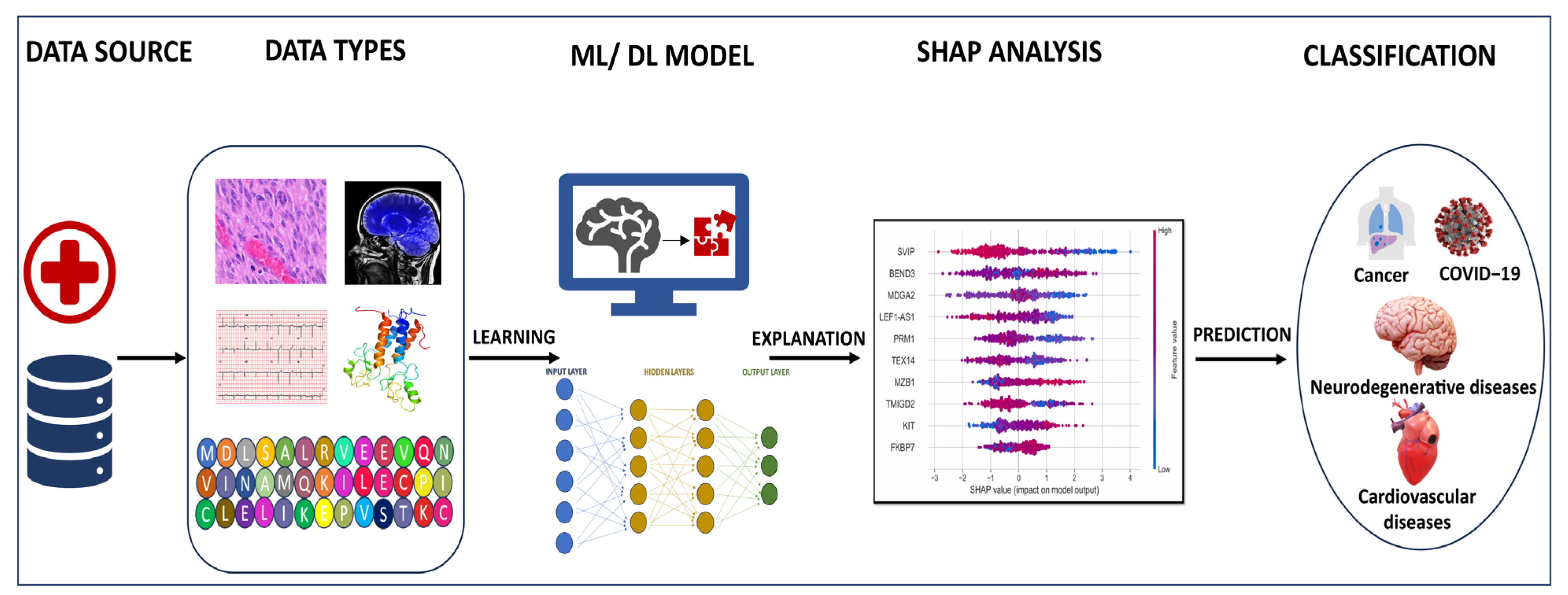
Figure 1. Workflow and application of AI in healthcare. The input data for training the ML/DL model can be obtained from databases and hospitals. These data may include next generation sequencing data, sequence and structural information of proteins, scan images, ECG, histopathology images and electronic health records of the patients. The model trained using these datasets may be able to identify or diagnose a disease condition for an unknown sample. Further, explainable AI techniques including SHAP analysis are used to identify the features responsible for the prediction.
Biomedinformatics 04 00030 g001
Disclaimer/Publisher’s Note: The statements, opinions and data contained in all publications are solely those of the individual author(s) and contributor(s) and not of MDPI and/or the editor(s). MDPI and/or the editor(s) disclaim responsibility for any injury to people or property resulting from any ideas, methods, instructions or products referred to in the content.