Article

Photochemical Redox Reactions of Catecholamines: Detection of Cyclized Oxidation Products and Boronate Esters

Department of Chemistry, College of William & Mary, Williamsburg, VA 23185, USA
*
Author to whom correspondence should be addressed.
Photochem 2026, 6(1), 11; https://doi.org/10.3390/photochem6010011
Submission received: 29 January 2026 / Revised: 28 February 2026 / Accepted: 5 March 2026 / Published: 9 March 2026

Abstract

Our recent work has focused on red light-mediated photoreduction of p-benzoquinones and both o-, and p-naphthoquinones using methylene blue and the chlorophyll metabolite, pheophorbide A as photosensitizers. Photoreduction of biologically relevant quinones mimics photoreduction of plastoquinone by chlorophyll in photosynthesis. We examined photo-oxidation and photoreduction reactions of catechols because their oxidation to o-quinones by reactive oxygen species is implicated in protein damage in neurodegeneration. Photo-oxidation of catecholamines including dopamine, epinephrine and norepinephrine required red light, methylene blue or pheophorbide A, and molecular oxygen. Their cyclized oxidation products, aminochrome, adrenochrome and noradrenochrome, were detected by UV/visible spectroscopy. Hydrogen peroxide was generated during photo-oxidation by singlet oxygen-dependent oxidation of catecholamines. Inclusion of tertiary amine electron donors decreased cyclized products but did not affect hydrogen peroxide yield consistent with concurrent photo-oxidation followed by photoreduction of the o-quinone intermediate. Unreacted dopamine and norepinephrine were quantified using 3-hydroxyphenyl boronic acid following photochemical reactions. Dopamine and norepinephrine boronate esters absorb at 417 and 550 nm. Photo-oxidation of dihydroxycaffeic acid and dihydroxyphenyl acetic acid was also evaluated by detecting their boronate esters at 475 nm. We hypothesize that photoreduction of transient o-quinones by the combination of red light and dietary chlorophyll metabolites may be a path to limit protein damage and to recycle catechol antioxidants.

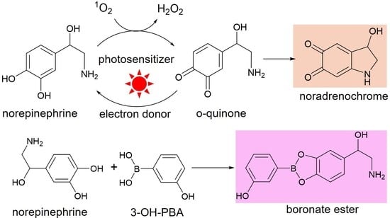
Graphical Abstract

1. Introduction

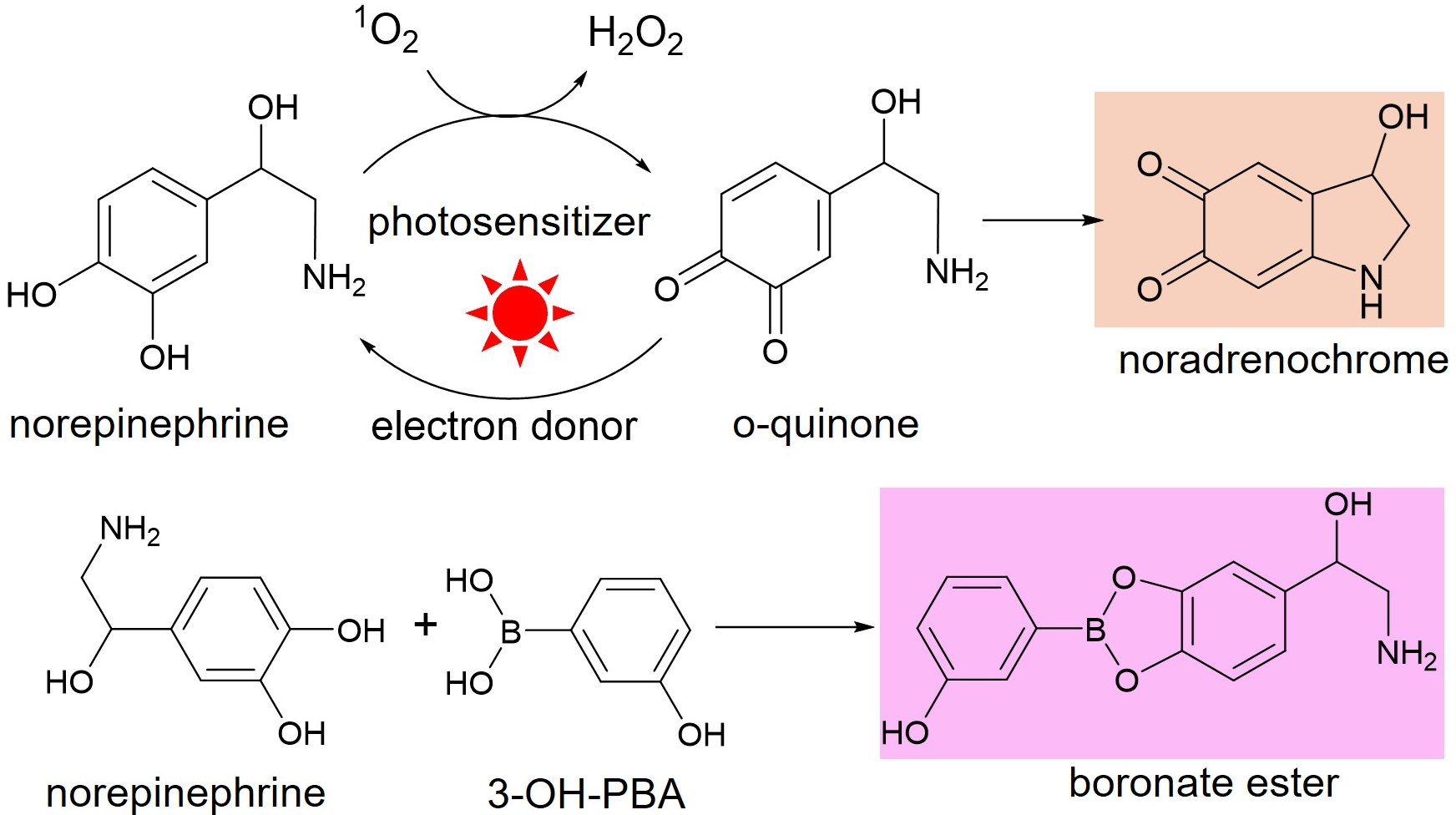
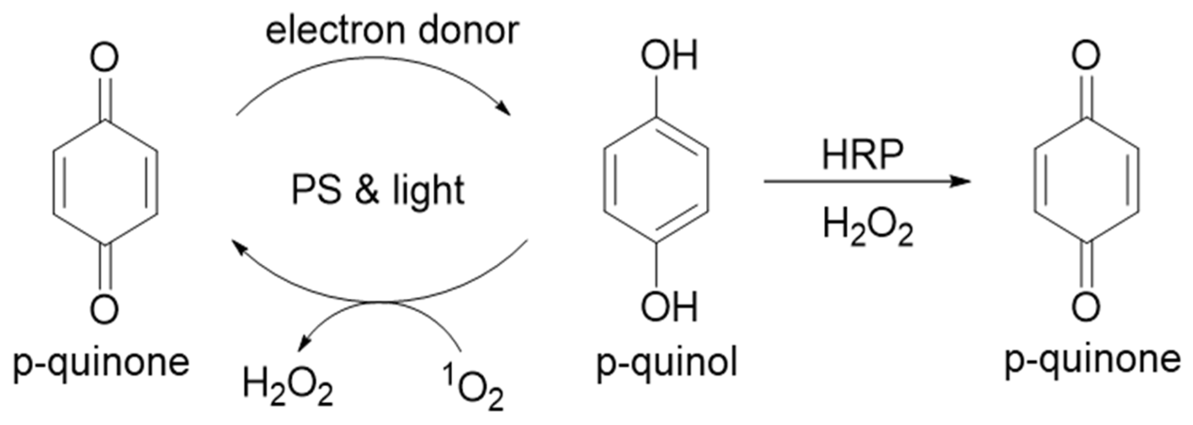
Employing the chemical photosensitizer methylene blue (MB) or the chlorophyll metabolite pheophorbide A (pheoA), we observed concurrent photoreduction of multiple quinone substrates and photo-oxidation of the resulting quinols (Scheme 1) in our previous work [1,2]. Photochemical redox cycling between the oxidized and reduced forms required singlet oxygen (1O2) and generated hydrogen peroxide (H2O2) as a byproduct. H2O2 was detected using horseradish peroxidase (HRP), an enzyme that oxidizes the quinol photoproduct to the quinone.
One objective has been to explore the range of physiological photochemical reactions that may be mediated by dietary chlorophyll. In photosynthesis, red light-mediated photoexcitation of chlorophyll initiates electron transfer to plastoquinone, a p-benzoquinone embedded in chloroplast membranes [3,4]. While considerable work has explored the photochemistry of chlorophyll in plants, only limited research has examined probable light-driven reactions of the chlorophyll we consume. PheoA used in our work is the primary metabolite produced from dietary chlorophyll in vivo [5,6].
Qu et al. reported that the combination of red light and chlorophyll metabolites photoreduced ubiquinone, a membrane-bound p-benzoquinone similar to plastoquinone that is an essential component of the electron-transport chain (ETC) that produces ATP in mitochondria [7]. Further, Zhang et al. reported that ATP yields in isolated mouse mitochondria increased when treated with chlorophyll metabolites and red light (670 nm) [8]. In the absence of light or chlorophyll metabolite, no increase in ATP was observed. In further support of a physiological role of chlorophyll and red light, C. elegans treated with chlorophyll metabolites and red light produced more ATP, and their life span increased [9].
These cellular examples and our experiments on p-quinones prompted us to re-visit work we reported on photochemical reactions of catechols [1,10]. Catechols include neurotransmitters, dopamine (DA) and epinephrine (EP), as well as plant-derived antioxidants like catechins and EGCG from green tea [11,12]. When catechols are oxidized by reactive oxygen species (ROS) including 1O2, a transient o-quinone is formed that resembles the p-quinones that we have employed as photoreduction substrates. O-benzoquinones, unlike p-benzoquinones, are not stable in aqueous solution and react rapidly to generate secondary products, often dimers and multimers [13,14,15,16]. In Parkinson’s disease, the o-quinone produced from DA by ROS reacts with protein cysteines in the substantia nigra [17,18]. Photoreduction of a transient o-quinone by the combination of red light and dietary chlorophyll metabolites would be an intriguing path to limit protein damage and to increase cellular antioxidant capacity. It is well-known that ultraviolet light exposure ensures adequate vitamin D biosynthesis [19]. Similarly, a red light-mediated photoreduction pathway for optimal health is worthy of investigation.
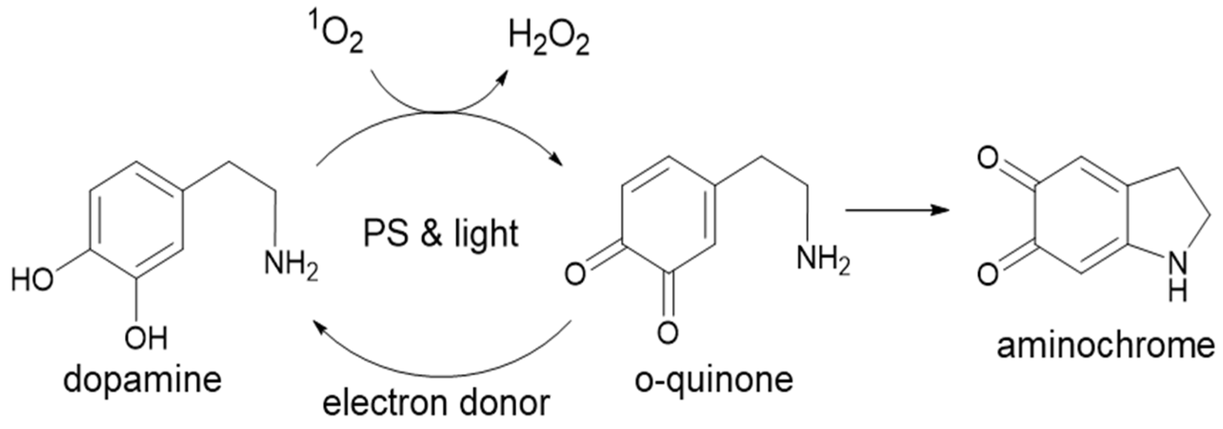
Photochemical reactions of catechols are challenging to study because a catechol must first be oxidized to its o-quinone intermediate by singlet oxygen that is generated by a photosensitizer, light and molecular oxygen (O2) [10,20,21]. The transient o-quinone is then photoreduced back to the catechol by the same photosensitizer, light and an electron donor as shown in Scheme 2. For catecholamines like DA, an intramolecular cyclization reaction of the o-quinone produces aminochrome, a product that absorbs in the visible range. Photo-oxidation of dopamine by 1O2 produces H2O2 and increasing concentrations of electron donors limit aminochrome formation [1].
Equipped with a greater understanding of the photochemical redox reactions of p-quinones and naphthoquinones mediated by MB and pheoA, we expanded our work to include the biologically relevant catecholamines L-dopa, EP and norepinephrine (NE). All photochemical reactions were performed in neutral, aqueous solution (phosphate buffer pH 7.4). Further, we employed new methodology to detect catechols using 3-hydroxyphenyl boronic acid (3-OH-PBA) [22]. Under basic conditions (50 mM carbonate, pH 11.5), several catechols reacted with 3-OH-PBA in minutes to yield products with visible absorbance. This method was used to quantify decreases in catechol concentrations following irradiation. This was especially important because we tested catechols that do not cyclize to colored products after photo-oxidation.

2. Materials and Methods

All chemicals were from Fisher Scientific (Waltham, MA, USA) or Millipore-Sigma (St. Louis, MO, USA) and were of the highest purity available. Pheophorbide A and TMB stock solutions were prepared in DMF and stored at −20 °C. Catechol stock solutions (20 mM) were prepared in water and stored at −20 °C. EDTA and TEOA stock solutions in water (25 or 250 mM, respectively) were adjusted to pH 7.4 with NaOH or HCl. All other solutions were prepared in water or 10 mM PB pH 7.4. Phosphate buffers were equilibrated to room temperature (20–22 °C) to ensure no variation in dissolved O2. Sodium carbonate (50 mM) was prepared fresh daily. 3-OH-PBA (20 mM) in water was stable at −20 °C indefinitely and at 4 °C for at least one week. Sodium periodate solutions (20–50 mM) in water were prepared fresh daily.

2.1. Red Light Specifications

A 36-watt red light composed of eighteen 2-watt LEDs (Wolezek LED, Amazon) was used for all photochemical experiments. The maximum wavelength of emitted light was 660 ± 10 nm. Samples were placed under the lamp on an aluminum foil-lined surface in a shallow box (11 cm from light source to surface). The irradiance directly under the center of the lamp was 31.5 mW/cm2. The total number of samples in plate wells was limited to 16 in a 4 × 4 array to minimize light intensity differences. Light intensity was measured regularly to ensure consistent exposure as described in [10]. Red light irradiation times did not exceed 2.5 min.

2.2. Photochemical Reactions of Catecholamines

Solutions contained 1 mM catecholamine and 2.5 mM MB or 12.5 mM pheoA in 10 mM PB pH 7.4. Due to solubility constraints, L-dopa reactions were in 25 mM PB pH 7.4. For MB/EDTA reactions, 0.625, 1.25 and 2.5 mM EDTA was used to achieve EDTA to MB ratios of 250, 500 and 1000. For pheoA/TEOA reactions, 6.25, 12.5 and 25 mM TEOA was used to achieve TEOA to pheoA ratios of 500, 1000 and 2000. All pheoA reactions with or without TEOA contained 20% DMF.
Absorbance was measured either as full scans (220–800 nm) in a cuvette (1 mL) or at select wavelengths in a 96-well plate (100 mL) prior to light exposure and after 2.5 min under the red light. Separate dark samples containing all reactants served as controls. For DA, L-dopa and EP, cyclized products were detected at 475 nm; for NE, 481 nm was used. To determine cyclized product concentrations from the plate assay, products were scanned immediately in a 1 cm pathlength microcuvette to determine a conversion factor. The pathlength in a 96-plate varies by sample volume. For 100 mL reactions in a 96-well plate, 30% of the published molar absorptivity for each catecholamine was used to convert to molarity. For this conversion, cyclized products were scanned in a cuvette and aliquots were immediately transferred to plate wells and the absorbance was measured. Catecholamines were also treated with sodium periodate or the combination of known amounts of H2O2 and HRP to ensure that their products were identical to the cyclized photo-oxidation products.

2.3. Detection of H2O2 in Photoreduction Samples

HRP (10 mL, 2 mg/mL) was added directly into the cuvette (1 mL) after irradiation and mixed by inverting 2–3 times. For 96-well plate reactions, 2 mL HRP solution was added to 100 mL reactions and samples were mixed by pipetting up and down 2–3 times. Absorbance increases indicative of H2O2 were monitored for up to 7 min until no further increase in absorbance occurred.

2.4. Detection of H2O2 with TMB and HRP

For reactions containing only photosensitizers and EDTA or TEOA, TMB and HRP were used to detect H2O2. Photo-oxidation reactions (100 mL) were prepared either in open microfuge tubes or in plate wells. Following irradiation for 2.5 min, aliquots (20 mL) were combined with 0.5 mM TMB and 0.5 mM HRP in 10 mM PB pH 7.4 (100 mL final) in a 96 well plate. After blue color development, 100 mL of 1 M HCl was added and absorbance was read at 450 nm. A standard curve from 0–40 mM H2O2 was used to calculate H2O2 concentrations in the photo-oxidation reactions. The HRP concentration was determined at 403 nm [23].

2.5. Detection of Catechols with 3-OH-Phenyl Boronic Acid (3-OH-PBA)

Catechol standards were combined with 3-OH-PBA in 50 mM sodium carbonate (200 mL) in 96-well plates. Reactions of all standards contained 200 mM 3-OH-PBA and typical catechol concentrations ranged from 12.5 to 50 mM (4–16-fold excess 3-OH-PBA). Color development was monitored by UV/visible spectroscopy (Perkin-Elmer Lambda 35 UV/Vis, Shelton, CT, USA) in a cuvette (1000 mL reaction) to determine optimal reaction times for each catechol used and absorbance maxima for each.
Photochemical reactions containing 1 mM catechol (100 mL) were prepared either in open microfuge tubes or in plate wells to ensure continuous oxygen exposure as described. Dark samples were also prepared as controls. After irradiation, aliquots (10 mL) were transferred to plate wells and combined with 190 mL 3-OH-PBA in 50 mM carbonate. Final concentrations of catechol and 3-OH-PBA in plate wells were 50 mM and 200 mM, respectively.

2.6. TLC Analysis of Catechol Reactions with 3-OH-PBA

Catechols, 3-OH-PBA standards, and reaction mixtures were separated in 75% ethyl acetate and 25% hexanes. Rf of 3-OH-PBA = 0.7; Rf values for all catechols were between 0.3 and 0.5. With 4-fold excess 3-OH-PBA, no catechol remained after the reaction times reported herein.

2.7. Data Analysis

For each treatment/condition, at least two-three independent experiments were performed in duplicate or triplicate. Mean values were calculated for each independent experiment (mean ± error). For figures showing error bars, mean values for independent experiments were averaged, and error calculated. Details for each are stated in the Figure Legends.

3. Results

We reported that p-benzoquinones as well as o- and p-naphthoquinones are photoreduced by the combination of photosensitizers, electron donors and red light in pH 7.4 phosphate buffer [1,2]. Because H2O2 was generated during photoreduction, we determined that newly photoreduced quinols reacted with 1O2 to regenerate quinones (Scheme 1). H2O2 generation proceeds via 1O2-mediated hydrogen atom abstraction from the quinol to yield a peroxyl radical (•OOH) and a semiquinone (RO•). At pH 7.4 used herein, •OOH will deprotonate to superoxide anion (O2−•) because its pKa is 4.8 [24]. Disproportionation of two superoxide anions yields O2 and H2O2 according to Equation (1). Further, two semiquinones disproportionate to yield a quinol and regenerate a quinone [25,26].
O2−• + O2−• + 2H+ → H2O2 + O2
Quinols and catechols are substrates for horseradish peroxidase (HRP), an enzyme that uses H2O2 to oxidize many organic molecules [15,16,27]. Quinols are oxidized by HRP only when H2O2 is available; therefore, p-quinone increased when HRP was added following irradiation (Scheme 1). When catalase was added prior to HRP, no increase in p-quinone was detected because it consumes H2O2 according to Equation (2):
2H2O2 → 2H2O + O2
Herein we applied this quinone photoreduction methodology to catechol photo-oxidation. Unlike p-quinones, a simple cycling mechanism between the oxidized and reduced forms in Scheme 1 is not possible because the o-quinone intermediates generated from catechols are not stable and will form a range of products depending on their substituents [28]. For example, catecholamines like DA will undergo an intramolecular Michael addition wherein the amine substituent adds to the o-quinone (Scheme 2). Catechins from green tea lack amine substituents and consequently dimerize or form multimers depending on reaction conditions of pH and concentration [13,14].
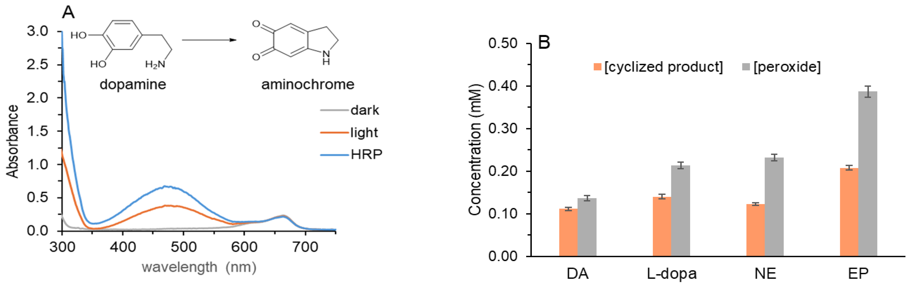
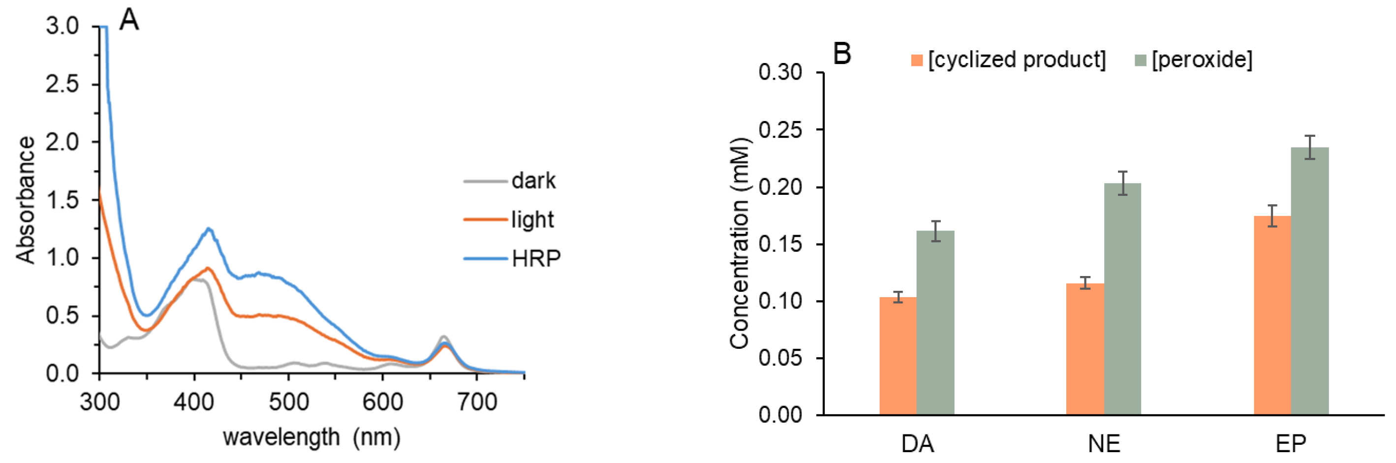
The combination of the photosensitizer, MB, DA, O2 and red light produced H2O2 and oxidized DA to aminochrome (Figure 1A). In prior work, we confirmed that 1O2 was required for this reaction because azide, a known 1O2 scavenger, decreased aminochrome [29]. Further, when identical reactions contained 20% D2O, thereby extending the lifetime of 1O2, aminochrome increased [10].
The absorbance spectrum in Figure 1A was obtained in a 1 mL quartz cuvette that was placed directly under a red light with maximum output at 660 nm. Two peaks are observed at wavelengths greater than 350 nm in Figure 1A after irradiation for 2.5 min: MB at 665 nm and that of aminochrome at 475 nm. Prior to light exposure, only MB was detected. Because the typical concentration of dissolved O2 in 10 mM phosphate pH 7.4 at 20–22 °C is 250–300 mM using an oxygen electrode, irradiation time was limited to 2.5 min to avoid O2 depletion. When irradiation time was increased to 5 min, only 10–15% more aminochrome formed. The absorbance increase at 300 nm has also been used to quantify aminochrome [20,21].
As for quinones, H2O2 was confirmed when HRP was added to the cuvette after irradiation. Aminochrome increased further because the solution contained sufficient unreacted DA to be oxidized by HRP (Figure 1A). When catalase was added prior to HRP, no increase in aminochrome was detected. The stoichiometry of the HRP reaction with DA was determined by adding known amount of H2O2 (Supplemental Figure S1). Addition of 200 mM H2O2 generated 100 mM aminochrome confirming that aminochrome is the four-electron oxidized product of DA [26,28]. A second 200 mM portion of H2O2 increased aminochrome further but only by ~85–90%. We suspect that the solution contains traces of additional DA oxidation products that are also HRP co-substrates.
Oxidation to aminochrome proceeds via the two-electron oxidized product, leukoaminochrome, which is also a catechol. Leukoaminochrome and a second oxidation product, 5,6-dihydroxyindole, do not absorb in the visible range [28]. Aminochrome is not stable and will polymerize within ~15–20 min to a grayish solution and ultimately produce a black precipitate. HRP assays using other peroxidase co-substrates to quantify H2O2 including ABTS; o-phenylenediamine and TMB cannot be used because excess DA, aminochrome, and other oxidation products in the solution interfered with their color formation.
DA was also oxidized to aminochrome with sodium periodate (Supplemental Figure S2). As for HRP/H2O2, two equivalents of periodate yielded one equivalent of aminochrome. When HRP was added after periodate, no increase in aminochrome was observed confirming that no H2O2 formed.
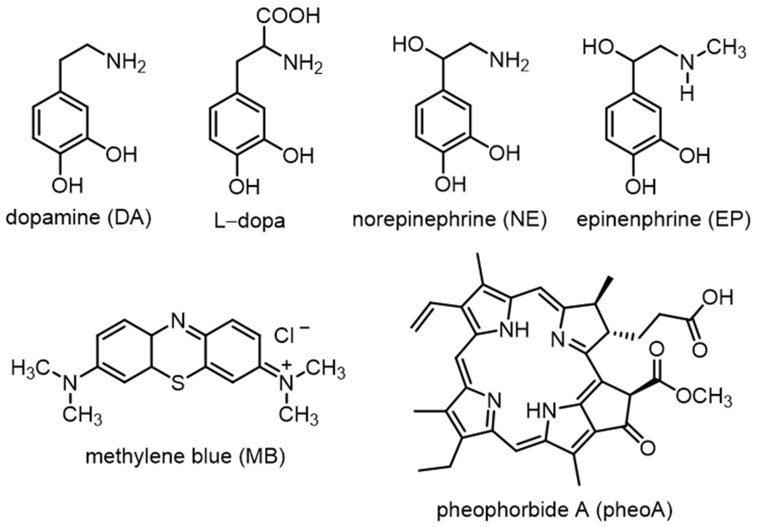
DA and several other catecholamines (Scheme 3) were assayed under the same conditions as in Figure 1A except in a 96-well plate format. In the open wells of the plate and with only 100 mL volumes, O2 does not become limiting as it might in a narrow 1 mL cuvette. Figure 1B summarizes the concentration of each cyclized oxidation product and the amount of H2O2 generated during photo-oxidation. A 2:1 ratio of H2O2 to cyclized product for HRP was used (Supplemental Figure S1).
DA photo-oxidation produced the least amount of cyclized product followed by NE, L-dopa, and EP. Indeed, the yield of cyclized EP product, adenochrome, was nearly 2-fold greater than DA as shown in Figure 1 with 0.11 mM aminochrome and 0.21 mM adenochrome. The enhanced reactivity of EP is attributed to the greater nucleophilicity of the methylated amine, not to increased reactivity of the catechol ring with 1O2. Yields of H2O2 for L-dopa, NE and EP were nearly double that of their cyclized product but for DA, H2O2 was only 30% greater. Based on stoichiometry of the HRP reaction and that of photo-oxidation, a 2:1 ratio was expected. These results support a common mechanism of photo-oxidation via the semiquinone (RO•) and H2O2 generation from superoxide anion.
Formation of cyclized products reported in Figure 1B requires oxidation of each to the reactive o-quinone intermediate. To study o-quinone photoreduction, EDTA was included as the tertiary amine electron donor. EDTA is an inexpensive and well-characterized electron donor used in many photochemical studies because its breakdown products have been characterized and are inert within the time frame of our experiments [30]. In all prior photoreduction work, the combination of MB and EDTA was very effective. At pH 7.4, MB is positively charged and EDTA has multiple negative charges. Favorable electrostatics facilitate electron transfer from EDTA to MB during cycles of photoreduction. We optimized reaction conditions using ratios of EDTA to MB of 250:1 to 1000:1.
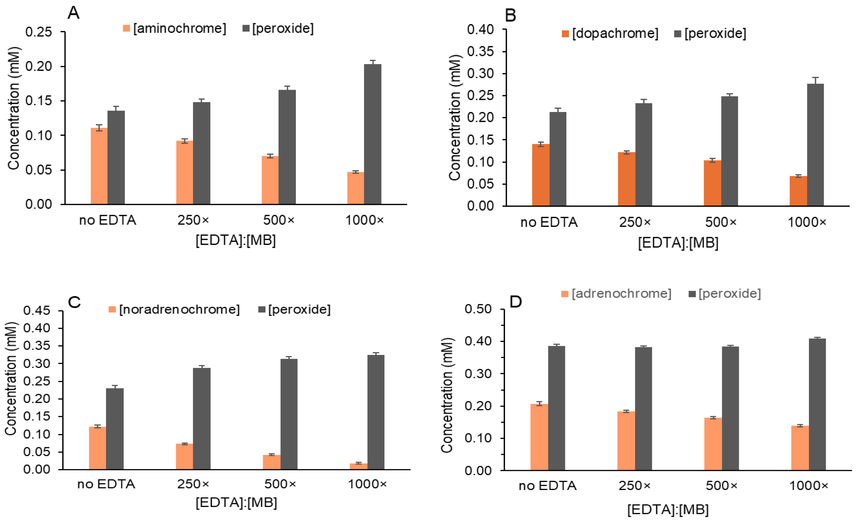
Each catecholamine was photo-oxidized with MB alone and with increasing EDTA concentrations to assess photoreduction (Figure 2A–D). The data for “no EDTA” in each graph is from Figure 1B. In Figure 2A, as EDTA increased, DA oxidation to aminochrome decreased. Despite less aminochrome, the yield of H2O2 did not decrease but increased as EDTA increased. H2O2 formation, even when EDTA was present, supports our hypothesis of photo-oxidation to the intermediate o-quinone followed by photoreduction (Scheme 2).
The reaction of tertiary amines including EDTA with 1O2 generates H2O2 as an end product [31,32,33]. Superoxide anion is produced when 1O2 abstracts an electron from a tertiary amine, R3N as shown in Equation (3) [33]. We recently reported H2O2 production from multiple common tertiary amines including EDTA and the biochemical buffers MES, MOPS, PIPES and HEPES [32].
1O2 + R3N: → O2−• + R3N+•
Rate constants for the reaction of catechols with 1O2 (107 M−1 s−1) are two orders of magnitude greater than those of tertiary amines with 1O2 [34,35]. In our photochemical reactions (Figure 2), 1 mM catecholamine is competing with up to 2.5 mM EDTA (in 1000:1 sample) for 1O2. We determined the H2O2 yields in reactions that contained MB, the three EDTA concentrations, and ambient O2 but no catecholamines. Only 33–40 mM H2O2 was produced photochemically from EDTA, well below the H2O2 concentrations reported in Figure 2A.
For L-dopa in Figure 2B, a similar decrease in the cyclized product, dopachrome, as EDTA increased was observed as well as the corresponding increase in H2O2. For NE, in Figure 2C, the decrease in cyclized product, noradrenochrome, as EDTA increased was greater than in Figure 2A,B,D. Noradrenochrome decreased by over 90% with 1000x EDTA:MB compared to decreases of 60%, 50% and 33% for aminochrome, dopachrome and adrenochrome, respectively. This difference in photoreduction under the same reaction conditions likely reflects differences in photoreduction potential.
Table 1 summarizes all H2O2 yields obtained in Figure 2A–D and in the absence of catechols for the three EDTA concentrations. For all catecholamines, the H2O2 yields were markedly greater than those for the EDTA reactions without catecholamines. This confirms that in reactions with catecholamines, H2O2 is formed primarily during the catechol photo-oxidation step, not via EDTA oxidation. Further, decreased cyclized products with increasing EDTA is evidence for o-quinone photoreduction.
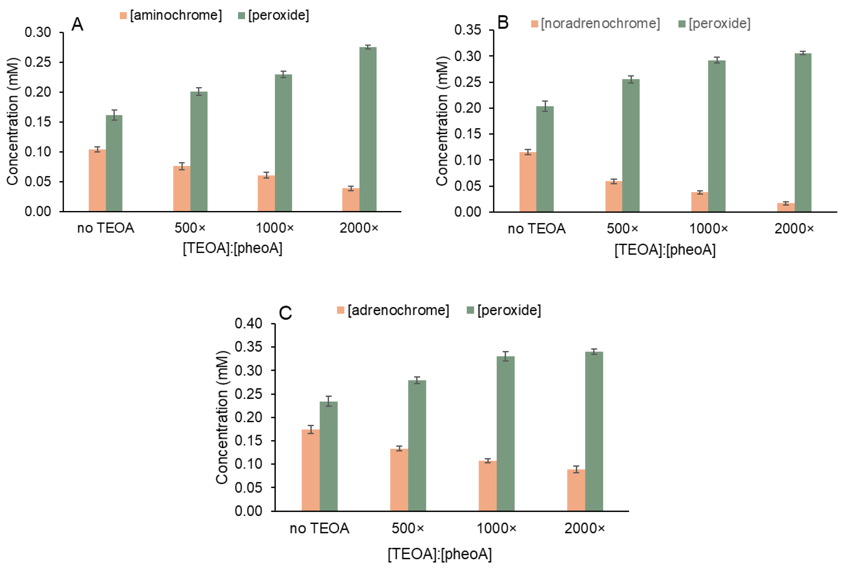
Photo-oxidation reactions were performed using pheoA as the photosensitizer (Figure 3A). For optimized pheoA solubility and reactivity, 20% DMF was included in all pheoA-mediated photochemical reactions [1,2]. Higher DMF concentrations were not tested because we did not want to compromise the activity of HRP and catalase, enzymes used to elucidate photo-oxidation and photoreduction pathways.
PheoA has two absorbance peaks in the visible range: one at 665 nm and another at 405–410 nm (Figure 3A—dark). After irradiation of a solution containing pheoA, 20% DMF and 1 mM DA, aminochrome is evident at 475 nm. When HRP was added, aminochrome increased further indicative of H2O2. Five-fold more pheoA was required to achieve approximately the same aminochrome yield as MB. This was expected and is consistent with our quinone photoreduction results.
PheoA does not absorb between 450 and 600 nm; therefore, 475 nm was used in a 96-well plate assay to measure both cyclized products and H2O2. Figure 3B summarizes the data for DA, NE and EP. L-dopa could not be assayed with pheoA due to its limited solubility in 20% DMF. The greater amount of the cyclized EP product, adenochrome (0.18 mM) relative to cyclized DA product, aminochrome (0.10 mM), is consistent with what we observed using MB in Figure 1B. NE photo-oxidation was modestly greater than that of DA.
PheoA has one carboxylate; therefore, EDTA was not an effective electron donor due to its multiple carboxylates. Triethanolamine (TEOA) with its pKa of 7.75 and no charges, is used routinely in our experiments as the electron donor with pheoA [1,2,30]. However, even with TEOA as the electron donor and no charge repulsion, pheoA photoreduction capacity calculated as mol quinone reduced per mol photosensitizer is always lower than the combination of MB and EDTA. Higher ratios of TEOA to pheoA than EDTA to MB were necessary to afford comparable photoreduction.
Figure 4A shows that pheoA-mediated photoreduction of DA increased with increasing TEOA. The highest TEOA concentration tested decreased aminochrome yield by 60% which is identical to photoreduction with MB and EDTA in Figure 2A. For NE, in Figure 4B, the cyclized product decreased by 85% also in good agreement with Figure 2C. Likewise, for EP in Figure 4C, the cyclized product decreased the least. Given that EP is most readily photo-oxidized, less photoreduction is expected.
Because higher TEOA concentrations were required for photoreduction with pheoA, the direct reaction of TEOA with 1O2 according to Equation (3) contributed more to the overall H2O2 yield. For example, 276 mM H2O2 was detected using HRP at the highest TEOA concentration in Figure 4A vs. 246 mM H2O2 in the absence of DA with the same TEOA. Table 2 summarizes all H2O2 yields obtained in Figure 4A–C and in the absence of catecholamines. For all catecholamines tested, the H2O2 yields in Figure 4 were greater than those for the TEOA reaction with 1O2. Although the difference is less striking than in Table 1, these results support a common mechanism of catecholamine photo-oxidation by 1O2 prior to photoreduction regardless of the photosensitizer and electron donor used.
Comparison of Figure 2 and Figure 4 highlight some differences in H2O2 yields after irradiation that warrant scrutiny. Although quantitation of the cyclized products is straightforward, it does not provide a full picture of all oxidation products. Moreover, molar absorptivity values for visible products are only in the 3000–4000 M−1 cm−1 and photochemical reactions of catechols that lack amine substituents cannot be studied by this method. Lastly, cyclized oxidation products are not stable in solution, which limits the use of some analytical methods to detect all products.
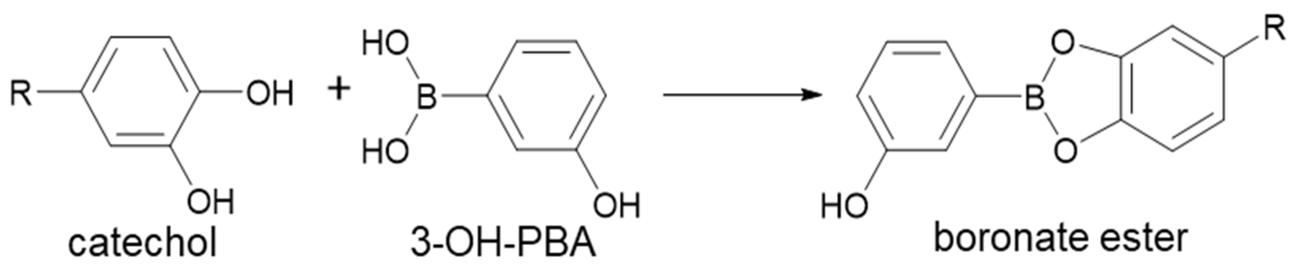
Catechols react avidly with boronic acids to form cyclic boronate esters under basic conditions (Scheme 4). Recently, Liu et al. reported a straightforward and sensitive assay using 3-hydroxyphenylboronic acid (3-OH-PBA) to detect DA [22]. The resulting boronate ester is fluorescent but also absorbs at 417 nm. We sought to measure remaining DA after the photochemical steps described in Figure 1, Figure 2, Figure 3 and Figure 4 by measuring absorbance at 417 nm in 50 mM carbonate solution (pH 11.5).
Initial evaluation of the DA reaction with 3-OH-PBA showed reliable detection of as little as 12.5 mM DA in 20 min at 417 nm (Supplemental Figure S3). A yellow product was observed within minutes of mixing; we read absorbance after 20 min when the reaction was about 90–95% complete. The absorbance at 417 nm was observed only at high pH in 50 mM carbonate because it is contingent on deprotonation of 3-OH-PBA. Acid addition shifted the absorbance from 417 nm to 385–390 nm. At pH 1–2, no absorbance greater than 300 nm was observed which is consistent with reversal of the reaction shown in Scheme 4.
Multiple controls were performed to ensure that both reactants and products in our photochemical reactions did not interfere with boronate ester quantitation. Up to 500 mM H2O2 was added to control, dark reactions with 1 mM DA; aliquots were mixed with 3-OH-PBA to achieve 50 mM DA (25 mM H2O2 final) and no change in absorbance at 417 nm was observed. DA was treated with sodium periodate to produce aminochrome and remaining DA was detected with 3-OH-PBA. Data in Supplemental Figure S4 confirms that aminochrome did not react with 3-OH-PBA. Similar control experiments with the other catecholamines were performed with identical outcomes.
Dark samples containing MB, EDTA and DA or pheoA, TEOA and 20% DMF were prepared. Supplemental Figure S5 shows that quantification was not affected by the relatively high concentrations of EDTA or TEOA we employ. Because pheoA absorbs at 417 nm, dark controls with pheoA were modestly higher than with MB. For photochemical reactions that contained pheoA, their DA concentration was calculated using a standard curve that also contained pheoA and DMF (Supplemental Figure S6).
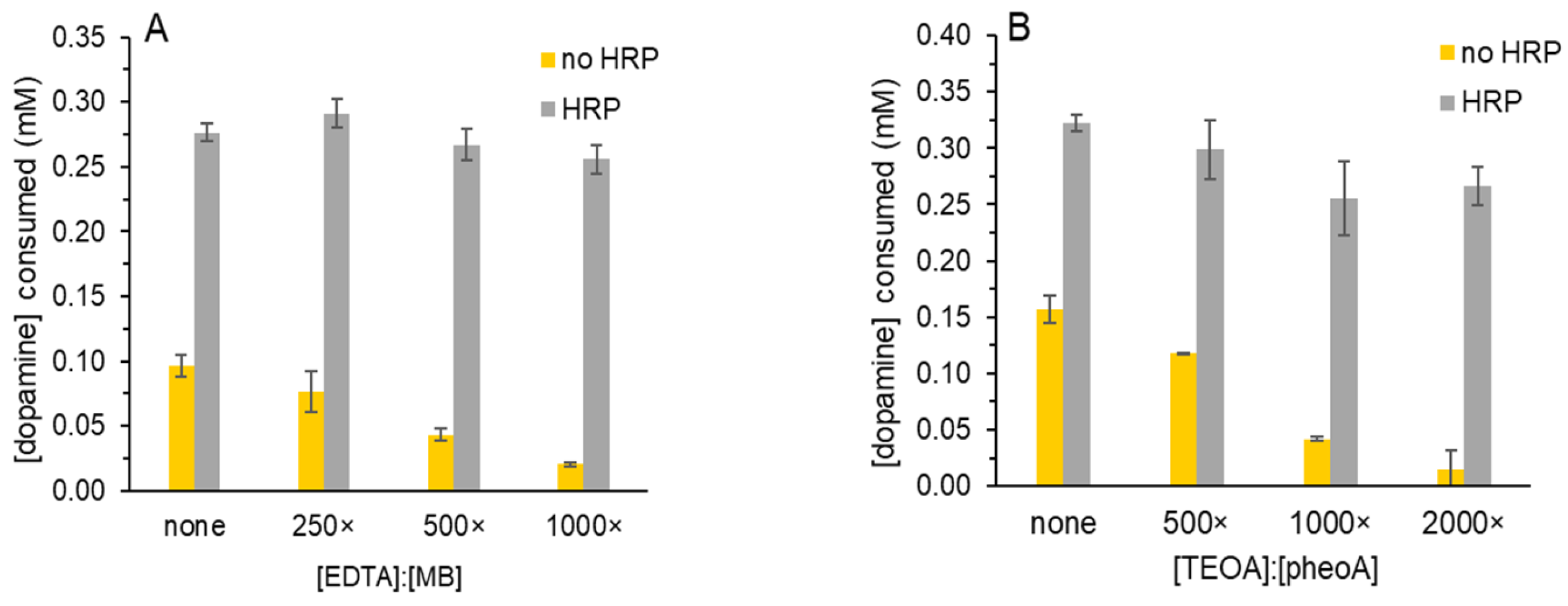
Reactions were prepared as in Figure 2A and DA was measured using 3-OH-PBA (Figure 5A). As the ratio of EDTA to MB increased, DA that was consumed decreased (in yellow). The change in DA consumed as EDTA increased parallels the decrease in aminochrome in Figure 2A. The decrease in DA consumed in Figure 5A with 1000:1 EDTA to MB was 80%, which is greater than the 50% decrease in aminochrome in Figure 2A. This suggests that not all DA was oxidized to aminochrome but that there are other oxidation products that do not absorb at 475 nm and do not react with 3-OH-PBA. Even though leukoaminochrome and dihydroxyindole are also catechols, we did not observe additional boronate esters with visible absorbance at the concentrations tested.
When HRP was added after irradiation, more DA was consumed because H2O2 that formed during photo-oxidation reacted with it. Unlike Figure 2A where aminochrome increased only 30% with HRP (no EDTA), Figure 5A shows a 2.5-fold increase in DA consumed (“no EDTA”). This also suggests that the HRP reaction of residual H2O2 and unreacted DA generates products other than aminochrome that do not absorb in the visible range.
When the DA boronate ester was detected after photochemistry with pheoA and TEOA, similar trends were observed. In Figure 5B, we observed a dose-dependent decrease in DA consumed as TEOA increased, that mirrors the decrease in aminochrome in Figure 4A. The resulting increase in DA consumed with HRP was less than that in Figure 5A but greater than in Figure 4A (no TEOA).
Other catecholamines were tested to determine if they reacted with 3-OH-PBA in a similar manner. L-dopa boronate ester was detected at 415 nm; however, its formation was much slower than that of DA. Even after an hour (vs. 20 min for DA), absorbance was still increasing. Given that catecholamines in basic solution oxidize more readily we did not use 3-OH-PBA to detect L-dopa after irradiation. EP did not react with 3-OH-PBA to form a boronate ester with visible absorbance.
NE reacted with 3-OH-PBA to yield a boronate ester that absorbed at 550 nm and a linear response was determined between 12.5 and 75 mM NE (Supplemental Figure S7). Reaction time was 25 min vs. 20 min for DA. Similar controls were performed to ensure that photochemical reactants and products did not interfere with the reaction.
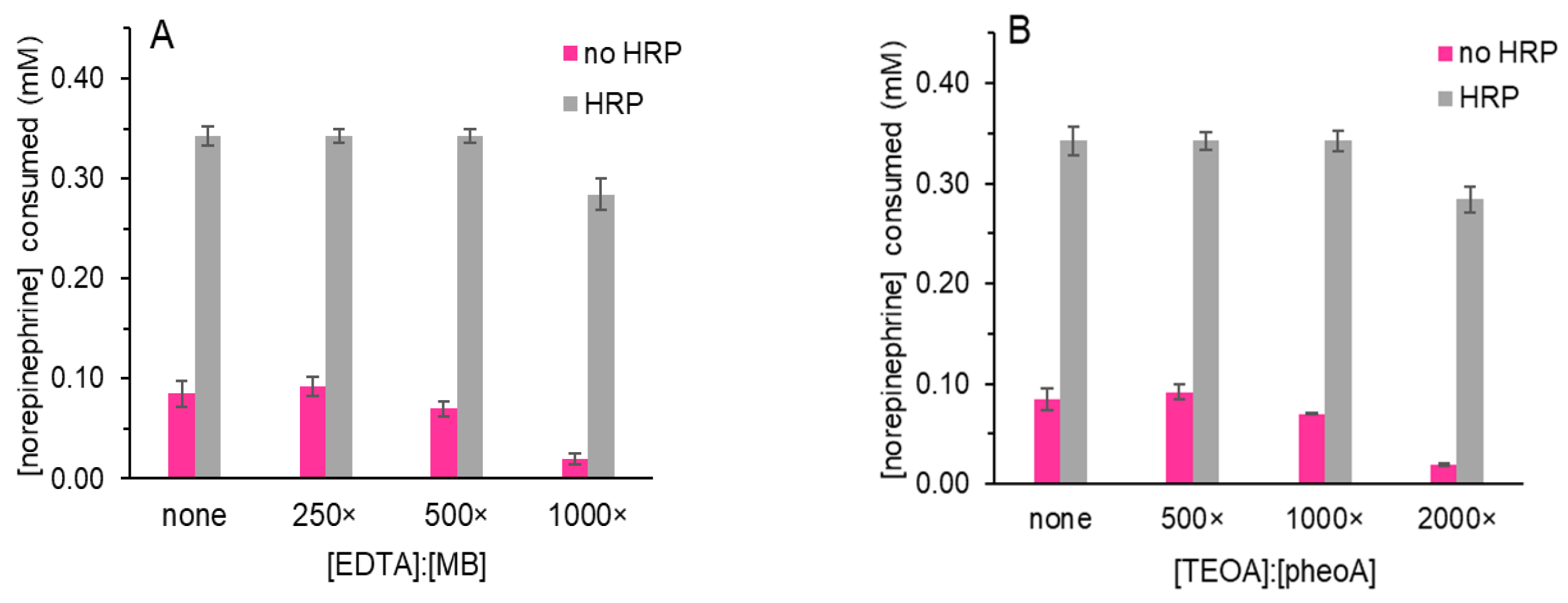
Photochemical reactions were prepared as in Figure 2C and NE was measured using 3-OH-PBA. As the ratio of EDTA to MB increased, NE that was consumed decreased (Figure 6A). This decrease in NE consumed as EDTA increased parallels the decrease in noradrenochrome in Figure 2C. The addition of HRP resulted in a nearly four-fold increase in NE consumed (no EDTA). As for DA in Figure 5A, this increase was greater than we observed when only the cyclized product was detected in Figure 2C. One concern is that additional products other than the cyclized products or H2O2 that form during the photochemical steps alter the rate or stability of the boronate esters.
When the NE boronate ester was detected after photochemistry with pheoA and TEOA, trends similar to those for the DA boronate ester were observed. In Figure 6B, we observed a dose-dependent decrease in NE consumed as TEOA increased that mirrors the decrease in noradrenochrome in Figure 4B. It is notable that of the four catecholamines in Figure 2 and Figure 4, NE treated with the highest ratios of electron donor to photosensitizer in Figure 2C and Figure 4B formed the least amount of cyclized product. Using 3-OH-PBA in Figure 6 to detect NE consumed, this was also observed.
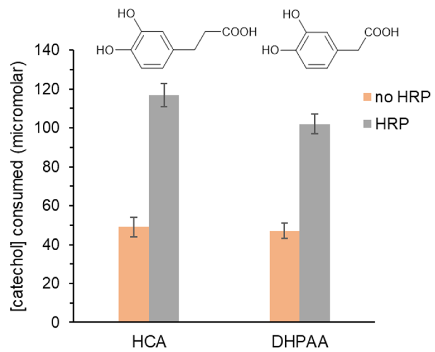
This methodology was applied to two catechols that do not have amines substituents that are necessary to form cyclized oxidation products. Hydrocaffeic acid (HCA) and dihydroxyphenylacetic acid (DHPAA) both contain carboxylic acid substituents and only differ by one methylene unit (Figure 7). Initial investigation showed that both reacted with 3-OH-PBA to yield products that absorb at 475 nm. However, the reaction of DHPAA with 3-OH-PBA was completed in 8 min vs. 20 min for HCA. Absorbance of the DHPAA boronate ester was also greater than that of HCA though both standard curves were linear from 12.5 to 50 mM (Supplemental Figures S8 and S9).
Figure 7 shows that both catechols (1 mM) were consumed to the same extent when mixed with MB and irradiated for 2.5 min. Of note, 5 mM MB was used to reliably detect a decrease in HCA or DHPAA vs. 2.5 mM in all other assays reported herein. Even with more MB, the amount of catechol consumed was less than that of DA or NE in Figure 5 and Figure 6. When HRP was added, more catechol was consumed. If catalase was added prior to HRP, no increase in catechol consumed was detected. Results in Figure 5, Figure 6 and Figure 7 confirm that boronate ester methodology can be employed to study catechol photochemical redox reactions.

4. Discussion

Previously, we evaluated multiple chlorophyll metabolites as photosensitizers for DA photo-oxidation [10]. To photo-oxidize DA to aminochrome, red light and O2 were required thereby implicating 1O2 as the oxidizing species. Inhibition by azide, a known 1O2 scavenger, and increased reactivity in 20% D2O confirmed the essential role of 1O2 [29].
Herein we used the photosensitizers MB and pheoA to assess photo-oxidation of multiple physiologically relevant catecholamine substrates in phosphate buffer at pH 7.4. Results in Figure 1 and Figure 3 confirm photo-oxidation based on formation of products resulting from rapid intramolecular cyclization of the intermediate o-quinone (Scheme 2). These initial products absorb in the visible range though they are not stable indefinitely in solution.
EP was more readily photo-oxidized than all other catecholamines with ~2-fold more cyclized product than DA regardless of the photosensitizer used (Figure 1B and Figure 3B). L-dopa and NE formed intermediate amounts of cyclized products. Kruk reported the same reactivity trend in methanol using MB, Rose Bengal and fluorescein as photosensitizers [20]. Further Kruk monitored changes in absorbance at 300 nm, not in the 475–480 nm range as we reported [20,21]. Several of the chlorophyll metabolites and catechols of interest that we use absorb at 300 nm. To avoid this interference, we used the visible wavelengths.
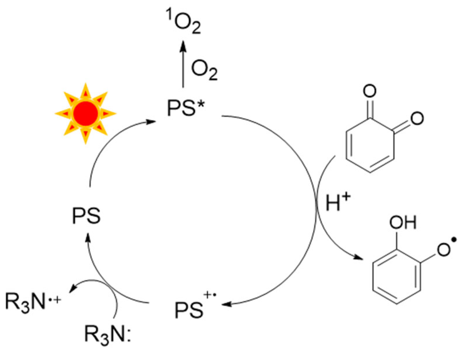
When electron donors were included in the photochemical reactions, a decrease in cyclized products was observed consistent with our prior work wherein photo-oxidation and photoreduction occur concurrently (Figure 2 and Figure 4) [1,2]. In Scheme 5, a photosensitizer (PS) is excited by red light to PS*, the triplet state. PS* reacts with O2 to form 1O2 or it transfers an electron to the intermediate o-quinone to yield the semiquinone and PS+•. The tertiary amine electron donor, R3N, reduces PS+• to regenerate the ground state PS. Because o-quinone is an intermediate, the rate of its photoreduction must exceed the rate of intramolecular cyclization [36].
In catechol reactions without electron donors, the only outcome is photo-oxidation by 1O2 that produces peroxyl radical followed by superoxide anion and ultimately H2O2. Catechols oxidized to semiquinones disproportionate to o-quinone and regenerates catechol. When an electron donor is present, both photo-oxidation and photoreduction occur. The amount of 1O2 formed by PS* is dependent on the O2 concentration. In the experiments described herein, O2 was not limiting. Once o-quinone is produced by photo-oxidation, it becomes a substrate for photoreduction as shown in Scheme 5. In prior experiments using p-benzoquinones and naphthoquinones, photoreduction increased when O2 was limited (Scheme 1).
Concurrent photochemical pathways also occur in intact chloroplasts. When light intensity is excessive, chlorophyll initiates 1O2 formation that is effectively scavenged by pigments within the leaves that act as antioxidants [37].
In photochemical catechol reactions containing tertiary amines, their competing reaction with 1O2 required scrutiny. According to Equation (3), tertiary amines react with 1O2 to generate superoxide anion and ultimately H2O2 [33]. Published rate constants for the reactions of 1O2 with catechols are at least two orders of magnitude greater than those of 1O2 with tertiary amines [34,35]. Data in Table 1 confirms that some H2O2 formed using MB and up to 2.5 mM EDTA (no catechol). However, at least four-fold more H2O2 formed in reactions that contained catecholamines. If EDTA were simply scavenging 1O2 to decrease cyclized products rather than serving as electron donors in o-quinone photoreduction, these high amounts of H2O2 would not have formed.
Even in pheoA reactions where up to 25 mM TEOA was employed, the yield of H2O2 was greater with catechol substrates than in their absence (Table 2). In our recently published work on the reaction of 1O2 with multiple tertiary amines, we detected micromolar H2O2 concentrations similar to those in Table 1 and Table 2 (no catechol) [32].
Because EP was most easily photo-oxidized with the highest yields of cyclized products (Figure 1B and Figure 3B), it is not surprising that increasing electron donors had less of an effect on cyclized product yields (Figure 2D and Figure 4C). This result is also consistent with concurrent photoredox processes rather than 1O2 scavenging by EDTA or TEOA as an explanation for decreased cyclized products in Figure 2 and Figure 4.
DA detection is an area of intense research that often requires elaborate synthetic strategies [38,39,40]. We employed novel methodology to detect unreacted catechols using the boronic acid, 3-OH-PBA. The resulting boronated esters yielded distinct colors detectable in the low micromolar range (Supplemental Figures S3 and S7–S9). The DA boronate ester absorbed at 417 nm whereas that of NE absorbed at 550 nm. Boronate ester products of HCA and DHPAA absorbed at 475 nm. In 50 mM carbonate solution (pH 11.5), catechols and 3-OH-PBA reacted in 25 min or less. Further, 3-OH-PBA is commercially available and inexpensive.
Figure 5 and Figure 6 represent a different detection method to understand the photochemical redox reactions in Figure 2 and Figure 4. It was gratifying to see the same decrease in cyclized DA and NE products as EDTA and TEOA increased. With HRP, catecholamines were consumed to a greater extent than in Figure 2A,C. By only detecting the cyclized, 4 electron-oxidized products in Figure 1, Figure 2, Figure 3 and Figure 4, quantitation is incomplete. Two-electron oxidized intermediates like leukoaminochrome and 1,2-dihydroxyindole (for DA) are known to form but they do not absorb in the visible range [28]. We expect that analogous products form for each of the catecholamines.
Our prior work confirmed photoreduction of quinones by these same combinations of photosensitizers and electron donors. Thus far, we have identified only one molecule, 2,6-dichlorophenolindophenol that was photoreduced by MB and EDTA, but not by pheoA and TEOA [41]. In our work, p-benzoquinones were easiest to photoreduce followed by o-naphthoquinones and lastly, p-naphthoquinones [1,2]. Reported reduction potentials are consistent with this ranking wherein o-quinones are more readily reduced than p-quinones [42,43,44]. The addition of an aromatic ring in naphthoquinones makes reduction more difficult. Thus, reduction potentials favor facile reduction of o-benzoquinones.
PheoA, the predominant chlorophyll metabolite produced in vivo, mediates formation of a triplet state, PS*, capable of producing 1O2 and participating in electron donation reactions—in this case, to an o-quinone. While tertiary amines are not physiologically relevant electron donors, their use allows up to explore catalytic photochemical processes. Even if a chlorophyll metabolite was photo-excited and performed only one electron transfer reaction, that has the potential to recycle intermediate o-quinones of dietary antioxidants produced by cellular ROS.


5. Conclusions

We describe red light-mediated photochemical redox reactions of catecholamines using methylene blue and pheophorbide A as photosensitizers. Dopamine, epinephrine, L-dopa and norepinephrine undergo singlet oxygen-dependent photo-oxidation to form intermediate o-quinones that react with their amine substituents to form cyclized products. Moreover, catechol photo-oxidation also generated hydrogen peroxide. The presence of tertiary amine electron donors suppressed the accumulation of cyclized products while maintaining hydrogen peroxide formation, supporting a mechanism involving initial photo-oxidation followed by photoreduction of transient o-quinones. A boronate ester assay using 3-hydroxyphenyl boronic acid was developed to quantify unreacted catechols and to extend the analysis to non-cyclizing catechols. This work provides valuable mechanistic insight into concurrent photo-oxidation and photoreduction processes.

Supplementary Materials

The following supporting information can be downloaded at: https://www.mdpi.com/article/10.3390/photochem6010011/s1, Figure S1: DA oxidized with H2O2/HRP. Figure S2: DA oxidized with sodium periodate. Figure S3: DA standard curve with 3-OH-PBA. Figure S4: DA oxidized with sodium periodate assayed with 3-OH-PBA. Figure S5: Dark controls with MB/EDTA or pheoA/TEOA plus 20% DMF. Figure S6: DA standard curve with 3-OH-PBA with pheoA and DMF. Figure S7: NE standard curve with 3-OH-PBA. Figure S8: HCA standard curve with 3-OH-PBA. Figure S9: DHPAA standard curve with 3-OH-PBA.

Author Contributions

Conceptualization, L.M.L.; methodology, A.T., H.M., A.M., B.Y. and L.M.L.; formal analysis, A.T., H.M. and L.M.L.; investigation, A.T., H.M., A.M., B.Y. and L.M.L.; writing—original draft preparation, L.M.L.; writing—review and editing, L.M.L.; supervision, L.M.L.; project administration, L.M.L. All authors have read and agreed to the published version of the manuscript.

Funding

This research received no external funding.

Institutional Review Board Statement

Not applicable.

Informed Consent Statement

Not applicable.

Data Availability Statement

Raw data, detailed procedures and all Excel files are available upon request from the corresponding author.

Acknowledgments

The authors acknowledge the William & Mary Green Fee for internal support.

Conflicts of Interest

The authors declare no conflicts of interest.

Abbreviations

The following abbreviations are used in this manuscript:
DAdopamine
DMFdimethylformamide
EDTAethylenediaminetetraacetic acid
EPepinephrine
HCAhydrocaffeic acid
DHPAAdihydroxyphenylacetic acid
H2O2hydrogen peroxide
HRPhorseradish peroxidase
MBmethylene blue
NEnorepinephrine
3-OH-PBA3-hydroxyphenyl boronic acid
PBphosphate buffer
pheoApheophorbide A
TEOAtriethanolamine
TMB3,3’,5,5’-tetramethylbenzidine

References

  1. Phan, K.; Lessard, E.E.; Reed, J.A.; Warsen, M.G.; Zimmer, S.; Landino, L.M. Concurrent Photooxidation and Photoreduction of Catechols and Para-Quinones by Chlorophyll Metabolites. Photochem 2024, 4, 346–360. [Google Scholar] [CrossRef]
  2. Landino, L.M.; Reed, J.A. Photochemical Redox Cycling of Naphthoquinones Mediated by Methylene Blue and Pheophorbide A. Molecules 2025, 30, 1351. [Google Scholar] [CrossRef] [PubMed]
  3. Havaux, M. Plastoquinone in and Beyond Photosynthesis. Trends Plant Sci. 2020, 25, 1252–1265. [Google Scholar] [CrossRef] [PubMed]
  4. Scheer, H. Chlorophyll Breakdown in Aquatic Ecosystems. Proc. Natl. Acad. Sci. USA 2012, 109, 17311–17312. [Google Scholar] [CrossRef]
  5. Ma, L.; Dolphin, D. The Metabolites of Dietary Chlorophylls. Phytochemistry 1999, 50, 195–202. [Google Scholar] [CrossRef]
  6. Ferruzzi, M.G.; Blakeslee, J. Digestion, Absorption, and Cancer Preventative Activity of Dietary Chlorophyll Derivatives. Nutr. Res. 2007, 27, 1–12. [Google Scholar] [CrossRef]
  7. Qu, J.; Ma, L.; Zhang, J.; Jockusch, S.; Washington, I. Dietary Chlorophyll Metabolites Catalyze the Photoreduction of Plasma Ubiquinone. Photochem. Photobiol. 2013, 89, 310–313. [Google Scholar] [CrossRef]
  8. Zhang, D.; Robinson, K.; Mihai, D.M.; Washington, I. Sequestration of Ubiquitous Dietary Derived Pigments Enables Mitochondrial Light Sensing. Sci. Rep. 2016, 6, 34320. [Google Scholar] [CrossRef]
  9. Xu, C.; Zhang, J.; Mihai, D.M.; Washington, I. Light-Harvesting Chlorophyll Pigments Enable Mammalian Mitochondria to Capture Photonic Energy and Produce ATP. J. Cell Sci. 2014, 127, 388–399. [Google Scholar] [CrossRef]
  10. Landino, L.M.; Shuckrow, Z.T.; Mooney, A.S.; Lauderback, C.O.; Lorenzi, K.E. Photo-Oxidation and Photoreduction of Catechols by Chlorophyll Metabolites and Methylene Blue. Chem. Res. Toxicol. 2022, 35, 1851–1862. [Google Scholar] [CrossRef]
  11. Bolton, J.L.; Dunlap, T.L.; Dietz, B.M. Formation and Biological Targets of Botanical O-Quinones. Food Chem. Toxicol. 2018, 120, 700–707. [Google Scholar] [CrossRef] [PubMed]
  12. Bolton, J.L.; Dunlap, T. Formation and Biological Targets of Quinones: Cytotoxic versus Cytoprotective Effects. Chem. Res. Toxicol. 2017, 30, 13–37. [Google Scholar] [CrossRef] [PubMed]
  13. Guyot, S.; Cheynier, V.; Souquet, J.-M.; Moutounet, M. Influence of pH on the Enzymic Oxidation of (+)-Catechin in Model Systems. J. Agric. Food Chem. 1995, 43, 2458–2462. [Google Scholar] [CrossRef]
  14. Guyot, S.; Vercauteren, J.; Cheynier, V. Structural Determination of Colourless and Yellow Dimers Resulting from (+)-Catechin Coupling Catalysed by Grape Polyphenoloxidase. Phytochemistry 1996, 42, 1279–1288. [Google Scholar] [CrossRef]
  15. Lopes, G.R.; Pinto, D.C.G.A.; Silva, A.M.S. Horseradish Peroxidase (HRP) as a Tool in Green Chemistry. RSC Adv. 2014, 4, 37244–37265. [Google Scholar] [CrossRef]
  16. Veitch, N.C. Horseradish Peroxidase: A Modern View of a Classic Enzyme. Phytochemistry 2004, 65, 249–259. [Google Scholar] [CrossRef]
  17. LaVoie, M.J.; Ostaszewski, B.L.; Weihofen, A.; Schlossmacher, M.G.; Selkoe, D.J. Dopamine Covalently Modifies and Functionally Inactivates Parkin. Nat. Med. 2005, 11, 1214–1221. [Google Scholar] [CrossRef]
  18. Segura-Aguilar, J.; Paris, I.; Muñoz, P.; Ferrari, E.; Zecca, L.; Zucca, F.A. Protective and Toxic Roles of Dopamine in Parkinson’s Disease. J. Neurochem. 2014, 129, 898–915. [Google Scholar] [CrossRef]
  19. Young, A.R.; Morgan, K.A.; Harrison, G.I.; Lawrence, K.P.; Petersen, B.; Wulf, H.C.; Philipsen, P.A. A Revised Action Spectrum for Vitamin D Synthesis by Suberythemal UV Radiation Exposure in Humans in Vivo. Proc. Natl. Acad. Sci. USA 2021, 118, e2015867118. [Google Scholar] [CrossRef]
  20. Kruk, I. Quenching of Singlet Oxygen by Catecholamines in DYE-Sensitized Photooxidation Reactions. Z. Für Phys. Chem. 1986, 267O, 569–575. [Google Scholar] [CrossRef]
  21. Kruk, I. The Participation of Singlet Oxygen in Dye-Sensitized Photooxidation Reaction of Catecholamines. Z. Für Phys. Chem. 1987, 268O, 607–613. [Google Scholar] [CrossRef]
  22. Liu, X.; Tian, M.; Gao, W.; Zhao, J. A Simple, Rapid, Fluorometric Assay for Dopamine by In Situ Reaction of Boronic Acids and Cis-Diol. J. Anal. Methods Chem. 2019, 2019, 6540397. [Google Scholar] [CrossRef] [PubMed]
  23. Aibara, S.; Yamashua, H.; Mori, E.; Kato, M.; Morita, Y. Isolation and Characterization of Five Neutral Isoenzymes of Horseradish Peroxidase. J. Biochem. 1982, 92, 531–539. [Google Scholar] [CrossRef]
  24. de Grey, A.D.N.J. HO2•: The Forgotten Radical. DNA Cell Biol. 2002, 21, 251–257. [Google Scholar] [CrossRef] [PubMed]
  25. Kalyanaraman, B.; Sealy, R.C.; Liehr, J.G. Characterization of Semiquinone Free Radicals Formed from Stilbene Catechol Estrogens: An ESR Spin Stabilization and Spin Trapping Study. J. Biol. Chem. 1989, 264, 11014–11019. [Google Scholar] [CrossRef] [PubMed]
  26. Kalyanaraman, B.; Felix, C.C.; Sealy, R.C. Peroxidatic Oxidation of Catecholamines. A Kinetic Electron Spin Resonance Investigation Using the Spin Stabilization Approach. J. Biol. Chem. 1984, 259, 7584–7589. [Google Scholar] [CrossRef] [PubMed]
  27. Ferrari, R.P.; Laurenti, E.; Casella, L.; Poli, S. Oxidation of Catechols and Catecholamines by Horseradish Peroxidase and Lactoperoxidase: ESR Spin Stabilization Approach Combined with Optical Methods. Spectrochim. Acta Part A Mol. Spectrosc. 1993, 49, 1261–1267. [Google Scholar] [CrossRef]
  28. Bisaglia, M.; Mammi, S.; Bubacco, L. Kinetic and Structural Analysis of the Early Oxidation Products of Dopamine: Analysis of the Interactions with α-Synuclein *. J. Biol. Chem. 2007, 282, 15597–15605. [Google Scholar] [CrossRef]
  29. Hasty, N.; Merkel, P.B.; Radlick, P.; Kearns, D.R. Role of Azide in Singlet Oxygen Reactions: Reaction of Azide with Singlet Oxygen. Tetrahedron Lett. 1972, 13, 49–52. [Google Scholar] [CrossRef]
  30. Pellegrin, Y.; Odobel, F. Les Donneurs d’électron Sacrificiels Pour La Production de Combustible Solaire. C. R. Chim. 2017, 20, 283–295. [Google Scholar] [CrossRef]
  31. Sun, J.; Peng, W.; Fan, B.; Gan, D.; Li, L.; Liu, P.; Shen, J. Tertiary Amines Convert 1O2 to H2O2 with Enhanced Photodynamic Antibacterial Efficiency. J. Hazard. Mater. 2022, 435, 128948. [Google Scholar] [CrossRef]
  32. Sliwinski, G.; Lessard, E.; Landino, L.M. Structural Effects on the Reaction of Singlet Oxygen with Tertiary Amines in Aqueous Solution. Oxygen 2025, 5, 22. [Google Scholar] [CrossRef]
  33. Ferroud, C.; Rool, P.; Santamaria, J. Singlet Oxygen Mediated Alkaloid Tertiary Amines Oxidation by Single Electron Transfer. Tetrahedron Lett. 1998, 39, 9423–9426. [Google Scholar] [CrossRef]
  34. Hessler, D.P.; Frimmel, F.H.; Oliveros, E.; Braun, A.M. Solvent Isotope Effect on the Rate Constants of Singlet-Oxygen Quenching by Edta and Its Metal Complexes. Helv. Chim. Acta 1994, 77, 859–868. [Google Scholar] [CrossRef]
  35. Sueishi, Y.; Hori, M.; Ishikawa, M.; Matsu-Ura, K.; Kamogawa, E.; Honda, Y.; Kita, M.; Ohara, K. Scavenging Rate Constants of Hydrophilic Antioxidants against Multiple Reactive Oxygen Species. J. Clin. Biochem. Nutr. 2014, 54, 67–74. [Google Scholar] [CrossRef] [PubMed]
  36. Tse, D.C.S.; McCreery, R.L.; Adams, R.N. Potential Oxidative Pathways of Brain Catecholamines. J. Med. Chem. 1976, 19, 37–40. [Google Scholar] [CrossRef] [PubMed]
  37. Krieger-Liszkay, A. Singlet Oxygen Production in Photosynthesis. J. Exp. Bot. 2005, 56, 337–346. [Google Scholar] [CrossRef]
  38. Secor, K.E.; Glass, T.E. Selective Amine Recognition:  Development of a Chemosensor for Dopamine and Norepinephrine. Org. Lett. 2004, 6, 3727–3730. [Google Scholar] [CrossRef]
  39. Zhang, L.; Liu, X.A.; Gillis, K.D.; Glass, T.E. A High-Affinity Fluorescent Sensor for Catecholamine: Application to Monitoring Norepinephrine Exocytosis. Angew. Chem. Int. Ed. 2019, 58, 7611–7614. [Google Scholar] [CrossRef]
  40. Maue, M.; Schrader, T. A Color Sensor for Catecholamines. Angew. Chem. Int. Ed. 2005, 44, 2265–2270. [Google Scholar] [CrossRef]
  41. Warsen, M.G.; Zimmer, S.; Phan, K.; Landino, L.M. Photochemical Redox Reactions of 2,6-Dichlorophenolindophenol and Its Use to Detect Photoreduced Quinones. Photochem 2025, 5, 19. [Google Scholar] [CrossRef]
  42. Huynh, M.T.; Anson, C.W.; Cavell, A.C.; Stahl, S.S.; Hammes-Schiffer, S. Quinone 1 e and 2 e/2 H+ Reduction Potentials: Identification and Analysis of Deviations from Systematic Scaling Relationships. J. Am. Chem. Soc. 2016, 138, 15903–15910. [Google Scholar] [CrossRef] [PubMed]
  43. Elhajj, S.; Gozem, S. First and Second Reductions in an Aprotic Solvent: Comparing Computational and Experimental One-Electron Reduction Potentials for 345 Quinones. J. Chem. Theory Comput. 2024, 20, 6227–6240. [Google Scholar] [CrossRef] [PubMed]
  44. Mukai, K.; Morimoto, H.; Kikuchi, S.; Nagaoka, S. Kinetic Study of Free-Radical-Scavenging Action of Biological Hydroquinones (Reduced Forms of Ubiquinone, Vitamin K and Tocopherol Quinone) in Solution. Biochim. Biophys. Acta (BBA) Gen. Subj. 1993, 1157, 313–317. [Google Scholar] [CrossRef]
Scheme 1. Photochemical redox reactions of p-quinones.
Photochem 06 00011 sch001
Scheme 2. Photochemical reactions of catechols.
Photochem 06 00011 sch002
Figure 1. Photo-oxidation of catecholamines with MB Reactions (1 mL) contained 1 mM DA and 2.5 mM MB in 10 mM PB pH 7.4. (A) UV/Vis scans were collected prior to (dark) and after irradiation. HRP (1 mM final) was added after 2.5 min light exposure and re-scanned. (B) Samples, 100 mL in a 96-well plate, were prepared as in (A). The increase in A475 for DA, L-dopa and EP or A481 for NE was used to calculate cyclized product concentration. The increase in absorbance after HRP addition was used to calculate [H2O2] (post irradiation). These data are the mean ± SD, n = 3 independent experiments.
Photochem 06 00011 g001
Scheme 3. Structures of catecholamines and photosensitizers.
Photochem 06 00011 sch003
Figure 2. Photochemical reactions of catecholamines with MB & EDTA Reactions (100 mL) contained 1 mM catecholamine, 2.5 mM MB and 0.625, 1.25 or 2.5 mM EDTA in 10 mM PB pH 7.4 to achieve the EDTA:MB ratios shown. The increase in A475 for DA, L-dopa and EP or A481 for NE was used to calculate cyclized product concentration. Further absorbance increases after HRP addition were used to calculate [H2O2]. (A) DA; (B) L-dopa; (C) NE; (D) EP. These data are the mean ± SD, n = 3 independent experiments.
Photochem 06 00011 g002
Figure 3. Photo-oxidation of catecholamines with pheoA Reactions (1 mL) contained 1 mM DA, 12.5 mM pheoA and 20% DMF in 10 mM PB pH 7.4. (A) UV/Vis scans were collected prior to (dark) and after irradiation. HRP (1 mM final) was added after 2.5 min light exposure and re-scanned. (B) Samples, 100 mL in a 96-well plate, were prepared as in (A). The increase in A475 for DA and EP or A481 for NE was used to calculate cyclized product concentration. Further absorbance increases after HRP addition were used to calculate [H2O2]. These data are the mean ± SD, n = 3 independent experiments.
Photochem 06 00011 g003
Figure 4. Photochemical reactions of catecholamines with pheoA & TEOA Reactions (100 mL) contained 1 mM catecholamine, 12.5 mM pheoA, 20% DMF, and 6.25, 12.5 or 25 mM TEOA in 10 mM PB pH 7.4 to achieve the TEOA:pheoA ratios shown. The increase in A475 for DA and EP or A481 for NE was used to calculate cyclized product concentration as in Figure 3B. Further absorbance increases after HRP addition were used to calculate [H2O2]. (A) DA; (B) NE; (C) EP. These data are the mean ± SD, n = 3 independent experiments.
Photochem 06 00011 g004
Scheme 4. Reaction of catechols with 3-OH-PBA.
Photochem 06 00011 sch004
Figure 5. Photochemical reactions of DA; detection with 3-OH-PBA Photochemical reactions were prepared as in Figure 2A or Figure 4A. Aliquots were combined with 3-OH-PBA in 50 mM carbonate, pH 11.5. The DA boronate ester was detected at 417 nm. (A) DA reactions with MB & EDTA; (B) DA reactions with pheoA and TEOA in 20% DMF. Concentrations were calculated from standard curves. These data are the mean ± SD, n = 3 independent experiments.
Photochem 06 00011 g005
Figure 6. Photochemical reactions of NE; detection with 3-OH-PBA Photochemical reactions were prepared as in Figure 2C and Figure 4B. Aliquots were combined with 3-OH-PBA in 50 mM carbonate and NE was measured at 550 nm. (A) NE reactions with MB & EDTA; (B) NE reactions with pheoA and TEOA in 20% DMF. Concentrations were calculated from standard curves. These data are the mean ± SD, n = 3 independent experiments.
Photochem 06 00011 g006
Figure 7. Photochemical reactions of HCA and DHPAA; detection with 3-OH-PBA Reactions contained 1 mM HCA or DHPAA and 5 mM MB in 10 mM PB pH 7.4. HRP (1 mM final) was added after irradiation. After irradiation, aliquots were combined with 3-OH-PBA in 50 mM carbonate and boronate esters of each were detected at 475 nm. Concentrations were calculated from standard curves. These data are the mean ± SD, n = 2 independent experiments.
Photochem 06 00011 g007
Scheme 5. Photochemical oxidation and reduction pathways.
Photochem 06 00011 sch005
Table 1. H2O2 yields (mM) in reactions with MB and EDTA.
[EDTA] (mM)No CatecholamineDA *L-DopaNEEP
000.136 0.2130.2310.387
0.6250.03330.1480.2340.2870.382
1.250.03640.166 0.3130.3130.384
2.50.03970.2040.3250.3250.409
* Average H2O2 values for each catecholamine are from Figure 2A–D.
Table 2. H2O2 yields (mM) in reactions with pheoA and TEOA.
[TEOA] (mM)No CatecholamineDA *NEEP
000.1610.2030.235
6.250.1450.2010.2550.280
12.50.2140.2300.2920.330
250.2460.2760.3060.340
* H2O2 values for each catecholamine are from Figure 4A–C.
Disclaimer/Publisher’s Note: The statements, opinions and data contained in all publications are solely those of the individual author(s) and contributor(s) and not of MDPI and/or the editor(s). MDPI and/or the editor(s) disclaim responsibility for any injury to people or property resulting from any ideas, methods, instructions or products referred to in the content.