1. Introduction
In response to increasing environmental concerns and the depletion of fossil-based raw materials, the development of sustainable, functional materials based on natural resources is an urgent priority. This need is particularly evident in the textile industry, where conventional synthetic dyes and finishing agents are associated with a high environmental impact, toxic effluents and limited biodegradability [
1,
2]. Consequently, there is growing interest in bio-based colourants and functional additives that enable the production of environmentally beneficial textiles while providing additional functionalities, such as antioxidant activity, UV protection or pH-responsive behaviour [
3,
4]. Among the bio-based compounds gaining attention, anthocyanins—naturally occurring pigments found in various fruits and vegetables—stand out due to their colouring properties, antioxidant activity, biocompatibility and biodegradability [
5,
6,
7]. One of their most notable properties is their ability to change colour significantly depending on the acidity or alkalinity (pH) of the surrounding medium [
8,
9]. For this reason, anthocyanins are considered to be natural pH indicators. This colour change is caused by a chemical transformation in the molecular structure of anthocyanins [
8,
9]. When the pH changes, the anthocyanin molecule gains or loses protons (H
+), altering its electronic structure and consequently the way it absorbs and reflects light. At pH values below 3, the molecule is highly protonated and the flavylium cation is the dominant form, exhibiting reddish colours. At pH 6–7, the molecule loses protons, resulting in the most stable structure being the quinoidal anhydrobase, which is associated with violet colours. Further deprotonation occurs at pH 7–8, resulting in blue colours and the most stable form being the anionic quinone. Finally, at pH values above 11, the molecule loses more protons, resulting in the least stable region where dianionic forms are produced [
8,
9].
These characteristics make anthocyanins promising candidates for use in sustainable textile colouration, smart textiles, and functional fabric finishes. However, their practical application in materials science and the textile industry is significantly limited by their susceptibility to environmental challenges, such as shifts in pH, light exposure, and changes in temperature [
10,
11], which can lead to colour fading, degradation, and poor durability during textile processing and use [
12,
13]. Therefore, improving the stability and fixation of anthocyanins is essential for their successful implementation in textiles, particularly in applications requiring resistance to laundering, light exposure, or variable environmental conditions.
To address these limitations and broaden their applicability, inorganic carriers, including clay minerals, have been extensively studied as stabilising and protective hosts for natural dyes. In this context, halloysite nanotubes (HNTs)—aluminosilicates found in nature that exhibit a unique tube-like morphology—have emerged as promising hosts for the encapsulation of active molecules, including pigments [
14,
15,
16,
17]. Structurally, halloysite is composed of rolled kaolinite-like layers with a distinct inner and outer surface chemistry. The internal lumen is rich in Al–OH groups, whereas the outer surface is dominated by Si–O functionalities, enabling selective interaction with various guest species [
17,
18,
19]. In previous studies, our research group successfully demonstrated that halloysite nanotubes can function as a stable matrix for copper chlorophyll, achieving an adsorption efficiency of 98%. This validates their potential for industrial use in textile dyeing and printing processes [
17]. While that study confirmed the feasibility of HA–dye hybrids as sustainable pigments, it focused on a commercially available chromophore with a relatively stable porphyrin structure. The present work constitutes a necessary continuation of that research, redirecting the focus towards anthocyanins extracted from pomegranate waste. Unlike chlorophyll, anthocyanins are highly sensitive molecules whose structural integrity and chromatic response depend heavily on the chemical environment. This characteristic necessitates a thorough investigation of synthesis variables, such as pH and the use of surfactants, which were not addressed in our previous studies. Moreover, current research demonstrates the potential of halloysite as a protective nanocarrier for stabilising dyes, drugs, enzymes, and other labile molecules. In textile systems, halloysite-based hybrids have shown promise in improving dye stability, enhancing colour fastness and imparting multifunctional properties, such as UV shielding, antimicrobial activity and sensing capabilities, when incorporated into fibres, coatings or surface treatments [
14,
15,
17,
18]. Studies have also shown that the surface modification of halloysite using surfactants or silane coupling agents can improve its compatibility with organic molecules, promote its dispersion in polar and nonpolar matrices, and enhance its functional performance in composite and coating systems relevant to textile finishing processes [
20,
21,
22]. Nevertheless, few studies have systematically explored the effects of combined chemical modification (surfactants and silane agents), environmental conditions (notably pH), and interaction with natural pigments such as anthocyanins on the structural, thermal, and optical features of the resulting hybrid materials. Furthermore, while some studies suggest that the dual-surface chemistry of halloysite enables the strong binding of bioactive compounds, others highlight issues such as aggregation, limited loading efficiency and desorption over time under unfavourable conditions [
22,
23], which may compromise the long-term performance of textile substrates. These diverging hypotheses highlight the need for a more comprehensive understanding of the interfacial phenomena governing pigment–nanoclay interactions and their implications for durability and functionality in textile systems.
This study examines the synthesis of hybrid materials based on anthocyanins extracted from pomegranate waste and halloysite nanotubes. The focus is on enhancing pigment retention and stability through surface modification techniques. A full factorial experimental design was employed to assess the impact of pH, surfactant type (cetylpyridinium bromide and sodium dodecyl sulfate) and silane treatment on the performance of the hybrid materials. The resulting materials were analysed using X-ray diffraction (XRD), Fourier-transform infrared spectroscopy (FTIR), scanning electron microscopy with energy-dispersive X-ray analysis (SEM/EDX), thermogravimetric analysis (TGA), Brunauer–Emmett–Teller (BET) surface area measurements, and UV–Vis/NIR reflectance spectroscopy. The results demonstrate that, when properly modified, halloysite is a highly effective support for natural anthocyanins. Surface treatment and synthesis conditions significantly influence the structural features and optical behaviour of the hybrids. These findings provide new insights into the design of sustainable hybrid pigments with potential applications in coatings, packaging and environmental sensing technologies, as well as eco-friendly textile colouration, functional finishes and smart textile systems.
3. Results
3.1. Thermogravimetric Analysis (TGA)
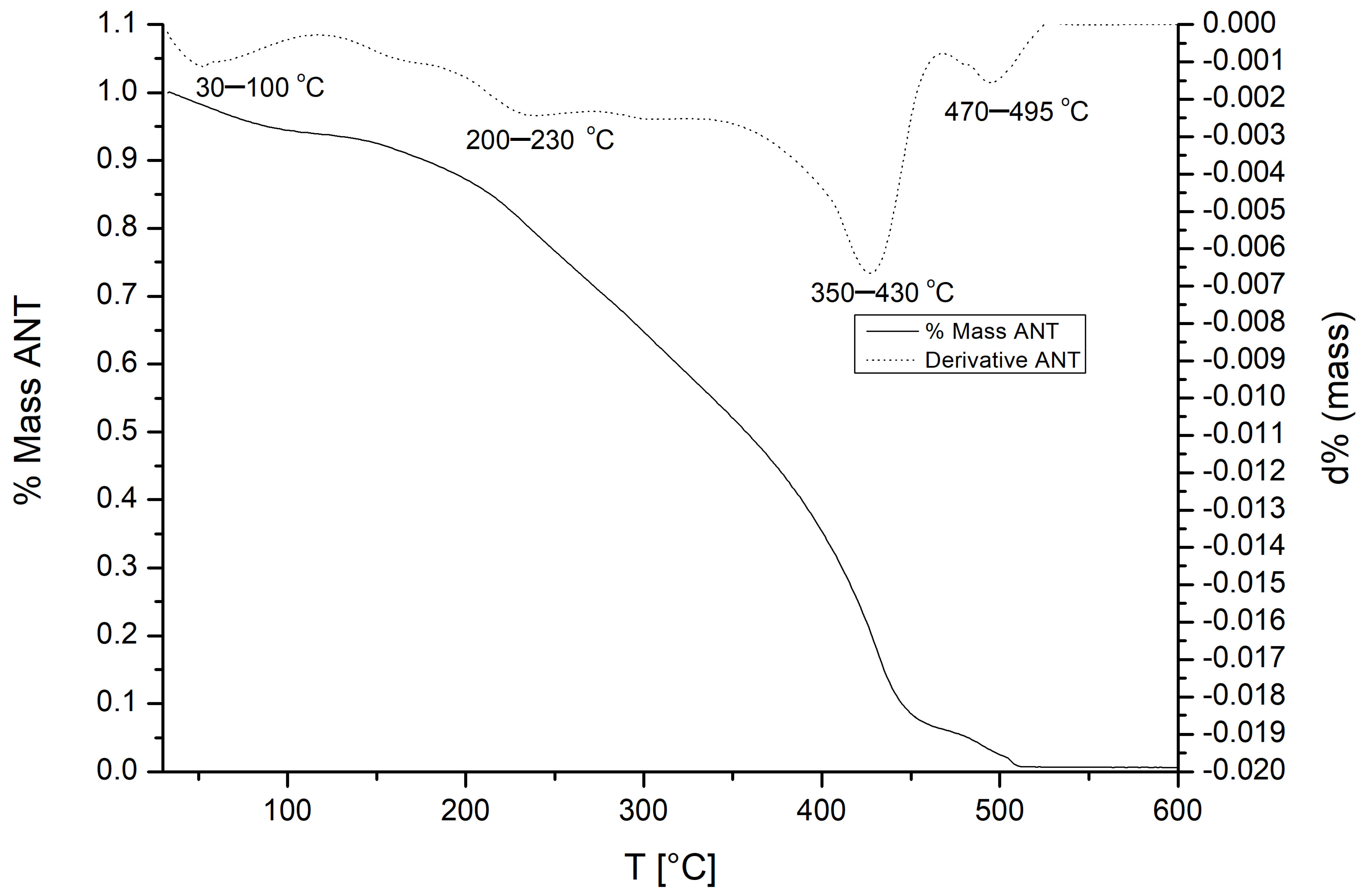
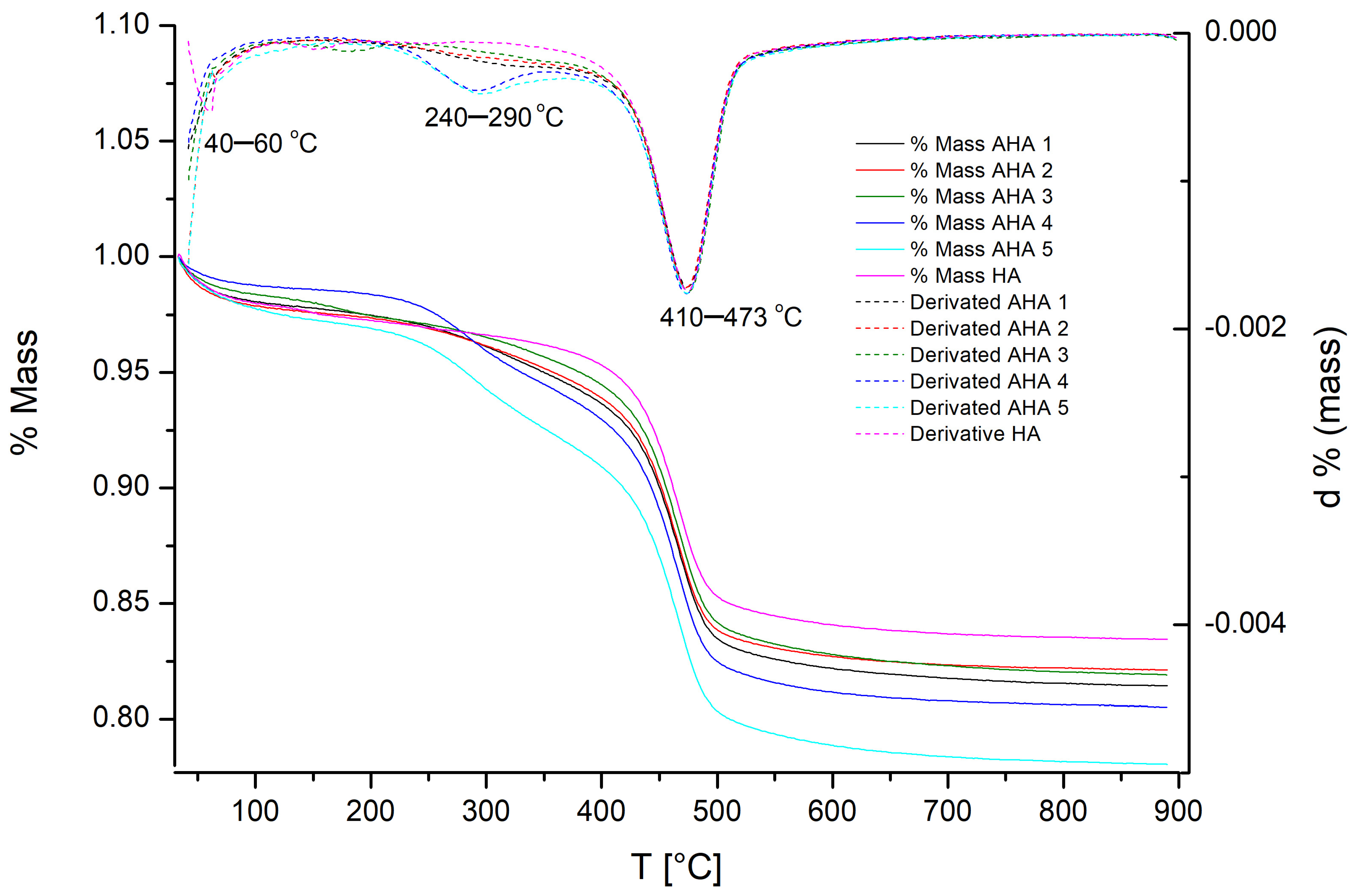
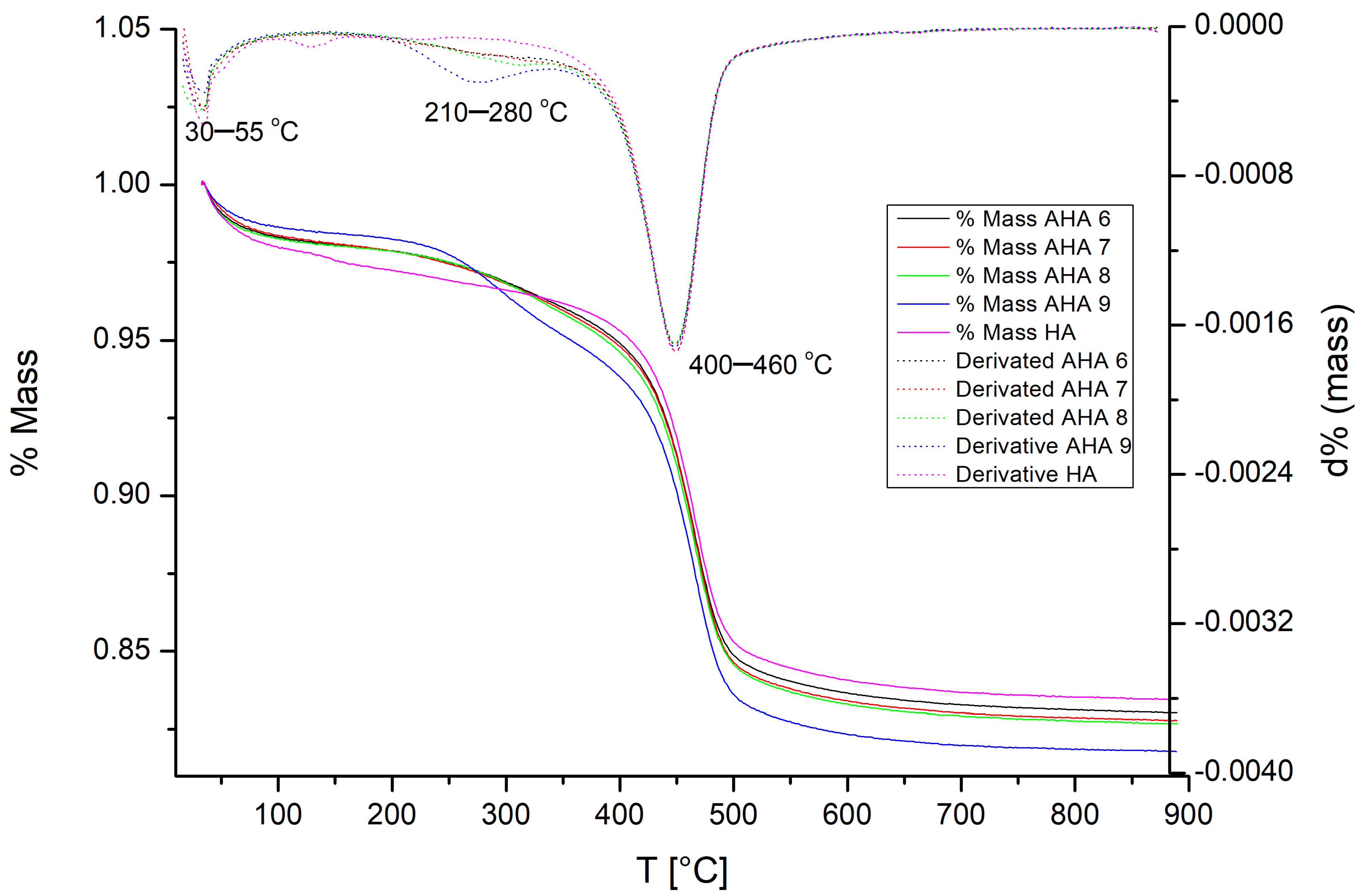
In TGA, the ‘T’ refers to the sample temperature recorded at a precise moment during thermogravimetric analysis, while ‘d%’ corresponds to the first derivative of the mass loss, expressed as a percentage change per degree Celsius (DTG signal).
Regarding the y-axis scale, the right y-axis represents the derivative thermogravimetric signal (dTG), expressed as the rate of mass change in percentage units. These values are not constrained between 0 and 1, as they correspond to differential mass loss rates rather than normalised fractions. Consequently, negative values are observed because the mass of the sample decreases with increasing temperature. This behaviour is inherent to DTG analysis and reflects the derivative d(mass)/dT, which is negative when mass loss occurs.
The left y-axis shows the cumulative mass loss as a normalised percentage. This axis is constrained between 0 and 1, where 0 corresponds to 0% mass loss and 1 corresponds to 100% mass loss.
The TGA curve of pure anthocyanins (
Figure 2), A significant mass loss is observed as the temperature increases, leading to complete degradation at around 500 °C. The thermal decomposition proceeds through several distinct stages, which are consistent with the features identified in the DTGA curve. The initial weight loss, which occurs between 30 and 100 °C, is attributed to the evaporation of water molecules that are adsorbed onto the surface of the anthocyanins [
36,
37]. Within this temperature range, the DTGA curve shows a small peak, which indicates an increased rate of mass loss in a localised area. Subsequent mass loss events occur within the temperature ranges of 200–230 °C and 350–430 °C. These are attributed to the degradation of organic matter, including the breakdown of phenolic acids and aldehydes resulting from deglycosylation and ring-cleavage reactions that are inherent to anthocyanin molecules [
36,
37]. Consequently, two distinct DTGA peaks are observed in these regions. The peak in the 350–430 °C range is the most intense, indicating the highest degradation rate and representing the main stage of anthocyanin decomposition.
The TGA curves of halloysite (HA) and its anthocyanin-based hybrids (AHA) are shown in
Figure 3 and
Figure 4. Overall, the hybrid materials exhibit thermal degradation patterns that closely resemble those of pristine halloysite, with TGA curves that nearly overlap. However, complete mass loss is not observed, with the decomposition process stabilising at around 500 °C. Closer analysis reveals an initial weight loss of 3–6% between 30 and 60 °C, which is associated with the desorption of surface-bound water molecules from the halloysite structure [
37,
38]. Within this temperature range, the DTGA curves show a small initial peak, indicating an increase in mass rate. A slight mass loss was detected in specific samples (AHA.4, AHA.5 and AHA.9) between 210 and 290 °C. Similarly, the DTGA curves of these samples show a weak peak in this range, indicating a localised increase in the degradation rate. These particular hybrids were prepared using a CPB surfactant as a surface modifier. The minor mass loss is believed to correspond to the release of hydrogen bromide (HBr), which is a component of the CPB surfactant [
39]. Similar mass loss profiles have been reported for other materials containing this surfactant [
39,
40]. Finally, a significant degradation step occurs between 400 and 473 °C, which corresponds to the dehydroxylation of the aluminium hydroxyl groups present in the clay structure [
37,
38]. This event is accompanied by the most intense DTGA peak across all samples, indicating the highest rate of mass loss. This process represents the main thermal degradation of the hybrids. Beyond this point, the mass stabilises, indicating the end of significant thermal decomposition, which is consistent with previous observations. Notably, the TGA and DTGA results suggest that halloysite nanotubes offer thermoprotection in hybrid systems by delaying dye degradation and maintaining structural integrity at elevated temperatures [
17,
40,
41]. Our previous research revealed that a very similar TGA and DTGA were observed [
17].
3.2. X-Ray Diffraction (XRD)
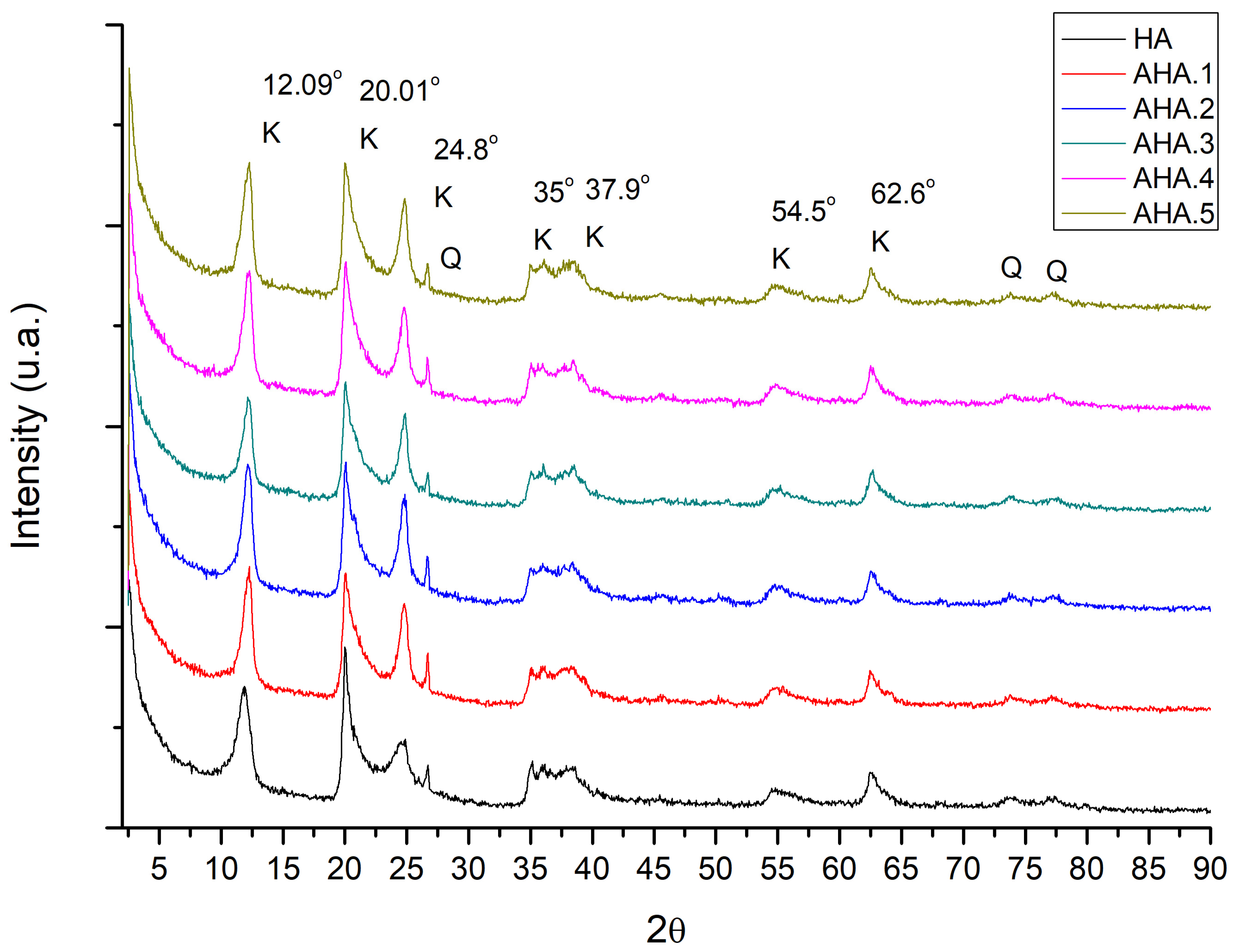
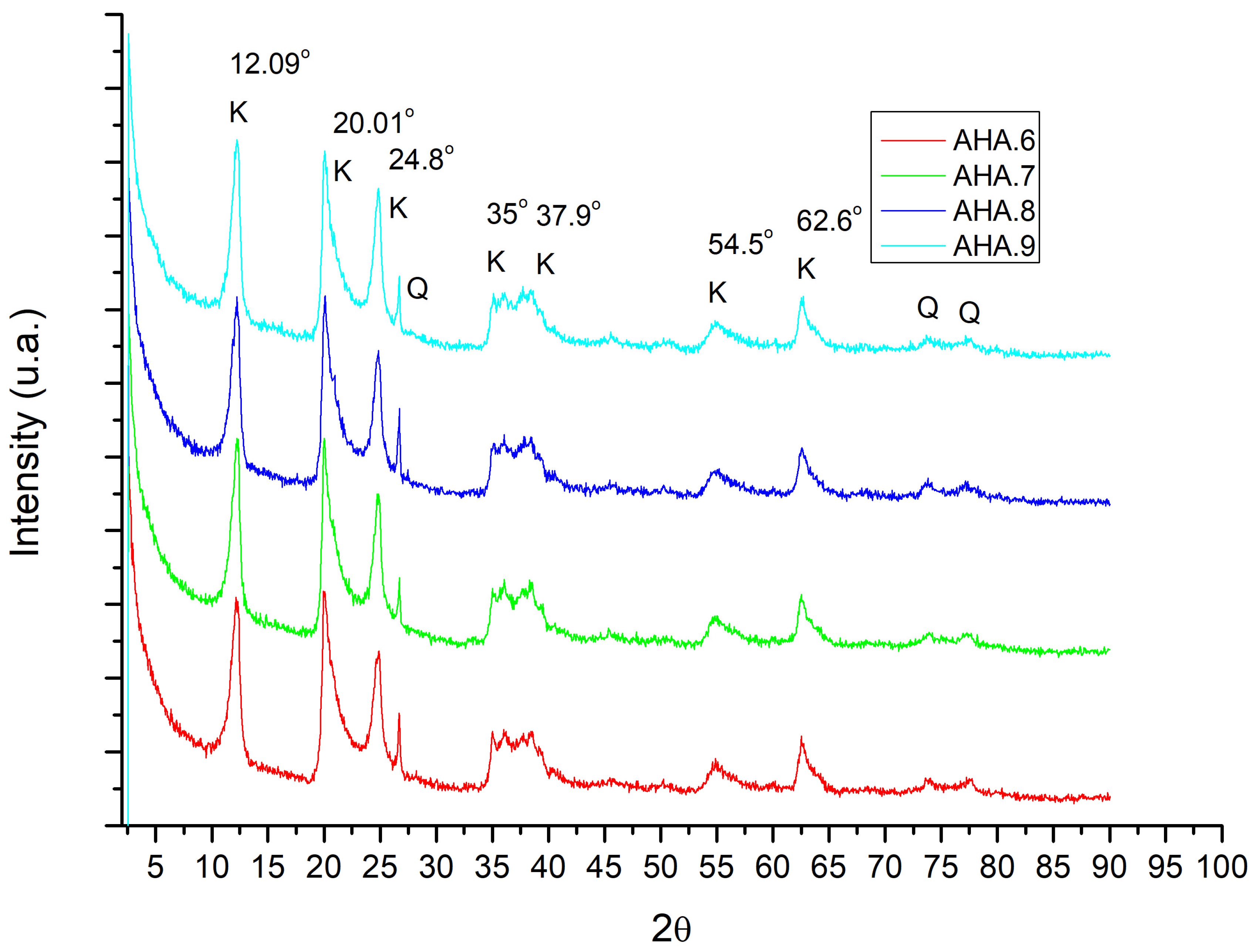
As shown in
Figure 5 and
Figure 6, the peak observed at 12.09° (2θ) reflects the basal spacing of the 7 Å HA form and is indicative of its dehydrated state. The presence of a peak at 24.85° further confirms the dehydrated state [
26,
42,
43].
A prominent peak at 62.78° confirms the tubular morphology characteristic of halloysite nanotubes [
26,
42,
43]. Overall, the diffraction peaks observed in all samples match the reference data for dehydrated halloysite (7 Å form), as per the JCPDS card no. 29-1487.
Table 2 summarises the main diffraction data.
In addition to the signals of halloysite, several diffraction peaks corresponding to monoclinic kaolinite (denoted as K) and cubic silicon oxide (denoted as Q) were also identified [
26]. As with the thermogravimetric analysis (TGA) results, the AHA hybrids closely mimic the diffraction profile of pristine halloysite, effectively reproducing its XRD pattern. A very similar XRD pattern was previously observed in our research [
17]. An important structural insight that can be obtained from XRD patterns, particularly from the basal interplanar spacing (d001), concerns the adsorption mode of the pigment within the clay system [
19,
44]. The intercalation of guest molecules into the layered structure of halloysite is expected to produce a shift in the (001) reflection towards lower 2θ values, or the appearance of an additional basal reflection due to interlayer expansion [
19,
44].
In the present case, however, the XRD patterns of the anthocyanin–halloysite hybrids show no significant change in the position of the basal reflection at approximately 12.1° 2θ compared to pristine halloysite. This indicates that the interplanar spacing remains unchanged after pigment loading, ruling out the intercalation of anthocyanins into the halloysite layers. Consequently, the results suggest that anthocyanins are primarily adsorbed onto the external and/or internal surfaces of the halloysite nanotubes without disrupting the clay crystal structure.
3.3. Fourier-Transform Infrared Spectroscopy (FTIR)
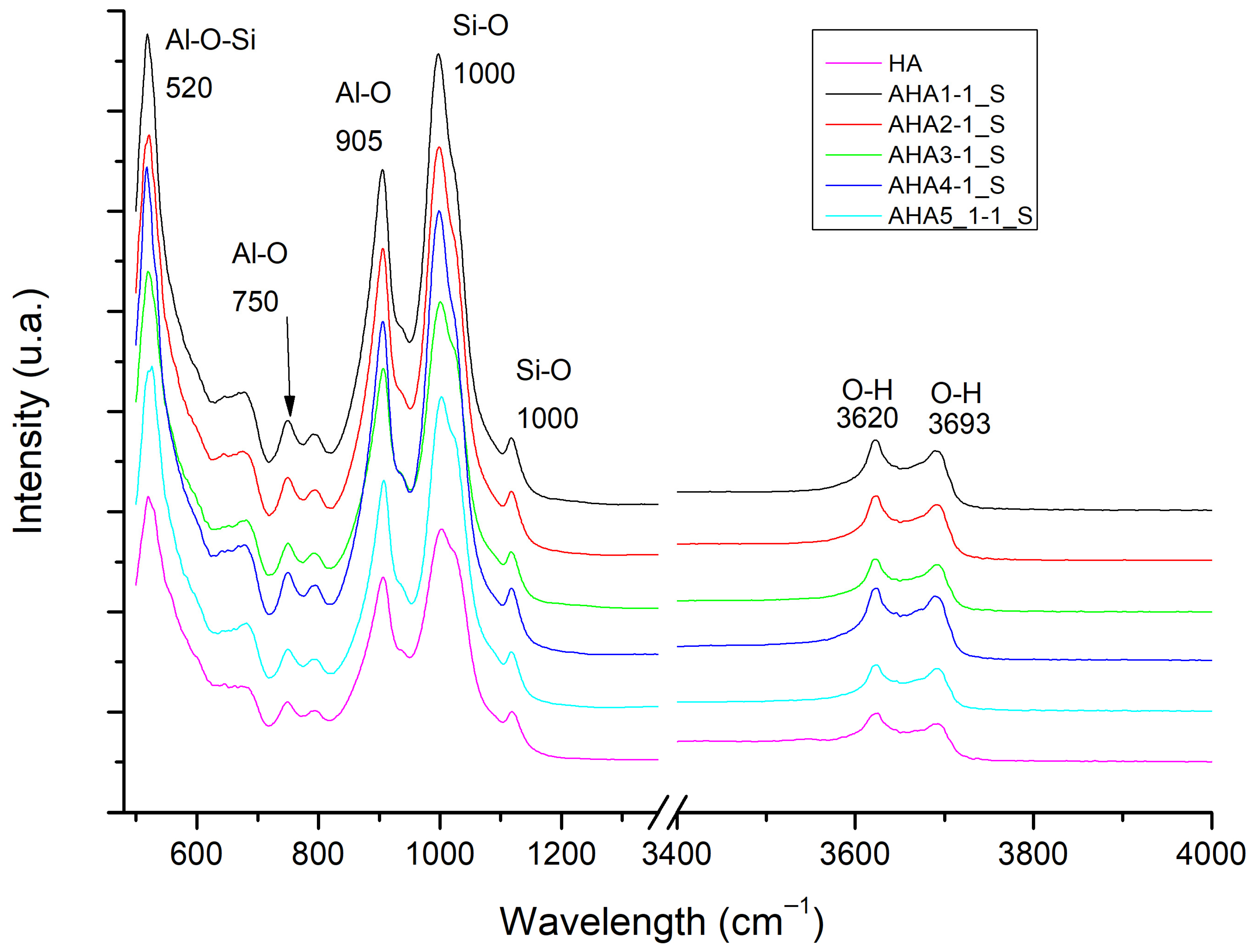
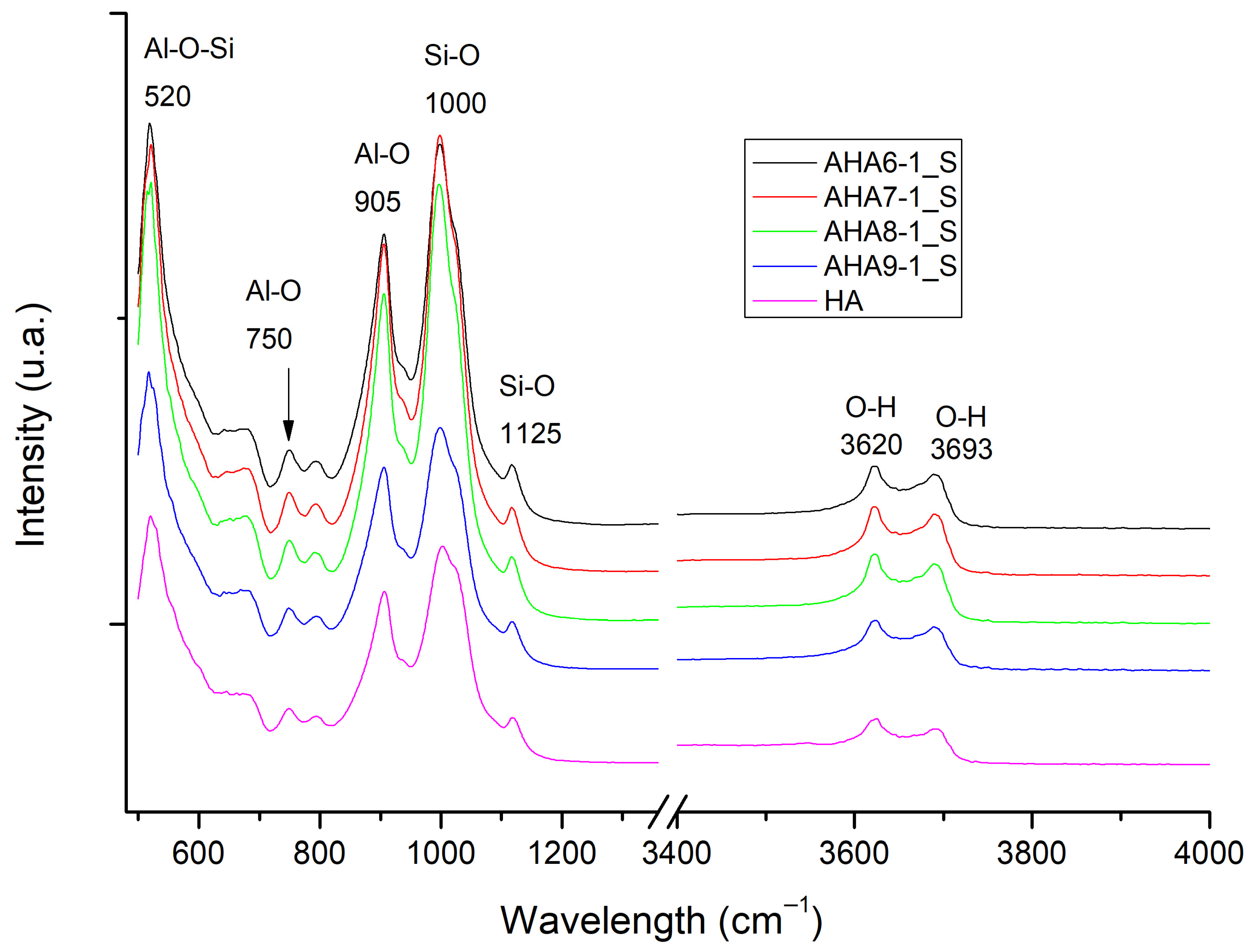
In this section, we present two FTIR spectra (
Figure 7 and
Figure 8) comparing the vibrational profiles of pristine halloysite (HA) and halloysite–dye hybrid materials (AHA) under different conditions.
Several characteristic absorption bands are observed in the spectra. A band at 520 cm
−1 corresponds to the Al–O–Si bond, while a peak at 1125 cm
−1 corresponds to the Si–O stretching mode. Additionally, a peak at 905 cm
−1 is due to the Al–O bond, while a peak near 1000 cm
−1 is assigned to the Si–O stretching mode. Furthermore, two distinct peaks are observed at 3693 cm
−1 and 3620 cm
−1 representing hydroxyl groups on the inner surface (–OH) and those belonging to the inner structure, respectively. These signals are characteristic of halloysite and consistent with those reported in previous structural studies of this nanoclay [
45,
46,
47].
The absence of bands at 3526 cm
−1 and 1637 cm
−1 in
Figure 7 and
Figure 8 confirms the dehydrated state of the halloysite, as these frequencies are typically associated with interlayer and adsorbed water [
45,
46,
47].
The FTIR analysis shows that all spectra corresponding to the different hybrid samples are essentially identical and perfectly match the spectral profile of pristine halloysite. This indicates that the dye incorporation process does not significantly alter the vibrational structure of the nanoclay, as seen in previous characterisations. Regarding our previous research, a very similar FTIR spectrum was observed [
17].
3.4. Total Solar Reflectance (TSR)
Figure 9 and
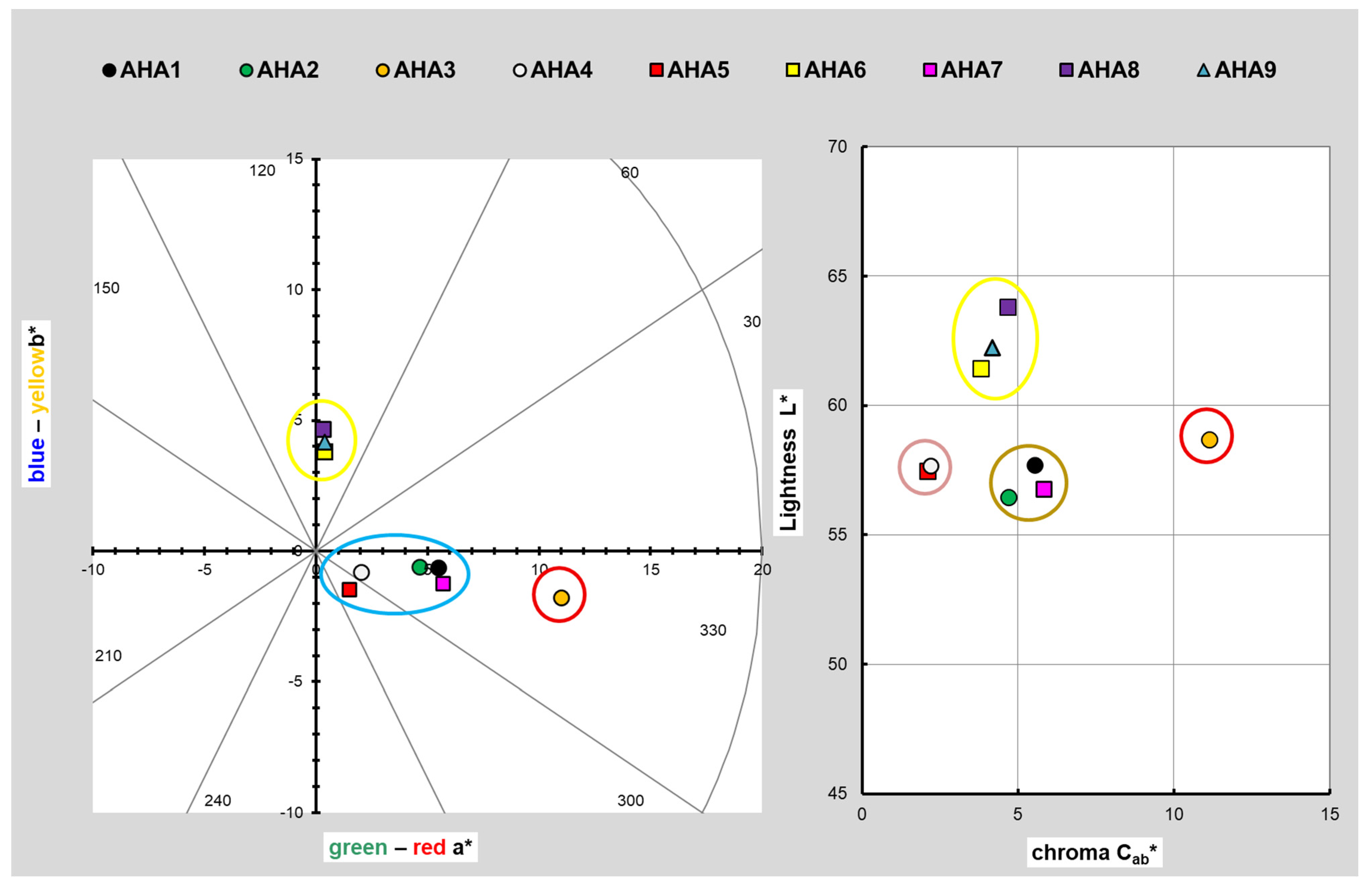
Table 3 illustrate the chromatic behaviour and the colour coordinates of samples AHA.1–AHA.9 in the CIELAB colour space. The left panel plots a* (green–red axis) versus b* (blue–yellow axis), while the right panel represents the lightness (L*) as a function of chroma (C*ab) On the a*-b* plot, all points fall in the first and fourth quadrants. In the left diagram we can see that the samples are located in the first and fourth quadrants. AHA.1, AHA.2, AHA.4, AHA.5 and AHA.7 are in the fourth quadrant (a*>0, b*>0), indicating reddish tones with bluish tendencies, positive a values* (tending toward red) and small or negative b values* (tending toward blue). Notably, by contrast, AHA.6, AHA.8, and AHA.9 deviate from this trend, exhibiting positive b* values, indicating a shift toward yellowish hues. These three are clearly distinguishable by their positions in the first quadrant. AHA.3 stands out for its high positive a* value (11.00), suggesting a strong red component, and the largest C*ab, i.e., it is the most saturated (strongly red). AHA.1, AHA.2 and AHA.7 are the next most saturated samples, have a lower a* (greener/less red) and consequently show a lower L* (darker), and have the lowest chroma, with a* and b* near zero (near-neutral/grey) and low L*. Among the yellowish set, AHA.8 exhibits the highest lightness (L* = 63.78) with relatively high chroma, giving a bright, saturated yellow; AHA.6 and AHA.9 are similar but slightly darker and less saturated.
In the right diagram, AHA.8 exhibits the highest lightness (L = 63.78) and elevated chroma values, indicating a bright and saturated yellow colour. AHA.6 and AHA.9 can be introduced in the same group as AHA.8, but they are slightly darker and saturated. Conversely, AHA.1, AHA.2 and AHA.7 can form another group, with lower lightness but similar chroma, indicating darker tones. AHA.3 has a strong chroma but moderate light, resulting in an intense colour and AHA.4 and AHA.5 have the lowest chroma and moderate light, resulting in dull colours.
Figure 9 shows that the trends in pH variation and surfactant type are consistent with the pH and surfactant conditions in
Table 1. This suggests that they may affect the optical behaviour of the AHA. Samples synthesised at a higher pH (e.g., AHA.6, AHA.8 and AHA.9) generally exhibited higher L* values, reflecting increased lightness, particularly for AHA.8 and AHA.9. This trend may be attributed to the improved dispersion of clay particles under alkaline conditions, as observed in similar halloysite-based systems [
48,
49]. Conversely, samples synthesised at acidic pH (AHA.1, AHA.2, AHA.3, AHA.4, AHA.5 and AHA.7) displayed similar brightness, but lower than samples synthesised at basic pH. Furthermore, as previously mentioned, they are all located in the fourth quadrant and tend towards blue. Among the acidic samples, differences in colour saturation can be seen due to the different surfactant surface modifiers. The CPB-containing samples (AHA.4 and AHA.5) showed the lowest saturation values, possibly due to surfactant–clay interactions altering the surface colour. Conversely, samples containing SDS (AHA.1) and samples without surfactant (AHA.7 and AHA.2) exhibited saturation levels similar to those of the basic pH samples but with darker tones, as indicated by lower L* values and moderate chroma values. In particular, AHA.3 showed the highest a* value (11.00), suggesting a strong red hue, which may result from acidic conditions promoting surface modification or surfactant retention on the halloysite structure [
50].
Regarding solar reflectance (TSR), values range from 8.88% (AHA.9) to 36.59% (AHA.4). Generally, samples with higher L* values and lower chroma values (e.g., AHA.4 and AHA.8) exhibit higher solar reflectance, which is consistent with the typical behaviour of lighter, less saturated colours. Despite its high chroma and intense red hue, AHA.3 shows a relatively high TSR value of 28.61%, which may be influenced by surface texture or particle scattering. Conversely, AHA.5 and AHA.9, which have darker or more saturated yellow tones, exhibit the lowest TSR values, both below 9%, indicating greater heat absorption. Interestingly, TSR correlated strongly with lightness (L*); AHA.4 and AHA.8, which were both prepared with CPB and SDS respectively, exhibited high TSR values (>30%), which coincided with higher L*. In contrast, despite having similar brightness, AHA.5 and AHA.9 had low TSR values (<9%), indicating that chromatic components (a*/b*) and surfactant-induced surface properties also influence reflectance behaviour [
18]. Overall, combining pH control with different surfactants offers a powerful strategy for tuning the visual and functional performance of halloysite-based materials, balancing aesthetic properties with solar energy reflectance efficiency.
3.5. Scanning Electron Microscopy (SEM) and Energy-Dispersive X-Ray Spectroscopy (EDX)
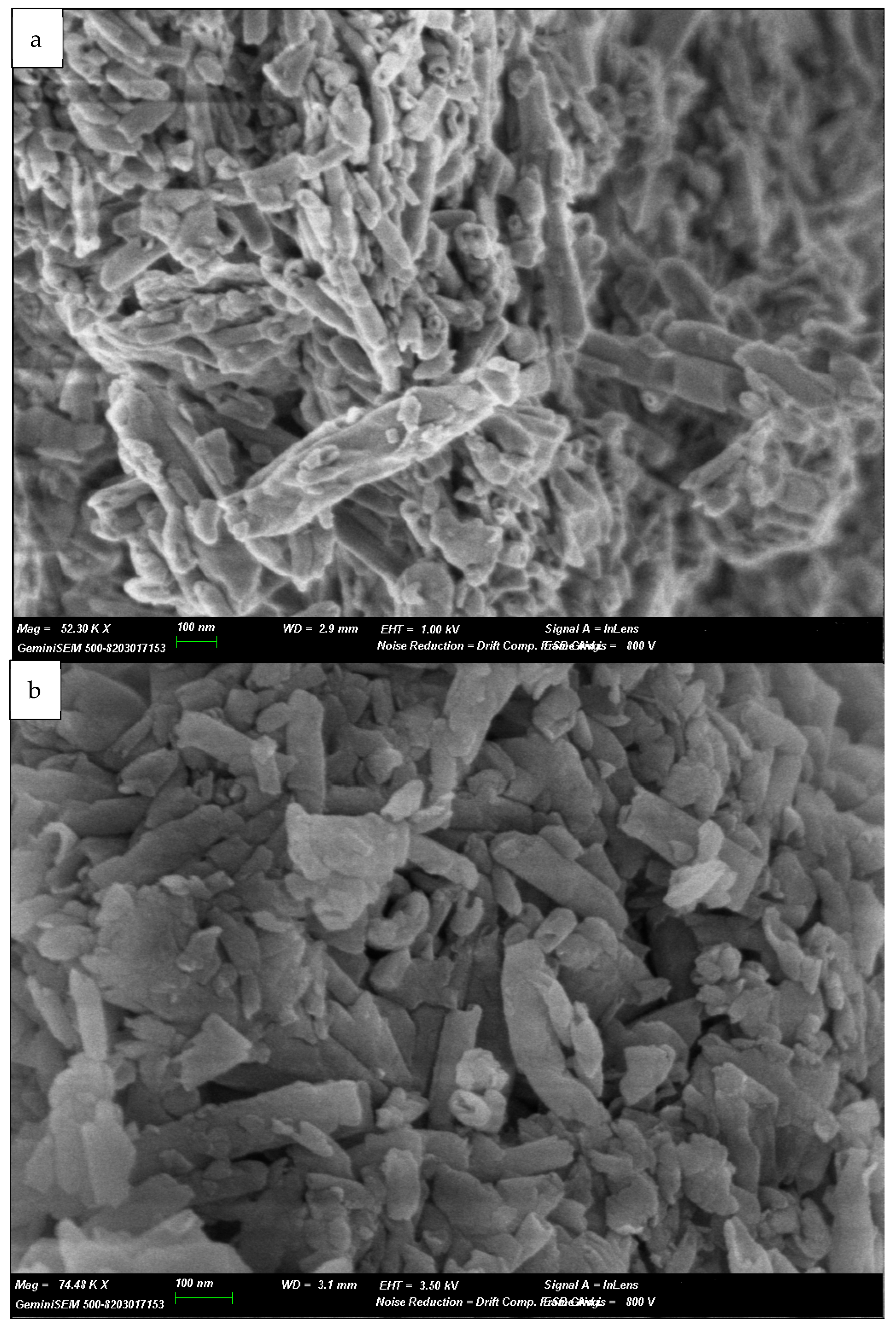
The SEM pictures of halloysite nanoclay (
Figure 10) reveal the characteristic tubular morphology of this 1:1 aluminosilicate. Natural halloysite forms these tubular structures due to the size mismatch between the octahedral Al-OH and tetrahedral Si-O sheets, which combine to form hollow cylinders. These nanotubes typically range in length from 200 nm to over 1 µm and in external diameter from 20 to 100 nm, depending on the source and degree of processing [
17,
19,
24,
25].
Figure 10 shows the halloysite before and after adsorption. Image a shows a reference sample of pure halloysite nanoclay, while image b corresponds to AHA1. Both images exhibit similar morphologies with clearly defined nanotubular structures. These tubes are aggregated and, in some areas, appear fragmented or partially collapsed—common artefacts due to SEM preparation under high vacuum or mechanical stress. Some flat or plate-like structures are also visible, possibly representing unrolled tubes or associated kaolinite impurities [
19]. Notably, no significant morphological differences can be observed between the halloysite nanoclay and the experimental sample, indicating that the halloysite structure has been preserved. This pattern is consistent across other samples analysed under the same conditions, confirming the stability of the halloysite morphology.
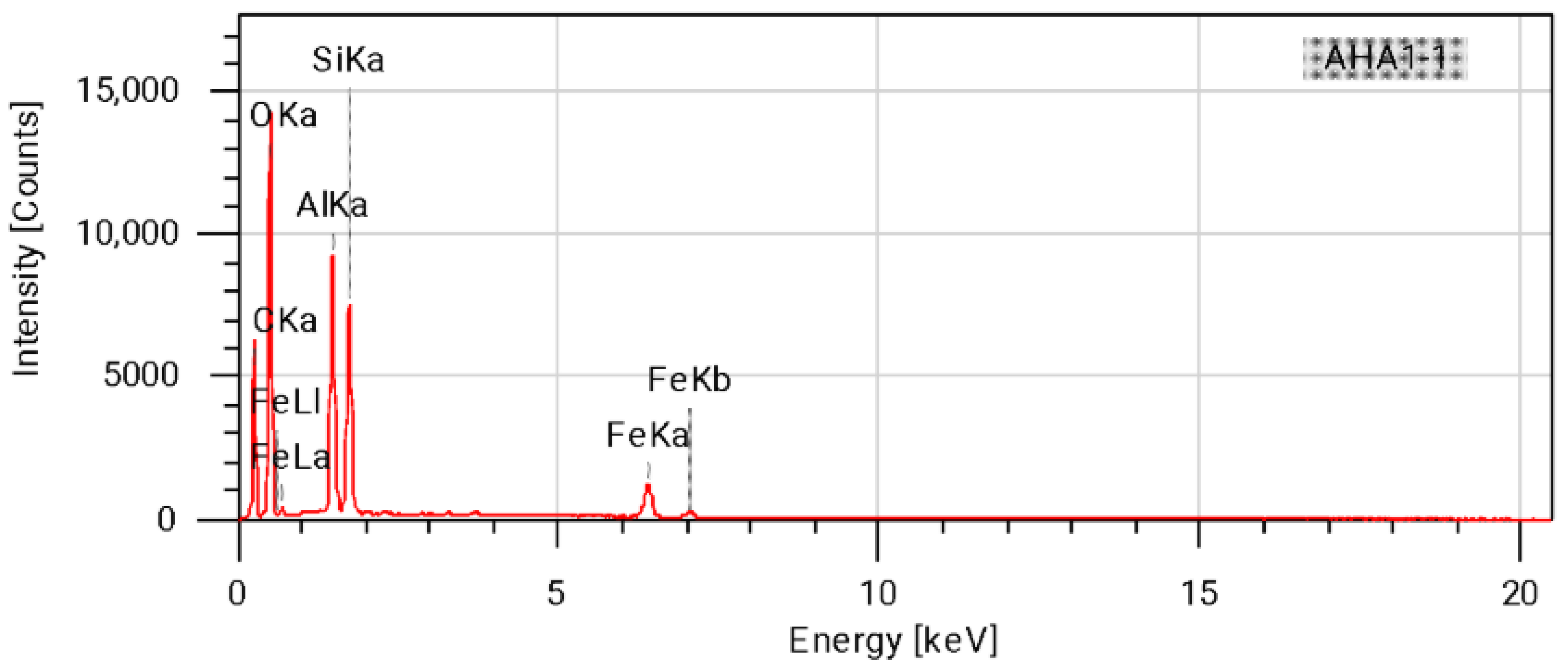
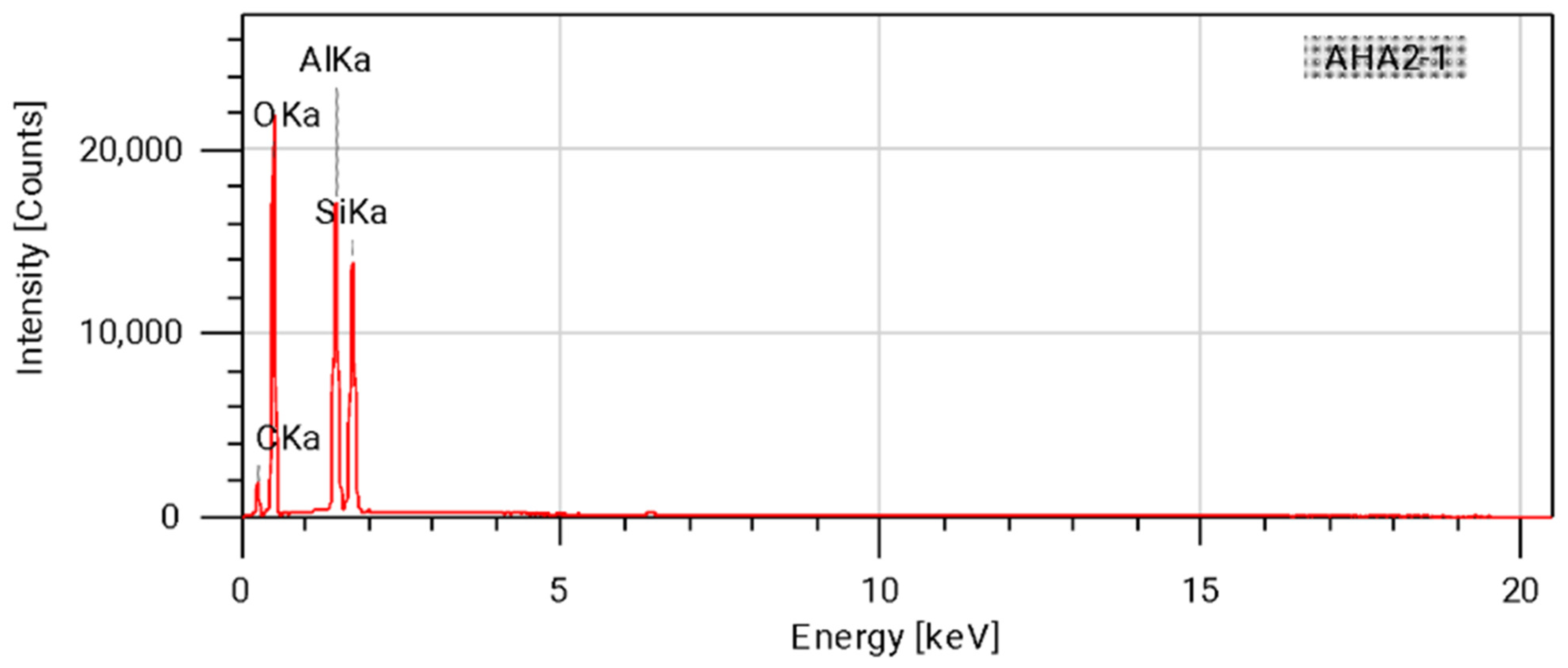
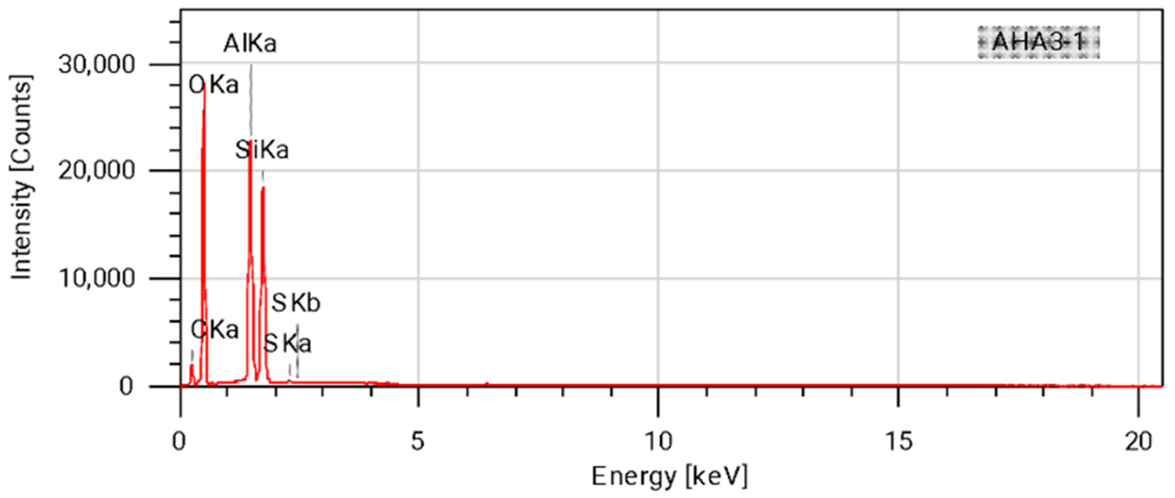
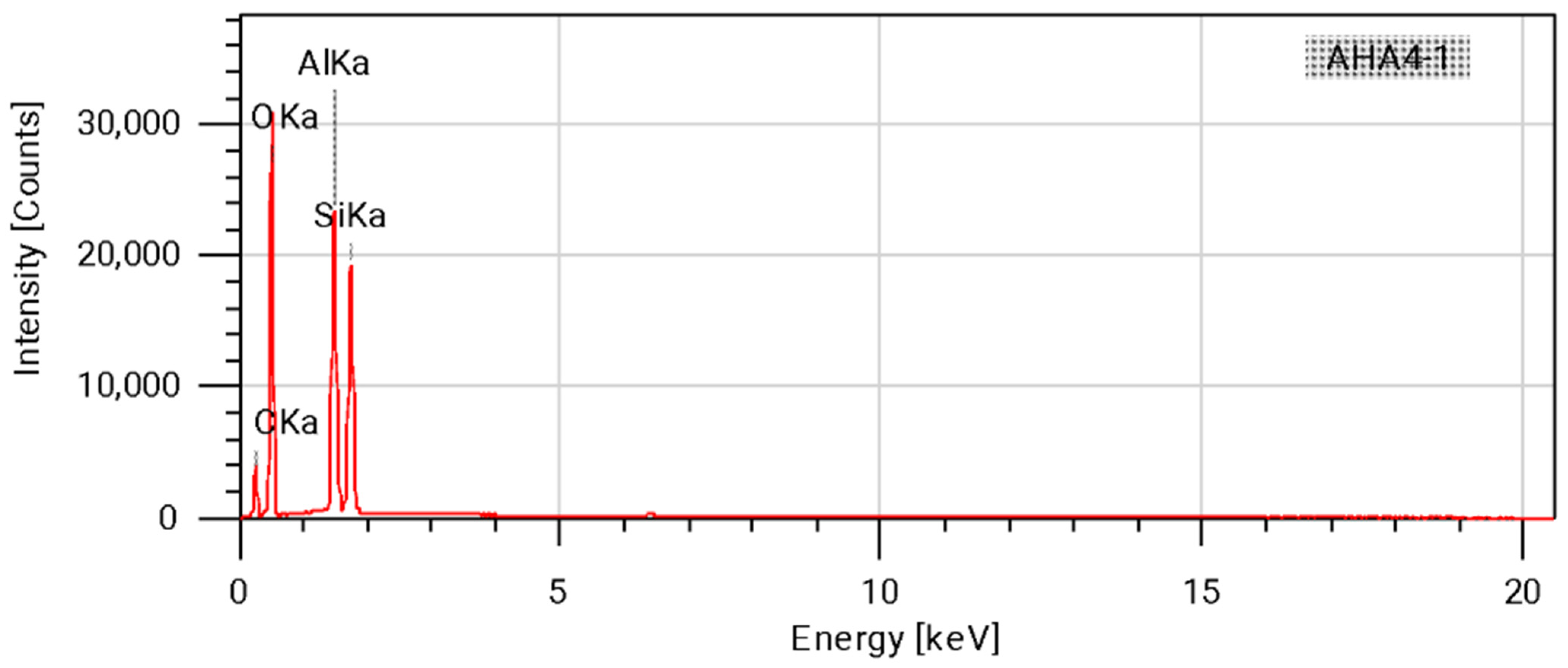
The energy-dispersive X-ray spectroscopy (EDX) spectra obtained for the hybrids are shown in
Figure 11,
Figure 12,
Figure 13 and
Figure 14 and in
Table 4, which show the mass and atomic percentages of each detected element. Only the first four samples were analysed, as the study focused on surface variability with different surfactants.
The presence of Al, O and Si is characteristic of halloysite nanoclay [
19,
24,
25,
51], as confirmed by previous FTIR analysis. However, other elements, such as C, S and Fe, were also detected. The presence of carbon can be attributed to the anthocyanins, which are primarily composed of carbon and oxygen [
6,
11,
36]. Sulphur likely originates from SDS residues, since samples 1 and 3 were prepared using SDS as a surfactant. Nevertheless, only sample 3 exhibits sulphur impurities, which may be due to subsequent treatments; in one case, the nanoclay may have been washed and filtered more thoroughly. Sample 4 was prepared using cetylpyridinium bromide (CPB) as the surfactant and no impurities were detected. Additionally, iron was detected exclusively in sample 1, which may be due to natural impurities or substitution within the halloysite structure [
19,
51]. Regarding our previous research, a very similar EDX spectra was observed [
17].
3.6. Brunauer–Emmett–Teller (BET) Surface Area and Porosity Measurements
Table 5 shows a comparison of the halloysite samples before and after dye adsorption. The halloysite data were taken from a previous scientific study [
17]. Following adsorption, a large reduction in pore size and depth was observed. As anthocyanin molecules accumulate, they progressively occupy the mineral’s internal pores. This pore filling leads to a decrease in the available space within the clay structure, ultimately limiting its capacity to absorb additional substances. Consequently, after anthocyanin adsorption, halloysite exhibits a lower effective surface area and reduced pore volume. These changes are consistently observed across all hybrid samples, indicating that a similar amount of anthocyanins is adsorbed under different experimental conditions. Regarding our previous research, a very similar BET was observed [
17].
3.7. ANOVA of Colour Results
A one-way analysis of variance (ANOVA) was performed, yielding the following results:
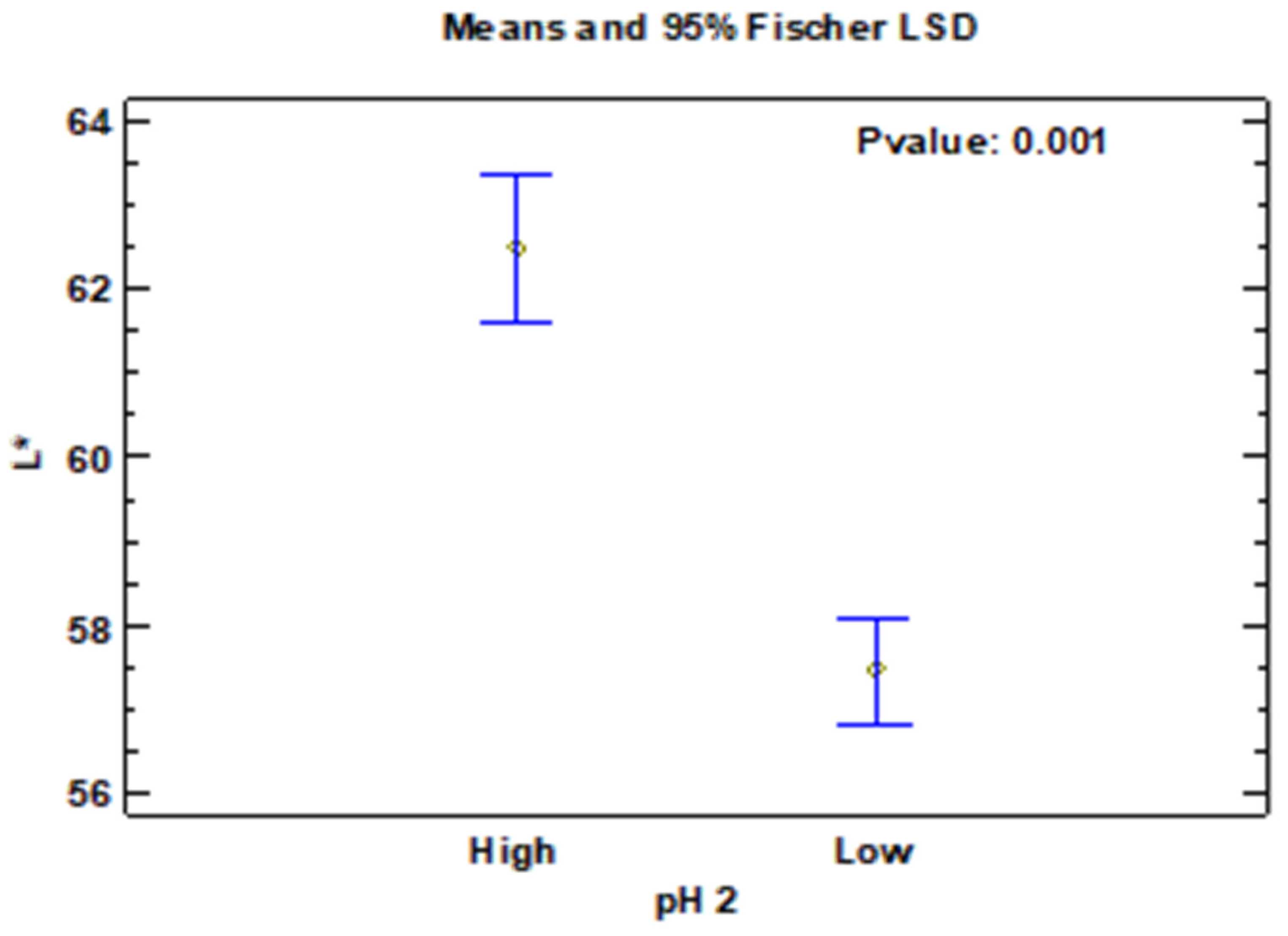
pH had a significant effect on lightness (L), as shown in
Figure 15 and
Table 6:
F(1, 7) = 59.53, p = 0.0001, ω2 = 0.88 [95% CI: 0.65–0.94].
The means ± error were clearly differentiated, indicating that pH adjustment substantially modifies the brightness of the pigment–clay hybrids.
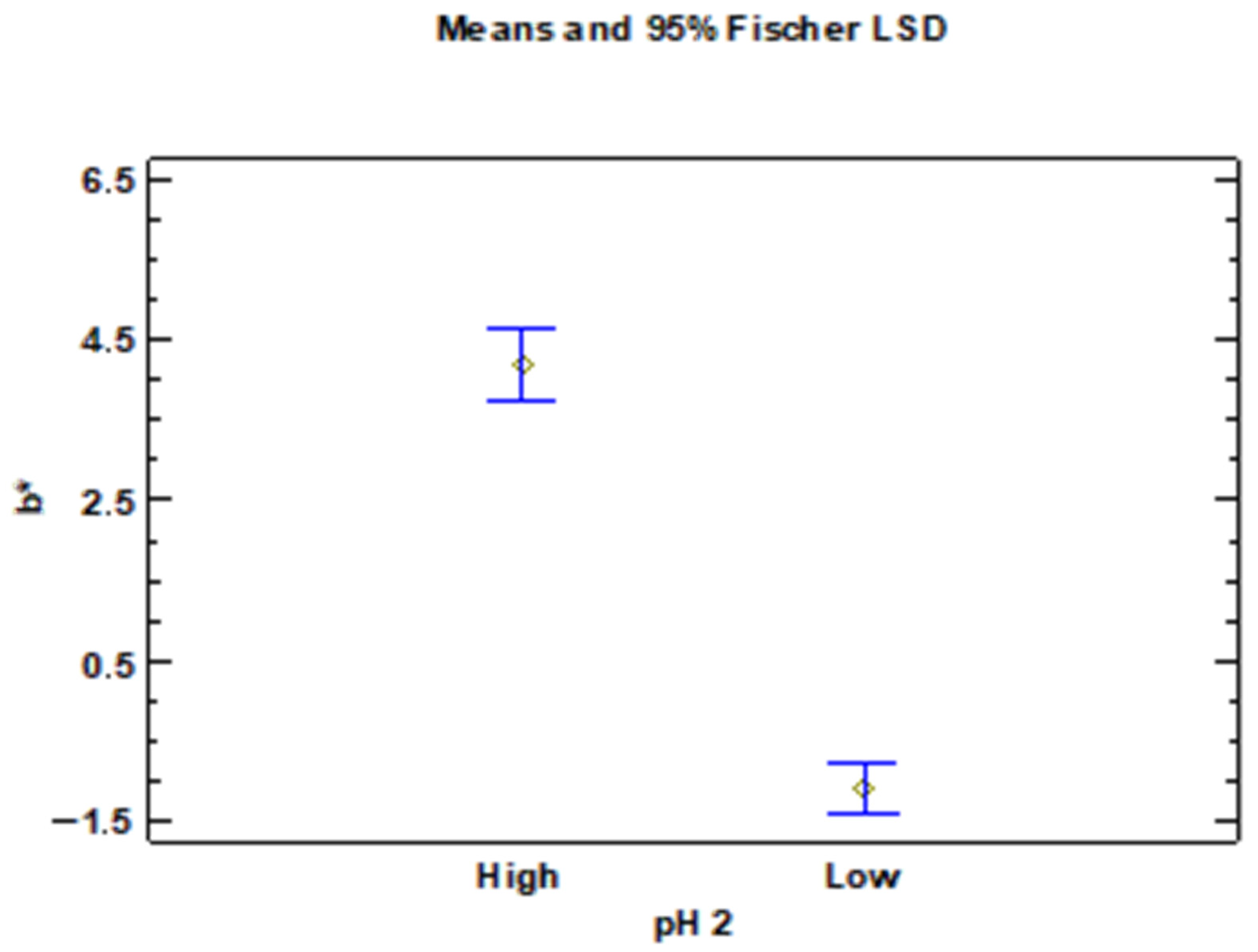
The effect of pH on the yellow–blue chromatic component (b*) was even more pronounced (
Figure 16 and
Table 7):
F(1, 7) = 253.50, p < 0.0001, ω2 = 0.97 [95% CI: 0.91–0.99].
The means showed a clear chromatic shift, with higher values under alkaline pH conditions.
Mean plots with 95% LSD intervals confirmed these differences, with the groups appearing separated in both L* and b* parameters and no overlap in the LSD bands. This supports the robustness of the observed contrasts.
Overall, the results indicate that pH is a critical factor determining both the lightness and chromatic tone of the HA–ANT hybrids. The very large effect sizes suggest practical relevance in addition to statistical significance.
4. Discussion
The results obtained confirm the potential of halloysite nanotubes as robust carriers for natural anthocyanins, offering thermal protection and structural integrity to hybrid materials. TGA showed that pure anthocyanins degrade completely at 500 °C, but the presence of halloysite delays this process, which is consistent with previous reports on the thermoprotective nature of clay nanotubes [
17,
19]. This enhanced thermal resistance suggests that anthocyanin–halloysite hybrids could better withstand textile finishing conditions than free pigments. This arises from the confinement of dye molecules within the halloysite lumen, as well as from interactions between the ACN–OH groups and the Al–OH/Si–OH sites of the clay. These interactions inhibit ACN degradation [
16,
52]. Notably, the thermal behaviour of the anthocyanin-loaded hybrids closely resembles that of pristine halloysite, with overlapping degradation curves and similar decomposition temperatures. This indicates that the incorporation of the pigment does not influence the thermal profile of the nanoclay, further highlighting the excellent thermal stability of the hybrid materials and the inert nature of the pigment–carrier interaction under heat stress. This behaviour is particularly relevant for textile applications, where colourants and functional additives are often subjected to thermal stress during processing steps such as drying, curing, calendaring or heat setting.
XRD analysis demonstrated that the crystalline structure of halloysite was preserved even with the inclusion of anthocyanins and surface treatments. The absence of diffraction peaks associated with hydrated forms of halloysite indicates that all samples were in the dehydrated 7 Å form, which is preferred for pigment adsorption due to its greater surface accessibility [
19]. The integrity of the tubular morphology further supports the material’s suitability as a nanocarrier.
FTIR revealed no notable spectral variations between the treated and untreated halloysite, indicating that the surfactants and anthocyanins did not disrupt the clay’s chemical structure.
In terms of colour performance, the pH of the synthesis medium was found to significantly influence the optical appearance of the hybrid pigments. Higher pH values led to increased luminosity (L*) and colour saturation, likely due to the improved dispersion of clay particles under alkaline conditions [
50]. While most samples exhibited similar overall hues, those synthesised at lower pH values displayed greater variability in colour parameters, potentially due to stronger interactions between the anthocyanins and surfactants in acidic environments. Specifically, samples synthesised at a low pH tended to exhibit reddish-blue tones, while those synthesised at a higher pH displayed more yellowish hues. This tunability could be useful for textile designers looking to achieve particular aesthetic effects using natural pigments.
Total solar reflectance (TSR) analysis revealed a general correlation between higher luminosity and increased solar reflectance, with one sample being an exception. This sample’s lower TSR may be attributed to surface texture effects. These findings suggest that adjusting the pH is a key strategy for tuning the visual and functional properties of the pigments. These results are particularly relevant for technical and outdoor textiles, where enhanced solar reflectance can contribute to thermal comfort and reduced heat absorption. Therefore, controlling the pH offers a simple and effective way to design hybrid pigments with tailored colour characteristics and improved solar management capabilities for textile applications.
Additionally, scanning electron microscopy (SEM) confirmed that the tubular morphology of halloysite was maintained after dye adsorption and surface treatments. Energy-dispersive X-ray spectroscopy (EDX) analysis revealed the presence of minor iron and sulphur residues on the surface of certain samples. These residues may originate from the natural composition of the halloysite or from interactions with the surfactants used, and they could potentially influence the final colour of the hybrids by introducing subtle shifts in tone or reflectance. In textile applications, such minor compositional variations may affect shade depth or undertone, underlining the importance of selecting the right raw materials and controlling the processing.
When these results are compared with our previous research on chlorophyll, it becomes evident that, although halloysite preserves its structural integrity in both systems, the inherent sensitivity of anthocyanins provides a level of chromatic versatility that chlorophyll does not exhibit. Furthermore, the reduction observed in the BET surface area—from 180 to approximately 46 m2/g—is consistent across both studies, confirming that occupation of the lumen and pores is the primary stabilisation mechanism for different types of natural dye. Finally, a comprehensive analysis using all characterisation techniques confirmed that adsorption conditions, namely pH and surfactant type, did not compromise the structural or thermal stability of the halloysite nanotubes. Nevertheless, these conditions significantly influenced the optical behaviour of the pigment, demonstrating that while the nanoclay functions as a stable and inert host matrix, the dye remains sensitive to the chemical environment during hybrid formation.



 ,
,