Article

Repurposing Clotrimazole for Pancreatic Ductal Adenocarcinoma: Comparative In Vitro Evaluation and In Silico ADMET Context


1
PerMed Research Group, RISE-Health, Faculty of Medicine, University of Porto, Alameda Professor Hernâni Monteiro, 4200-319 Porto, Portugal
2
School of Life and Environmental Sciences, University of Trás-os-Montes and Alto Douro (UTAD), Edifício de Geociências, 5000-801 Vila Real, Portugal
3
Laboratory of Personalized Medicine, Department of Community Medicine, Health Information and Decision (MEDCIDS), Faculty of Medicine, University of Porto, Rua Doutor Plácido da Costa, 4200-450 Porto, Portugal
4
RISE-Health, Department of Community Medicine, Health Information and Decision (MEDCIDS), Faculty of Medicine, University of Porto, Rua Doutor Plácido da Costa, 4200-450 Porto, Portugal
*
Author to whom correspondence should be addressed.
Physchem 2026, 6(1), 17; https://doi.org/10.3390/physchem6010017
Submission received: 12 January 2026 / Revised: 12 February 2026 / Accepted: 4 March 2026 / Published: 10 March 2026
(This article belongs to the Section Biophysical Chemistry)

Abstract

Background: Clotrimazole (CLZ) is an approved antifungal with reported pleiotropic effects. Beyond its antifungal use, CLZ can perturb glycolytic flux and ionic homeostasis, motivating its evaluation as a repurposing candidate in oncology. Objective: We aimed to evaluate CLZ and nitazoxanide (NTZ) as drug repurposing candidates for pancreatic ductal adenocarcinoma (PDAC) in comparison with standard chemotherapeutics gemcitabine (GEM) and 5-fluorouracil (5-FU). Methods: T3M4 PDAC cells were treated (0.1–100 µM; 48–72 h) with 5-FU, GEM, CLZ, and NTZ. Cell viability (MTT) and morphology were assessed, and CLZ-based combinations were analyzed by the Chou–Talalay method. In silico studies provided physicochemical descriptors and ADMET profiles, along with predicted interactions with relevant bioorganic targets (e.g., KCa3.1/KCNN4 ion channels). Results: CLZ produced marked cytotoxicity at 72 h (IC50 ≈ 9 µM) and achieved a greater reduction in cell viability at higher concentrations compared to 5-FU and GEM under identical conditions, whereas NTZ showed modest and inconsistent effects. CLZ combinations with 5-FU or GEM were mainly antagonistic. In silico analyses indicated high membrane permeability and suggested potential interactions with KCa3.1, supporting a hypothesis-generating interpretation of the observed in vitro effects. Conclusions: Within a drug repurposing framework, CLZ exhibited consistent cytotoxic activity as a single agent in a PDAC cell model, whereas NTZ revealed limited effects and CLZ-based combinations were not beneficial under the tested conditions. These findings position CLZ as a monotherapy-oriented repurposing candidate for PDAC and motivate further mechanistic and translational studies to clarify the biological basis of its in vitro activity.

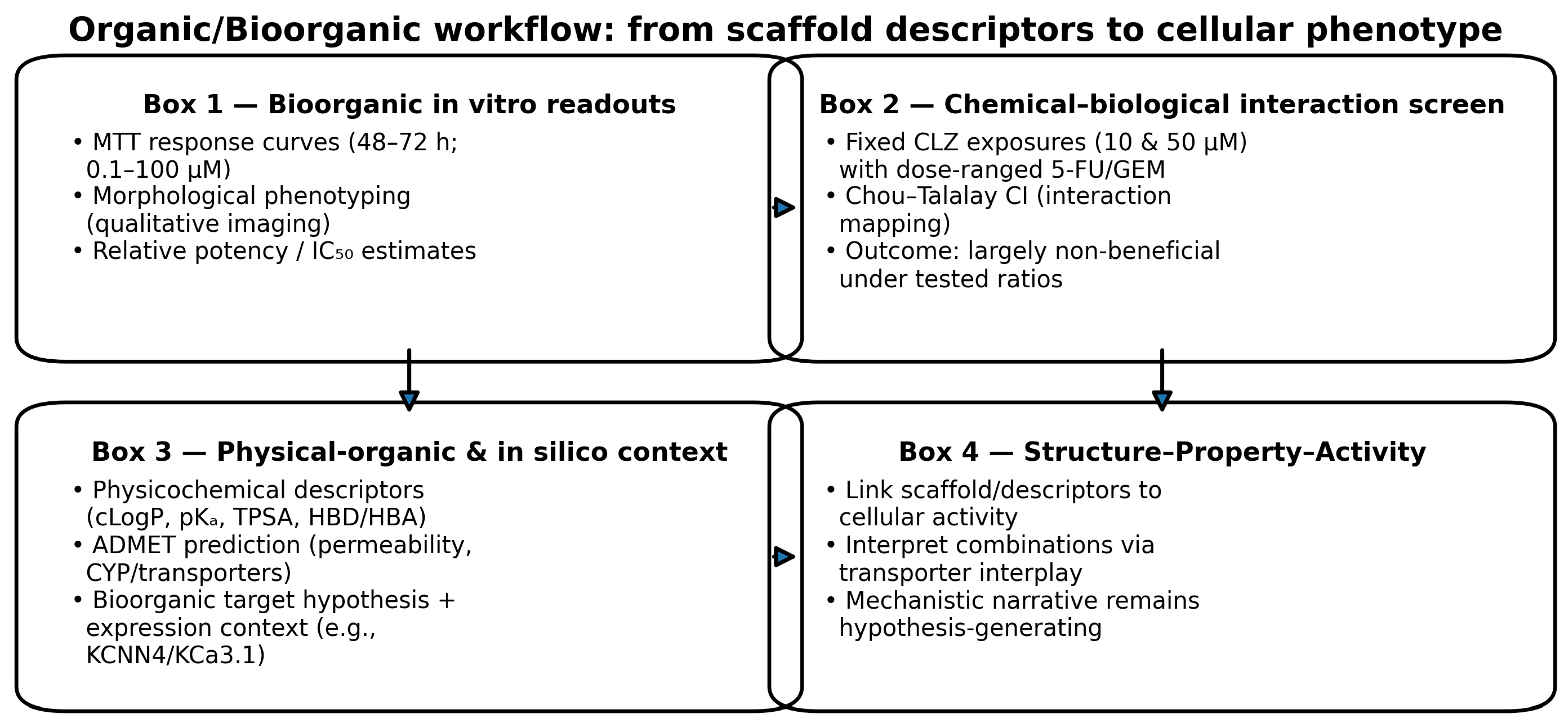
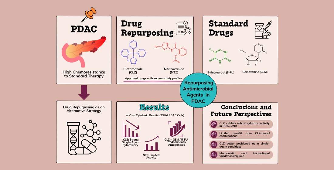
Graphical Abstract

1. Introduction

Drug repurposing, also known as “drug repositioning,” “drug reprofiling,” or “drug rediscovery,” is a strategy in the drug discovery field aimed at identifying new therapeutic applications for previously approved or investigational drugs beyond their initial clinical indications. This methodology represents a reliable alternative to traditional drug development strategies by overcoming key challenges such as high costs, lengthy timelines, and substantial risk of failure [1,2,3]. Historically, the practice of drug repurposing has primarily been driven by opportunistic and serendipitous occurrences. Indeed, the most successful examples of drug repurposing to date have often lacked a systematic approach [1]. However, recent developments in genomic and proteomic technologies, bioinformatics, and machine learning tools have allowed the systematic discovery of novel drug candidates, particularly in oncology, where drug repurposing has gained noteworthy attention. Numerous drugs have a pleiotropic nature; therefore, drug repurposing represents a way of exploring multiple biological pathways that drugs can target, thereby accelerating the development of novel cancer therapeutics [2,3,4].
Pancreatic ductal adenocarcinoma (PDAC) is a significant global health concern, recognized for its aggressive nature and high mortality rates [5]. This malignancy is particularly challenging due to the tendency for late-stage diagnosis, rapid disease progression, and the limited availability of effective treatment options. Current standard chemotherapy for PDAC, including gemcitabine (GEM, Scheme 1) and 5-fluorouracil (5-FU, Scheme 1), is responsible for a modest survival rate due to intrinsic and acquired resistance mechanisms [6,7,8,9,10,11]. These poor treatment outcomes highlighted the relevance of drug repurposing as a viable strategy to identify new therapeutic alternatives with known safety profiles [12]. Different investigations have utilized omics-based approaches, bioinformatics, and machine learning to find repurposed agents for PDAC treatment. For example, transcriptomic analyses have identified differentially expressed genes associated with PDAC, leading to potential therapeutic candidates [13]. Machine learning has also been demonstrated to be a powerful tool. For example, eravacycline, an antibacterial agent, was identified through machine learning-based screening for its ability to inhibit PDAC cell proliferation and migration [14].
Various antimicrobial agents have been investigated as potential candidates for drug repurposing within oncology. This exploration is due to their anticancer properties, known safety profiles, and drug-repurposing potential. A review by Pfab et al. [15] presents their ability to disrupt cancer cell metabolism, induce apoptosis, and limit tumor progression, supporting their feasibility as cost-effective therapeutic options. Among these, Clotrimazole (CLZ, Scheme 1) and Nitazoxanide (NTZ, Scheme 1) have been described as promising candidates. CLZ, an imidazole antifungal, inhibits glycolysis by targeting hexokinase, disrupts intracellular calcium homeostasis, and impairs mitochondrial function, leading to apoptosis in multiple cancers, including breast cancer, glioblastoma, multiple myeloma, and melanoma [16,17,18,19,20,21,22,23]. The documented effects of CLZ are particularly significant in the context of PDAC, a malignancy characterized by its metabolic plasticity [24] and resistance to apoptosis [25]. Given the pronounced reliance of PDAC cells on glycolytic metabolism to sustain their aggressive characteristics, the ability of CLZ to disrupt energy production and induce metabolic stress positions it as a highly promising therapeutic candidate for the treatment of PDAC. While CLZ’s anticancer effects are well-documented, it has yet to be extensively investigated in the context of PDAC. Similarly, NTZ, developed initially as an antiprotozoal agent, has revealed broad-spectrum activity against bacteria, viruses, and parasites, along with promising anticancer potential [26]. It induces cancer cell death through different mechanisms, including proteasomal inhibition, metabolic disruption, and suppression of vital survival pathways. NTZ effectively inhibited multiple catalytic subunits of the 20S proteasome, resulting in cell cycle arrest and inducing cell death in colon cancer cells [27]. Wang et al. [28] demonstrated that NTZ inhibits late-stage autophagy and enhances the induction of cell cycle arrest by the inhibitor of growth family member 1 (ING1) in glioblastoma. The ability to specifically target autophagy, which is a well-known resistance mechanism in PDAC [29], highlights the potential of NTZ as a promising therapeutic agent. In addition, NTZ has important anti-inflammatory effects, suppressing the nuclear factor kappa B (NF-κB) and mitogen-activated protein kinase (MAPK) pathways, which may contribute to its anticancer potential [30]. While these effects have been observed in other malignancies, NTZ’s therapeutic potential in PDAC remains unexplored.
Here, we evaluate CLZ and NTZ alongside standard PDAC agents (5-FU and GEM) in T3M4 cells, and we integrate in vitro cytotoxicity with in silico target/ADMET analyses within a drug repurposing framework.

2. Material and Methods

2.1. Cell Culture Conditions, Reagents and Drugs

Human T3M4 pancreatic ductal adenocarcinoma cells were utilized to assess cellular viability, proliferation, and cytotoxicity of the 5-FU, GEM, NTZ, and CLZ in T3M4 cells. The T3M4 cells were cultured in Dulbecco’s Modified Eagle’s Medium (DMEM) supplemented with 10% fetal bovine serum (FBS) and 1% penicillin (1000 U/mL)/streptomycin (10 mg/mL). The culture conditions involved an environment at 37 °C with 95% humidified air and 5% CO2. As T3M4 cells are adherent, they were maintained in a monolayer in T25 cm2 flasks and subcultured upon reaching a 75–80% confluence. The culture medium was replaced twice a week, with one replacement coinciding with cell splitting. All procedures were conducted in a vertical laminar flow hood using sterilized materials. Cell reagents were sourced from Millipore Sigma (Merck KGaA, Darmstadt, Germany).
The drugs utilized in this study were 5-FU, Gemcitabine, Nitazoxanide, and Clotrimazole, and they were obtained from Merck Life Sciences (Algés, Portugal), while the T3M4 cells were offered by the American Type Culture Collection (ATCC, Manassas, VA, USA).
Given CLZ’s low aqueous solubility and high lipophilicity, solubility limitations and precipitation at higher concentration ranges may influence the effective exposure and should be controlled (e.g., visual inspection, mixing time, or analytical confirmation).

2.2. Cell Seeding

At approximately 75–80% cell confluence, the culture medium was first aspirated, followed by a wash with 4 mL of PBS (Sigma-Aldrich; Merck KGaA, Darmstadt, Germany) and subsequent removal of the medium. For each experiment, cell detachment was initiated using 500 μL of 0.25% trypsin-EDTA (Gibco; Thermo Fisher Scientific, Inc., Waltham, MA, USA) solution. The cells were then incubated at 37 °C in a 5% carbon dioxide atmosphere for 7–10 min. Following this, the trypsin effect was neutralized by adding the culture medium. The resulting cell suspension was centrifuged for 5 min at 1100 rpm using a Hettich centrifuge (Tuttlingen, Germany). The supernatant was then replaced with a fresh culture medium, and the cells were re-suspended. Subsequently, viable cell counting was performed using trypan blue, a stain that exclusively targets cells with compromised cell membranes. This process was carried out utilizing a Neubauer chamber and the Leica DMI 6000B microscope equipped with a Leica DFC350 FX camera, (Leica Microsystems, Wetzlar, Germany).
T3M4 cells were seeded at a density of 5000 cells per well in 96-well plates and then incubated at 37 °C for 24 h before drug exposure.

2.3. Cell Treatment of T3M4 Cells

The assessment of cytotoxicity in T3M4 cells involved evaluating the effects of GEM and 5-FU after 48 h and 72 h, utilizing concentrations of 0.1, 1, 10, 25, 50, and 100 µM. Similarly, the individual evaluation of CLZ and NTZ was undertaken after 72 h, employing the same concentrations. The IC50 values for 5-FU and GEM were determined at both 48 h and 72 h. T3M4 cells were concurrently treated with both drugs in a combination therapy approach, without prior pretreatment. Specifically, 5-FU was administered in combination with CLZ and incubated for durations of 48 and 72 h. Additionally, GEM was also combined with CLZ and subjected to the same incubation periods. The concentrations used for the combination treatments of 5-FU and GEM were 0.1, 1, 10, 25, 50, and 100 µM, while CLZ was utilized at concentrations of 10 µM and 50 µM. Control cells were exposed to 0.1% dimethyl sulfoxide (DMSO), which was also used as a solvent for dissolving GEM, 5-FU, CLZ, and NTZ.

2.4. Morphological Analysis of T3M4 Cells

After drug exposure, morphological assessments of the T3M4 cells were conducted for each experiment using a Leica DMI 6000B microscope equipped with a Leica DFC350 FX camera. Image acquisition was performed utilizing the Leica LASX software (v3.7.4) developed by Leica Microsystems, Wetzlar, Germany. In order to ensure accurate size reference and maintain consistency in measurements and comparisons, a scale bar of 50 μm has been included in all microscopic images.

2.5. MTT Assay: Colorimetric Assay in T3M4 Cells

The MTT (thiazolyl blue tetrazolium bromide) colorimetric assay is widely utilized for evaluating cellular viability, proliferation, and cytotoxicity related to specific drugs. This method specifically focuses on assessing the mitochondrial function of T3M4 cells by comparing cell viability post-drug treatment with a control group. When the mitochondria are functional, MTT is reduced by mitochondrial dehydrogenases, leading to the formation of formazan crystals and the production of a purple-colored product. The color intensity directly correlates to cellular viability and, subsequently, to the absorbance value. Therefore, the assessment of cell viability involves comparing the absorbance values between the experimental and control groups.
After 48 h or 72 h, 100 µL of an MTT solution (0.5 mg/mL) in PBS (Sigma-Aldrich; Merck KGaA, Darmstadt, Germany) was added to the cells after aspirating the medium from the wells. The MTT assay was conducted in the absence of light. Subsequently, the plate was incubated for two hours at 37 °C with 5% carbon dioxide to allow for the formation of formazan crystals. After incubation, the MTT was removed, and 100 µL of DMSO was added to each well to dissolve the formazan crystals. The plate was then subjected to absorbance measurement at 570 nm using an automatic plate reader (Tecan Infinite M200, Tecan Group Ltd., Männedorf, Switzerland).

2.6. Statistical Analysis of the Results

The cell viability graph results include the mean ± SEM (standard error of the mean) values calculated using GraphPad Prism 9 software (GraphPad Software Inc., San Diego, CA, USA). A one-way ANOVA test with Dunnett’s multiple comparisons method was employed to assess the control group versus the experimental group for each drug concentration. The symbol (*) was utilized to denote significant results. * Statistically significant vs. control at p < 0.05; ** Statistically significant vs. control at p < 0.01; *** Statistically significant vs. control at p < 0.001; **** Statistically significant vs. control at p < 0.0001. Viability results were normalized to the control group and illustrated using logarithmized drug concentrations with non-linear regression analysis for generating dose–response curves. The IC50 value was determined using the same software based on the dose–response curves generated.

2.7. Drug Combination Analysis

Drug interaction studies were performed using CompuSyn software (version 1.0, ComboSyn Inc., Paramus, NJ, USA), based on the Chou–Talalay method. Combination Index (CI) values were generated across a range of drug concentrations and time points. CI < 1, CI = 1, and CI > 1 were interpreted as synergistic, additive and antagonistic interactions, respectively.

2.8. Target Expression Analysis

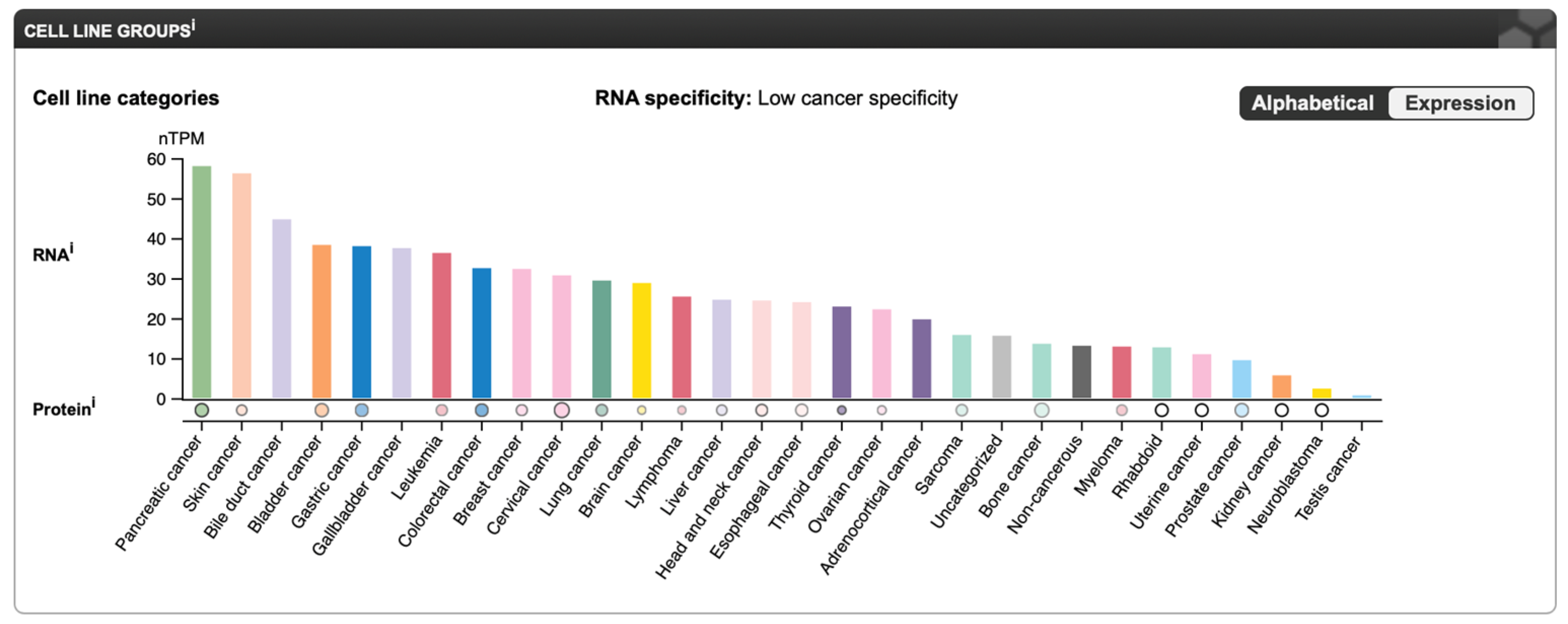
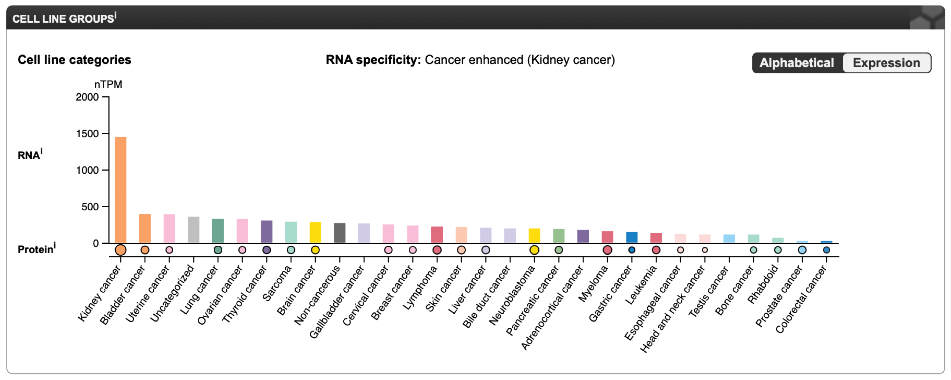
To evaluate the relevance of the targets in pancreatic cancer, expression data were obtained from the Human Protein Atlas (HPA) (https://www.proteinatlas.org, accessed on 25 August 2025). RNA expression levels of KCNN4 (KCa3.1) and AKR1B1 were retrieved for pancreatic ductal adenocarcinoma cell lines. Expression plots were downloaded directly from HPA, and results were integrated with in vitro cytotoxicity findings.

2.9. Computational Modeling of ADMET Properties

The physicochemical and absorption, distribution, metabolism, excretion, and toxicity (ADMET) properties of NTZ, CLZ, 5-FU, and GEM were predicted using ADMET Predictor® v11.0 (SimulationPlus, Lancaster, CA, USA). The chemical structures of all compounds were generated in MedChem Designer® v9.0 (SimulationPlus, Lancaster, CA, USA) and subsequently imported into ADMET Predictor® for in silico analysis. Key physicochemical descriptors, including molecular weight, lipophilicity (logP), acid dissociation constant (pKa), aqueous solubility, diffusion coefficient, and effective permeability (Peff), were estimated, along with additional ADMET-related parameters.
In addition, comprehensive predictions of metabolic pathways and transporter interactions were performed. Cytochrome P450 (CYP) enzyme involvement was assessed by identifying each compound as a potential substrate and/or inhibitor of major CYP isoforms, while phase II metabolism via uridine diphosphate-glucuronosyltransferases (UGTs) was also evaluated. Drug-transporter interactions were predicted for key uptake and efflux transporters. These in silico predictions were used to characterize the absorption potential, intracellular exposure, and drug–drug interaction liability of all drugs, providing a mechanistic framework to support the interpretation of the in vitro cytotoxicity and combination treatment results (Scheme 2).

3. Results

3.1. Drugs Tested Alone in T3M4 Cells

3.1.1. Cytotoxic Effect of the Antineoplastic Drugs 5-FU and GEM

In this study, T3M4 pancreatic ductal adenocarcinoma cancer cells were exposed to varying concentrations (0.1, 1, 10, 25, 50, and 100 µM) of 5-FU and GEM for periods of 48 and 72 h. 5-FU and GEM, commonly used for PDAC management, were assessed as part of this research effort to compare with the results of repurposed drugs. Cell viability was determined through the utilization of an MTT assay, a widely used method for evaluating mitochondrial activity and overall cell viability. The cell morphology was observed at 48-h and 72-h intervals to gain further insight into the cytotoxic effects of 5-FU and GEM on PDAC cell morphology.
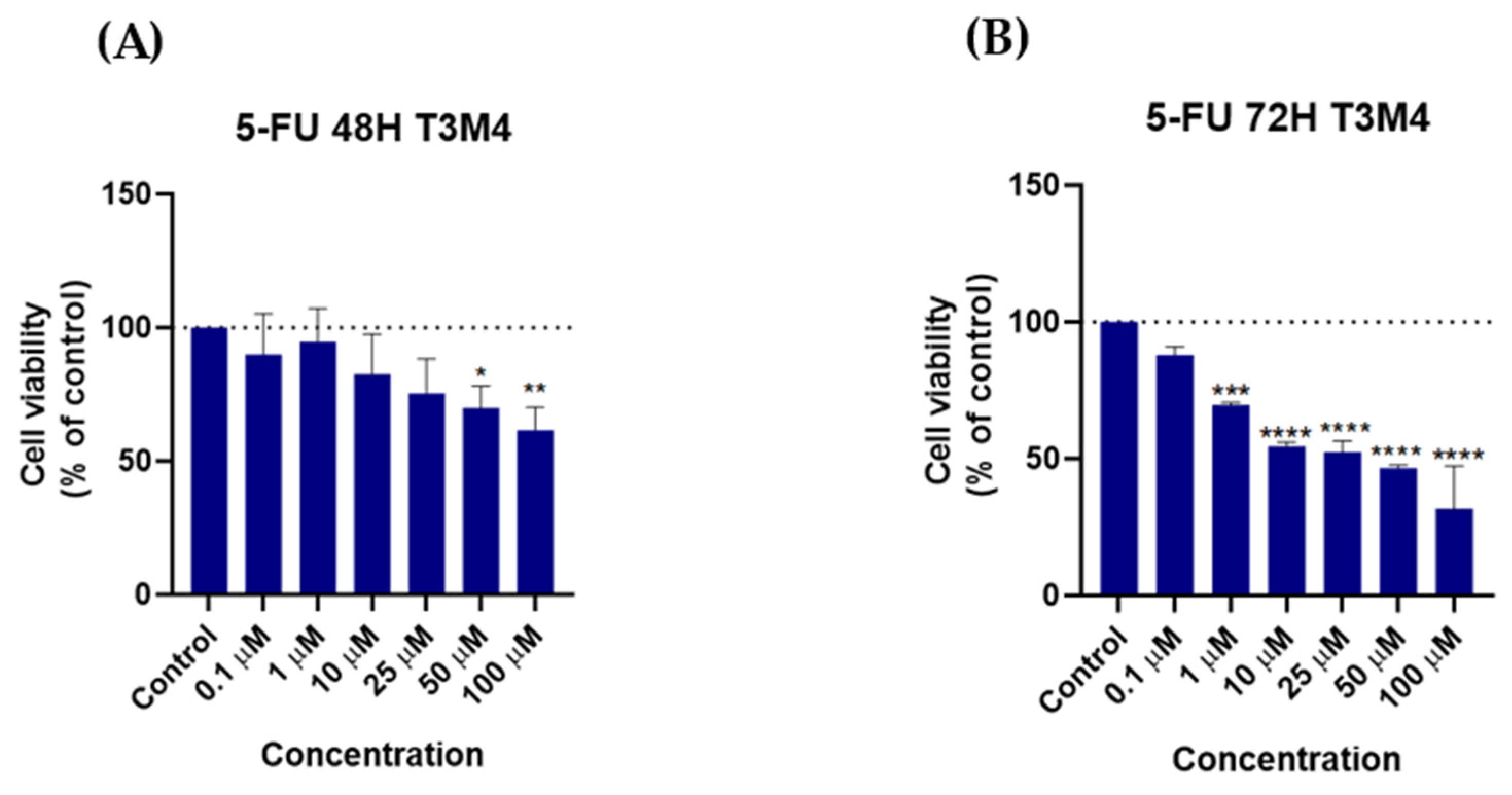
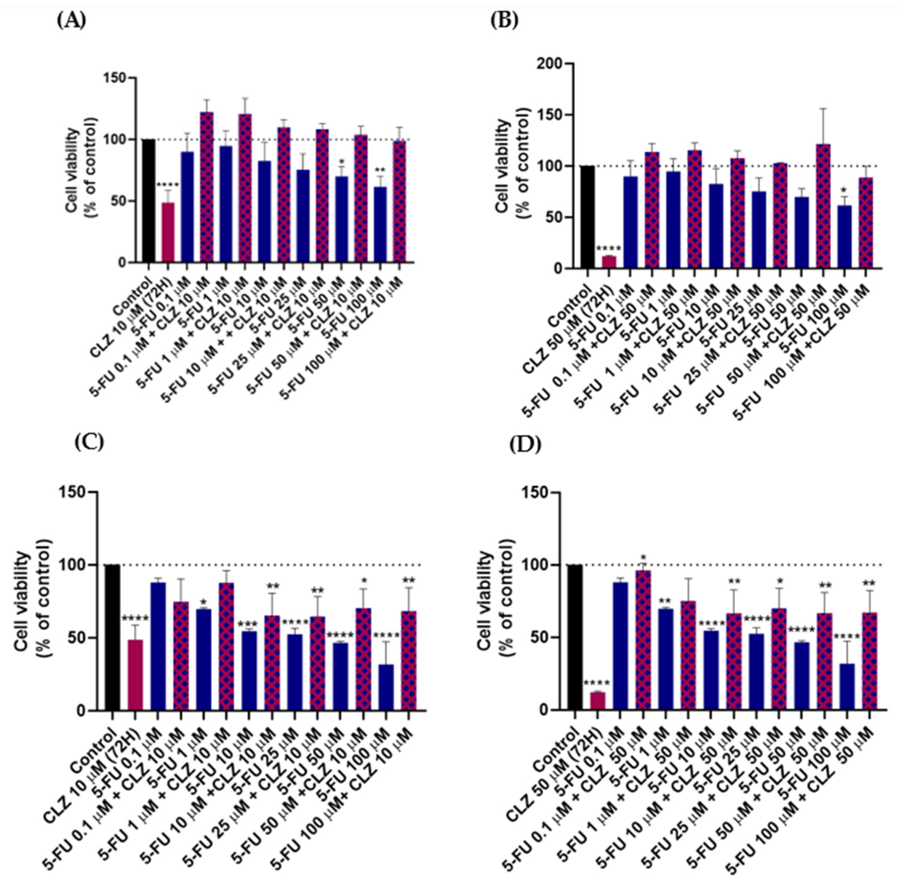
Figure 1 and Figure 2 present the results of administering 5-FU alone, demonstrating cell viability and morphological changes. The results show that, after 48 h, 5-FU does not significantly reduce the cell viability of T3M4 cells (Figure 1A). Additionally, the reduction in cell viability remains inconsistent and never reaches 50%. However, the data indicates that after 72 h, 5-FU leads to a notable decrease in cell viability (Figure 1B). After 72 h of exposure to 5-FU, there is a proportional decrease in T3M4 cell viability corresponding to an increase in 5-FU concentration. Lower concentrations of 10 µM and 25 µM demonstrate cell viability of 54.51% and 52.38% respectively. Elevated concentrations of 50 µM and 100 µM reduced cell viability to 46.58% and 31.78% respectively. The MTT assay data for the T3M4 cell line, illustrated in Figure 1, indicates that 5-FU exhibits significant anti-cancer activity after 72 h; these results suggest that the drug is more active after 72 h in the T3M4 cell line.
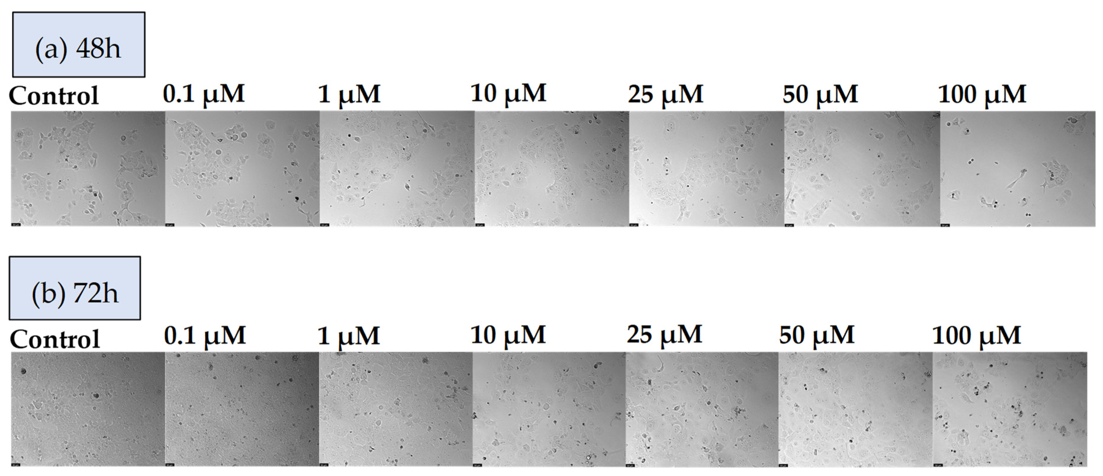
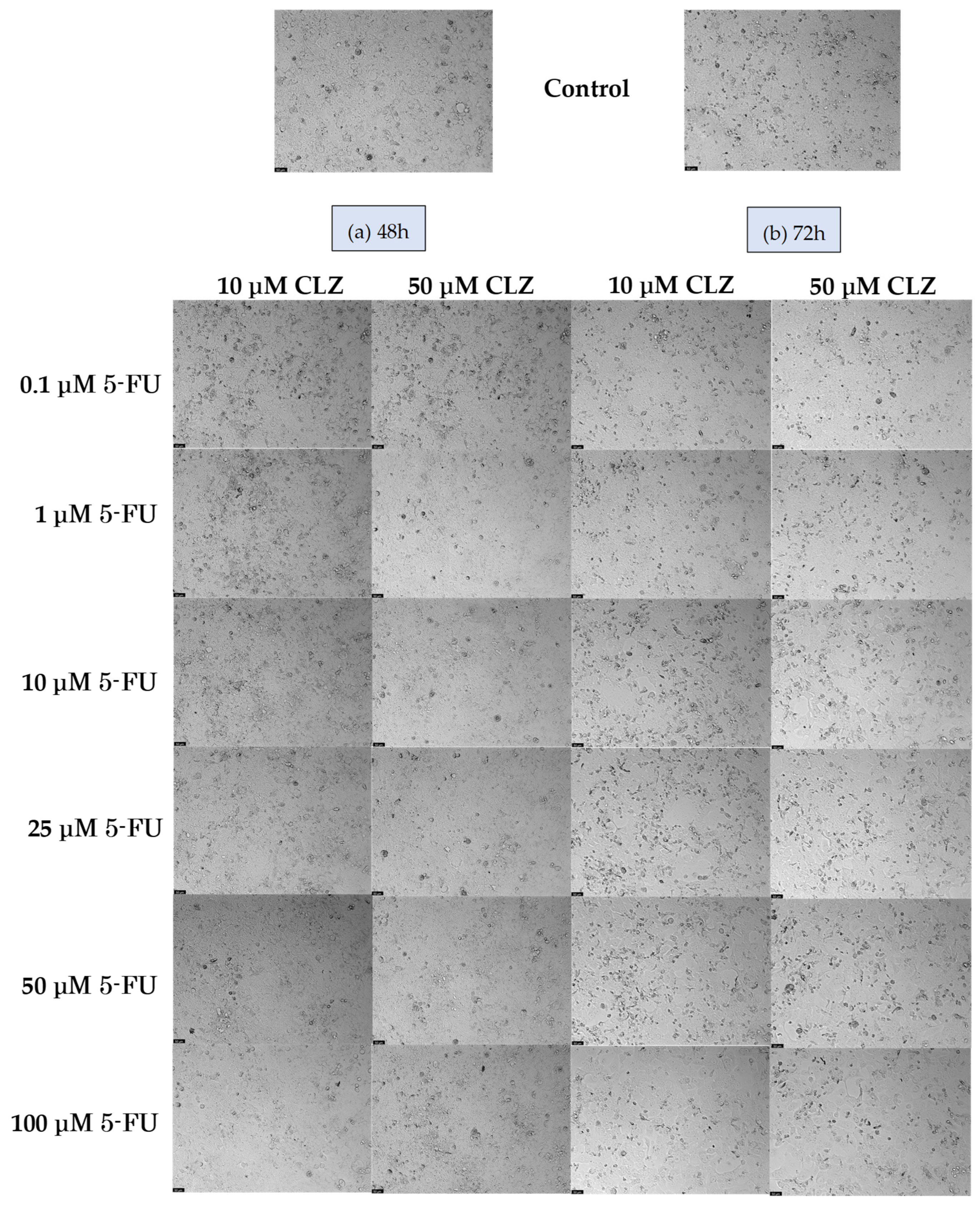
In the morphological analysis (Figure 2), it is evident that exposure to 5-FU resulted in changes in cell phenotype when compared to the control cells. This effect was particularly pronounced after 72 h. Additionally, a discernible reduction in cell density was observed following treatment with 5-FU. These findings are consistent with the results of the MTT assay, as higher concentrations of 5-FU corresponded to reduced cell density.
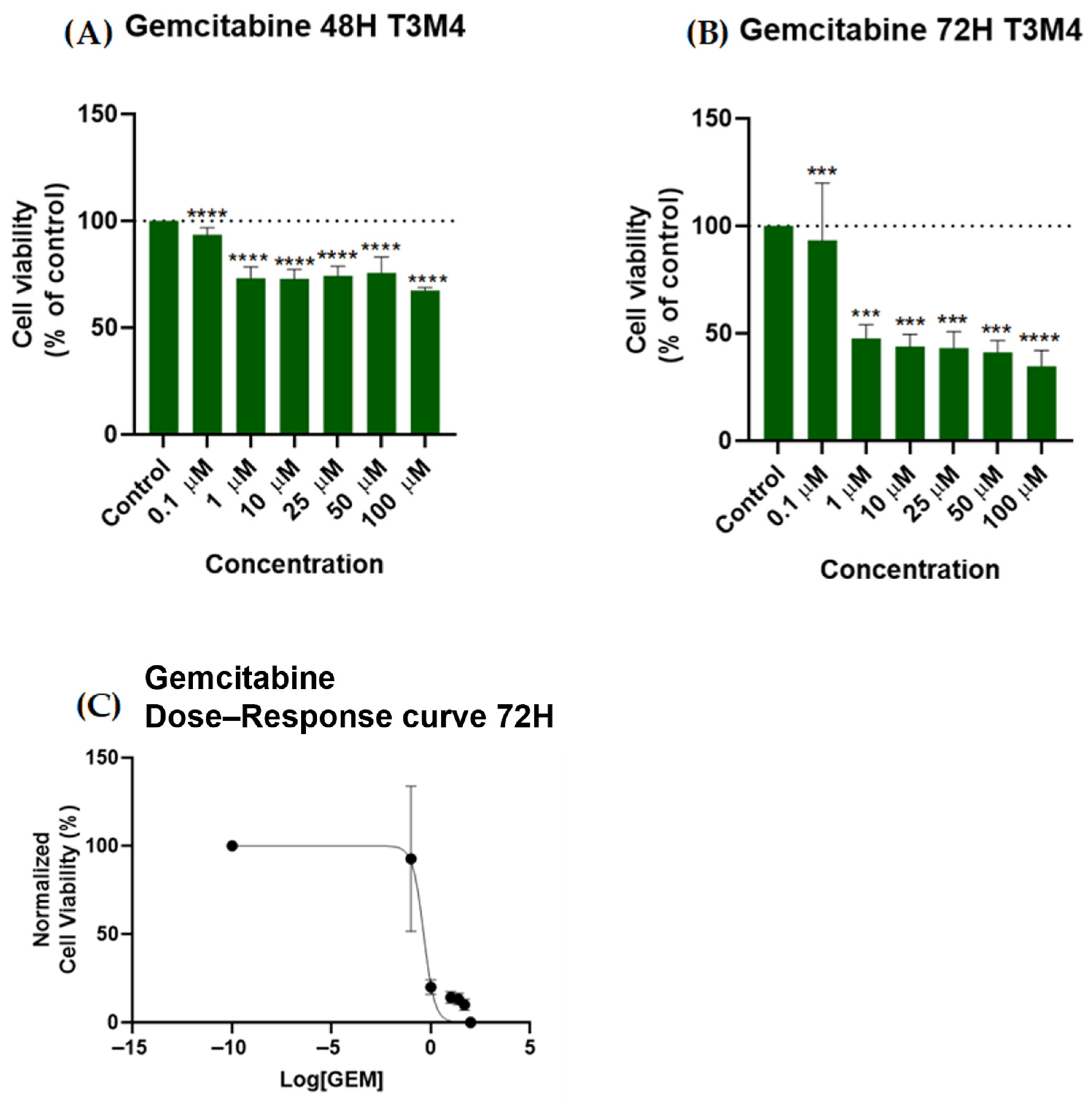
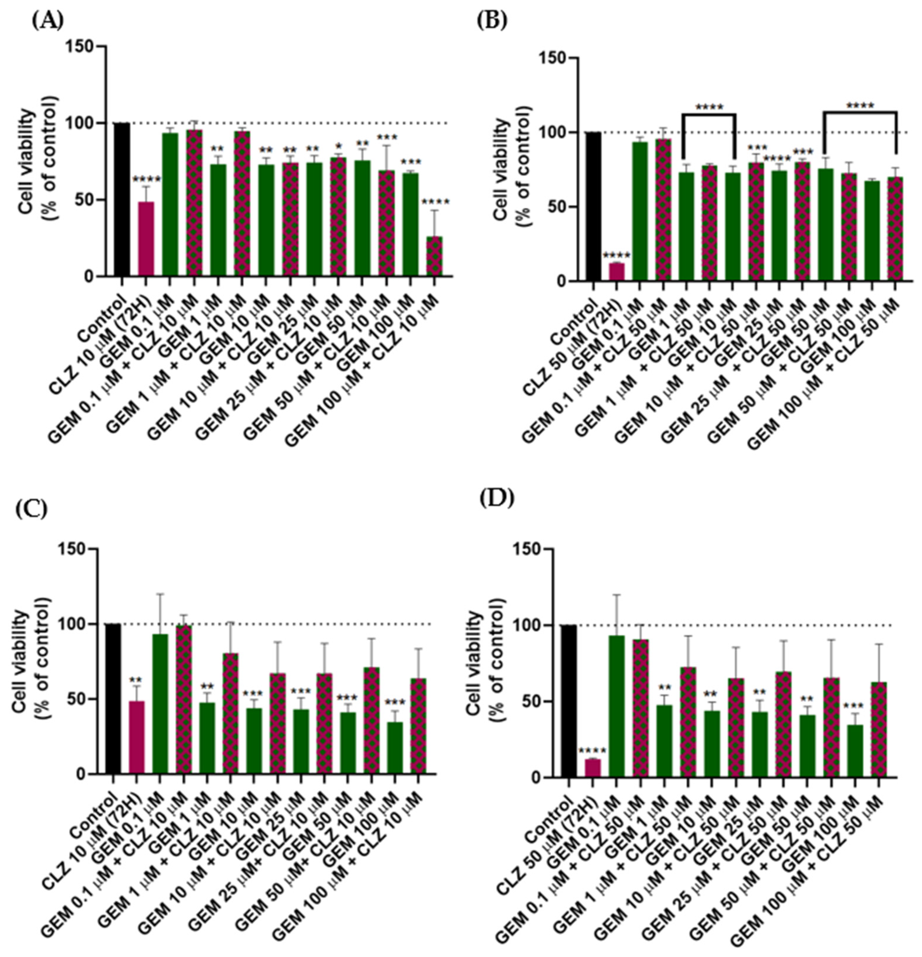
GEM is recognized as the primary chemotherapy agent for treating PDAC [31]. To conduct comparative tests with repurposed drugs and evaluate their efficacy as alternative treatments for drug resistance associated with standard chemotherapies such as GEM, we have tested the efficacy of T3M4 cells exposed to various concentrations (0.1, 1, 10, 25, 50, and 100 µM) of GEM alone after 48 h and 72 h (Figure 3 and Figure 4). In Figure 3A, it is possible to observe that after 48 h, GEM did not induce a significant reduction in the cell viability of the T3M4 cells. Notably, none of the concentrations tested resulted in a cell viability below 50%. Interestingly, the cytotoxic effect of 100 µM GEM was comparable to 1 µM at 48 h, indicating that a higher concentration did not translate into increased cell death. However, after 72 h, a more pronounced dose-dependent response was observed, with 100 µM exhibiting significantly higher cytotoxicity compared to lower concentrations (Figure 3B). GEM concentrations of 1, 10, 25, 50, and 100 µM resulted in cell viabilities of 47.69%, 43.87%, 43.18%, 41.23%, and 34.63%, respectively, highlighting a time-dependent drug response where prolonged exposure of 72 h increased cytotoxicity in a dose-dependent manner.
The dose–response correlation of GEM at 72 h was further analyzed by constructing a dose–response curve using the data obtained from the MTT assay, as shown in Figure 3C. The IC50 value, which indicates the concentration of GEM required to inhibit the growth of T3M4 cells by 50%, was determined from this curve. It was calculated within the range defined by the upper and lower plateaus of the normalized dose–response curve, using non-linear regression analysis. The dose–response curve for GEM at 72 h demonstrated an IC50 value of 0.4384 μM. Overall, significant and consistent antineoplastic activity was observed after 72 h of T3M4 cell exposure to GEM.
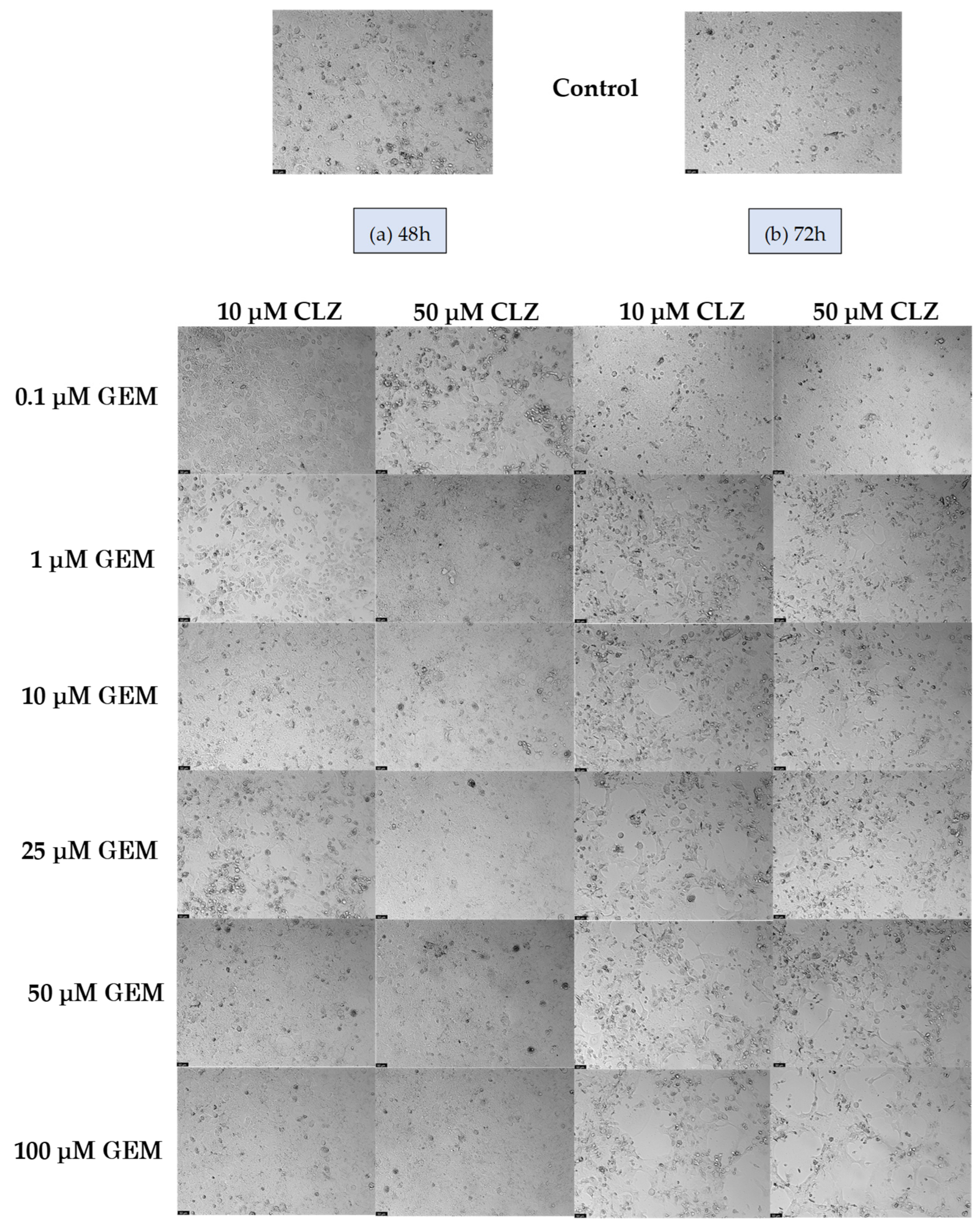
The T3M4 cell line was subjected to varying concentrations of GEM for 48 and 72 h, and subsequent morphological changes were examined, as depicted in Figure 4a,b correspondingly. The treated cells exhibited distinct morphological features compared to the control cells. Notably, at concentrations of 1 µM or higher and a 72-h exposure, the cells displayed a notably elongated morphology, significantly differing from the control cells. In comparison, at 48 h, the morphological changes in the treated cells were less pronounced when compared to the control. These observations align with the data obtained from the MTT assay.
Given the superior antineoplastic activity demonstrated by both standard therapies, 5-FU and GEM, at the 72-h mark, we have opted to explore the repurposed drugs suggested in this article during the same exposure period. This enables us to conduct a comparative analysis of the results.

3.1.2. Cytotoxic Effect of the Repurposed Drugs NTZ and CLZ

The potential antitumor effects of two repurposed drugs, NTZ and CLZ, as individual agents, were assessed in T3M4 cells. The cell line was exposed to increasing concentrations of each drug, ranging from 0.1 to 100 µM, to examine cell viability 72 h post-treatment. This approach allows for a comparison of the effectiveness of these drugs with the optimal results achieved from standard drugs for PDAC. The objective is to determine whether the repurposed drugs presented here may offer a new therapeutic opportunity to combat drug resistance to GEM and 5-FU.
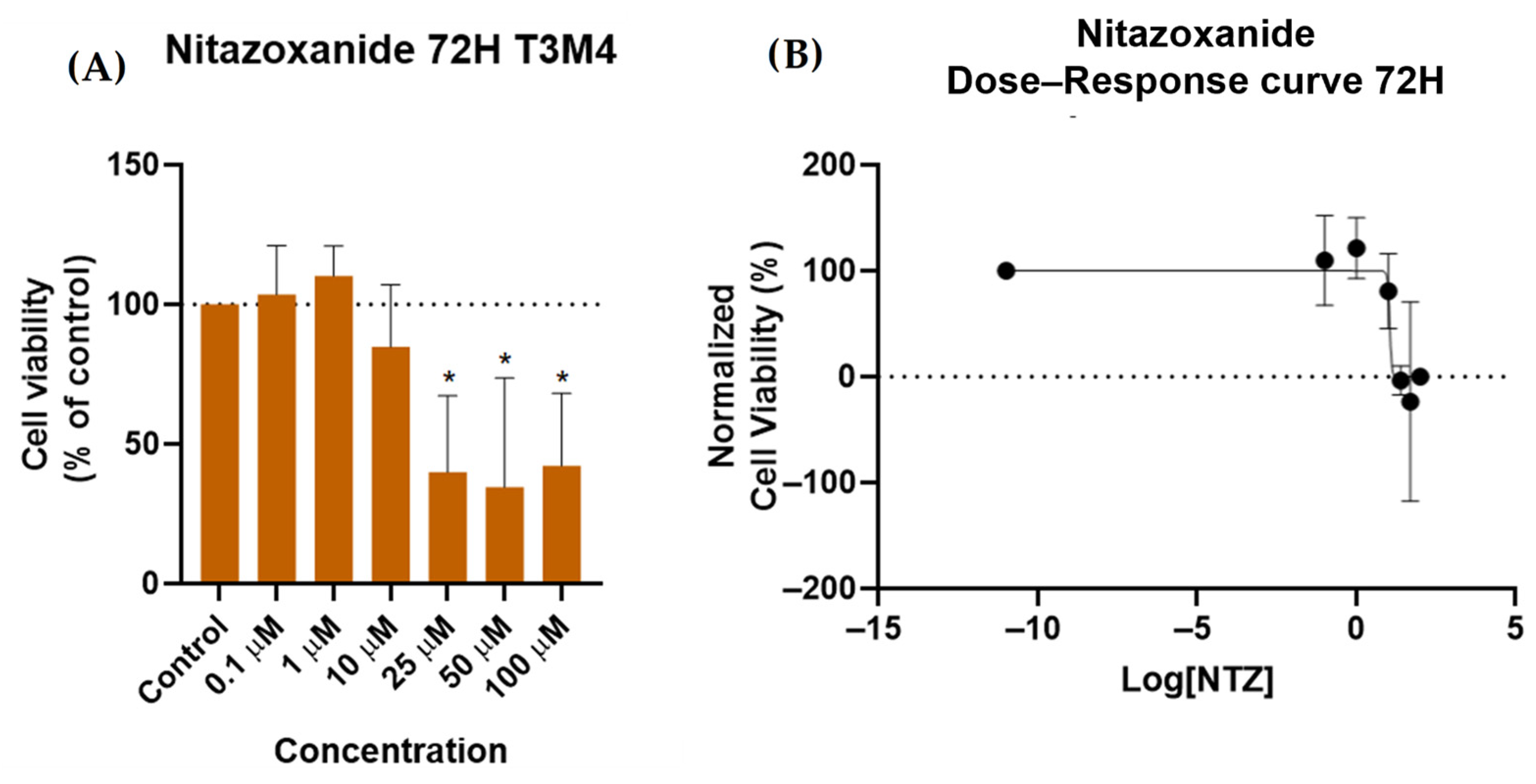
Based on the results obtained from the MTT assay, it was observed that NTZ did not exhibit a significant effect in T3M4 cells (Figure 5A). At concentrations equal to or greater than 25 µM, a reduction in cell viability of less than 50% was observed. However, at a concentration of 100 µM, the decrease in cell viability was less significant compared to the 50 µM concentration. The dose–response curve for NTZ at 72 h indicated an IC50 value of 11.14 µM (Figure 5B). These findings suggest the drug’s performance was inconsistent, indicating a limited antitumor effect in T3M4 cells.
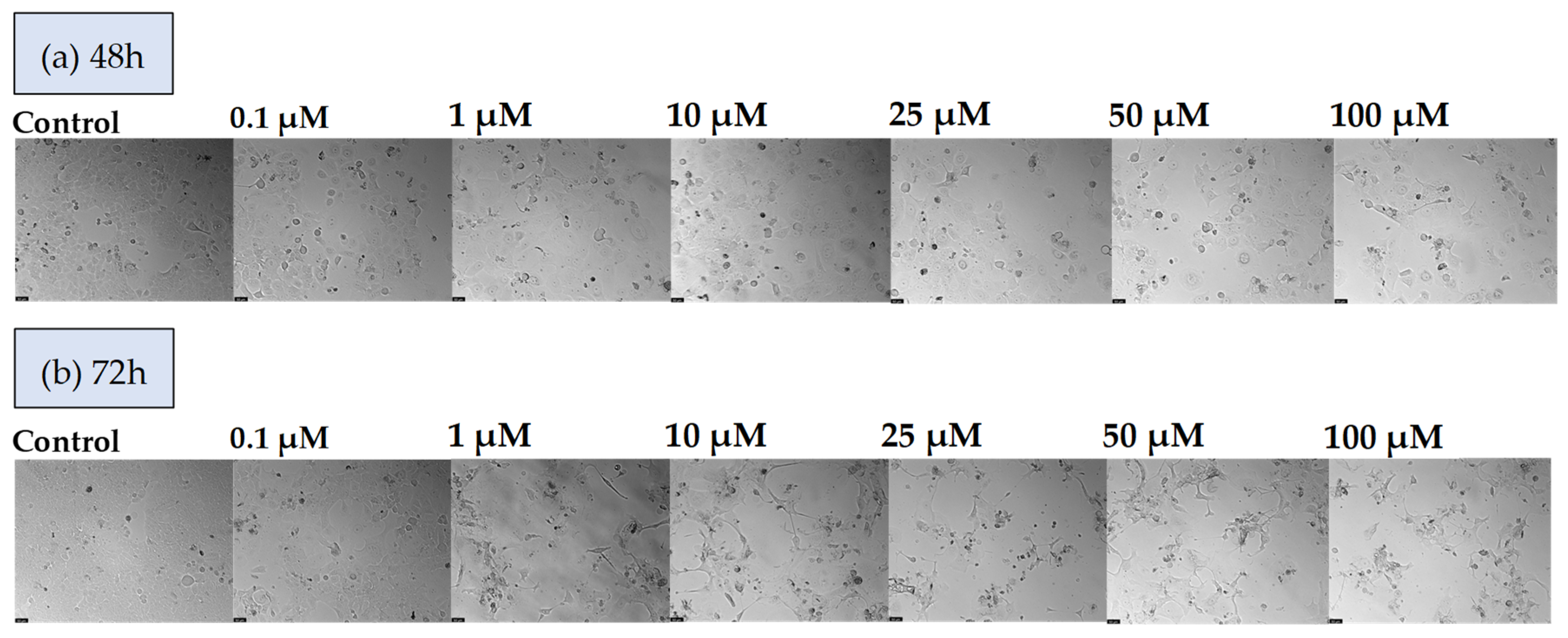
The morphological characteristics of T3M4 cells following 72 h of exposure to the repurposed drug NTZ and the control are depicted in Figure 6. The observed morphological changes align with the findings of the MTT assay. Notably, at concentrations equal to or higher than 25 µM, alterations in cell morphology compared to the control were evident, although the reduction in cell density was generally inconsistent. The results suggest that there is a restricted antitumor effect with potential resistance to NTZ in the T3M4 cell line.
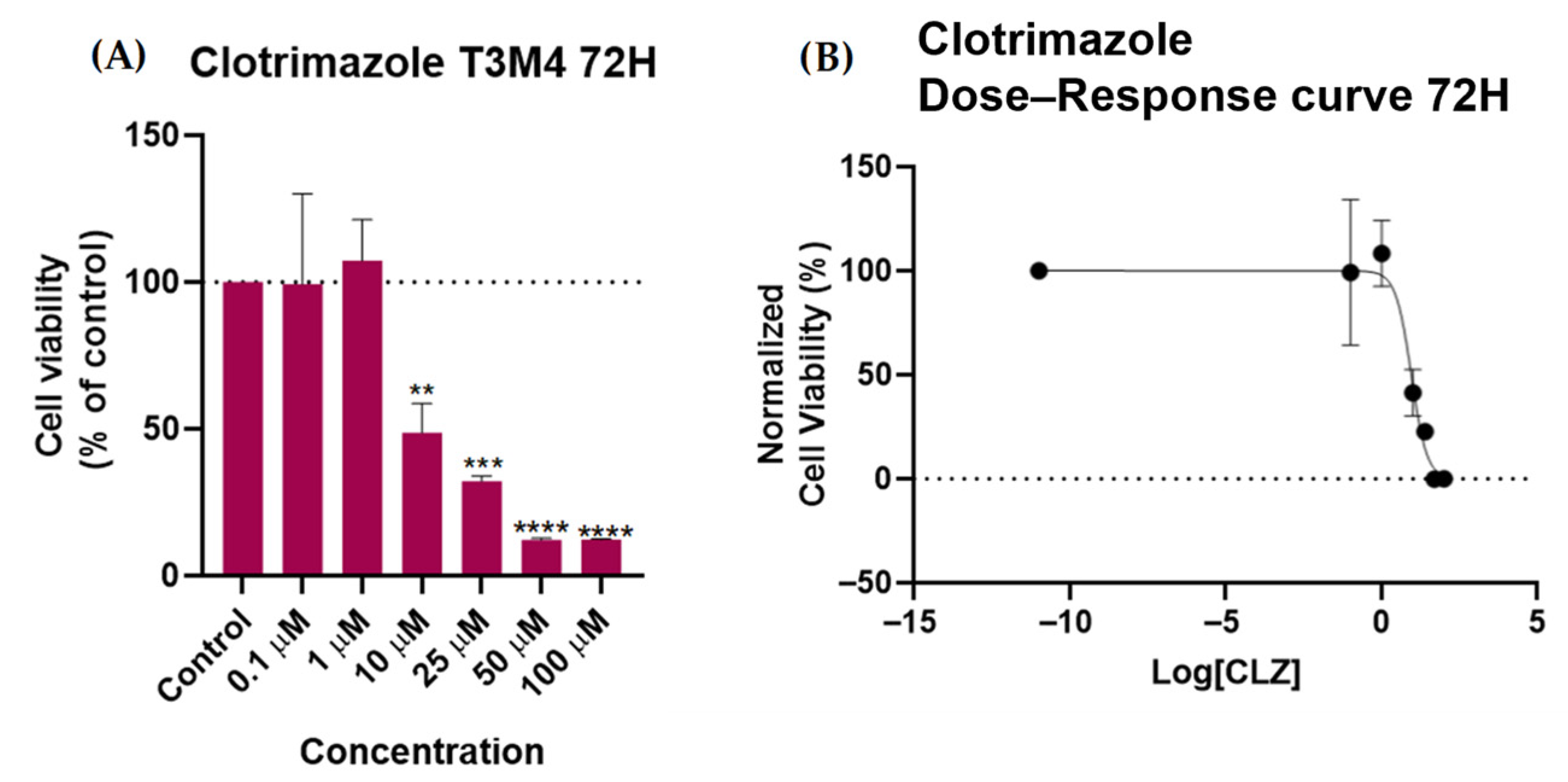
The MTT assay results following the 72-h treatment of T3M4 cells with CLZ are presented in Figure 7. At concentrations of 0.1 µM and 1 µM, no discernible antitumor effects were observed; rather, cells displayed resistance to CLZ. However, at a concentration equal to or greater than 10 µM, cell viability decreased by less than 50% in a concentration-dependent manner (Figure 7A). Specifically, concentrations of 10 µM and 25 µM resulted in cell viabilities of 48.6% and 32.19%, respectively. Higher concentrations of 50 µM and 100 µM yielded cell viabilities of 12.14% and 12.28%, respectively. The dose–response curve for CLZ at 72 h revealed an IC50 value of 9.011 μM (Figure 7B), indicating a potent antitumor effect of this repurposed drug on T3M4 cells after 72 h.
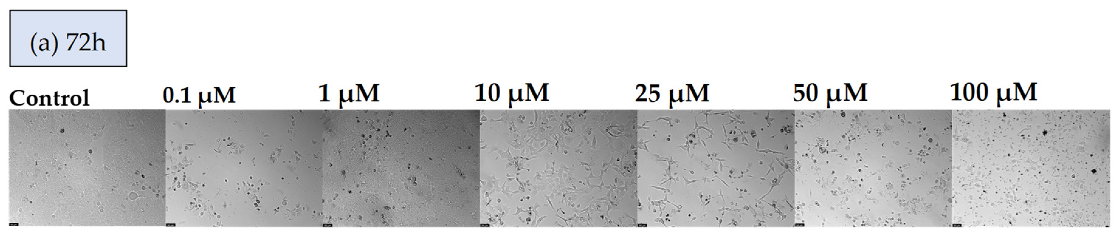
The MTT assay results indicate that CLZ may possess substantial antineoplastic activity in T3M4 cells. Additionally, an evaluation of CLZ’s impact on T3M4 cells over a 72-h exposure period revealed significant morphological effects (Figure 8). At lower concentrations, a resistance effect was observed at 1 µM compared to 0.1 µM, resulting in increased cell density. Consistent with the MTT assay, a decrease in cell density was noted at concentrations between 10 and 100 µM. Notably, at 10 µM, elongation of cells was observed, while at 25 µM, an elongated morphology resembling neuronal cells, distinct from the T3M4 PDAC control cells, was evident. At concentrations of 50 and 100 µM, CLZ induced substantial cell lysis and the formation of cellular debris.

3.2. Drugs Combination in T3M4 Cells

This study evaluated the antitumor efficacy of two repurposed drugs, CLZ and NTZ, and compared them with standard therapies for PDAC. The effects of each drug were assessed individually, and CLZ demonstrated the most significant cytotoxic activity, with a dose-dependent reduction in cell viability at concentrations ranging from 10 µM to 100 µM after 72 h. Based on these findings, CLZ was selected for combination studies to investigate potential synergistic effects with standard therapies. To optimize treatment efficacy, 10 µM and 50 µM concentrations of CLZ were chosen for combinatorial assessments. These concentrations were selected to achieve a significant reduction in tumor cell viability while mitigating excessive cytotoxicity that could compromise therapeutic applicability. The cytotoxic effects of CLZ in combination with standard therapies were evaluated at 48 h and 72 h.

3.2.1. 5-FU and CLZ

As demonstrated above, CLZ was selected based on its consistent antitumor activity in the T3M4 cell line. We assessed cell viability by employing the MTT assay to evaluate the combined impact of 5-FU and CLZ (Figure 9). Upon the combined treatment of T3M4 cells with 5-FU and CLZ, a notable reduction in cytotoxicity was observed compared to treatment with 5-FU alone, as evidenced in Figure 9. Furthermore, the combined treatment did not induce a higher level of cytotoxicity when compared to CLZ treatment alone at 72H. In summary, the combination of these drugs resulted in a less pronounced decrease in cell viability. In some cases, resistance to this treatment approach was observed in the T3M4 cell line.
The morphological analysis depicted in Figure 10 illustrates the effects of the combination of 5-F and CLZ on the morphological characteristics of T3M4 cells. This combination did not significantly impact the morphology or cell density of T3M4 cells.

3.2.2. GEM and CLZ

The results in Figure 11 illustrate the outcomes of the MTT assay conducted to evaluate the impact of combining GEM and CLZ on T3M4 cells over 48 and 72 h. It is noteworthy that, at 48 h, the combination of 100 µM GEM and 10 µM CLZ resulted in cell viability of 26.06%, which is an improvement compared to the individual treatments of GEM (67.44%) and CLZ (48.66%) (Figure 11A). However, the overall findings indicate that the cell viability is higher when the drugs are combined.
The morphological analysis presented in Figure 12 demonstrates the influence of the combined application of GEM and CLZ on the morphological features of T3M4 cells. Although some concentrations showed a reduction in cell density, overall, this combination did not exert a significant impact on the morphology or cell density of T3M4 cells.

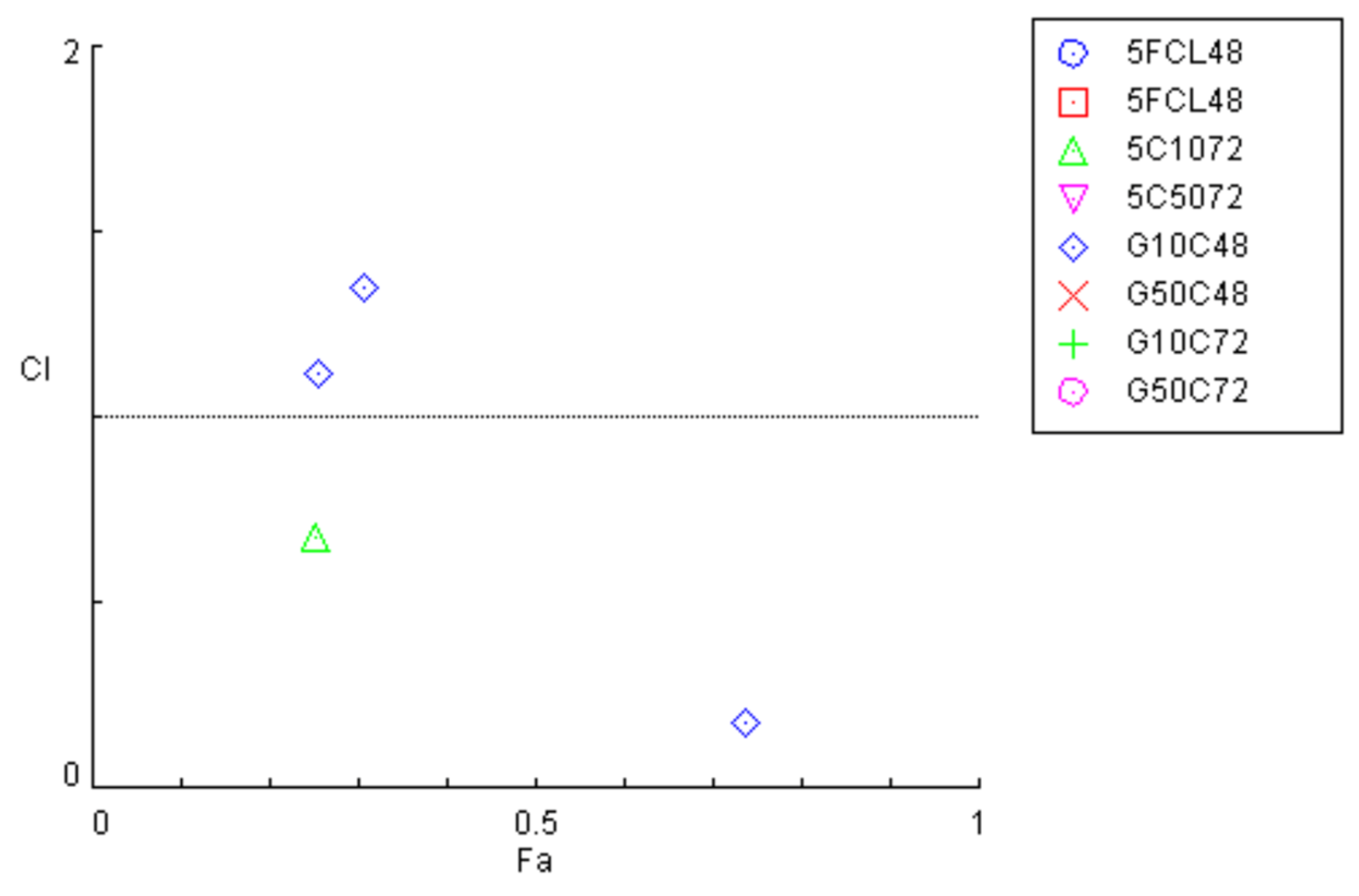
3.3. Combination Index Analysis

The combination index (CI) analysis obtained through CompuSyn demonstrated that, in most cases, the combinations of CLZ, NTZ, GEM, and 5-FU did not result in synergistic interactions in T3M4 pancreatic cancer cells (Figure 13). Although the majority of combinations yielded CI values > 1, indicating antagonism, two exceptions were observed: at intermediate effect levels, the combination of CLZ and 5-FU at 48 h presented a CI < 1, suggesting weak synergy, and at higher effect levels, GEM combined with 10 µM CLZ also displayed a CI slightly below 1. However, these effects were isolated and not consistently reproduced across concentrations or time points, limiting their translational significance.
Importantly, the cytotoxic activity of the single agents was consistently greater than that observed for the combinations under the tested conditions, particularly in the case of CLZ, which showed marked activity when used alone. These results suggest that the antitumoral potential of the tested drugs is more effectively captured in monotherapy, rather than in combination, under the conditions tested.

3.4. In Silico Target Prediction

In silico target predictions and transcript-level expression data support a plausible KCNN4/KCa3.1 hypothesis, but functional validation will be required to establish causality in this model.
Among the predicted targets, two stood out due to their known relevance in pancreatic cancer: the calcium-activated potassium channel KCa3.1 (KCNN4) and the aldo-keto reductase family member AKR1B1. Previous studies have shown that KCNN4 is highly expressed in pancreatic tumors, where it promotes proliferation, migration, and chemoresistance [32,33]. In contrast, although AKR1B1 is expressed at lower levels compared with KCNN4, its upregulation in pancreatic cancer cells has been shown to promote proliferation, inhibit apoptosis, and associate with increased metastatic potential and reduced patient survival [34,35]. Expression analysis using the Human Protein Atlas corroborated these findings, revealing high KCNN4 expression and moderate expression of AKR1B1.

3.5. Target Expression in PDAC

To validate the predicted targets, we analyzed their expression profiles using the Human Protein Atlas. The results showed that KCa3.1 (KCNN4) is highly expressed in pancreatic cancer (Figure 14), strongly supporting its relevance as a functional target of CLZ. In contrast, AKR1B1 was also expressed, but at lower levels compared with KCNN4 (Figure 15). This differential expression pattern provides a molecular explanation for the stronger efficacy of CLZ relative to NTZ in T3M4 cells.
These findings are consistent with the CI analysis, which indicated limited or absent synergy between drugs, and further support the notion that single-agent treatments, especially CLZ, may represent more effective therapeutic strategies in PDAC than drug combinations.

3.6. In Silico ADMET Profiling to Support In Vitro Antitumor Effects

To complement the in vitro cytotoxicity results, an in silico ADMET profiling was conducted for the four studied drugs (NTZ, CLZ, 5-FU, and GEM). Physicochemical properties were estimated using the compound prediction software ADMET Predictor® v11.0 (Table 1). A comparative analysis of these descriptors was performed against reference values obtained from relevant drug databases and other established ADME prediction platforms, including SwissADME and pkCSM. The close agreement between the predicted and reference values supports the robustness of the in silico analysis.
CLZ demonstrated higher predicted lipophilicity and reduced predicted aqueous solubility in comparison to NTZ, 5-FU, and GEM. NTZ exhibited an intermediate level of lipophilicity while maintaining low aqueous solubility. In contrast, both 5-FU and GEM were characterized by lower predicted lipophilicity and increased aqueous solubility. These differences in physicochemical properties were further associated with enhanced predicted permeability for CLZ relative to the other compounds.
Predicted metabolic interactions revealed comparative differences between repurposed drugs and standard chemotherapeutics. CLZ demonstrated a wider predicted engagement with various CYP isoforms, functioning as both a substrate and an inhibitor. In contrast, NTZ presented predicted interactions with a more limited selection of CYP enzymes, generally accompanied by lower confidence levels. Standard chemotherapeutic agents, on the other hand, displayed more restricted predicted metabolic involvement. For instance, 5-FU was predicted to interact with a limited number of CYP isoforms, while GEM was not expected to undergo phase I metabolism; rather, it is associated with phase II conjugation via UGT2B7.
Transporter interaction predictions provided valuable insights into the behavior of the compounds. Both NTZ and CLZ were identified as having potential interactions with multiple uptake and efflux transporters, in contrast to 5-FU and GEM, which exhibited simpler transporter interaction profiles. These predictions were evaluated within the context of differences in intracellular exposure and the observed outcomes from in vitro combination studies.
Overall, the in silico ADMET results offer a succinct comparative analysis of the physicochemical properties, predicted metabolic pathways, and transporter interactions of the evaluated compounds. These findings serve as supportive, comparative, and hypothesis-generating data that complement the experimental results.

4. Discussion

PDAC presents a challenging clinical scenario with poor prognosis. The mortality rate closely aligns with the incidence rate [36], and untreated patients typically have a survival period of only a few months. Surgical resection of the affected organ portion represents the most effective therapeutic approach. However, this option is available to only approximately 20% of patients. Furthermore, even among eligible patients, recurrence commonly occurs within a relatively brief timeframe [37]. The current standard of care for PDAC patients involves the use of conventional cytotoxic agents, with GEM being the established gold standard for treating advanced PDAC [38].
In our study, we observed that T3M4 cells treated with GEM exhibited spindle-shaped morphology, loss of cell–cell adhesion, and the presence of pseudopodia-like structures, which were not as prominent in cells treated with 5-FU. These findings are corroborated by previous studies reporting that GEM-resistant cells undergo distinct morphological changes, including an elongated shape, increased pseudopodia formation, and features resembling transformed fibroblasts [39,40]. In contrast, cells treated with 5-FU primarily displayed a more compact and adherent morphology, suggesting that these two chemotherapeutic agents may induce different cellular responses. This difference in morphological changes may be related to different mechanisms of action, with GEM affecting DNA synthesis [9] and 5-FU primarily targeting RNA processing and thymidylate synthase inhibition [41]. Moreover, our findings indicate that GEM outperformed 5-FU as it demonstrated greater efficacy in reducing cell viability of T3M4 cells at lower concentrations. Additionally, it exhibited a decrease in cell viability with an increase in GEM concentration at 72 h. In light of the aggressive and highly chemoresistant characteristics of PDAC, the primary biological endpoint of this study was to evaluate the reduction in cancer cell viability following drug exposure. To effectively illustrate this, we utilized cell viability graphs as the main experimental readout, which provide direct insights into the time- and concentration-dependent cytotoxic effects, including the maximal reduction in viable cells. Additionally, IC50 values were incorporated as a complementary measure to describe the relative potency of the treatments; however, it is important to note that these values do not necessarily indicate maximal cytotoxic efficacy. This distinction is particularly significant when assessing the comparative effects of drugs with different pharmacodynamic profiles, such as GEM and CLZ. Nonetheless, the emergence of drug resistance to both drugs (5-FU and GEM) presents a significant challenge, resulting in high mortality rates for patients with PDAC. As a result, our focus has been on identifying a potential repurposed drug candidate that may offer superior efficacy compared to the standard drugs.
The development of novel anti-cancer drugs constitutes a costly and time-consuming endeavor, necessitating comprehensive cell- and animal-based studies, followed by human clinical trials to validate preclinical findings regarding safety and efficacy [42]. It is important to recognize that the process, with an average duration of 13 years, is highly demanding. A promising new chemical entity may encounter setbacks during the necessary clinical trial phases before its approval as a drug. These setbacks may arise from unforeseen safety issues or a lack of efficacy in patients [42,43,44]. In the current scenario, repurposing approved drugs for cancer therapy represents an appealing and alternative approach. This strategy has the potential to address several issues linked to the discovery of new drugs [1,3,45].
Numerous studies have demonstrated the inhibitory effects of the drug CLZ on the proliferation of cancer cells through diverse mechanisms. For example, Liu et al. found that CLZ suppresses migration and invasion of hepatocellular carcinoma cells by inhibiting ERK phosphorylation and epithelial–mesenchymal transition [46]. In breast cancer cell lines (MCF-7 and MDA-MB-231), Bae et al. observed that CLZ induces apoptosis and G1 phase arrest while inhibiting MMP9, thereby reducing cell proliferation and invasiveness [47]. Additionally, Wang et al. conducted further research showing that CLZ significantly reduces cell viability, colony formation, and tumor growth in oral squamous cell carcinoma, both in vitro and in vivo, primarily through G0/G1 cell cycle arrest and modulation of apoptosis-related proteins [48]. In studies involving melanoma models, Ochioni et al. and Adinolf et al. observed that CLZ not only hinders tumor growth but also influences the tumor microenvironment by reprogramming tumor-associated macrophages and prompting apoptosis through hexokinase inhibition [23,49].
Zuccolini et al. highlighted the potential off-target effects of CLZ on IK channels, affecting the viability and migration of melanoma and pancreatic cancer cells [50]. Our study corroborates their findings, showing that CLZ reduced the number of viable pancreatic cancer cells compared with DMSO-treated controls after 72 h. While their research primarily focused on ion channels, specifically IK channel blockers, and evaluated the efficacy of CLZ as a channel blocker at 30 µM for 72 h, we tested concentrations ranging from 0.1 to 100 µM and found that CLZ, when administered alone, displays substantial antineoplastic activity, resulting in a significant reduction in cell viability. It is important to note that although GEM demonstrates a higher relative potency based on IC50 values, CLZ induced a greater reduction in cell viability at higher concentrations after 72 h of exposure.
The results of this study highlight variations in pharmacodynamic profiles, rather than suggesting a superior intrinsic potency of CLZ.
The results derived from the in silico ADMET assessment of CLZ exhibit a notable concordance with the in vitro findings. CLZ exhibits considerable lipophilicity and permeability, attributes that may facilitate its intracellular accumulation and elucidate its pronounced cytotoxic effects in PDAC cells. The predicted interactions with various CYP enzymes and drug transporters are therefore regarded as supportive evidence, contributing to the generation of hypotheses for further investigation, rather than serving as definitive indicators of in vivo mechanisms of action.
The existing epidemiological and preclinical data indicate that certain CYP isoforms, notably CYP2A6 [51] and CYP2C9 [52], may be implicated in pathways associated with cancer. Within this framework, the predicted inhibition of these enzymes by CLZ presents a plausible, albeit indirect, basis for further exploration of its potential repurposing as a therapeutic agent for PDAC. Nevertheless, it is essential to underscore that direct evidence linking these pathways to the progression of PDAC remains limited. Consequently, a careful and thoughtful interpretation of this data is warranted. Importantly, the in vitro findings obtained in PDAC cell models must be interpreted with caution, as drug behavior may differ substantially following systemic administration in humans, a factor underlying many translational failures [53]. Indeed, CLZ is known to undergo rapid hepatic metabolism via the CYP system, resulting in the formation of inactive metabolites that are readily excreted [54]. This extensive first-pass metabolism leads to very low systemic exposure and minimal circulating levels of the parent compound, which may compromise target engagement in vivo. The ability to achieve the antitumor activity observed in PDAC cells at clinically relevant concentrations remains an open question that necessitates further investigation.
Nitazoxanide (NTZ) has demonstrated various pharmacological effects in infectious and neoplastic diseases [55,56,57]. However, the therapeutic potential of NTZ in PDAC is unknown, as it has not been previously evaluated in PDAC to the best of our knowledge. The efficacy of NTZ in gastric cancer cell lines was previously investigated by Ribeiro et al. [58], who found promising results indicating its potential as a therapeutic agent for GC. However, our findings reveal that NTZ, when administered as a monotherapy to T3M4 cells over a 72-h period, does not demonstrate significant antitumor activity. The observed nonlinear dose–response curve indicates potential limitations in its efficacy, which may reflect adaptive cellular responses or resistance mechanisms at higher concentrations. These observations are consistent with the in silico ADMET predictions. Although NTZ displays moderate lipophilicity, which could in principle favor cellular distribution, its very low aqueous solubility and limited unbound fraction may restrict effective intracellular concentrations in T3M4 cells.
From the perspective of clinical efficacy, NTZ may emerge as a candidate for repurposing in the treatment of PDAC under particular conditions. Its drug–transporter interaction profile identified NTZ as a substrate of P-gp, a feature of particular relevance in PDAC, where overexpression of efflux transporters is common and contributes, at least in part, to the intrinsic chemoresistance of this malignancy [59]. Conversely, NTZ was also predicted to be a substrate of the OATP superfamily, which has been reported to be overexpressed in pancreatic tumors compared with normal pancreatic tissue [60,61,62]. As OATPs mediate drug uptake, their overexpression could potentially enhance NTZ disposition within pancreatic cancer cells. However, this characteristic also raises concerns in the context of polypharmacy, as the concomitant administration of multiple xenobiotics that share the same OATP substrates may lead to competitive interactions [63]. Although NTZ exhibits significant mechanistic interest, its translational applicability in the context of PDAC appears to be substantially limited under the conditions assessed.
Notwithstanding these limitations, the predicted CYP-mediated interactions of NTZ are considered exploratory and hypothesis-generating. The in silico analysis has identified NTZ as a potential inhibitor of CYP1A2, CYP2C9, and CYP2C19, while also acting as a substrate of the latter two enzymes. However, the relevance of these predicted interactions within the context of PDAC remains to be definitively elucidated. CYP1A2 and CYP2C9 have been described as having potential associations with cancer-related metabolic pathways across various tumor models [52,64]. Nevertheless, their precise roles in PDAC have not been clearly established. Thus, it is essential to exercise appropriate caution when considering the potential contributions of NTZ-mediated CYP modulation to the antitumor effects observed in PDAC.
Notably, the literature describing NTZ metabolism is inconsistent. Regulatory documentation from the Food and Drug Administration (FDA) reports inhibitory potential toward CYP2C9 [65], in agreement with our predictions. Other in vitro drug metabolism studies have described that tizoxanide, the active metabolite of NTZ, does not exert significant inhibitory effects on CYP enzymes [66]. These discrepancies highlight the need for further dedicated in vitro studies to clarify the speculative mechanisms proposed here and to better define the metabolic and translational potential of NTZ in PDAC.
After assessing the anticancer potential of each repurposed drug alone in the T3M4 cell line, we investigated the cytotoxic effects of combining the most promising repurposed drug, CLZ, with GEM and 5-FU. The concept of drug combinations has garnered significant attention as a potential strategy for addressing complex diseases such as cancer, inflammation, and type 2 diabetes [67,68,69]. However, the interactions between drugs in combination can result in a wide range of unexpected outcomes [70]. Of particular interest are drug synergy and antagonism. Drug synergy, which refers to the enhanced efficacy of drugs when used in combination, is a key objective in combinational drug development [68]. Synergistic drug combinations have demonstrated high efficacy and greater therapeutic specificity [71]. Conversely, drug antagonism is often undesirable but may have utility in combating drug-resistant mutations [72]. The findings indicate that there is no synergistic effect between CLZ and GEM or 5-FU.
These findings suggest that CLZ may be better suited as a single-gent repurposing candidate rather than as part of combination regimens with standard chemotherapeutics in this PDAC model.
The antagonistic effect observed for the combined use of CLZ and 5-FU may be explained by CLZ-mediated interference with 5-FU uptake, leading to a pharmacokinetic-pharmacodynamic (PK-PD) disconnect. Our in silico predictions indicate that CLZ inhibits the transporters OAT1 and OAT3, which are partially responsible for 5-FU cellular uptake.
Inhibition of these transporters is therefore expected to reduce intracellular exposure to 5-FU and, consequently, attenuate its cytotoxic activity. Moreover, both compounds are substrates of P-gp, and their concurrent administration may result in competitive interactions at this efflux transporter. These hypotheses are consistent with the predominantly antagonistic CI values observed experimentally.
From a Structure–Property–Activity (SPAR) perspective, the imidazole scaffold of CLZ offers clear directions for rational structural optimization and activity tuning, with a focus on enhancing single-agent efficacy rather than combination performance.
The chlorinated imidazole scaffold of CLZ suggests tractable vectors for tuning the lipophilic–ionizable balance and membrane/channel engagement. For example, imidazole N-substitutions may modulate pK_a and intracellular retention, while selective halogen editing on the phenyl rings could rebalance lipophilicity and mitigate potential metabolic liabilities without compromising potency. Bioisosteric replacement of the imidazole ring may further allow fine-tuning of physicochemical properties and off-target interactions. These considerations provide a framework for future SAR-driven optimization rather than definitive mechanistic claims.
The observed attenuation in combinations containing CLZ is consistent with competition at the transporter level. In contrast, the significant cytotoxic effects of CLZ when administered as a single agent, alongside the high expression of its predicted target, KCNN4/KCa3.1, in PDAC cells, indicate that CLZ’s antitumoral efficacy may be more effectively demonstrated when utilized as a monotherapy under the tested conditions.
The presence of glycolytic ATP is crucial for powering ATP-dependent plasma membrane calcium ATPases (PMCAs), which are accountable for maintaining low intracellular calcium ([Ca2+]i). Inhibition of glycolysis, rather than mitochondrial metabolism, results in the diminishment of PMCA function, leading to cytotoxic calcium overload and subsequent cell death in PDAC cells [73]. Furthermore, the reversal of the Warburg effect has been demonstrated to preserve PMCA function in PDAC cells treated with glycolytic inhibitors [74]. Consequently, the glycolytic dependency of PMCAs may offer a promising therapeutic target in PDAC. It is noteworthy that in our study, CLZ demonstrated pronounced antineoplastic effects in this in vitro model, distinct from those observed for the individual standard therapeutics. The anticancer efficacy of CLZ has been associated with its ability to reduce tumor cell viability by inhibiting glycolytic flux, leading to a decrease in intracellular ATP levels. CLZ is believed to affect cell glycolysis and ATP production by detaching glycolytic enzymes from the cytoskeleton. Zancan et al. [75] demonstrated that CLZ directly inhibits the key glycolytic enzyme 6-phosphofructo-1-kinase (PFK). Their findings suggest the role of CLZ as a negative regulator of glycolytic flux through direct inhibition of the key enzyme PFK [75].
The development of chemotherapy resistance to GEM and 5-FU may trigger metabolic reprogramming that plays a role in the regulatory mechanisms associated with chemotherapy resistance in PDAC cells. This may elucidate the findings observed with ClZ, suggesting that this drug may target key PDAC hallmarks related to metabolic reprogramming. The hypothesis proposed warrants consideration in future studies, particularly in the context of repurposing CLZ for the treatment of PDAC. It is imperative to conduct thorough testing to elucidate the drug’s mechanism of action in targeting PDAC.

4.1. Perspectives for Further Investigation

4.1.1. The Use of a Single Cell Line T3M4

Because CLZ was tested at fixed concentrations (10 and 50 µM) while 5-FU/GEM were dose-ranged, the CI analysis should be interpreted as an initial interaction screen; ratio-dependent effects may warrant follow-up using constant-ratio designs anchored to IC50 values. Accordingly, CI results should be regarded as exploratory rather than definitive. For the present study, we utilized a single PDAC cell line, specifically T3M4. In the T3M4 cell line, a comprehensive analysis has revealed a total of 88 mutated genes, as delineated in the CCLE Cell Line Gene Mutation Profiles dataset [76]. Among the identified mutations of significant interest are those affecting the KRAS, TP53, and CDK11B genes [76]. The T3M4 cell line is defined by the presence of the KRAS Q61H mutation [77]. It is noteworthy that this particular mutation constitutes approximately 5.5% of the KRAS mutation subtypes typically identified in cases of pancreatic cancer and the clinical features associated with this subtype of KRAS mutation remain largely unexplored. The predominant KRAS mutations observed within pancreatic cancer are KRAS G12D, KRAS G12V, and KRAS G12R [78].
The significance of KRAS mutation alleles and clinical outcomes in PDAC remains unclear and warrants further investigation. Recently, Yousef et al. [79] undertook a comprehensive analysis of a substantial cohort sourced from the MD Anderson Cancer Center, which included 803 patients diagnosed with stages I–IV of pancreatic cancer. Their investigation revealed that both the KRAS G12D and KRAS Q61H mutant subtypes are significantly correlated with inferior overall survival when compared to the wild-type KRAS. Furthermore, in their assessment of the PanCAN “Know Your Tumor” (KYT) dataset, encompassing 408 patients from an external cohort, the authors discerned that the KRAS G12R subtype demonstrates a notable survival advantage relative to the KRAS G12D and KRAS Q61H subtypes [79]. This study reveals that the KRAS Q61H subtype, while not the most prevalent KRAS mutation identified in PDAC, might be associated with poorer overall survival compared to more frequently observed mutations and the wild-type KRAS. Consequently, it is important to explore potential therapeutic strategies in the context of KRAS Q61H, as we have undertaken in our research. Nevertheless, the exclusive utilization of one cell line may not adequately capture the heterogeneity of PDAC phenotypes, necessitating the validation of our findings in additional cell lines. It is recommended that key experimental procedures be replicated in one to two supplementary cell lines that exhibit more frequent mutations, such as KRAS G12D and KRAS G12V, to further substantiate the findings of this study. Furthermore, it would be advantageous to evaluate the pharmacological effects of CLZ and NTZ on normal pancreatic cell lines, including HPDE and HPNE. In addition, the incorporation of a spectrum of cell lines, categorized according to varying degrees of differentiation—namely poorly, moderately, and well-differentiated pancreatic cancer cell lines—would significantly enhance the comprehensive understanding of these therapeutic agents across diverse PDAC differentiation statuses. Future investigations should also consider the inclusion of GEM- or 5-FU-resistant cell lines.

4.1.2. Potential Cytotoxic Effects

The present in vitro dataset provides a clear comparative activity profile across CLZ, NTZ, 5-FU, and GEM under standardized conditions. Nevertheless, the primary readout (MTT) reflects metabolic competence and is therefore an indirect proxy for viability, while the associated morphological inspection remains qualitative. Thus, the results are best interpreted in terms of exposure-dependent effects on cell viability rather than definitive cell-death mechanisms.
We examined the effects of CLZ and NTZ on the T3M4 PDAC cell line using cell viability assays and morphological analyses. While these methodologies provided valuable preliminary insights into the cytotoxic effects of these agents, they do not fully capture the long-term consequences or the underlying mechanisms of action. The MTT assay [80], despite its widespread use for assessing cell viability, has notable limitations. Its reliance on mitochondrial dehydrogenase activity as a viability indicator can lead to misinterpretations, as mitochondrial function may be altered independently of actual cell survival. Factors such as metabolic adaptations, drug-induced mitochondrial stress, and variations in cell type can influence MTT reduction, leading to inaccurate assessments of cytotoxic effects [81,82]. Additionally, the MTT assay does not differentiate between cytostatic and cytotoxic effects, making it insufficient for determining whether a reduction in viability results from cell cycle arrest or induction of cell death [83]. While it has been validated for proliferative and antiproliferative assays, discrepancies between MTT and other viability assays highlight its limitations [84,85]. As a result, the observed decrease in viability could be due to either a cytostatic effect (cell cycle arrest) or a cytotoxic effect (induction of apoptosis or necrosis). Although morphological changes, such as loss of cell adhesion and cellular debris formation, were observed following CLZ treatment, these findings remain qualitative and do not confirm the specific mode of cell death.
To further expand these promising findings, future investigations can integrate biochemical and functional assays to gain deeper mechanistic insights into the effects of CLZ. Techniques such as Annexin V and propidium iodide (PI) co-staining will allow clear distinction between apoptotic and necrotic pathways [86], while caspase-3/7 activation assays can confirm the involvement of apoptosis. Complementary approaches, including LDH release assays, will provide valuable information on membrane integrity and cell death mechanisms. In parallel, Western blotting or immunofluorescence targeting proliferation markers such as Ki-67 or proliferating cell nuclear antigen (PCNA) can help determine whether CLZ primarily exerts its effects through inhibition of cell proliferation. Altogether, these methodologies offer powerful tools to unravel the precise biological processes underlying the strong cytotoxic activity of CLZ, thereby reinforcing its potential as a novel therapeutic option in pancreatic ductal adenocarcinoma [87,88,89,90,91,92].
An important avenue for further exploration is the incorporation of direct biochemical assays, such as ATP quantification, to strengthen mechanistic insights into the effects of CLZ on glycolysis and its interactions with GEM and 5-FU. While the current results clearly demonstrate significant cytotoxic activity and reveal antagonistic interactions in certain combinations, complementing these findings with ATP measurements would provide valuable confirmation of the impact of CLZ on cellular energy metabolism. Assessing intracellular ATP levels in PDAC cells treated with CLZ, either as a standalone therapy or in combination with GEM and 5-FU, will help clarify whether glycolytic inhibition directly drives the observed biological responses. This knowledge will expand our understanding of the metabolic dynamics at play and refine the interpretation of drug–drug interactions in this context.
Equally important, the MTT assay continues to be widely recognized as a reliable and robust method for initial screening of drug cytotoxicity. In this study, it has served as a powerful tool to uncover the strong therapeutic potential of CLZ against PDAC. Building on this solid foundation, future studies can benefit from integrating additional quantitative biochemical assays and automated high-throughput analytical platforms. Such complementary approaches will enhance precision, reproducibility, and mechanistic clarity, enabling a more comprehensive characterization of CLZ’s mode of action. By advancing in these directions, future research will not only validate and expand the current findings but also accelerate the translational pathway of CLZ from in vitro promise to preclinical development. Altogether, this progress could position CLZ as an innovative and effective therapeutic option for pancreatic ductal adenocarcinoma, addressing one of the most urgent unmet needs in oncology.

4.1.3. Considerations Regarding the 2D Monolayer Cell Culture Model

This study was performed in a 2D monolayer cell culture model, which provides a controlled and reproducible system for assessing drug effects. While such models are highly informative, they do not fully capture the complexity of the tumor microenvironment in vivo, including stromal interactions, immune components, and extracellular matrix dynamics that may shape drug response and resistance. To further increase translational relevance, complementary approaches such as 3D spheroid systems, organoid cultures, and ultimately in vivo studies could provide additional insights and validation of the therapeutic potential of CLZ in PDAC [93,94,95].
Importantly, despite the simplified nature of the 2D setting, our results clearly show that CLZ induces significant, dose-dependent cytotoxic effects in T3M4 cells, accompanied by morphological alterations consistent with impaired cell viability. As a first step, these findings establish a strong foundation and underscore the importance of future mechanistic and translational investigations.
Drug concentrations were intentionally optimized to maximize interpretability in vitro, delineating exposure–response profiles and supporting mechanistic hypotheses. These results therefore establish a practical activity window for subsequent exposure-matching work. Future pharmacokinetic and translational evaluations can map the observed effects to clinically relevant exposure metrics (e.g., Cmax, AUC, unbound fraction), strengthening therapeutic feasibility assessments.

4.1.4. Additional Considerations for Interpretation and Translation

The interaction profiles observed for CLZ-containing combinations should be interpreted in light of the combination design employed in this study. Because CLZ was evaluated at fixed concentrations (10 and 50 μM) while 5-FU and GEM were dose-ranged, the resulting CI estimates provide an initial interaction screen that is informative for prioritization, but may not fully capture ratio-dependent effects. A complementary constant-ratio design anchored to IC50 values would be well suited to refine interaction mapping while preserving the comparative framework established here. From a physical–organic perspective, CLZ’s pronounced lipophilicity and low aqueous solubility are integral to its membrane-partitioning behaviour, yet they can also influence effective exposure at the upper concentration range through precipitation, adsorption, or micro-heterogeneity. Ensuring consistent formulation and exposure (e.g., controlled solvent fraction, mixing time, and visual/analytical confirmation of solubility) strengthens the interpretability of concentration–response data and facilitates alignment with PK-informed translation.
Finally, the integrated in vitro/in silico approach supports a coherent bioorganic hypothesis linking CLZ’s scaffold-driven properties to membrane-proximal targets (including KCNN4/KCa3.1) and metabolic stress, but mechanistic attribution remains appropriately hypothesis-generating at this stage. In this context, the predicted ADMET and metabolic features should be interpreted qualitatively and warrant experimental validation, particularly through in vivo metabolic stability assays in human liver microsomes or hepatocytes. Targeted functional validation—such as pharmacological modulation and channel-activity readouts for KCa3.1, alongside ATP, apoptosis, and metabolic stability assays—would efficiently consolidate the proposed structure–property–activity rationale without expanding the experimental scope beyond the core questions addressed here.

5. Conclusions

Clotrimazole, a chlorinated imidazole, showed marked, dose-dependent cytotoxicity in T3M4 PDAC cells and induced a pronounced reduction in cell viability at 72 h compared to 5-FU and GEM at higher concentrations, whereas NTZ displayed limited activity. In combination settings, CLZ-containing regimens were largely non-beneficial under the tested ratios, a pattern consistent with transporter-mediated interplay and supporting CLZ as a monotherapy-oriented repurposing candidate in this in vitro model. From a physical–organic/bioorganic standpoint, CLZ’s imidazole scaffold and lipophilic–ionizable balance provide a coherent rationale for membrane-proximal target engagement (including a KCNN4/KCa3.1 hypothesis) and metabolic stress, aligning with the observed cytotoxic effects and the in silico ADMET/target context. Overall, these findings support a structure–property–activity (SPAR) perspective to guide imidazole-scaffold optimization (e.g., N-substitution and halogen topology) and a focused set of orthogonal mechanistic readouts (ATP, apoptosis, and KCa3.1 functional assays) to refine the bioorganic positioning of CLZ-based chemistry for PDAC repurposing.

Author Contributions

Conceptualization, N.V.; methodology, I.M., L.M. and E.R.; formal analysis, I.M. and N.V.; investigation, I.M.; L.M. and E.R.; writing—original draft preparation, I.M.; writing—review and editing, N.V.; supervision, N.V.; project administration, N.V.; funding acquisition, N.V. All authors have read and agreed to the published version of the manuscript.

Funding

This research was funded by Fundação para a Ciência e a Tecnologia (FCT), Portugal, through project 2024.18026.PEX.

Institutional Review Board Statement

Not applicable.

Informed Consent Statement

Not applicable.

Data Availability Statement

The original contributions presented in this study are included in the article. Further inquiries can be directed to the corresponding author.

Acknowledgments

I.M. acknowledges the CHAIR in Onco-Innovation/FMUP for funding her project. The authors acknowledge support from the European Regional Development Fund (FEDER), through COMPETE 2020—Operational Programme for Competitiveness and Internationalisation (POCI), under the Portugal 2020 Partnership Agreement, and from national funds through FCT, within the scope of CINTESIS—R&D Unit (UIDB/4255/2020) and the Associate Laboratory RISE (LA/P/0053/2020). N.V. further acknowledges support from FCT and FEDER through grants IF/00092/2014/CP1255/CT0004 and PRR-09/C06-834I07/2024.P11721, as well as support from the CHAIR in Onco-Innovation of the Faculty of Medicine, University of Porto (FMUP).

Conflicts of Interest

The authors declare no conflicts of interest.

Correction Statement

This article has been republished with a minor correction to the Funding and Acknowledgments statement. This change does not affect the scientific content of the article.

References

  1. Pushpakom, S.; Iorio, F.; Eyers, P.A.; Escott, K.J.; Hopper, S.; Wells, A.; Doig, A.; Guilliams, T.; Latimer, J.; McNamee, C.; et al. Drug repurposing: Progress, challenges and recommendations. Nat. Rev. Drug Discov. 2019, 18, 41–58. [Google Scholar] [CrossRef] [Scilit]
  2. Pantziarka, P.; Verbaanderd, C.; Huys, I.; Bouche, G.; Meheus, L. Repurposing drugs in oncology: From candidate selection to clinical adoption. Semin. Cancer Biol. 2021, 68, 186–191. [Google Scholar] [CrossRef] [Scilit]
  3. Zhang, Z.; Zhou, L.; Xie, N.; Nice, E.C.; Zhang, T.; Cui, Y.; Huang, C. Overcoming cancer therapeutic bottleneck by drug repurposing. Signal Transduct. Target. Ther. 2020, 5, 113. [Google Scholar] [CrossRef] [Scilit]
  4. Dinic, J.; Efferth, T.; Garcia-Sosa, A.T.; Grahovac, J.; Padron, J.M.; Pajeva, I.; Rizzolio, F.; Saponara, S.; Spengler, G.; Tsakovska, I. Repurposing old drugs to fight multidrug resistant cancers. Drug Resist. Updat. 2020, 52, 100713. [Google Scholar] [CrossRef] [Scilit]
  5. Cabasag, C.J.; Ferlay, J.; Laversanne, M.; Vignat, J.; Weber, A.; Soerjomataram, I.; Bray, F. Pancreatic cancer: An increasing global public health concern. Gut 2022, 71, 1686–1687. [Google Scholar] [CrossRef] [Scilit] [PubMed]
  6. Binenbaum, Y.; Na’ara, S.; Gil, Z. Gemcitabine resistance in pancreatic ductal adenocarcinoma. Drug Resist. Updat. 2015, 23, 55–68. [Google Scholar] [CrossRef] [Scilit] [PubMed]
  7. Jain, A.; Bhardwaj, V. Therapeutic resistance in pancreatic ductal adenocarcinoma: Current challenges and future opportunities. World J. Gastroenterol. 2021, 27, 6527–6550. [Google Scholar] [CrossRef] [Scilit] [PubMed]
  8. Koltai, T.; Reshkin, S.J.; Carvalho, T.M.A.; Di Molfetta, D.; Greco, M.R.; Alfarouk, K.O.; Cardone, R.A. Resistance to Gemcitabine in Pancreatic Ductal Adenocarcinoma: A Physiopathologic and Pharmacologic Review. Cancers 2022, 14, 2486. [Google Scholar] [CrossRef] [Scilit] [PubMed]
  9. Mendes, I.; Vale, N. Overcoming Microbiome-Acquired Gemcitabine Resistance in Pancreatic Ductal Adenocarcinoma. Biomedicines 2024, 12, 227. [Google Scholar] [CrossRef] [Scilit] [PubMed]
  10. Shi, X.; Liu, S.; Kleeff, J.; Friess, H.; Büchler, M.W. Acquired resistance of pancreatic cancer cells towards 5-Fluorouracil and gemcitabine is associated with altered expression of apoptosis-regulating genes. Oncology 2002, 62, 354–362. [Google Scholar] [CrossRef] [Scilit] [PubMed]
  11. Wang, W.B.; Yang, Y.; Zhao, Y.P.; Zhang, T.P.; Liao, Q.; Shu, H. Recent studies of 5-fluorouracil resistance in pancreatic cancer. World J. Gastroenterol. 2014, 20, 15682–15690. [Google Scholar] [CrossRef] [Scilit] [PubMed]
  12. Rebelo, R.; Polónia, B.; Santos, L.L.; Vasconcelos, M.H.; Xavier, C.P.R. Drug Repurposing Opportunities in Pancreatic Ductal Adenocarcinoma. Pharmaceuticals 2021, 14, 280. [Google Scholar] [CrossRef] [Scilit] [PubMed]
  13. Sunildutt, N.; Ahmed, F.; Salih, A.R.C.; Kim, H.C.; Choi, K.H. Unraveling new avenues in pancreatic cancer treatment: A comprehensive exploration of drug repurposing using transcriptomic data. Comput. Biol. Med. 2024, 185, 109481. [Google Scholar] [CrossRef] [Scilit]
  14. Jabarin, A.; Shtar, G.; Feinshtein, V.; Mazuz, E.; Shapira, B.; Ben-Shabat, S.; Rokach, L. Eravacycline, an antibacterial drug, repurposed for pancreatic cancer therapy: Insights from a molecular-based deep learning model. Brief. Bioinform. 2024, 25, bbae108. [Google Scholar] [CrossRef] [Scilit] [PubMed]
  15. Pfab, C.; Schnobrich, L.; Eldnasoury, S.; Gessner, A.; El-Najjar, N. Repurposing of Antimicrobial Agents for Cancer Therapy: What Do We Know? Cancers 2021, 13, 3193. [Google Scholar] [CrossRef] [Scilit]
  16. Crowley, P.D.; Gallagher, H.C. Clotrimazole as a pharmaceutical: Past, present and future. J. Appl. Microbiol. 2014, 117, 611–617. [Google Scholar] [CrossRef] [Scilit]
  17. Milne, L.J. The antifungal imidazoles: Clotrimazole and miconazole. Scott. Med. J. 1978, 23, 149–152. [Google Scholar] [CrossRef] [Scilit] [PubMed]
  18. Kadavakollu, S.; Stailey, C.; Kunapareddy, C.S.; White, S. Clotrimazole as a Cancer Drug: A Short Review. Med. Chem. 2014, 4, 722–724. [Google Scholar] [CrossRef]
  19. Aktas, H.; Flückiger, R.; Acosta, J.A.; Savage, J.M.; Palakurthi, S.S.; Halperin, J.A. Depletion of intracellular Ca2+ stores, phosphorylation of eIF2α, and sustained inhibition of translation initiation mediate the anticancer effects of clotrimazole. Proc. Natl. Acad. Sci. USA 1998, 95, 8280–8285. [Google Scholar] [CrossRef] [Scilit]
  20. Coelho, R.G.; Calaça Ide, C.; Celestrini Dde, M.; Correia, A.H.; Costa, M.A.; Sola-Penna, M. Clotrimazole disrupts glycolysis in human breast cancer without affecting non-tumoral tissues. Mol. Genet. Metab. 2011, 103, 394–398. [Google Scholar] [CrossRef] [Scilit]
  21. Liu, H.; Li, Y.; Raisch, K.P. Clotrimazole induces a late G1 cell cycle arrest and sensitizes glioblastoma cells to radiation in vitro. Anticancer Drugs 2010, 21, 841–849. [Google Scholar] [CrossRef] [Scilit]
  22. Song, Y.; Zhang, H.; Geng, J.; Chen, H.; Bo, Y.; Lu, X. Clotrimazole inhibits growth of multiple myeloma cells in vitro via G0/G1 arrest and mitochondrial apoptosis. Sci. Rep. 2024, 14, 15406. [Google Scholar] [CrossRef] [Scilit]
  23. Adinolfi, B.; Carpi, S.; Romanini, A.; Da Pozzo, E.; Castagna, M.; Costa, B.; Martini, C.; Olesen, S.P.; Schmitt, N.; Breschi, M.C.; et al. Analysis of the Antitumor Activity of Clotrimazole on A375 Human Melanoma Cells. Anticancer Res. 2015, 35, 3781–3786. [Google Scholar]
  24. Ghiglione, N.; Abbo, D.; Bushunova, A.; Costamagna, A.; Porporato, P.E.; Martini, M. Metabolic plasticity in pancreatic cancer: The mitochondrial connection. Mol. Metab. 2025, 92, 102089. [Google Scholar] [CrossRef] [Scilit]
  25. Modi, S.; Kir, D.; Banerjee, S.; Saluja, A. Control of Apoptosis in Treatment and Biology of Pancreatic Cancer. J. Cell. Biochem. 2016, 117, 279–288. [Google Scholar] [CrossRef] [Scilit] [PubMed]
  26. Hemphill, A.; Mueller, J.; Esposito, M. Nitazoxanide, a broad-spectrum thiazolide anti-infective agent for the treatment of gastrointestinal infections. Expert Opin. Pharmacother. 2006, 7, 953–964. [Google Scholar] [CrossRef] [Scilit] [PubMed]
  27. Lü, Z.; Li, X.; Li, K.; Ripani, P.; Shi, X.; Xu, F.; Wang, M.; Zhang, L.; Brunner, T.; Xu, P.; et al. Nitazoxanide and related thiazolides induce cell death in cancer cells by targeting the 20S proteasome with novel binding modes. Biochem. Pharmacol. 2022, 197, 114913. [Google Scholar] [CrossRef] [Scilit] [PubMed]
  28. Wang, X.; Shen, C.; Liu, Z.; Peng, F.; Chen, X.; Yang, G.; Zhang, D.; Yin, Z.; Ma, J.; Zheng, Z.; et al. Nitazoxanide, an antiprotozoal drug, inhibits late-stage autophagy and promotes ING1-induced cell cycle arrest in glioblastoma. Cell Death Dis. 2018, 9, 1032. [Google Scholar] [CrossRef] [Scilit] [PubMed]
  29. Reyes-Castellanos, G.; Abdel Hadi, N.; Carrier, A. Autophagy Contributes to Metabolic Reprogramming and Therapeutic Resistance in Pancreatic Tumors. Cells 2022, 11, 426. [Google Scholar] [CrossRef] [Scilit] [PubMed]
  30. Shou, J.; Kong, X.; Wang, X.; Tang, Y.; Wang, C.; Wang, M.; Zhang, L.; Liu, Y.; Fei, C.; Xue, F.; et al. Tizoxanide Inhibits Inflammation in LPS-Activated RAW264.7 Macrophages via the Suppression of NF-κB and MAPK Activation. Inflammation 2019, 42, 1336–1349. [Google Scholar] [CrossRef] [Scilit]
  31. Fujiwara-Tani, R.; Sasaki, T.; Takagi, T.; Mori, S.; Kishi, S.; Nishiguchi, Y.; Ohmori, H.; Fujii, K.; Kuniyasu, H. Gemcitabine Resistance in Pancreatic Ductal Carcinoma Cell Lines Stems from Reprogramming of Energy Metabolism. Int. J. Mol. Sci. 2022, 23, 7824. [Google Scholar] [CrossRef] [Scilit] [PubMed]
  32. Jiang, S.; Zhu, L.; Yang, J.; Hu, L.; Gu, J.; Xing, X.; Sun, Y.; Zhang, Z. Integrated expression profiling of potassium channels identifys KCNN4 as a prognostic biomarker of pancreatic cancer. Biochem. Biophys. Res. Commun. 2017, 494, 113–119. [Google Scholar] [CrossRef] [Scilit] [PubMed]
  33. Mo, X.; Zhang, C.F.; Xu, P.; Ding, M.; Ma, Z.J.; Sun, Q.; Liu, Y.; Bi, H.K.; Guo, X.; Abdelatty, A.; et al. KCNN4-mediated Ca2+/MET/AKT axis is promising for targeted therapy of pancreatic ductal adenocarcinoma. Acta Pharmacol. Sin. 2022, 43, 735–746. [Google Scholar] [CrossRef] [Scilit]
  34. Ji, J.; Jin, D.; Xu, M.; Jiao, Y.; Wu, Y.; Wu, T.; Lin, R.; Zheng, W.; Liu, Z.; Jiang, F.; et al. AKR1B1 promotes pancreatic cancer metastasis by regulating lysosome-guided exosome secretion. Nano Res. 2022, 15, 5279–5294. [Google Scholar] [CrossRef] [Scilit]
  35. Khayami, R.; Hashemi, S.R.; Kerachian, M.A. Role of aldo-keto reductase family 1 member B1 (AKR1B1) in the cancer process and its therapeutic potential. J. Cell. Mol. Med. 2020, 24, 8890–8902. [Google Scholar] [CrossRef] [Scilit]
  36. Franck, C.; Müller, C.; Rosania, R.; Croner, R.S.; Pech, M.; Venerito, M. Advanced Pancreatic Ductal Adenocarcinoma: Moving Forward. Cancers 2020, 12, 1955. [Google Scholar] [CrossRef] [Scilit]
  37. Roth, S.; Michalski, C.; Hoheisel, J.D. A systemic look at pancreatic cancer patients: Predicting metastasis by studying the liver. Signal Transduct. Target. Ther. 2024, 9, 246. [Google Scholar] [CrossRef] [Scilit]
  38. Lambert, A.; Schwarz, L.; Borbath, I.; Henry, A.; Van Laethem, J.L.; Malka, D.; Ducreux, M.; Conroy, T. An update on treatment options for pancreatic adenocarcinoma. Ther. Adv. Med. Oncol. 2019, 11, 1758835919875568. [Google Scholar] [CrossRef] [Scilit]
  39. Shah, A.N.; Summy, J.M.; Zhang, J.; Park, S.I.; Parikh, N.U.; Gallick, G.E. Development and characterization of gemcitabine-resistant pancreatic tumor cells. Ann. Surg. Oncol. 2007, 14, 3629–3637. [Google Scholar] [CrossRef] [Scilit] [PubMed]
  40. Wang, Z.; Li, Y.; Kong, D.; Banerjee, S.; Ahmad, A.; Azmi, A.S.; Ali, S.; Abbruzzese, J.L.; Gallick, G.E.; Sarkar, F.H. Acquisition of epithelial-mesenchymal transition phenotype of gemcitabine-resistant pancreatic cancer cells is linked with activation of the notch signaling pathway. Cancer Res. 2009, 69, 2400–2407. [Google Scholar] [CrossRef] [Scilit] [PubMed]
  41. Mendes, I.; Vale, N. The Use of Personalized Medicine in Pancreatic Ductal Adenocarcinoma (PDAC): New Therapeutic Opportunities. Future Pharmacol. 2024, 4, 934–954. [Google Scholar] [CrossRef] [Scilit]
  42. Anonymous. A decade in drug discovery. Nat. Rev. Drug Discov. 2012, 11, 3. [Google Scholar] [CrossRef] [Scilit]
  43. Scannell, J.W.; Blanckley, A.; Boldon, H.; Warrington, B. Diagnosing the decline in pharmaceutical R&D efficiency. Nat. Rev. Drug Discov. 2012, 11, 191–200. [Google Scholar] [CrossRef] [Scilit] [PubMed]
  44. Maeda, H.; Khatami, M. Analyses of repeated failures in cancer therapy for solid tumors: Poor tumor-selective drug delivery, low therapeutic efficacy and unsustainable costs. Clin. Transl. Med. 2018, 7, 11. [Google Scholar] [CrossRef] [Scilit]
  45. Parvathaneni, V.; Kulkarni, N.S.; Muth, A.; Gupta, V. Drug repurposing: A promising tool to accelerate the drug discovery process. Drug Discov. Today 2019, 24, 2076–2085. [Google Scholar] [CrossRef] [Scilit]
  46. Liu, X.; Gao, J.; Sun, Y.; Zhang, F.; Guo, W.; Zhang, S. Clotrimazole Inhibits HCC Migration and Invasion by Modulating the ERK-p65 Signaling Pathway. Drug Des. Dev. Ther. 2022, 16, 863–871. [Google Scholar] [CrossRef] [Scilit] [PubMed]
  47. Bae, S.H.; Park, J.H.; Choi, H.G.; Kim, H.; Kim, S.H. Imidazole Antifungal Drugs Inhibit the Cell Proliferation and Invasion of Human Breast Cancer Cells. Biomol. Ther. 2018, 26, 494–502. [Google Scholar] [CrossRef] [Scilit] [PubMed]
  48. Wang, J.; Jia, L.; Kuang, Z.; Wu, T.; Hong, Y.; Chen, X.; Leung, W.K.; Xia, J.; Cheng, B. The in vitro and in vivo antitumor effects of clotrimazole on oral squamous cell carcinoma. PLoS ONE 2014, 9, e98885. [Google Scholar] [CrossRef] [Scilit] [PubMed]
  49. Ochioni, A.C.; Imbroisi Filho, R.; Esteves, A.M.; Leandro, J.G.B.; Demaria, T.M.; do Nascimento Júnior, J.X.; Pereira-Dutra, F.S.; Bozza, P.T.; Sola-Penna, M.; Zancan, P. Clotrimazole presents anticancer properties against a mouse melanoma model acting as a PI3K inhibitor and inducing repolarization of tumor-associated macrophages. Biochim. Biophys. Acta Mol. Basis Dis. 2021, 1867, 166263. [Google Scholar] [CrossRef] [Scilit] [PubMed]
  50. Zuccolini, P.; Barbieri, R.; Sbrana, F.; Picco, C.; Gavazzo, P.; Pusch, M. IK Channel-Independent Effects of Clotrimazole and Senicapoc on Cancer Cells Viability and Migration. Int. J. Mol. Sci. 2023, 24, 16285. [Google Scholar] [CrossRef] [Scilit]
  51. Kadlubar, S.; Anderson, J.P.; Sweeney, C.; Gross, M.D.; Lang, N.P.; Kadlubar, F.F.; Anderson, K.E. Phenotypic CYP2A6 variation and the risk of pancreatic cancer. JOP 2009, 10, 263–270. [Google Scholar] [PubMed]
  52. Sausville, L.N.; Gangadhariah, M.H.; Chiusa, M.; Mei, S.; Wei, S.; Zent, R.; Luther, J.M.; Shuey, M.M.; Capdevila, J.H.; Falck, J.R.; et al. The Cytochrome P450 Slow Metabolizers CYP2C9*2 and CYP2C9*3 Directly Regulate Tumorigenesis via Reduced Epoxyeicosatrienoic Acid Production. Cancer Res. 2018, 78, 4865–4877. [Google Scholar] [CrossRef] [Scilit] [PubMed]
  53. Fogel, D.B. Factors associated with clinical trials that fail and opportunities for improving the likelihood of success: A review. Contemp. Clin. Trials Commun. 2018, 11, 156–164. [Google Scholar] [CrossRef] [Scilit] [PubMed]
  54. National Institute of Diabetes and Digestive and Kidney Diseases. LiverTox: Clinical and Research Information on Drug-Induced Liver Injury [Internet]; Clotrimazole; National Institute of Diabetes and Digestive and Kidney Diseases: Bethesda, MD, USA, 2012. Available online: https://www.ncbi.nlm.nih.gov/books/NBK548320/ (accessed on 29 December 2025).
  55. Fox, L.M.; Saravolatz, L.D. Nitazoxanide: A new thiazolide antiparasitic agent. Clin. Infect. Dis. 2005, 40, 1173–1180. [Google Scholar] [CrossRef] [Scilit]
  56. Rossignol, J.F. Nitazoxanide: A first-in-class broad-spectrum antiviral agent. Antivir. Res. 2014, 110, 94–103. [Google Scholar] [CrossRef] [Scilit]
  57. Di Santo, N.; Ehrisman, J. A functional perspective of nitazoxanide as a potential anticancer drug. Mutat. Res. 2014, 768, 16–21. [Google Scholar] [CrossRef] [Scilit]
  58. Ribeiro, E.; Araújo, D.; Pereira, M.; Lopes, B.; Sousa, P.; Sousa, A.C.; Coelho, A.; Rêma, A.; Alvites, R.; Faria, F.; et al. Repurposing Benztropine, Natamycin, and Nitazoxanide Using Drug Combination and Characterization of Gastric Cancer Cell Lines. Biomedicines 2023, 11, 799. [Google Scholar] [CrossRef] [Scilit] [PubMed]
  59. O’Driscoll, L.; Walsh, N.; Larkin, A.; Ballot, J.; Ooi, W.S.; Gullo, G.; O’Connor, R.; Clynes, M.; Crown, J.; Kennedy, S. MDR1/P-glycoprotein and MRP-1 drug efflux pumps in pancreatic carcinoma. Anticancer Res. 2007, 27, 2115–2120. [Google Scholar]
  60. Abe, T.; Unno, M.; Onogawa, T.; Tokui, T.; Kondo, T.N.; Nakagomi, R.; Adachi, H.; Fujiwara, K.; Okabe, M.; Suzuki, T.; et al. LST-2, a human liver-specific organic anion transporter, determines methotrexate sensitivity in gastrointestinal cancers. Gastroenterology 2001, 120, 1689–1699. [Google Scholar] [CrossRef] [Scilit]
  61. Kounnis, V.; Chondrogiannis, G.; Mantzaris, M.D.; Tzakos, A.G.; Fokas, D.; Papanikolaou, N.A.; Galani, V.; Sainis, I.; Briasoulis, E. Microcystin LR Shows Cytotoxic Activity Against Pancreatic Cancer Cells Expressing the Membrane OATP1B1 and OATP1B3 Transporters. Anticancer Res. 2015, 35, 5857–5865. [Google Scholar]
  62. Buxhofer-Ausch, V.; Secky, L.; Wlcek, K.; Svoboda, M.; Kounnis, V.; Briasoulis, E.; Tzakos, A.G.; Jaeger, W.; Thalhammer, T. Tumor-specific expression of organic anion-transporting polypeptides: Transporters as novel targets for cancer therapy. J. Drug Deliv. 2013, 2013, 863539. [Google Scholar] [CrossRef] [Scilit]
  63. Schulte, R.R.; Ho, R.H. Organic Anion Transporting Polypeptides: Emerging Roles in Cancer Pharmacology. Mol. Pharmacol. 2019, 95, 490–506. [Google Scholar] [CrossRef] [Scilit] [PubMed]
  64. Cheng, Z.N.; Shu, Y.; Liu, Z.Q.; Wang, L.S.; Ou-Yang, D.S.; Zhou, H.H. Role of cytochrome P450 in estradiol metabolism in vitro. Acta Pharmacol. Sin. 2001, 22, 148–154. [Google Scholar]
  65. U.S. Food and Drug Administration (FDA). Clinical Pharmacology & Biopharmaceutics Review: NDA 21-497—Alinia (Nitazoxanide); U.S. FDA: Silver Spring, MD, USA, 2004. Available online: https://www.accessdata.fda.gov/drugsatfda_docs/nda/2004/21-497_Alinia_BioPharmr.pdf (accessed on 29 December 2025).
  66. U.S. Food and Drug Administration (FDA). Alinia (Nitazoxanide) Prescribing Information; U.S. FDA: Silver Spring, MD, USA, 2005. Available online: https://s3-us-west-2.amazonaws.com/drugbank/fda_labels/DB00507.pdf?1265922810 (accessed on 29 December 2025).
  67. Feala, J.D.; Cortes, J.; Duxbury, P.M.; Piermarocchi, C.; McCulloch, A.D.; Paternostro, G. Systems approaches and algorithms for discovery of combinatorial therapies. Wiley Interdiscip. Rev. Syst. Biol. Med. 2010, 2, 181–193. [Google Scholar] [CrossRef] [Scilit] [PubMed]
  68. Fitzgerald, J.B.; Schoeberl, B.; Nielsen, U.B.; Sorger, P.K. Systems biology and combination therapy in the quest for clinical efficacy. Nat. Chem. Biol. 2006, 2, 458–466. [Google Scholar] [CrossRef] [Scilit]
  69. Keith, C.T.; Borisy, A.A.; Stockwell, B.R. Multicomponent therapeutics for networked systems. Nat. Rev. Drug Discov. 2005, 4, 71–78. [Google Scholar] [CrossRef] [Scilit] [PubMed]
  70. Yeh, P.J.; Hegreness, M.J.; Aiden, A.P.; Kishony, R. Drug interactions and the evolution of antibiotic resistance. Nat. Rev. Microbiol. 2009, 7, 460–466. [Google Scholar] [CrossRef] [Scilit] [PubMed]
  71. Lehár, J.; Krueger, A.S.; Avery, W.; Heilbut, A.M.; Johansen, L.M.; Price, E.R.; Rickles, R.J.; Short, G.F., 3rd; Staunton, J.E.; Jin, X.; et al. Synergistic drug combinations tend to improve therapeutically relevant selectivity. Nat. Biotechnol. 2009, 27, 659–666. [Google Scholar] [CrossRef] [Scilit] [PubMed]
  72. Chait, R.; Craney, A.; Kishony, R. Antibiotic interactions that select against resistance. Nature 2007, 446, 668–671. [Google Scholar] [CrossRef] [Scilit] [PubMed]
  73. James, A.D.; Chan, A.; Erice, O.; Siriwardena, A.K.; Bruce, J.I. Glycolytic ATP fuels the plasma membrane calcium pump critical for pancreatic cancer cell survival. J. Biol. Chem. 2013, 288, 36007–36019. [Google Scholar] [CrossRef] [Scilit]
  74. James, A.D.; Patel, W.; Butt, Z.; Adiamah, M.; Dakhel, R.; Latif, A.; Uggenti, C.; Swanton, E.; Imamura, H.; Siriwardena, A.K.; et al. The Plasma Membrane Calcium Pump in Pancreatic Cancer Cells Exhibiting the Warburg Effect Relies on Glycolytic ATP. J. Biol. Chem. 2015, 290, 24760–24771. [Google Scholar] [CrossRef] [Scilit]
  75. Zancan, P.; Rosas, A.O.; Marcondes, M.C.; Marinho-Carvalho, M.M.; Sola-Penna, M. Clotrimazole inhibits and modulates heterologous association of the key glycolytic enzyme 6-phosphofructo-1-kinase. Biochem. Pharmacol. 2007, 73, 1520–1527. [Google Scholar] [CrossRef] [Scilit] [PubMed]
  76. Ma’ayan Laboratory. T3M4 Gene Set—CCLE Cell Line Gene Mutation Profiles. Harmonizome. Available online: https://maayanlab.cloud/Harmonizome/gene_set/T3M4/CCLE+Cell+Line+Gene+Mutation+Profiles (accessed on 14 January 2025).
  77. Ma, Y.; Schulz, B.; Trakooljul, N.; Al Ammar, M.; Sekora, A.; Sender, S.; Hadlich, F.; Zechner, D.; Weiss, F.U.; Lerch, M.M.; et al. Inhibition of KRAS, MEK and PI3K Demonstrate Synergistic Anti-Tumor Effects in Pancreatic Ductal Adenocarcinoma Cell Lines. Cancers 2022, 14, 4467. [Google Scholar] [CrossRef] [Scilit]
  78. Nusrat, F.; Khanna, A.; Jain, A.; Jiang, W.; Lavu, H.; Yeo, C.J.; Bowne, W.; Nevler, A. The Clinical Implications of KRAS Mutations and Variant Allele Frequencies in Pancreatic Ductal Adenocarcinoma. J. Clin. Med. 2024, 13, 2103. [Google Scholar] [CrossRef] [Scilit] [PubMed]
  79. Yousef, A.; Yousef, M.; Chowdhury, S.; Abdilleh, K.; Knafl, M.; Edelkamp, P.; Alfaro-Munoz, K.; Chacko, R.; Peterson, J.; Smaglo, B.G.; et al. Impact of KRAS mutations and co-mutations on clinical outcomes in pancreatic ductal adenocarcinoma. npj Precis. Oncol. 2024, 8, 27. [Google Scholar] [CrossRef] [Scilit]
  80. Mosmann, T. Rapid colorimetric assay for cellular growth and survival: Application to proliferation and cytotoxicity assays. J. Immunol. Methods 1983, 65, 55–63. [Google Scholar] [CrossRef] [Scilit]
  81. Berridge, M.V.; Herst, P.M.; Tan, A.S. Tetrazolium dyes as tools in cell biology: New insights into their cellular reduction. Biotechnol. Annu. Rev. 2005, 11, 127–152. [Google Scholar] [CrossRef] [Scilit]
  82. Surin, A.M.; Sharipov, R.R.; Krasil’nikova, I.A.; Boyarkin, D.P.; Lisina, O.Y.; Gorbacheva, L.R.; Avetisyan, A.V.; Pinelis, V.G. Disruption of Functional Activity of Mitochondria during MTT Assay of Viability of Cultured Neurons. Biochemistry 2017, 82, 737–749. [Google Scholar] [CrossRef] [Scilit] [PubMed]
  83. van Tonder, A.; Joubert, A.M.; Cromarty, A.D. Limitations of the 3-(4,5-dimethylthiazol-2-yl)-2,5-diphenyl-2H-tetrazolium bromide (MTT) assay when compared to three commonly used cell enumeration assays. BMC Res. Notes 2015, 8, 47. [Google Scholar] [CrossRef] [Scilit]
  84. Ghasemi, M.; Liang, S.; Luu, Q.M.; Kempson, I. The MTT Assay: A Method for Error Minimization and Interpretation in Measuring Cytotoxicity and Estimating Cell Viability. Methods Mol. Biol. 2023, 2644, 15–33. [Google Scholar] [CrossRef] [Scilit] [PubMed]
  85. Loveland, B.E.; Johns, T.G.; Mackay, I.R.; Vaillant, F.; Wang, Z.X.; Hertzog, P.J. Validation of the MTT dye assay for enumeration of cells in proliferative and antiproliferative assays. Biochem. Int. 1992, 27, 501–510. [Google Scholar] [PubMed]
  86. Crowley, L.C.; Marfell, B.J.; Scott, A.P.; Waterhouse, N.J. Quantitation of Apoptosis and Necrosis by Annexin V Binding, Propidium Iodide Uptake, and Flow Cytometry. Cold Spring Harb. Protoc. 2016, 2016. [Google Scholar] [CrossRef] [Scilit] [PubMed]
  87. Shim, M.K.; Yoon, H.Y.; Lee, S.; Jo, M.K.; Park, J.; Kim, J.H.; Jeong, S.Y.; Kwon, I.C.; Kim, K. Caspase-3/-7-Specific Metabolic Precursor for Bioorthogonal Tracking of Tumor Apoptosis. Sci. Rep. 2017, 7, 16635. [Google Scholar] [CrossRef] [Scilit] [PubMed]
  88. Kabakov, A.E.; Gabai, V.L. Cell Death and Survival Assays. Methods Mol. Biol. 2018, 1709, 107–127. [Google Scholar] [CrossRef] [Scilit] [PubMed]
  89. Kurien, B.T.; Scofield, R.H. Western blotting: An introduction. Methods Mol. Biol. 2015, 1312, 17–30. [Google Scholar] [CrossRef] [Scilit]
  90. Eminaga, S.; Teekakirikul, P.; Seidman, C.E.; Seidman, J.G. Detection of Cell Proliferation Markers by Immunofluorescence Staining and Microscopy Imaging in Paraffin-Embedded Tissue Sections. Curr. Protoc. Mol. Biol. 2016, 115, 14.25.1–14.25.14. [Google Scholar] [CrossRef] [Scilit] [PubMed]
  91. Miller, I.; Min, M.; Yang, C.; Tian, C.; Gookin, S.; Carter, D.; Spencer, S.L. Ki67 is a Graded Rather than a Binary Marker of Proliferation versus Quiescence. Cell Rep. 2018, 24, 1105–1112.e5. [Google Scholar] [CrossRef] [Scilit]
  92. González-Magaña, A.; Blanco, F.J. Human PCNA Structure, Function and Interactions. Biomolecules 2020, 10, 570. [Google Scholar] [CrossRef] [Scilit]
  93. Gündel, B.; Liu, X.; Löhr, M.; Heuchel, R. Pancreatic Ductal Adenocarcinoma: Preclinical in vitro and ex vivo Models. Front. Cell Dev. Biol. 2021, 9, 741162. [Google Scholar] [CrossRef] [Scilit]
  94. Moreira, L.; Bakir, B.; Chatterji, P.; Dantes, Z.; Reichert, M.; Rustgi, A.K. Pancreas 3D Organoids: Current and Future Aspects as a Research Platform for Personalized Medicine in Pancreatic Cancer. Cell Mol. Gastroenterol. Hepatol. 2017, 5, 289–298. [Google Scholar] [CrossRef] [Scilit]
  95. Steins, A.; Bijlsma, M.F.; van Laarhoven, M. The role of the tumor microenvironment in pancreatic ductal adenocarcinoma and preclinical models to study it. In Conn’s Handbook of Models for Human Aging; Academic Press: Cambridge, MA, USA, 2018; pp. 735–748. [Google Scholar]
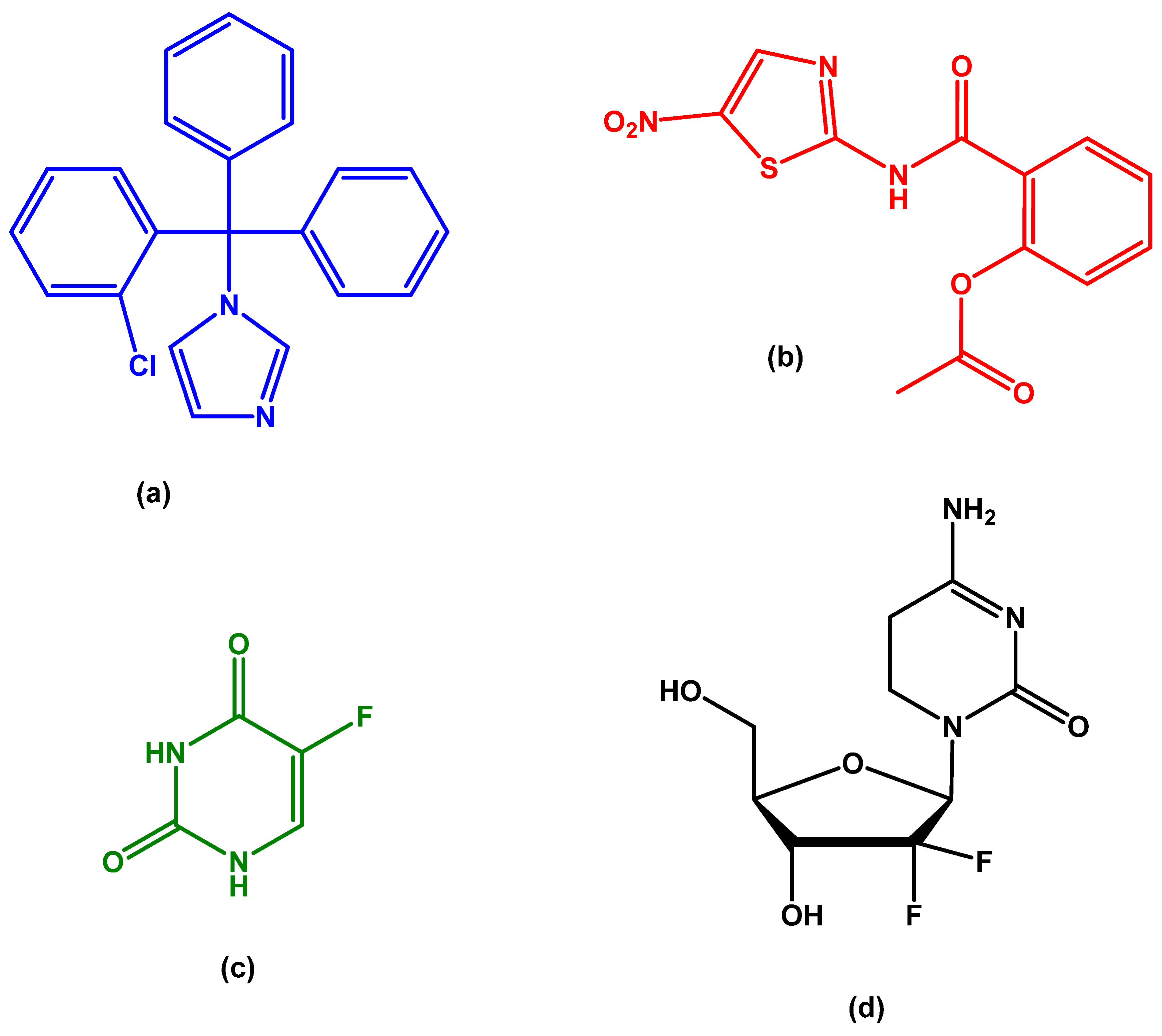
Scheme 1. Chemical structures of clotrimazole (CLZ) (a), nitazoxanide (NTZ) (b), 5-fluorouracil (5-FU) (c), and gemcitabine (GEM) (d).
Physchem 06 00017 sch001
Scheme 2. Organic/bioorganic experimental–computational workflow integrating in vitro phenotyping with physical–organic descriptors, ADMET context, and structure–property–activity (SPAR) interpretation.
Physchem 06 00017 sch002
Figure 1. The cell viability results of T3M4 cells following exposure to 5-FU at increasing concentrations (0.1–100 µM) for 48 h (A) and 72 h (B). Control cells were treated with 0.1% DMSO. Cell viability was assessed using the MTT assay, and the results are presented as percentages of the control and are expressed as means ± SEM. Each experiment was independently repeated three times (n = 3). Statistical significance was determined compared to the control at p < 0.05 (*); p < 0.01 (**); p < 0.001 (***) and p < 0.0001 (****).
Physchem 06 00017 g001
Figure 2. Following exposure to the cytotoxic effects of the antineoplastic drug 5-FU, the impact on the morphology of T3M4 cells was observed through microscopic images at 48- and 72-h post-exposure.
Physchem 06 00017 g002
Figure 3. The cell viability results of T3M4 cells following exposure to GEM at increasing concentrations (0.1–100 µM) for 48 h (A) and 72 h (B) and the 72 h dose–response (C). Control cells were treated with 0.1% DMSO. Cell viability was assessed using the MTT assay, and the results are presented as percentages of the control and are expressed as means ± SEM. Each experiment was independently repeated three times (n = 3). Statistical significance was determined compared to the control at p < 0.001 (***) and p < 0.0001 (****).
Physchem 06 00017 g003
Figure 4. The microscopic analysis of the impact of exposure to the cytotoxic properties of the antineoplastic GEM on the morphological characteristics of T3M4 cells over a period of 48 and 72 h.
Physchem 06 00017 g004
Figure 5. The cell viability results of T3M4 cells following exposure to NTZ at increasing concentrations (0.1–100 µM) for 72 h (A) and the dose–response (B). Control cells were treated with 0.1% DMSO. Cell viability was assessed using the MTT assay, and the results are presented as percentages of the control and are expressed as means ± SEM. Each experiment was independently repeated three times (n = 3). Statistical significance was determined compared to the control at p < 0.05 (*).
Physchem 06 00017 g005
Figure 6. Following a 72-h exposure of T3M4 to the repurposed drug NTZ, a microscopic examination was conducted to assess potential cytotoxic effects on T3M4 morphology.
Physchem 06 00017 g006
Figure 7. The cell viability results of T3M4 cells after exposure to CLZ at increasing concentrations (0.1–100 µM) for 72 h (A) and the dose–response (B). Control cells were treated with 0.1% DMSO. Cell viability was assessed using the MTT assay, and the results are presented as percentages of the control and are expressed as means ± SEM. Each experiment was independently repeated three times (n = 3). Statistical significance was determined compared to the control at p < 0.01 (**); p < 0.001 (***) and p < 0.0001 (****).
Physchem 06 00017 g007
Figure 8. The microscopic examination reveals the impact of exposure to the cytotoxic effects of the repurposed drug CLZ on the morphological features of T3M4 cells after 72 h.
Physchem 06 00017 g008
Figure 9. The cell viability results for T3M4 cells following exposure to 5-FU at increasing concentrations (0.1–100 µM) and 10 µM of CLZ or 50 µM of CLZ for 48 h (A,B) and 72 h (C,D). The results of the standard drug 5-FU for the same analysis period were included to facilitate comparative analysis. It is important to clarify that the assessments conducted at 72 h for CLZ alone were included solely to offer a visual comparison to the monotherapy repurposed drug that demonstrated the most favorable results. Control cells were treated with 0.1% DMSO. Cell viability was assessed using the MTT assay, and the results are presented as percentages of the control and are expressed as means ± SEM. Each experiment was independently repeated three times (n = 3). Statistical significance was determined compared to the control at p < 0.05 (*); p < 0.01 (**); p < 0.001 (***) and p < 0.0001 (****).
Physchem 06 00017 g009
Figure 10. The microscopic examination reveals the impact of exposure to the cytotoxic effects of the combination of CLZ and 5-FU on T3M4 cells after 48 and 72 h.
Physchem 06 00017 g010
Figure 11. The cell viability results for T3M4 cells following exposure to GEM at increasing concentrations (0.1–100 µM) and 10 µM of CLZ or 50 µM of CLZ for 48 h (A,B) and 72 h (C,D). The results of the standard drug GEM for the same analysis period were included to facilitate comparative analysis. It is important to clarify that the assessments conducted at 72 h for CLZ alone were included solely to offer a visual comparison to the monotherapy repurposed drug that demonstrated the most favorable results. Control cells were treated with 0.1% DMSO. Cell viability was assessed using the MTT assay, and the results are presented as percentages of the control and are expressed as means ± SEM. Each experiment was independently repeated three times (n = 3). Statistical significance was determined compared to the control at p < 0.05 (*); p < 0.01 (**); p < 0.001 (***) and p < 0.0001 (****).
Physchem 06 00017 g011
Figure 12. The microscopic examination reveals the impact of exposure to the cytotoxic effects of the combination of CLZ and GEM on T3M4 cells after 48 and 72 h.
Physchem 06 00017 g012
Figure 13. Combination Index (CI) plots generated using CompuSyn for the different drug combinations tested in T3M4 pancreatic ductal adenocarcinoma cells. The CI–Fa curves illustrate the interaction between drugs at increasing fractions affected (Fa). A CI value < 1 indicates synergism, CI = 1 denotes an additive effect, and CI > 1 indicates antagonism.
Physchem 06 00017 g013
Figure 14. RNA expression of the calcium-activated potassium channel KCa3.1 (KCNN4) in pancreatic cancer cell lines, retrieved from the Human Protein Atlas database. KCNN4 shows high expression levels in PDAC, supporting its role as a potential functional target of clotrimazole. Data retrieved from the Human Protein Atlas (www.proteinatlas.org, accessed on 25 August 2025).
Physchem 06 00017 g014
Figure 15. RNA expression of aldo-keto reductase family 1 member B1 (AKR1B1) in pancreatic cancer cell lines, obtained from the Human Protein Atlas database. AKR1B1 is expressed at lower levels compared with KCNN4, consistent with the weaker cytotoxic activity of nitazoxanide observed in vitro. Data retrieved from the Human Protein Atlas (www.proteinatlas.org, accessed on 25 August 2025).
Physchem 06 00017 g015
Table 1. Estimated physicochemical properties of NTZ, CLZ, 5-FU, and GEM.
DrugPhysicochemical PropertiesPredicted ValueOptimized ValueReference
NTZLogP2.672.14
pKa0.34/6.44−4.20/10.62
Molecular Weight (g/mol)307.27307.28
Water Solubility (mg/mL)0.0090.007
Diff. Coeff. (cm2/s·105)0.83NA
Peff (cm/s·104)2.85NA
Fup (%)3.98~1
B:P0.71NA
CLZLogP5.235.48
pKa6.196.26
Molecular Weight (g/mol)344.85344.80
Water Solubility (mg/mL)0.0020.001
Diff. Coeff. (cm2/s·105)0.69NA
Peff (cm/s·104)8.61NA
Fup (%)3.79NA
B:P0.71NA
5-FULogP−0.78−0.66
pKa11.08/7.878.02
Molecular Weight (g/mol)130.08130.08
Water Solubility (mg/mL)11.465.86
Diff. Coeff. (cm2/s·105)1.46NA
Peff (cm/s·104)2.71NA
Fup (%)85.2288–92
B:P1.24NA
GEMLogP−1.33−1.40
pKa3.42/−2.963.60
Molecular Weight (g/mol)263.20263.20
Water Solubility (mg/mL)4.7015.30
Diff. Coeff. (cm2/s·105)0.94NA
Peff (cm/s·104)0.57NA
Fup (%)87.05NA
B:P1.19NA
LogP, lipophilicity; pKa, ionization constant; Diff. Coeff., diffusion coefficient; Peff, effective human jejunal permeability; Fup, fraction unbound in plasma; B:P, blood to plasma ratio; NA, not available.
Disclaimer/Publisher’s Note: The statements, opinions and data contained in all publications are solely those of the individual author(s) and contributor(s) and not of MDPI and/or the editor(s). MDPI and/or the editor(s) disclaim responsibility for any injury to people or property resulting from any ideas, methods, instructions or products referred to in the content.