Article

Exploring New Conservation Methods: Isolation and Characterization of Algicidal Bacteria from Ornamental Fountains in the Alhambra and Generalife (Granada, Spain)


by
Isabel Calvo-Bayo
1,
Sandy Fillet
2,
Oana A. Cuzman
3,
Lorena Cuberos-Cáceres
1,
Manuel González-del-Valle
4,
Fernando Bolívar-Galiano
1 and
Julio Romero-Noguera
5,*
1
Painting Department, Faculty of Fine Arts, University of Granada, Avda. Andalucía s/n, 18071 Granada, Spain
2
Sanbiotec Laboratory S.L., Street Los Olivos, 18630 Granada, Spain
3
Institute of Heritage Science (ISPC)-National Research Council of Italy (CNR), Via Madonna del Piano, 10, 50019 Sesto Fiorentino, Italy
4
G2G Algae Solutions, Gabriel Lupiañez Gely, 32, 41120 Seville, Spain
5
Painting Department, Faculty of Fine Arts, University of Seville, Laraña 3, 41003 Seville, Spain
*
Author to whom correspondence should be addressed.
Conservation 2026, 6(2), 70; https://doi.org/10.3390/conservation6020070

Submission received: 17 April 2026 / Revised: 24 May 2026 / Accepted: 28 May 2026 / Published: 10 June 2026

Abstract

Ornamental fountains in the Alhambra and Generalife (Granada, Spain) constitute complex socio-ecological systems where water, stone, and biological communities interact, making them highly vulnerable to biodeterioration caused by phototrophic microorganisms such as cyanobacteria, green algae, and diatoms. Conventional chemical biocides, although widely applied, present significant drawbacks including toxicity, material degradation, ecological imbalance, and limited long-term effectiveness. In this context, this study evaluated the potential of algicidal bacteria as a sustainable alternative for controlling phototrophic growth in heritage environments. Water samples from eight ornamental fountains were analyzed using 16S ribosomal RNA (16S rRNA) gene sequencing to characterize bacterial communities and identify taxa previously reported with algicidal activity. Statistical analyses were conducted to assess relationships between microbial community structure and biofilm development. In parallel, functional screening assays using filtered fountain waters against Chlorella vulgaris were performed to evaluate intrinsic inhibitory capacity. The most active sample was selected for bacterial isolation and further validation through co-culture assays, cell density measurements, and pulse-amplitude-modulated (PAM) fluorometry. A total of 18 genera with reported algicidal capacity were detected, representing a substantial fraction of the microbiome across all samples. However, no significant association was found between these taxonomic metrics and biofilm development, highlighting a decoupling between taxonomic composition and functional activity. The most active isolate, identified as Stenotrophomonas maltophilia strain LIG25, caused a rapid decline in photosynthetic efficiency and achieved more than 98% inhibition of algal growth. These findings demonstrate that ornamental fountain microbiomes represent a reservoir of native biocontrol agents and support the development of eco-friendly strategies for cultural heritage conservation.

1. Introduction

Ornamental fountains constitute complex interfaces where water, stone, and living communities interact within historically shaped open landscapes [1]. Rather than isolated architectural elements, they function as socio-ecological systems embedded in gardens and semi-natural environments where material preservation depends on the stability of surrounding biological and environmental dynamics [2,3]. In such contexts, preventive conservation must reconcile structural integrity with ecological balance, particularly under increasing climatic and anthropogenic pressures [4].
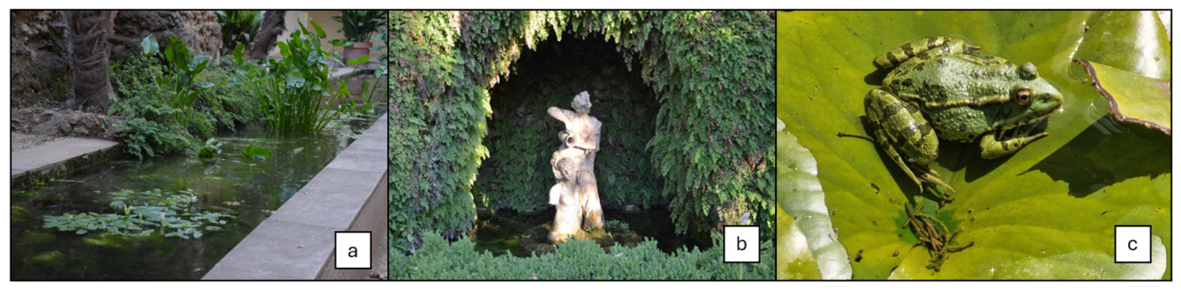
In open monumental spaces such as the Alhambra and Generalife (Granada, Spain), ornamental fountains coexist with aquatic invertebrates, amphibians, birds, and diverse plant communities that depend on stable water quality (Figure 1). These hydraulic systems are integrated into biologically active landscapes where ecological and cultural values are inseparable [5,6]. Consequently, strategies aimed at controlling phototrophic colonization must not only preserve stone substrates but also remain compatible with surrounding biodiversity. Interventions that alter microbial equilibrium may have cascading ecological consequences in these open-air systems [7].
One of the most persistent threats to ornamental fountains is biodeterioration caused by phototrophic microorganisms, including cyanobacteria, green algae, and diatoms [8,9]. These organisms colonize porous stone surfaces and form biofilms that induce discoloration, surface roughening, loss of polish, and progressive material weakening [10]. Their metabolic activity modifies local microenvironments, enhances moisture retention, and facilitates secondary colonization, thereby compromising both aesthetic and structural values [11,12,13].
Control strategies have traditionally relied on chemical biocides such as benzalkonium chloride, sodium hypochlorite, and chlorine-based formulations. Although effective in the short term, these agents present substantial ecological and material drawbacks [14]. Their non-selective toxicity disrupts native microbial communities, may affect non-target fauna and flora, and can alter the physicochemical quality of fountain waters [15,16]. Repeated applications can oxidize metallic elements, degrade protective patinas, and react with carbonate matrices, increasing porosity and surface fragility [15,17]. Moreover, several phototrophic taxa exhibit adaptive resistance to conventional biocides, reducing long-term efficacy [10]. In cyanobacterial systems, chemical stress may stimulate the release of intracellular toxins such as microcystins from Microcystis aeruginosa, further deteriorating water quality and increasing ecological risk [18]. Thus, chemical suppression may destabilize ecological equilibrium without ensuring sustainable control [19].
Research on harmful algal blooms (HABs) offers a transferable ecological framework for addressing this challenge. Bloom-forming species such as Microcystis aeruginosa, Anabaena sp., Cladophora sp., Spirogyra sp., Chlorella, among others, proliferate not only in natural water bodies but also in artificial aquatic systems, including ornamental fountains [8,19,20]. Increasingly frequent heat waves accelerate algal metabolism and promote bloom recurrence, while reduced oxygen availability limits microbial competition [21]. Beyond aesthetic degradation, ingestion of toxin-contaminated water has been associated with mortality events in birds and small mammals inhabiting ornamental gardens [22,23,24]. Managing phototrophic proliferation in heritage fountains is therefore not only a conservation issue but also an environmental one.
Within HAB management, algicidal bacteria have emerged as biological regulators capable of inhibiting or lysing microalgae and cyanobacteria through direct cell interactions or the secretion of extracellular bioactive metabolites such as lipopeptides, indoles, phenolics, and cyclodipeptides [25,26,27]. Many of these bacteria contribute to water quality improvement through complementary mechanisms such as denitrification and microcystin degradation, reducing nutrient loads and toxin accumulation. These processes not only regulate algal proliferation but also mitigate secondary physicochemical alterations that accelerate stone biodeterioration in ornamental fountains [19,28]. Commercial formulations based on Bacillus consortia have demonstrated their capacity to reduce algal biomass and stabilize physicochemical parameters in aquaculture systems [29,30]. However, despite their proven effectiveness in engineered aquatic environments, the translation of algicidal biocontrol strategies to historic ornamental fountains remains largely unexplored. Recent freshwater studies further suggested that surface-associated microbiomes in open aquatic systems may represent an ecologically relevant source of biocontrol potential. In submerged macrophytes such as Vallisneria natans, epiphytic microbiomes have been shown to restructure under cyanobacterial stress, with increased bacterial diversity and selective enrichment of potentially algicidal genera. These findings support the idea that antagonistic bacteria may form part of the natural ecological dynamics of surface-associated microbiomes in open freshwater environments, although isolation and functional validation remain necessary to distinguish taxonomic occurrence from active biocontrol [31,32].
In cultural heritage conservation, microbial applications have primarily focused on carbonate precipitation and biocleaning of organic deposits, frequently employing Bacillus species [33,34]. Functional studies specifically targeting phototrophic biodeterioration are scarce, and no research has systematically investigated whether the native microbiota of historic fountains contains strains with intrinsic algicidal capacity that can be experimentally isolated and validated on stone substrates under environmentally relevant conditions. Consequently, a critical gap persists between ecological knowledge derived from HAB research and its implementation in water-exposed heritage conservation.
The Monumental Complex of the Alhambra and Generalife, a UNESCO World Heritage Site (1984), represents a paradigmatic case in which hydraulic heritage structures operate within biodiverse open landscapes [7,8]. Its ornamental fountains constitute long-term microbial reservoirs shaped by local physicochemical selection. It was hypothesized that these native microbial communities harbor bacterial taxa with functional algicidal potential adapted to local environmental conditions, capable of regulating phototrophic growth while preserving ecosystem stability and reducing biosecurity and ecological risks associated with the introduction of exogenous microorganisms. In this context, a central objective of the study was to determine whether the bacterial community composition of ornamental fountains is associated with reduced phototrophic biofilm development, in order to identify potential bacteria–alga biocontrol processes operating under natural conditions. To address this question, statistical analyses were performed to evaluate the relationships among bacterial community structure, biofilm development, and the occurrence of inhibitory patterns, and the detected genera were further contrasted with the scientific literature to determine whether algicidal activity had been previously reported. Working with microbiota naturally adapted to the local environmental conditions of heritage fountains may offer important advantages over exogenous microbial applications, including improved ecological compatibility, greater persistence under in situ conditions, and reduced risks of ecological imbalance or unintended impacts on associated biodiversity.
To test this hypothesis, the present study: (i) characterized the bacterial communities inhabiting multiple fountains of the Alhambra and Generalife through 16S rRNA gene sequencing; (ii) performed statistical analyses to explore the relationship between bacterial community structure and phototrophic biofilm development; (iii) screened fountain water samples for inhibitory activity against biodeteriorative phototrophs; (iv) identified bacterial taxa associated with inhibitory samples in order to characterize members of the native microbiota with putative algicidal capacity; and (v) assessed the inhibitory effects of selected bacterial cultures on representative microalgal assays using PAM fluorometry and Neubauer chamber cell counts. By integrating microbiome characterization, ecological analysis, literature-based functional assessment, and experimental validation, this study provides a framework for investigating site-adapted bacterial taxa and their potential role in biocontrol processes in open-air heritage fountains.

2. Materials and Methods

2.1. Study Site and Sampling Strategy

Water samples were collected from eight ornamental hydraulic systems located within and around the Monumental Complex of the Alhambra and Generalife (Granada, Spain), including fountains situated both within the protected monumental enclosure and in surrounding open garden areas. The selected sites were: the water outlet at the Cueva, located on the Cuesta de los Chinos, one of the historic access routes to the Monumental Complex (S1); the water inlet supplied by the historic Acequia del Sultán (S2); the Horizontal Greek Cross Fountain in the Generalife Gardens (S3); the inhibition halo associated with the Fountain of Lindaraja (S4); the submerged zone of the Lindaraja Fountain (S5); the Fountain in the Patio de la Sultana (S6); the Fountain of Emperor Charles V (S7); and the Fountain of the Pimiento and the Tomate (S8).
Approximately 1 L of water was collected from each site in sterile containers and transported to the laboratory under refrigerated conditions for immediate processing.
The physicochemical parameters of this fountain water were measured in situ to characterize the environmental conditions of the sampling site. Electrical conductivity was determined using a PCE-PHD 1 portable water analyzer (PCE Ibérica S.L., Tobarra, Albacete, Spain). While pH and additional water quality parameters were measured using a XS REVio Portable multiparameter meter equipped with the corresponding electrodes (Giorgio Bormac S.r.l., Carpi, Italy). The water exhibited a slightly alkaline pH between 7.2 and 7.35 and an electrical conductivity of 237 µS·cm−1 at 20 °C, indicating low mineralization and conditions compatible with carbonate stone substrates typical of the Alhambra ornamental fountains.

2.2. Microbiome Characterization

2.2.1. Filtration and DNA Extraction

Each water sample was initially filtered through conventional filter paper to remove coarse particles. Subsequent sequential filtration was performed sterile polycarbonate membranes of decreasing pore sizes (8–11 μm, 5 μm, 0.47 μm, and 0.22 μm) with a sterile vacuum filtration system equipped with an Aldrich® Essentials membrane filtration apparatus (Z741031; Chemrus, distributed by Sigma-Aldrich/Merck KGaA, Darmstadt, Germany). The 0.22 μm membranes, retaining the bacterial fraction, were preserved at −20 °C or stored in nucleic acid stabilizing solution until further processing. Genomic DNA was extracted from the membranes using mechanical disruption with zirconia beads followed by column-based purification according to the manufacturer’s protocol.

2.2.2. 16S rRNA Gene Amplification and Sequencing

DNA was extracted from the 0.22 μm filters via mechanical disruption with zirconia beads, followed by column-based purification. For bacterial characterization, the full-length 16S rRNA gene was amplified using primers 27F/1492R or the 16S Barcoding Kit SQK-16S024. Barcodes from the EXP-NBD114 Native Barcoding Kit were ligated using the Ligation Sequencing Kit SQK-LSK109, following the manufacturer’s protocol (Oxford Nanopore Technologies, Oxford, UK). A summary of the microbiome reports provided by Sanbiotec S.L. (Granada, Spain), including sequencing metrics and genus-level taxonomic results, is available in Supplementary File S1.

2.2.3. Bioinformatic Processing

Amplicons were sequenced on a MinION device (Oxford Nanopore Technologies, Oxford, UK, FLO-MIN106 flow cell), generating a minimum of 20,000 reads per sample. Raw reads were basecalled using Guppy v6.0 (Oxford Nanopore Technologies) with high-accuracy mode. Quality filtering was performed to retain reads with Q-score ≥ 9 and length between 1200 and 1700 bp. Chimeric sequences were removed using UCHIME implemented in USEARCH v11.0.667.
Operational Taxonomic Units (OTUs) were assigned using the USEARCH software v11.0.667 against the Ribosomal Database Project (RDP, Michigan State University) reference database (version 18) with a minimum identity threshold of 97%. Taxonomic proportions were calculated from the resulting OTU tables. Diatoms (phylum Bacillariophyta) were removed from the dataset prior to analysis. Relative abundances were normalized to 100% for each sample to account for differences in sequencing depth.

2.3. Statistical and Ecological Analyses

All statistical and ecological analyses of the microbiome dataset were performed in R version 4.4.3 using the packages vegan, ggplot2, dplyr, Hmisc, and pheatmap. The R code used for statistical analyses and graphical processing is provided in File S7.

2.3.1. Biofilm Categorization and Statistical Grouping

Samples were classified by visual assessment of phototrophic biofilm coverage at the specific sampling area as High biofilm (n = 3: S1, S5, S7; >50% visible surface coverage by algal/cyanobacterial growth) or Low biofilm (n = 5: S2, S3, S4, S6, S8; <20% visible coverage). Accordingly, S4 was included in the Low biofilm group because the specific area sampled showed limited visible phototrophic growth, despite being located in a fountain where biofilm development was evident in other areas.

2.3.2. Taxonomic Composition and Heatmap Visualization

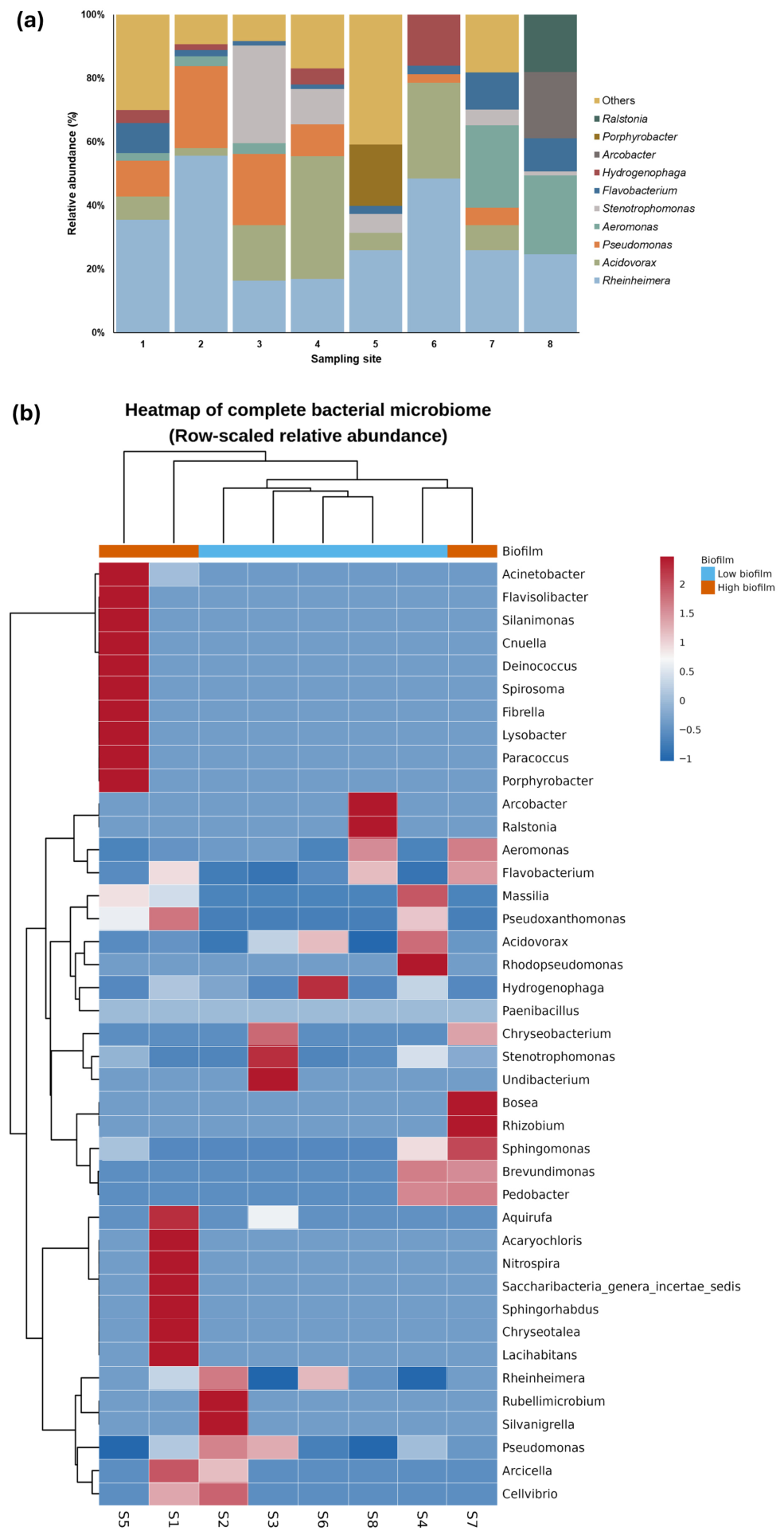
Taxonomic composition was visualized using stacked bar plots showing the 10 most abundant bacterial genera in each sample. Genera outside the top 10 were grouped as “Others”.
To explore compositional similarity among samples and genera, a heatmap including all detected bacterial genera was generated from genus-level relative abundance data using hierarchical clustering with Euclidean distance and Ward.D2 linkage. Heatmap values were row-scaled (z-score transformation) to highlight relative enrichment and depletion patterns across samples. Because row scaling standardizes each genus independently, the heatmap was used to interpret relative compositional patterns rather than absolute taxonomic dominance.
The core microbiome was defined as bacterial genera detected in at least 75% of samples (n ≥ 6), using a minimum relative abundance threshold of 0.1%.

2.3.3. Alpha Diversity Analysis

Alpha diversity was assessed using four indices: (i) observed richness, (ii) Shannon diversity, (iii) Simpson diversity, and (iv) Pielou’s evenness. These indices were calculated from genus-level relative abundance data.
Comparisons between High and Low biofilm categories were performed using the Kruskal–Wallis test, selected as a non-parametric approach suitable for small sample sizes and non-normal distributions. To control for multiple testing across the four alpha-diversity indices, Benjamini–Hochberg false discovery rate (FDR) correction was applied. FDR-adjusted p-values (q-values) < 0.05 were considered statistically significant.

2.3.4. Beta Diversity Analysis

Beta diversity was assessed using Bray–Curtis dissimilarity, calculated from genus-level relative abundance data. Community composition was visualized by Principal Coordinates Analysis (PCoA), and the proportion of variance explained by each axis was obtained from the corresponding eigenvalues.
Hierarchical clustering of samples was performed using Ward.D2 linkage to identify groups of compositionally similar samples.
Differences in community composition between High and Low biofilm categories were evaluated using Permutational Multivariate Analysis of Variance (PERMANOVA) with 999 permutations. To verify the assumption of homogeneous multivariate dispersion, PERMDISP was also performed with 999 permutations. ANOSIM was used as a complementary descriptive multivariate test.

2.3.5. Identification and Analysis of Genera with Reported Algicidal Activity

Bacterial genera with reported algicidal activity were identified through a literature review of peer-reviewed publications describing algicidal effects against phototrophic microorganisms. Searches were conducted in PubMed, Web of Science, and Google Scholar using combinations of the terms algicidal bacteria, algae-lysing bacteria, anti-algal bacteria, bacterial control of algae, and the names of target genera or phototrophic organisms.
Based on this review, 18 bacterial genera with previously reported algicidal capacity were compiled. For each sample, the following metrics were calculated: (i) total relative abundance of genera with reported algicidal activity, (ii) proportion of the community represented by these genera, and (iii) algicidal richness, defined as the number of reported algicidal genera detected per sample.
Comparisons between High and Low biofilm categories were performed using the Kruskal–Wallis test. To control for multiple testing across algicidal-genera metrics, Benjamini–Hochberg false discovery rate (FDR) correction was applied. Adjusted p-values (q-values) < 0.05 were considered statistically significant.
Heatmaps of genera with reported algicidal activity were generated using row-scaled relative abundance values to identify enrichment and depletion patterns among samples. A core algicidal microbiome was defined as genera with reported algicidal activity detected in at least 75% of samples (n ≥ 6) and with a minimum relative abundance of 0.1%.

2.4. Functional Screening of Fountain Waters

To determine whether the microbiota present in the ornamental fountains exhibited intrinsic algicidal activity, water samples were subjected to a functional screening assay against Chlorella vulgaris. The algal strain was provided by the Institute of Marine Sciences of Andalusia (ICMAN-CSIC, Puerto Real, Cádiz, Spain). This strain was selected because it has been reported as a tolerant phototrophic model, capable of surviving or recolonizing surfaces under chemical stress conditions and showing high adaptability in disturbed systems, particularly when associated bacterial communities are present [10,35,36,37]. Prior to inoculation, each water sample was filtered through sterile 3.0 μm MF-Millipore™ membranes (Merck KGaA, Darmstadt, Germany) using a sterile vacuum filtration system equipped with an Aldrich® Essentials membrane filtration apparatus (Z741031; Chemrus, distributed by Sigma-Aldrich/Merck KGaA, Darmstadt, Germany). to remove phototrophic organisms while retaining the bacterial fraction.
Screening assays were performed in 24-well microplates. Each well contained 750 μL of exponentially growing C. vulgaris culture and 750 μL of the corresponding filtered fountain water (final volume 1.5 mL) (File S2). The algal inoculum was adjusted to an initial density of 1 × 10⁴ cells·mL−1, in accordance with internationally recognized algal toxicity test guidelines [38,39,40]. Untreated algal cultures maintained in standard growth medium BG-11 served as controls [41]. For each well, three independent Neubauer chamber counts were performed, and the mean value was used to calculate the final cell concentration of that well.
For each fountain sample, three independent biological replicates were prepared (n = 3). Plates were incubated for 14 days at 25 ± 1 °C under a 12:12 h light/dark photoperiod (4000 lux) without agitation. Algal cell density was quantified using a Neubauer chamber at 5, 7 and 14 days after inoculation to construct growth curves for both treated and control cultures.
The area under the growth curve (AUC) was calculated using the trapezoidal integration method to integrate cell density over time:
A = N 1 N 0 2 t 1 + N 1 + N 2 2 N 0 2 t 2 t 1 + N n 1 + N n 2 N 0 2 t n t n 1
where N 0 represents the initial cell density, N i   represents the measured cell density at time t i , and A corresponds to the baseline-corrected area under the growth curve. Percentage growth inhibition was calculated from AUC values as:
G r o w t h   i n h i b i t i o n   % =   A C   A t A C · 100
where A c corresponds to the area under the curve of the control and A t to that of the treated cultures.
Data were processed and graphically represented using SigmaPlot 10.0, while statistical analyses were performed with IBM SPSS Statistics 30. Normality and homogeneity of variance were assessed using the Shapiro–Wilk and Levene’s tests, respectively. Statistical comparisons were performed on replicate AUC values, from which growth inhibition percentages were derived. When these assumptions were met, one-way ANOVA (p ≤ 0.05) was applied, followed by Dunnett’s post hoc test to compare each treatment with the control and Tukey’s test to evaluate differences among treatments. Results are expressed as mean ± SD (n = 3) and presented as percentage variation relative to the control (0%).
The sample exhibiting the highest growth inhibition was selected for bacterial isolation. The same normalization approach was subsequently applied to the co-culture assays performed with the isolated bacterial strain. The layout of the 24-well microplate design is provided in File S3.

2.5. Isolation and Molecular Identification

The fountain water sample showing the highest algicidal activity in the screening assay was selected for bacterial isolation. An aliquot of this sample was transferred and streaked onto Tryptone Soy Agar (TSA), a general-purpose, non-selective medium that supports the growth of a broad range of heterotrophic bacteria [42], in order to obtain isolated colonies. Plates were incubated at 30 °C for 48 h.
Isolation of the predominant bacterial morphotype was performed using the standard streak plate method to ensure separation of individual colonies. Colonies displaying the most abundant and consistent morphology across replicate plates were selected and repeatedly subcultured until axenic cultures were achieved.
Genomic DNA was extracted from pure bacterial cultures using the DNeasy Blood & Tissue Kit (Qiagen, Hilden, Germany) according to the manufacturer’s instructions. The nearly full-length 16S rRNA gene was amplified using universal primers 16SF1 (5′-GASTTTGATCCTGGCTYAG-3′) and 1492R (5′-GGAAACAGCTATGACCATG-3′) [43]. PCR amplification was performed with TaqGold DNA polymerase under the following cycling conditions: initial denaturation at 95 °C for 15 min; 35 cycles of 95 °C for 30 s, 56 °C for 40 s, and 72 °C for 1 min; followed by a final extension at 72 °C for 8 min.
PCR products were visualized by agarose gel electrophoresis, purified using the QIAquick PCR Purification Kit (Qiagen), and subjected to bidirectional Sanger sequencing (Macrogen Europe, Amsterdam, The Netherlands). Forward and reverse sequences were inspected, trimmed, and assembled into consensus sequences using ChromasPro 2.1.10. Taxonomic identification of the bacterial isolates was performed by comparing the consensus 16S rRNA gene sequences against the NCBI GenBank nucleotide database using BLASTn v2.16.0+. The closest matches were evaluated according to percentage identity, query coverage, E-value, and alignment length. Species-level assignment was accepted when sequences showed high identity and coverage with reference sequences deposited in GenBank.

2.6. Algicidal Co-Culture Assays

The isolated bacterial strain was cultured in Luria–Bertani (LB) medium [44] at 25 °C under constant agitation (190 rpm) until the culture reached and was adjusted to an optical density of OD600 = 1.5, corresponding to 2.20 × 10⁸ CFU·mL−1, following the protocol described by Pokrzywinski et al. [45]. Optical density at 600 nm was measured every 8 h using the cuvette mode of a Zuzi 4260/50 UV/Vis spectrophotometer over a period of 72 h.
Co-culture assays were performed by mixing the bacterial suspension and algal culture in a 1:1 volumetric ratio (v/v). The selected biodeteriorative microalga was C. vulgaris, the same strain used in the initial screening assays. The algal inoculum was adjusted to an initial density of 1 × 10⁴ cells·mL−1.
Experiments were conducted in triplicate (n = 3 biological replicates) using 100 mL working volumes in 250 mL flasks at 20 ± 1 °C under continuous illumination. Cultures were manually shaken twice daily to maintain homogeneity. Control cultures without bacterial addition were maintained under the same experimental conditions. Cell density was quantified daily for 72 h using a Neubauer chamber, following internationally recognized algal toxicity test guidelines [38,39,40]. area under the growth curve (AUC) and the percentage of growth inhibition under these conditions were subsequently calculated using Equation (1) and Equation (2), respectively. Phototrophic cultures were also monitored microscopically throughout the experiment using a Nikon ECLIPSE TS100 inverted microscope (Nikon Corporation, Tokyo, Japan).
Photosynthetic performance was evaluated during a 72 h exposure assay using an FP 110 FluorPen portable PAM fluorometer (Photon Systems Instruments, Drásov, Czech Republic). For each measurement, 3 mL aliquots were transferred to 10 mm polystyrene spectrophotometer cuvettes and dark-adapted for 15–30 min prior to fluorescence determination [46]. An additional 24 h assay was conducted at the Institute of Heritage Science, National Research Council of Italy (ISPC-CNR, Florence, Italy), using a PAM-2000 Portable Chlorophyll Fluorometer (Heinz Walz GmbH, Effeltrich, Germany) to assess the short-term physiological response at higher temporal resolution. This assay was performed under constant agitation and darkness, with fluorescence measurements recorded every minute during the first hour and every 10 min thereafter. Parallel controls containing BG-11 medium, LB medium, and LB medium adjusted to pH 8.4, corresponding to the pH of the bacterial culture, were included to determine whether medium composition or pH, in the absence of bacterial cells, affected photosystem II (PSII) photochemical efficiency independently of the bacterial treatment.

3. Results

3.1. Microbiome Characterization of Ornamental Fountains

The bacterial microbiome associated with the ornamental fountains comprised 41 genera distributed across the eight analysed samples (File S1). Taxonomic composition showed marked heterogeneity among sampling sites, with clear differences in both the identity and relative abundance of the dominant genera. Overall, Rheinheimera was the most abundant genus in the dataset, with a mean relative abundance of 31.13%, followed by Acidovorax (13.68%), Pseudomonas (9.70%), and Aeromonas (7.45%).
The relative abundance barplot showed that several samples were dominated by one or two main genera. Rheinheimera predominated particularly in S2 (55.57%), S6 (48.46%), and S1 (35.44%), whereas S3 exhibited a high abundance of Stenotrophomonas (30.69%) and S4 was dominated by Acidovorax (38.64%). Sample S8 displayed a distinct compositional profile, characterized by relatively high abundances of Aeromonas (24.69%), Arcobacter (20.76%), and Ralstonia (18.08%). In contrast, S5 showed the highest proportion of taxa grouped as “Others”, reflecting a more heterogeneous community than the remaining samples (Figure 2a).
The heatmap of the complete microbiome confirmed the marked compositional heterogeneity among samples. Hierarchical clustering clearly separated S5 from the remaining samples, whereas the other samples were distributed into two broader compositional groups. One cluster included S1, whereas the second grouped S2, S3, S4, S6, S7, and S8, although without a clear separation according to biofilm category.
The heatmap also revealed sample-specific enrichment patterns at the genus level. S5 was distinguished by the exclusive or predominant relative enrichment of several genera, including Acinetobacter, Cnuella, Deinococcus, Lysobacter, Spirosoma, Silanimonas, Porphyrobacter, Paracoccus, Flavisolibacter, and Fibrella. In contrast, Rheinheimera showed higher relative representation in S2 and S6, whereas Stenotrophomonas was mainly enriched in S3. Acidovorax and Rhodopseudomonas displayed their highest relative enrichment in S4, while Arcobacter and Ralstonia were predominantly associated with S8. In addition, S7 showed marked enrichment in genera such as Bosea, Rhizobium, Sphingomonas, Brevundimonas, and Pedobacter (Figure 2b).

3.2. Bacterial Diversity Patterns and Community Structure

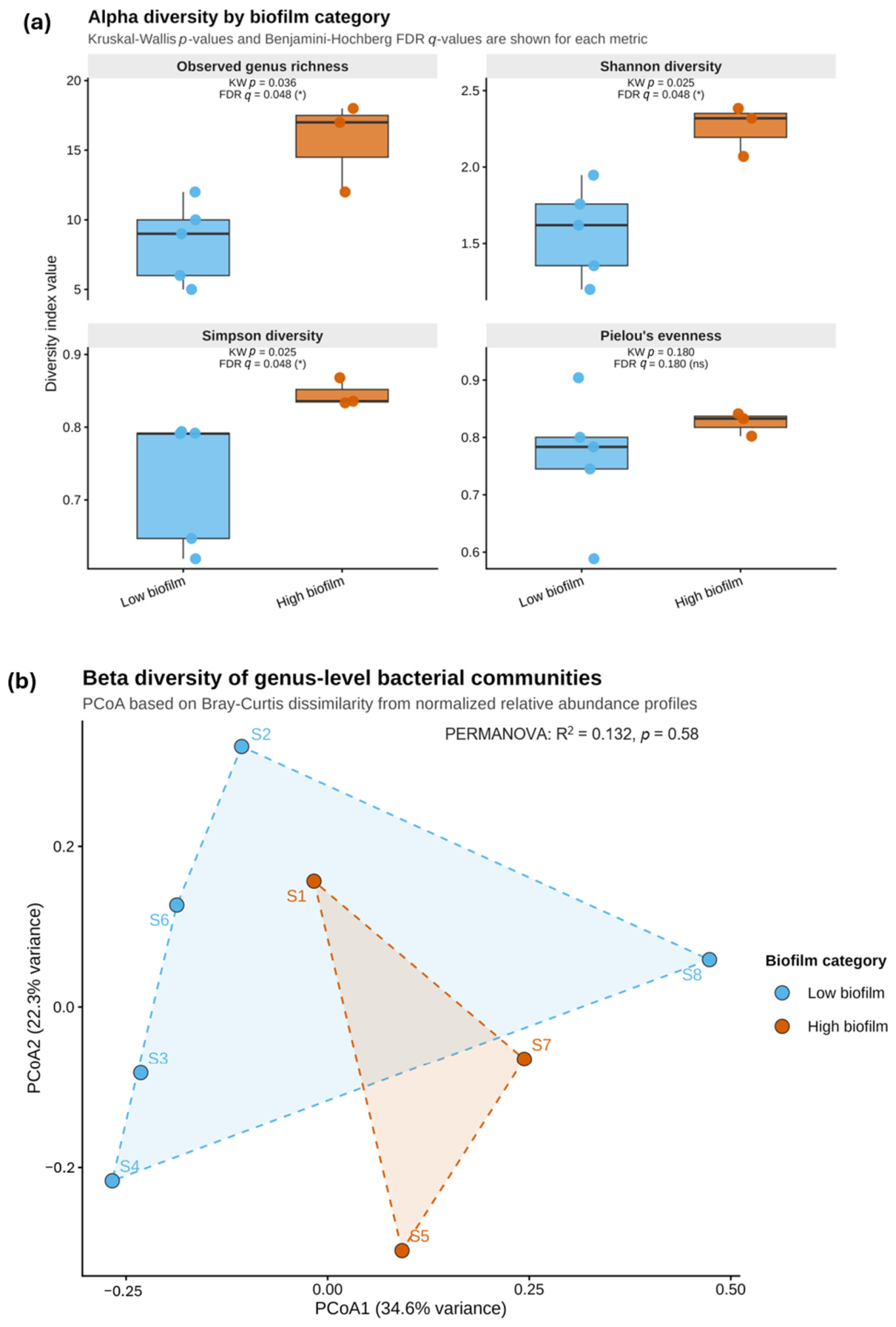
Alpha diversity indices revealed differences between samples with high and low phototrophic colonization (Figure 3a). Observed richness was significantly higher in high-biofilm samples than in low-biofilm samples (15.67 vs. 8.40 genera; χ2 = 4.408, p = 0.036, q = 0.048). Similarly, the Shannon index was higher in highly colonized samples (2.257) than in low-colonization samples (1.576; χ2 = 5.000, p = 0.025, q = 0.048), as was the Simpson index (0.846 vs. 0.729; χ2 = 5.000, p = 0.025, q = 0.048). In contrast, Pielou’s evenness did not differ significantly between categories (0.825 in high and 0.764 in low; χ2 = 1.800, p = 0.180, q = 0.180).
Taken together, these results indicate that samples with greater phototrophic colonization were associated with higher bacterial diversity, whereas samples with lower coverage exhibited reduced richness and diversity values. However, this pattern did not translate into a clear differentiation in overall community composition between biofilm categories.
Regarding beta diversity, PCoA based on Bray–Curtis dissimilarity (Figure 3b) showed that the first two axes explained 56.9% of the total compositional variance, with 34.6% attributed to PC1 and 22.3% to PC2. Samples were distributed across the ordination space without forming defined clusters according to biofilm category. S5 occupied a relatively distinct position within the ordination space, whereas the remaining samples showed partial overlap between categories.
This pattern was consistent with the multivariate analyses. PERMANOVA indicated that biofilm category explained only 13.2% of the compositional variance and was not significant (R2 = 0.132, pseudo-F = 0.915, p = 0.579). PERMDISP detected no significant differences in multivariate dispersion between groups (p = 0.527), indicating that dispersion differences were not detected between biofilm categories. Likewise, ANOSIM did not show separation between categories (R = −0.077, p = 0.647). As a complementary analysis, hierarchical clustering, included as Figure S1, showed a pattern consistent with the absence of clear grouping according to biofilm category.

3.3. Occurrence of Genera with Reported Algicidal Activity

A total of 18 genera previously reported in the literature as having algicidal activity were identified within the analysed bacterial microbiome (Table S1). These genera constituted a predominant fraction of the bacterial community in all samples, accounting for 61% to 100% of the total relative abundance, with a mean of 81.85% ± 15.10%. The highest proportion was recorded in S6 (100%), whereas S5 and S8 showed the lowest values, both close to 61%.
The richness of genera with reported algicidal activity varied among samples, ranging from 4 to 10 detected genera per site (mean = 7.0 ± 1.9). High-biofilm samples showed a descriptively higher richness of reported algicidal genera than low-biofilm samples; however, this difference was not statistically significant after FDR correction (Kruskal–Wallis, p = 0.17, q = 0.17) (Figure S3).
Among the identified genera, Flavobacterium and Rheinheimera were present in 100% of the samples, Acidovorax in 88%, and Pseudomonas in 75%, thus constituting the most recurrent taxonomic core within the set of reported algicidal genera. In terms of relative abundance, Rheinheimera was the most represented genus (mean 31.13%), followed by Acidovorax (13.68%), Pseudomonas (9.70%), and Aeromonas (7.45%) (Figure S2).
Comparison between biofilm categories did not reveal significant differences in the overall proportion of reported algicidal genera. Although low-biofilm samples showed a descriptively higher total relative abundance of genera with reported algicidal activity, this difference was not statistically significant after FDR correction (Kruskal–Wallis, p = 0.10, q = 0.17). Similarly, the richness of reported algicidal genera tended to be higher in high-biofilm samples, but this pattern was also not statistically significant (Kruskal–Wallis, p = 0.17, q = 0.17) (Figure S3).
The heatmap of reported algicidal genera showed differential compositional patterns among samples (Figure S4). S5 displayed a distinct profile, characterized by the relative enrichment of Acinetobacter and Lysobacter. In contrast, S3 showed a marked relative enrichment of Stenotrophomonas, whereas S4 was characterized by the relative enrichment of Acidovorax and Pseudomonas. In addition, Hydrogenophaga showed its highest relative representation in S6, whereas Rheinheimera was more abundant in S2 and S6. Aeromonas and Flavobacterium showed comparatively higher relative representation in S7 and S8. Overall, these results show that genera with reported algicidal activity were widely represented across all samples, although their distribution varied among sampling sites.

3.4. Inhibitory Effects of Fountain Water Samples on Chlorella vulgaris

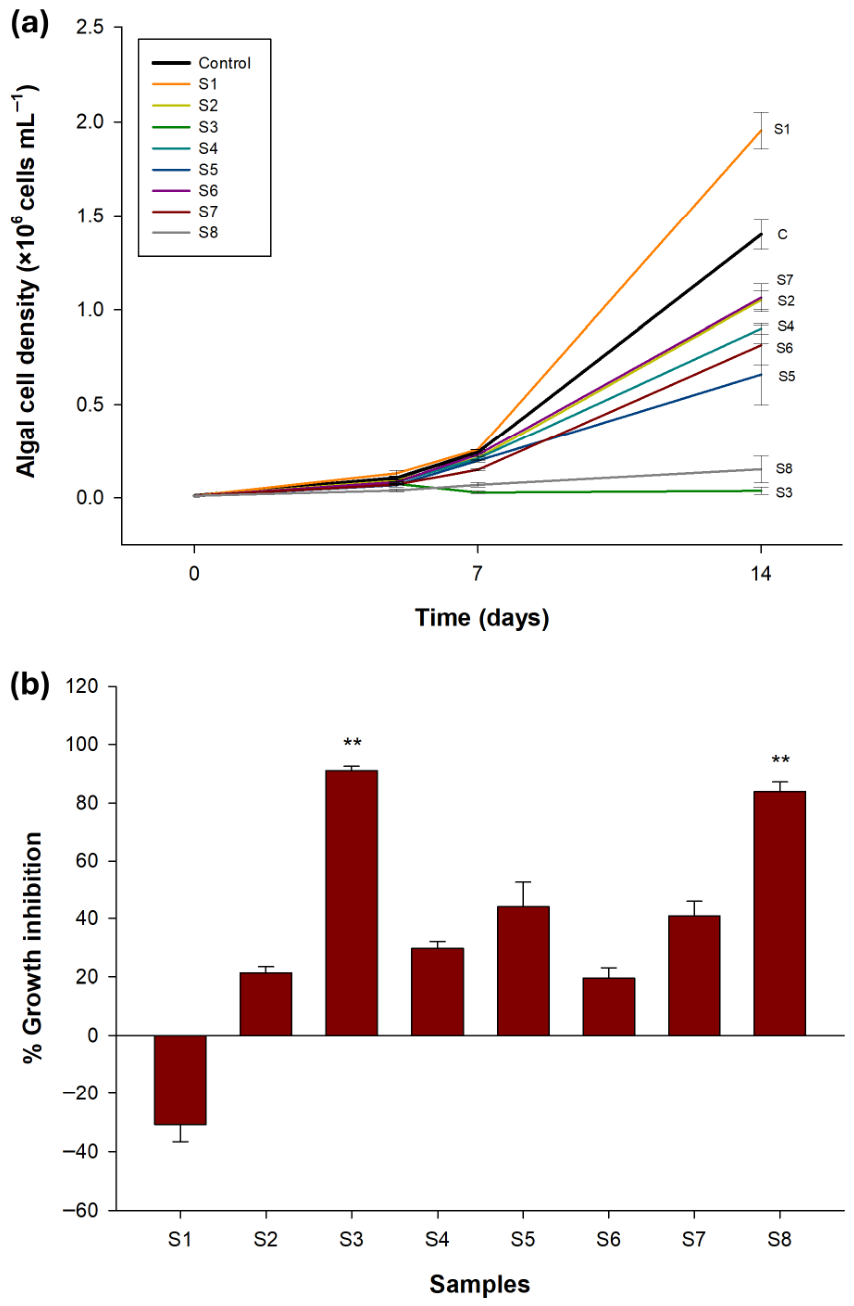
Growth curves revealed marked differences in microalgal dynamics among treatments (Figure 4a). Under control conditions, C. vulgaris showed progressive growth, increasing from the initial inoculum to 1.06 × 10 5 cells·mL−1 on day 5, 2.44 × 10 5 cells·mL−1 on day 7, and 1.40 × 10 6 cells·mL−1 on day 14 (File S3). In contrast, S1 showed an enhanced growth pattern, reaching 1.95 × 10 6 cells·mL−1 on day 14, thereby exceeding the control and corresponding to a negative inhibition value of approximately −30%, which indicates a stimulatory rather than an inhibitory effect (Figure 4b).
The strongest suppression of algal growth was observed in S3 (Figure S5). Cell density remained extremely low throughout the assay, with mean values of 4.03 × 10 4 cells·mL−1 on day 5, 2.86 × 10 4 cells·mL−1 on day 7, and only 3.75 × 10 4 cells·mL−1 on day 14 (File S3), showing no recovery over time. This pattern was consistent with the highest inhibition percentage recorded among all treatments, approximately 90% (Figure 4b). S8 also showed a strong inhibitory effect, with cell densities of 3.89 × 10 4 , 6.98 × 10 4 , and 1.53 × 10 5 cells·mL−1 on days 5, 7, and 14 (File S3), respectively, corresponding to an inhibition of approximately 84–85%. Although some increase in cell density was observed over time in S8, growth remained well below that of the control throughout the experiment. Statistical comparison of replicate AUC values confirmed that only S3 and S8 differed significantly from the untreated control (Dunnett’s test, p = 0.001 and p = 0.002, respectively).
The remaining samples showed intermediate responses, with final cell densities lower than the control but still maintaining an increasing trend over time (Figure 4a). S5 and S7 showed moderate inhibition, reaching 6.59 × 10 5 and 8.15 × 10 5 cells·mL−1 on day 14, corresponding to approximately 44% and 41% inhibition, respectively (Figure 4b). By contrast, S2, S4, and S6 showed comparatively weaker effects, with final cell densities of 8.18 × 10 5 , 9.01 × 10 5 , and 7.68 × 10 5 cells·mL−1 (File S3), corresponding to inhibition values of approximately 20%, 30%, and 20%, respectively.

3.5. Isolation and Identification of the Predominant Inhibitory Bacterium

The predominant bacterial isolate, which also showed the highest algicidal activity in the screening assays, was identified as Stenotrophomonas maltophilia strain LIG25. The obtained 16S rRNA gene sequence was deposited in the NCBI GenBank database under accession number PX631962.1.
Two additional isolates, Microbacterium oxydans strain LIG25 and Microbacterium foliorum strain LIG25, were also identified from the same sample. Their 16S rRNA gene sequences were deposited in the NCBI GenBank database under accession numbers PZ365384 and PZ365432, respectively. However, neither strain showed algicidal activity in the screening assays. Co-culture experiments with C. vulgaris showed sustained microalgal growth in the presence of both bacteria. Similarly, assays performed with Leptolyngbya sp. to evaluate possible cyanolytic activity also showed cyanobacterial growth. These two isolates were therefore discarded from further algicidal assays. The corresponding screening results are shown in File S4.

3.6. Algicidal Activity of the Isolated Strain S.maltophilia LIG 25 in Co-Culture

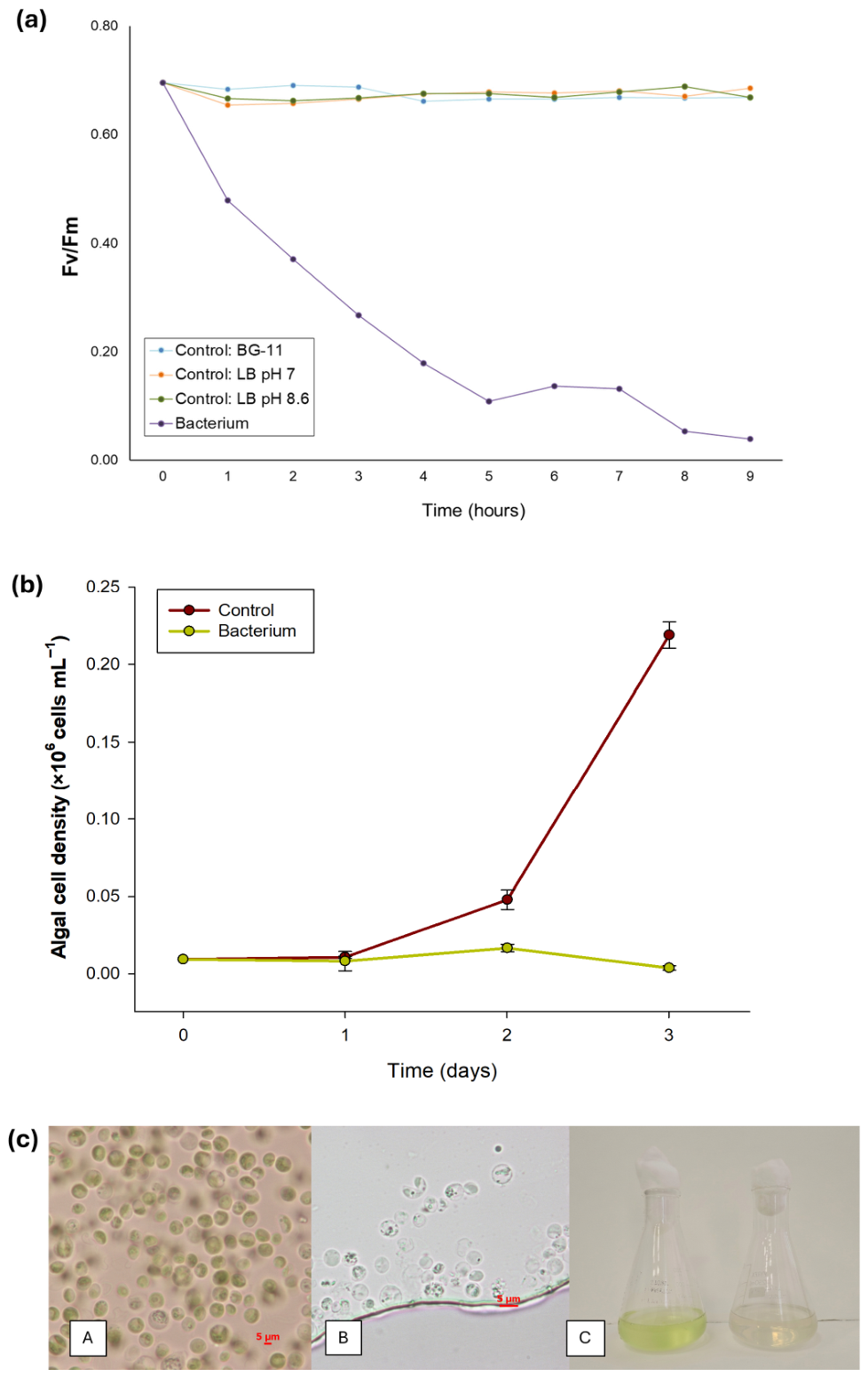
Microalgal photosynthetic activity was evaluated by PAM fluorometry during a 72 h assay. The processed cell density and PAM fluorescence data are available in File S5, whereas the raw maximum quantum yield of photosystem II (Fv/Fm) dataset is provided in File S6. The results showed a rapid decline in photosynthetic efficiency (Fv/Fm) during the initial stages of the experiment. In the 72 h assay, Fv/Fm decreased from approximately 0.700 to 0.020 within the first 24 h (Figure S6). In contrast, the controls conducted under different experimental conditions (BG-11; C. vulgaris + LB; C. vulgaris + LB adjusted to pH 8.4) remained stable throughout the assay, with Fv/Fm values ranging between 0.690 and 0.650. (Figure 5a) To characterize this process at higher temporal resolution, an additional 24 h assay was performed. In this case, the initial Fv/Fm value (0.695) decreased to approximately half within the first hour, reaching a value close to 0.350. Thereafter, Fv/Fm continued to decline progressively until reaching a minimum value of 0.016 at 9 h. From this point onward, values remained low, with fluctuations of approximately 0.010. For this reason, Figure 5a shows only the first 9 h of the assay.
Cell counts confirmed these physiological results (Figure 5b). In the controls, exponential growth was observed during the first 72 h, reaching a maximum density of (2.27 ± 0.08) × 105 cells·mL−1. In contrast, the bacterial co-culture showed only slight growth during the first 48 h, with a mean density of (1.67 ± 0.24) × 104 cells·mL−1. At 72 h, cell density decreased to (3.80 ± 1.46) × 103 cells·mL−1, corresponding to an approximate inhibition of 98.3% relative to the control.
Microscopic observations supported these results (Figure 5c). C. vulgaris cells exhibited evident structural alterations, including deformation of the characteristic spherical morphology, loss of cellular integrity, and disorganization of intracellular contents. A marked cellular bleaching was also observed, consistent with the loss of photosynthetic chromophores, together with the disappearance or loss of definition of the pyrenoid in numerous cells. In several cases, collapsed cells and dispersed cellular debris were observed, consistent with cell lysis processes.

4. Discussion

The ornamental fountains of the Alhambra and Generalife harbour spatially heterogeneous bacterial communities, with differences in taxonomic composition, diversity, and functional response against C. vulgaris. Taken together, these results indicate that the bacterial microbiome of these heritage systems does not constitute a uniform community, but rather an assemblage shaped by local environmental characteristics and, likely, by the availability of niches generated by phototrophic colonization. This point is especially relevant in an open heritage context, where biodeterioration develops under constant interaction with water, the stone substrate, radiation, nutrient input, and the indigenous microbiota of the system [9,13,37].
The variability among samples confirms that these fountains should not be interpreted as a single homogeneous microbial ecosystem. Some communities were dominated by recurrent genera, such as Rheinheimera, Flavobacterium, Acidovorax, or Pseudomonas, whereas others showed more singular profiles, as in the case of S5. This heterogeneity is consistent with the open and environmentally complex nature of these systems [6,47,48]. Comparable studies on outdoor stone heritage epilithic biofilms have likewise shown that marked differences in community composition may arise even under the same broader macroenvironment, supporting the relevance of local microenvironmental conditions in shaping microbiome structure [9]. However, in the absence of direct physicochemical measurements, the data only support the existence of relevant local differences, not the precise identification of their causes.
The relationship between visible biofilm and bacterial diversity is also informative. Samples with greater phototrophic colonization showed significantly higher alpha diversity [49], whereas the overall community composition did not display clear separation among biofilm categories. This pattern is consistent with the idea that phototrophic biofilms may increase local habitat complexity by providing colonization space, organic substrates, and microscale protection, thereby favouring the incorporation of additional bacterial taxa. In broader ecological terms, phototrophic microorganisms may also sustain part of the diversity and multifunctionality of the associated heterotrophic bacteriome through bottom-up trophic effects [26,50]. This suggests that greater phototrophic biomass may favour the incorporation of additional bacterial taxa and increase habitat complexity without generating radically different microbiomes. Thus, more heavily colonized surfaces appear to sustain more diverse communities, though not necessarily compositionally distinct ones. This interpretation also agrees with studies showing that, under increased primary productivity, biofilm communities may respond mainly through greater biomass and shifts in subdominant taxa, while dominant taxa remain relatively stable [51]. This finding indicates that visible biofilm abundance should not be overinterpreted as the main structuring factor of the bacterial microbiome. At the same time, this relationship should be interpreted cautiously, since other heritage studies have not consistently detected alpha-diversity differences between visually contrasting colonization categories, underscoring the context dependence of this pattern [52].
Within this framework, the broad representation of genera previously described in the literature as algicidal is a relevant finding, although it must be interpreted cautiously. The presence of 18 genera with reported algicidal capacity within the microbiome (Table S1) suggests that bacteria-microalga interactions may form part of the normal ecological dynamics of these fountains. However, genus-level taxonomic identification does not allow the assumption that all detected strains retain such activity or express it under the specific conditions of this system. Therefore, the mere presence of genera reported as algicidal cannot be regarded as functional evidence of biocontrol. This limitation is reinforced by the absence of significant differences among biofilm categories in the total proportion and richness of these genera after FDR correction, despite the descriptive trend toward higher relative abundance of reported algicidal genera in low-biofilm samples and higher algicidal genus richness in high-biofilm samples. Consequently, genus-level characterization is useful as an initial approximation, but insufficient to interpret the functional potential of the system accurately. This distinction is particularly important in light of current knowledge on bacteria-microalga interactions, which emphasizes that these relationships are highly context-dependent and often taxon- or strain-specific rather than generalizable at broad taxonomic levels [26]. Since algicidal activity may vary markedly among species and even among strains within the same genus, moving towards finer taxonomic resolution will be essential to determine which bacteria are actually involved in antagonistic and biocontrol processes.
This decoupling between taxonomic composition and functional activity constitutes one of the main conceptual contributions of the study. Sample S6 illustrates this clearly: although 100% of its bacterial community consisted of genera previously reported as algicidal, it belonged to the low-biofilm category and nevertheless did not show one of the strongest inhibitory responses in the functional screening. This indicates that the mere presence of potentially antagonistic bacteria is not sufficient to explain biofilm status or to predict actual biocontrol capacity. Other factors are likely involved, including the effective abundance of each taxon, variation at the species and strain level, environmental regulation of bioactive metabolite production, interactions among bacteria, and the physical structure of the biofilm matrix itself, which may modulate the diffusion or efficacy of inhibitory compounds. In addition, studies on heritage microbiomes suggest that environmental filters such as moisture, temperature, and local geochemical conditions may exert stronger control over community assembly than visible colonization alone [9,53]. Thus, taxonomic analysis is useful as an initial screening tool, but not as conclusive evidence of function.
Among the dominant genera, Rheinheimera deserves particular consideration. Its high abundance in several samples, including both high- and low-biofilm contexts, suggests that it is well adapted to the conditions of ornamental fountains and likely plays an important role as a persistent colonizer of aquatic surfaces. This interpretation is consistent with recent studies describing Rheinheimera strains with a strong capacity for biofilm formation, low nutrient requirements, and stable extracellular matrices rich in proteins and polysaccharides [54,55]. In the present study, however, its taxonomic dominance was not associated with a measurable reduction in phototrophic biofilm development. Therefore, although Rheinheimera has previously been described as antagonistic toward certain microorganisms [56], the current results do not support its relative abundance alone as a reliable indicator of effective algicidal activity in this system. Instead, its prevalence appears to reflect a strong adaptation to biofilm-associated and surface-persistent ecological niches.
The presence of Stenotrophomonas in samples with low phototrophic colonization, such as S3, is noteworthy, as is its occurrence in S4, which corresponded to a localized discontinuity of algal growth within an otherwise developed biofilm. This pattern is relevant because S4 also showed relative enrichment of Acidovorax and Pseudomonas, suggesting a bacterial assemblage potentially associated with localized inhibitory conditions. Acidovorax has previously been described as a genus with algicidal capacity, although, according to the available literature, such activity has only been reported against the planktonic microalga Chlamydomonas reinhardtii [57], a taxon frequently found in ornamental fountains [8]. Several Pseudomonas species have also been investigated for their algicidal activity against different freshwater phototrophs (Table S1) [25], supporting the relevance of this genus in the interpretation of the S4 microbial profile. In addition, among the genera with reported algicidal capacity most frequently detected in low-biofilm fountains were Aeromonas, Acidovorax, Hydrogenophaga, Pseudomonas, and Stenotrophomonas. These taxa were also accompanied by Flavobacterium and Rheinheimera (File S1); however, both genera were consistently abundant across nearly all samples, regardless of the degree of phototrophic biofilm development. Therefore, the co-occurrence of Stenotrophomonas, Acidovorax, and Pseudomonas in S4 may support the hypothesis that localized interruptions of algal growth are associated with bacterial assemblages with antagonistic potential. However, this interpretation remains hypothetical, since the present study does not demonstrate synergy among these genera or establish a causal role in the observed inhibitory process.
The functional screening using filtered fountain waters confirmed that not all aquatic microbiota have the same biocontrol potential. Some samples showed strong intrinsic inhibitory activity against C. vulgaris, whereas others produced only moderate or limited effects, or even stimulated growth, as observed for S1. This result demonstrates that bacterial presence alone is insufficient to explain the observed activity. This is consistent with current understanding of bacteria-microalga interactions, according to which associated microorganisms may either inhibit or promote phototrophic growth depending on taxonomic identity, metabolite exchange, and environmental context [26]. The stimulation observed in S1 therefore suggests that certain components of the water or microbial community may, under certain conditions, promote algal growth rather than inhibit it [58,59]. Conversely, the strong effect observed in S3 and S8 indicates that some indigenous microbiota contain bacterial combinations or metabolites sufficiently active to suppress C. vulgaris growth strongly and persistently. This functional heterogeneity reinforces the need to select candidates on the basis of experimental evidence rather than taxonomy alone.
The selection of Stenotrophomonas maltophilia LIG25 from the sample showing the highest inhibitory activity was based precisely on this functional criterion. Microbacterium oxydans and Microbacterium foliorum were isolated from the same sample, but both strains allowed the growth of C. vulgaris and Leptolyngbya sp. and were therefore not considered suitable candidates for validating algicidal activity (File S4). This observation reinforces a key point of the manuscript: the presence of a bacterium within an inhibitory microbiota does not necessarily imply that it is responsible for the observed effect. In this case, the selection of S. maltophilia was supported by its predominance in the selected sample and, above all, by the experimental validation of its activity against C. vulgaris. Although M. foliorum was isolated from the selected sample, neither the results of the present study nor the available literature support its involvement in algicidal activity. Rather, its occurrence in surface-associated habitats and biofilm communities suggests that it may be related to adhesion, persistence, or coexistence within complex microbial consortia, rather than to direct antagonism against phototrophic microorganisms [60]. Likewise, M. oxydans has been reported in Chlorella-associated consortia linked to enhanced algal growth [61,62]. Taken together, these observations suggest that both isolates are more likely associated with adhesion, coexistence, or neutral interactions within microbial communities than with effective algicidal activity.
The co-culture assays, PAM fluorometry, and microscopy converge on the same interpretation: S. maltophilia LIG25 exerts a rapid, intense, and multifaceted inhibitory effect on C. vulgaris (Figure 5a). The abrupt decline in Fv/Fm within less than 24 h indicates that physiological disruption of PSII constitutes an early event in the inhibitory process. Decreases in Fv/Fm are widely recognized as indicators of PSII photodamage and impaired electron transport under stress conditions [63,64,65]. In healthy microalgae, Fv/Fm values are typically ≥0.3–0.4, whereas the values recorded in this study (0.020–0.100) indicate severe PSII dysfunction and a near-complete loss of photosynthetic activity rather than a mild physiological limitation. This type of impairment of the photosynthetic apparatus is also among the mechanisms most frequently reported in algicidal bacteria [66,67]. The collapse of photosynthetic efficiency precedes and accompanies the subsequent decline in cell counts, suggesting that functional disruption of the photosynthetic system forms part of the initial mechanism of damage. Cell counts confirm that this deterioration does not remain limited to transient growth inhibition, but rather progresses towards extreme suppression of proliferation and, ultimately, towards a progressive loss of cell density (Figure 5b).
Microscopic observations reinforce this interpretation by revealing deformation of the spherical morphology, disorganization of intracellular contents, loss of photosynthetic chromophores, disappearance or poor definition of the pyrenoid, cell collapse, and lysis (Figure 5c). Taken together, these results point to an inhibitory process affecting both photosynthetic function and cellular structural integrity. Although the present study did not allow the responsible metabolite or mechanism to be identified precisely, the rapid decline in Fv/Fm and the observed damage pattern are consistent with an indirect action mediated by extracellular compounds. Since the slight stimulation observed at 48 h in co-culture was not maintained and cell density dropped sharply by 72 h, a transient hormetic response to a toxic agent could be cautiously considered [68,69]; however, this possibility does not alter the overall conclusion that the final effect is strongly inhibitory. In addition, the LB and pH-adjusted LB controls did not reproduce the decline observed in Fv/Fm, allowing the effect to be ruled out as a simple consequence of the bacterial medium or pH changes and reinforcing the view that it is attributable to biological activity associated with the bacterium or its extracellular products (Figure 5a).
The relevance of S. maltophilia extends beyond its specific effect on C. vulgaris. It is an environmentally versatile bacterium, widely distributed in soils, sediments, rhizospheres, and aquatic environments, which helps explain why it was relatively easy to detect and isolate in this context. Considerable attention has been paid to it because of its capacity to inhibit phytopathogenic fungi and its applications in soil and aquatic bioremediation. From an applied perspective, S. maltophilia produces a diverse array of extracellular metabolites and enzymes, including solvent-tolerant alkaline proteases, serine proteases, and antifungal compounds such as maltophilin [70,71,72,73]. It also stands out for its ability to degrade organic matter and xenobiotic compounds, including hydrocarbons, pesticides, and aromatic molecules, as well as for its involvement in heavy metal detoxification and phosphate removal from wastewater through iron-reducing activity, thereby contributing to the mitigation of eutrophication [74,75,76,77,78]. Taken together, these properties reinforce its interest for remediation strategies in sensitive aquatic systems, where the use of conventional chemical treatments may be undesirable.
Its potential against microalgae and cyanobacteria, however, has been much less explored than its role in other biocontrol and bioremediation contexts. Studies have shown that other Stenotrophomonas strains, such as strain F6, produce extracellular compounds such as cyclo-(Gly-Pro) and hydroquinone with strong algicidal activity against Microcystis sp., Chlamydomonas sp., C. vulgaris, and Oscillatoria sp. [79]. More recently, S. maltophilia has also been described as causing thallus degradation in the marine macroalga Kappaphycus alvarezii through direct contact of live cells, associated with bleaching and tissue stiffening [80]. Taken together, these precedents suggest that Stenotrophomonas may act through both indirect and direct mechanisms. In addition, it belongs to the Gammaproteobacteria, a group that includes many of the algicidal bacteria described to date [25,27,81].
Moreover, S. maltophilia has already been used in the conservation and restoration of cultural heritage. A notable example is its application in the cleaning of the wall paintings of the Casina Farnese in Rome, where strain UI3E was successfully applied in Laponite® microcompresses (CTS S.r.l., Briosco, Italy) to remove aged proteinaceous deposits, including animal glue and casein, without affecting either the pictorial layer or the substrate and without leaving bacterial residues [82]. This precedent demonstrates that, at least under certain conditions and formulations, the species can act selectively on unwanted organic matter in sensitive heritage materials. This knowledge reinforces the opportunity to develop biocontrol and biocleaning strategies for sensitive heritage objects, transferring to the heritage field a biotechnological approach widely developed in HAB control but still scarcely explored in cultural assets exposed to water.
Nevertheless, it will also be necessary to test whether this behaviour is maintained against other phototrophs involved in biodeterioration. A particularly relevant next step will therefore be to assess its activity against cyanobacteria, diatoms, and endolithic green algae characteristic of stone surfaces, as well as against mixed communities closer to the real conditions of ornamental fountains. This broader evaluation will help define its spectrum of action more accurately and assess its integration into more complete biocontrol strategies based on bacterial combinations or consortia, in which different strains may act through complementary mechanisms and thereby broaden the control spectrum across different phototrophic groups [83,84].
The accumulated experience with Bacillus-based formulated products demonstrates that bacteria can be incorporated into commercial biotechnological solutions that are both effective and environmentally compatible [30]. However, the results of this study suggest that it is also worth exploring indigenous genera adapted to the local heritage ecosystem, especially those that do not raise biosafety concerns.
Any applied projection, however, must be formulated cautiously. S. maltophilia is also an opportunistic pathogen, so its direct use as a living organism in open heritage environments or publicly accessible spaces may raise regulatory, ecological, and biosafety constraints [85,86]. A particularly reasonable line of future research is therefore to identify the metabolites, enzymes, or extracellular fractions responsible for the algicidal activity and to shift development towards cell-free filtrates or formulations based on excreted products. This approach would be relevant not only for S. maltophilia, but also for other bacteria with algicidal potential, as it would preserve biological efficacy while minimizing the risks associated with the deliberate release of living microorganisms [87,88]. It also aligns with the growing interest in “green” conservation strategies aimed at more selective, sustainable solutions compatible with the ecological balance of the surrounding environment [33].

5. Conclusions

The ornamental fountains of the Alhambra and Generalife harbour bacterial microbiomes with potential inhibitory activity against phototrophic microorganisms involved in biodeterioration. The combined microbiome analysis and functional screening presented here indicate that these heritage aquatic systems may represent a valuable source of native bacterial candidates and, therefore, a relevant basis for the development of more sustainable management strategies. The detection of inhibitory activity in several samples, together with the isolation and experimental validation of Stenotrophomonas maltophilia LIG25 as an algicidal bacterium, provides initial evidence that autochthonous bacteria present in these systems may contribute to microbial interactions involved in phototrophic growth regulation. Although this does not yet constitute a validated management tool, it establishes a solid experimental basis for future research on microbiota-inspired biocontrol strategies for ornamental fountains. Overall, this work provides a starting point for the development of more selective, sustainable, and heritage-compatible approaches to limit the overgrowth of harmful phototrophic microorganisms while preserving both historical materials and the ecological balance of their surrounding environment.

Supplementary Materials

The following supporting information can be downloaded at: https://www.mdpi.com/article/10.3390/conservation6020070/s1. File S1: Normalized table of bacterial genera detected in the fountain microbiome, including the list of genera grouped under the “Others” category in the stacked bar plot of Figure 2a. File S2: Schematic layout of the functional screening assay of fountain waters. File S3: Cell density counts dataset from the multi-well screening assay. File S4: Results from multi-well co-culture assays with isolated and identified bacteria. File S5: Co-culture cell density data and PAM fluorescence datasets (72 h and 24 h assays). File S6: Raw PAM fluorometry dataset of photosynthetic efficiency (Fv/Fm). File S7: R Code. Figure S1: Hierarchical clustering of samples based on Bray–Curtis dissimilarity. Hierarchical clustering of sampling sites based on Bray–Curtis distance using the Ward.D2 method. The dendrogram shows the similarity relationships among microbial communities across the eight ornamental fountains. Figure S2: Relative abundance of algicidal vs. non-algicidal bacterial genera across sampling sites. Figure S3: Relative abundance and richness of genera with reported algicidal activity across biofilm categories. Boxplots show the total relative abundance (%) of genera previously reported as algicidal and the richness of reported algicidal genera in Low and High biofilm samples. Points represent individual samples. Statistical annotations indicate Kruskal–Wallis p-values and Benjamini–Hochberg FDR-adjusted q-values. ns, not significant. Figure S4: Heatmap of algicidal genera across sampling sites. Figure S5: Experimental setup and visual assessment of algal growth inhibition. Figure S6: Changes in photosynthetic efficiency (Fv/Fm) of Chlorella vulgaris under control conditions and in co-culture with Stenotrophomonas maltophilia LIG25. Table S1: Algicidal bacteria reported in the literature, their target microorganisms, bioactive compounds and corresponding references [89,90,91,92,93,94,95,96,97,98,99,100,101,102,103,104,105,106,107,108,109,110,111,112,113,114,115,116,117,118].

Author Contributions

I.C.-B.: Writing—original draft preparation, data curation, conceptualization, methodology, software, formal analysis, investigation; S.F.: Investigation, supervision, methodology, data curation, formal analysis, resources; O.A.C.: Validation, investigation, methodology; L.C.-C.: Methodology and technical work; M.G.-d.-V.: Methodology, validation, resources; F.B.-G.: Project administration and funding acquisition; J.R.-N.: Writing—review and editing, supervision, validation and funding acquisition. All authors have read and agreed to the published version of the manuscript.

Funding

This research was funded by project BIOALHAMBRA, grant number PID2022-143064OB-I00, supported by MCIN/AEI/10.13039/501100011033 «Proyectos de Generación de Conocimiento» (Spain) and project SOL2024-30779, (VII) Plan Propio de Investigación-Programa al estímulo de áreas con necesidades investigadoras y de la actividad investigadora emergente—Modalidad A1—by University of Seville.

Institutional Review Board Statement

Not applicable.

Informed Consent Statement

Not applicable.

Data Availability Statement

The original contributions presented in this study are included in the article/Supplementary Material. Further inquiries can be directed to the corresponding author.

Acknowledgments

The authors gratefully acknowledge Fernando Delgado-Chaves, from Galagos and affiliated with the University of Hamburg, for his valuable bioinformatics expertise and for providing essential analytical support and tools. The authors also thank Ignacio Moreno-Garrido, from the Institute of Marine Sciences of Andalusia (ICMAN-CSIC), for his valuable scientific guidance and training in microalgal culture and ecotoxicological assessment.

Conflicts of Interest

S.F. is affiliated with Sanbiotec Laboratory S.L. and M.G.-d.-V. is affiliated with G2G Algae Solutions. The authors declare that these commercial affiliations did not influence the design, analysis, interpretation, or reporting of the study. The remaining authors declare no conflicts of interest.

References

  1. Bender, A.; Guerreiro, M.; Agapito, D.; Sequeira, B.D.; Mendes, J. Sensory Experiences in Heritage Contexts: A Qualitative Approach. Eur. J. Tour. Res. 2024, 36, 3604. [Google Scholar] [CrossRef]
  2. Chen, X.; Duan, W.; Liu, Y.; Li, S.; Sui, S. Spatial Structure of Yangzhou’s Old Public Bathrooms: A Space Syntax Analysis of the Topological Characteristics of Their Unique Sensory Space. J. Asian Archit. Build. Eng. 2023, 22, 3455–3475. [Google Scholar] [CrossRef]
  3. Galan, J. Urban Typologies and Urban Sustainability: A Comparative and Landscape-Based Study in the City of Valencia. Cities 2024, 154, 105344. [Google Scholar] [CrossRef]
  4. Zhang, Z.; Xiong, K.; Huang, D. Natural World Heritage Conservation and Tourism: A Review. Herit. Sci. 2023, 11, 55. [Google Scholar] [CrossRef]
  5. García Montes, J.M. Flora Ornamental de la Alhambra y el Generalife. Doctoral Dissertation, Universidad de Granada, Granada, Spain, 1997. [Google Scholar]
  6. Yadav, S.; Purchase, D. Biodeterioration of Cultural Heritage Monuments: A Review of Their Deterioration Mechanisms and Conservation. Int. Biodeterior. Biodegrad. 2025, 201, 106066. [Google Scholar] [CrossRef]
  7. Bolívar Galiano, F.; Calvo-Bayo, I. Diagnóstico de los efectos del turismo en la calidad del agua de las fuentes ornamentales ubicadas en el paisaje de la Alhambra y el Valle del Darro. In El Observatorio en Turismo Patrimonial Sostenible en Andalucía: Análisis, Diagnóstico, Adecuación, Innovación y Transferencia; Ediciones Complutense: Madrid, Spain, 2023; Volume 22, pp. 131–150. [Google Scholar]
  8. Bolívar-Galiano, F.; Abad-Ruiz, C.; Sánchez-Castillo, P.; Toscano, M.; Romero-Noguera, J. Frequent Microalgae in the Fountains of the Alhambra and Generalife: Identification and Creation of a Culture Collection. Appl. Sci. 2020, 10, 6603. [Google Scholar] [CrossRef]
  9. Meng, S.; Qian, Y.; Liu, X.; Wang, Y.; Wu, F.; Wang, W.; Gu, J.-D. Community Structures and Biodeterioration Processes of Epilithic Biofilms Imply the Significance of Micro-Environments. Sci. Total Environ. 2023, 876, 162665. [Google Scholar] [CrossRef] [PubMed]
  10. Bolívar-Galiano, F.; Cuzman, O.A.; Abad-Ruiz, C.; Sánchez-Castillo, P. Facing Phototrophic Microorganisms That Colonize Artistic Fountains and Other Wet Stone Surfaces: Identification Keys. Appl. Sci. 2021, 11, 8787. [Google Scholar] [CrossRef]
  11. Suchy, H.; Zalar, P.; Macedo, M.F. Microbial Diversity of Biodeteriorated Limestone Cultural Heritage Assets Identified Using Molecular Approaches—A Literature Review. Appl. Sci. 2024, 14, 7429. [Google Scholar] [CrossRef]
  12. Golubić, S.; Pietrini, A.M.; Ricci, S. Euendolithic Activity of the Cyanobacterium Chroococcus lithophilus Erc. in Biodeterioration of the Pyramid of Caius Cestius, Rome, Italy. Int. Biodeterior. Biodegrad. 2015, 100, 7–16. [Google Scholar] [CrossRef]
  13. Favero-Longo, S.E.; Viles, H.A. A Review of the Nature, Role and Control of Lithobionts on Stone Cultural Heritage: Weighing-Up and Managing Biodeterioration and Bioprotection. World J. Microbiol. Biotechnol. 2020, 36, 100. [Google Scholar] [CrossRef]
  14. Pinna, D. Can We Do without Biocides to Cope with Biofilms and Lichens on Stone Heritage? Int. Biodeterior. Biodegrad. 2022, 172, 105437. [Google Scholar] [CrossRef]
  15. Sanmartín, P.; Bosch-Roig, P.; Pangallo, D.; Kraková, L.; Serrano, M. Unraveling Disparate Roles of Organisms, from Plants to Bacteria, and Viruses on Built Cultural Heritage. Appl. Microbiol. Biotechnol. 2023, 107, 2027–2037. [Google Scholar] [CrossRef]
  16. Komar, M.; Derese, N.; Szymczak, K.; Nowicka-Krawczyk, P.; Gutarowska, B. Natural Plant Oils as Anti-Algae Biocides for Sustainable Application in Cultural Heritage Protection. Sustainability 2025, 17, 6996. [Google Scholar] [CrossRef]
  17. Pascale, M.R.; Roggio, D.S.; Barbieri, E.; Marino, F.; Derelitto, C.; Girolamini, L.; Bragalli, C.; Bitelli, G.; Cristino, S. New Frontiers in Water Distribution System Management and Monitoring: First Development of a Water Safety Plan Based on Heritage Building Information Modeling (HBIM) in Neptune Fountain, Bologna, Italy. Water 2024, 16, 2075. [Google Scholar] [CrossRef]
  18. Qian, Y.; He, Y.; Li, H.; Yi, M.; Zhang, L.; Zhang, L.; Liu, L.; Lu, Z. Benzalkonium Chlorides (C12) Inhibits Growth but Motivates Microcystins Release of Microcystis aeruginosa Revealed by Morphological, Physiological, and iTRAQ Investigation. Environ. Pollut. 2022, 292, 118305. [Google Scholar] [CrossRef]
  19. Calvo-Bayo, I.; Bolívar-Galiano, F.; Romero-Noguera, J. Algicidal Bacteria: A Sustainable Proposal to Control Microalgae in the Conservation and Restoration of Stone Cultural Heritage. Sustainability 2025, 17, 10610. [Google Scholar] [CrossRef]
  20. Pal, M.; Yesankar, P.J.; Dwivedi, A.; Qureshi, A. Biotic Control of Harmful Algal Blooms (HABs): A Brief Review. J. Environ. Manag. 2020, 268, 110687. [Google Scholar] [CrossRef] [PubMed]
  21. He, S.; Zhang, Y.; Li, N.; Shi, K.; Zhang, Y.; Qin, B.; Zhu, G.; Liu, M.; Shao, K. Summer Heatwaves Promote Harmful Algal Blooms in the Fuchunjiang Reservoir, an Important Drinking Water Source. J. Environ. Manag. 2024, 359, 121056. [Google Scholar] [CrossRef]
  22. Rattner, B.A.; Wazniak, C.E.; Lankton, J.S.; McGowan, P.C.; Drovetski, S.V.; Egerton, T.A. Review of Harmful Algal Bloom Effects on Birds with Implications for Avian Wildlife in the Chesapeake Bay Region. Harmful Algae 2022, 120, 102319. [Google Scholar] [CrossRef] [PubMed]
  23. Cadaillon, A.M.; Mattera, B.; Albizzi, A.; Montoya, N.; Maldonado, S.; Raya Rey, A.; Riccialdelli, L.; Almandoz, G.O.; Schloss, I.R. Multispecies Mass Mortality in the Beagle Channel Associated with Paralytic Shellfish Toxins. Harmful Algae 2024, 132, 102581. [Google Scholar] [CrossRef]
  24. Messineo, V.; Bruno, M.; De Pace, R. The Role of Cyano-HAB (Cyanobacteria Harmful Algal Blooms) in the One Health Approach to Global Health. Hydrobiology 2024, 3, 238–262. [Google Scholar] [CrossRef]
  25. Coyne, K.J.; Wang, Y.; Johnson, G. Algicidal Bacteria: A Review of Current Knowledge and Applications to Control Harmful Algal Blooms. Front. Microbiol. 2022, 13, 871177. [Google Scholar] [CrossRef] [PubMed]
  26. Abate, R.; Oon, Y.-L.; Oon, Y.-S.; Bi, Y.; Mi, W.; Song, G.; Gao, Y. Diverse Interactions between Bacteria and Microalgae: A Review for Enhancing Harmful Algal Bloom Mitigation and Biomass Processing Efficiency. Heliyon 2024, 10, e36503. [Google Scholar] [CrossRef]
  27. Wang, J.; Xu, B.; Huang, L. Molecular Mechanisms of Algicidal Bacteria in Controlling Harmful Algal Blooms: Advances in Bacteria-Algae Interactions. Environ. Microbiol. Rep. 2026, 18, e70305. [Google Scholar] [CrossRef]
  28. Ma, B.; Li, A.; Chen, S.; Guo, H.; Li, N.; Pan, S.; Chen, K.; Liu, H.; Kosolapov, D.B.; Liu, X.; et al. Algicidal Activity Synchronized with Nitrogen Removal by Actinomycetes: Algicidal Mechanism, Stress Response of Algal Cells, Denitrification Performance, and Indigenous Bacterial Community Co-Occurrence. J. Hazard. Mater. 2024, 470, 134117. [Google Scholar] [CrossRef] [PubMed]
  29. Atef, S.; Ahmed, O.M.; Said, M.M.; Abo-Al-Ela, H.G. Dietary Bacillus Species Modulate Lipid Metabolism-Related Parameters, Growth, Water Quality, and Bacterial Load in Nile Tilapia (Oreochromis niloticus). Anim. Feed Sci. Technol. 2024, 310, 115943. [Google Scholar] [CrossRef]
  30. Hlordzi, V.; Kuebutornye, F.K.A.; Afriyie, G.; Abarike, E.D.; Lu, Y.; Chi, S.; Anokyewaa, M.A. The Use of Bacillus Species in Maintenance of Water Quality in Aquaculture: A Review. Aquac. Rep. 2020, 18, 100503. [Google Scholar] [CrossRef]
  31. Gao, Y.; Wei, Y.; Zeng, D.; Zhang, J.; Dong, J.; Gao, X.; Yuan, H.; Li, X.; Qiu, D.; Burford, M. Restructuring of the Epiphytic Microbiome and Recruitment of Algicidal Bacteria by Vallisneria natans for the Suppression of Microcystis. Front. Plant Sci. 2026, 16, 1731742. [Google Scholar] [CrossRef]
  32. Inaba, N.; Kodama, I.; Nagai, S.; Shiraishi, T.; Matsuno, K.; Yamaguchi, A.; Imai, I. Distribution of Harmful Algal Growth-Limiting Bacteria on Artificially Introduced Ulva and Natural Macroalgal Beds. Appl. Sci. 2020, 10, 5658. [Google Scholar] [CrossRef]
  33. Caldeira, A.T. Green Mitigation Strategy for Cultural Heritage Using Bacterial Biocides. In Microorganisms in the Deterioration and Preservation of Cultural Heritage; Joseph, E., Ed.; Springer International Publishing: Cham, Switzerland, 2021; pp. 137–154. [Google Scholar]
  34. Fidanza, M.R.; Caneva, G. Natural Biocides for the Conservation of Stone Cultural Heritage: A Review. J. Cult. Herit. 2019, 38, 271–286. [Google Scholar] [CrossRef]
  35. Xue, X.; Su, X.; Zhou, L.; Ji, J.; Qin, Z.; Liu, J.; Li, K.; Wang, H.; Wang, Z. Antibiotic-Induced Recruitment of Specific Algae-Associated Microbiome Enhances the Adaptability of Chlorella vulgaris to Antibiotic Stress and Incidence of Antibiotic Resistance. Environ. Sci. Technol. 2023, 57, 13336–13345. [Google Scholar] [CrossRef] [PubMed]
  36. Nikolić, N.; Simić, G.S.; Golić, I.; Popović, S. The Effects of Biocides on the Growth of Aerophytic Green Algae (Chlorella sp.) Isolated from a Cave Environment. Arch. Biol. Sci. 2021, 73, 341–351. [Google Scholar] [CrossRef]
  37. Correa, M.V.; Rastelli, S.E.; Gómez de Saravia, S.G. Assessing Preventive Surface Treatments against Microalgae on Marble for Sustainable Building Conservation. Built Herit. 2026, 10, 17. [Google Scholar] [CrossRef]
  38. APHA; AWWA. Standard Methods for the Examination of Water and Wastewater, 17th ed.; APHA: Washington, DC, USA; AWWA: Denver, CO, USA, 1989. [Google Scholar]
  39. ISO. Water Quality—Fresh Water Algal Growth Inhibition Test with Unicellular Green Algae; International Organization for Standardization: Geneva, Switzerland, 2012. [Google Scholar]
  40. OECD. Guidelines for the Testing of Chemicals, Section 201: Alga, Growth Inhibition Test; Organisation for Economic Co-operation and Development: Paris, France, 2011. [Google Scholar]
  41. Pandey, S.; Narayanan, I.; Vinayagam, R.; Selvaraj, R.; Varadavenkatesan, T.; Pugazhendhi, A. A Review on the Effect of Blue Green 11 Medium and Its Constituents on Microalgal Growth and Lipid Production. J. Environ. Chem. Eng. 2023, 11, 109984. [Google Scholar] [CrossRef]
  42. Yang, X. Moraxellaceae. In Encyclopedia of Food Microbiology; Elsevier: Amsterdam, The Netherlands, 2014; pp. 826–833. [Google Scholar]
  43. Weisburg, W.G.; Barns, S.M.; Pelletier, D.A.; Lane, D.J. 16S Ribosomal DNA Amplification for Phylogenetic Study. J. Bacteriol. 1991, 173, 697–703. [Google Scholar] [CrossRef]
  44. Wang, H.; Guo, J.; Chen, X.; He, H. The Metabolomics Changes in Luria–Bertani Broth Medium under Different Sterilization Methods and Their Effects on Bacillus Growth. Metabolites 2023, 13, 958. [Google Scholar] [CrossRef] [PubMed]
  45. Pokrzywinski, K.L.; Tilney, C.L.; Modla, S.; Caplan, J.L.; Ross, J.; Warner, M.E.; Coyne, K.J. Effects of the Bacterial Algicide IRI-160AA on Cellular Morphology of Harmful Dinoflagellates. Harmful Algae 2017, 62, 127–135. [Google Scholar] [CrossRef]
  46. Perri, K.A.; Manning, S.R.; Watson, S.B.; Fowler, N.L.; Boyer, G.L. Dark Adaptation and Ability of Pulse-Amplitude Modulated (PAM) Fluorometry to Identify Nutrient Limitation in the Bloom-Forming Cyanobacterium, Microcystis aeruginosa (Kützing). J. Photochem. Photobiol. B Biol. 2021, 219, 112186. [Google Scholar] [CrossRef]
  47. Zhang, Y.; Wu, F.; Su, M.; He, D.; Gu, J.-D.; Guo, Q.; Feng, H.; Wang, W.; Kakakhel, M.A.; Yang, Y. Spatial and Temporal Distributions of Microbial Diversity under Natural Conditions on the Sandstone Stelae of the Beishiku Temple in China. Int. Biodeterior. Biodegrad. 2021, 163, 105279. [Google Scholar] [CrossRef]
  48. Tang, X.; Xie, G.; Shao, K.; Tian, W.; Gao, G.; Qin, B. Aquatic Bacterial Diversity, Community Composition and Assembly in the Semi-Arid Inner Mongolia Plateau: Combined Effects of Salinity and Nutrient Levels. Microorganisms 2021, 9, 208. [Google Scholar] [CrossRef]
  49. Li, Q.; Zhang, B.; He, Z.; Yang, X. Distribution and Diversity of Bacteria and Fungi Colonization in Stone Monuments Analyzed by High-Throughput Sequencing. PLoS ONE 2016, 11, e0163287. [Google Scholar] [CrossRef]
  50. Yu, Y.; Zhang, J.; Chen, R.; Coleine, C.; Liu, W.; Delgado-Baquerizo, M.; Feng, Y. Unearthing the Global Patterns of Cultural Heritage Microbiome for Conservation. Int. Biodeterior. Biodegrad. 2024, 190, 105784. [Google Scholar] [CrossRef]
  51. Bengtsson, M.M.; Wagner, K.; Schwab, C.; Urich, T.; Battin, T.J. Light Availability Impacts Structure and Function of Phototrophic Stream Biofilms across Domains and Trophic Levels. Mol. Ecol. 2018, 27, 2913–2925. [Google Scholar] [CrossRef] [PubMed]
  52. Janakiev, T.; Dimkić, I.; Aleksić, J.; Grbić, M.L.; Knežević, A.; Kosel, J.; Tavzes, Č.; Unković, N. Beneficial Bacteria-Based Bioformulations as Potential Biocontrol and Biocleaning Solutions for Stone Heritage Conservation. World J. Microbiol. Biotechnol. 2025, 41, 200. [Google Scholar] [CrossRef]
  53. Ding, S.; Liu, W.; Li, X.; Feng, Y. The Ecology of Microbiome on Cultural Relics: The Linkage of Assembly, Composition and Biodeterioration. J. Cult. Herit. 2025, 71, 412–418. [Google Scholar] [CrossRef]
  54. Chen, W.M.; Chen, W.T.; Young, C.C.; Sheu, S.Y. Rheinheimera riviphila sp. nov., Isolated from a Freshwater Stream. Arch. Microbiol. 2019, 201, 919–926. [Google Scholar] [CrossRef] [PubMed]
  55. Yadav, V.; Manjhi, A.; Vadakedath, N. Mercury Remediation Potential of Mercury-Resistant Strain Rheinheimera metallidurans sp. nov. Isolated from a Municipal Waste Dumping Site. Ecotoxicol. Environ. Saf. 2023, 257, 114888. [Google Scholar] [CrossRef]
  56. Chen, W.M.; Lin, C.Y.; Sheu, S.Y. Investigating Antimicrobial Activity in Rheinheimera sp. Due to Hydrogen Peroxide Generated by L-Lysine Oxidase Activity. Enzym. Microb. Technol. 2010, 46, 487–493. [Google Scholar] [CrossRef]
  57. Ren, S.; Jin, Y.; Ma, J.; Zheng, N.; Zhang, J.; Peng, X.; Xie, B. Isolation and Characterization of Algicidal Bacteria from Freshwater Aquatic Environments in China. Front. Microbiol. 2023, 14, 1156291. [Google Scholar] [CrossRef]
  58. Sassano, G.; Del Mondo, A.; Pinto, G.; Pollio, A.; De Natale, A. Co-Culture of Microalgae and Bacteria for the Production of Bioactive Compounds. Ann. Microbiol. 2025, 75, 13. [Google Scholar] [CrossRef]
  59. Lian, J.; Schimmel, P.; Sanchez-Garcia, S.; Wijffels, R.H.; Smidt, H.; Sipkema, D. Different Co-Occurring Bacteria Enhance or Decrease the Growth of the Microalga Nannochloropsis sp. CCAP211/78. Microb. Biotechnol. 2021, 14, 1159–1170. [Google Scholar] [CrossRef] [PubMed]
  60. Behrendt, U.; Ulrich, A.; Schumann, P. Description of Microbacterium foliorum sp. nov. and Microbacterium phyllosphaerae sp. nov., Isolated from the Phyllosphere of Grasses and the Surface Litter after Mulching the Sward, and Reclassification of Aureobacterium resistens (Funke et al. 1998) as Microbacterium resistens comb. nov. Int. J. Syst. Evol. Microbiol. 2001, 51, 1267–1276. [Google Scholar] [CrossRef] [PubMed]
  61. Bandaranayake, U.S.; Ikarashi, T.; Mizuta, Y.; Watari, T.; Yamaguchi, T.; Hatamoto, M. Advanced Isolation Strategies for Mimicking the Actual Environment to Reveal Key Biofilm-Forming Bacteria in an Actual Sewage-Treating Membrane Bioreactor. Environ. Technol. Innov. 2025, 40, 104519. [Google Scholar] [CrossRef]
  62. Chen, C.; Wang, Y.; Dai, Q.; Du, W.; Zhao, Y.; Song, Q. Screening of Bacteria Promoting Carbon Fixation in Chlorella vulgaris under High Concentration CO2 Stress. Biology 2025, 14, 157. [Google Scholar] [CrossRef]
  63. Xu, Y.; Peng, B.-Y.; Zhang, X.; Xu, Q.; Yang, L.; Chen, J.; Zhou, X.; Zhang, Y. The Aging of Microplastics Exacerbates the Damage to Photosynthetic Performance and Bioenergy Production in Microalgae (Chlorella pyrenoidosa). Water Res. 2024, 259, 121841. [Google Scholar] [CrossRef]
  64. Parkhill, J.-P.; Maillet, G.; Cullen, J.J. Fluorescence-Based Maximal Quantum Yield for PSII as a Diagnostic of Nutrient Stress. J. Phycol. 2001, 37, 517–529. [Google Scholar] [CrossRef]
  65. Mallick, N.; Mohn, F.H. Use of Chlorophyll Fluorescence in Metal-Stress Research: A Case Study with the Green Microalga Scenedesmus. Ecotoxicol. Environ. Saf. 2003, 55, 64–69. [Google Scholar] [CrossRef]
  66. Hadjoudja, S.; Vignoles, C.; Deluchat, V.; Lenain, J.-F.; Le Jeune, A.-H.; Baudu, M. Short Term Copper Toxicity on Microcystis aeruginosa and Chlorella vulgaris Using Flow Cytometry. Aquat. Toxicol. 2009, 94, 255–264. [Google Scholar] [CrossRef]
  67. Deng, Y.; Beadham, I.; Ren, H.-Y.; Ji, M.-M.; Ruan, W.-Q. A Study into the Species Sensitivity of Green Algae towards Imidazolium-Based Ionic Liquids Using Flow Cytometry. Ecotoxicol. Environ. Saf. 2020, 194, 110392. [Google Scholar] [CrossRef]
  68. Esteves, S.M.; Keck, F.; Almeida, S.F.P.; Figueira, E.; Bouchez, A.; Rimet, F. Can We Predict Diatoms Herbicide Sensitivities with Phylogeny? Influence of Intraspecific and Interspecific Variability. Ecotoxicology 2017, 26, 1065–1077. [Google Scholar] [CrossRef]
  69. Klátyik, S.; Takács, E.; Barócsi, A.; Lenk, S.; Kocsányi, L.; Darvas, B.; Székács, A. Hormesis, the Individual and Combined Phytotoxicity of the Components of Glyphosate-Based Formulations on Algal Growth and Photosynthetic Activity. Toxics 2024, 12, 257. [Google Scholar] [CrossRef] [PubMed]
  70. Allafchian, A.; Jalali, M.; Yazdani, F.; Shahabi, S. Exploring Alkaline Serine Protease Production and Characterization in Proteolytic Bacteria Stenotrophomonas maltophilia: Insights from Real-Time PCR and Fermentation Techniques. Biocatal. Agric. Biotechnol. 2024, 58, 103186. [Google Scholar] [CrossRef]
  71. Georgieva, M.L.; Lebedeva, S.A.; Bilanenko, E.N. Production, Characterization, Gene Cloning, and Nematocidal Activity of the Extracellular Protease from Stenotrophomonas maltophilia N4. J. Biosci. Bioeng. 2016, 121, 614–618. [Google Scholar] [CrossRef]
  72. Waghmare, S.R.; Gurav, A.A.; Mali, S.A.; Nadaf, N.H.; Jadhav, D.B.; Sonawane, K.D. Purification and Characterization of Novel Organic Solvent Tolerant 98 kDa Alkaline Protease from Isolated Stenotrophomonas maltophilia Strain SK. Protein Expr. Purif. 2015, 107, 1–6. [Google Scholar] [CrossRef]
  73. Wang, Q.; Ji, F.; Wang, J.; Jiang, B.; Li, L.; An, L.; Li, Y.; Bao, Y. Characterization of a Salt-Activated Protease with Temperature-Dependent Secretion in Stenotrophomonas maltophilia FF11 Isolated from Frozen Antarctic Krill. J. Ind. Microbiol. Biotechnol. 2016, 43, 829–840. [Google Scholar] [CrossRef]
  74. Wang, X.; Liu, Y.; Zhang, Y.; Li, J.; Chen, Z. Efficient Pyridine Biodegradation by Stenotrophomonas maltophilia J2: Degradation Performance, Mechanism, and Immobilized Application for Wastewater. J. Hazard. Mater. 2024, 461, 132220. [Google Scholar] [CrossRef]
  75. Biswas, A.; Saha, P.; Saha, B.; Chowdhury, R.; Das, S. Unravelling the Mechanism of Arsenic Resistance and Bioremediation in Stenotrophomonas maltophilia: A Molecular Approach. Environ. Pollut. 2024, 363, 125066. [Google Scholar] [CrossRef]
  76. Farooq, A.; Kanwal, R.; Bashir, K.; Tian, X.; Chen, Y.; Liu, D.; Zhang, L.; Xiang, Q.; Zhao, K.; Yu, X.; et al. Multi-Omics Analysis of Stenotrophomonas maltophilia S-11 Reveals Its Potential for Pb2+ Bioremediation in Contaminated Soil. J. Hazard. Mater. 2025, 495, 138867. [Google Scholar] [CrossRef] [PubMed]
  77. Lara-Moreno, A.; Morillo, E.; Merchán, F.; Villaverde, J. A Comprehensive Feasibility Study of Effectiveness and Environmental Impact of PAH Bioremediation Using an Indigenous Microbial Degrader Consortium and a Novel Strain Stenotrophomonas maltophilia CPHE1 Isolated from an Industrial Polluted Soil. J. Environ. Manag. 2021, 289, 112512. [Google Scholar] [CrossRef] [PubMed]
  78. Bhatt, V.; Koringa, P.; Konda, A.K.; Iyer, B. Biodegradation of Glyphosate by Stenotrophomonas maltophilia GP-1 Involves C-P Lyase Pathway. Biodegradation 2025, 36, 73. [Google Scholar] [CrossRef]
  79. Lin, S.; Geng, M.; Liu, X.; Tan, J.; Yang, H. On the Control of Microcystis aeruginosa and Synechococcus Species Using an Algicidal Bacterium, Stenotrophomonas F6, and Its Algicidal Compounds Cyclo-(Gly-Pro) and Hydroquinone. J. Appl. Phycol. 2016, 28, 345–355. [Google Scholar] [CrossRef]
  80. Achmad, M. Effect of Stenotrophomonas maltophilia Bacteria and Environmental Factors on the Thallus of Red Seaweed Kappaphycus alvarezii. Int. J. Agric. Biol. 2025, 34, 340208. [Google Scholar] [CrossRef]
  81. Chen, Y.; Luo, G.; Chen, S.; Zhang, D.; Xie, W.; Wang, Z.; Zheng, W.; Xu, H. The Potential of Prodigiosin for Control of Prorocentrum donghaiense Blooms: Algicidal Properties and Acute Toxicity to Other Marine Organisms at Various Trophic Levels. Ecotoxicol. Environ. Saf. 2021, 228, 112913. [Google Scholar] [CrossRef] [PubMed]
  82. Mazzoni, M.; Alisi, C.; Tasso, F.; Cecchini, A.; Marconi, P.; Sprocati, A.R. Laponite Micro-Packs for the Selective Cleaning of Multiple Coherent Deposits on Wall Paintings: The Case Study of Casina Farnese on the Palatine Hill (Rome, Italy). Int. Biodeterior. Biodegrad. 2014, 94, 1–11. [Google Scholar] [CrossRef]
  83. Pal, M.; Pal, S.; Qureshi, A.; Sangolkar, L. Perspective of Cyanobacterial Harmful Algal Bloom (HAB) Mitigation: Microcystis Toxin Degradation by Bacterial Consortia. Indian J. Exp. Biol. 2018, 56, 511–518. [Google Scholar]
  84. He, L.; Lin, Z.; Wang, Y.; He, X.; Zhou, J.; Guan, M.; Zhou, J. Facilitating Harmful Algae Removal in Fresh Water via Joint Effects of Multi-Species Algicidal Bacteria. J. Hazard. Mater. 2021, 403, 123662. [Google Scholar] [CrossRef]
  85. Mikhailovich, V.; Heydarov, R.; Zimenkov, D.; Chebotar, I. Stenotrophomonas maltophilia Virulence: A Current View. Front. Microbiol. 2024, 15, 1385631. [Google Scholar] [CrossRef]
  86. Santevecchi, B.A.; Smith, T.T.; MacVane, S.H. Stenotrophomonas maltophilia: The Landscape in Critically Ill Patients and Optimising Management Approaches. Antibiotics 2024, 13, 577. [Google Scholar] [CrossRef]
  87. Drumond, M.M.; Tapia-Costa, A.P.; Neumann, E.; Nunes, Á.C.; Barbosa, J.W.; Kassuha, D.E.; Mancha-Agresti, P. Cell-Free Supernatant of Probiotic Bacteria Exerted Antibiofilm and Antibacterial Activities against Pseudomonas aeruginosa: A Novel Biotic Therapy. Front. Pharmacol. 2023, 14, 1152588. [Google Scholar] [CrossRef] [PubMed]
  88. Nataraj, B.H.; Ali, S.A.; Behare, P.V.; Yadav, H. Postbiotics as Metabolites and Their Biotherapeutic Potential. Int. J. Mol. Sci. 2024, 25, 5441. [Google Scholar] [CrossRef] [PubMed]
  89. Bedoshvili, Y.; Bayramova, E.; Sudakov, N.; Klimenkov, I.; Kurilkina, M.; Likhoshway, Y.; Zakharova, Y. Impact of Algicidal Bacillus mycoides on Diatom Ulnaria acus from Lake Baikal. Diversity 2021, 13, 469. [Google Scholar] [CrossRef]
  90. Li, H.; Ai, H.; Kang, L.; Sun, X.; He, Q. Simultaneous Microcystis Algicidal and Microcystin Degrading Capability by a Single Acinetobacter Bacterial Strain. Environ. Sci. Technol. 2016, 50, 11903–11911. [Google Scholar] [CrossRef]
  91. Su, J.F.; Shao, S.C.; Huang, T.L.; Ma, F.; Lu, J.S.; Zhang, K. Algicidal Effects and Denitrification Activities of Acinetobacter sp. J25 against Microcystis aeruginosa. J. Environ. Chem. Eng. 2016, 4, 1002–1007. [Google Scholar] [CrossRef]
  92. Yi, Y.-L.; Yu, X.-B.; Zhang, C.; Wang, G.-X. Growth Inhibition and Microcystin Degradation Effects of Acinetobacter guillouiae A2 on Microcystis aeruginosa. Res. Microbiol. 2015, 166, 93–101. [Google Scholar] [CrossRef]
  93. Zakharova, Y.R.; Galachyants, Y.P.; Kurilkina, M.I.; Likhoshvay, A.V.; Petrova, D.P.; Shishlyannikov, S.M.; Ravin, N.V.; Mardanov, A.V.; Beletsky, A.V.; Likhoshway, Y.V. The Structure of Microbial Community and Degradation of Diatoms in the Deep Near-Bottom Layer of Lake Baikal. PLoS ONE 2013, 8, e59977. [Google Scholar] [CrossRef]
  94. Guo, X.; Liu, X.; Wu, L.; Pan, J.; Yang, H. The Algicidal Activity of Aeromonas sp. Strain GLY-2107 against Bloom-forming Microcystis aeruginosa Is Regulated by N-acyl Homoserine Lactone-mediated Quorum Sensing. Environ. Microbiol. 2016, 18, 3867–3883. [Google Scholar] [CrossRef]
  95. Lenneman, E.M.; Barney, B.M. Draft Genome Sequences of the Alga-Degrading Bacteria Aeromonas hydrophila Strain AD9 and Pseudomonas pseudoalcaligenes Strain AD6. Genome Announc. 2014, 2, e00709-14. [Google Scholar] [CrossRef]
  96. Nishu, S.D.; Kang, Y.; Han, I.; Jung, T.Y.; Lee, T.K. Nutritional Status Regulates Algicidal Activity of Aeromonas sp. L23 against Cyanobacteria and Green Algae. PLoS ONE 2019, 14, e0213370. [Google Scholar] [CrossRef]
  97. Park, B.S.; Park, C.-S.; Shin, Y.; Yoon, S.; Han, M.-S.; Kang, Y.-H. Different Algicidal Modes of the Two Bacteria Aeromonas bestiarum HYD0802-MK36 and Pseudomonas syringae KACC10292T against Harmful Cyanobacteria Microcystis aeruginosa. Toxins 2022, 14, 128. [Google Scholar] [CrossRef]
  98. Weiss, G.; Kovalerchick, D.; Lieman-Hurwitz, J.; Murik, O.; De Philippis, R.; Carmeli, S.; Sukenik, A.; Kaplan, A. Increased Algicidal Activity of Aeromonas veronii in Response to Microcystis aeruginosa: Interspecies Crosstalk and Secondary Metabolites Synergism. Environ. Microbiol. 2019, 21, 1140–1150. [Google Scholar] [CrossRef]
  99. Zhang, H.; Wang, Y.; Huang, J.; Fan, Q.; Wei, J.; Wang, F.; Jia, Z.; Xiang, W.; Liang, W. Inhibition of Microcystis aeruginosa Using Brevundimonas sp. AA06 Immobilized in Polyvinyl Alcohol-Sodium Alginate Beads. Desalin. Water Treat. 2018, 111, 192–200. [Google Scholar] [CrossRef]
  100. Guo, X.; Liu, X.; Pan, J.; Yang, H. Synergistic Algicidal Effect and Mechanism of Two Diketopiperazines Produced by Chryseobacterium sp. Strain GLY-1106 on the Harmful Bloom-Forming Microcystis aeruginosa. Sci. Rep. 2015, 5, 14720. [Google Scholar] [CrossRef]
  101. Zhang, C.; Massey, I.Y.; Liu, Y.; Huang, F.; Gao, R.; Ding, M.; Xiang, L.; He, C.; Wei, J.; Li, Y.; et al. Identification and Characterization of a Novel Indigenous Algicidal Bacterium Chryseobacterium Species against Microcystis aeruginosa. J. Toxicol. Environ. Health A 2019, 82, 845–853. [Google Scholar] [CrossRef] [PubMed]
  102. Li, Y.; Zhu, H.; Lei, X.; Zhang, H.; Cai, G.; Chen, Z.; Fu, L.; Xu, H.; Zheng, T. The Death Mechanism of the Harmful Algal Bloom Species Alexandrium tamarense Induced by Algicidal Bacterium Deinococcus sp. Y35. Front. Microbiol. 2015, 6, 992. [Google Scholar] [CrossRef] [PubMed]
  103. Li, Y.; Zhu, H.; Lei, X.; Zhang, H.; Guan, C.; Chen, Z.; Zheng, W.; Xu, H.; Tian, Y.; Yu, Z.; et al. The First Evidence of Deinoxanthin from Deinococcus sp. Y35 with Strong Algicidal Effect on the Toxic Dinoflagellate Alexandrium tamarense. J. Hazard. Mater. 2015, 290, 87–95. [Google Scholar] [CrossRef] [PubMed]
  104. Afi, L.; Metzger, P.; Largeau, C.; Connan, J.; Berkaloff, C.; Rousseau, B. Bacterial Degradation of Green Microalgae: Incubation of Chlorella emersonii and Chlorella vulgaris with Pseudomonas oleovorans and Flavobacterium aquatile. Org. Geochem. 1996, 25, 117–130. [Google Scholar] [CrossRef]
  105. Li, Y.; Zhu, H.; Guan, C.; Zhang, H.; Guo, J.; Chen, Z.; Cai, G.; Lei, X.; Zheng, W.; Tian, Y.; et al. Towards Molecular, Physiological, and Biochemical Understanding of Photosynthetic Inhibition and Oxidative Stress in the Toxic Alexandrium tamarense Induced by a Marine Bacterium. Appl. Microbiol. Biotechnol. 2014, 98, 4637–4652. [Google Scholar] [CrossRef]
  106. Inaba, N.; Trainer, V.L.; Onishi, Y.; Ishii, K.-I.; Wyllie-Echeverria, S.; Imai, I. Algicidal and Growth-Inhibiting Bacteria Associated with Seagrass and Macroalgae Beds in Puget Sound, WA, USA. Harmful Algae 2017, 62, 136–147. [Google Scholar] [CrossRef]
  107. Kang, Y.-K.; Cho, S.-Y.; Kang, Y.-H.; Katano, T.; Jin, E.-S.; Kong, D.-S.; Han, M.-S. Isolation, Identification and Characterization of Algicidal Bacteria against Stephanodiscus hantzschii and Peridinium bipes for the Control of Freshwater Winter Algal Blooms. J. Appl. Phycol. 2008, 20, 375–386. [Google Scholar] [CrossRef]
  108. Flaherty, K.W.; Walker, H.L.; Britton, C.H.; Lembi, C.A. Response of Cylindrospermopsis raciborskii and Pseudanabaena limnetica to a Potential Biological Control Agent, Bacterium SG-3 (Lysobacter cf. brunescens). Lake Reserv. Manag. 2007, 23, 255–263. [Google Scholar] [CrossRef]
  109. Ivanova, J.; Stoyancheva, G.; Pouneva, I. Lysis of Antarctic Algal Strains by Bacterial Pathogen. Antonie van Leeuwenhoek 2014, 105, 997–1005. [Google Scholar] [CrossRef]
  110. Lu, Q.; Zhou, X.; Liu, R.; Shi, G.; Zheng, N.; Gao, G.; Wang, Y. Impacts of a Bacterial Algicide on Metabolic Pathways in Chlorella vulgaris. Ecotoxicol. Environ. Saf. 2023, 249, 114451. [Google Scholar] [CrossRef]
  111. Liu, F.; Zhu, S.; Qin, L.; Feng, P.; Xu, J.; Zhou, W.; Wang, Z. Isolation, Identification of Algicidal Bacteria and Contrastive Study on Algicidal Properties against Microcystis aeruginosa. Biochem. Eng. J. 2022, 185, 108525. [Google Scholar] [CrossRef]
  112. Zhang, F.; Ye, Q.; Chen, Q.; Yang, K.; Zhang, D.; Chen, Z.; Lu, S.; Shao, X.; Fan, Y.; Yao, L.; et al. Algicidal Activity of Novel Marine Bacterium Paracoccus sp. Strain Y42 against a Harmful Algal-Bloom-Causing Dinoflagellate, Prorocentrum donghaiense. Appl. Environ. Microbiol. 2018, 84, e01015-18. [Google Scholar] [CrossRef]
  113. Yang, L.; Maeda, H.; Yoshikawa, T.; Zhou, G. Algicidal Effect of Bacterial Isolates of Pedobacter sp. against Cyanobacterium Microcystis aeruginosa. Water Sci. Eng. 2012, 5, 375–382. [Google Scholar] [CrossRef]
  114. Chen, S.; Haga, M.; Imai, I.; Sakai, R.; Fujita, M.J. Function of the Algicidal Bacterium Pseudomonas sp. Go58 Isolated from the Biofilm on a Water Plant, and Its Active Compounds, Pyoluteorins. Sci. Total Environ. 2023, 872, 162088. [Google Scholar] [CrossRef]
  115. Kodani, S.; Imoto, A.; Mitsutani, A.; Murakami, M. Isolation and Identification of the Antialgal Compound, Harmane (1-Methyl-β-carboline), Produced by the Algicidal Bacterium, Pseudomonas sp. K44-1. J. Appl. Phycol. 2002, 14, 109–114. [Google Scholar] [CrossRef]
  116. Shi, S.; Tang, D.; Liu, Y. Effects of an Algicidal Bacterium Pseudomonas mendocina on the Growth and Antioxidant System of Aphanizomenon flos-aquae. Curr. Microbiol. 2009, 59, 107–112. [Google Scholar] [CrossRef]
  117. Wijesooriya, M.M.; Masakorala, K.; Widana Gamage, S.M.K. A Novel Cyanolytic Bacterium, Pseudomonas fluorescens BG-E as a Potential Biological Control Agent for Freshwater Bloom-Forming Cyanobacteria Pseudanabaena spp. J. Phycol. 2023, 59, 570–589. [Google Scholar] [CrossRef]
  118. Zhou, S.; Yin, H.; Tang, S.; Peng, H.; Yin, D.; Yang, Y.; Liu, Z.; Dang, Z. Physiological Responses of Microcystis aeruginosa against the Algicidal Bacterium Pseudomonas aeruginosa. Ecotoxicol. Environ. Saf. 2016, 127, 214–221. [Google Scholar] [CrossRef] [PubMed]
Figure 1. Ornamental fountains of the Alhambra and Generalife as open socio-ecological systems coexisting with aquatic vegetation and fauna. (a,b) Fountains located in the “Carmen de los Mártires” garden area; (c) aquatic fauna (amphibian species) inhabiting a fountain within the monumental complex of the Alhambra (Granada, Spain).
Conservation 06 00070 g001
Figure 2. Taxonomic composition of the bacterial microbiome associated with ornamental fountains of the Alhambra and Generalife. (a) Stacked bar plot showing the relative abundance of the 10 most abundant bacterial genera across the eight sampling sites; genera outside the top 10 were grouped as “Others” and are listed in File S1, Sheet 3. (b) Heatmap of the complete bacterial microbiome at genus level based on row-scaled relative abundance values, showing sample-specific compositional patterns. Rows represent bacterial genera and columns represent sampling sites. Hierarchical clustering was applied to both genera and samples. The annotation bar indicates biofilm condition.
Conservation 06 00070 g002
Figure 3. Alpha and beta diversity of bacterial communities associated with ornamental fountains of the Alhambra and Generalife. (a) Alpha diversity indices calculated from normalized genus-level relative abundance data in High and Low biofilm samples. Statistical annotations indicate Kruskal–Wallis p-values and Benjamini–Hochberg FDR-adjusted q-values (* q < 0.05; ns, not significant). (b) Principal Coordinate Analysis (PCoA) based on Bray–Curtis dissimilarities calculated from normalized genus-level relative abundance data. Dashed lines in the PCoA plot delineate samples belonging to the same biofilm category and are intended only to facilitate visual interpretation.
Conservation 06 00070 g003
Figure 4. Functional screening of filtered fountain-water samples against C. vulgaris. (a) Growth curves of C. vulgaris exposed to the different filtered water samples. (b) Growth inhibition percentages calculated from AUC values relative to the untreated control. Bars represent mean ± SD (n = 3). Asterisks indicate statistically significant differences in replicate AUC values compared with the untreated control according to one-way ANOVA followed by Dunnett’s post hoc test (** p < 0.01).
Conservation 06 00070 g004
Figure 5. Effects of bacterial co-culture on Chlorella vulgaris: (a) changes in photosynthetic efficiency (Fv/Fm) under bacterial treatment and control conditions during the first 9 h of exposure; (b) changes in algal cell density in control and bacterial co-culture during the 72 h assay; (c) microscopic and macroscopic observations of C. vulgaris under bacterial treatment, showing (A) control cells with normal spherical morphology and intact pigmentation, (B) cells exposed to S. maltophilia LIG25 after five days of exposure, and (C) visual comparison between control and bacterial co-culture after one week of exposure. Scale bar = 5 µm.
Conservation 06 00070 g005
Disclaimer/Publisher’s Note: The statements, opinions and data contained in all publications are solely those of the individual author(s) and contributor(s) and not of MDPI and/or the editor(s). MDPI and/or the editor(s) disclaim responsibility for any injury to people or property resulting from any ideas, methods, instructions or products referred to in the content.