Article

Salicylic Acid with NaCl Acts as a Stressor and Alters Root Traits and the Estimated Root Surface Area of Rapeseed (Brassica napus L.) Genotypes in Hydroponic Culture

Department of Genetics and Plant Breeding, Bangladesh Agricultural University, Mymensingh 2202, Bangladesh
*
Author to whom correspondence should be addressed.
These authors contributed equally to this work.
Stresses 2025, 5(3), 48; https://doi.org/10.3390/stresses5030048 (registering DOI)
Submission received: 24 May 2025 / Revised: 19 July 2025 / Accepted: 22 July 2025 / Published: 1 August 2025
(This article belongs to the Section Plant and Photoautotrophic Stresses)

Abstract

Understanding the alterations to the shoot and root traits of rapeseed (Brassica napus) in response to salt stress is vital for improving its ability to thrive in saline-prone regions. This research aims to evaluate the responses of shoot and root traits of rapeseed at the vegetative stage under salt- and salicylic acid-induced stress in hydroponic culture. Five parents and ten F3 segregants of rapeseed were subjected to three treatments: T1: control, T2: 8 dSm−1 salt, and T3: 8 dSm−1 salt + 0.1 mM salicylic acid at 21 days of age. Salinity stress significantly reduced the estimated root surface area by 54% compared to control, highlighting the plasticity of roots under stress. The simultaneous application of salt and SA did not alleviate the salinity stress, but rather reinforced the degree of stress and decreased the number of leaves, diameter of the main axis, chlorophyll content, and estimated root surface area by 18.5%, 15.4%, 38.8%, and 23%, respectively, compared to T2. The parental genotype M-245 followed by F3 genotype M-232×M-223 accounted for the higher overall estimated root surface area. These results provide novel insights into the responses of root traits in rapeseed breeding lines under dual treatment, which hold promising implications for future rapeseed breeding efforts focused on sustainable rapeseed production.

1. Introduction

Salinity is considered one of the major abiotic stresses that substantially reduces the amount of cultivable land around the world and hampers the quality and productivity of crops [1,2]. It is predicted that half of the world’s total arable land will be affected by increasing levels of salinity by 2050, as the amount of salt-inundated land area is soaring by 10% every year [3]. About 30% of Bangladesh’s arable land is located along the coast, where tidal flooding during the rainy season, direct flooding from storm surges, and the flow of salty ground and surface water during the dry season have a detrimental impact on gross agricultural activity [4,5]. The economic and employment structure of Bangladesh predominantly relies on agriculture [6]. Therefore, to sustain agricultural activity in a developing country like Bangladesh, it is imperative to introduce salt-resilient crop varieties to the coastal regions.
Rapeseed holds third position globally [7,8] and first position in Bangladesh [9] for vegetable oil production. Salinity stress significantly alters the key yield-contributing traits of different Brassica species and genotypes and ultimately hampers overall production [10,11,12]. Salinity interferes with the accumulation of essential nutrients in plants [13]. Salinity stress inhibits cell division, hampers cell expansion, alters stomatal opening and closing, reduces turgor pressure, and causes an imbalance in ionic homeostasis [14].
As the ‘hidden half’ of a plant, the roots are exposed to salinity from the very early stage of development, exerting plasticity in response to excessive Na+ [15]. Salinity stress leads to significant changes in root morphology, including reduced root length, root diameter, and overall root biomass [16]. A simultaneous reduction in root length and root fresh weight leads to the retardation of root growth in Brassica juncea [17]. In response to salinity, tolerant genotypes often display a range of alterations in root morphology. Some intrinsic architectural modifications in response to salinity include expanding the root volume and surface area by increasing the length and density of root hairs and root tips at the expense of root diameter [16,18]. These dynamic modifications enable genotypes to thrive in saline-prone regions. The level of tolerance through root architecture modification largely depends on the specific trait [16,19] and individual genotype of a crop species [20].
A number of previous studies have demonstrated the positive role of salicylic acid—a phenolic phytohormone—in alleviating abiotic stresses like salinity, drought, and cold stress [21,22,23,24,25]. In most of these previous studies, SA was applied either via foliar application or seed priming. Therefore, to date, it has not been comprehensively explained how SA regulates the shoot and root morphology of rapeseed under salinity stress while being absorbed through root systems directly. It is evident that SA regulates the root growth in a concentration-dependent manner [26,27,28,29]. In Brassica juncea, spraying plants with 0.1 mM of SA did not provide a higher seed yield or dry weight, but opposite results were found with the application of 10 µM of SA [30], which indicates that at higher concentrations, SA acts as a growth inhibitor [26]. The molarity gradient of SA also regulates the formation and growth of secondary root formation. For example, in Arabidopsis, the application of 0.1–0.2 mM of SA retarded the development of adventitious root formation, but 3–50 µM of SA promoted it [31]. Additionally, the foliar application of SA often accelerated the deleterious effects of salt stress in melon plants cultivated via a hydroponic system [32]. Therefore, it may be hypothesized that the role of SA depends on its dose, and also on the test species of the plant [27,30,33,34,35].
To develop salt-compatible genotypes of rapeseed, it is essential to understand how salt stress and hormones change shoot and root traits, so that the stress-responsive traits can be selected for future breeding programs. The F2 and F3 generations produced immediately after selfing the hybrids are considered the most segregating population. As the genetic combination in an early segregating population (F3) is not fixed, phenotyping those generations will help to identify the desired traits from various homozygous and heterozygous genetic combinations.
The goal of the present study was to dissect the responses of the shoot and root traits of parents and F3 segregants exposed to 8 dSm−1 salt as well as 8 dSm−1 salt + 0.1 mM SA. The aim was to assess the treatment effects and genotypic variation in shoot and root traits among rapeseed parental lines and F3 segregants under salinity stress, identify promising salt-tolerant breeding lines, evaluate the role of exogenous SA application with NaCl, and elucidate the relationship between the stress-responsive traits. By evaluating shoot and root traits across different genotypes under the aforementioned conditions, this study will contribute to a better understanding of the dynamic root architecture of B. napus in response to salinity stress and salicylic acid.

2. Results

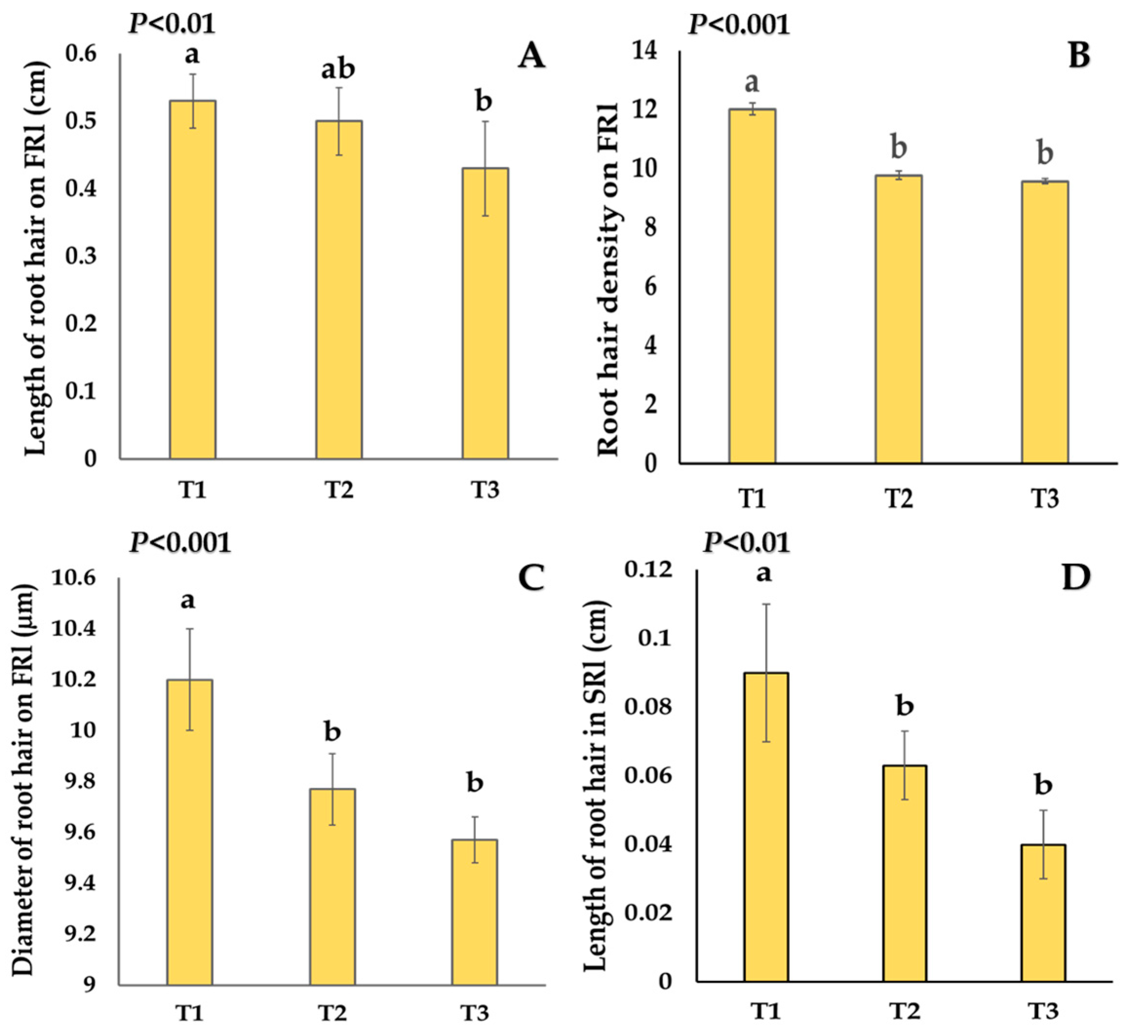
2.1. Effects of Salt Stress and Salicylic Acid on Shoot and Root Traits

The number of live leaves per plant displayed a gradually diminishing trend from T1 to T3 (p < 0.001, Table 1, Figure 1A) due to premature senescence. This early senescence led to the significant (p < 0.001) upsurge in both death of leaves (Figure 1B) and visual injury scores under T2 and T3 compared to control (Figure 1C). Salinity stress reduced the SPAD value significantly (p < 0.001, Table 1, Figure 1D), which was further accelerated significantly (p < 0.001, Table 1) under T3 (Table 1, Figure 1D). As an inhibitor of biomass accumulation, both T2 and T3 reduced the shoot dry weight by 33.8% and 84.6%, respectively, (Figure 1E), and root dry weight by 33.3% and 55.5%, respectively, (Figure 1F), compared to T1.
Salinity stress (T2) significantly reduced the diameter of individual root hairs and the number of root hairs formed per unit area (density) of first-order lateral roots (Figure 2B,C). This stress also reduced the length of the root hairs on second-order lateral roots (Figure 2D). By contrast, the length of root hairs of first-order lateral roots was not significantly altered by T2, although it was reduced by T3 (p < 0.05, Figure 2A).
The root hair length on first-order lateral roots (RHlf), which influences nutrient and water uptake, ranged from 0.35 cm (M-205×M-232) to 0.63 cm (M-245), indicating variation in root hair extension (Table 2). Salinity stress reduced the root elongation and expansion by significantly reducing the length and diameter of the main axis (p < 0.001, Table 2, Figure 3A,D), the length of first-order lateral roots (p < 0.01, Table 1, Figure 3C), and the length and diameter of second-order lateral roots (p < 0.001, Table 1, Figure 3B,E) compared to control. The application of salicylic acid (T3) in addition to salt significantly (p < 0.001, Table 1) shortened the length of the main axis, while the other lateral root traits remained statistically similar to T2 (Figure 3). The estimated root surface area (RSA) decreased significantly (p < 0.001, Table 1) to 155.4 cm2 under salt stress (T2) compared to 338.4 cm2 in control (T1) (Figure 3F). The addition of 0.1 mM of salicylic acid with salt (T3) failed to alleviate this stress, rather significantly (p < 0.001, Table 1) reducing the estimated root surface area to 76.8 cm2 compared to T2 (Figure 3F).

2.2. Genotypic Variations

The genotype M-232×M-223 reported the maximum estimated RSA values of 421 cm2, whereas the genotype M-245×M-206 had the minimum estimated RSA value of 55.8 cm2, thus showing considerable genetic differences in root absorption capacity (Table 2). In case of T×G interaction, the F3 genotype M-232×M-223 displayed the highest estimated RSA under T1; however, under T2 and T3 it significantly reduced (Figure S1). Only in the genotype M-206 did the estimated RSA increase under salinity stress (Figure S1). The estimated RSA did not decline in the genotypes M-206×M-232, M-206, or M-245 under T2, but it decreased significantly under T3 (Figure S1). The highest shoot dry weight was estimated to be 0.98 mg in the genotype M-245 and the root dry weight was maximum 0.137 mg in the genotype M-206, indicating genotypic variation in biomass accumulation (Table 2).

2.3. Trait Associations

The first two principal components explained 100% of the data variation (Table S1) for the PCA conducted with treatments. The PC1 had the highest contribution, explaining 93.1% of the variation, with strong positive coefficients for most of the traits (Figure 4A). The PC1 scores were highly significant (p < 0.001, Table S1) for treatments and distinguished T1 from T3 for higher positive coefficients of the traits, e.g., estimated root surface area, dry weight of roots, length of first-order lateral roots, and lower negative coefficients of the visual injury score (Figure 4A).
In another PCA with genotypic means, the first five PCs were identified more than unity and explained 80.7% of the data variation (Table S2). PC1 clearly separated the genotypes M-245, M-232×M-223, and M-205×M-245 from M-223×M-206, M-245×M-206, and M-223×M-205 for their higher PC scores and higher and positive coefficients of the traits, e.g., estimated root surface area, dry weight of shoots, length of main axis, and lower negative coefficients of the traits—visual injury score and number of dead leaves (Figure 4B). The estimated root surface had a significant (p < 0.001) positive correlation with the traits, e.g., length of main axis, diameter of main axis, length of first- and second-order lateral roots, diameter of second-order lateral roots, and dry weight of shoots and roots (Table S3).

3. Discussion

Plants’ reactions to salinity stress lead to a reduction in shoot growth, which occurs in two distinct phases. The first phase is comparatively shorter and involves a rapid increase in the external osmotic pressure. The second phase is comparatively slower and culminates in the accumulation of Na+ in the leaves [36]. The accumulation of salt in plant tissues often varies between roots and shoots. To better understand the basis of salinity tolerance, it is important to understand the degree of modifications in the main axis, lateral roots, and root hairs under salinity stress. In this section, the key findings of this study have been discussed.

3.1. Salinity Stress Disturbed Shoot Growth and Photosynthetic Capacity

Since salt stress prevents proper cell growth and development and hinders the uptake and transfer of water and nutrients, growth is inhibited as a consequence [37]. In this study, the significant reduction in the number of leaves (Figure 1A) and upsurge in the number of dead leaves (Figure 1B) under 8 dSm−1 salt stress (T2) compared to control (T1) were associated with early leaf maturation and senescence in plants, triggered by toxic Na+ accumulation in cell membranes [38]. This result corresponds with the observations of Azari et al. [39] in Brassica plants.
In previous studies, salinity stress has significantly reduced the chlorophyll content [40,41] and shoot dry weight [40,42] in Brassica genotypes. In this study, the photosynthetic efficiency (Figure 1D) and shoot biomass (Figure 1E) were reduced in T2 compared to T1—indicating the perturbation of the photosystem (thylakoid membranes) and carbon assimilation (by disturbing the RUBISCO activity)—ultimately leading to a reduced chlorophyll content and shoot biomass, respectively [43,44,45,46]. The overall leaf and shoot growth and chlorophyll content were comparatively better in the genotype M-245 than the other genotypes (Table 2). Therefore, the genotype M-245 could be further exploited in future breeding programs for salinity tolerance.

3.2. Salinity Stress Retarded the Development of Roots and New Root Hair Formation

Plant roots are not only primarily responsible for nutrient and water uptake, but they also confront any stress signals immediately, which ultimately triggers genetic responses to combat stress [47]. The elevated salinity impacts the root system through two primary modifications: firstly, it disrupts the root’s absorption capacity, and secondly, it alters the root’s dynamics [48]. Roots display dynamic morphological plasticity and play a substantial role in tolerance to numerous edaphic stresses [49,50]. Long-term salinity stress causes the excess lignification of the root tip area, which makes the root tip harder, ultimately ceasing root growth [51,52], and as a result, root branches become shorter in length. In this study, the elongation and girth of the main axis (Figure 3A,D) and laterals (Figure 3B,C) diminished significantly, while the root dry weight (Figure 1F) declined by 33.3% under T2 compared to T1, indicating clear retardation of root elongation and enlargement [53] due to salinity. By contrast, Sun et al. [54] and Robin et al. [55] found that higher salinity positively affected root growth and new root formation in Tamarix chinensis and wheat, respectively, indicating that a number of different factors including genotypic responses alter root growth under salinity stress.
Root hairs are minute, cylindrical outgrowths of root epidermal cells that increase the absorptive area of roots and immediately sense any stress near their vicinity [56]. Minimizing root hair density and growth is considered an early adaptive mechanism of plants to reduce toxic ion (Na+) uptake for mitigating the damage caused by forthcoming stress [57]. This study showed that the density of root hair on first-order lateral roots (Figure 2B) and the length of root hair on second-order lateral roots (Figure 2D) decreased by 19% and 30%, respectively, in T2 compared to T1, as a strategy to overcome the over-absorption of Na+ through the vascular system. By considering the responses of root hair as a model, these findings may contribute to a better understanding of cell responses to salinity stress.

3.3. Salinity Stress Significantly Altered Estimated Root Surface Area (RSA)

NaCl-induced estimated root surface area reductions were evident in wheat [55], faba beans (Vicia faba) [58], and radish [59]. In this study, a significant reduction of 54% in the estimated root surface area under T2 was attributed to the reduction in the diameter of the main axis and second-order lateral roots and the length of first-order lateral roots (Figure 3F, Table S3). Moreover, as a predominant contributor of surface area [19], the density of root hairs was also reduced by 18.7% under T2 compared to T1 (Figure 2B). By contrast, a study by Arif et al. [16] demonstrated that salinity imposition for a one-week duration in the reproductive phase increased the estimated RSA by 20% by enhancing the RSA-contributing parameters in two rapeseed varieties. The reason behind this discrepancy could be the age of the plants when the stress was imposed. In this study, the stress treatments were applied in the early vegetative phase (21 days of age) of the plants, whereas Arif et al. [16] imposed the stress treatments in the reproductive phase of the plant (at 55 days of age). Therefore, it could be hypothesized that plants might exhibit growth stage-specific root plasticity to positively modify their root dynamics to withstand prolonged stress. During the very early vegetative period, plant roots pass through the developing stage, which is why salt stress drastically reduces cell division and expansion, leading to a reduced estimated root surface area. On the contrary, with the advancement of age, the root system sees robust growth, which is capable of compensating for improper growth and development under salt stress.

3.4. Salicylic Acid (SA) Acted as Stressor in Combination with NaCl upon Vascular Uptake

In previous studies conducted on maize [60], wheat [61], chamomile [62], and soybeans [63], SA demonstrated a stress-alleviating role and promoted shoot and root growth. For instance, shoot and root growth was increased by 20% and 45%, respectively, in soybean plants when treated with different levels of exogenous SA. In the present study, SA was added simultaneously with NaCl to the nutrient solution to absorb and translocate through the vascular system of B. napus plants. The shoot and root traits that were greatly altered (p < 0.05) under salinity stress (T2) did not show a notable revival after SA was applied with salt (T3); rather, some of the variables—number of leaves (by 18.5%, Figure 1A), chlorophyll content (by 38.8%, Figure 1D), diameter of main axis (by 15%, Figure 3D), and estimated RSA (by 23%, Figure 3F)—further decreased compared to T2 as a reinforcement of stress, which opposes the previous findings in different crops. SA applied with NaCl might have induced oxidative stress in the plants and made the shoot and root traits deteriorate [64,65,66]. This oxidative damage to plant tissues gradually hampered photosynthetic activity (Figure 1D) and caused the early senescence of leaves, which ultimately resulted in leaf death and a poor visual appearance (Figure 1C) [33,67,68,69].
In this study, 0.1 mM of SA with 8 dSm−1 of NaCl reduced both the length and diameter of secondary roots by 35.8% and 27.7%, respectively, compared to control (Figure 3B,E), suggesting that this particular level of SA (0.1 mM), along with 8 dSm−1 of NaCl, might have disrupted auxin (a regulator of root primordia development) transportation in roots via PIN protein phosphorylation through the activation of PID—a serine/threonine-specific protein kinase [70]. As auxin transportation might have hampered, the elongation and girth of secondary roots were retarded. This particular level of SA (0.1 mM) alone was also proven to be a retardant of adventitious root growth in Arabidopsis thaliana [31]. By contrast, the similar dose and procedure of application of SA in Azalea increased the proportion of lateral roots [71]. The individual plant species and specific physiological phases of the plant also regulate the role of SA under stress [35]. Therefore, SA might act differently if the experiment were to be conducted in the reproductive phase rather than in the early vegetative phase. Experiments with multiple doses (low to medium) of SA and in different growth phases of the plants could be recommended to elucidate the comprehensive uptake-related responses of Brassica plants.

3.5. Trait Associations Clearly Showed Relationships Among the Traits

Associations among root traits provide the basis of the understanding of the developmental process and regulatory mechanisms that control primary and secondary root formation in plants [72,73]. The PCA distinctly separated the responses of 15 rapeseed genotypes and three treatments, highlighting the importance of different traits in determining their performance. In the case of PCA with treatments, strong associations with traits like the lateral root length, root hair density, root dry weight, and shoot dry weight, along with estimated surface area, indicated that a breeding strategy targeting those traits would be judicious to increase the tolerance of rapeseed under salt stress (Figure 4A). Similar findings were also observed in wheat [19,74]. The separation of genotypes along PC1 and PC2 suggested variability in stress tolerance and biomass allocation, which could be valuable for selecting the individual genotypes. Genotypes such as M-232×M-223 and M-245 clustered closely with desirable traits, suggesting their comparatively better root growth under salinity stress (Figure 4B) and their potential to be exploited for salt resilience. These findings can serve as a foundation for future research aimed at developing salinity-tolerant rapeseed varieties by selecting the desired contributing traits to the root surface area.
As the salinity levels increase in coastal regions around the world, this study provides potential avenues for further rapeseed breeding for salt stress resilience by delving into particular root component responses under salinity and a combination of salt and SA. This will allow us to visualize the changes in the root system during stress and to develop more sustainable genotypes with better root plasticity under stress. This study also challenged the conventional assumption that SA only acts as a ‘therapeutic agent’ against abiotic stress. Therefore, future studies targeting the dose, tissue, and species-specific effects of SA depending on the developmental stage and method of application are recommended.
Despite highlighting some novel findings about dual stressor effects on the root and shoot traits of B. napus, this study focused only on limited areas. Firstly, the experiment considered a single dose of SA and was performed only during the vegetative stage of plants. Secondly, the stress period lasted only for 7 days, which means that there is a lack of proper visualization of the stress response in the later stages, e.g., the flowering, pod bearing, and maturation periods. Finally, the study lacks molecular investigations, such as gene expression profiling or hormonal assays regarding roots’ architectural alterations in response to salt and salicylic acid.

4. Materials and Methods

4.1. Culture of Plants and Management of Hydroponic System

The experiment was conducted in the culture room of the Department of Genetics and Plant Breeding (GPB), Bangladesh Agricultural University (BAU). The seeds of five parental genotypes and ten F3 segregants were collected from the existing germplasm of the Department of GPB, BAU (Table 3).
Seeds were germinated in Petri dishes in a plant culture room. The temperature was maintained at 22 ± 2 °C, relative humidity was 70%, and the day–night ratio was 16:8 h in the culture room [16,75]. Uniform, healthy, dark green seedlings were transferred to a hydroponic culture media one week after germination. Plants were provided with a modified Hoagland solution—1 mM NH4NO3, 0.6 mM MgCl2 × H2O, 0.6 mM NaH2PO4 × H2O, 0.3 mM CaCl2 × H2O, 0.3 mM K2SO4, 50 μM H3BO3, 90 μM Fe-EDTA, 9 μM MnSO4 × 4H2O, 0.7 μM ZnSO4 × 7H2O, 0.3 μM CuSO4 × 5H2O, and 0.1 μM NaMoO4 × 2H2O—dissolved thoroughly in water [16,55,75]. The pH of the hydroponic system was checked at regular intervals and maintained between 5.8 and 6.3 by adding 1 M of NaOH solution as a buffering agent. Treatments were imposed on plants at 21 days of age and continued for 7 days. A completely randomized design (CRD) was followed, consisting of three treatments and three replications for each genotype. The treatments were control (T1), 8 dSm-1 salt (NaCl—EMSURE-ACS, ISO, Reag. Ph Eur) stress (T2), and 8 dSm−1 salt + 0.1 mM salicylic acid (T3), which were imposed after placing the plants in a perforated cork sheet in such a way that the roots of young plants touched the underneath solution (10 L bucket). To prepare the 8 dSm−1 salt solution (T2), 51.2 g of NaCl was mixed thoroughly with 10 L of tap water. Additionally, 0.138 g of salicylic acid was added into this solution to obtain the 8 dSm−1 salt + 0.1 mM SA solution (T3). Aquarium air pumps were provided to make sure that the hydroponics system received the optimal oxygen supply. The solution was discarded and refreshed once per week.

4.2. Data Collection

The chlorophyll content of leaves was measured in the standing plants with a chlorophyll meter (SPAD-502 PLUS, 3 V; 200 Mw; Konica Minolta Inc., Osaka, Japan) after three days of imposing treatments. The parameters of shoots and roots (Text S1) were measured and calculated at the destructive harvest in 28-day-old plants and 7 days after treatment imposition. Visual injury levels were scored on a 1–9 scale (low score indicates healthy plant) considering the appearance of the plant (Table 4).
To properly visualize the root components under a microscope, the test samples of roots were stained with 0.5% acetocarmine (prepared with 45% glacial acetic acid) solution. The length and diameter of the main axis and the length of lateral roots were measured using a centimeter ruler, and the diameter of first- and second-order lateral roots was measured with an ocular micrometer under a light microscope at 100× magnification. The density of roots was determined by counting on a marked scale area of 0.25 mm. The root hair length was determined by averaging four randomly chosen root hairs. Finally, the shoots and roots were separately wrapped and sealed in a paper bag and placed in an oven for 72 h to measure the dry weights.
The root surface area was estimated following the equation developed and modified by Robin et al. [55] and Arif et al. [16], respectively (Text S2)). The equation was as follows [16,55]:
Estimated root surface area = π Dm Lm (1 + a1 n1 π D1 L1 (1 + arh1 nrh1 π Drh1 Lrh1
+ a2 n2 πD2 L2 (1 + arh2 nrh2 π Drh2 Lrh2)))
Here, Dm and Lm denote the diameter and length of the main axis, respectively; Di and Li are the diameter and length of the ith-order lateral roots, respectively; Drhi and Lrhi are the diameter and length of the root hair of the ith-order lateral roots, respectively; ai is the percentage of the length of roots that bear ith-order lateral roots; arhi denotes the percentage of the length of ith-order lateral roots that bear root hairs; ni is the density of ith-order lateral roots; and nrhi denotes the density of the root hair of ith-order lateral roots.

4.3. Data Analysis

Data analysis was performed using Minitab (version 17) statistical software (Minitab Inc., State College, PA, USA). All the parameters were tested for normality by conducting Shapiro–Wilk tests and found to be normal [76]. Analysis of variance and Tukey’s pairwise comparison were performed to find out the significance levels of treatments, genotypes, and treatment × genotype interactions. Principal component analysis (PCA) was performed to establish associations among the most significant stress-responsive traits, and analysis of variance of the principal component (PC) scores of the first two principal components was carried out using the same model. Graphical illustrations were prepared using Microsoft Excel version 2016.

5. Conclusions

The responses of root and shoot traits under different stress make up an intricate process. This study dived into dual treatment responses that provided new insights for understanding how SA in association with NaCl modulates the root system architecture and estimated root surface area of five parental lines and ten F3 genotypes of B. napus. The results suggested that the estimated root surface area along with its contributing traits were retarded under both the T2 and T3. The addition of salicylic acid with NaCl (T3) to the nutrient solution failed to rescue the compromised plants; it rather turned out to be a stress accelerator, as the estimated root surface area decreased by 23% compared to under T2. This phenomenon gave rise to a new controversial role of SA in stress regulation in plants and needs further investigation. The F3 genotype M-232×M-223 and the parental genotype M-245 performed better than the others. The trait association of shoot and root traits revealed that the estimated root surface area had a strong association with the length of the main axis, first-order lateral roots, second-order lateral roots, the diameter of the main axis, and second-order lateral roots. Though there are previous documents about the stress alleviation capacity of SA, the results obtained in this study necessitate more intense future studies into this phenomenon, where SA has been taken up through the vascular pathway. The overall findings provide an improved understanding of salinity- and SA-induced modifications of the root system architecture in B. napus cultured in hydroponics that will add value to the research on salt-resilient rapeseed breeding programs.

Supplementary Materials

The following supporting information can be downloaded at: https://www.mdpi.com/article/10.3390/stresses5030048/s1. Figure S1: Root surface area (cm2) across 15 genotypes under three treatments. T1 = control, T2 = 8 dSm−1 salt stress and T3 = 8 dSm−1 salt + 0.1 mM salicylic acid. Vertical bars indicate standard error of mean; letters denote significant differences.; Table S1: Coefficients of principal components for shoot and root traits under 3 treatments; Table S2: Principal components with eigenvalue for shoot and root traits of 15 genotypes; Table S3: Pearson correlation coefficients among the shoot and root traits of 15 rapeseed genotypes; Text S1: Description about the parameters collected and Text S2: Breakdown of equation 1 for estimating the potential root surface area of component roots.

Author Contributions

Conceptualization, J.A. and A.H.K.R.; methodology, A.H.K.R., J.A. and R.S.; software, N.C. and J.A.; validation, N.C., J.A. and R.S.; formal analysis, N.C. and J.A.; investigation, A.H.K.R.; data curation, J.A. and R.S.; writing—original draft preparation, N.C. and J.A.; writing—review and editing, N.C. and A.H.K.R.; supervision, A.H.K.R. and J.N.; J.A. and N.C. contributed equally to this manuscript. All authors have read and agreed to the published version of the manuscript.

Funding

This research was funded by the Bangladesh Agricultural University Research Systems (Project no. 2023/110/BAU).

Data Availability Statement

The data are available upon reasonable request from the authors.

Acknowledgments

The authors acknowledge the National Science and Technology Fellowship (2022-23) from the Ministry of Science and Technology, Government of Bangladesh.

Conflicts of Interest

The authors declare no conflicts of interest.

Abbreviations

The following abbreviations are used in this manuscript:
ANOVAAnalysis of Variance
PCAPrincipal Component Analysis
RSAEstimated Root Surface Area
NaClSodium Chloride
SASalicylic Acid

References

  1. Dhankher, O.P.; Foyer, C.H. Climate resilient crops for improving global food security and safety. Plant Cell Environ. 2018, 41, 877–884. [Google Scholar] [CrossRef] [PubMed]
  2. Shahbaz, M.; Ashraf, M. Improving salinity tolerance in cereals. Crit. Rev. Plant Sci. 2013, 32, 237–249. [Google Scholar] [CrossRef]
  3. Ullah, A.; Bano, A.; Khan, N. Climate change and salinity effects on crops and chemical communication between plants and plant growth-promoting microorganisms under stress. Front. Sustain. Food Syst. 2021, 5, 618092. [Google Scholar] [CrossRef]
  4. Dasgupta, S.; Huq, M.; Khan, Z.H.; Murshed, M.; Ahmed, Z.; Mukherjee, N.; Khan, M.F.; Pandey, K. Cyclones in a changing climate: The case of Bangladesh. Clim. Dev. 2014, 6, 96–110. [Google Scholar] [CrossRef]
  5. Kabir, M.E.; Sarker, B.C.; Gosh, A.K.; Maiuddin, M.; Bell, R.W. Effect of sowing dates for wheat grown in excess water and salt-affected soils in southwestern coastal Bangladesh. J. Indian Soc. Coast. Agric. Res. 2019, 37, 51–59. [Google Scholar]
  6. Raihan, S. Economic Reforms and Agriculture in Bangladesh: Assessment of Impacts Using Economy-Wide Simulation Models; MPRA Paper; University Library of Munich: Munich, Germany, 2011; pp. 1–55. [Google Scholar]
  7. Chakroborty, N.; Kabir, M.A.; Hossen, M.M.; Afrin, J.; Islam, M.M.; Robin, A.H.K. Species-Specific Variation in Oil Content and Fatty Acid Profiles of Short-Duration Rapeseed and Mustard. ACS Food Sci. Technol. 2025, 5, 1082–1090. [Google Scholar] [CrossRef]
  8. Shen, J.; Liu, Y.; Wang, X.; Bai, J.; Lin, L.; Luo, F.; Zhong, H. A comprehensive review of health-benefiting components in rapeseed oil. Nutrients 2023, 15, 999. [Google Scholar] [CrossRef]
  9. Helal, M.M.U.; Islam, D.M.N.; Biswas, D.M.; Kadir, D.M.; Miah, D.M.N.H. Performance of rapeseed and mustard (Brassica sp.) Varieties/lines in north-east region (Sylhet) of Bangladesh. Sci. J. Seoul Sci. 2022, 4, 6–13. [Google Scholar]
  10. Shahzad, B.; Rehman, A.; Tanveer, M.; Wang, L.; Park, S.K.; Ali, A. Salt stress in Brassica: Effects, tolerance mechanisms, and management. J. Plant Growth Regul. 2022, 41, 781–795. [Google Scholar] [CrossRef]
  11. Liu, Y.; Ding, X.; Lv, Y.; Cheng, Y.; Li, C.; Yan, L.; Zou, X. Exogenous serotonin improves salt tolerance in rapeseed (Brassica napus L.) seedlings. Agronomy 2021, 11, 400. [Google Scholar] [CrossRef]
  12. Chakraborty, K.; Sairam, R.K.; Bhaduri, D. Effects of different levels of soil salinity on yield attributes, accumulation of nitrogen, and micronutrients in Brassica spp. J. Plant Nutr. 2015, 39, 1026–1037. [Google Scholar] [CrossRef]
  13. Munns, R.; Tester, M. Mechanisms of salinity tolerance. Annu. Rev. Plant Biol. 2008, 59, 651–681. [Google Scholar] [CrossRef]
  14. Miller, G.A.D.; Suzuki, N.; Ciftci-Yilmaz, S.; Mittler, R. Reactive oxygen species homeostasis and signaling during drought and salinity stresses. Plant Cell Environ. 2010, 33, 453–467. [Google Scholar] [CrossRef] [PubMed]
  15. Chaurasia, S.; Kumar, S.; Garg, S.; Pandey, R.; Anand, A.; Dikshit, H.K.; Bansal, R. Exploring the genetic variability for root traits in mung bean under salinity stress. Plant Physiol. Rep. 2024, 29, 651–659. [Google Scholar] [CrossRef]
  16. Arif, M.R.; Islam, M.T.; Robin, A.H.K. Salinity stress alters root morphology and root hair traits in Brassica napus. Plants 2019, 8, 192. [Google Scholar] [CrossRef]
  17. Hayat, S.; Maheshwari, P.; Wani, A.S.; Irfan, M.; Alyemeni, M.N.; Ahmad, A. Comparative effect of 28-homobrassinolide and salicylic acid in the amelioration of NaCl stress in Brassica juncea L. Plant Physiol. Biochem. 2012, 53, 61–68. [Google Scholar] [CrossRef]
  18. Khan, M.O.; Farooq, N.; Nawaz, M.A.; Fatima, S.; Islam, E.; Mukhtar, Z.; Ahmad, N. Evaluation of the salt tolerance potential of commercial Brassica cultivars. Commun. Soil Sci. Plant Anal. 2024, 55, 498–516. [Google Scholar] [CrossRef]
  19. Robin, A.H.K.; Uddin, M.J.; Bayazid, K.N. Polyethylene glycol (PEG)-treated hydroponic culture reduces length and diameter of root hairs of wheat varieties. Agronomy 2015, 5, 506–518. [Google Scholar] [CrossRef]
  20. Flowers, T.J.; Colmer, T.D. Salinity tolerance in halophytes. New Phytol. 2008, 179, 945–963. [Google Scholar] [CrossRef]
  21. El-Hawary, M.M.; Hashem, O.S.; Hasanuzzaman, M. Seed priming and foliar application with ascorbic acid and salicylic acid mitigate salt stress in wheat. Agronomy 2023, 13, 493. [Google Scholar] [CrossRef]
  22. Chakma, R.; Biswas, A.; Saekong, P.; Ullah, H.; Datta, A. Foliar application and seed priming of salicylic acid affect growth, fruit yield, and quality of grape tomato under drought stress. Sci. Hortic. 2021, 280, 109904. [Google Scholar] [CrossRef]
  23. Ahmad, I.; Basra, S.M.A.; Wahid, A. Exogenous application of ascorbic acid, salicylic acid and hydrogen peroxide improves the productivity of hybrid maize at low temperature stress. Int. J. Agric. Biol. 2014, 16, 825–830. [Google Scholar]
  24. Peng, Y.; Yang, J.; Li, X.; Zhang, Y. Salicylic acid: Biosynthesis and signaling. Annu. Rev. Plant Biol. 2021, 72, 761–791. [Google Scholar] [CrossRef]
  25. Pirasteh-Anosheh, H.; Ranjbar, G.; Hasanuzzaman, M.; Khanna, K.; Bhardwaj, R.; Ahmad, P. Salicylic acid-mediated regulation of morpho-physiological and yield attributes of wheat and barley plants in deferring salinity stress. J. Plant Growth Regul. 2022, 41, 1291–1303. [Google Scholar] [CrossRef]
  26. Hayat, Q.; Hayat, S.; Irfan, M.; Ahmad, A. Effect of exogenous salicylic acid under changing environment: A review. Environ. Exp. Bot. 2010, 68, 14–25. [Google Scholar] [CrossRef]
  27. Singh, P.K.; Chaturvedi, V.K.; Bose, B. Effects of salicylic acid on seedling growth and nitrogen metabolism in cucumber (Cucumis sativus L.). J. Stress Physiol. Biochem. 2010, 6, 102–113. [Google Scholar]
  28. Pour, A.P.; Farahbakhsh, H.; Saffari, M.; Keramat, B. Effects of seed priming on germination and seedling growth under salinity stress in fenugreek. Int. J. Agric. Crop Sci. 2012, 4, 779–786. [Google Scholar]
  29. Bouallègue, A.; Souissi, F.; Nouairi, I.; Souibgui, M.; Abbes, Z.; Mhadhbi, H. Salicylic acid and hydrogen peroxide pretreatments alleviate salt stress in faba bean (Vicia faba) seeds during germination. Seed Sci. Technol. 2017, 45, 675–690. [Google Scholar] [CrossRef]
  30. Fariduddin, Q.; Hayat, S.; Ahmad, A. Salicylic acid influences net photosynthetic rate, carboxylation efficiency, nitrate reductase activity, and seed yield in Brassica juncea. Photosynthetica 2003, 41, 281–284. [Google Scholar] [CrossRef]
  31. Pasternak, T.; Groot, E.P.; Kazantsev, F.V.; Teale, W.; Omelyanchuk, N.; Kovrizhnykh, V.; Palme, K.; Mironova, V.V. Salicylic acid affects root meristem patterning via auxin distribution in a concentration-dependent manner. Plant Physiol. 2019, 180, 1725–1739. [Google Scholar] [CrossRef] [PubMed]
  32. Oliveira, V.K.N.; Lima, G.D.; Soares, M.D.M.; Soares, L.D.A.; Gheyi, H.R.; Silva, A.D.; Fernandes, P.D. Salicylic acid does not mitigate salt stress on the morphophysiology and production of hydroponic melon. Braz. J. Biol. 2022, 82, 262664. [Google Scholar] [CrossRef]
  33. Bagautdinova, Z.Z.; Omelyanchuk, N.; Tyapkin, A.V.; Kovrizhnykh, V.V.; Lavrekha, V.V.; Zemlyanskaya, E.V. Salicylic acid in root growth and development. Int. J. Mol. Sci. 2022, 23, 2228. [Google Scholar] [CrossRef] [PubMed]
  34. Christianson, M.L.; Duffy, S.H. Dose-dependent effect of salicylates in a moss, Funaria hygrometrica. J. Plant Growth Regul. 2002, 21, 200–208. [Google Scholar] [CrossRef]
  35. Arfan, M.; Athar, H.R.; Ashraf, M. Does exogenous application of salicylic acid through the rooting medium modulate growth and photosynthetic capacity in two differently adapted spring wheat cultivars under salt stress? J. Plant Physiol. 2007, 164, 685–694. [Google Scholar] [CrossRef] [PubMed]
  36. Munns, R. Comparative physiology of salt and water stress. Plant Cell Environ. 2002, 25, 239–250. [Google Scholar] [CrossRef]
  37. Ashraf, M.A.; Ashraf, M.; Ali, Q. Response of two genetically diverse wheat cultivars to salt stress at different growth stages: Leaf lipid peroxidation and phenolic contents. Pak. J. Bot. 2010, 42, 559–565. [Google Scholar]
  38. Saddiq, M.S.; Iqbal, S.; Hafeez, M.B.; Ibrahim, A.M.; Raza, A.; Fatima, E.M.; Ciarmiello, L.F. Effect of salinity stress on physiological changes in winter and spring wheat. Agronomy 2021, 11, 1193. [Google Scholar] [CrossRef]
  39. Azari, A.; Sanavi, S.M.; Askari, H.; Ghanati, F.; Naji, A.M.; Alizadeh, B. Effect of salt stress on morphological and physiological traits of two species of rapeseed (Brassica napus and B. rapa). Iran J. Crop Sci. 2012, 14, 121–135. [Google Scholar]
  40. Jan, S.A.; Shinwari, Z.K.; Rabbani, M.A. Agro-morphological and physiological responses of Brassica rapa ecotypes to salt stress. Pak. J. Bot. 2016, 48, 1379–1384. [Google Scholar]
  41. Singh, M.P.; Pandey, S.K.; Singh, M.; Ram, P.C.; Singh, B.B. Photosynthesis, transpiration, stomatal conductance and leaf chlorophyll content in mustard genotypes grown under sodic conditions. Photosynthetica 1990, 24, 623–627. [Google Scholar]
  42. Shah, S.S.; Mohammad, F.; Shafi, M.; Bakht, J.; Zhou, W. Effects of cadmium and salinity on growth and photosynthesis parameters of Brassica species. Pak. J. Bot. 2011, 43, 333–340. [Google Scholar]
  43. Sudhir, P.; Murthy, S.D.S. Effects of salt stress on basic processes of photosynthesis. Photosynthetica 2004, 42, 481–486. [Google Scholar] [CrossRef]
  44. Tseng, M.J.; Liu, C.W.; Yiu, J.C. Enhanced tolerance to sulfur dioxide and salt stress of transgenic Chinese cabbage plants expressing both superoxide dismutase and catalase in chloroplasts. Plant Physiol. Biochem. 2007, 45, 822–833. [Google Scholar] [CrossRef] [PubMed]
  45. Galmés, J.; Molins, A.; Flexas, J.; Conesa, M.À. Coordination between leaf CO2; diffusion and Rubisco properties allows maximizing photosynthetic efficiency in Limonium species. Plant Cell Environ. 2017, 40, 2081–2094. [Google Scholar] [CrossRef]
  46. Hussain, T.; Huchzermeyer, B.; Koyro, H.W.; Khan, M.A. Linkage between leaf development and photosynthetic response at hyperosmotic salinity in the C4 grass Panicum antidotale. Flora 2019, 256, 52–60. [Google Scholar] [CrossRef]
  47. Lynch, D.R.; Foroud, N.; Kozub, G.C.; Fames, B.C. The effect of moisture stress at three growth stages on the yield, components of yield and processing quality of eight potato varieties. Am. Potato J. 1995, 72, 375–385. [Google Scholar] [CrossRef]
  48. Hu, Y.; Schmidhalter, U. Drought and salinity: A comparison of their effects on mineral nutrition of plants. J. Plant Nutr. Soil Sci. 2005, 168, 541–549. [Google Scholar] [CrossRef]
  49. Bell, D.L.; Sultan, S.E. Dynamic phenotypic plasticity for root growth in Polygonum: A comparative study. Am. J. Bot. 1999, 86, 807–819. [Google Scholar] [CrossRef]
  50. Kano, M.; Inukai, Y.; Kitano, H.; Yamauchi, A. Root plasticity as the key root trait for adaptation to various intensities of drought stress in rice. Plant Soil 2011, 342, 117–128. [Google Scholar] [CrossRef]
  51. Neumann, P.M.; Azaizeh, H.; Leon, D. Hardening of root cell-walls—A growth-inhibitory response to salinity stress. Plant Cell Environ. 1994, 17, 303–309. [Google Scholar] [CrossRef]
  52. Neves, G.Y.S.; Marchiosi, R.; Ferrarese, M.L.L.; Siqueira-Soares, R.C.; Ferrarese, O. Root growth inhibition and lignification induced by salt stress in soybean. J. Agron. Crop Sci. 2010, 196, 467–473. [Google Scholar] [CrossRef]
  53. Zou, Y.; Zhang, Y.; Testerink, C. Root dynamic growth strategies in response to salinity. Plant Cell Environ. 2022, 45, 695–704. [Google Scholar] [CrossRef]
  54. Sun, J.; Xia, J.; Shao, P.; Ma, J.; Gao, F.; Lang, Y.; Li, C. Response of the fine root morphological and chemical traits of Tamarix chinensis to water and salt changes in coastal wetlands of the Yellow River Delta. Front. Plant Sci. 2022, 13, 952830. [Google Scholar] [CrossRef]
  55. Robin, A.H.K.; Matthew, C.; Uddin, M.J.; Bayazid, K.N. Salinity-induced reduction in root surface area and changes in major root and shoot traits at the phytomer level in wheat. J. Exp. Bot. 2016, 67, 3719–3729. [Google Scholar] [CrossRef]
  56. Wang, Y.; Zhang, W.; Li, K.; Sun, F.; Han, C.; Wang, Y.; Li, X. Salt-induced plasticity of root hair development is caused by ion disequilibrium in Arabidopsis thaliana. J. Plant Res. 2008, 121, 87–96. [Google Scholar] [CrossRef]
  57. Jin, D.; Li, S.; Li, Z.; Yang, L.; Han, X.; Hu, Y.; Jiang, Y. Arabidopsis ABRE-binding factors modulate salinity-induced inhibition of root hair growth by interacting with and suppressing RHD6. Plant Sci. 2023, 332, 111728. [Google Scholar] [CrossRef]
  58. Srinivasarao, C.H.; Benzioni, A.; Eshel, A.; Waisel, Y. Effects of salinity on root morphology and nutrient acquisition by faba beans (Vicia faba L.). J. Indian Soc. Soil Sci. 2004, 52, 184–191. [Google Scholar]
  59. Waisel, Y.; Breckle, S.W. Differences in responses of various radish roots to salinity. Plant Soil 1987, 104, 191–194. [Google Scholar] [CrossRef]
  60. Gunes, A.; Inal, A.; Alpaslan, M.; Eraslan, F.; Bagci, E.G.; Cicek, N. Salicylic acid induced changes on some physiological parameters symptomatic for oxidative stress and mineral nutrition in maize (Zea mays L.) grown under salinity. J. Plant Physiol. 2007, 164, 728–736. [Google Scholar] [CrossRef] [PubMed]
  61. Shakirova, F.M.; Sakhabutdinova, A.R.; Bezrukova, M.V.; Fatkhutdinova, R.A.; Fatkhutdinova, D.R. Changes in the hormonal status of wheat seedlings induced by salicylic acid and salinity. Plant Sci. 2003, 164, 317–322. [Google Scholar] [CrossRef]
  62. Kováčik, J.; Grúz, J.; Bačkor, M.; Strnad, M.; Repčák, M. Salicylic acid-induced changes to growth and phenolic metabolism in Matricaria chamomilla plants. Plant Cell Rep. 2009, 28, 135–143. [Google Scholar] [CrossRef]
  63. Gutiérrez-Coronado, M.A.; Trejo-López, C.; Larqué-Saavedra, A. Effects of salicylic acid on the growth of roots and shoots in soybean. Plant Physiol. Biochem. 1998, 36, 563–565. [Google Scholar] [CrossRef]
  64. Luo, J.P.; Jiang, S.T.; Pan, L.J. Enhanced somatic embryogenesis by salicylic acid of Astragalus adsurgens Pall.: Relationship with H2O2 production and H2O2 metabolizing enzyme activities. Plant Sci. 2001, 161, 125–132. [Google Scholar] [CrossRef]
  65. Kawano, T.; Muto, S. Mechanism of peroxidase actions for salicylic acid-induced generation of active oxygen species and an increase in cytosolic calcium in tobacco cell suspension culture. J. Exp. Bot. 2000, 51, 685–693. [Google Scholar] [CrossRef] [PubMed]
  66. Durner, J.; Klessig, D.F. Inhibition of ascorbate peroxidase by salicylic acid and 2,6-dichloroisonicotinic acid, two inducers of plant defense responses. Proc. Natl. Acad. Sci. USA 1995, 92, 11312–11316. [Google Scholar] [CrossRef]
  67. Ashraf, M. Effect of salicylic acid applied through rooting medium on drought tolerance of wheat. Pak. J. Bot. 2006, 38, 1127–1136. [Google Scholar]
  68. Moharekar, S.T.; Lokhande, S.D.; Hara, T.; Tanaka, R.; Tanaka, A.; Chavan, P.D. Effect of salicylic acid on chlorophyll and carotenoid contents of wheat and moong seedlings. Photosynthetica 2003, 41, 315–317. [Google Scholar] [CrossRef]
  69. Anandhi, S.; Ramanujam, M.P. Effect of salicylic acid on black gram (Vigna mungo) cultivars. Indian J. Plant Physiol. 1997, 2, 138–141. [Google Scholar]
  70. Michniewicz, M.; Zago, M.K.; Abas, L.; Weijers, D.; Schweighofer, A.; Meskiene, I.; Heisler, M.G.; Ohno, C.; Zhang, J.; Huang, F. Antagonistic regulation of PIN phosphorylation by PP2A and PINOID directs auxin flux. Cell 2007, 130, 1044–1056. [Google Scholar] [CrossRef] [PubMed]
  71. Hou, P.C.; Lin, K.H.; Huang, Y.J.; Wu, C.W.; Chang, Y.S. Evaluation of vegetation indices and plant growth regulator use on the rooting of azalea cuttings. Hortic. Bras. 2020, 38, 153–159. [Google Scholar] [CrossRef]
  72. Bellini, C.; Pacurar, D.I.; Perrone, I. Adventitious roots and lateral roots: Similarities and differences. Annu. Rev. Plant Biol. 2014, 65, 639–666. [Google Scholar] [CrossRef]
  73. Gruber, B.D.; Giehl, R.F.H.; Friedel, S.; von Wirén, N. Plasticity of the Arabidopsis root system under nutrient deficiencies. Plant Physiol. 2013, 163, 161–179. [Google Scholar] [CrossRef] [PubMed]
  74. Narayanan, S.; Mohan, A.; Gill, K.S.; Prasad, P.V. Variability of root traits in spring wheat germplasm. PLoS ONE 2014, 9, e100317. [Google Scholar] [CrossRef] [PubMed]
  75. Robin, A.H.K.; Ghosh, S.; Shahed, M.A. PEG-induced osmotic stress alters root morphology and root hair traits in wheat genotypes. Plants 2021, 10, 1042. [Google Scholar] [CrossRef] [PubMed]
  76. Shapiro, S.S.; Wilk, M.B. An analysis of variance test for normality (complete samples). Biometrika 1965, 52, 591–611. [Google Scholar] [CrossRef]
Figure 1. Responses of shoot traits and root dry weight of rapeseed genotypes to three different treatments: T1 = control, T2 = 8 dSm−1 salt stress, and T3 = 8 dSm−1 salt + 0.1 mM salicylic acid. (A) No. of leaves, (B) no. of leaf deaths, (C) visual injury scores, (D) chlorophyll content (SPAD unit), (E) shoot dry weight (g), and (F) root dry weight (g). Each data point (yellow bar) represents the average of 45 observations. Vertical bars indicate standard error; different letters denote significant differences. Total sample number, n = 135. As the visual injury score (C) was non-parametric data, boxplot was included for each treatment.
Stresses 05 00048 g001
Figure 2. Responses of root hair traits of rapeseed genotypes to three different treatments: T1 = control, T2 = 8 dSm−1 salt stress, and T3 = 8 dSm−1 salt + 0.1 mM salicylic acid. (A) Length of root hair in FRl (first-order lateral roots) (cm), (B) length of root hair in SRl (second-order lateral roots) (cm) (C) diameter of root hair in FRl (first-order lateral roots) (µm), and (D) root hair density in FRl. Each data point (yellow bar) represents the average of 45 observations. Vertical bars indicate standard error; different letters denote significant differences. Total sample number, n = 135.
Stresses 05 00048 g002
Figure 3. Responses of main axis, laterals, and estimated root surface area of rapeseed genotypes to three different treatments: T1 = control, T2 = 8 dSm−1 salt stress, and T3 = 8 dSm−1 salt + 0.1 mM salicylic acid. (A) Length of main axis (cm), (B) diameter of main axis (cm), (C) length of FRl (first-order lateral roots) (cm), (D) length of SRl (second-order lateral roots) (cm), (E) diameter of SRl (second-order lateral roots) (cm), and (F) root surface area (cm2). Each data point (yellow bar) represents an average of 45 observations. Vertical bars indicate standard error; different letters denote significant differences. Total sample number, n = 135.
Stresses 05 00048 g003
Figure 4. Biplot for rapeseed genotypes under treatments T1 = control, T2 = 8 dSm−1 salt stress, and T3 = 8 dSm−1 salt + 0.1 mM salicylic acid for the first two principal components, PC1 and PC2. Blue color represents the traits. (A) treatment variation. (B) genotypic variation. Blue color represents the abbreviations of variables. NL = Number of leaves, NDL = no. of leaf deaths, VIS = visual injury score, MAL = length of main axis (cm), Mad = diameter of main axis (cm), FRl = length of first-order lateral roots (cm), FRd = diameter of first-order lateral roots (mm), RHDf = density of root hair of first-order lateral roots, RHLf = length of root hair of first-order lateral roots (cm), RHdf = diameter of root hair of first-order lateral roots (µm), SRl = length of second-order lateral roots (cm), SRd = diameter of second-order lateral roots (mm), RHDs = density of root hair of second-order lateral roots, RHls = length of root hair of second-order lateral roots (cm), RHds = diameter of root hair of second-order lateral roots (µm), DWS = shoot dry weight (g), DWR = root dry weight (g), Cc = chlorophyll content (SPAD unit), and RSA = estimated root surface area (cm2).
Stresses 05 00048 g004
Table 1. Analysis of variance (mean squares) for shoot and root traits of fifteen rapeseed genotypes, with the level of significance grown under salt and salicylic acid-induced hydroponic culture. Total sample number, n = 135.
Source of Variation df FS NL NDL MAL MAd FRl NFR FRd RHDf RHlf RHdf
Genotype (G)140.64 ***33.85 ***2.49 NS135.63 **0.34 ***41.67 NS4.36 ***0.045 ***30.18 ***0.092 ***4.55 ***
Treatment (T)20.02 NS100.02 ***214.03 ***515.54 ***1.23 ***203.92 **2.69 NS0.033 NS21.3 ***0.12 **4.54 ***
G × T280.19 NS5.25 **1.81 *109.23 ***0.75 ***63.34 ***2.16 ***0.014 ***7.58 ***0.033 **0.38 ***
Error900.192.811.0829.340.01221.700.780.0061.860.0160.12
SRlNSRSRdRHDsRHlsRHdsDWSDWRCcRSA
Genotype (T)1423.16 ***2.39 *0.008 ***43.72 ***0.101 ***3.082 ***0.53 ***0.008 ***142.74 ***132,827 ***
Treatment (T)284.7 ***2.29 NS0.021 ***1.92 NS0.1 **0.95 NS1.48 ***0.031 ***3476.62 ***811,345 ***
G × T2824.1 ***1.54 NS0.003 ***3.25 **0.035 **1.12 NS0.145 ***0.0042 ***47.50 ***98,839 ***
Error905.131.040.00071.3480.0180.960.0370.001211.6323,613
*, **, and *** indicate significant at ≤5%, ≤1%, and ≤0.1% levels of probability, respectively. NS: non-significant. Here, FS = flower status, NL = number of leaves, NDL = no. of leaf deaths, MAL = length of main axis (cm), MAd = diameter of main axis (cm), FRl = length of first-order lateral roots (cm), NFR = no. of first-order lateral roots, FRd = diameter of first-order lateral roots (mm), RHDf = root hair density of first-order lateral roots, RHlf = length of root hair of first-order lateral roots (mm), RHdf = diameter of root hair of first-order lateral roots (mm), SRl = length of second-order lateral roots (cm), NSR = no. of second-order lateral roots, SRd = diameter of second-order lateral roots (mm), RHDs = density of root hair of second-order lateral roots, RHls = length of root hair of second-order lateral roots (cm), RHds = diameter of root hair of second-order lateral roots (µm), DWS = shoot dry weight (g), DWR = root dry weight (g), Cc = chlorophyll content (SPAD unit), and RSA = estimated root surface area (cm2).
Table 2. Genotypic variation with average performance of shoot and root traits among the 15 genotypes of rapeseed. Each of the values represents the average data of 9 plants of individual genotypes (Table 1) under 3 different treatments. Total sample number, n = 135.
Traits G1 G2 G3 G4 G5 G6 G7 G8 G9 G10 G11 G12 G13 G14 G15 Mean
FS0.44 ab0.0 b0.55 ab0.33 ab0.55 ab0.67 ab0.44 ab0.89 a0.56 ab0.56 ab0.67 ab0.89 a1.0 a0.78 a1.0 a0.62
NL7.78 b–d6.44 d7.78 b–d6.56 cd7.67 b–d8.78 b–d9.22 bc8.11 b–d8.00 b–d8.67 b–d8.67 b–d9.89 b9.56 b8.78 b–d14.78 a8.711
MAL (cm)26.78 ab25.33 a–d21.24 b–d22.18 a–d20.06 b–d30.32 a27.64 ab21.12 b–d26.42 a–c17.17 d26.29 a–c23.73 a–d22.07 a–d17.56 cd27.43 ab23.690
MAd (mm)1.05 c–f0.93 e–h0.82 h0.85 gh1.02 d–g1.42 a1.21 bc0.917 f–h1.38 ab0.974 e–h0.997 e–h1.22 bc1.2 b–d1.12 c–e1.38 ab1.10
NFR3.78 b5.00 ab4.22 b3.78 b4.89 ab4.44 b4.00 a–c4.22 b5.11 ab4.22 b3.89 b3.89 b4.56 b5.11 ab6.33 a4.496
FRd (mm)0.48 a–d0.34 e0.39 c–e0.42 b–e0.42 b–e0.40 b–e0.49 a–c0.38 c–e0.59 a0.43 b–e0.34 e0.53 ab0.42 b–e0.35 de0.38 c–e0.424
RHDf8.67 a8.67 a9.33 de9.78 c–e11.00 b–d11.56 a–d9.33 de12.44 ab12.11 ab12.67 ab13.44 a13.33 a11.78 a–c13.78 a13.33 a11.415
RHlf (cm)0.63 a0.35 f0.37 d–f0.60 ab0.56 a–e0.57 a–d0.46 a–f0.41 b–f0.44 a–f0.39 c–f0.36 ef0.62 a0.60 a–c0.51 a–f0.49 a–f0.491
RHdf11.11 a9.89 b10.44 ab8.33 c8.33 c10.44 ab9.78 b9.78 b10 b10 b10 b10 b10 b9.67 b10 b9.852
SRl (cm)10.61 a5.89 b6.64 b5.86 b7.23 ab6.58 b5.17 b4.43 b4.30 b4.57 b5.73 b6.12 b7.03 ab4.43 b6.97 ab6.105
SRd (mm)0.22 a–d0.16 d0.25 a–c0.25 a–c0.24 a–c0.19 b–d0.20 a–d0.16 d0.22 a–d0.19 b–d0.19 cd0.21 a–d0.20 a–d0.21 a–d0.26 a0.209
RHDs9.11 d9.11 d10.67 cd9.78 d12.22 bc10.67 cd13.67 ab13.11 ab15.00 a14.67 a14.00 ab14.11 ab15.00 a14.00 ab14.78 a12.659
RHls (cm) 0.463 a–c0.321 c0.370 bc0.549 a–c0.618 ab0.464 a–c0.432 a–c0.323 c0.379 bc0.422 a–c0.324 c0.60 ab0.632 a0.392 a–c0.468 a–c0.451
RHds (µm) 10.11 a8.78 a–c9.78 ab8.00 c8.00 c9.22 a–c9.44 a–c8.33 bc9.00 a–c8.67 a–c9.33 a–c9.00 a–c9.00 a–c9.00 a–c9.00 a–c8.978
DWS (mg)0.384 cd0.267 cd0.180 d0.160 d0.277 cd0.573 bc0.544 bc0.308 cd0.571 bc0.271 cd0.787 ab0.736 ab0.539 bc0.294 cd0.985 a0.458
DWR (mg)0.059 b–e0.076 b–e0.041 c–e0.03 de0.053 c–e0.057 b–e0.074 b–e0.041 c–e0.085 a–d0.025 e0.089 a–c0.137 a0.062 b–e0.036 c–e0.110 ab0.065
Cc (SPAD unit)33.13 a–c34.74 a24.71 f25.72 ef24.12 f27.02 d–f29.09 b–f26.26 ef31.07 a–e32.33 a–d32.86 a–c27.50 c–f35.20 a33.61 ab35.00 a30.158
RSA (cm2)421.05 a143.6 bc86.97 c80.95 c152.46 bc268.08 a–c146.93 bc100.97 bc344.64 ab55.85 c110.9 bc207.96 a–c222.26 a–c92.29 bc418.47 a177.27
Here, FS = flower status, NL = number of leaves, DL = no. of leaf deaths, VIS = visual injury score, MAL = length of main axis (cm), MAd = diameter of main axis (cm), FRl = length of first-order lateral roots (cm), NFR = no. of first-order lateral roots, FRd = diameter of first-order lateral roots (mm), RHDf = root hair density of first-order lateral roots, RHlf = length of root hair of first-order lateral roots (mm), RHdf = diameter of root hair of first-order lateral roots, SRl = length of second-order lateral roots, NSR = no. of second-order lateral roots, SRd = diameter of second-order lateral roots, RHDs = density of root hair of second-order lateral roots, RHls = length of root hair of second-order lateral roots (cm), RHds = diameter of root hair of second-order lateral roots (µm), DWS = shoot dry weight (g), DWR = root dry weight (g), Cc = chlorophyll content (SPAD unit), and RSA = estimated root surface area (cm2). G1 = M-232×M-223, G2 = M-205×M-232, G3 = M-205×M-223, G4 = M- 223×M-206, G5 = M-223×M-205, G6 = M-205×M-245, G7 = M-206×M-223, G8 = M-232×M-245, G9 = M-206×M-232, G10 = M-245×M-206, G11 = M-205, G12 = M-206, G13 = M-223, G14 = M-232, and G15 = M-245. The superscript letters denote the tukey’s letters for significant variation among the values.
Table 3. Accession names and generation status of the rapeseed genotypes considered for this experiment. M-xyz×M-pqr denotes F3 segregants of the parent M-xyz and M-pqr. M denotes mustard/rapeseed.
Sl. No. Genotypes Generation
1M-205Parents
2M-206
3M-223
4M-232
5M-245
6M-232×M-223F3 segregants
7M-205×M-232
8M-205×M-223
9M-223×M-206
10M-223×M-205
11M-205×M-245
12M-206×M-223
13M-232×M-245
14M-206×M-232
15M-245×M-206
Table 4. Criteria for visual injury scoring of rapeseed plants after destructive harvest under three different treatments T1 = control, T2 = 8 dSm−1 salt stress, and T3 = 8 dSm−1 salt + 0.1 mM salicylic acid, total n = 135 (Arif et al. [16]).
Level of Injury Visual Status of Leaves Visual Status of Flowers Visual Status of Siliquae
1Normal pigmentation and growthHealthy and normal color, blossoms properlyNormal color and growth
3Almost normal, but the tip becomes pale and wilting initiatesBud does not blossom properly; blossomed bud starts shrinkingNearly normal, but slight discoloration is observed
5Leaves become rolled, a large, discolored proportion start dryingCompacted or twisted petals; young bud starts to die instead of blossomingNo further growth or very slow growth, almost discolored
7Mostly dry, pigmentation can hardly be observedUnopened flower bud dies; fertilization is totally stuntedSiliqua dries and growth is totally stunted
9On the verge of deathMost of the buds dieSiliqua dead or about to die
Disclaimer/Publisher’s Note: The statements, opinions and data contained in all publications are solely those of the individual author(s) and contributor(s) and not of MDPI and/or the editor(s). MDPI and/or the editor(s) disclaim responsibility for any injury to people or property resulting from any ideas, methods, instructions or products referred to in the content.

